

Rosemary (Rosmarinus officinalis L.) Glycolic Extract Protects Liver Mitochondria from Oxidative Damage and Prevents Acetaminophen-Induced Hepatotoxicity
 ,
,  and
and
Abstract
:1. Introduction
2. Materials and Methods
2.1. Chemicals, Plant Source, and Extract Preparation
2.2. Isolation of Mitochondria, Animal Treatments, and Sample Preparation
2.3. Preparation of PCPECL Liposomes
2.4. Total Phenols and Flavonoids
2.5. DPPH Assay, Fe2+, and Superoxide Scavenger Activity
2.6. Reactive Oxygen Species (ROS)
2.7. Lipid Oxidation
2.8. GSH and Protein Thiol Groups
2.9. Cell Culture and Cellular Assays
2.10. Blood Biochemical Analyses
2.11. Untargeted Metabolomics Analysis by HPLC-ESI-MS/MS
2.12. Gas Chromatography-Mass Spectrometry (GC-MS) Analysis
2.13. Statistical Analyses
3. Results
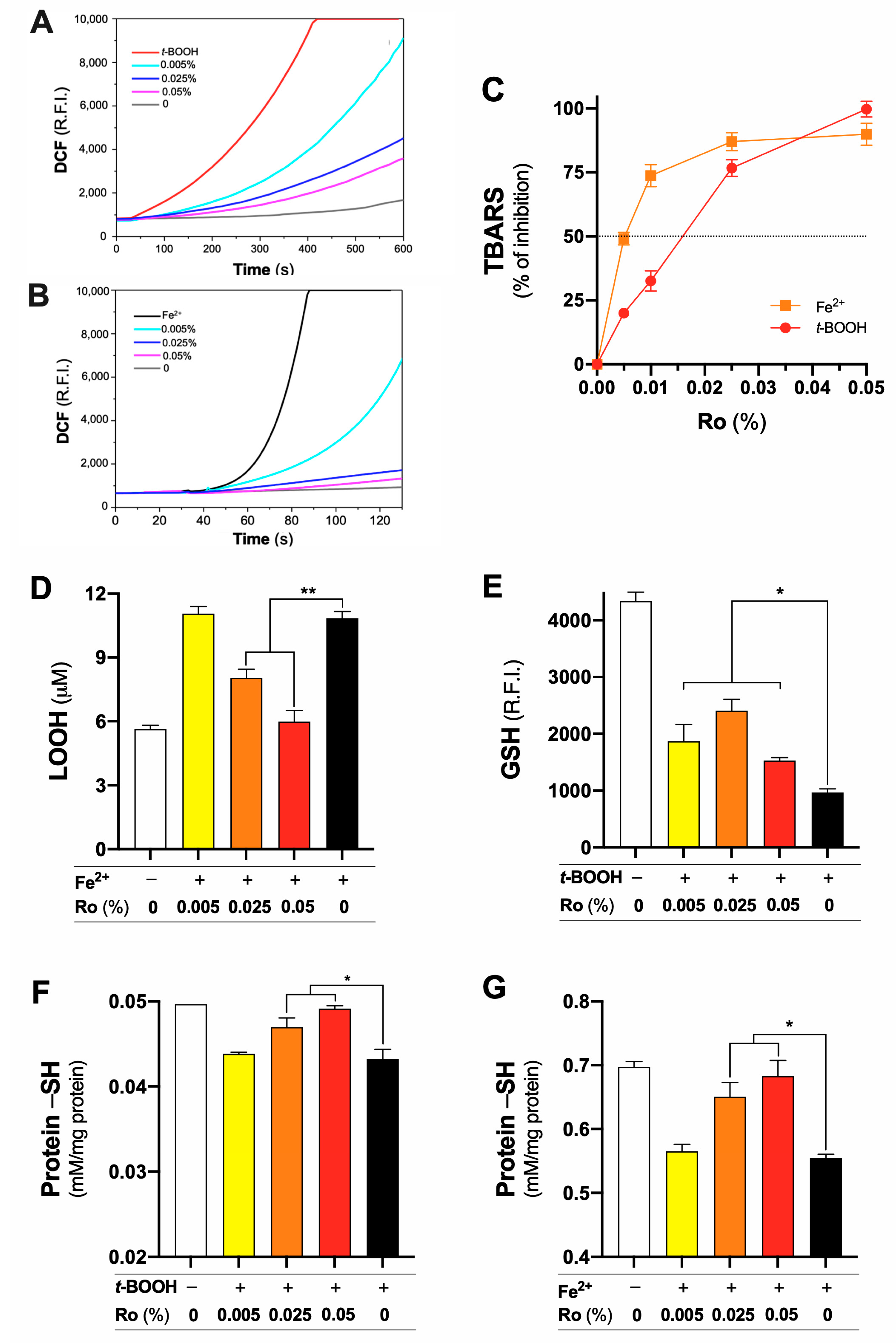
3.1. Rosmarinus officinalis Glycolic Extract (Ro) Protects Mitochondrial Lipids and Proteins from Oxidation
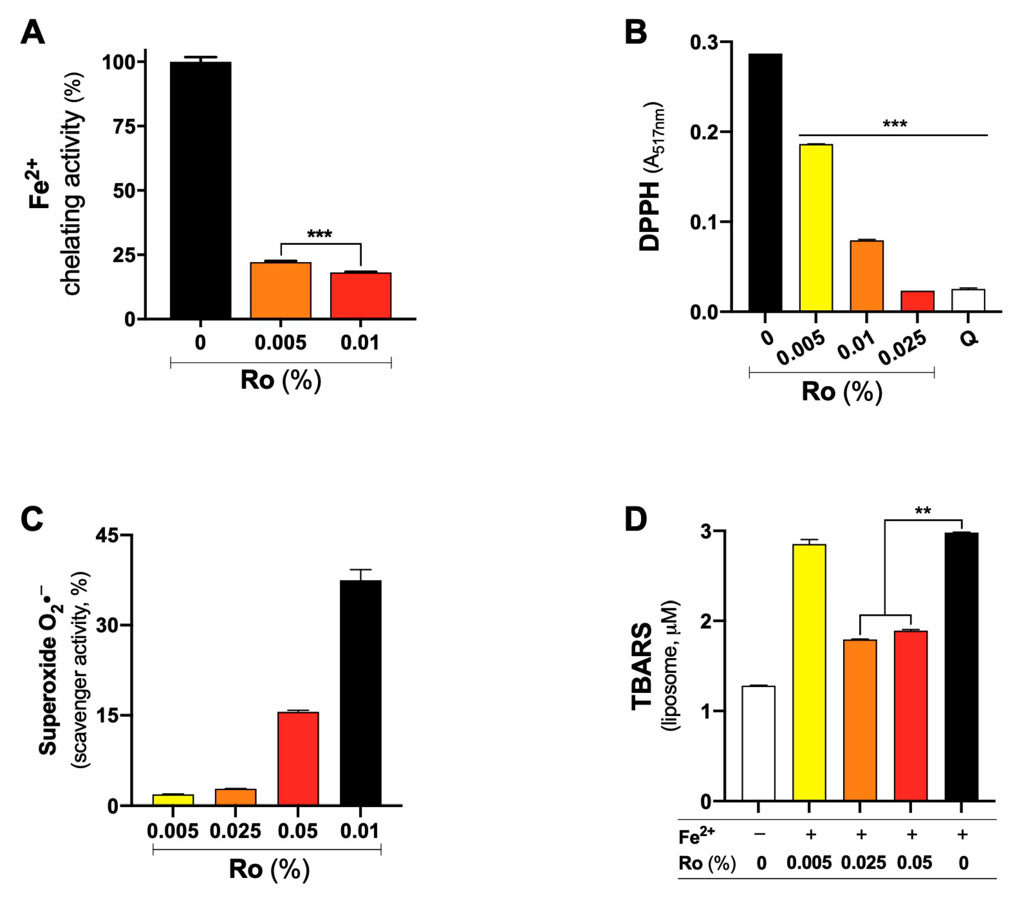
3.2. Iron (II) Chelating and Free Radical Scavenger Activity Account to the Antioxidant Protection Exhibited by Ro
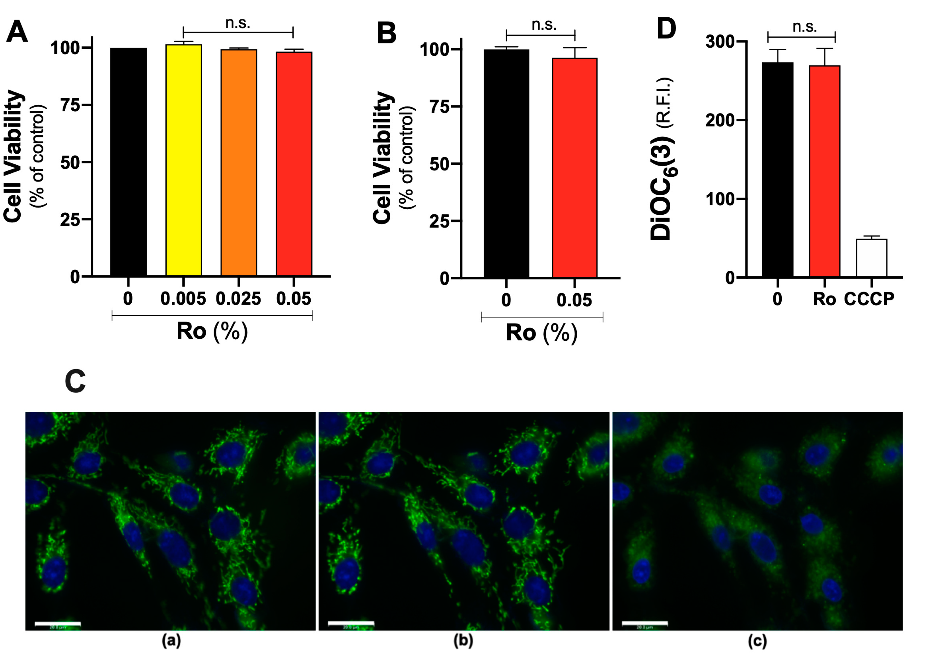
3.3. Ro Does Not Affect Viability or Mitochondrial Membrane Potential (∆Ψ) of Liver Fibroblasts
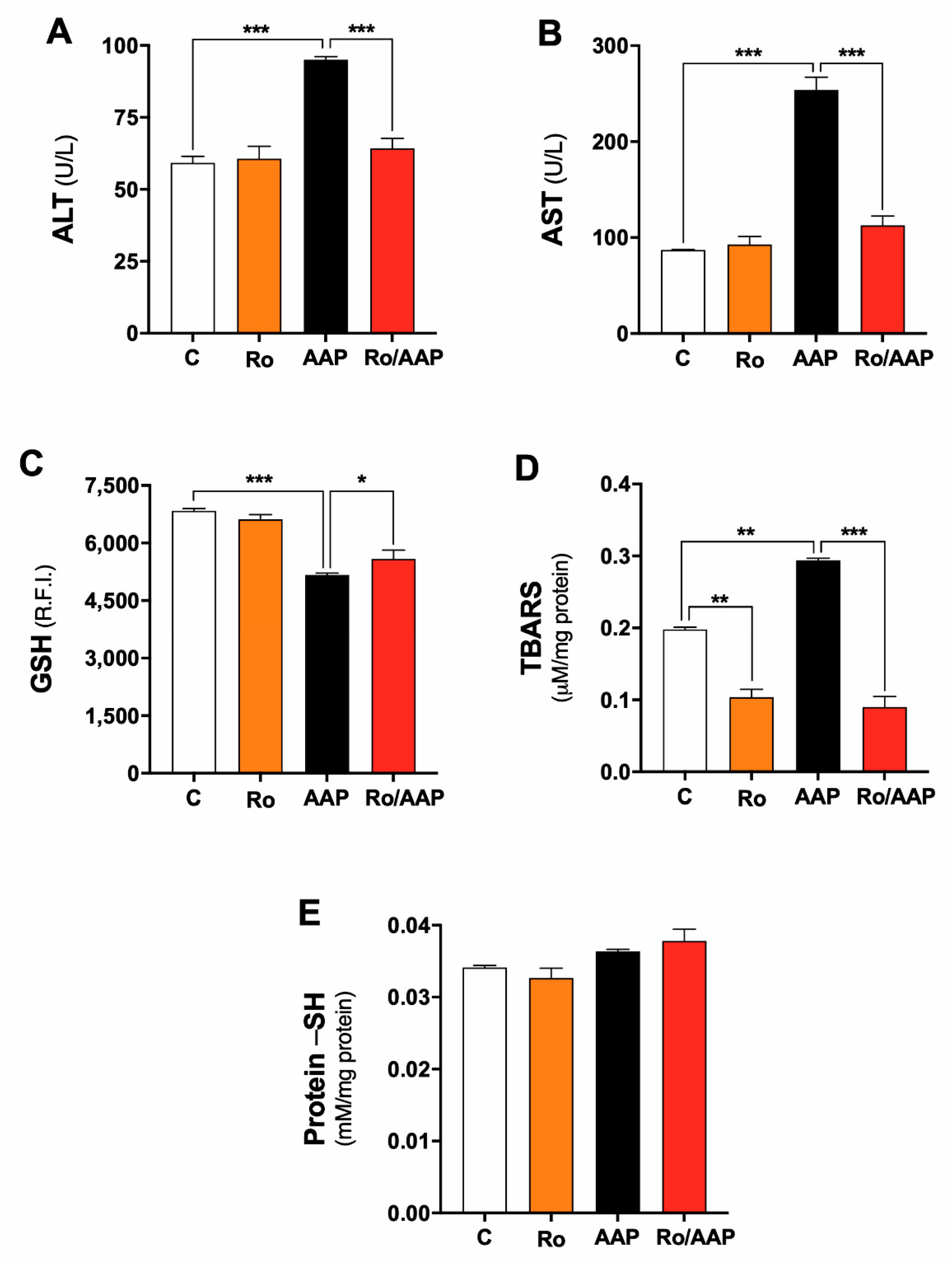
3.4. Ro Protection against Acetaminophen-Induced Hepatotoxicity in Rats
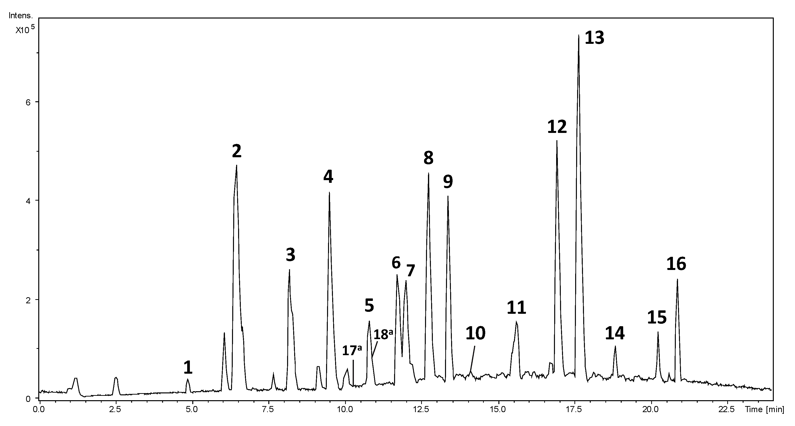
3.5. Chemical Composition of Rosmarinus officinalis L. Glycolic Extract
4. Discussion
5. Conclusions
Author Contributions
Funding
Institutional Review Board Statement
Informed Consent Statement
Data Availability Statement
Acknowledgments
Conflicts of Interest
References
- Ramos Da Silva, L.R.; Ferreira, O.O.; Cruz, J.N.; De Jesus Pereira Franco, C.; Oliveira Dos Anjos, T.; Cascaes, M.M.; Almeida Da Costa, W.; Helena De Aguiar Andrade, E.; Santana De Oliveira, M. Lamiaceae Essential Oils, Phytochemical Profile, Antioxidant, and Biological Activities. Evid.-Based Complement. Altern. Med. 2021, 2021, 6748052. [Google Scholar] [CrossRef]
- Veenstra, J.P.; Johnson, J.J. Rosemary (Salvia rosmarinus): Health-Promoting Benefits and Food Preservative Properties. Int. J. Nutr. 2021, 6, 1. [Google Scholar] [CrossRef] [PubMed]
- Gurib-Fakim, A. Medicinal Plants: Traditions of Yesterday and Drugs of Tomorrow. Mol. Asp. Med. 2006, 27, 1–93. [Google Scholar] [CrossRef] [PubMed]
- Boldi, A.M. Libraries from Natural Product-like Scaffolds. Curr. Opin. Chem. Biol. 2004, 8, 281–286. [Google Scholar] [CrossRef]
- Bouyahya, A.; Et-Touys, A.; Bakri, Y.; Talbaui, A.; Fellah, H.; Abrini, J.; Dakka, N. Chemical Composition of Mentha pulegium and Rosmarinus officinalis Essential Oils and Their Antileishmanial, Antibacterial and Antioxidant Activities. Microb. Pathog. 2017, 111, 41–49. [Google Scholar] [CrossRef] [PubMed]
- Mahmoud, A.A.; Al-Shihry, S.S.; Son, B.W. Diterpenoid Quinones from Rosemary (Rosmarinus officinalis L.). Phytochemistry 2005, 66, 1685–1690. [Google Scholar] [CrossRef]
- Borges, R.S.; Ortiz, B.L.S.; Pereira, A.C.M.; Keita, H.; Carvalho, J.C.T. Rosmarinus officinalis Essential Oil: A Review of Its Phytochemistry, Anti-Inflammatory Activity, and Mechanisms of Action Involved. J. Ethnopharmacol. 2019, 229, 29–45. [Google Scholar] [CrossRef] [PubMed]
- Bai, N.; He, K.; Roller, M.; Lai, C.S.; Shao, X.; Pan, M.H.; Ho, C.T. Flavonoids and Phenolic Compounds from Rosmarinus officinalis. J. Agric. Food Chem. 2010, 58, 5363–5367. [Google Scholar] [CrossRef]
- Jordán, M.J.; Castillo, J.; Bañón, S.; Martínez-Conesa, C.; Sotomayor, J.A. Relevance of the Carnosic Acid/Carnosol Ratio for the Level of Rosemary Diterpene Transfer and for Improving Lamb Meat Antioxidant Status. Food Chem. 2014, 151, 212–218. [Google Scholar] [CrossRef]
- Ibrahim, N.; Abbas, H.; El-Sayed, N.S.; Gad, H.A. Rosmarinus officinalis L. Hexane Extract: Phytochemical Analysis, Nanoencapsulation, and in Silico, in Vitro, and in Vivo Anti-Photoaging Potential Evaluation. Sci. Rep. 2022, 12, 13102. [Google Scholar] [CrossRef]
- Borrás-Linares, I.; Stojanović, Z.; Quirantes-Piné, R.; Arráez-Román, D.; Švarc-Gajić, J.; Fernández-Gutiérrez, A.; Segura-Carretero, A. Rosmarinus officinalis Leaves as a Natural Source of Bioactive Compounds. Int. J. Mol. Sci. 2014, 15, 20585–20606. [Google Scholar] [CrossRef] [PubMed] [Green Version]
- De Oliveira, G.G.; Carnevale Neto, F.; Demarque, D.P.; de Sousa Pereira-Junior, J.A.; Sampaio Peixoto Filho, R.C.; de Melo, S.J.; da Silva Almeida, J.R.G.; Lopes, J.L.C.; Lopes, N.P. Dereplication of Flavonoid Glycoconjugates from Adenocalymma imperatoris-maximilianii by Untargeted Tandem Mass Spectrometry-Based Molecular Networking. Planta Med. 2017, 83, 636–646. [Google Scholar] [CrossRef] [PubMed] [Green Version]
- Demarque, D.P.; Dusi, R.G.; de Sousa, F.D.M.; Grossi, S.M.; Silvério, M.R.S.; Lopes, N.P.; Espindola, L.S. Mass spectrometry-based metabolomics approach in the isolation of bioactive natural products. Sci Rep. 2020, 10, 1051. [Google Scholar] [CrossRef] [Green Version]
- Guaratini, T.; Vessecchi, R.L.; Lavarda, F.C.; Campos, P.M.B.G.M.; Naal, Z.; Gates, P.J.; Lopes, N.P. New chemical evidence for the ability to generate radical molecular ions of polyenes from ESI and HR-MALDI mass spectrometry. Analyst 2004, 129, 1223–1226. [Google Scholar] [CrossRef]
- Guaratini, T.; Vessecchi, R.; Pinto, E.; Collepicolo, P.; Lopes, N.P. Balance of xanthophylls molecular and protonated molecular ions in electrospray ionization. J. Mass Spectrom. 2005, 40, 963–968. [Google Scholar] [CrossRef]
- Bozin, B.; Mimica-Dukic, N.; Samojlik, I.; Jovin, E. Antimicrobial and Antioxidant Properties of Rosemary and Sage (Rosmarinus officinalis L. and Salvia officinalis L., Lamiaceae) Essential Oils. J. Agric. Food Chem. 2007, 55, 7879–7885. [Google Scholar] [CrossRef]
- Wang, W.; Li, N.; Luo, M.; Zu, Y.; Efferth, T. Antibacterial Activity and Anticancer Activity of Rosmarinus officinalis L. Essential Oil Compared to That of Its Main Components. Molecules 2012, 17, 2704–2713. [Google Scholar] [CrossRef] [Green Version]
- Moreno, S.; Scheyer, T.; Romano, C.S.; Vojnov, A.A. Antioxidant and Antimicrobial Activities of Rosemary Extracts Linked to Their Polyphenol Composition. Free. Radic. Res. 2006, 40, 223–231. [Google Scholar] [CrossRef] [PubMed]
- Waller, S.B.; Cleff, M.B.; Dalla Lana, D.F.; de Mattos, C.B.; Guterres, K.A.; Freitag, R.A.; Sallis, E.S.V.; Fuentefria, A.M.; de Mello, J.R.B.; de Faria, R.O.; et al. Can the Essential Oil of Rosemary (Rosmarinus officinalis Linn.) Protect Rats Infected with Itraconazole-Resistant Sporothrix Brasiliensis from Fungal Spread? J. Med. Mycol. 2021, 31, 101199. [Google Scholar] [CrossRef]
- Karpiński, T.M. Essential Oils of Lamiaceae Family Plants as Antifungals. Biomolecules 2020, 10, 103. [Google Scholar] [CrossRef] [Green Version]
- Lucarini, R.; Bernardes, W.A.; Ferreira, D.S.; Tozatti, M.G.; Furtado, R.; Bastos, J.K.; Pauletti, P.M.; Januário, A.H.; Silva, M.L.A.E.; Cunha, W.R. In Vivo Analgesic and Anti-Inflammatory Activities of Rosmarinus officinalis Aqueous Extracts, Rosmarinic Acid and Its Acetyl Ester Derivative. Pharm. Biol. 2013, 51, 1087–1090. [Google Scholar] [CrossRef] [PubMed] [Green Version]
- Ghasemzadeh Rahbardar, M.; Amin, B.; Mehri, S.; Mirnajafi-Zadeh, S.J.; Hosseinzadeh, H. Anti-Inflammatory Effects of Ethanolic Extract of Rosmarinus officinalis L. and Rosmarinic Acid in a Rat Model of Neuropathic Pain. Biomed. Pharmacother. 2017, 86, 441–449. [Google Scholar] [CrossRef] [PubMed]
- Hsieh, C.L.; Peng, C.H.; Chyau, C.C.; Lin, Y.C.; Wang, H.E.; Peng, R.Y. Low-Density Lipoprotein, Collagen, and Thrombin Models Reveal That Rosemarinus officinalis L. Exhibits Potent Antiglycative Effects. J. Agric. Food Chem. 2007, 55, 2884–2891. [Google Scholar] [CrossRef] [PubMed]
- Rodrigues, A.P.S.; Souza, B.S.F.E.; Barros, A.S.A.; de Oliveira Carvalho, H.; Duarte, J.L.; Boettger, L.E.M.; Barbosa, R.; Ferreira, A.M.; Ferreira, I.M.; Fernandes, C.P.; et al. The Effects of Rosmarinus officinalis L. Essential Oil and Its Nanoemulsion on Dyslipidemic Wistar Rats. J. Appl. Biomed. 2020, 18, 126–135. [Google Scholar] [CrossRef] [PubMed]
- López-Jiménez, A.; García-Caballero, M.; Medina, M.Á.; Quesada, A.R. Anti-Angiogenic Properties of Carnosol and Carnosic Acid, Two Major Dietary Compounds from Rosemary. Eur. J. Nutr. 2013, 52, 85–95. [Google Scholar] [CrossRef]
- Kayashima, T.; Matsubara, K. Antiangiogenic Effect of Carnosic Acid and Carnosol, Neuroprotective Compounds in Rosemary Leaves. Biosci. Biotechnol. Biochem. 2012, 76, 115–119. [Google Scholar] [CrossRef]
- Karthik, D.; Viswanathan, P.; Anuradha, C.V. Administration of Rosmarinic Acid Reduces Cardiopathology and Blood Pressure through Inhibition of P22phox NADPH Oxidase in Fructose-Fed Hypertensive Rats. J. Cardiovasc. Pharmacol. 2011, 58, 514–521. [Google Scholar] [CrossRef]
- Amaral, G.P.; de Carvalho, N.R.; Barcelos, R.P.; Dobrachinski, F.; de Lima Portella, R.; da Silva, M.H.; Lugokenski, T.H.; Dias, G.R.M.; da Luz, S.C.A.; Boligon, A.A.; et al. Protective Action of Ethanolic Extract of Rosmarinus officinalis L. in Gastric Ulcer Prevention Induced by Ethanol in Rats. Food Chem. Toxicol. 2013, 55, 48–55. [Google Scholar] [CrossRef]
- Samarghandian, S.; Borji, A.; Farkhondeh, T. Evaluation of Antidiabetic Activity of Carnosol (Phenolic Diterpene in Rosemary) in Streptozotocin-Induced Diabetic Rats. Cardiovasc. Haematol. Disord.-Drug Targets 2017, 17, 11–17. [Google Scholar] [CrossRef]
- Bakirel, T.; Bakirel, U.; Keleş, O.Ü.; Ülgen, S.G.; Yardibi, H. In Vivo Assessment of Antidiabetic and Antioxidant Activities of Rosemary (Rosmarinus officinalis) in Alloxan-Diabetic Rabbits. J. Ethnopharmacol. 2008, 116, 64–73. [Google Scholar] [CrossRef]
- Tai, J.; Cheung, S.; Wu, M.; Hasman, D. Antiproliferation Effect of Rosemary (Rosmarinus officinalis) on Human Ovarian Cancer Cells in Vitro. Phytomedicine 2012, 19, 436–443. [Google Scholar] [CrossRef] [PubMed]
- De Oliveira, J.R.; Camargo, S.E.A.; De Oliveira, L.D. Rosmarinus officinalis L. (Rosemary) as Therapeutic and Prophylactic Agent. J. Biomed. Sci. 2019, 26, 5. [Google Scholar] [CrossRef] [PubMed]
- Sharifi-Rad, J.; Ezzat, S.M.; El Bishbishy, M.H.; Mnayer, D.; Sharopov, F.; Kılıç, C.S.; Neagu, M.; Constantin, C.; Sharifi-Rad, M.; Atanassova, M.; et al. Rosmarinus Plants: Key Farm Concepts towards Food Applications. Phytother. Res. 2020, 34, 1474–1518. [Google Scholar] [CrossRef] [PubMed]
- Nieto, G.; Ros, G.; Castillo, J. Antioxidant and Antimicrobial Properties of Rosemary (Rosmarinus officinalis, L.): A Review. Medicines 2018, 5, 98. [Google Scholar] [CrossRef] [Green Version]
- Dorta, D.J.; Pigoso, A.A.; Mingatto, F.E.; Rodrigues, T.; Pestana, C.R.; Uyemura, S.A.; Santos, A.C.; Curti, C. Antioxidant Activity of Flavonoids in Isolated Mitochondria. Phytother. Res. 2008, 22, 1213–1218. [Google Scholar] [CrossRef] [PubMed]
- Okamura, N.; Haraguchi, H.; Hashimoto, K.; Yagi, A. Flavonoids in Rosmarinus officinalis Leaves. Phytochemistry 1994, 37, 1463–1466. [Google Scholar] [CrossRef]
- Forman, H.J.; Zhang, H. Targeting Oxidative Stress in Disease: Promise and Limitations of Antioxidant Therapy. Nat. Rev. Drug Discov. 2021, 20, 689–709. [Google Scholar] [CrossRef]
- El-Demerdash, F.M.; Abbady, E.A.; Baghdadi, H.H. Oxidative Stress Modulation by Rosmarinus officinalis in Creosote-Induced Hepatotoxicity. Environ. Toxicol. 2016, 31, 85–92. [Google Scholar] [CrossRef]
- Amin, A.; Hamza, A.A. Hepatoprotective Effects of Hibiscus, Rosmarinus and Salvia on Azathioprine-Induced Toxicity in Rats. Life Sci. 2005, 77, 266–278. [Google Scholar] [CrossRef]
- El-Demerdash, F.M.; El-Sayed, R.A.; Abdel-Daim, M.M. Hepatoprotective Potential of Rosmarinus officinalis Essential Oil against Hexavalent Chromium-Induced Hematotoxicity, Biochemical, Histological, and Immunohistochemical Changes in Male Rats. Environ. Sci. Pollut. Res. 2021, 28, 17445–17456. [Google Scholar] [CrossRef]
- Ramadan, K.S.; Khalil, O.A.; Danial, E.N.; Alnahdi, H.S.; Ayaz, N.O. Hypoglycemic and Hepatoprotective Activity of Rosmarinus officinalis Extract in Diabetic Rats. J. Physiol. Biochem. 2013, 69, 779–783. [Google Scholar] [CrossRef] [PubMed]
- El-Naggar, S.A.; Abdel-Farid, I.B.; Germoush, M.O.; Elgebaly, H.A.; Alm-Eldeen, A.A. Efficacy of Rosmarinus officinalis Leaves Extract against Cyclophosphamide-Induced Hepatotoxicity. Pharm. Biol. 2016, 54, 2007–2016. [Google Scholar] [CrossRef] [Green Version]
- Bunchorntavakul, C.; Reddy, K.R. Acetaminophen-Related Hepatotoxicity. Clin. Liver Dis. 2013, 17, 587–607. [Google Scholar] [CrossRef] [PubMed]
- Mazaleuskaya, L.L.; Sangkuhl, K.; Thorn, C.F.; Fitzgerald, G.A.; Altman, R.B.; Klein, T.E. PharmGKB Summary: Pathways of Acetaminophen Metabolism at the Therapeutic versus Toxic Doses. Pharm. Genom. 2015, 25, 416. [Google Scholar] [CrossRef] [Green Version]
- Figueira, T.R.; Barros, M.H.; Camargo, A.A.; Castilho, R.F.; Ferreira, J.C.B.; Kowaltowski, A.J.; Sluse, F.E.; Souza-Pinto, N.C.; Vercesi, A.E. Mitochondria as a Source of Reactive Oxygen and Nitrogen Species: From Molecular Mechanisms to Human Health. Antioxid. Redox Signal. 2013, 18, 2029–2074. [Google Scholar] [CrossRef]
- Sies, H. Oxidative Stress: Oxidants and Antioxidants. Exp. Physiol. 1997, 82, 291–295. [Google Scholar] [CrossRef]
- Cruz, T.S.; Faria, P.A.; Santana, D.P.; Ferreira, J.C.; Oliveira, V.; Nascimento, O.R.; Cerchiaro, G.; Curti, C.; Nantes, I.L.; Rodrigues, T. On the Mechanisms of Phenothiazine-Induced Mitochondrial Permeability Transition: Thiol Oxidation, Strict Ca2+ Dependence, and Cyt c Release. Biochem. Pharmacol. 2010, 80, 1284–1295. [Google Scholar] [CrossRef] [PubMed]
- Ainsworth, E.A.; Gillespie, K.M. Estimation of total phenolic content and other oxidation substrates in plant tissues using Folin-Ciocalteu reagent. Nat. Protoc. 2007, 2, 875–877. [Google Scholar] [CrossRef] [PubMed]
- Guimarães, N.S.S.; Mello, J.C.; Paiva, J.S.; Bueno, P.C.P.; Berretta, A.A.; Torquato, R.J.; Nantes, I.L.; Rodrigues, T. Baccharis dracunculifolia, the Main Source of Green Propolis, Exhibits Potent Antioxidant Activity and Prevents Oxidative Mitochondrial Damage. Food Chem. Toxicol. 2012, 50, 1091–1097. [Google Scholar] [CrossRef] [Green Version]
- Cowart, R.E.; Singleton, F.L.; Hind, J.S. A Comparison of Bathophenanthrolinedisulfonic Acid and Ferrozine as Chelators of Iron(II) in Reduction Reactions. Anal. Biochem. 1993, 211, 151–155. [Google Scholar] [CrossRef]
- Lebel, C.P.; Ischiropoulos, H.; Bondy, S.C. Evaluation of the probe 2′,7′-dichlorofluorescin as an indicator of reactive oxygen species formation and oxidative stress. Chem. Res. Toxicol. 1992, 5, 227–231. [Google Scholar] [CrossRef] [PubMed] [Green Version]
- Mello, J.C.; Guimarães, N.S.S.; Gonzalez, M.V.D.; Paiva, J.S.; Prieto, T.; Nascimento, O.R.; Tiago Rodrigues, T. Hydroxyl scavenging activity accounts for differential antioxidant protection of Plantago major against oxidative toxicity in isolated rat liver mitochondria. J. Pharm. Pharmacol. 2012, 64, 1177–1187. [Google Scholar] [CrossRef] [PubMed]
- Mello, J.C.; Gonzalez, M.V.D.; Moraes, V.W.R.; Prieto, T.; Nascimento, O.R.; Rodrigues, T. Protective Effect of Plantago major Extract against t-BOOH-Induced Mitochondrial Oxidative Damage and Cytotoxicity. Molecules 2015, 20, 17747–17759. [Google Scholar] [CrossRef] [PubMed] [Green Version]
- Borges, M.B.D.; Dos Santos, C.G.; Yokomizo, C.H.; Sood, R.; Vitovic, P.; Kinnunen, P.K.J.; Rodrigues, T.; Nantes, I.L. Characterization of Hydrophobic Interaction and Antioxidant Properties of the Phenothiazine Nucleus in Mitochondrial and Model Membranes. Free. Radic. Res. 2010, 44, 1054–1063. [Google Scholar] [CrossRef] [PubMed]
- Hissin, P.J.; Hilf, R. A Fluorometric Method for Determination of Oxidized and Reduced Glutathione in Tissues. Anal. Biochem. 1976, 74, 214–226. [Google Scholar] [CrossRef]
- Cálgaro-Helena, A.F.; Devienne, K.F.; Rodrigues, T.; Dorta, D.J.; Raddi, M.S.G.; Vilegas, W.; Uyemura, S.A.; Santos, A.C.; Curti, C. Effects of Isocoumarins Isolated from Paepalanthus Bromelioides on Mitochondria: Uncoupling, and Induction/Inhibition of Mitochondrial Permeability Transition. Chem. Biol. Interact. 2006, 161, 155–164. [Google Scholar] [CrossRef]
- Jocelyn, P.C. Spectrophotometric Assay of Thiols. Methods Enzymol. 1987, 143, 44–67. [Google Scholar] [CrossRef]
- Mello, J.C.; Moraes, V.W.R.; Watashi, C.M.; Silva, D.C.; Cavalcanti, L.P.; Franco, M.K.K.D.; Yokaichiya, F.; Araujo, D.R.; Rodrigues, T. Enhancement of chlorpromazine antitumor activity by Pluronics F127/L81 nanostructured system against human multidrug resistant leukemia. Pharmacol. Res. 2016, 111, 102–112. [Google Scholar] [CrossRef]
- Pilon, A.C.; Del Grande, M.; Silvério, M.R.S.; Silva, R.R.; Albernaz, L.C.; Vieira, P.C.; Lopes, J.L.C.; Espindola, L.S.; Lopes, N.P. Combination of GC-MS Molecular Networking and Larvicidal Effect against Aedes aegypti for the Discovery of Bioactive Substances in Commercial Essential Oils. Molecules 2022, 27, 1588. [Google Scholar] [CrossRef]
- Liu, Y.; Guo, M. Studies on Transition Metal-Quercetin Complexes Using Electrospray Ionization Tandem Mass Spectrometry. Molecules 2015, 20, 8583–8594. [Google Scholar] [CrossRef] [Green Version]
- Hyung, S.C.; Jun, W.K.; Cha, Y.N.; Kim, C. A Quantitative Nitroblue Tetrazolium Assay for Determining Intracellular Superoxide Anion Production in Phagocytic Cells. J. Immunoass. Immunochem. 2006, 27, 31–44. [Google Scholar] [CrossRef]
- Pessoto, F.S.; Faria, P.A.; Cunha, R.L.O.R.; Comasseto, J.V.; Rodrigues, T.; Nantes, I.L. Organotellurane-Promoted Mitochondrial Permeability Transition Concomitant with Membrane Lipid Protection against Oxidation. Chem. Res. Toxicol. 2007, 20, 1453–1461. [Google Scholar] [CrossRef] [PubMed]
- Simunkova, M.; Barbierikova, Z.; Jomova, K.; Hudecova, L.; Lauro, P.; Alwasel, S.H.; Alhazza, I.; Rhodes, C.J.; Valko, M. Antioxidant vs. Prooxidant Properties of the Flavonoid, Kaempferol, in the Presence of Cu(II) Ions: A ROS-Scavenging Activity, Fenton Reaction and DNA Damage Study. Int. J. Mol. Sci. 2021, 22, 1619. [Google Scholar] [CrossRef] [PubMed]
- Jeffery, E.H.; Haschek, W.M. Protection by dimethylsulfoxide against acetaminophen-induced hepatic, but not respiratory toxicity in the mouse. Toxicol. Appl. Pharmacol. 1988, 93, 452–461. [Google Scholar] [CrossRef]
- Göksel Sener, G.; Sehirli, O.; Cetinel, S.; Yeğen, B.G.; Gedik, N.; Ayanoğlu-Dülger, G. Protective effects of MESNA (2-mercaptoethane sulphonate) against acetaminophen-induced hepatorenal oxidative damage in mice. J. Appl. Toxicol. 2005, 25, 20–29. [Google Scholar] [CrossRef]
- Lopes, N.P.; Stark, C.B.W.; Gates, P.J.; Staunton, J. Fragmentation studies on monensin A by sequential electrospray mass spectrometry. Analyst 2002, 127, 503–506. [Google Scholar] [CrossRef]
- Pilon, A.C.; Gu, H.; Raftery, D.; Bolzani, V.D.S.; Lopes, N.P.; Castro-Gamboa, I.; Carnevale Neto, F. Mass Spectral Similarity Networking and Gas-Phase Fragmentation Reactions in the Structural Analysis of Flavonoid Glycoconjugates. Anal. Chem. 2019, 91, 10413–10423. [Google Scholar] [CrossRef]
- Mena, P.; Cirlini, M.; Tassotti, M.; Herrlinger, K.A.; Dall’Asta, C.; Del Rio, D. Phytochemical Profiling of Flavonoids, Phenolic Acids, Terpenoids, and Volatile Fraction of a Rosemary (Rosmarinus officinalis L.) Extract. Molecules 2016, 21, 1576. [Google Scholar] [CrossRef]
- Sharma, Y.; Velamuri, R.; Fagan, J.; Schaefer, J. Full-Spectrum Analysis of Bioactive Compounds in Rosemary (Rosmarinus officinalis L.) as Influenced by Different Extraction Methods. Molecules 2020, 25, 4599. [Google Scholar] [CrossRef]
- Kontogianni, V.G.; Tomic, G.; Nikolic, I.; Nerantzaki, A.; Sayyad, A.; Stosic-Grujicic, N.; Stojanovic, S.; Gerothanassis, I.P.; Tzakos, A.G. Phytochemical profile of Rosmarinus officinalis and Salvia officinalis extracts and correlation to their antioxidant and anti-proliferative activity. Food Chem. 2013, 136, 120–129. [Google Scholar] [CrossRef]
- Wang, M.; Carver, J.J.; Phelan, V.V.; Sanchez, L.M.; Garg, N.; Peng, Y.; Nguyen, D.D.; Watrous, J.; Kapono, C.A.; Luzzatto-Knaan, T.; et al. Sharing and community curation of mass spectrometry data with Global Natural Products Social Molecular Networking. Nat. Biotechnol. 2016, 34, 828–837. [Google Scholar] [CrossRef] [PubMed] [Green Version]
- Green, D.R.; Reed, J.C. Mitochondria and Apoptosis. Science 1998, 281, 118. [Google Scholar] [CrossRef] [PubMed]
- Aruoma, O.I.; Halliwell, B.; Aeschbach, R.; Löligers, J. Antioxidant and Pro-Oxidant Properties of Active Rosemary Constituents: Carnosol and Carnosic Acid. Xenobiotica 1992, 22, 257–268. [Google Scholar] [CrossRef]
- Pérez-Fons, L.; Garzón, M.T.; Micol, V. Relationship between the Antioxidant Capacity and Effect of Rosemary (Rosmarinus officinalis L.) Polyphenols on Membrane Phospholipid Order. J. Agric. Food Chem. 2010, 58, 161–171. [Google Scholar] [CrossRef]
- Harlina, P.W.; Ma, M.; Shahzad, R.; Khalifa, I. Effect of Rosemary Extract on Lipid Oxidation, Fatty Acid Composition, Antioxidant Capacity, and Volatile Compounds of Salted Duck Eggs. Food Sci. Anim. Resour. 2022, 42, 689–711. [Google Scholar] [CrossRef]
- Nazem, F.; Farhangi, N.; Neshat-Gharamaleki, M. Beneficial Effects of Endurance Exercise with Rosmarinus officinalis Labiatae Leaves Extract on Blood Antioxidant Enzyme Activities and Lipid Peroxidation in Streptozotocin-Induced Diabetic Rats. Can. J. Diabetes 2015, 39, 229–234. [Google Scholar] [CrossRef] [PubMed]
- Karimi, G.; Hassanzadeh, M.; Mehri, S. Protective Effect of Rosmarinus officinalis L. Essential Oil against Free Radical-Induced Erythrocyte Lysis. Iran. J. Pharm. Sci. 2005, 1, 231–236. [Google Scholar]
- Kaliora, A.C.; Andrikopoulos, N.K. Effect of Alkanna Albugam Root on LDL Oxidation. A Comparative Study with Species of the Lamiaceae Family. Phytother. Res. 2005, 19, 1077–1079. [Google Scholar] [CrossRef]
- Haraguchi, H.; Saito, T.; Okamura, N.; Yagi, A. Inhibition of Lipid Peroxidation and Superoxide Generation by Diterpenoids from Rosmarinus officinalis. Planta Med. 1995, 61, 333–336. [Google Scholar] [CrossRef]
- Sueishi, Y.; Sue, M.; Masamoto, H. Seasonal Variations of Oxygen Radical Scavenging Ability in Rosemary Leaf Extract. Food Chem. 2018, 245, 270–274. [Google Scholar] [CrossRef]
- Horvathova, E.; Navarova, J.; Galova, E.; Sevcovicova, A.; Chodakova, L.; Snahnicanova, Z.; Melusova, M.; Kozics, K.; Slamenova, D. Assessment of Antioxidative, Chelating, and DNA-Protective Effects of Selected Essential Oil Components (Eugenol, Carvacrol, Thymol, Borneol, Eucalyptol) of Plants and Intact Rosmarinus officinalis Oil. J. Agric. Food Chem. 2014, 62, 6632–6639. [Google Scholar] [CrossRef]
- Lin, C.; Zhang, X.; Su, Z.; Xiao, J.; Lv, M.; Cao, Y.; Chen, Y. Carnosol Improved Lifespan and Healthspan by Promoting Antioxidant Capacity in Caenorhabditis elegans. Oxidative Med. Cell. Longev. 2019, 2019, 59580431619. [Google Scholar] [CrossRef] [PubMed] [Green Version]
- Moreira, E.A.; Pilon, A.C.; Andrade, L.E.; Lopes, N.P. New Perspectives on Chlorogenic Acid Accumulation in Harvested Leaf Tissue: Impact on Traditional Medicine Preparations. ACS Omega 2018, 3, 12. [Google Scholar] [CrossRef]
- Zegura, B.; Dobnik, D.; Niderl, M.H.; Filipi, M. Antioxidant and antigenotoxic effects of rosemary (Rosmarinus officinalis L.) extracts in Salmonella typhimurium TA98 and HepG2 cells. Environ. Toxicol. Pharmacol. 2011, 32, 296–305. [Google Scholar] [CrossRef] [PubMed]
- Visentin, A.; Rodríguez-Rojo, S.; Navarrete, A.; Maestri, D.; Cocero, M.J. Precipitation and encapsulation of rosemary antioxidants by supercritical antisolvent process. J. Food Eng. 2012, 109, 9–15. [Google Scholar] [CrossRef]
- Sasaki, K.; El Omri, A.; Kondo, S.; Han, J.; Isoda, H. Rosmarinus officinalis polyphenols produce anti-depressant like effect through monoaminergic and cholinergic functions modulation. Behav. Brain Res. 2013, 238, 86–94. [Google Scholar] [CrossRef]
- Soares, M.S.; da Silva, D.F.; Forim, M.R.; da Silva, M.F.; Fernandes, J.B.; Vieira, P.C.; Silva, D.B.; Lopes, N.P.; de Carvalho, S.A.; de Souza, A.A.; et al. Quantification and localization of hesperidin and rutin in Citrus sinensis grafted on C. limonia after Xylella fastidiosa infection by HPLC-UV and MALDI imaging mass spectrometry. Phytochemistry 2015, 115, 161–170. [Google Scholar] [CrossRef] [PubMed]
- Kluska, M.; Juszczak, M.; Żuchowski, J.; Stochmal, A.; Woźniak, K. Effect of Kaempferol and Its Glycoside Derivatives on Antioxidant Status of HL-60 Cells Treated with Etoposide. Molecules 2022, 27, 333. [Google Scholar] [CrossRef] [PubMed]
- Hassanen, N.H.M.; Fahmi, A.; Shams-Eldin, E.; Abdur-Rahman, M. Protective Effect of Rosemary (Rosmarinus officinalis) against Diethylnitrosamine-Induced Renal Injury in Rats. Biomarkers 2020, 25, 281–289. [Google Scholar] [CrossRef]
- El-Demerdash, F.M.; El-Sayed, R.A.; Abdel-Daim, M.M. Rosmarinus officinalis Essential Oil Modulates Renal Toxicity and Oxidative Stress Induced by Potassium Dichromate in Rats. J. Trace Elem. Med. Biol. 2021, 67, 126791. [Google Scholar] [CrossRef]
- Rašković, A.; Milanović, I.; Pavlović, N.; Ćebović, T.; Vukmirović, S.; Mikov, M. Antioxidant Activity of Rosemary (Rosmarinus officinalis L.) Essential Oil and Its Hepatoprotective Potential. BMC Complement. Med. Ther. 2014, 14, 225. [Google Scholar] [CrossRef] [PubMed] [Green Version]
- Sotelo-Félix, J.I.; Martinez-Fong, D.; Muriel, P.; Santillán, R.L.; Castillo, D.; Yahuaca, P. Evaluation of the Effectiveness of Rosmarinus officinalis (Lamiaceae) in the Alleviation of Carbon Tetrachloride-Induced Acute Hepatotoxicity in the Rat. J. Ethnopharmacol. 2002, 81, 145–154. [Google Scholar] [CrossRef] [PubMed]
- Gutiérrez, R.; Alvarado, J.L.; Presno, M.; Pérez-Veyna, O.; Serrano, C.J.; Yahuaca, P. Oxidative Stress Modulation by Rosmarinus officinalis in CCl4-Induced Liver Cirrhosis. Phytother. Res. 2010, 24, 595–601. [Google Scholar] [CrossRef]
- Ielciu, I.; Sevastre, B.; Olah, N.K.; Turdean, A.; Chișe, E.; Marica, R.; Oniga, I.; Uifălean, A.; Sevastre-Berghian, A.C.; Niculae, M.; et al. Evaluation of Hepatoprotective Activity and Oxidative Stress Reduction of Rosmarinus officinalis L. Shoots Tincture in Rats with Experimentally Induced Hepatotoxicity. Molecules 2021, 26, 1737. [Google Scholar] [CrossRef] [PubMed]
- Joyeux, M.; Rolland, A.; Fleurentin, J.; Mortier, F.; Dorfman, P. Tert-Butyl Hydroperoxide-Induced Injury in Isolated Rat Hepatocytes: A Model for Studying Anti-Hepatotoxic Crude Drugs. Planta Med. 1990, 56, 171–174. [Google Scholar] [CrossRef]
- Siemionow, K.; Teul, J.; Drągowski, P.; Pałka, J.; Miltyk, W. New Potential Biomarkers of Acetaminophen-Induced Hepatotoxicity. Adv. Med. Sci. 2016, 61, 325–330. [Google Scholar] [CrossRef]
- Botros, M.; Sikaris, K.A. The De Ritis Ratio: The Test of Time. Clin. Biochem. Rev. 2013, 34, 117. [Google Scholar]
- Corcoran, G.B.; Wong, B.K. Role of Glutathione in Prevention of Acetaminophen-Induced Hepatotoxicity by N-Acetyl-L-Cysteine in Vivo: Studies with N-Acetyl-D-Cysteine in Mice. J. Pharmacol. Exp. Ther. 1986, 238, 54–61. [Google Scholar]
- Hjelle, J.J.; Brzeznicka, E.A.; Klaassen, C.D. Comparison of the Effects of Sodium Sulfate and N-Acetylcysteine on the Hepatotoxicity of Acetaminophen in Mice. J. Pharmacol. Exp. Ther. 1986, 236, 526–534. [Google Scholar]
- Perron, N.R.; Brumaghim, J.L. A Review of the Antioxidant Mechanisms of Polyphenol Compounds Related to Iron Binding. Cell Biochem. Biophys. 2009, 53, 75–100. [Google Scholar] [CrossRef]


| Concentration (μM) | |
|---|---|
| Total phenols | 12.4 ± 0.2 |
| Flavonoids | 7.2 ± 0.1 |
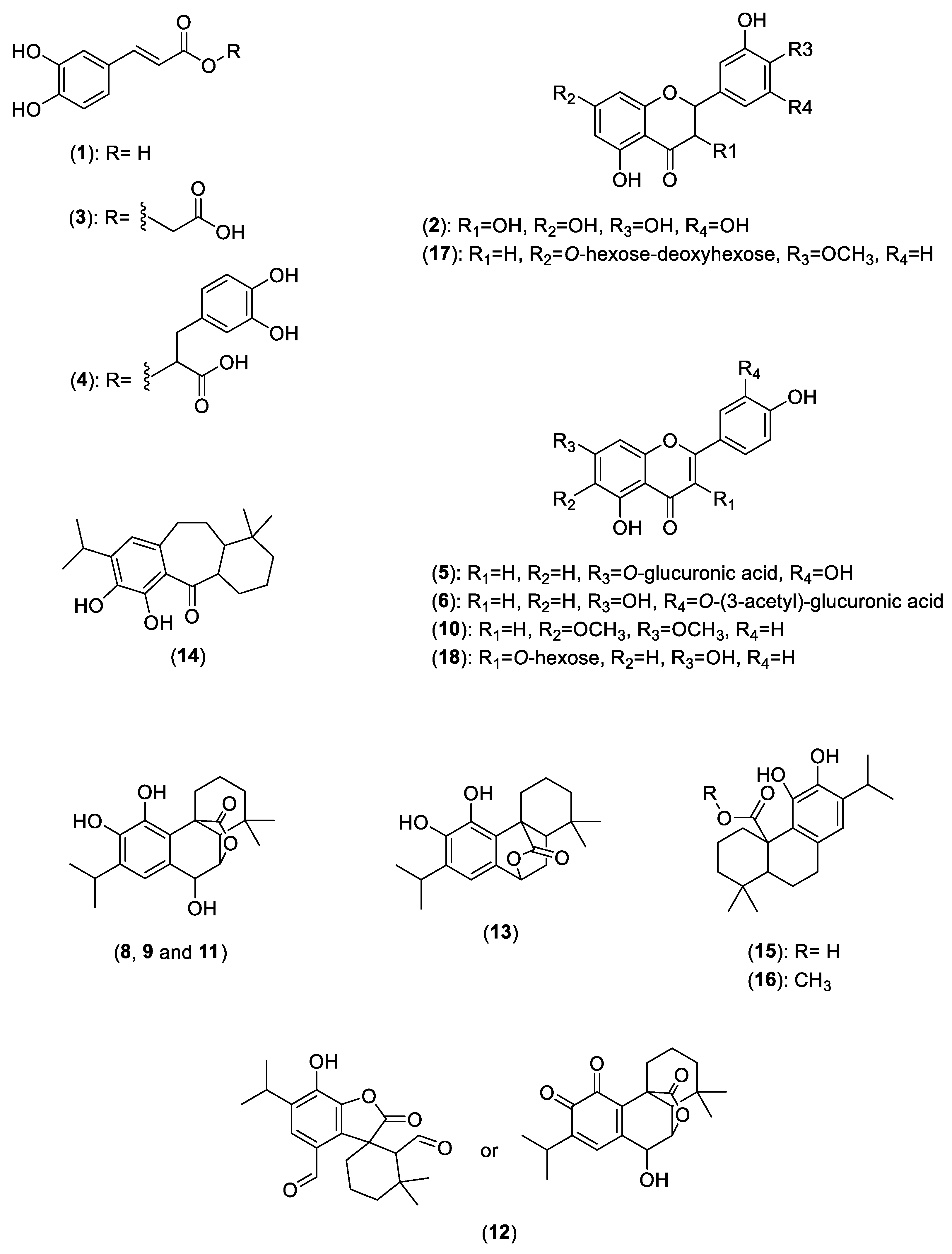
| ID | RT (min) | MS m/z [M − H]− | MS/MS m/z | Compound | Reference |
|---|---|---|---|---|---|
| 1 | 4.9 | 179 | 135 | Caffeic acid | [68] |
| 2a | 6.5 | 305 | 225 | Gallocatechin | [69] |
| 3 | 8.2 | 237 | 179, 161 | Caffeoylglycolic acid | - |
| 4a | 9.4 | 359 | 161(100), 179, 197 | Rosmarinic acid | [70] |
| 5 | 10.8 | 461 | 285(100), 199, 151 | Luteolin-7-O-glucuronide | [68] |
| 6 | 11.7 | 503 | 399, 285(100), 255 | Luteolin-3′-O-(3-acetyl)-glucuronide | [11] |
| 7 | 12.0 | 315 | 300(100), 227, 199 | Methoxy-tetra-hydroxy-flavone | - |
| 8a | 12.8 | 345 | 301, 283, 267, 217 | Rosmanol isomer | [70] |
| 9a | 13.4 | 345 | 283, 267, 227 | Rosmanol isomer/epirosmanol | [70] |
| 10 | 14.1 | 313 | 298, 283, 255, 227, 164 | Cirsimaritin | [71] (CCMSLIB00004718157) |
| 11 | 15.6 | 345 | 283, 227 | Rosmanol isomer/epirosmanol | [70] |
| 12a | 16.9 | 343 | 299, 243, 216 | Rosmadial or Rosmanol quinone | [68] |
| 13a | 17.7 | 329 | 285(100), 201 | Carnosol | [70] |
| 14 | 18.9 | 315 | 285, 201 | Rosmaridiphenol | [68] |
| 15 | 20.3 | 331 | 287(100), 272, 244 | Carnosic acid | [68] |
| 16 | 20.9 | 345 | 301(100), 286, 271 | Methyl carnosate | [70] |
| RT (min) | MS m/z [M + H]+ | MS/MS m/z | Compound | Reference | |
| 17 | 10.3 | 611 | 547, 449, 287(100) | Hesperidin | [71] (CCMSLIB00000214340) |
| 18 | 10.8 | 449 | 287(100) | Astragalin | [71] (CCMSLIB00003136634) |
Disclaimer/Publisher’s Note: The statements, opinions and data contained in all publications are solely those of the individual author(s) and contributor(s) and not of MDPI and/or the editor(s). MDPI and/or the editor(s) disclaim responsibility for any injury to people or property resulting from any ideas, methods, instructions or products referred to in the content. |
© 2023 by the authors. Licensee MDPI, Basel, Switzerland. This article is an open access article distributed under the terms and conditions of the Creative Commons Attribution (CC BY) license (https://creativecommons.org/licenses/by/4.0/).
Share and Cite
Guimarães, N.S.S.; Ramos, V.S.; Prado-Souza, L.F.L.; Lopes, R.M.; Arini, G.S.; Feitosa, L.G.P.; Silva, R.R.; Nantes, I.L.; Damasceno, D.C.; Lopes, N.P.; et al. Rosemary (Rosmarinus officinalis L.) Glycolic Extract Protects Liver Mitochondria from Oxidative Damage and Prevents Acetaminophen-Induced Hepatotoxicity. Antioxidants 2023, 12, 628. https://0-doi-org.brum.beds.ac.uk/10.3390/antiox12030628
Guimarães NSS, Ramos VS, Prado-Souza LFL, Lopes RM, Arini GS, Feitosa LGP, Silva RR, Nantes IL, Damasceno DC, Lopes NP, et al. Rosemary (Rosmarinus officinalis L.) Glycolic Extract Protects Liver Mitochondria from Oxidative Damage and Prevents Acetaminophen-Induced Hepatotoxicity. Antioxidants. 2023; 12(3):628. https://0-doi-org.brum.beds.ac.uk/10.3390/antiox12030628
Chicago/Turabian StyleGuimarães, Natalia S. S., Vyctória S. Ramos, Laura F. L. Prado-Souza, Rayssa M. Lopes, Gabriel S. Arini, Luís G. P. Feitosa, Ricardo R. Silva, Iseli L. Nantes, Debora C. Damasceno, Norberto P. Lopes, and et al. 2023. "Rosemary (Rosmarinus officinalis L.) Glycolic Extract Protects Liver Mitochondria from Oxidative Damage and Prevents Acetaminophen-Induced Hepatotoxicity" Antioxidants 12, no. 3: 628. https://0-doi-org.brum.beds.ac.uk/10.3390/antiox12030628