Article

Phytochemical Characterization, Antioxidant, and Anti-Proliferative Activities of Wild and Cultivated Nigella damascena Species Collected in Sicily (Italy)

1
Department of Environmental, Biological and Pharmaceutical Sciences and Technologies (DiSTABiF), University of Campania “Luigi Vanvitelli”, Via Vivaldi 43, 81100 Caserta, Italy
2
Department of Biology, University of Naples “Federico II”, 80138 Naples, Italy
3
Department of Biological, Chemical and Pharmaceutical Sciences and Technologies (STEBICEF), University of Palermo, Viale delle Scienze, Ed. 17, 90128 Palermo, Italy
4
NBFC, National Biodiversity Future Center, 90133 Palermo, Italy
5
Centro Interdipartimentale di Ricerca “Riutilizzo Bio-Based Degli Scarti da Matrici Agroalimentari” (RIVIVE), University of Palermo, 90128 Palermo, Italy
6
Department of Biology and Pharmaceutical Botany, Medical University of Gdańsk, 80210 Gdańsk, Poland
7
Bona Furtuna LLC, Los Gatos, CA 95030, USA
8
Department of Precision Medicine, University of Campania “Luigi Vanvitelli”, Via L. De Crecchio 7, 80138 Naples, Italy
*
Authors to whom correspondence should be addressed.
These authors contributed equally to this work.
Submission received: 31 January 2024 / Revised: 22 March 2024 / Accepted: 26 March 2024 / Published: 27 March 2024
(This article belongs to the Special Issue Antioxidant and Protective Effects of Plant Extracts)

Abstract

:
The use of Nigella damascena seeds in the culinary field or as aerial parts infusions in the pharmaceutical and cosmetic fields is widely reported. The biological activity of this plant, as demonstrated over the years, is closely related to its phytochemical content. This investigation focused on the comparative study of the same plants of N. damascena, one totally wild (WND), while the other two, one with white flowers (CWND) and the other with blue flowers (CBND), were subject to cultivation, irrigation, and manual weeding. Using the potential of 1D and 2D-NMR spectroscopy, coupled with MS/MS spectrometric studies, the three methanolic extracts of N. damascena were investigated. Chemical studies have highlighted the presence of triterpene saponin compounds and various glycosylated flavonoids. Finally, the in vitro antiproliferative and antioxidant activities of the three individual extracts were evaluated. The antiproliferative activity performed on U-937, HL-60, and MCF-7 tumor cell lines highlighted a greater anticancer effect of the CBND and CWND extracts compared to the data obtained using WND. The antioxidant activity, however, performed to quantify ROS generation is comparable among the extracts used.

1. Introduction

Nigella damascena L., commonly love-in-a-mist, is a species with a Euro-Mediteranean distribution (Figure 1), growing spontaneously from Macaronesia to the Mediterranean basin (excluding Egypt and Israel) up to northwestern Iran [1].
This species has a single, erect stem, which may branch at the top. The basal leaves are linear–spatulate, and the flowers are terminal, solitary, and surrounded by an involucre of filiform bractiform leaves (Figure 2a). The fruit of N. damascena are ovoid capsules, smooth and ribbed. When ripe, the fruit opens at the top to release numerous black, triangular, wrinkled, and aromatic seeds [3].
Commercially, N. damascena continues to be used in herbal medicine, with its seeds and derived oil being marketed as “black cumin”, a term encompassing both N. damascena and N. sativa. The primary interest in N. damascena today is as an ornamental garden plant and for its flowering beauty [4]. It is extensively cultivated in gardens for the aesthetic appeal of its numerous hybrid varieties [5], which feature double petaloid flowers in colors ranging from white to pink and various shades from blue to purple (Figure 2b) [6].
In its natural distribution range, this plant grows spontaneously in anthropogenic ecosystems: in fact, it tends to colonize disturbed areas, such as dry uncultivated fields, vineyards, arid fields and meadows, and on-the-road margins, from sea level to 1000 m a.s.l. [7].
This species is one of the 26 accepted taxa belonging to the genus Nigella, described by Linnaeus in Species plantarum [8] as one of the smaller genera of the Ranunculaceae family [4], which includes the better-known Nigella sativa (black cumin), a species widely consumed as a seasoning in North Africa, the Arabian Peninsula, and India.
Thus, if for N. sativa, the ancient medico-botanical literature of the past gives extensive treatment, N. damascena is considered of lesser interest [9]. The seeds of N. damascena L. are used as ingredients in a wide range of foods [10], for example, as flavorings in bread and cheese [11], and are widely present in the folk medicine of various countries and in traditional medicine, N. damascena seeds have been employed for menstrual regulation, indicating possible estrogenic effects [12], for catarrhal conditions, [13], for their pain-relieving, anti-edema, fever-reducing, worm-expelling, and disinfectant properties [14]. In Sicilian folk medicine, a seed infusion, referred to as vaccareddi or lampiuneddi, is used as a milk-production stimulant [15], but several cases of intoxication have been recorded [16].
The phytochemical analyses carried out on the extracts obtained from the different parts of N. damascena allowed us to distinguish two main classes of predominant metabolites, the alkaloids and the terpene derivatives. Carrying out an exhaustive analysis of the research on N. damascena, to date, 57 compounds have been isolated, and it is essential among these to number the various dolabbelanic diterpenes, complex structures often esterified with phenylacetyl, benzoyl, acetyl, nicotinoyl, and isobutyryl portions [3]. It should be noted that dolabellans are mostly products of marine organisms, such as mollusks or several algae [17], and that these are present in a few terrestrial plants [18,19,20], including N. damascena [21,22]. Over the years, different activities have been evaluated for this compound and among these, antimicrobial, anti-inflammatory [23], and anti-tumor [24] activities have been carried out.
The intention of this work aims to evaluate a qualitative diversity of polar extracts obtained from cultivated samples of N. damascena “Oxford Blue” (CBND), cultivated samples of N. damascena (CWND), and spontaneous plants collected from the wild (WND). Specifically, using 1D and 2D NMR techniques, coupled with UHPLC-ESI-QqTOF HR MS/MS experiments, the possible chemical diversity in qualitative terms on flavonoid, dolabellane, and triterpene compounds was evaluated. Subsequently, antioxidant and anticancer tests were carried out. The tests performed did not show the promising activity of the plants studied here, but they highlighted how phytochemical differences play a fundamental role in biological activity.

2. Materials and Methods

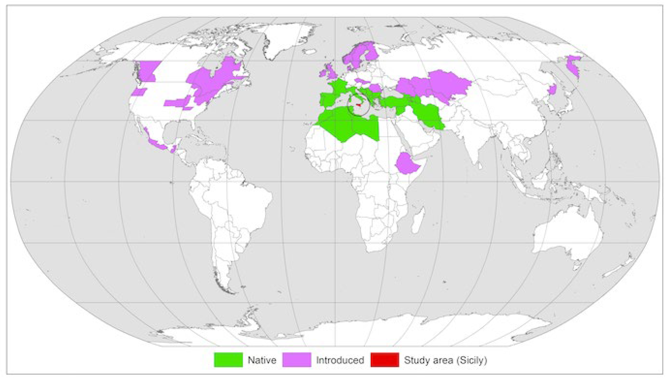
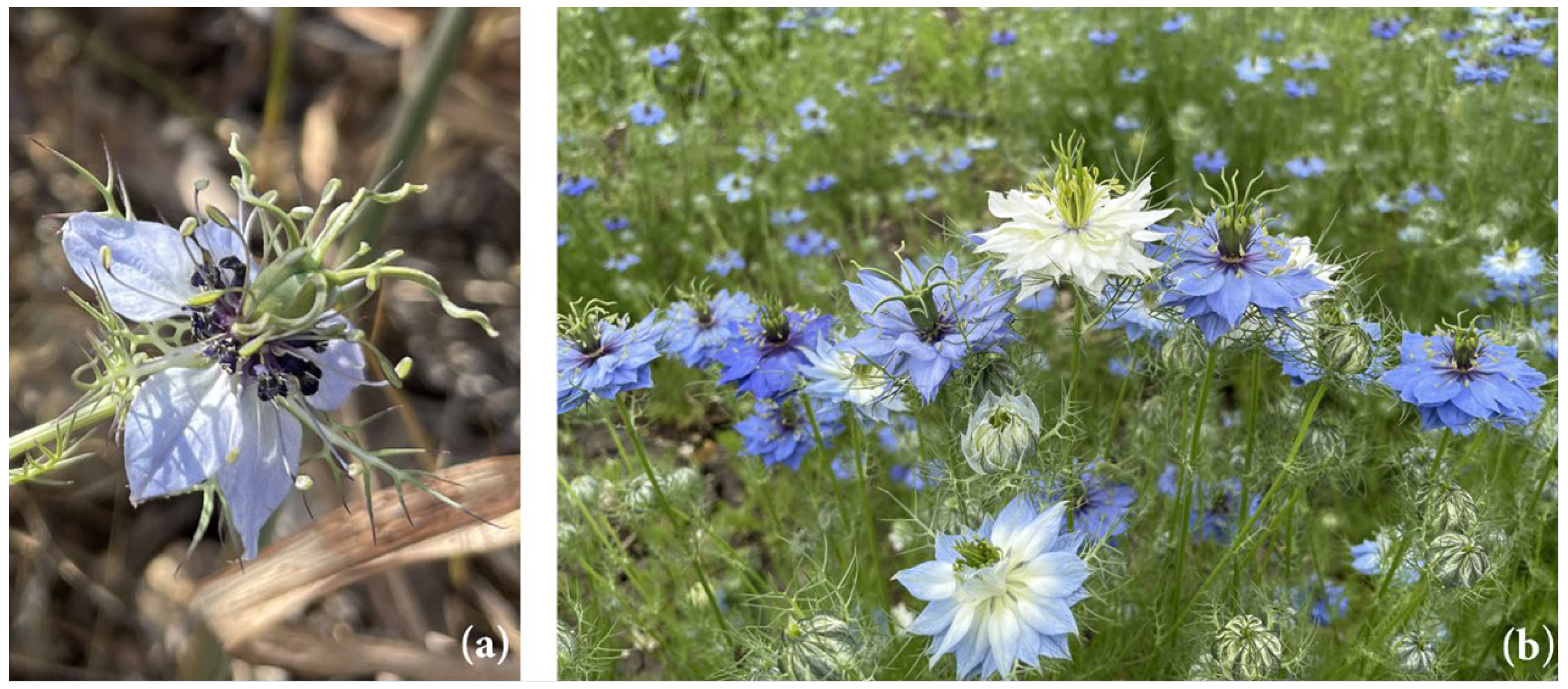
2.1. Plant Materials

The plant material consists of plants N. damascena, both wild and cultivated, at the full flowering stage. The samples were collected in May 2022 within the Bona Furtuna farm (Corleone, Italy), which funded the “Extremophytes Potential” project. This project aims to understand how extreme environmental conditions affect the production of biologically active compounds and how cultivation influences the synthesis of bioactive compounds that plants produce under stressful conditions. The collection site represents a significant example of the traditional rural landscape, characterized by a high degree of naturalness comprising elements of considerable naturalistic and environmental value.
The wild samples were taken from the natural population of N. damascena (WND) (37°45′49″ N 13°18′34″ E) located at an altitude of 770 m above sea level, growing in the Mediterranean dry grassland that settles at the base of Monte Barraù, a Special Conservation Zone (ZCS) of the Monti Sicani district.
The cultivated samples were taken from a cultivation plot within the Bona Furtuna farm (37°45′49″ N 13°18′23″ E) at an altitude of 760 m above sea level, about 300 m away from the natural population. These were the N. damascena “Oxford Blue” (CBND) and N. damascena (CWND), cultivated using traditional low-intensity methods to bring the plant to normal conditions of stress induced by the Mediterranean climate.

2.2. Extraction Procedure

For each sample (WND, CWND, and CBND), flowers, leaves, and stems, weighed using a technical scale (~88 g), once frozen (−20 °C), were subjected to a freeze-drying process until they reached a constant weight. Therefore, the freeze-dried plant material from each group (~20 g) was finely ground and then subjected to an extraction process at room temperature using methanol (50 mL × 3 times) (Sigma-Aldrich Corporation, St. Louis, MO, USA). After extraction procedures, the extracts were filtered, reduced in volume at low pressures and at low temperatures, and freeze-dried again until a constant weight was reached. The final yield was 27.95% for WND, 20.30% for CWND, and 19.96% for CBND.

2.3. Metabolite Profiling

Dried and powdered plant material (50 mg) for each sample type (WND, CWND, and CBND) was transferred to a 2 mL microtube and suspended in a mixture of 1.5 mL of phosphate buffer (Fluka Chemika, Buchs, Switzerland; 90 mM; pH 6.0) in D2O (Cambridge Isotope Laboratories, Andover, MA, USA)—containing 0.1% w/w trimethylsilylpropionic-2,2,3,3-d4 acid sodium salt (TMSP, Sigma-Aldrich, St. Louis, MO, USA)—and CD3OD (Sigma-Aldrich, St. Louis, MO, USA) (1:1). The mixture was vortexed at room temperature for 1 min, ultrasonicated (Elma Transsonic Digital, Hohentwiel, Germany) for 40 min, and then centrifuged (Beckman Allegra™ 64R, F2402H rotor; Beckman Coulter, Fullerton, CA, USA) at 13,000 rpm for 10 min. A volume of 0.60 mL was transferred to a 5-mm NMR tube and analyzed by NMR and LC-MS.
Partial purification of extracts was performed by dissolving the different dried extracts in water and partitioning them in ethyl acetate.

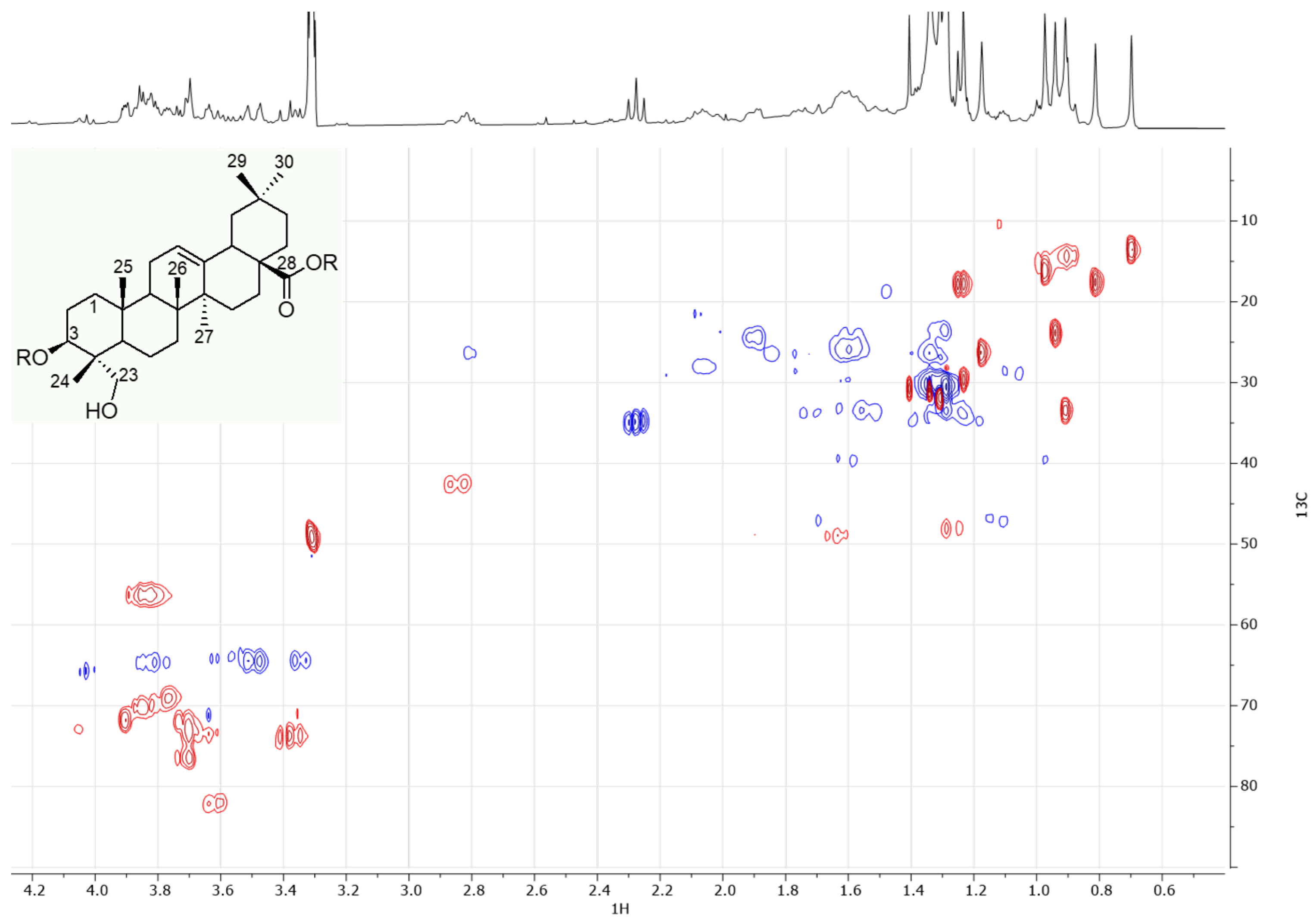
2.3.1. NMR Experiments

NMR spectra were recorded at 25 °C on a 300.03 MHz for 1H and 75.45 MHz for 13C on a Bruker Fourier transform NMR. CD3OD was used as the internal lock. The 1H-NMR was acquired using a 1d-NOESY sequence to suppress the residual H2O signal. Each 1H-NMR spectrum consisted of 32 scans with the following parameters: acquisition time (AQ) = 4.54 s; relaxation delay (RD) = 2 s. Free induction decays (FIDs) were Fourier-transformed, and the resulting spectra were manually phased, baseline-corrected, and calibrated to TMSP. COSY, HSQC, and HMBC spectra were acquired using standard sequences from the Bruker library. On the partially purified fraction, NMR analyses were performed using standard zg30, HSQC, and HMBC sequences from the Bruker library.

2.3.2. UHPLC-ESI-QqTOF HR MS/MS Experiments

Extracts were analyzed using a NEXERA UHPLC system (Shimadzu, Tokyo, Japan) and the Luna® Omega C-18 column (Shimadzu, Tokyo, Japan). The linear gradient for separation purposes consisted of water (A) and acetonitrile (B), both with 0.1% formic acid. B eluent was held at 5% for 0.5 min and was allowed to reach 17.5% at 5 min; then, it was increased to 45% in 5 min and held at this % for the other 2 min. Finally, it was augmented to 95% in 1 min, and following another min at 95%, initial conditions were restored in 1 min, allowing for the system to re-equilibrate for 1 min. The flow rate was 0.5 mL/min. The injection volume was 2.0 μL. MS analysis was performed using the AB SCIEX Triple TOF® 4600 (AB Sciex, Concord, ON, Canada), equipped with a DuoSprayTM ion source, operating in negative ESI mode. A full scan TOF survey (170–1700 Da) and eight IDA (information-dependent acquisition) experiments were performed using the following parameters: curtain gas 35 psi; nebulizer gas 60 psi; heated gas 60 psi; ion spray voltage −4.5 kV; interface heater temperature 600 °C; declustering potential −80V; collision energy −45 ± 10 V. The instrument was controlled by Analyst® TF 1.7.1 software. Data processing was carried out by PeakView® software version 2.2.

2.4. Cell Lines and Culture Conditions

The cell lines HaCat, U-937, HL-60, and MCF-7 were obtained from the American Type Culture Collection (ATCC, Rockville, MD, USA). U-937 and HL-60 cells were grown in Roswell Park Memorial Institute (RPMI)-1640 medium (Gibco, New York, NY, USA) (Euroclone, Pero, Italy), while HaCat and MCF-7 in Dulbecco’s Modified Eagle Medium (DMEM) were grown at 37 °C in 5% CO2 atmosphere, then supplemented with 10% heat-inactivated fetal bovine serum (FBS), 1% l-glutamine, 1% ampicillin or streptomycin, and 0.1% gentamicin. All cell lines were treated with extract of CWND, CBND, and WND at different concentrations ranging from 1.25 mg/mL to 5 mg/mL from 24 h to 72 h. For the biological assays, N. damascena extracts were resuspended in dimethyl sulfoxide (DMSO) and used at the final concentrations indicated in each experiment. The DMSO was used as a negative control.

2.4.1. MTT Assay

Cell vitality was determined by MTT assay. A total of 2 × 10⁴ cells/well were plated in a 96-well plate and treated, in triplicates, with extract of WND, CWND, and CBND at different concentrations and times. Thiazolyl Blue Tetrazolium Bromide [3-(4,5-dimethylthiazol-2-yl)-2,5-diphenyltetrazolium bromide] (MTT; Sigma-Aldrich) solution was added at 0.5 mg/mL. After 4 h, for the adhesion cells HaCat and MCF-7, the supernatant was simply removed, whereas, for the suspension cells U-937 and HL-60, the plates were first centrifuged. The purple formazan crystals were dissolved in isopropanol (Carlo Erba Reagents, Cornaredo, Italy), and the absorbance was read at a wavelength of 570 nm with a TECAN M-200 reader (Tecan, Männedorf, Switzerland) [25].

2.4.2. FACS Analysis

The BD FACS Celesta flow cytometer from BD Biosciences was used to evaluate the percentage of cell death and the distribution of cells in the different phases of the cell cycle. After treatment with extracts, cells were harvested, and after washing with phosphate-buffered saline (1× PBS), cells were treated with PI buffer (1× PBS and 2 mg/mL propidium iodide) to evaluate the cell death rate. Subsequently, after a further wash with 1× PBS, the cells were treated with Cycle Buffer (1× PBS, 10% NP-40, 10% sodium citrate, and 2 mg/mL propidium iodide) for 15 min at room temperature to evaluate the percentage of cells in different phases of the cell cycle.

2.4.3. Polymorphonuclear Leukocytes (PMN) Isolation

Whole blood was obtained with informed consent from healthy volunteers at the University “Federico II” in Naples, Italy. Polymorphonuclear leukocytes were isolated following the protocol described by Badalamenti et al. [26]. The isolated PMNs were measured in the presence or absence of extract of WND, CWND, and CBND, without or with Opsonised zymosan (OZ).

2.5. Reactive Oxygen Species (ROS) Generation and Antioxidant Enzymes Measured in PMN Cells

Dichlorofluorescein (DCF) assay was performed to quantify ROS generation, according to Manna et al. [27]. ROS quantity was monitored by fluorescence (excitation wavelength of 350 nm and an emission wavelength of 600 nm) on a microplate reader. Results were expressed as fluorescence intensity. A commercial kit (BioAssay System, San Diego, CA, USA) was used to determine superoxide dismutase (SOD) and catalase (CAT) enzymatic activity in PMN cells according to the manufacturer’s recommendations. The activity of enzymes was expressed as U/L [28]. The PMN was treated with extracts of WND, CWND, and CBND at the concentration of 0.5 mg/mL without or with OZ (0.5 mg/mL).

2.6. Statistics

All data and statistics were analyzed by GraphPad PrismTM 7.0 software (Graph PadPrismTM Software Inc., San Diego, CA, USA). Values are mean ± standard deviation (SD) of biological triplicates.

3. Results

3.1. Chemical Characterization of the Extracts

The 1H-NMR spectra of the wild and cultivated N. damascena extracts (Figure S1) were analyzed. In all of the samples, traces of phenolic compounds were detected, but their relative abundance compared to the other components of the extracts was low. However, many signals in the region between 6.0 and 8.5 ppm were detected and suggested the presence of flavonoid derivatives [29,30]. Two doublets due to the meta-coupled protons of the A-ring of flavonoids (J = 2.0 Hz) were detected at δH 6.33 and 6.54, respectively. These two signals, which showed COSY correlations with each other (Figure S2), also showed HSQC correlations with carbons at δC 99.0 and 94.2, respectively (Figure S3). Furthermore, ABX and AA’BB’ aromatic systems were recognizable and suggested the presence of quercetin and kaempferol derivatives. The main quercetin derivative detected in the extracts was characterized by a doublet (J = 8.1 Hz) at δH 7.02 (δC 115.4), a doublet of a doublet at δH 7.56 (J = 8.1, 2.0 Hz; δC 122.0), and a doublet (J = 2.0 Hz) at δH 7.67 (δC 116.9). On the other hand, the main kaempferol derivative was characterized by two doublets (J = 8.5 Hz) at δH 7.00 (partially overlapped with the signal of quercetin at 7.02) and 8.10, correlating with each other in the COSY experiment. Long-range NMR experiments confirmed the presence of these flavonoids, but further discrimination of the different derivatives was not possible; the samples were, therefore, further analyzed by UHPLC-ESI QqTOF-MS/MS.
Beyond sweetener compounds (Figure S4), such as hexosyl hexitol (e.g., maltitol, C12H24O11), identifiably based on its [M-H] ion at m/z 343.1247, and the TOF MS/MS fragment ions at m/z 181.07 and 179.06, and N-(deoxyfructosyl)leucine ([M-H] ion at m/z 292.1402; C12H23NO7), the great part of the identified compounds in WND, CWND, and CBND extracts belong to flavonol glycosides (Table 1). In fact, the relative content of these compounds was 59.8, 87.1, and 60.5% in WND, CWND, and CBND extracts, respectively. As already suggested by NMR analyses, the derivatives of quercetin and kaempferol were the most representative. Quercetin glycosides accounted for 81.4, 74.5, and 83.2% of the extracts.
Compound 1, with the [M-H] ion at m/z 949.2459, underwent, in the TOF-MS/MS experiment, neutral loss of 648.2 (162.05 × 4) Da to provide the radical aglycone at m/z 300.0281, according to glycosylation occurrence at C-3 position (Figure S5a). Thus, the quercetin tetrahexoside was likely quercetin 3-O-gentiotetroside [31].
Compounds 2, 7, 8, and 16 were quercetin triglycosides (Figure S5b), mainly differing in their glyconic moiety. The deprotonated molecular ion of compound 2 underwent the neutral loss of 146.06 Da (deoxyhexose—H2O) to achieve the ion at m/z 625.14 or a homolytic cleavage to provide the radical ion at m/z 446.09. Both ions were in accordance with a dihexosyl moiety at the quercetin C-3 position, while deoxyhexose was positioned at C-7 carbon. According to this hypothesis, quercetin 3-O-sophoroside-7-O-rhamnoside was previously found in N. sativa [32]. The neutral loss of 486.15 Da from the [M-H] ion of compound 7, and the high abundance of the TOF-MS/MS quercetin radical ion were in accordance with quercetin 3-O-trihexoside occurrence, while compound 8 was likely quercetin 3-O-hexosylpentoside-7-O-deoxyhexoside. In fact, the loss of dehydrated deoxyhexose was detectable, together with the radical ion at m/z 446.09. Finally, compound 16 was likely a quercetin 3-O-dihexosylpentoside.
Compounds 10, 15, and 20 were quercetin 3-O-diglycosides, showing as sugar moiety, dihexose (−324.11 Da), hexosylpentose (−294.10 Da), and hexosyldeoxyhexose (−308.11 Da), respectively (Figure S6), while quercetin hexoside (21) and quercetin deoxyhexoside (31) were the identified monoglycosyl derivatives. Compound 31 showed as the base peak of the ion at m/z 301.0368, according to glycosylation at the C-7 position.
Interestingly, hydroxymethyl glutaryl derivatives of the above-described quercetin glycosides, as well as acylated derivatives, were also identified (Figure S7). Compounds 9, 13, 18, and 24 were hydroxymethyl glutaryl derivatives of compounds 2, 7, 8, and 16, respectively. The neutral losses of 62.00 Da (CO2 + H2O), 102.03 Da, and 144.04 Da suggested the presence of the hydroxymethyl glutaryl residue. The TOF-MS/MS spectra of compounds 9 and 13 highlighted that these neutral losses occurred from their relative deprotonated molecular ions and from the [M-H-(deoxyhexose-H2O)] ion, suggesting that hydroxymethyl glutaryl was located on C-3 glyconic residue. Analogously, a similar fragmentation pattern was shared from compound 23, which was putatively quercetin (hydroxymethylglutaryl)hexosylpentoside.
Hydroxycinnamoyl quercetin triglycosides were also detected (Figure S8), distinguishable based on the hydroxycinnamoyl moiety in caffeoyl- (19 and 22), feruloyl- (25 and 28), and p-coumaroyl- (26 and 27) derivatives.
Similarly, kaempferol glycosides identified differed in the length of the glyconic moiety, and their identity (Figures S9 and S10). Briefly, compounds 3 and 6 were constitutional isomers, sharing the neutral loss of 146.05 Da and the formation through the loss of a dihexosyl radical of the radical form of kaempferol deoxyhexose. Compounds 11 and 14 were likely the hydroxymethyl glutaryl derivatives of the previous ones, while compounds 12 and 30 were tentatively kaempferol 3-O-trihexoside and its feruloyl derivative, respectively. Two kaempferol dihexosides (4 and 17) and kaempferol deoxyhexoside (32) were also recognized. Among kaempferol glycosides, kaempferol-3-O-[β-d-glucopyranosyl-(1 → 2)-β-d-galactopyranosyl-(1 → 2)-β-d-glucopyranoside was previously reported as an abundant constituent of N. sativa, but it was only at trace levels in other Nigella species, so much so that it was hypothesized as a marker for identifying N. sativa-based products [33]. Furthermore, myricetin dihexosylpentoside (5) and isorhamnetin dihexoside (29) were putatively identified.
The other identified compounds were triterpene saponins, showing as aglycone hederagenin or oleanolic acid. This was confirmed both by the MS (Table 2, Figure S11) and NMR analyses. Concerning the NMR analyses, in order to simplify the identification of compounds in the mixture, a partial purification was carried out. While the more polar saponins were identified by MS (Table 2), the attention here was focused on the less polar and relatively abundant saponin, which was detected in the ethyl acetate fractions obtained from the partitioning of the whole extract with ethyl acetate/water. This fraction was analyzed by NMR. In the 1H and HSQC spectra (Figure 3 and Figure S12), a methyl singlet was detected at δH 0.70/δC 13.5. This signal showed long-range correlations (Figure S13) with two carbinol carbons at δC 81.8 and 64.0. The latter was identified as methylene, showing HSQC correlations with two diasterotopic protons at δH 3.36 and 3.50. These observations allowed us to attribute the signals at δH 0.70/δC 13.5 and that at δH 3.36–3.50/δC 64.0 to the positions 24 and 23 of the aglycone structure (Figure 3), respectively. The previously mentioned correlation observed for H-24 with the other carbinol carbon at δC 81.8 also suggested the presence of a hydroxyl group at the C-3 carbon of the aglycone. The H-24 protons also showed a long-range correlation with a quaternary carbon at δC 47.9, shared by another methyl signal at δH 0.97/δC 16.3. This allowed us to attribute the latter signals to the methyl-25. The signals belonging to the methyl-27 (δH 1.17/δC 26.3) were identified thanks to several correlations, with the diagnostic one being the correlation with the quaternary olefinic carbon (C-13) at δC 144.5. Methyl-26 was instead identified thanks to the correlations shared with methyl-25 and -27. Finally, the methyl-29 and methyl-30 at δH 0.90/δC 33.0 and at δH 0.94/δC 23.8, respectively, were identified based on their typically shared correlations. These data suggested that hederagenin was the aglycone of the most abundant saponin [34]. However, as highlighted in the MS analyses, the samples are characterized by the presence of several different saponins, and therefore, future studies could be aimed at the isolation and structural elucidation of these compounds.

3.2. Biological Properties

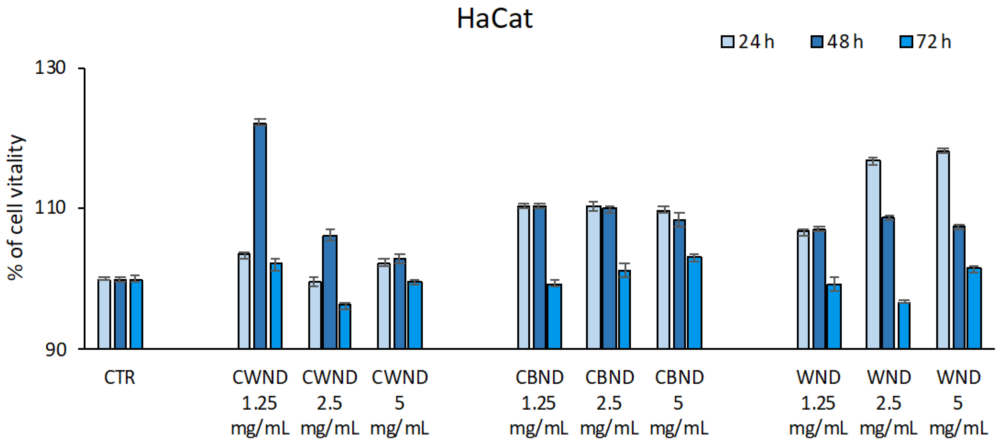
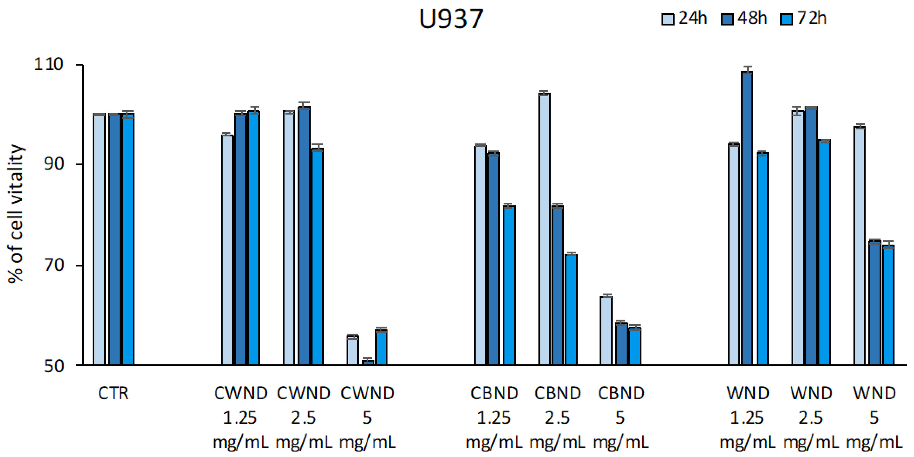
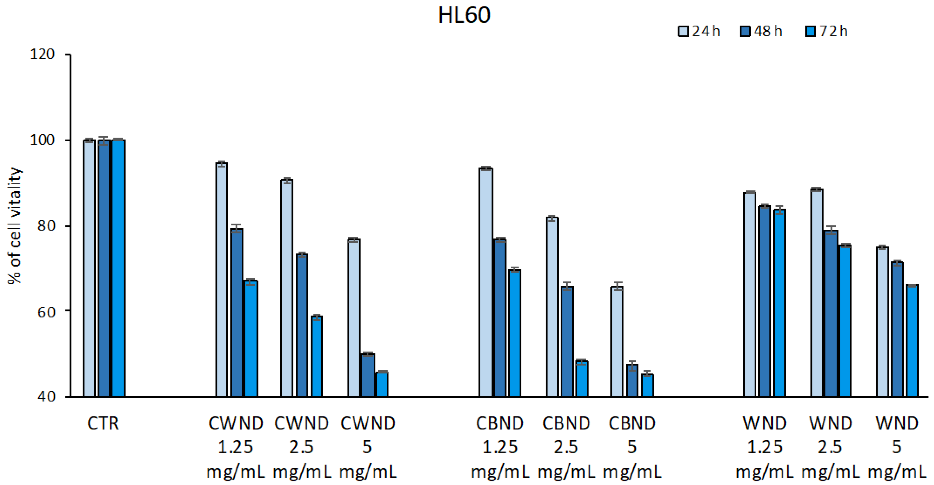
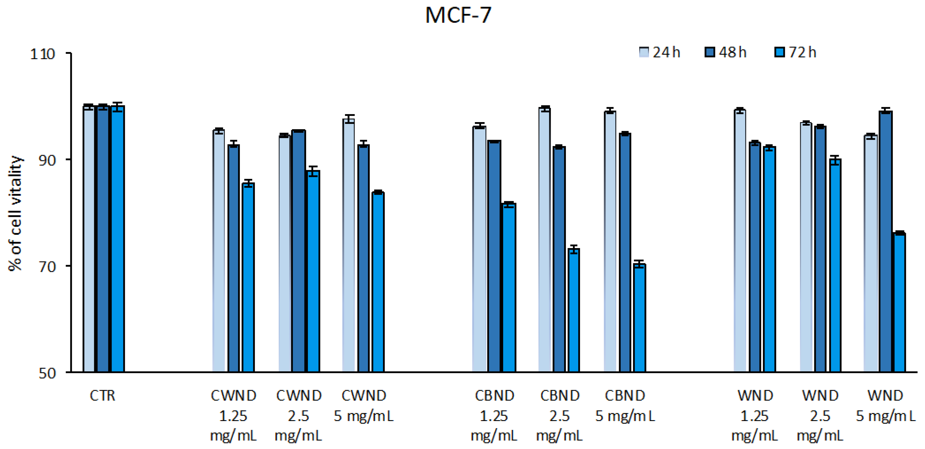
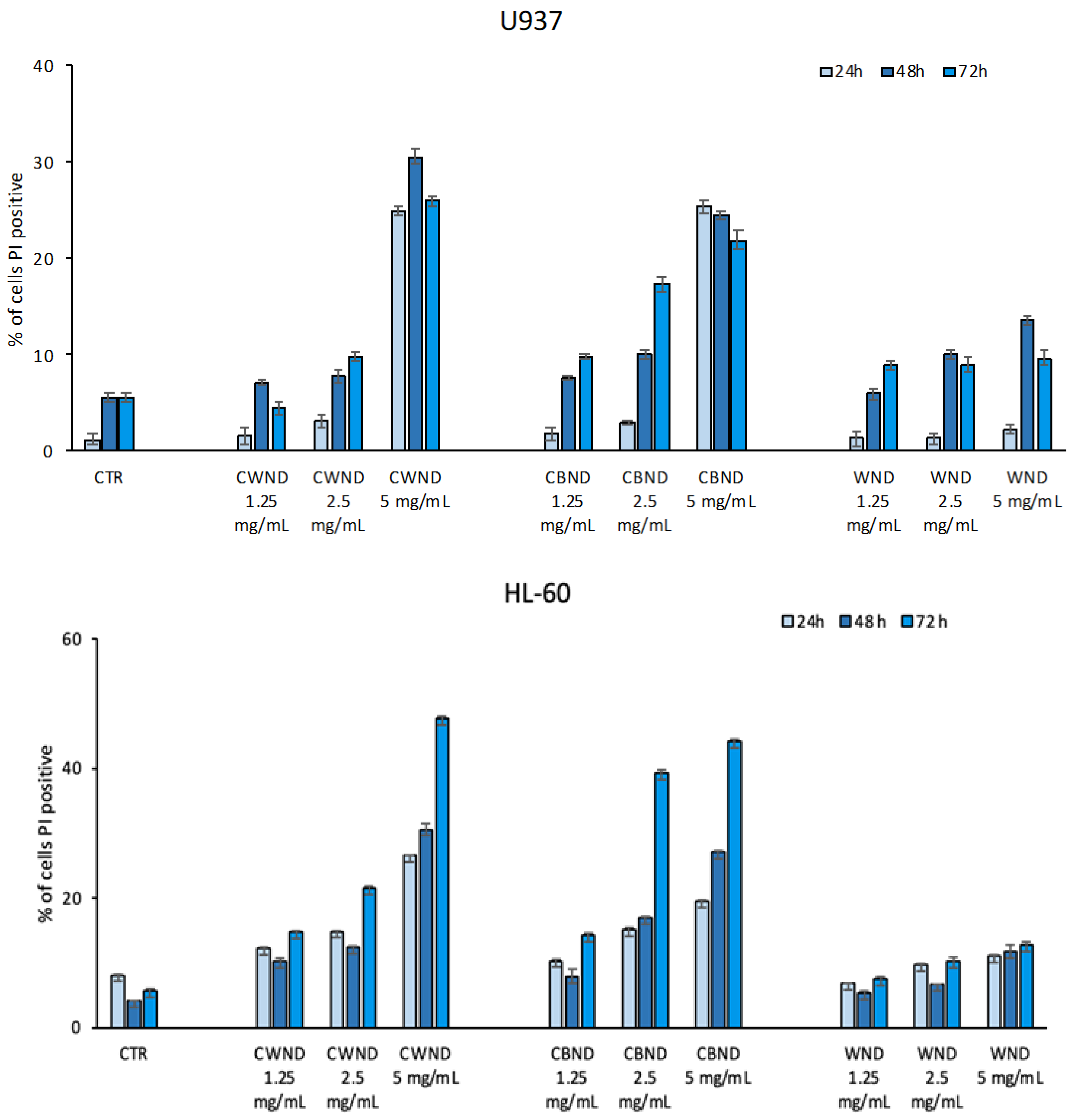
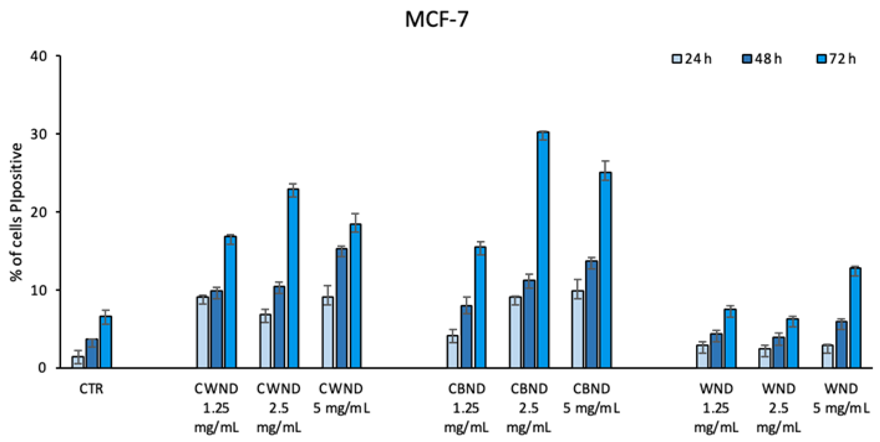
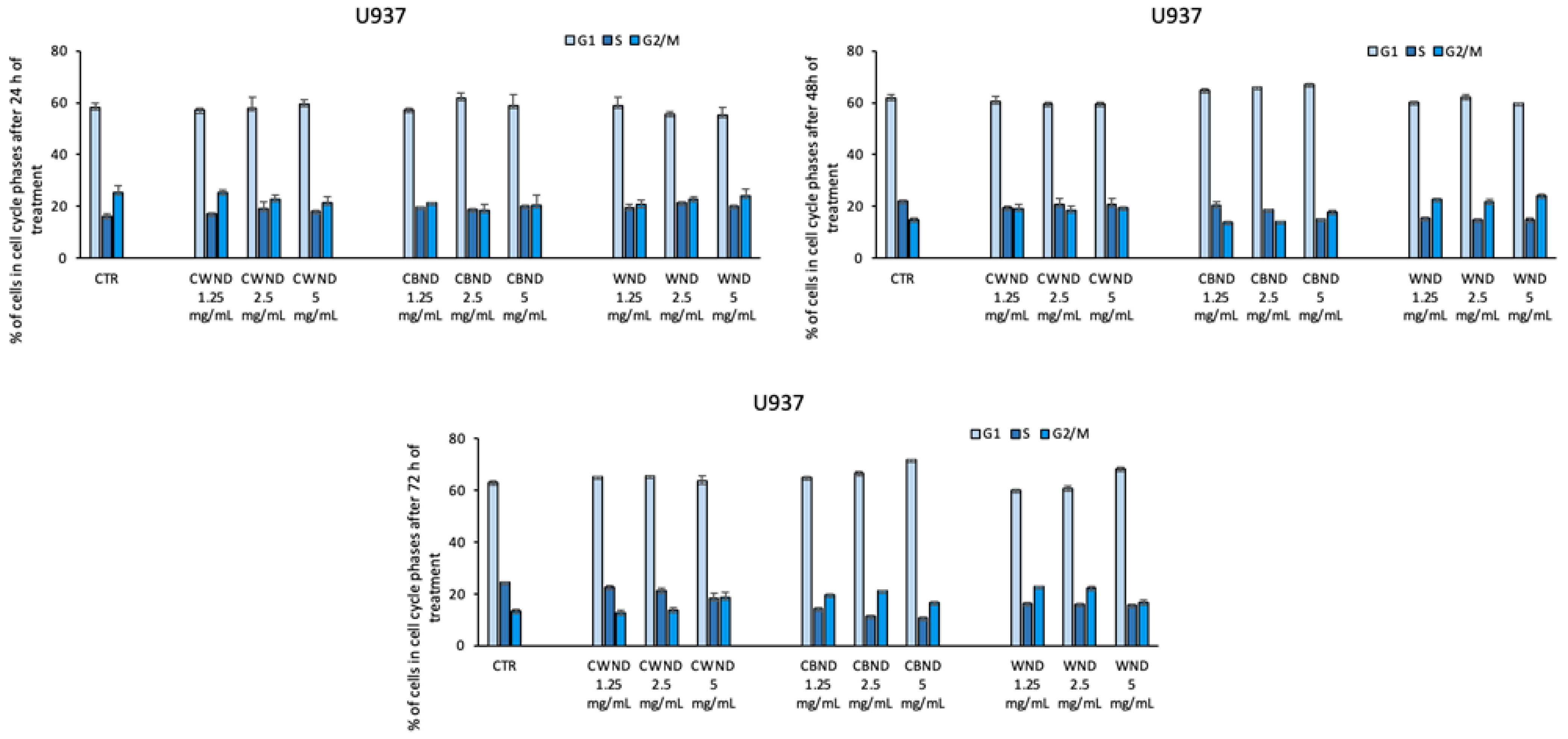
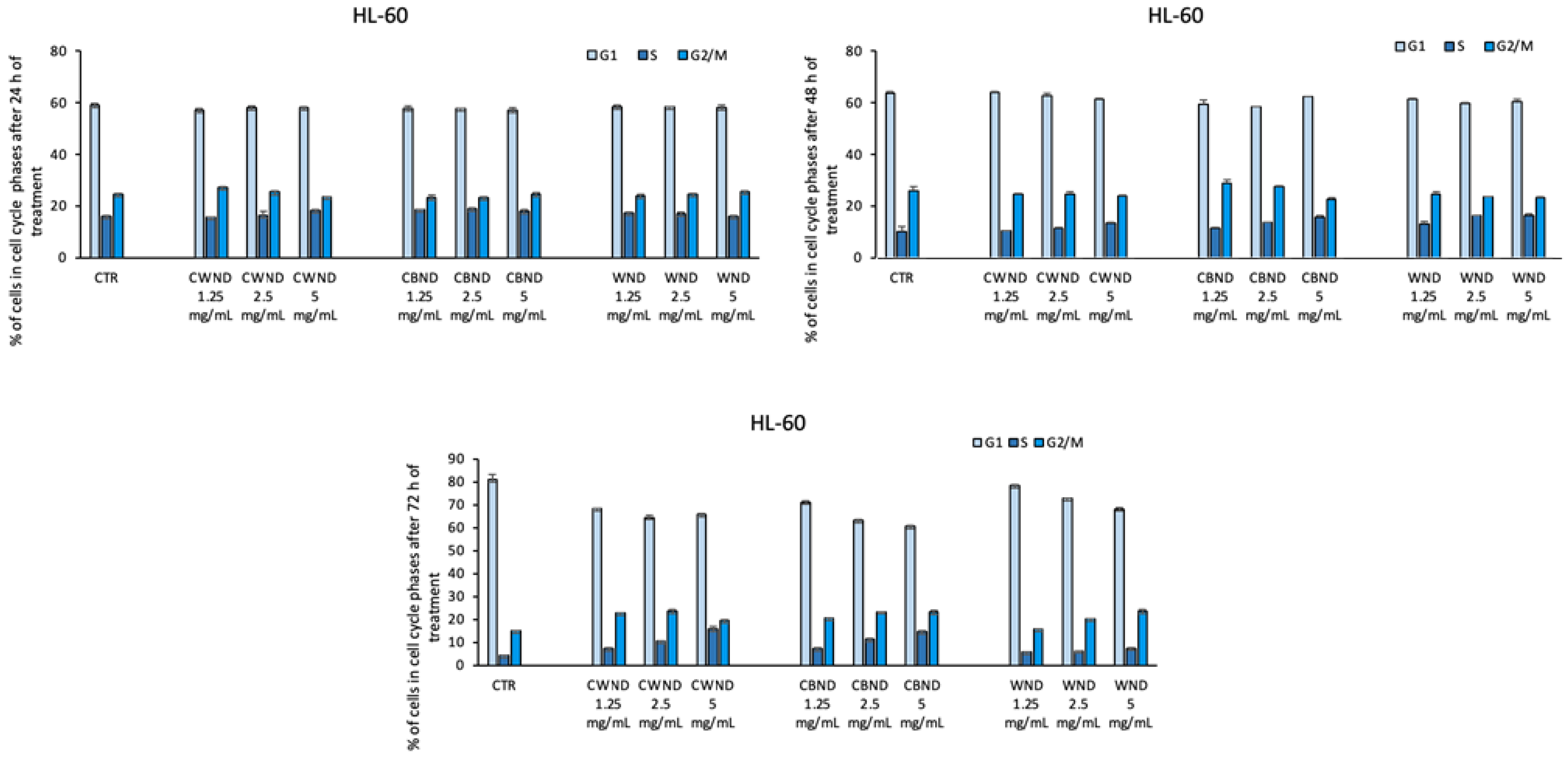
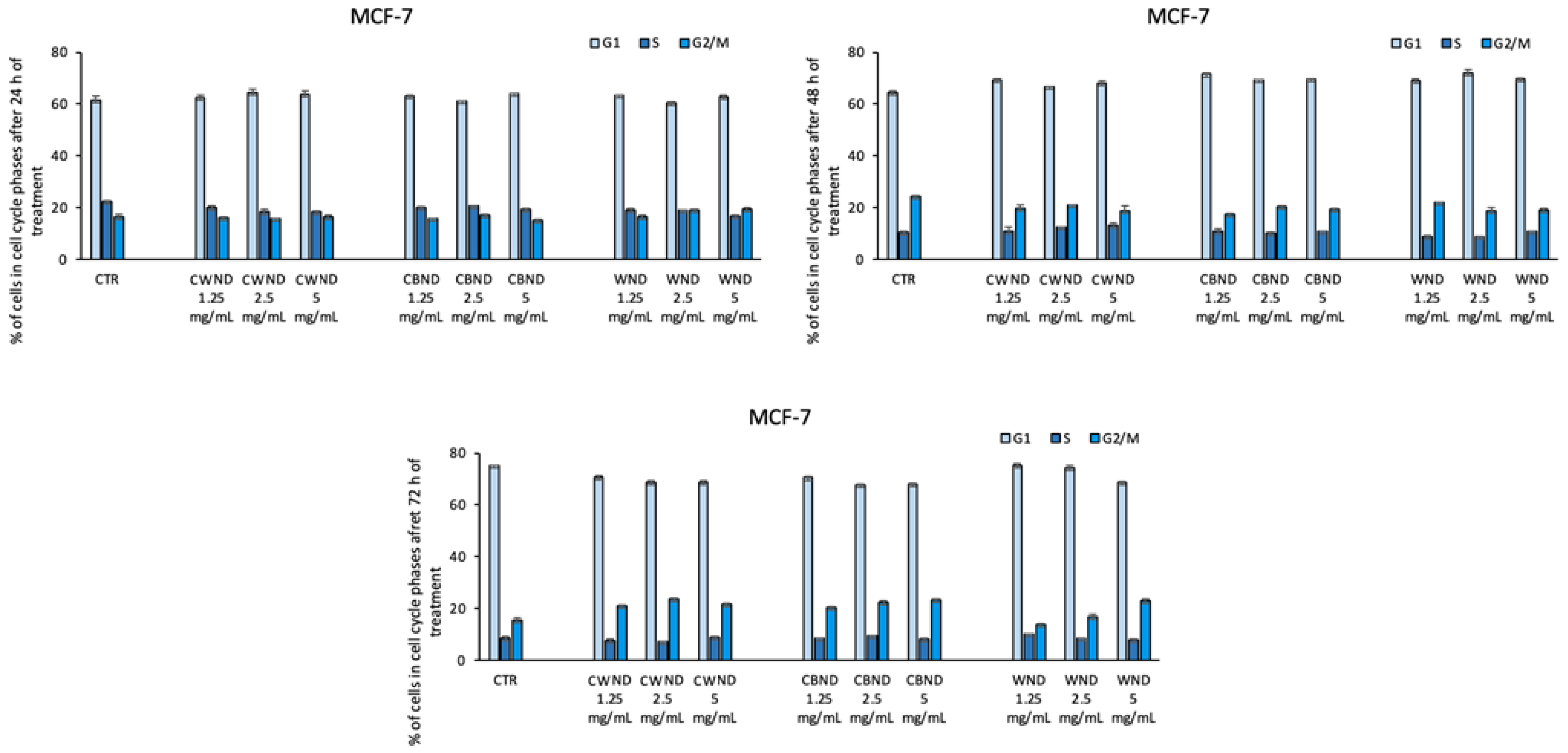
3.2.1. Biological Effect of CWND, CBND, and WND in Hematological and Solid Cancer Cells

To evaluate the biological activity of the extracts of three varieties of N. damascena, their antiproliferative effect was initially evaluated in normal keratinocytes (HaCaT). The HaCat cell line was treated with CWND, CBND, and WND extracts at three different concentrations (1.25, 2.5, and 5 mg/mL) for 24, 48, and 72 h. The results showed that the three Nigella extracts were not cytotoxic in normal cells because no significant effect on cell viability was displayed (Figure 4).
On the contrary, in cancer cell lines, the three extracts showed a different biological efficacy. Specifically, in two leukemic cell lines, U-937 and HL-60, a reduction in cell viability was observed. In U-937, treatment with CWND was able to induce a reduction in cell viability (~40%) only at the highest concentration of 5 mg/mL; a similar effect was also observed after treatment with CBND, although a slight reduction was already appreciable at 1.25 and 2.5 mg/mL. A minor effect was achieved by WND treatment at 5 mg/mL. HL-60 cells appeared more sensitive to the treatments with CWND and CBND; a greater time-dose-dependent reduction in cell viability was observed, with a greater effect after 72 h at the maximum concentration used (~50%). No significant effect was obtained by WND treatment in this cell line (Figure 5).
In the MCF7 breast cancer cell line, the treatment with CBND induced a reduction in cell viability (~30%) only at 72 h for all three doses. A poor effect was noticed in both CWND and WND (Figure 6).
Taken together, the data suggest that CWND and CBND extracts have greater antiproliferative potential than WND. This effect is more evident in hematological tumor cell lines.
To better characterize the potential antiproliferative effect of the three extracts in the same cell lines, their biological effect on cell death and the cell cycle was tested. As shown in Figure 7, in line with what was observed from the cell viability analysis, we observed a greater effect on hematological cell lines than on epithelial cancer cell lines. The U-937 cells displayed an increase in the percentage of cell death (~30%) after treatment with CWND and CBND at a concentration of 5 mg/mL already after 24 h. The HL-60 cell line confirmed a greater sensitivity than U-937 to treatment with CWND and CBND, with a percentage of cell death up to ~50%. WND showed no significant effect (Figure 7).
In MFC-7 cells, treatment with CBND at 2.5 and 5 mg/mL after 72 h induces an increase in cell death (~30%) (Figure 8).
The data obtained are in line with what was highlighted by the cell proliferation analysis conducted using the MTT assay.
Finally, cell cycle analysis also displayed that the treatment with CWND, CBND, and WND was not able to induce significant variations in cell cycle progression (Figure 9, Figure 10 and Figure 11).

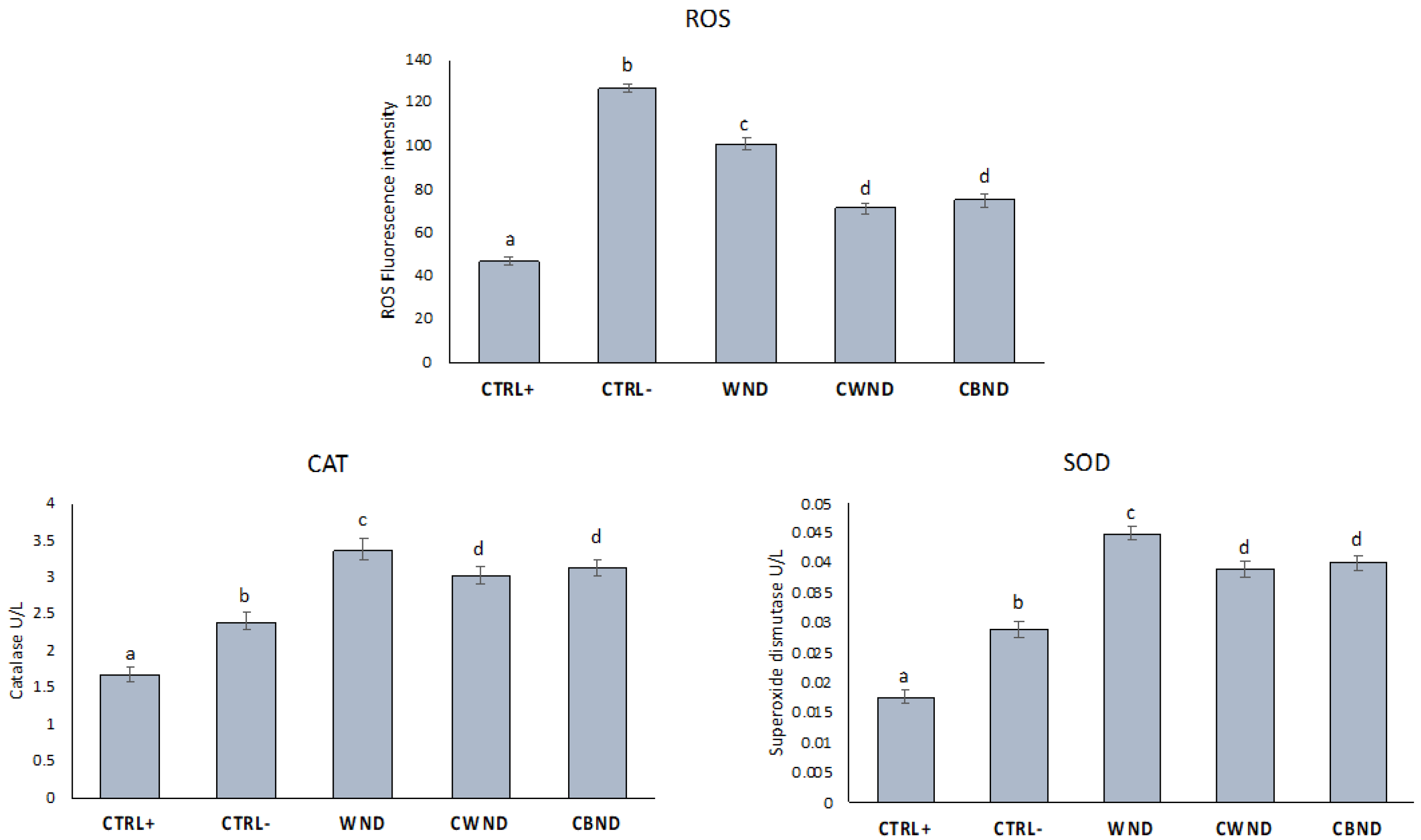
3.2.2. ROS Generation and Antioxidant Enzymes

PMNs were treated with CWND, CBND, and WND extracts at a concentration of 0.5 mg/mL without or with OZ (0.5 mg/mL). As can be seen from Figure 12, ROS levels decreased after treatment with the three extracts compared to control. Furthermore, CWND and CBND extracts were more efficient than WND ones, while no significant differences were observed between the two cultivated plants. Furthermore, the extracts increased the activity of antioxidant enzymes compared to Ctrl+ and Ctrl−, with greater efficiency from the CWND and CBND extracts compared to WND.
In this study, we tried to elucidate how N. damascena extracts influenced two key aspects of cellular oxidative stress response: the generation of reactive oxygen species (ROS) and the activities of antioxidant enzymes. Specifically, they focused on polymorphonuclear leukocytes (PMN), a type of white blood cell crucial for immune defense and inflammation regulation.
Oxidative stress, characterized by an imbalance between ROS production and the body’s antioxidant defenses, plays a pivotal role in various pathological conditions, including inflammation and cancer. Antioxidant enzymes such as superoxide dismutase and catalase act as frontline defenders against ROS-mediated damage by neutralizing harmful free radicals.

4. Discussion

Spectroscopic studies carried out with the main aid of two-dimensional NMR spectra, coupled with the mass spectrometry technique, confirmed the clear presence of flavonoids and triterpene saponins (Table 1 and Table 2). Regarding triterpenoids, four have been described for the first time from the methanolic extract of the aerial parts of several N. damascena plants cultivated in Japan. These triterpene glycosides, also called nigellosides A-D, were identified with eight other triterpene glycosides known in the literature [34]. The latter isolated saponins are not chemotaxonomic markers of the Nigella genus because they have been isolated from various species such as Blighia welwitschii (Hiern) Radlk. [35], Dipsacus azureus Schrenk [36], Schefflera rotundifolia [37], Patrinia scabiosaefolia Link, a Chinese drug whose extract has shown hepatotoxic activities [38], as well as anti-tumor and anti-inflammatory effects [39], Lonicera japonica Thunb. [40], and Anemone anhuiensis roots [41]. While there are few scientific studies in which the isolation of triterpenes from N. damascena has been reported [34], the situation is different for the relative N. sativa. Recently, N. sativa aerial parts were phytochemically investigated to isolate, among the others, a new compound, namely, 3β,23,28-trihydroxyolean-12-ene-3-O-α-l-arabinopyranosyl-(1 → 4)-α-l rhamno pyranosyl-(1 → 4)-β-d-gluco-pyranoside [42]. In this work, spectroscopic data allowed for the provisional and qualitative identification of eleven compounds (1′–11′), which mainly shared glycosylation at the C-3 carbon and esterification at the C-28 carbon of the aglycone core. This is totally in line with the compounds identified by Yoshimitsu et al. [34], who reported the glycosylation site on C-3 and a C12-C13 double bond. Their relative quantitation highlighted that these compounds were poorly abundant in CWND, while they were similarly present in WND and CBND extracts. The TOF-MS/MS spectra of all the compounds are reported in Figure S11.
The blue, pink, and light-blue color of N. damascena flowers is due to the presence of various flavonoids, in particular, glycosylated anthocyanins. The flavonoids contained in the blue flowers of N. damascena, grown in Japan, were studied and led to the isolation of the anthocyanin petunidin 3-O-(6′-O-α-rhamnopyranosyl-2′-O-β-xylopyranosyl-β-glucopyranosides [43]. From the HPLC-MS analysis of the flowers of “Miss Jekyll Rose Shade”, structure, such as the cyanidin 3-O-[2-O-(β-glucopyranosyl)-6-O-(α-rhamnopyranosyl)-β-glucopyranoside]) [44], was identified, while from the flowers of two cultivars, “Miss Jekyll Blue” and “Miss Jekyll White”, the presences of delphinidin 3-[2-(xylosyl)-6-(rhamnosyl)-glucoside] and 7-O-methyldelphinidin were highlighted [44]. It is, therefore, essential to carry out further studies to confirm the exact molecular structure of the compounds identified here and understand possible differences between the cultivars already studied.
Different studies in the literature have investigated the anti-tumor potential of N. damascena essential oil and extract. This is because it would seem that the activity is mainly due to the presence of the sesquiterpene hydrocarbon β-elemene, whose presence can reach up to 73.0% [45,46]. It seems capable of modifying the permeability of the cell membrane, increasing cellular absorption of the drug. This compound can hardly be present in these polar extracts, so the demonstrated activity is to be found toward the identified triterpene structures or in the presence of dolabellane metabolites. For example, 3-O-β-D-glucopyranosyl-(1→3)-α-L-rhamnopyranosyl-(1→2)-α-L-arabinopyranosyl hederagenin 28-β-D-glucopyranosyl ester, a glycosidic triterpene isolated from the methanolic extract of N. damascena aerial parts, presented excellent antiproliferative activity against three different murine and human cell lines such as J774. A1, WEHI-164 e HEK-293 (IC50 = 0.51–1.8 μM) [37], while dipsacoside B did not show relevant activity neither against A549 and SGC-7901 tumor cells nor against the human liver cell line (HL-7702) [47]. In turn, dolabellane, a compound belonging to the sesquiterpene’s class, isolated from the aerial parts of N. glandulifera seeds but also presented in the aerial parts, showed moderate cytotoxic activity, reducing the viability of T98G, U87, U251, and GL261 glioma cancer cell lines by only 29% [48].
It has been widely demonstrated that the biological effects of extracts are the result of both the different compositions of the extracts themselves and the synergistic action of the compounds contained in them. It is also known that the composition and concentration of the different bioactive compounds in plants and, consequently, the biological effectiveness of the extracts can be influenced by numerous factors, among which, certainly, the genotype and environmental factors play an important role [49]. The data obtained in this study revealed that the three tested extracts had different antiproliferative potentials. In particular, CWNB and CBND showed a stronger anti-tumor effect than WND, especially against U937 and HL-90 tumor cell lines, and this effect could be ascribed to the peculiar content of bioactive compounds present in each extract. However, in our case, the observed differences in the bioactivity of the extracts cannot be attributed to a single metabolite or class of compounds. The synergistic effect of the different compounds on the antiproliferative activity is very likely, and further efforts and energy must be made to confirm the real mechanism of action by isolating the different metabolites.
A study by Salmani et al. [50] examined the cytotoxic effects of different Nigella damascena seed extracts on breast cancer cells (MDA-MB-231 and MCF-7) and colon cancer cells (HT-29). They found that the ethyl acetate extract exhibited significant cytotoxic activity against both breast and colon cancer cell lines, suggesting its potential as a therapeutic agent against these cancers [50]. Another study conducted by Karimi et al. [51] investigated the antiproliferative effects of Nigella damascena seed extract on human glioblastoma multiforme (GBM) cell lines. The results demonstrated that the extract inhibited the proliferation of GBM cells and induced apoptosis, indicating its potential as a therapeutic agent for the treatment of GBM [51].
By investigating the effects of N. damascena extracts on ROS production and the activities of these antioxidant enzymes in PMN, this study aimed to shed light on the potential therapeutic properties of the extract in mitigating oxidative stress-related disorders. Understanding how natural compounds like N. damascena modulate oxidative stress pathways in immune cells could pave the way for developing novel treatments targeting inflammatory and oxidative stress-related conditions.
The antioxidant activity of natural extracts is very often linked to the presence of hydrolyzed aromatic compounds [52] and to the presence of phenolic acids [53]. Alu’datt and colleagues [54] demonstrated that several protein fractions obtained from N. damascena aerial parts, characterized by a notable presence of linked phenolic compounds, were able to increase the content of free phenolics, demonstrating excellent antioxidant activity. The phenolic content, influenced by a possible qualitative diversity of compounds such as polyphenols and flavonoids present in CWND, CBND, and WND, influences the antioxidant activity of the investigated extracts. Further studies are needed to evaluate quantitative differences in the studied extracts, isolating the individual metabolisms present.
Antioxidants play a crucial role in protecting cells from oxidative stress-induced damage, which is implicated in various diseases, including cancer and neurodegenerative disorders. Several studies have explored the antioxidant activity of N. damascena extracts. A study conducted by Hosseinzadeh et al. [55] evaluated the antioxidant activity of N. damascena seed extract using various in vitro assays, including DPPH (2,2-diphenyl-1-picrylhydrazyl) radical scavenging and ferric reducing antioxidant power (FRAP) assays. The results demonstrated significant antioxidant activity of the seed extract, suggesting its potential in scavenging free radicals and reducing oxidative stress [55].
The use of natural agents to regulate antioxidant properties and tumorigenesis is increasing. For example, the anti-tumor potential of other plants of the Nigella genus has also been highly described. In fact, the anti-tumor effects of Nigella sativa have been characterized, together with antiproliferative, pro-apoptotic, antioxidant, cytotoxic, anti-mutagenic, and antimetastatic properties [56].
The main molecular mechanisms of action implicated in the described activities are known, and the results reported over the last two decades strongly suggest that N. sativa fractions could serve, alone or in combination with known chemotherapeutic drugs, as effective agents to control the oxidative procession and the onset of the tumor, growth, and metastasis of a wide range of tumors [57].
Our preliminary data, and the evidence in the literature of the antiproliferative and antioxidant activities of Nigella extracts, suggest that further studies are certainly necessary to shed more light on the molecular and cellular aspects relating to the mechanisms underlying the N. damascena properties. We hope that further research efforts will clarify the mechanisms involved in its potential suppressive role in tumorigenesis and cancer. Experimental evidence suggests potent anticancer effects of N. damascena extracts, but preventive and clinical studies directly indicating the anticancer potential of its extracts are still lacking.

5. Conclusions

In this study, the chemical profile of Nigella damascena plants, subjected to different pedoclimatic conditions, was evaluated. Based studies performed via 1D- and 2D-NMR and UHPLC-ESI-QqTOF HR MS/MS have demonstrated the presence of metabolites such as sugars, free amino acids, organic acids, saponins, and flavonoid compounds. A qualitative difference was found between the three extracts studied regarding glycosylated triterpenes. Their relative quantitation highlighted that these compounds were poorly abundant in CWND, while they were similarly present in WND and CBND extracts.
The biological results suggest that the three Nigella extracts are able to induce antiproliferative effects in cancer cell lines, with a major effect in hematological malignant cells and any effects in normal cells. In the antiproliferative tests, CWNB and CBND displayed a stronger anticancer effect than WND. This effect was more evident in hematological cancer cell lines U-937 and HL-60.
At present, there is ambiguity regarding whether certain beneficial effects of numerous natural compounds, like their anticancer properties, require direct transcriptional activity or are primarily influenced by epigenetic mechanisms. Consequently, additional research is warranted to assess whether these observed biological activities might stem from potential epigenetic effects exerted by secondary metabolites, as evidenced in prior studies on other natural compounds [58]. This preliminary study is the basis for further research in which we will try to isolate the preponderant metabolites individually, thus evaluating their contribution to the anti-tumor and antioxidant activity.

Supplementary Materials

The following supporting information can be downloaded at https://0-www-mdpi-com.brum.beds.ac.uk/article/10.3390/antiox13040402/s1, Figure S1. 1H-NMR spectra, acquired at 300 MHz in CD3OD, of WND, CBND, and CWND N. damascena extracts; Figure S2. COSY spectrum of WND extract: aromatic region; Figure S3. HSQC spectrum of WND extract: aromatic region; Figure S4. TOF-MS/MS spectra of hexosyl hexitol (a) and N-(deoxyfructosyl)leucine (b), tentatively identified in N. damascena extracts. Theoretical m/z values are reported below each structure; Figure S5. TOF-MS/MS spectra of (a) quercetin tetrahexoside (1) and (b) quercetin triglycosides (i) 2, (ii) 7, (iii) 8, and (iv) 16. Theoretical m/z values are reported below each structure; Figure S6. TOF-MS/MS spectra of quercetin diglycosides (a–c) and monoglycosides (d,e); Figure S7. TOF-MS/MS spectra (i) of quercetin hydroxymethylglutaryl triglycosides 9 (a), 13 (b), 18 (c), and 24 (d). The fragmentation pattern hypothesized for compound 9 is depicted (ii); theoretical m/z values are below each structure; Figure S8. TOF-MS/MS spectra of quercetin hydroxycinnamoyl glycosides 19 (a), 22 (b), 25 (c), 28 (d), 26 (e), and 27 (f); Figure S9. TOF-MS/MS spectra of kaempferol triglycosides 3 (a), 6 (b), 12 (c); the hydroxymethylglutaryl derivatives 11 (d), and 14 (e), and kaempferol feruloyl trihexoside 30 (f); Figure S10. TOF-MS/MS spectra of kaempferol glycosides 4 (a), 17 (b), and 32 (c), and of myricetin triglycoside (5) (d) and isorhamnetin dihexoside (29) (e); Figure S11. TOF-MS/MS spectra of triterpene saponins 1′11′ (a–l). The TIC (Total Ion Chromatogram) region in which the compounds eluted is shown in the upper part of the figure; Figure S12. HSQC spectrum of the ethyl acetate fraction obtained from the L/L partitioning of the crude polar extract; Figure S13. HMBC spectrum of the ethyl acetate fraction obtained from the L/L partitioning of the crude polar extract.

Author Contributions

Conceptualization, N.B. and M.B.; methodology, N.B., M.S., M.K., S.P., V.M. and L.C.; software M.S., S.P., V.M., P.B. and L.C.; validation, N.B., M.B., V.M., M.S. and P.B.; formal analysis, N.B., V.M. and M.S.; investigation, N.B., M.S., V.M., L.C., P.M. and S.P.; resources, N.B., M.B., P.M. and G.B.; data curation, N.B., G.B. and M.S. and V.M.; writing—original draft preparation, G.B. and N.B.; writing—review and editing, G.B., N.B., M.B., M.S., V.M., A.F. and A.B.; visualization, N.B., G.B. and P.M.; supervision, N.B., M.B, M.S. and V.M. All authors have read and agreed to the published version of the manuscript.

Funding

This research received external funding by National Biodiversity Future Center S.c.a.r.l., Piazza Marina 61 (c/o Palazzo Steri) Palermo, Italy, C.I. CN00000033—CUP UNIPA B73C22000790001. This research received external funding from the European Union—Next Generation EU, PRIN-PNRR Project Code: P2022CKMPW; CUP: B53D23025620001. This work was supported by a grant from MIUR-ITALY PRIN 2017 (Ministry of Education, University, and Research, Project N. 2017A95NCJ) and by the “EXTREMOPHYTES POTENTIAL Environmental stress and community effects on plants biologically active compounds” project, promoted and funded by Bona Furtuna LLC in collaboration with Furtuna Skin LLC (Corleone, Sicily, Italy).

Institutional Review Board Statement

Not applicable.

Informed Consent Statement

Not applicable.

Data Availability Statement

All data and materials are available upon request from the corresponding author.

Conflicts of Interest

The authors declare no conflicts of interest. The authors declare that this study received funding from Bona Furtuna LLC and Furtuna Skin LLC. The funder was not involved in the study design, collection, analysis, interpretation of data, the writing of this article or the decision to submit it for publication.

References

  1. POWO. Plants of the World Online. Facilitated by the Royal Botanic Gardens, Kew. Published on the Internet [WWW Document]. 2023. Available online: http://www.plantsoftheworldonline.org/ (accessed on 22 November 2023).
  2. ESRI. ESRI Data & Maps Environmental Systems Research Institute, Redlands (CA) USA. 2002. Available online: https://www.esri.com/en-us/home (accessed on 22 November 2023).
  3. Badalamenti, N.; Modica, A.; Bazan, G.; Marino, P.; Bruno, M. The ethnobotany, phytochemistry, and biological properties of Nigella damascena—A review. Phytochemistry 2022, 198, 113165. [Google Scholar] [CrossRef]
  4. Heiss, A.G.; Stika, H.P.; De Zorzi, N.; Jursa, M. Nigella in the mirror of time a brief attempt to draw a genus’ ethnohistorical portrait. In Festschrift für Helmut Johannes Kroll Zum 65; Geburtstag; Wachholtz Verlag: Hamburg, Germany, 2012; pp. 147–169. [Google Scholar]
  5. Jabbour, F.; Pasquier, P.E.D.; Chazalviel, L.; Guilloux, M.L.; Conde E Silva, N.; Deveaux, Y.; Manicacci, D.; Galipot, P.; Heiss, A.G.; Damerval, C. Evolution of the distribution area of the Mediterranean Nigella damascena and a likely multiple molecular origin of its perianth dimorphism. Flora 2021, 274, 151735. [Google Scholar] [CrossRef]
  6. Huxley, A.; Griffiths, M. The New Royal Horticultural Society Dictionary of Gardening. In Dictionary of Gardening; MacMillan, P., Ed.; MacMillan Press: London, UK, 1992; Volume 4, pp. 154–168. [Google Scholar]
  7. Pignatti, S. Flora D’italia, 2nd ed.; Editor Edagricole: Milan, Italy, 2017; Volume 1. [Google Scholar]
  8. Linné, C.V. Species Plantarum, Exhibentes Plantas Rite Cognitas, ad Genera Relatas, Cum Differentiis Specificis, Nominibus Trivialibus, Synonymis Selectis, Locis Natalibus, Secundum Systema Sexuale Digestas; Impensis Laurentii Salvii, Holmiae: Stockholm, Sweden, 1753. [Google Scholar] [CrossRef]
  9. Fiori, A. Nuova Flora Analitica D’italia; Tipografia di M. Ricci: Firenze, Italy, 1923. [Google Scholar] [CrossRef]
  10. Toma, C.C.; Olah, N.K.; Vlase, L.; Mogoșan, C.; Mocan, A. Comparative studies on polyphenolic composition, antioxidant and diuretic effects of Nigella sativa L. (Black Cumin) and Nigella damascena L. (lady-in-a-Mist) seeds. Molecules 2015, 20, 9560–9574. [Google Scholar] [CrossRef]
  11. Ballero, M.; Fresu, I. Application of medicinal plants in Seui’s Barbagia (Central Sardinia, Italy). Fitoterapia 1993, 64, 141–150. [Google Scholar]
  12. Agradi, E.; Fico, G.; Cillo, F.; Francisci, C.; Tomè, F. Estrogenic activity of phenolic compounds from Nigella damascena evaluated using a recombinant yeast screen. Planta Med. 2001, 67, 553–555. [Google Scholar] [CrossRef] [PubMed]
  13. Fico, G.; Braca, A.; Tomè, F.; Morelli, I. Phenolic derivatives from Nigella damascena seeds. Pharm. Biol. 2000, 38, 371–373. [Google Scholar] [CrossRef]
  14. Heiss, A.G.; Oeggl, K. The oldest evidence of Nigella damascena L. (Ranunculaceae) and its possible introduction to central Europe. Veget. Hist. Archaeobot. 2005, 14, 562–570. [Google Scholar] [CrossRef]
  15. Geraci, A.; Polizzano, V.; Schicchi, R. Ethnobotanical uses of wild taxa as galactagogues in Sicily (Italy). Acta Soc. Bot. Pol. 2018, 87, 1–18. [Google Scholar] [CrossRef]
  16. Aleo, N.; Amato, F.; Aleo, M. Le piante tossiche della flora trapanese (Sicilia). Quad. Bot. Amb. Appl. 2011, 11, 31–49. [Google Scholar]
  17. Rodrìguez, A.D.; González, E.; Ramìrez, C. The structural chemistry, reactivity, and total synthesis of dolabellane diterpenes. Tetrahedron 1998, 54, 11683–11729. [Google Scholar] [CrossRef]
  18. Mohamed, K.M.; Ohtani, K.; Kasai, R.; Yamadi, K. Dolabellane diterpene glucosides from Chrozophora obliqua. Phytochemistry 1994, 37, 495–500. [Google Scholar] [CrossRef] [PubMed]
  19. Mohamed, K.M.; Ohtani, K.; Kasai, R.; Yamasaki, K. 3-Hydroxy-3-methylglutaryl dolabellane diterpenes from Chrozophora obliqua. Phytochemistry 1995, 39, 151–161. [Google Scholar] [CrossRef]
  20. Cao, X.W.; Wang, F.J.; Liew, O.W.; Zhao, J. Analysis of triterpenoid saponins reveals insights into structural features associated with potent protein drug enhancement effects. Mol. Pharm. 2020, 17, 683–694. [Google Scholar] [CrossRef] [PubMed]
  21. Ogawa, K.; Nakamura, S.; Asada, Y.; Yamashita, M.; Matsuda, H. Oxazonigelladine and dolabellane-type diterpene constituents from Nigella damascena seeds. Tetrahedron 2017, 73, 7054–7060. [Google Scholar] [CrossRef]
  22. Ogawa, K.; Nakamura, S.; Hosokawa, K.; Ishimaru, H.; Saito, N.; Ryu, K.; Fujimuro, M.; Nakashima, S.; Matsuda, H. New diterpenes from Nigella damascena seeds and their antiviral activities against herpes simplex virus type-1. J. Nat. Med. 2018, 72, 439–447. [Google Scholar] [CrossRef] [PubMed]
  23. Sieniawska, E.; Michel, P.; Mroczek, T.; Granica, S.; Skalicka-Wožniak, K. Nigella damascena L. essential oil and its main constituents, damascenine and β-elemene modulate inflammatory response of human neutrophils ex vivo. Food Chem. Toxicol. 2019, 125, 161–169. [Google Scholar] [CrossRef] [PubMed]
  24. Edris, A.E. Anti-cancer properties of Nigella spp. essential oils and their major constituents, thymoquinone and β-elemene. Curr. Clin. Pharmacol. 2009, 4, 43–46. [Google Scholar] [CrossRef] [PubMed]
  25. Sarno, F.; Pepe, G.; Termolino, P.; Carafa, V.; Massaro, C.; Merciai, F.; Altucci, L. Trifolium repens blocks proliferation in chronic myelogenous leukemia via the BCR-ABL/STAT5 pathway. Cells 2020, 9, 379. [Google Scholar] [CrossRef] [PubMed]
  26. Badalamenti, N.; Russi, S.; Bruno, M.; Maresca, V.; Vaglica, A.; Ilardi, V.; Zanfardino, A.; Di Napoli, M.; Varcamonti, M.; Cianciullo, P.; et al. Dihydrophenanthrenes from a Sicilian accession of Himantoglossum robertianum (Loisel.) P. delforge showed antioxidant, antimicrobial, and antiproliferative activities. Plants 2021, 10, 2776. [Google Scholar] [CrossRef] [PubMed]
  27. Manna, A.; Saha, P.; Sarkar, A.; Mukhopadhyay, D.; Bauri, A.K.; Kumar, D.; Das, P.; Chattopadhyay, S.; Chatterjee, M. Malabaricone-A induces a redox imbalance that mediates apoptosis in U937 Cell Line. PLoS ONE 2012, 7, e36938. [Google Scholar] [CrossRef] [PubMed]
  28. Barbosa, P.O.; Pala, D.; Silva, C.T.; de Souza, M.O.; do Amaral, J.F.; Vieira, R.A.L.; de Freitas Folly, G.A.; Volp, A.C.P.; de Freitas, R.N. Açai (Euterpe oleracea Mart.) pulp dietary intake improves cellular antioxidantenzymes and biomarkers of serum in healthy women. Nutrition 2016, 32, 674–680. [Google Scholar] [CrossRef] [PubMed]
  29. Scognamiglio, M.; Fiumano, V.; D’Abrosca, B.; Esposito, A.; Choi, Y.H.; Verpoorte, R.; Fiorentino, A. Chemical interactions between plants in Mediterranean vegetation: The influence of selected plant extracts on Aegilops geniculata metabolome. Phytochemistry 2014, 106, 69–85. [Google Scholar] [CrossRef] [PubMed]
  30. Scognamiglio, M.; Schneider, B. Identification of potential allelochemicals from donor plants and their synergistic effects on the metabolome of Aegilops geniculata. Front. Plant Sci. 2020, 11, 1046. [Google Scholar] [CrossRef] [PubMed]
  31. Sayaka, M.; Kazuyoshi, T.; Yukie, O.; Seiji, O.; Tunehiro, M.; Hajime, M. Functional and structural characterization of a flavonoid glucoside 1,6-glucosyltransferase from Catharanthus roseus. Plant Cell Physiol. 2009, 50, 1401–1415. [Google Scholar] [CrossRef]
  32. El-Beltagi, H.S.; Aziz, S.M.S.A.; Aboshady, A.I.; Ibrahim, M.A.R.; Ibrahim, M.F.M.; Alenezi, M.A.; Darwish, D.B.E.; Al-Qahtani, S.M.; Al-Harbi, N.A.; Darwish, H.; et al. Isolation and identification of flavonoids from black cumin (Nigella sativa) by HPLC-MS and in silico molecular interactions of their major compounds with Fusarium oxysporum trypsin-like serine protease. Separations 2023, 10, 360. [Google Scholar] [CrossRef]
  33. Farag, M.A.; Haidy, A.; Gad, A.G.H.; Ludger, A.W. Metabolomics driven analysis of six Nigella species seeds via UPLC-qTOF-MS and GC–MS coupled to chemometrics. Food Chem. 2014, 151, 333–342. [Google Scholar] [CrossRef] [PubMed]
  34. Yoshimitsu, H.; Nishida, M.; Okawa, M.; Nohara, T. Four new triterpene glycosides from Nigella damascena. Chem. Pharm. Bull. 2007, 55, 488–491. [Google Scholar] [CrossRef] [PubMed]
  35. Pendes, A.; Delaude, C. Structure elucidation of an acetylated saponin of Blighia welwitschii by NMR spectroscopy. Carbohydr. Res. 1994, 263, 79–88. [Google Scholar] [CrossRef] [PubMed]
  36. Mukhamedziev, M.M.; Alimbaeva, P.K.; Gorovits, T.T.; Abubakirov, N.K. Structure of dipsacoside B—A triterpene glycoside from Dipsacus azureus. Chem. Nat. Compd. 1971, 7, 145–149. [Google Scholar] [CrossRef]
  37. Braca, A.; Autore, G.; De Simone, F.; Marzocco, S.; Morelli, I.; Venturella, F.; De Tommasi, N. Cytotoxic saponins from Schfflera rotundifolia. Planta Med. 2004, 70, 960–966. [Google Scholar] [CrossRef] [PubMed]
  38. Choi, J.S.; Woo, W.S. Triterpene glycosides from the roots of Patrinia scabiosifolia. Planta Med. 1987, 52, 62–65. [Google Scholar] [CrossRef] [PubMed]
  39. Hwang, D.I.; Won, K.J.; Kim, D.Y.; Kim, H.B.; Li, Y.; Lee, H.M. Chemical composition of Patrinia scabiosifolia flower absolute and its migratory and proliferative activities in human keratinocytes. Chem. Biodivers. 2019, 16, e1900252. [Google Scholar] [CrossRef] [PubMed]
  40. Kawai, H.; Kuroyanagi, M.; Umehara, K.; Akira Ueno, A.; Satake, M. Studies on the saponins of Lonicera japonica Thunb. Chem. Pharm. Bull. 1988, 36, 4769–4775. [Google Scholar] [CrossRef] [PubMed]
  41. Ye, W.C.; Zhang, Q.W.; Zhao, S.X.; Che, C.T. Four new oleanane saponins from Anemone anhuiensis. Chem. Pharm. Bull. 2001, 49, 632–634. [Google Scholar] [CrossRef] [PubMed]
  42. Parveen, A.; Farooq, M.A.; Kyunn, W.W. A new oleanane type saponin from the aerial parts of Nigella sativa with antioxidant and anti-diabetic potential. Molecules 2020, 25, 2171. [Google Scholar] [CrossRef] [PubMed]
  43. Toki, K.; Saito, N.; Nogami, A.; Tatsuzawa, F.; Shigihara, A.; Honda, T. Flavonoid glycosides isolated from the blue flowers of Nigella damascena. Heterocycles 2009, 78, 2287–2294. [Google Scholar] [CrossRef]
  44. Tanikawa, N.; Homma, K.; Tatsuzawa, F. Flavonoids of the rose-pink, blue, and white flowers of Nigella damascena L. (Ranunculaceae). Sci. Hortic. 2019, 257, 108609. [Google Scholar] [CrossRef]
  45. Kokoska, L.; Havlik, J.; Valterova, I.; Nepovim, A.; Rada, V.; Vanek, T. Chemical composition of the essential oil of Nigella orientalis L. seeds. Flavour Fragr. J. 2005, 20, 419–420. [Google Scholar] [CrossRef]
  46. Moretti, A.; D’Antuono, L.F.; Elementi, S. Essential oils of Nigella sativa L. and Nigella damascena L. seed. J. Essent. Oil Res. 2004, 16, 182–183. [Google Scholar] [CrossRef]
  47. Xu, K.; Zhan Shu, Z.; Xu, Q.M.; Liu, Y.L.; Li, X.R.; Wang, Y.L.; Yang, S.L. Cytotoxic activity of Pulsatilla chinensis saponins and their structure—Activity relationship. J. Asian Nat. Prod. Res. 2013, 15, 680–686. [Google Scholar] [CrossRef] [PubMed]
  48. Gao, J.B.; Zhang, X.J.; Zhang, R.H.; Zhu, L.L.; Pu, D.B.; Li, X.L.; Li, H.L.; Xu, M.; Xiao, W.L. In vitro human dihydroorotate dehydrogenase inhibitory, antiinflammatory and cytotoxic activities of alkaloids from the seeds of Nigella glandulifera. Planta Med. 2018, 84, 1013–1021. [Google Scholar] [CrossRef]
  49. Al-Jumaili, A.; Kumar, A.; Bazaka, K.; Jacob, M.V. Plant secondary metabolite-derived polymers: A potential approach to develop antimicrobial films. Polymers 2018, 10, 515. [Google Scholar] [CrossRef] [PubMed]
  50. Salmani, J.M.; Asgharpanah, J.; Zendehdel, K. Cytotoxic effects of Nigella damascena Extract on Human Breast and Colorectal Cancer Cell Lines. Asian Pac. J. Cancer Prev. 2014, 15, 3829–3833. [Google Scholar]
  51. Karimi, A.; Moradi, M.T.; Alidadi, S.; Hashemi, L.; Ghaffari, S.H. The effect of Nigella sativa extract on human glioblastoma multiforme cells: A laboratory study. Iran. J. Cancer Prev. 2013, 6, 218–224. [Google Scholar]
  52. Badalamenti, N.; Ilardi, V.; Rosselli, S.; Bruno, M. The ethnobotany, phytochemistry and biological properties of genus Ferulago—A review. J. Ethnopharmacol. 2021, 274, 114050. [Google Scholar] [CrossRef] [PubMed]
  53. Sytar, O. Phenolic acids in the inflorescences of different varieties of buckwheat and their antioxidant activity. J. King Saud Univ. Sci. 2015, 27, 136–142. [Google Scholar] [CrossRef]
  54. Alu’datt, M.H.; Rababah, T.; Alhamad, N.M.; Gammoh, S.; Ereifej, K.; Alodat, M.; Hussein, N.M.; Kubow, S.; Torley, P.J. Antioxidant and antihypertensive properties of phenolic–protein complexes in extracted protein fractions from Nigella damascena and Nigella arvensis. Food Hydrocoll. 2016, 56, 84–92. [Google Scholar] [CrossRef]
  55. Hosseinzadeh, H.; Ramezani, M.; Salmani, G.A. Antinociceptive, anti-inflammatory and acute toxicity effects of Zataria multiflora Boiss extracts in mice and rats. J. Ethnopharmacol. 2007, 109, 393–400. [Google Scholar] [CrossRef] [PubMed]
  56. Dalli, M.; Bekkouch, O.; Azizi, S.E.; Azghar, A.; Gseyra, N.; Kim, J. Nigella sativa L. Phytochemistry and pharmacological activities: A Review. Biomolecules 2021, 12, 20. [Google Scholar] [CrossRef]
  57. Majdalawieh, A.F.; Fayyad, M.W. Recent advances on the anti-cancer properties of Nigella sativa, a widely used food additive. Integr. Med. 2016, 7, 173–180. [Google Scholar] [CrossRef]
  58. Miceli, M.; Bontempo, P.; Nebbioso, A.; Altucci, L. Natural compounds in epigenetics: A current view. Food Chem. Toxicol. 2014, 73, 71–83. [Google Scholar] [CrossRef]
Figure 1. Global distribution of N. damascena. The black circle highlights the study area at the center of the natural distribution range [1,2].
Antioxidants 13 00402 g001
Figure 2. (a) The N. damascena flower, in which dark petals (reduced to nectaries) and white petaloids are evident, lends the species its ancient name, Melanthium, derived from the Greek μέλαν (melan) meaning “black” and άνθος (anthos) meaning “flower”. (b) In cultivated forms of N. damascena “Oxford Blue”, the flowers are characterized by having multiple verticils of petaloids ranging from white to blue during the development phase, which gives the species a high aesthetic value. The photos refer to the wild populations (a) and cultivated populations (b) sampled in Corleone for this study.
Antioxidants 13 00402 g002
Figure 3. Detail of the HSQC spectrum of the ethyl acetate fraction obtained from the liquid/liquid partitioning of the crude polar extracts and structure of the hederagenin aglycone.
Antioxidants 13 00402 g003
Figure 4. Cell vitality rates determined by MTT assay of HaCat cells after treatment with extracts of N. damascena with white flowers (CWND), blue flowers (CBND), and wild (WND) at the indicated concentrations and time. Values are mean ± standard deviation (SD) of biological triplicates.
Antioxidants 13 00402 g004
Figure 5. Cell vitality rates determined by MTT assay of U-937 and HL-60 cells after treatment with extracts of N. damascena with white flowers (CWND), blue flowers (CBND), and wild (WND) at the indicated concentrations and time. Values are mean ± standard deviation (SD) of biological triplicates.
Antioxidants 13 00402 g005aAntioxidants 13 00402 g005b
Figure 6. Cell vitality rates determined by MTT assay of MCF-7 cells after treatment with extracts of N. damascena with white flowers (CWND), blue flowers (CBND), and wild (WND) at the indicated concentrations and time. Values are mean ± standard deviation (SD) of biological triplicates.
Antioxidants 13 00402 g006
Figure 7. Cell death assay determined by FACS analysis of U-937 and HL-60 cells after treatment with extracts of N. damascena with white flowers (CWND), blue flowers (CBND), and wild (WND) at the indicated concentrations and time. Values are mean ± standard deviation (SD) of biological triplicates.
Antioxidants 13 00402 g007
Figure 8. Cell death assay determined by FACS analysis of MCF-7 cells after treatment with extracts of N. damascena with white flowers (CWND), blue flowers (CBND), and wild (WND) at the indicated concentrations and time. Values are mean ± standard deviation (SD) of biological triplicates.
Antioxidants 13 00402 g008
Figure 9. Cell cycle assay determined by FACS analysis of U-937 cells after treatment with extracts of N. damascena with white flowers (CWND), blue flowers (CBND), and wild (WND) at the indicated concentrations and time. Values are mean ± standard deviation (SD) of biological triplicates.
Antioxidants 13 00402 g009
Figure 10. Cell cycle assay determined by FACS analysis of HL-60 cells after treatment with extracts N. damascena with white flowers (CWND), blue flowers (CBND), and wild (WND) at the indicated concentrations and time. Values are mean ± standard deviation (SD) of biological triplicates.
Antioxidants 13 00402 g010
Figure 11. Cell cycle assay determined by FACS analysis of MCF-7 cells after treatment with extracts N. damascena with white flowers (CWND), blue flowers (CBND), and wild (WND) at the indicated concentrations and time. Values are mean ± standard deviation (SD) of biological triplicates.
Antioxidants 13 00402 g011
Figure 12. Reactive Oxygen Species (ROS) and the activities of antioxidant enzymes (superoxide dismutase; catalase) in PMN (Ctrl+), PMN with OZ (Ctrl−), PMN treated with white flowers (CWND), blue flowers (CBND), and wild (WND) at the concentration of 0.5 mg/mL and with OZ. Data were presented as mean and standard error and were analyzed with a paired t-test. Bars not accompanied by the same letter were significantly different at p < 0.05.
Antioxidants 13 00402 g012
Table 1. Chromatographic and TOF MS data of flavonoid glycosides tentatively identified in WND, CBND, and CWND N. damascena extracts. RDB = Ring Double Bonds.
PeakRtTentative AssignmentMolecular Formula[M-H] Found[M-2H]2−RDBError (ppm)
13.791Quercetin tetrahexosideC39H50O27949.2459 15−0.8
24.010Quercetin dihexoside deoxyhexosideC33H40O21771.1993 140.5
34.269Kaempferol dihexoside deoxyhexoside (1)C33H40O20755.2046 140.8
44.308Kaempferol dihexosideC27H30O16609.1460 13−0.2
54.315Myricetin dihexosylpentosideC32H38O22773.1751 14−4.0
64.373Kaempferol dihexoside deoxyhexoside (2)C33H40O20755.2046 14−1.6
74.409Quercetin trihexosideC33H40O22787.1940 140.2
84.450Quercetin hexosylpentoside deoxyhexosideC32H38O20741.1884 140.9
94.452Quercetin (hydromethylglutaryl) dihexoside deoxyhexosideC39H48O25915.2399 16−1.4
104.562Quercetin dihexosideC27H30O17625.1426 132.5
114.740Kaempferol (hydromethylglutaryl) dihexoside deoxyhexoside (1)C39H48O24899.2499 161.6
124.883Kaempferol trihexosideC33H40O21771.1979 14−1.3
134.885Quercetin (hydromethylglutaryl) hexosylpentoside deoxyhexosideC38H46O24885.2285 16−2.4
144.889Kaempferol (hydromethylglutaryl) dihexoside deoxyhexoside (2)C39H48O24899.2452 16−1.2
154.954Quercetin hexosylpentosideC26H28O16595.1308 130.6
164.957Quercetin dihexosylpentosideC32H38O21757.1839 140.8
175.032Kaempferol dihexosideC27H30O16609.1463 130.3
185.113Quercetin (hydromethylglutaryl) trihexosideC39H48O26931.2346 16−1.4
195.231Quercetin caffeoyltrihexoside (1)C42H46O25949.2246474.109124−1.0
205.339Quercetin hexosyldeoxyhexosideC27H30O16609.1463 130.3
215.471Quercetin hexosideC21H20O12463.0881 12−0.2
225.505Quercetin caffeoyltrihexoside (2)C42H46O25949.2248474.109424−0.8
235.560Quercetin (hydroxymethylglutaryl)
hexosylpentoside
C32H36O20739.1730 15−1.9
245.572Quercetin (hydroxymethylglutaryl) dihexosylpentosideC38H46O25901.2248 16−0.8
255.861Quercetin feruloyltrihexoside (1)C43H48O25963.2393 20−2.0
266.065Quercetin coumaroyltrihexoside (1)C42H46O24933.2306466.111820−1.9
276.058Quercetin coumaroyltrihexoside (2)C42H46O24933.2303 20−0.4
286.142Quercetin feruloyl trihexoside (2)C43H48O25963.2393 20−0.5
296.209Isorhamnetin dihexosideC28H32O16623.1623 130.9
306.338Kaempferol feruloyl trihexosideC43H48O24947.2463 200
316.941Quercetin deoxyhexosideC21H20O11447.0942 122.0
327.525Kaempferol deoxyhexosideC21H20O10431.0993 122.2
When two isomers of the same compound are detected and cannot be distinguished from each other, they are indicated by a number in brackets.
Table 2. Chromatographic and TOF MS data of triterpenoid saponins tentatively identified in WND, CBND, and CWND N. damascena extracts. RDB = Ring Double Bonds.
PeakRtTentative AssignmentMolecular Formula[M-H] Found[M-H]2− FoundRDBError (ppm)
1′7.7343- hexosyl deoxyhexosyl hederagenin 28-hexosyl esterC48H78O18941.5114 10−0.1
2′8.0613-hexosyl pentosyl hederagenin 28-hexosyl esterC47H76O18927.4963 100.4
3′8.3503-hexosyl deoxyhexosyl pentosyl hederagenin 28-dihexosyl esterC61H100O311235.6059663.3057100.8
4′8.4903-deoxyhexosyl pentosyl hederagenin 28-hexosyl esterC47H76O17911.5018 100.9
5′8.9283-hexosyl deoxyhexosyl pentosyl oleanolic acidC47H76O16895.5038 10−1.4
6′9.1403-deoxyhexosyl pentosyl oleanolic acid 28-hexosyl esterC47H76O16895.5055 10−0.6
7′9.4163-deoxyhexosyl pentosyl oleanolic acid 28-dihexosyl esterC53H86O211057.5585 11−0.4
8′9.8133-hexosyl deoxyhexosyl hederagenin 28-hexosyl esterC48H78O18941.5119 100.4
9′10.3653-hexosyl deoxyhexosyl hederagenin pentosyl ester (1)C47H76O17911.5000 10−1.1
10′10.5023-hexosyl deoxyhexosyl hederagenin pentosyl ester (2)C47H76O17911.5017 100.8
11′10.7693-hexosyl deoxyhexosyl dehydrohederagenin pentosyl esterC47H74O17909.4855 100.2
When two isomers of the same compound are detected and cannot be distinguished from each other, they are indicated by a number in brackets.
Disclaimer/Publisher’s Note: The statements, opinions and data contained in all publications are solely those of the individual author(s) and contributor(s) and not of MDPI and/or the editor(s). MDPI and/or the editor(s) disclaim responsibility for any injury to people or property resulting from any ideas, methods, instructions or products referred to in the content.

