Article

Molecular Lipopolysaccharide Di-Vaccine Protects from Shiga-Toxin Producing Epidemic Strains of Escherichia coli O157:H7 and O104:H4

by
Ivan A. Dyatlov
1,
Edward A. Svetoch
1,
Anna A. Mironenko
2,*,
Boris V. Eruslanov
1,
Victoria V. Firstova
1,
Nadezhda K. Fursova
1,
Alexander L. Kovalchuk
2,
Vyacheslav L. Lvov
2,3 and
Petr G. Aparin
2,3
1
State Research Center for Applied Microbiology and Biotechnology, Rospotrebnadzor, 142279 Obolensk, Moscow Region, Russia
2
National Research Center Institute of Immunology Federal Medical-Biology Agency (FMBA of Russia), 115522 Moscow, Russia
3
ATVD-TEAM Co., Ltd., 115522 Moscow, Russia
*
Author to whom correspondence should be addressed.
Submission received: 30 September 2022 / Revised: 25 October 2022 / Accepted: 29 October 2022 / Published: 1 November 2022
(This article belongs to the Collection Vaccines against Infectious Diseases)

Abstract

:
Background: Shiga toxin-producing Escherichia coli (STEC) O157:H7 and O104:H4 strains are important causative agents of food-borne diseases such as hemorrhagic colitis and hemolytic–uremic syndrome, which is the leading cause of kidney failure and death in children under 5 years as well as in the elderly. Methods: the native E. coli O157:H7 and O104:H4 lipopolysaccharides (LPS) were partially deacylated under alkaline conditions to obtain apyrogenic S-LPS with domination of tri-acylated lipid A species—Ac3-S-LPS. Results: intraperitoneal immunization of BALB/c mice with Ac3-S-LPS antigens from E. coli O157:H7 and O104:H4 or combination thereof (di-vaccine) at single doses ranging from 25 to 250 µg induced high titers of serum O-specific IgG (mainly IgG1), protected animals against intraperitoneal challenge with lethal doses of homologous STEC strains (60–100% survival rate) and reduced the E. coli O157:H7 and O104:H4 intestinal colonization under an in vivo murine model (6–8-fold for monovalent Ac3-S-LPS and 10-fold for di-vaccine). Conclusions: Di-vaccine induced both systemic and intestinal anti-colonization immunity in mice simultaneously against two highly virulent human STEC strains. The possibility of creating a multivalent STEC vaccine based on safe Ac3-S-LPS seems to be especially promising due to a vast serotype diversity of pathogenic E. coli.

1. Introduction

Escherichia coli O157:H7 and O104:H4 are members of a group of pathogenic Shiga toxin-producing E. coli (STEC), which is defined by capacity to produce Shiga toxin (Stx) type 1 or 2, or both (as well as variants of these) [1,2,3]. Bacteria E. coli O157:H7 was first recognized as a human pathogen in the early 1980s [4,5,6], later it has been identified as the infectious agent causing worldwide morbidity and the largest STEC outbreaks [4,7,8,9,10,11,12,13,14,15,16,17,18,19]. E. coli O157:H7 causes various severe infections ranging from watery diarrhea to hemorrhagic colitis (HC), and Stx production determines the development of hemolytic uremic syndrome (HUS) [4,20,21], which is characterized by hemolytic anemia, thrombocytopenia, and renal failure [7,8,22]. Approximately 10% of patients with E. coli O157:H7 infection develop HUS complication with mortality rate up to 5% [7,22] and it is presently the most common cause of acute renal failure in children under 5 years and in the elderly [23,24].
The dangerous outbreak of food-borne infection in Germany and France (2011) caused by a relatively new highly virulent hybrid Stx type 2-producing E. coli O104:H4 strain [25] affected 3816 persons, 845 (22%) of treated patients developed HUS and among them 54 patients died [26]. Risk increase of HUS development in patients with HC and aggravation of the disease may be provoked by antibiotic therapy [27,28]. Today, 40 years after the first outbreak caused by STEC, an effective treatment for HUS is yet to be developed. Thus, the modern approaches to treatment of patients with HUS are unsuccessful and cannot prevent fatality. In view of this, the development of an effective vaccine against STEC is of particular relevance.
Main vaccine strategies against STEC infection are targeted on induction of anti-toxin and/or anti-whole cell immunity. Nontoxic subunits, recombinant proteins, and fusion proteins of various design developed on the basis of Stx molecule represent anti-toxin vaccine constructs, which induce strong humoral immune response as well as protection against STEC O157:H7 challenge [29,30,31].
Effective activation of immunity against bacterial cells is unlikely without induction of immune response to cell wall lipopolysaccharide (LPS). LPS O-polysaccharide domain, referred to as the O-antigen, determines the specificity of anti-STEC protective immunity [32,33].
LPS, the endotoxin of E. coli, along with Shiga toxins, is an important pathogenic factor, which participates in initiation of infection (invasion) as well as in the pathogenesis of HUS [16,34,35,36]. Numerous animal studies demonstrated that modelling of clinical symptoms of HUS is archived only with the combined administration of endotoxin (LPS) and Stx2 [37,38,39,40]. The importance of LPS in the pathogenesis of HUS provides a solid basis for the study of these macromolecules as target antigens for the development of vaccines against STEC. Additionally, high titers of serum antibodies against LPS of STEC pathogens are found in convalescent adult patients with HC and in children with acute HUS, which indicates the suppression of a high pathogenic activity of LPS [41,42,43]. Thus, LPS-specific immune response is advantageous during STEC infection and supports simultaneous formation of anti-toxin immunity, as well as immunity against cell wall antigens.
Endotoxicity, however, precludes LPS from clinical use. As LPS is highly heterogenous, we hypothesized that more homogenous pools of LPS might be less toxic. We developed a method to generate a homogenous LPS subfraction, Ac3-S-LPS, containing long chain O-specific polysaccharide (S-LPS) and mainly tri-acylated lipid A, with no penta- and hexa-acylated, and rare tetra-acylated lipid A [44]. Ac3-S-LPS had drastically reduced pyrogenicity and endotoxicity. In a clinical trial it was found that Ac3-S-LPS as a candidate vaccine against S. flexneri 2a was safe in volunteers at a dose of 50 µg, with low pyrogenicity, no major and few minor adverse events, and did not induce pro-inflammatory cytokines. In spite of the profound lipid A modification, the vaccine induced a prevalence of IgG and IgA antibodies [44].
In this manuscript, we report the development of a di-vaccine composed of two mono-vaccines based on the original molecular LPS form obtained from E. coli O157:H7 and O104:H4. Each LPS in di-vaccine is highly homogenous and represents Ac3-S-LPS molecules, in contrast to natural heterogeneous endotoxic LPS, which is a mixture of different pools of LPS molecules. Ac3-S-LPS from E. coli O157:H7 or O104:H4 is similar to clinically applicable Ac3-S-LPS vaccine from S. flexneri 2a and could be suitable for human parenteral administration.

2. Results

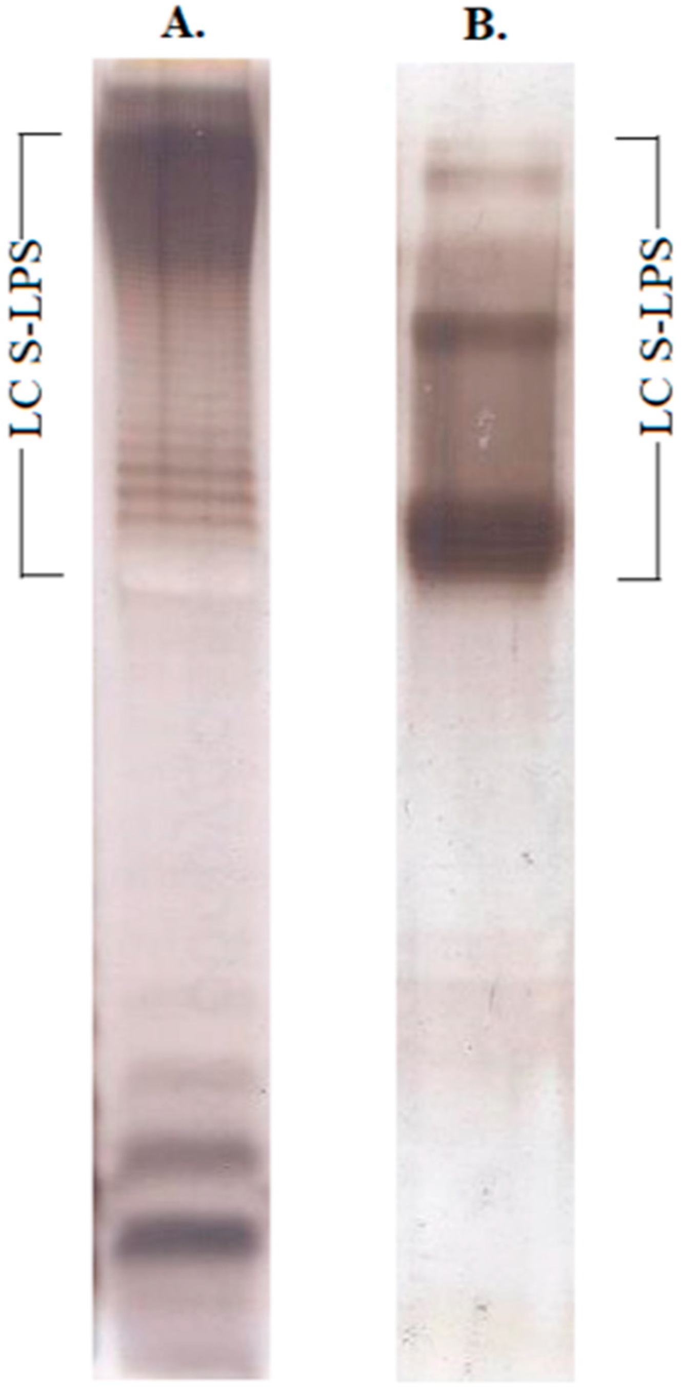
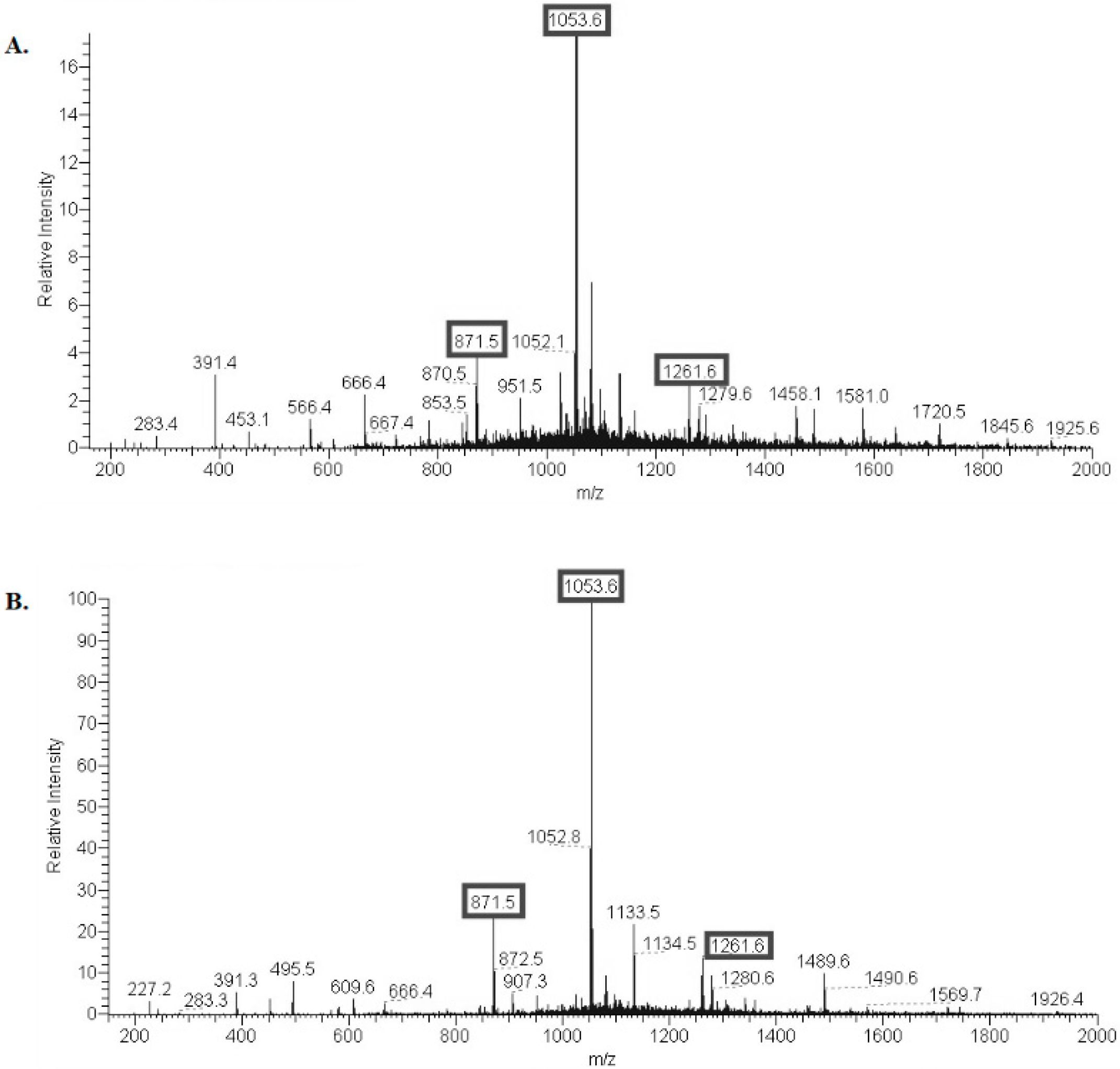
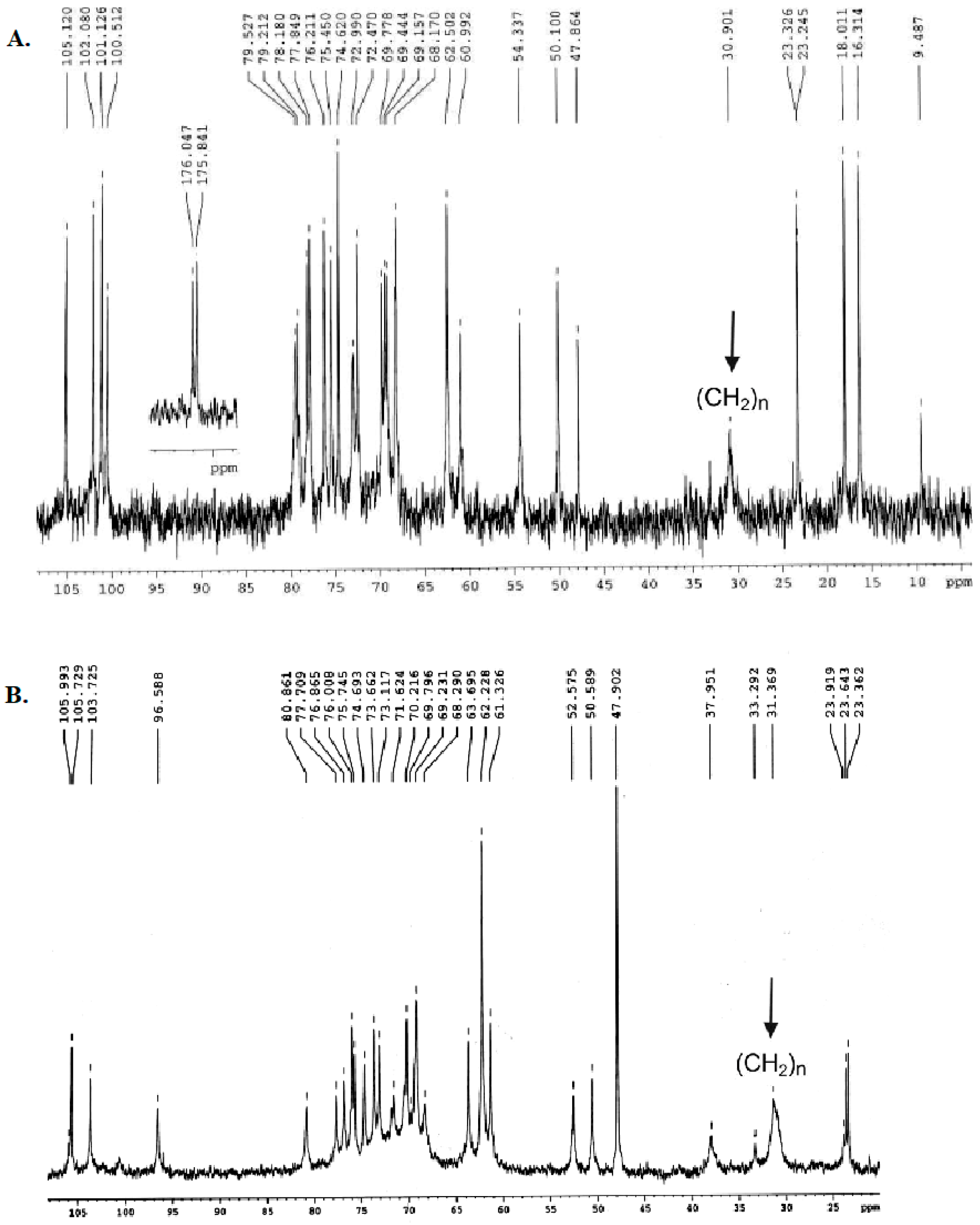
2.1. Structural Characterization of Ac3-S-LPS from STEC Strains

LPS was purified from E. coli O157 and O104 by the Westphal method [45]. S-LPS fraction with a long-chain O-polysaccharide domain was isolated from the intact E. coli O157:H7 and O104:H4 LPS by Sephadex G-150 gel-permeation chromatography in the presence of Na-deoxycholate, which was confirmed by electrophoresis in an SDS-polyacrylamide gel (Figure 1 and Figure S1).
S-LPS preparations were partially deacylated under alkaline conditions to obtain modified S-LPS. Lipid A (LA) was released from the modified S-LPS by mild acid hydrolysis. The negative ion mode electrospray ionization mass spectrum of the released LA showed peaks for (M-H) ions of various acyl variants (Figure 2). E. coli O157 and E. coli O104 modified S-LPS contain mainly tri-acylated LA with small admixtures of di- and tetra-acylated LA—Ac3-S-LPS. No peaks for penta- and hexa-acyl derivatives were present in the mass spectra of the modified LA.
13C NMR spectra of E. coli O157 and E. coli O104 Ac3-S-LPS (Figure 3) were in agreement with published data [46,47] indicating that deacylation did not alter the O-polysaccharide (O-PS) repeating unit structures. The O-PS domains of E. coli O157 and E. coli O104 Ac3-S-LPS contain unusual, rare sugars—α-D-perosamine and acetylneuraminic acid, respectively (Figures S2 and S3).

2.2. Immunobiological Study of Modified LPS

The serological specificity of the modified LPS was confirmed by a direct ELISA using rabbit sera obtained against E. coli O157 and E. coli O104 strains (data not shown).
Pyrogenicity of E. coli O157 and E. coli O104 Ac3-S-LPS was examined in rabbits by intravenous injection at a WHO approved Vi-vaccine pyrogenicity test dose of 0.025 μg/kg [48]. Both preparations were apyrogenic (∑Δ t °C = 0.4 °C for E. coli O157 Ac3-S-LPS and ∑Δ t °C = 0.3 °C for E. coli O104 Ac3-S-LPS).
The levels of endotoxin in intact LPS preparations from E. coli O157 and E. coli O104, estimated by the LAL assay, were high and constituted 584 and 3606 EU/µg, respectively. E. coli O157 Ac3-S-LPS had 29.86 EU/µg (≈19-fold reduction), and E. coli O104 Ac3-S-LPS had 33.24 EU/µg (≈108-fold reduction) of endotoxin content.
In the acute toxicity test, the dose of safe administration of the E. coli O157 and E. coli O104 Ac3-S-LPS exceeded 500 mg/kg per mouse after a single intraperitoneal (i.p.) injection.

2.3. Immunization Provides Effective Protection of Mice against Intraperitoneal STEC Infection

Intraperitoneal immunization of BALB/c mice with Ac3-S-LPS E. coli O157 or E. coli O104 at a repeated dose of 25 µg provided effective protection of animals against i.p. challenge with minimal lethal doses (LD100) of homologous STEC strains: 2.6 × 10⁷ CFU/mouse of E. coli O157 and 1 × 10⁸ CFU/mouse of E. coli O104 (Table 1). The preparation efficacy depended on the immunization scheme: the third injection provided 70% and 100% protection against E. coli O157 and O104 challenge, respectively, versus 60% protection rate for both strains following the second injection. All intact (control) mice died after i.p. injection of the same STEC strains at LD100, which were 2.6 × 10⁷ (E. coli O157) and 1 × 10⁸ (E. coli O104) CFU/mouse.
The protective properties of Ac3-S-LPS E. coli O157 or E. coli O104 correlated with their ability to elicit E. coli O157 or O104 LPS-specific IgG response, and both preparations were similar by immunogenic potency. Ac3-S-LPS E. coli O157 or E. coli O104 elicited serum O-specific IgG both after second and third injection at a dose of 25 µg (Figure 4A). The third injection of preparations elicited four-fold IgG increases over those following the second injection (the titers were 1:5120 versus 1:1280 for E. coli O157 Ac3-S-LPS and 1:10,240 versus 1:2560 for E. coli O104 Ac3-S-LPS), and these rises were statistically significant (p < 0.05, ANOVA). Thus, the well-known mechanism of serotype-specific anti-bacterial protection associated with rises in anti-O antibodies has been confirmed for modified LPS immunogens from STEC strains.

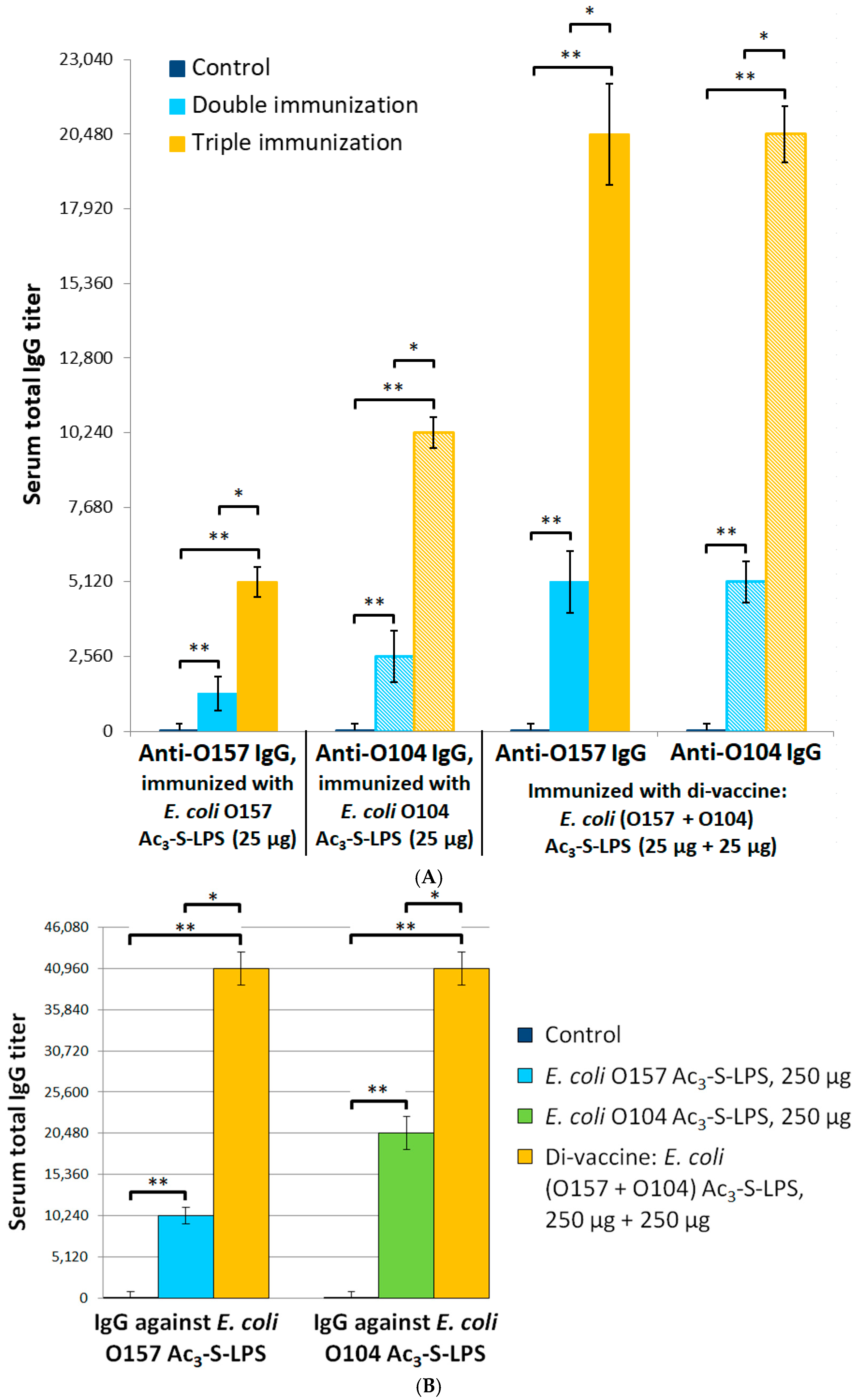
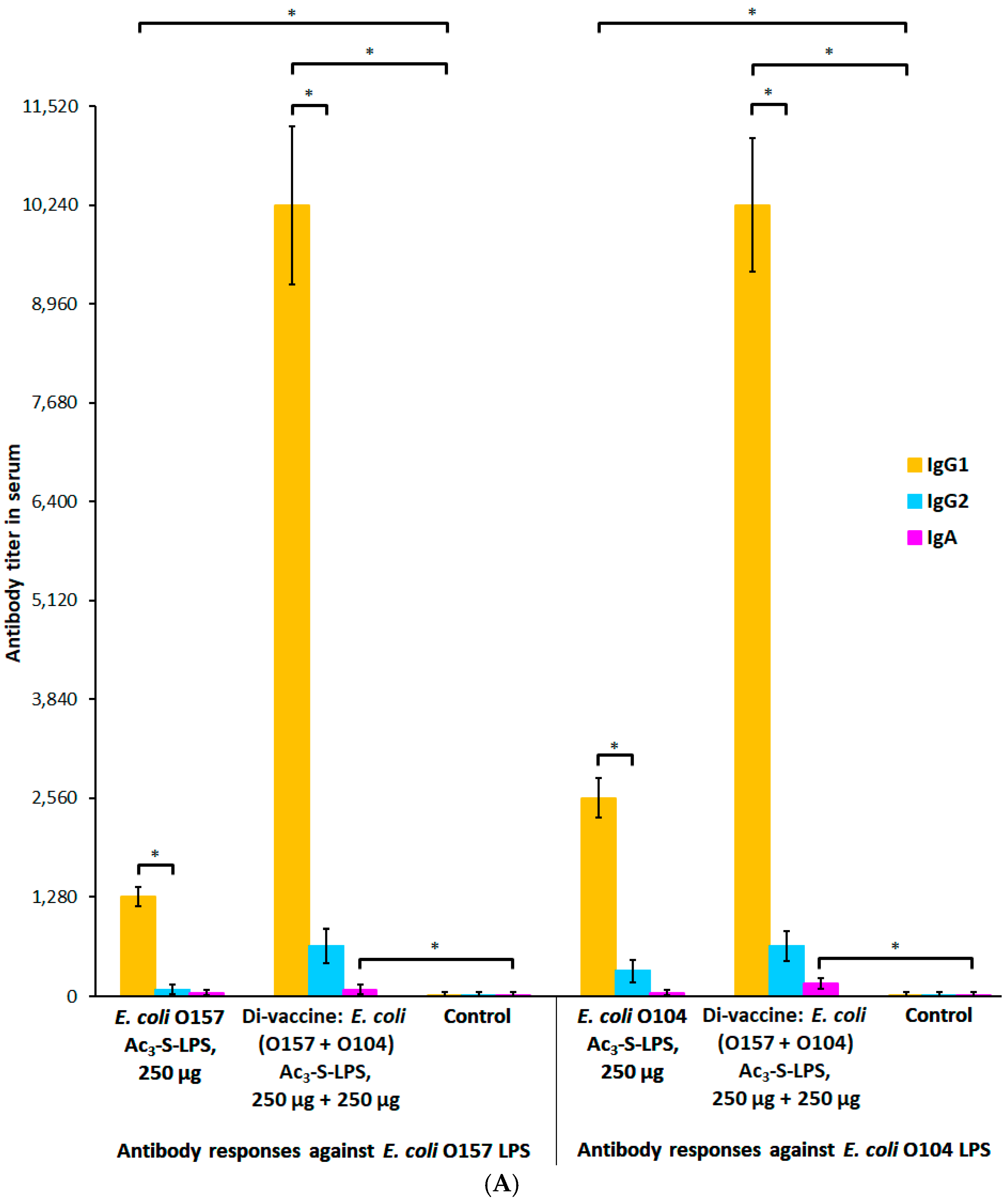
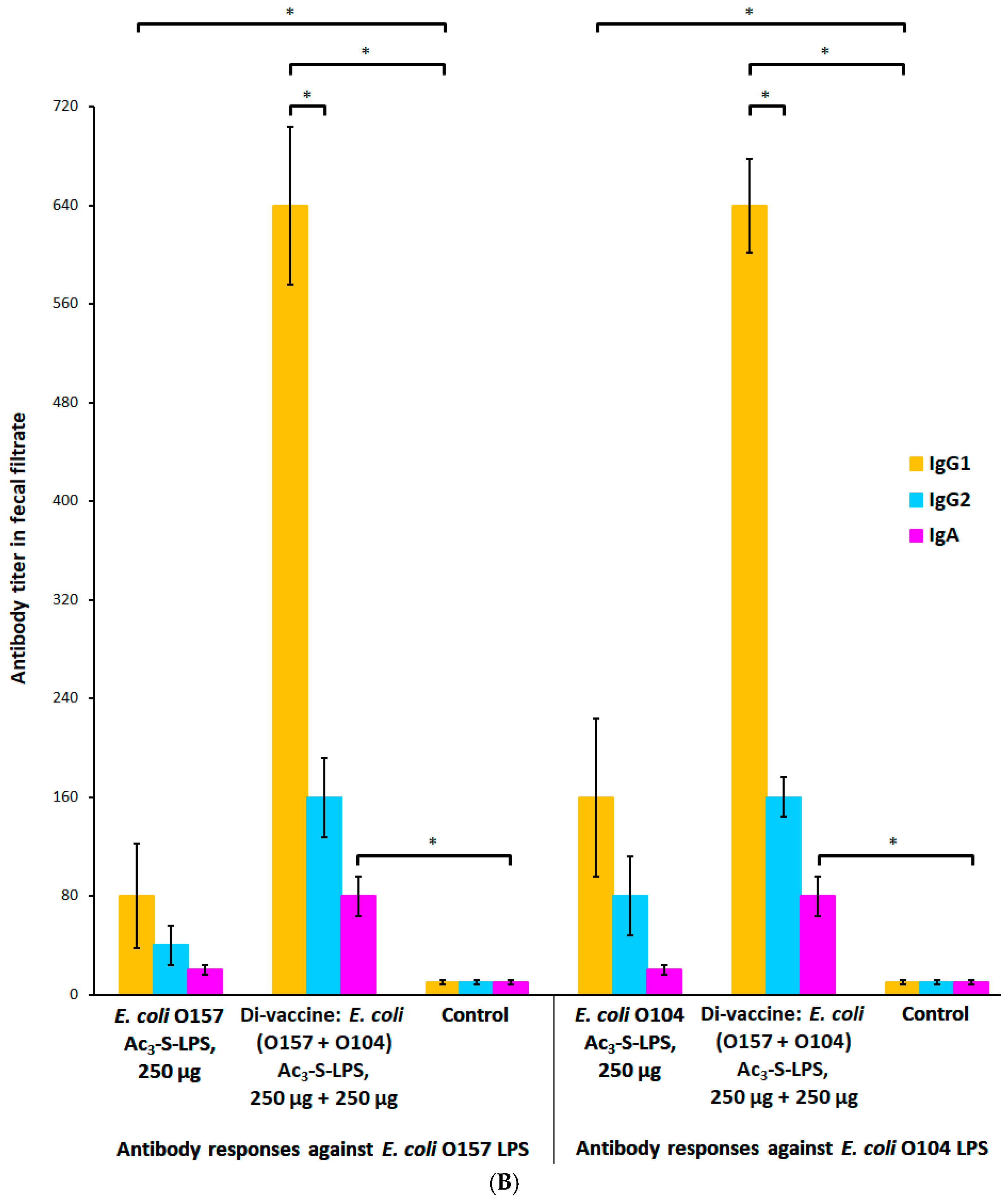
2.4. Protective and Immunogenic Properties of the STEC Di-Vaccine

The di-vaccine against E. coli O157 or O104 infection was obtained by mixing two antigens, Ac3-S-LPS E. coli O157 and Ac3-S-LPS E. coli O104, in a ratio of 1:1 (w/w). Both components were highly purified modified LPS with a minimal content of protein and nucleic acid impurities (less than 2% w/w each).
BALB/c mice were i.p. immunized with 25 µg of each modified LPS antigen (25 µg + 25 µg), i.e., the total dose of di-vaccine was 50 µg. The triple immunization with di-vaccine provided the highest protection (100%) against i.p. challenges with both highly virulent human STEC strains E. coli O157 or E. coli O104 (Table 1). In groups of mice immunized with di-vaccine and infected with E. coli O157 or E. coli O104, no bacteria were isolated from parenchymal organs 15 days after infection. These observations indicate the complete elimination of pathogens in vaccinated mice. Control mice died in all experiments.
In addition, the highest levels of serum IgG were elicited by immunization with di-vaccine preparation (Figure 4A): the end-point titer was 1:20,480 for both antigens after third injection, which was four-fold higher than IgG titer registered after the second injection–1:5120 for both antigens (p < 0.05, ANOVA). The IgG titers of control sera were less than 1:20 for each antigen. Thus, both Ac3-S-LPS components of di-vaccine induced 256-fold and higher increase in serum O-specific IgG-titers (p < 0.05, ANOVA). Notably, specific IgA in the serum and feces of immune mice was not detected under this protocol.

2.5. Non-Lethal Intragastric Colonization Study in Mice Immunized with E. coli (O157 + O104) Ac3-S-LPS Di-Vaccine and Its Individual Components

Bacterial counts in fecal samples of BALB/c mice double or triple immunized with a 25 µg dose of E. coli O157 and O104 Ac3-S-LPS determined on days 2, 5, 7, 9, and 12 after intragastric inoculation of 1 × 10⁹ CFU/mouse of homologous STEC strains resistant to streptomycin (E. coli O157Str and E. coli O104Str) were not significantly different from those in the control group (data not shown). Therefore, a 25 µg dose of E. coli O157 and O104 Ac3-S-LPS provided no intestinal protective effect.
Due to the high safety profile of Ac3-S-LPS preparations, we decided to increase the experimental vaccination dose. After a 10-fold increase in a single immunizing dose of each Ac3-S-LPS antigen to 250 µg and to 500 µg for di-vaccine a significant decrease in bacterial counts in fecal samples of triple immunized mice was detected (Table 2) compared to the control group (p < 0.05, ANOVA). The highest decreases in E. coli O157 and E. coli O104 cell counts in the feces were observed in mice immunized with E. coli (O157 + O104) Ac3-S-LPS di-vaccine (p < 0.05, ANOVA). Reductions in bacterial counts in the feces of immunized mice were 8–9-fold for E. coli O157 and 5–7-fold for E. coli O104 in the early stages of the experiment on days 2 and 5 and reached 10-fold maximum on day 7.

2.6. Dose-Dependent Modification of the Systemic and Mucosal Immune Response with Ac3-S-LPS STEC Antigens

Of particular interest was the study of the immune response in mice immunized with a high dose of vaccine preparations and demonstrating mucosal immunity to STEC infection. E. coli O157 and O104 Ac3-S-LPS and E. coli (O157 + O104) Ac3-S-LPS di-vaccine elicited serum O-specific IgG after third injection at a dose of 250 µg of each antigen (Figure 4B). The highest levels of serum E. coli O157 and O104 LPS-specific IgG were again elicited by E. coli (O157 + O104) Ac3-S-LPS di-vaccine and the titer was 1:40,960 for both antigens. These were two- to four-fold higher (p < 0.05, ANOVA) than IgG titers following immunization with single-antigen preparations (1:10,240 for Ac3-S-LPS E. coli O157 or 1:20,480 for Ac3-S-LPS E. coli O104). The titers of control sera were <1:20 for each antigen, thus all Ac3-S-LPS preparations induced at least 512-fold rise in serum O-specific IgG-titers (p < 0.05, ANOVA). We found that IgG1 was the dominant subclass in the sera and feces (Figure 5A,B). The highest levels of IgG1 were elicited by Ac3-S-LPS E. coli (O157 + O104) di-vaccine and these titers for both antigens were 1:10,240 in the sera and 1:640 in the feces, which were 1024- and 64-fold higher (p < 0.05, ANOVA), respectively, than those in control sera and coprofiltrates (<1:10 for both antigens). Vaccine antigen Ac3-S-LPS E. coli O157 elicited 128- and 8-fold increases in serum or fecal IgG1 titers, respectively, compared to the control group (p < 0.05, ANOVA). Another antigen, Ac3-S-LPS E. coli O104, elicited 256 and 16-fold higher titers in the sera and feces, respectively, after triple injection, compared to the control group (p < 0.05, ANOVA). It should be noted that Ac3-S-LPS E. coli (O157 + O104) di-vaccine under the 10-fold dose escalation induced similar IgA response for both antigens in the sera (1:160 titer) and in the feces (1:80 titer) which were 16- and 8-fold higher (p < 0.05, ANOVA), respectively, than those for the control group (<1:10 for each antigen). Single Ac3-S-LPS E. coli O157 or Ac3-S-LPS E. coli O104 preparations were less immunogenic and elicited two- to four-fold increases in IgA titers in the sera and feces compared to the control group (p < 0.05, ANOVA). Modification of the serological response profile with increasing vaccine dose is an interesting feature of these antigens. Ac3-S-LPS immunogen becomes capable of the simultaneous activation of both the systemic and local specific immune responses.

3. Discussion

In this manuscript, we describe new antigens—modified lipopolysaccharides which were obtained from highly virulent human STEC strains E. coli O157:H7 and E. coli O104:H4. The choice of LPS as a protective antigen for the E. coli vaccine is not accidental. E. coli possess a plasticity in their genome that has allowed them to evolve into pathogenic strains able to cause clinically important diseases in humans [49]. In addition, genomic plasticity of E. coli is the reason for the wide antigenic variability of bacteria and the presence of many variations of both protein and polysaccharide antigens. Many E. coli antigens are bacterial proteins. Unlike them, LPS is a carbohydrate antigen, which is localized in the bacterial cell wall and can be considered as the most conserved E. coli antigen. According to the opinion of experts in the field of enteric infections, the development of a broad-spectrum E. coli vaccine is highly desirable. For that purpose, an interesting approach would be the use of immunologically cross-reactive polysaccharide antigens such as Poly-N-Acetylglucosamine (PNAG) [50]. Passive immunity to various strains of E. coli can be achieved by administration of immune sera from PNAG-injected animals. A vaccine consisting of PNAG conjugated with protein carrier has been developed [51,52].
Our approach aims to create a polyvalent E. coli vaccine based on clinically applicable 3-acyl derivatives of LPS. Partial deacylation of native LPS did not alter the O-polysaccharide repeating unit structures, but significantly reduced LAL clotting potency of Ac3-S-LPS E. coli O157 and Ac3-S-LPS E. coli O104 and both preparations met pyrogenicity requirements for a capsular polysaccharide vaccine [48]. This article describes a divalent LPS vaccine against the most epidemically significant E. coli strains O157:H7 and E. coli O104:H4. However, the LPS vaccine preparation also contains the cross-reactive core domain common to all E. coli strains belonging to the R2 type. This fact can serve as the basis for the formation of a wider spectrum of protection against various E. coli strains than O-serotype specific protective immunity.
We found that double and triple i.p. immunization with Ac3-S-LPS E. coli O157 or Ac3-S-LPS E. coli O104 or di-vaccine combination thereof at a dose of 25 µg of each antigen elicited E. coli O157 and O104 LPS-specific IgG response in mice, and IgG titers were 64-fold and higher than those for the control group. Thus, E. coli O157 and O104 LPS-specific IgG confer systemic protective immunity against i.p. challenge with homologous STEC strains at a lethal dose (60–100% protective efficacy), which agrees with similar results of other researchers [53,54,55]. However, protection against the oral administration of pathogenic E. coli O157 and E. coli O104 in mice vaccinated three times with LPS monocomponents, or di-vaccine, at doses of 25 or 50 μg, has not been achieved.
A number of researchers have questioned the suitability of parenteral immunization for the induction of intestinal immunity. Neither the E. coli O157 O-polysaccharide-based vaccine nor i.p. injection of E. coli O157-specific monoclonal antibodies protected animals against intragastric infection with E. coli O157:H7. E. coli O157-specific IgG was not detected in feces after subcutaneous immunization [56,57]. In this regard, interesting data were obtained when we increased the dose of LPS immunogens 10-fold. We found that triple i.p. immunization with 250 µg of Ac3-S-LPS E. coli O157 or Ac3-S-LPS E. coli O104 or di-vaccine based on antigen compounds at a dose of 500 µg induced local intestinal immunity in mice. Limitation of the colonization activity of E. coli O157:H7 and E. coli O104:H4 after intragastric infection with homologous STEC strains (at a non-lethal dose of ~1 × 10⁹ CFU/mouse) were registered in immune mice. The concentrations of pathogens in the feces of immunized mice during the entire study period (2, 5, 7, 9, and 12 days after infection) were significantly lower (up to 10-fold) than in the control group. The obtained protective effect is consistent with the study by Ademokoya et al. [58], which showed that i.p. immunization of rats with native E. coli O157 LPS equally effectively reduced the concentration of pathogen in the liver, spleen, ileum, and in the feces and provided 70% protection against intragastric infections with lethal doses of E. coli O157:H7 compared with 100% lethality in control mice.
The decrease in STEC pathogen’s CFU counts in the feces correlated with high levels of E. coli O157 and O104 LPS-specific IgG in the sera (512-fold and higher than those for the control group) and feces with domination of IgG1 subclass in mice triple i.p. immunized with 250 µg of Ac3-S-LPS E. coli O157 or Ac3-S-LPS E. coli O104 and di-vaccine combination thereof at a dose of 250 µg of each antigen. Moreover, E. coli O157 and O104 LPS-specific IgA were detected in immunized mice sera and feces. The detection of E. coli O157 and O104 LPS-specific IgG in the feces confirms the suggestions by Konadu et al. [32] regarding the possible transudation of IgG from the blood into the intestinal lumen. Our data are also in agreement with Robbins et al.’s [59] hypothesis, according to which anti-LPS IgG is a major immunoglobulin component of secretory fluids, including those of the small intestine, that may inactivate the inoculum of the pathogen on epithelial surfaces of the intestine. An important protective role of antibodies against STEC was demonstrated by Paton et al. [60], who showed that polyclonal antibodies against E. coli O111 and O157 blocked the adhesion of homologous strains to human intestinal epithelial cells by 95%.
Our findings demonstrate that parenteral (i.p.) immunization with E. coli (O157 + O104:H4) Ac3-S-LPS di-vaccine induces both systemic and intestinal anti-colonization immunity in mice simultaneously against two epidemically significant human STEC strains; and this protects against i.p. and intragastric infection with homologous STEC strains. The immunogenic and protective properties of Ac3-S-LPS di-vaccine E. coli (O157 + O104) were higher in comparison with single-antigen preparations. This phenomenon may be explained by the synergistic action of two antigenically different STEC Ac3-S-LPS on the immune system of mice, and absence of intermolecular antigenic competition. It is also plausible to propose that the higher level of protection is due to the induction of antibodies to the core domain.
The clinical safety level of the original LPS immunogens opens up the possibility of developing a polyvalent Ac3-S-LPS vaccine. These findings are the basis for our further studies of the immunogenic and protective properties of preparations containing Ac3-S-LPS antigens isolated from other STEC strains O26, O45, O103, O111, O121, and O145, causative agents of HUS.

4. Materials and Methods

4.1. Bacterial Strains and Growth Conditions

E. coli O157:H7 strain 717 51658 and E. coli O104:H4 strain RKI#112027 were provided by the State Collection of Pathogenic Microorganisms and Cell Cultures of the State Research Center for Applied Microbiology and Biotechnology (SRC AMB), Obolensk, Russia. Strains of E. coli O157:H7 and E. coli O104:H4 resistant to streptomycin—E. coli O157Str and E. coli O104Str—were generated by spontaneous mutagenesis and used in this study both for intragastric and i.p. infection. Biomass of E. coli O157:H7 and E. coli O104:H4 were obtained through cultivation in SORBITOL nutrient medium (SRC AMB, Obolensk, Russia) in a 60 L BioR fermenter (Prointech, Saratov, Russia) for 18 h at 37 °C with stirring and aeration. Bacterial cells inactivated by 1.5% formaldehyde were separated from the liquid phase by flow centrifugation (Z-41, Cepa, Lahr, Germany). Wet cells were subsequently washed with filter-sterilized saline and distilled water, and resuspended in sterile water. Then 200 mL aliquots were loaded into 1200 mL cylindrical glass vials (Fast-Freeze, Labconco, Kansas City, MO, USA) and frozen in a freezer (Shell Freezer, Labconco, Kansas City, MO, USA) at −40 °C for 40 min. Then vials were transferred to a lyophilizer (FreeZone, Labconco, Kansas City, MO, USA) and freeze-dried at −50 °C for 12 h.

4.2. Preparation of S-LPS

LPS was obtained from E. coli O157 and O104 by the Westphal method [58]. The crude LPS was purified according to Ledov et al. [48]. Briefly, LPS was dissolved in 0.05 M TRIS-buffer and treated with RNAse, DNAse, and Proteinase K (Sigma-Aldrich, Burlington, MA, USA). The suspension was dialyzed using ultrafiltration with a 50 kDa cut-off membrane (Vladisart, Vladimir, Russia), concentrated, and lyophilized to give a purified LPS preparation. LPS was applied to a column of Sephadex G-150 in 0.2 M NaCl containing 0.001 M EDTA, 0.01 M TRIS, 0.01% NaN3, 2% Na-deoxycholate (Sigma-Aldrich, Burlington, MA, USA), and then eluted with the same buffer containing 0.25% Na-deoxycholate [61]. Fractions that contained S-LPS were combined and freeze-dried.

4.3. Partial Deacylation of S-LPS

A solution of S-LPS was heated with stirring in 8.3% aqueous ammonia and water (100 mL) containing 100 mg of Na-deoxycholate at 30 °C for 8 h, then cooled to 5–10 °C, diluted with sterile water, neutralized with AcOH, and freeze-dried. The product was treated with 100% ethanol, the precipitate was separated by centrifugation, washed twice with 100% ethanol, vacuum-dried, dissolved in sterile water and freeze-dried to give S-LPS with mainly a tri-acylated lipid A—Ac3-S-LPS moiety. Immediately before injection into animals, the freeze-dried Ac3-S-LPS preparations were suspended and diluted in sterile saline.

4.4. Analyses

Protein and nucleic acid contents in the LPS preparations were determined by the methods of Bradford [62] and Spirin [63], respectively. S-LPSs were analyzed using sodium dodecyl sulfate (SDS)-polyacrylamide gel (12%) electrophoresis with silver staining [64]. 1H and 13C nuclear magnetic resonance (NMR) spectroscopy of Ac3-S-LPS was performed for solutions in 99.95% D2O at 323K on a Bruker DRX-500 spectrometer (Bruker Daltonics, Billerica, MA, USA) using sodium 3-trimethylsilylpropanoate-2,2,3,3-d4H 0) and acetone (δC 31.45) as references for calibration. Prior to analysis, samples were freeze-dried from 99.5% D2O. The Bruker Topspin 2.1 program was employed to acquire and process the NMR data. Electrospray ionization high-resolution mass spectra were recorded in the negative ion mode using a micrOTOF II mass spectrometer (Bruker Daltonics, Billerica, MA, USA). To obtain lipid A, Ac3-S-LPS was treated with 2% AcOH at 100 °C for 1 h. Samples (~50 ng μL−1) were dissolved in a 1:1 (v/v) water-acetonitrile mixture and sprayed at a flow rate of 3 μL min−1. End plate offset voltage was set to −0.5 kV and capillary voltage to −4.5 kV. Drying gas temperature was 180 °C. Mass range was from m/z 50 to 3000.

4.5. Safety Characteristics of Ac3-S-LPS

The level of endotoxin in LPS and Ac3-S-LPS was assayed by the Limulus polyphemus amebocyte lysate (LAL) method with FDA-licensed Endosafe® chromogenic LAL reagent (Charles River, Wilmington, MA, USA), according to the manufacturer’s instructions. The results were expressed in endotoxin units (EU) per µg. Pyrogen tests were conducted with doses at 0.025 µg/kg on Chinchilla rabbits weighing 2.8–3.05 kg in accordance with the European Pharmacopoeia requirements [65] and WHO requirements for the Vi-polysaccharide vaccine [48]. A substance was considered to be apyrogenic if the cumulative temperature rises of three rabbits did not exceed 1.2 °C.

4.6. Mice

Pathogen-free 8-week-old female 20 to 22 g BALB/c mice were obtained from a colony at branch “Stolbovaya” of the Federal State Institution of Science “Scientific Center of Biomedical Technologies of FMBA”, Russia. All animal experiments were performed in accordance with protocol 5512 approved by the Institutional Animal Care and Use Committee of the NRC-Institute of Immunology, Moscow, Russia. This protocol is in compliance with the NIH guidelines for the humane use and care of laboratory animals.

4.7. Immunization

To investigate the immunogenicity and efficacy of E. coli O157 and E. coli O104 Ac3-S-LPS, BALB/c mice (15 mice per group) were immunized i.p. twice with 30-day interval or three times with 10-day intervals, respectively. Single immunizing doses of E. coli O157 and E. coli O104 Ac3-S-LPS and di-vaccine in different experiments ranged from 25 to 250 µg. Collection of blood and fecal samples was carried out 15 days after the final immunization. Control mice were i.p. injected with sterile saline.

4.8. Serology

LPS antibody levels in sera and feces from 5 mice were assayed by enzyme-linked immunosorbent assay (ELISA) using 96-well high-binding plates (Greiner, Sigma-Aldrich, Burlington, MA, USA) coated with native LPS of E. coli O157:H7 or E. coli O104:H4 (40 µg/mL) in PBS (Sigma-Aldrich, Burlington, MA, USA). Peroxidase-conjugated secondary antibodies specific for mouse IgG, IgG1, IgG2, and IgA (Invitrogen) were used for detection. The LPS antibody levels were expressed in end-point titers (optical density ≥ 0.4).

4.9. Intraperitoneal Challenge Model

To reproduce the lethal infection, mice (10 per group) were i.p. challenged with the minimum lethal dose of homologous STEC strains: 2.6 × 10⁷ CFU/mouse of E. coli O157 and 1 × 10⁸ CFU/mouse of E. coli O104. The infected mice were observed for two weeks. Surviving mice were euthanized with CO2. All animals, including the dead, were necropsied and their livers and spleens were removed, homogenized, and plated on GRM agar with streptomycin (100 mg/L) (Ecolan, Moscow, Russia). Plates were incubated at 37 °C for 18–24 h. Cultures of E. coli O157 and E. coli O104 were identified by latex agglutination using a homemade kit.

4.10. Non-Lethal Intragastric Colonization Study

To reproduce a non-lethal colonization model, BALB/c mice (10 per group) were immunized i.p. twice with a 30-day interval or three times with 10-day intervals, respectively. On day 45 mice, were intragastrically infected with E. coli O157Str or O104Str (1 × 10⁹ CFU/mouse) by the method proposed by Wadolkowski et al. [66]. Fecal bacterial counts (CFU/g) were determined by preparing serial ten-fold dilutions (up to 1:1,000,000 times) of homogenized stool samples in 0.8% NaCl (Sigma-Aldrich, Burlington, MA, USA) and plating them on GRM agar with streptomycin (100 mg/L) (Ecolan, Moscow, Russia).

4.11. Statistical Analyses

Statistical analyses were performed with STATISTICA 10.0 software (StatSoft, Tulsa, OK, USA). Antibody levels are expressed as geometric mean titer (GMT). Levels below the sensitivity of the ELISA were assigned the value of one-half of that level. Comparison of GMTs were performed by ANOVA. p-values less than 0.05 were considered significant.

Supplementary Materials

The following supporting information can be downloaded at: https://0-www-mdpi-com.brum.beds.ac.uk/article/10.3390/vaccines10111854/s1, Figure S1: Uncropped image of silver-stained SDS-12% polyacrylamide gel electrophoresis of S-LPS; Figure S2: 1H-NMR spectrum (500 MHz, 50 °C) of the O-chain of the 3-O-Acyl-LPS from E. coli O157; Figure S3: 1H-NMR spectrum (500 MHz, 50 °C) of the O-chain of the 3-O-Acyl-LPS from E. coli O104.

Author Contributions

Conceptualization, I.A.D., P.G.A.; investigation, I.A.D., E.A.S., A.A.M., B.V.E., V.V.F., N.K.F., V.L.L. and P.G.A.; data curation, B.V.E.; validation, A.L.K., writing—original draft preparation, I.A.D., E.A.S., A.A.M. and P.G.A.; writing—review and editing, A.A.M., A.L.K. and P.G.A. All authors have read and agreed to the published version of the manuscript.

Funding

This study was financially supported by a grant of the Ministry of Science and Higher Education of the Russian Federation (Agreement No. 07-5-15-2019-1671 of 31 October 2019).

Institutional Review Board Statement

The study was conducted according to the guidelines of the Declaration of Helsinki and approved by the Institutional Animal Care and Use Committee of the NRC-Institute of Immunology, Moscow, Russia (protocol 5512).

Informed Consent Statement

Not applicable.

Data Availability Statement

The data presented in this study are available on request from the corresponding author.

Acknowledgments

We are grateful to Biana I. Alkhazova for the chemical characterization of E. coli O157 and O104 Ac3-S-LPS structure and Aleksandra Yu. Ryazanova for preparation of the figures.

Conflicts of Interest

The authors declare no conflict of interest.

References

  1. Aleksic, S.; Karch, H.; Bockemuhl, J. A biotyping scheme for Shiga-like (Vero) toxin-producing Escherichia coli 0157 and a list of serological cross-reactions between 0157 and other Gram-negative bacteria. Int. J. Med. Microbiol. 1992, 276, 221–230. [Google Scholar] [CrossRef]
  2. Chart, H.; Law, D.; Rowe, B.; Acheson, D.W.K. Patients with haemolytic uraemic syndrome caused by Escherichia coli 0157: Absence of antibodies to Vero cytotoxin 1 (VT1) or VT2. J. Clin. Pathol. 1993, 46, 1053–1054. [Google Scholar] [CrossRef] [PubMed] [Green Version]
  3. Cleary, T.G.; Mathewson, J.J.; Faris, E.; Pickering, L.K. Shiga-like cytotoxin production by enteropathogenic Escherichia coli serogroups. Infect. Immun. 1985, 47, 335–337. [Google Scholar] [CrossRef] [PubMed] [Green Version]
  4. Riley, L.W.; Remis, R.S.; Helgerson, S.D.; McGee, H.B.; Wells, J.G.; Davis, B.R.; Hebert, R.J.; Olcott, E.S.; Johnson, L.M.; Hargrett, N.T.; et al. Hemorrhagic colitis associated with a rare Escherichia coli serotype. N. Engl. J. Med. 1983, 308, 681–685. [Google Scholar] [CrossRef]
  5. Johnson, W.M.; Lior, H.; Bezanson, G.S. Cytotoxic Escherichia coli 0157:H7 associated with hemorrhagic colitis in Canada. Lancet 1983, 1, 76. [Google Scholar] [CrossRef]
  6. MacDonald, K.L.; O’Leary, M.J.; Cohen, M.L.; Norris, P.; Wells, J.G.; Noll, E.; Kobayashi, J.M.; Blake, P.A. E. coli 0157:H7: An emerging gastrointestinal pathogen. JAMA 1988, 259, 3567–3570. [Google Scholar] [CrossRef]
  7. Tarr, P.I. Escherichia coli 0157:H7: Clinical, diagnostic, and epidemiological aspects of human infection. Clin. Infect. Dis. 1995, 20, 1–8. [Google Scholar] [CrossRef]
  8. Griffin, P.M.; Tauxe, R.V. The epidemiology of infections caused by E. coli 0157:H7, other enterohemorrhagic E. coli, and the associated hemolytic uremic syndrome. Epidemiol. Rev. 1991, 13, 60–98. [Google Scholar] [CrossRef]
  9. Bitzan, M.; Ludwig, K.; Klemt, M.; Konig, H.; Buren, J.; Muller-Wiefel, D.E. The role of Escherichia coli 0 157 infections in the classical (enteropathic) haemolytic uremic syndrome: Results of a central European, multicentre study. Epidemiol. Infect. 1993, 110, 183–196. [Google Scholar] [CrossRef] [Green Version]
  10. Michino, H.; Araki, K.; Minami, S.; Takaya, S.; Sakai, N.; Miyazaki, M.; Ono, A.; Yanagawaet, H. Massive outbreak of Escherichia coli 0157:H7 infection in schoolchildren in Sakai City, Japan, associated with consumption of white radish sprouts. Am. J. Epidemiol. 1999, 150, 797–803. [Google Scholar] [CrossRef]
  11. Chart, H.; Rowe, B.; van der Kar, N.; Monnens, L.A. Serological identification of Escherichia coli 0157 as a cause of haemolytic uraemic syndrome in Netherlands. Lancet 1991, 337, 437. [Google Scholar] [CrossRef]
  12. Carter, A.O.; Borczyk, A.A.; Carlson, J.A.; Harvey, B.; Hockin, J.C.; Karmali, M.A.; Krishnan, C.; Korn, D.A.; Lior, H. A severe outbreak of Escherichia coli 0157:H7- associated hemorrhagic colitis in a nursing home. N. Engl. J. Med. 1987, 317, 1496–1500. [Google Scholar] [CrossRef] [PubMed]
  13. Lopez, E.L.; Diaz, M.; Grinstein, S.; Devoto, S.; Mendilaharzu, F.; Murray, B.E.; Ashkenazi, S.; Rubeglio, E.; Woloj, M.; Vasquez, M.; et al. Hemolytic uremic syndrome and diarrhea in Argentine children: The role of Shiga-like toxins. J. Infect. Dis. 1989, 160, 469–475. [Google Scholar] [CrossRef] [PubMed]
  14. Seriwatana, J.; Brown, J.E.; Echeverria, P.; Taylor, D.N.; Suthienkul, O.; Newland, J. DNA probes to identify Shiga-like toxin I and II producing enteric bacterial pathogens isolated from patients with diarrhea in Thailand. J. Clin. Microbiol. 1988, 26, 1614–1615. [Google Scholar] [CrossRef] [Green Version]
  15. Smith, H.R.; Rowe, B.; Gross, R.J.; Fry, N.K.; Scotland, S.M. Hemorrhagic colitis and vero-cytotoxin-producing Escherichia coli in England and Wales. Lancet 1987, 329, 1062–1065. [Google Scholar] [CrossRef]
  16. Tarr, P.I.; Gordon, C.A.; Chandler, W.L. Shiga-toxin-producing Escherichia coli and hemolytic uremic syndrome. Lancet 2005, 365, 1073–1086. [Google Scholar]
  17. Watanabe, H.; Wada, A.; Inagak, Y.; Tamura, K. Outbreaks of enterohaemorragic Escherichia coli O157:H7 infection by two different genotype strains in Japan. Lancet 1996, 348, 831–832. [Google Scholar] [CrossRef]
  18. Launders, N.; Locking, M.E.; Hanson, M.; Willshaw, G.; Charlett, A.; Salmon, R.; Cowden, J.; Harker, K.S.; Adak, G.K. A large Great Britain-wide outbreak of STEC O157 phage type 8 linked to handling of raw leeks and potatoes. Epidemiol. Infect. 2016, 144, 171–181. [Google Scholar] [CrossRef]
  19. Onishchenko, G.G.; Dyatlov, I.A.; Svetoch, E.A.; Volozhantsev, N.V.; Bannov, V.A.; Kartsev, N.N.; Borzenkov, V.N.; Fursova, N.K.; Shemyakin, I.G.; Bogun, A.G.; et al. Molecular-genetic characterization of shiga-toxin producing Escherichia coli isolated during a food-borne outbreak in St. Petersburg in 2013. Vestn. Ross. Akad. Med. Nauk 2015, 1, 70–81. (In Russian) [Google Scholar] [CrossRef] [Green Version]
  20. Karmali, M.A.; Petric, M.; Lim, C. The association between idiopathic hemolytic uremic syndrome and infection by verotoxin-producing Escherichia coli. J. Infect. Dis. 1985, 151, 775–782. [Google Scholar] [CrossRef]
  21. Karmali, M.A.; Steele, B.T.; Petric, M.; Lim, C. Sporadic cases of haemolytic-uraemic syndrome associated with faecal cytotoxin and cytotoxin-producing Escherichia coli in stools. Lancet 1983, 1, 619–620. [Google Scholar] [CrossRef]
  22. Pavia, A.T.; Nichols, C.R.; Green, D.P.; Tauxe, R.V.; Mottice, S.; Greene, K.D.; Wells, J.G.; Siegler, R.L.; Brewer, E.D.; Hannon, D.; et al. Hemolytic-uremic syndrome during an outbreak of Escherichia coli 0157:H7 infections in institutions for mentally retarded persons: Clinical and epidemiologic observations. J. Pediatr. 1990, 116, 544–551. [Google Scholar] [CrossRef]
  23. Caprioli, A.; Morabito, S.; Brugère, H.; Oswald, E. Enterohaemorrhagic Escherichia coli: Emerging issues on virulence and modes of transmission. Vet. Res. 2005, 36, 289–311. [Google Scholar] [CrossRef] [PubMed] [Green Version]
  24. Karmali, M.A. Infection by verocytotoxin-producing Escherichia coli. Clin. Microbiol. Rev. 1989, 2, 15–38. [Google Scholar] [CrossRef]
  25. Muniesa, M.; Hammerl, J.A.; Hertwi, S.; Appel, B.; Brüssow, H. Shiga toxin-producing Escherichia coli O104:H4: A new challenge for microbiology. Appl. Environ. Microbiol. 2012, 12, 40654073. [Google Scholar] [CrossRef] [Green Version]
  26. Frank, C.; Cramer, J.P.; Askar, M.; Faber, M.; Heiden, M.; Bernard, H.; Fruth, A.; Prager, R.; Spode, A.; Wadl, M.; et al. Epidemic profile of shiga-toxin-producing Escherichia coli O104:H4 outbreak in Germany. N. Engl. J. Med. 2011, 365, 1771–1780. [Google Scholar] [CrossRef] [Green Version]
  27. Safdar, N.; Said, A.; Gangnon, R.E.; Maki, D.G. Risk of hemolytic uremic syndrome after antibiotic treatment of Escherichia coli O157:H7 enteritis: A meta-analysis. J. Am. Med. Assoc. 2002, 288, 996–1001. [Google Scholar] [CrossRef]
  28. Wong, C.S.; Mooney, J.C.; Brandt, J.R.; Staples, A.O.; Jelacic, S.; Boster, D.R.; Watkins, S.L.; Tarr, P.I. Risk factors for the hemolytic uremic syndrome in children infected with Escherichia coli O157:H7: A multivariable analysis. Clin. Infect. Dis. 2012, 55, 33–41. [Google Scholar] [CrossRef] [Green Version]
  29. Amani, J.; Salmanian, A.H.; Rafati, S.; Mousavi, S.L. Immunogenic properties of chimeric protein from espA, eae and tir genes of Escherichia coli O157:H7. Vaccine 2010, 28, 6923–6929. [Google Scholar] [CrossRef]
  30. Cai, K.; Gao, X.; Li, T.; Wang, Q.; Hou, X.; Tu, W.; Xiao, L.; Tian, M.; Liu, Y.; Wang, H. Enhanced immunogenicity of a novel Stx2Am-StxlB fusion protein in a mice model of enterohemorrhagic Escherichia coli O157:H7 infection. Vaccine 2011, 29, 946–952. [Google Scholar] [CrossRef]
  31. Lin, R.; Zhu, B.; Zhang, Y.; Bai, Y.; Zhi, F.; Long, B.; Li, Y.; Wu, Y.; Wu, X.; Fan, H. Intranasal immunization with novel EspA-Tir-M fusion protein induces protective immunity against enterohemorrhagic Escherichia coli O157:H7 challenge in mice. Microb. Pathog. 2017, 105, 19–24. [Google Scholar] [CrossRef] [PubMed]
  32. Konadu, E.; Robbins, J.B.; Shiloach, J.; Bryla, D.A.; Szu, S.C. Preparation, characterization, and immunological properties in mice of Escherichia coli O157 O-specific polysaccharide-protein conjugate vaccines. Infect. Immun. 1994, 62, 5048–5054. [Google Scholar] [CrossRef] [PubMed] [Green Version]
  33. Robbins, J.B.; Chu, C.; Schneerson, R. Hypothesis for vaccine development: Protective immunity to enteric diseases caused by nontyphoidal Salmonellae and Shigellae may be conferred by serum IgG antibodies to the O-specific polysaccharide of their lipopolysaccharides. Clin. Infect. Dis. 1992, 15, 346–361. [Google Scholar] [CrossRef] [PubMed]
  34. O’Brien, A.D.; Holmes, R.K. Shiga and Shiga-like toxins. Microbiol. Rev. 1987, 51, 206–220. [Google Scholar] [CrossRef]
  35. Siegler, R.; Oakes, R. Hemolytic uremic syndrome; pathogenesis, treatment, and outcome. Curr. Opin. Pediatr. 2005, 17, 200–204. [Google Scholar] [CrossRef]
  36. Arenas, J. The role of bacterial lipopolysaccharides as immune modulator in vaccine and drug development. Endocr. Metab. Immune Disord. Drug Targets 2012, 12, 221–235. [Google Scholar] [CrossRef]
  37. Barrett, T.J.; Potter, M.E.; Wachsmuth, I.K. Bacterial endotoxin both enhances and inhibits the toxicity of Shiga-like toxin II in rabbits and mice. Infect. Immun. 1989, 57, 3434–3437. [Google Scholar] [CrossRef] [Green Version]
  38. Karpman, D.; Connell, H.; Svensson, M.; Scheutz, F.; Alm, P.; Svanborg, C. The role of lipopolysaccharide and Shiga-like toxin in a mouse model of Escherichia coli O157:H7 infection. J. Infect. Dis. 1997, 175, 611–620. [Google Scholar] [CrossRef] [Green Version]
  39. Ikeda, M.; Ito, S.; Honda, M. Hemolytic uremic syndrome induced by lipopolysaccharide and Shiga-like toxin. Pediatr. Nephrol. 2004, 19, 485–489. [Google Scholar] [CrossRef]
  40. Keepers, T.R.; Psotka, M.A.; Gross, L.K.; Obrig, T.G. A Murine Model of HUS: Shiga Toxin with Lipopolysaccharide Mimics the Renal Damage and Physiologic Response of Human Disease. J. Am. Soc. Nephrol. 2006, 17, 3404–3414. [Google Scholar] [CrossRef] [Green Version]
  41. Bitzan, M.; Moebius, E.; Ludwig, K.; Müller-Wiefel, D.E.; Heesemann, J.; Karch, H. High incidence of serum antibodies to Escherichia coli O157 lipopolysaccharide in children with hemolytic-uremic syndrome. J. Pediatr. 1991, 119, 380–385. [Google Scholar] [CrossRef]
  42. Luzzi, I.; Tozzi, A.E.; Rizzoni, G.; Niccolini, A.; Benedetti, I.; Minelli, F.; Caprioli, A. Detection of serum antibodies to the lipopolysaccharide of Escherichia coli O103 in patients with hemolytic-uremic syndrome. J. Infect. Dis. 1995, 171, 514–515. [Google Scholar] [CrossRef] [PubMed]
  43. Li, Y.; Frey, E.; Mackenzie, A.M.; Finlay, B.B. Human response to Escherichia coli O157:H7 infection: Antibodies to secreted virulence factors. Infect. Immun. 2000, 68, 5090–5095. [Google Scholar] [CrossRef] [PubMed] [Green Version]
  44. Ledov, V.A.; Golovina, M.E.; Markina, A.A.; Knirel, Y.A.; L’vov, V.L.; Kovalchuk, A.L.; Aparin, P.G. Highly homogenous tri-acylated S-LPS acts as a novel clinically applicable vaccine against Shigella flexneri 2a infection. Vaccine 2019, 37, 1062–1072. [Google Scholar] [CrossRef]
  45. Westphal, O. Bacterial lipopolysaccharide-extraction with phenol water and further application of procedure. Met. Carbohydr. Chem. 1965, 1, 83–91. [Google Scholar]
  46. Perry, M.B.; MacLean, L.; Griffith, D.W. Structure of the O-chain polysaccharide of the phenol-phase soluble lipopolysaccharide of Escherichia coli O:157:H7. Biochem. Cell Biol. 1986, 64, 21–28. [Google Scholar] [CrossRef]
  47. Gamian, A.; Romanowska, E.; Ulrich, J.; Defaye, J. The structure of the sialic acid-containing Escherichia coli O104 O-specific polysaccharide and its linkage to the core region in lipopolysaccharide. Carbohydr. Res. 1992, 236, 195–208. [Google Scholar] [CrossRef]
  48. WHO. Requirements for Vi-Polysaccharide Typhoid Vaccine; Technical Report Series No.840 Annex 1; WHO: Geneva, Switzerland, 1994.
  49. Rojas-Lopez, M.; Monterio, R.; Pizza, M.; Desvaux, M.; Rosini, R. Intestinal pathogenic Escherichia coli: Insights for vaccine development. Front. Microb. 2018, 9, 1–17. [Google Scholar] [CrossRef]
  50. Lu, X.; Skurnik, D.; Pozzi, C.; Roux, D.; Cywes-Bentley, C.; Ritchie, J.M.; Munera, D.; Gening, M.L.; Tsvetkov, Y.E.; Nifantiev, N.E.; et al. A Poly-N-Acetylglucosamine − Shiga toxin broad-spectrum conjugate vaccine for Shiga toxin-producing Escherichia coli. mBio 2014, 5, e00974. [Google Scholar] [CrossRef] [Green Version]
  51. Gening, M.L.; Pier, G.B.; Nifantiev, N.E. Broadly protective semi-synthetic glycoconjugate vaccine against pathogens capable of producing poly-(1→6)-N-acetyl-D-glucosamine exopolysaccharide. Drug Discov. Today Technol. 2020, 35–36, 13–21. [Google Scholar] [CrossRef]
  52. Maira-Litrán, T.; Bentancor, L.V.; Bozkurt-Guzel, C.; O’Malley, J.M.; Cywes-Bentley, C.; Pier, G.B. Synthesis and Evaluation of a Conjugate Vaccine Composed of Staphylococcus aureus Poly-N-Acetyl-Glucosamine and Clumping Factor A. PLoS ONE 2012, 7, e43813. [Google Scholar] [CrossRef] [PubMed]
  53. Konadu, E.; Donohue-Rolfe, A.; Calderwood, S.B.; Pozsgay, V.; Shiloach, J.; Robbins, J.B.; Szu, S.C. Syntheses and immunologic properties of Escherichia coli O157 O-specific polysaccharide and Shiga toxin 1 B subunit conjugates in mice. Infect. Immun. 1999, 67, 6191–6193. [Google Scholar] [CrossRef] [PubMed]
  54. Rokhsartalab-Azar, S.; Shapouri, R.; Rahnema, M.; Najafzadeh, F.; Kianmehr, A. Synthesis, characterization and immunological properties of Escherichia coli 0157:H7 lipopolysaccharide-diphtheria toxoid conjugate vaccine. Iran. J. Microbiol. 2015, 7, 150–155. [Google Scholar] [PubMed]
  55. Konadu, E.Y.; Parke, J.C., Jr.; Tran, H.T.; Bryla, D.A.; Robbins, J.B.; Szu, S.C. Investigational vaccine for Escherichia coli O157: Phase 1 study of O157 O-specific polysaccharide Pseudomonas aeruginosa recombinant exoprotein A conjugates in adults. J. Infect. Dis. 1998, 177, 383–387. [Google Scholar] [CrossRef] [Green Version]
  56. Conlan, J.W.; Cox, A.D.; KuoLee, R.; Webb, A.; Perry, M.B. Parenteral immunization with a glycoconjugate vaccine containing the O157 antigen of Escherichia coli O157:H7 elicits a systemic humoral immune response in mice, but fails to prevent colonization by the pathogen. Can. J. Microbiol. 1999, 45, 279–286. [Google Scholar] [CrossRef]
  57. Conlan, J.W.; KuoLee, R.; Webb, A.; Cox, A.D.; Perry, M.B. Oral immunization of mice with a glycoconjugate vaccine containing the O157 antigen of Escherichia coli O157:H7 admixed with cholera toxin fails to elicit protection against subsequent colonization by the pathogen. Can. J. Microbiol. 2000, 46, 283–290. [Google Scholar] [CrossRef]
  58. Ademokoya, A.A.; Adebolu, T.T.; Ogundare, A.O. Evaluation of the Lipopolysaccharide of Escherichia coli O157:H7 for prophylactic ability against diarrhea caused by homologous organism in Wistar albino rats. Glob. Adv. Res. J. Med. Sci. 2015, 4, 117–120. [Google Scholar]
  59. Robbins, J.B.; Schneerson, R.; Szu, S.C. Perspective: Hypothesis: Serum IgG antibody is sufficient to confer protection against infectious diseases by inactivating the inoculum. J. Infect. Dis. 1995, 171, 1387–1398. [Google Scholar] [CrossRef]
  60. Paton, A.W.; Voss, E.; Manning, P.A.; Paton, J.C. Antibodies to lipopolysaccharide block adherence of Shiga toxin-producing Escherichia coli to human intestinal epithelial (Henle 407) cells. Microb. Pathog. 1998, 24, 57–63. [Google Scholar] [CrossRef]
  61. Peterson, A.A.; McGroarty, E.J. High-molecular-weight components in lipopolysaccharides of Salmonella typhimurium, Salmonella minnesota, and Escherichia coli. J. Bacteriol. 1985, 162, 738–745. [Google Scholar] [CrossRef] [Green Version]
  62. Bradford, M.M. A rapid and sensitive method for the quantitation of microgram quantities of protein utilizing the principle of protein-dye binding. Anal. Biochem. 1976, 72, 248–254. [Google Scholar] [CrossRef]
  63. Spirin, A.S. Spectrophotometric determination of total nucleic acids. Biokhimiia 1958, 23, 656–662. [Google Scholar] [PubMed]
  64. Tsai, C.M. The analysis of lipopolysaccharide (endotoxin) in meningococcal polysaccharide vaccines by silver staining following SDS-PAGE. J. Biol. Stand. 1986, 14, 25–33. [Google Scholar] [CrossRef]
  65. Pyrogens (2.6.8.). European Pharmacopoeia, 8th ed.; European Directorate for the Quality of Medicines & Healthcare, Council of Europe: Strasbourg, France, 2013; pp. 183–184. [Google Scholar]
  66. Wadolkowski, E.A.; Burris, J.A.; O’Brien, A.D. Mouse model for colonization and disease caused by enterohemorrhagic Escherichia coli O157:H7. Infect. Immun. 1990, 58, 2438–2445. [Google Scholar] [CrossRef] [PubMed]
Figure 1. Silver-stained SDS polyacrylamide gel of S-LPS from E. coli O157 (A) and E. coli O104 (B). LC S-LPS indicate S-LPS with long-chain O-polysaccharide.
Vaccines 10 01854 g001
Figure 2. Mass spectra of lipid A (LA) of Ac3-S-LPS E. coli O157 (A) and of Ac3-S-LPS E. coli O104 (B). The peak at m/z 1053.6 (major) belongs to tri-acylated LA; peaks at m/z 871.5 and 1261.6 belong to di- and tetra-acylated LA, respectively. All species of LA are monophosphoryl.
Vaccines 10 01854 g002
Figure 3. 13C NMR spectra of Ac3-S-LPS E. coli O157 (A) and Ac3-S-LPS E. coli O104 (B). (CH2)n indicates methylene groups of fatty acids.
Vaccines 10 01854 g003
Figure 4. Total IgG serum antibody response in mice induced by E. coli (O157 + O104) Ac3-S-LPS di-vaccine and its individual components. (A) Mice were immunized i.p. two or three times with 25 µg of each Ac3-S-LPS E. coli O157 or Ac3-S-LPS E. coli O104 antigen or with 50 µg (25 µg + 25 µg) of di-vaccine. The third injection of preparations elicited increases in titers over those following the second injection, and these rises were significant (p < 0.05, ANOVA). The highest levels of serum total IgG were elicited by immunization with di-vaccine preparation. The titers of control sera did not exceed less than 1:20 for both antigens, and the increases in total IgG titers over those in control sera were highly significant (p < 0.01, ANOVA). (B) Total IgG serum antibody titers in mice immunized i.p three times with 250 µg of each Ac3-S-LPS E. coli O157 or Ac3-S-LPS E. coli O104 antigen or with 500 µg (250 µg + 250 µg) of di-vaccine. The di-vaccine elicited two- to four-fold IgG increases over those following the immunization with single-antigen preparations, and these rises were significant (p < 0.05, ANOVA). All Ac3-S-LPS preparations induced 512-fold and higher rises in serum O-specific IgG-titers and these rises were significant (p < 0.01, ANOVA). The results shown are means ± SD. Significant differences between values are indicated (* p < 0.05, ** p < 0.01, ANOVA).
Vaccines 10 01854 g004
Figure 5. Systemic and mucosal humoral immune responses in mice after triple immunization with high dose of E. coli (O157 + O104) Ac3-S-LPS di-vaccine and its individual components. (A) Titers of specific IgG subclasses in sera increased in mice immunized i.p. three times with 250 µg of each Ac3-S-LPS E. coli O157 or Ac3-S-LPS E. coli O104 antigen or with 500 µg (250 µg + 250 µg) of di-vaccine. IgG1 was the dominant subclass in the sera. Differences between IgG1 and IgG2 titer increases were significant (p < 0.05, ANOVA). Ac3-S-LPS preparations induced 128-fold and higher rises in serum O-specific IgG1-titers, and these rises were significant (p < 0.05, ANOVA). Only di-vaccine elicited IgA response for both antigens in the sera and the increases in IgA titers over those in control sera were significant (p < 0.05, ANOVA). (B) Titers of specific IgG subclasses in the feces increased in mice immunized i.p. three times with 250 µg of each Ac3-S-LPS E. coli O157 or Ac3-S-LPS E. coli O104 antigen or with 500 µg (250 µg + 250 µg) of di-vaccine. IgG1 was the dominant subclass in the feces. Differences between IgG1 and IgG2 titer increases were significant only for di-vaccine (p < 0.05, ANOVA). Di-vaccine induced 64-fold rises in fecal O-specific IgG1-titers for both antigens and these rises were significant. Di-vaccine also elicited IgA response for both antigens in the feces, and the 8-fold increases in IgA titers over those in control feces were significant. The results shown are means ± SD. Significant difference between values are indicated (* p < 0.05, ANOVA).
Vaccines 10 01854 g005aVaccines 10 01854 g005b
Table 1. E. coli (O157 + O104) Ac3-S-LPS di-vaccine and its individual components protect mice against intraperitoneal challenge with homologous STEC strains. BALB/c mice (10 per group) were i.p. immunized with 25 µg of each Ac3-S-LPS E. coli O157 or E. coli O104 antigen or with 50 µg (25 µg + 25 µg) of di-vaccine. LD100 was 2.6 × 10⁷ CFU/mouse for E. coli O157 and 1 × 10⁸ CFU/mouse for E. coli O104.
PreparationImmunization SchemeInfection on Day 45,
Strain of E. coli
Survival Rate (%)
1st Injection2nd Injection3rd Injection
E. coli O157
Ac3-S-LPS
Day 0Day 30-O15760
Day 0Day 10Day 20O15770
E. coli O104
Ac3-S-LPS
Day 0Day 30-O10460
Day 0Day 10Day 20O104100
E. coli (O157+O104)
Ac3-S-LPS
Day 0Day 30-O157100
Day 0Day 10Day 20O104100
Control
(sterile saline)
Day 0--O1570
Day 0--O1040
Table 2. E. coli (O157 + O104) Ac3-S-LPS di-vaccine and its individual components reduce bacterial counts in the feces of mice intragastrically inoculated with homologous STEC strains. BALB/c mice (10 per group) were immunized i.p. three times with 10-day intervals with 250 µg of each Ac3-S-LPS E. coli O157 or E. coli O104 antigen or with 500 µg (250 µg + 250 µg) of di-vaccine and intragastrically challenged with 1 × 10⁹ CFU/mouse of homologous STEC strains resistant to streptomycin (E. coli O157Str and E. coli O104Str). The decreases in bacterial counts in fecal samples of triple immunized mice compared to control group were statistically significant (* p < 0.05, ANOVA).
PreparationInfection on Day 45,
Strain of
E. coliStr
E. coli O157 or O104 Fecal Cell Count (lg CFU/g), Mean ± SD
2nd Day5th Day7th Day9th Day12th Day
E. coli O157
Ac3-S-LPS
O1578.83 ± 0.03 *8.52 ± 0.02 *8.48 ± 0.04 *8.34 ± 0.02 *8.16 ± 0.01 *
E. coli O104
Ac3-S-LPS
O1049.13 ± 0.02 *8.98 ± 0.04 *9.00 ± 0.01 *8.79 ± 0.02 *8.57 ± 0.03 *
E. coli (O157+O104)
Ac3-S-LPS
O1578.60 ± 0.01 *8.51 ± 0.01 *8.29 ±0.01 *8.09 ± 0.03 *8.04 ± 0.03 *
O1049.3 ± 0.01 *9.0 ± 0.02 *8.6 ±0.03 *8.4 ± 0.04 *8.2 ± 0.02 *
Control
(sterile saline)
O1579.55 ± 0.029.33 ± 0.039.27 ± 0.028.99 ± 0.058.81 ± 0.01
O1049.8 ± 0.049.5 ± 0.039.5± 0.029.0 ± 0.018.9 ± 0.04
Publisher’s Note: MDPI stays neutral with regard to jurisdictional claims in published maps and institutional affiliations.

