
Pretherapeutic Imaging for Axillary Staging in Breast Cancer: A Systematic Review and Meta-Analysis of Ultrasound, MRI and FDG PET

 and
and
Abstract
:1. Introduction
2. Materials and Methods
2.1. Search Strategy
2.2. Inclusion and Exclusion Criteria
2.3. Data Extraction and Quality Assessment
2.4. Data Synthesis and Statistical Analysis
2.5. Subgroup Analyses
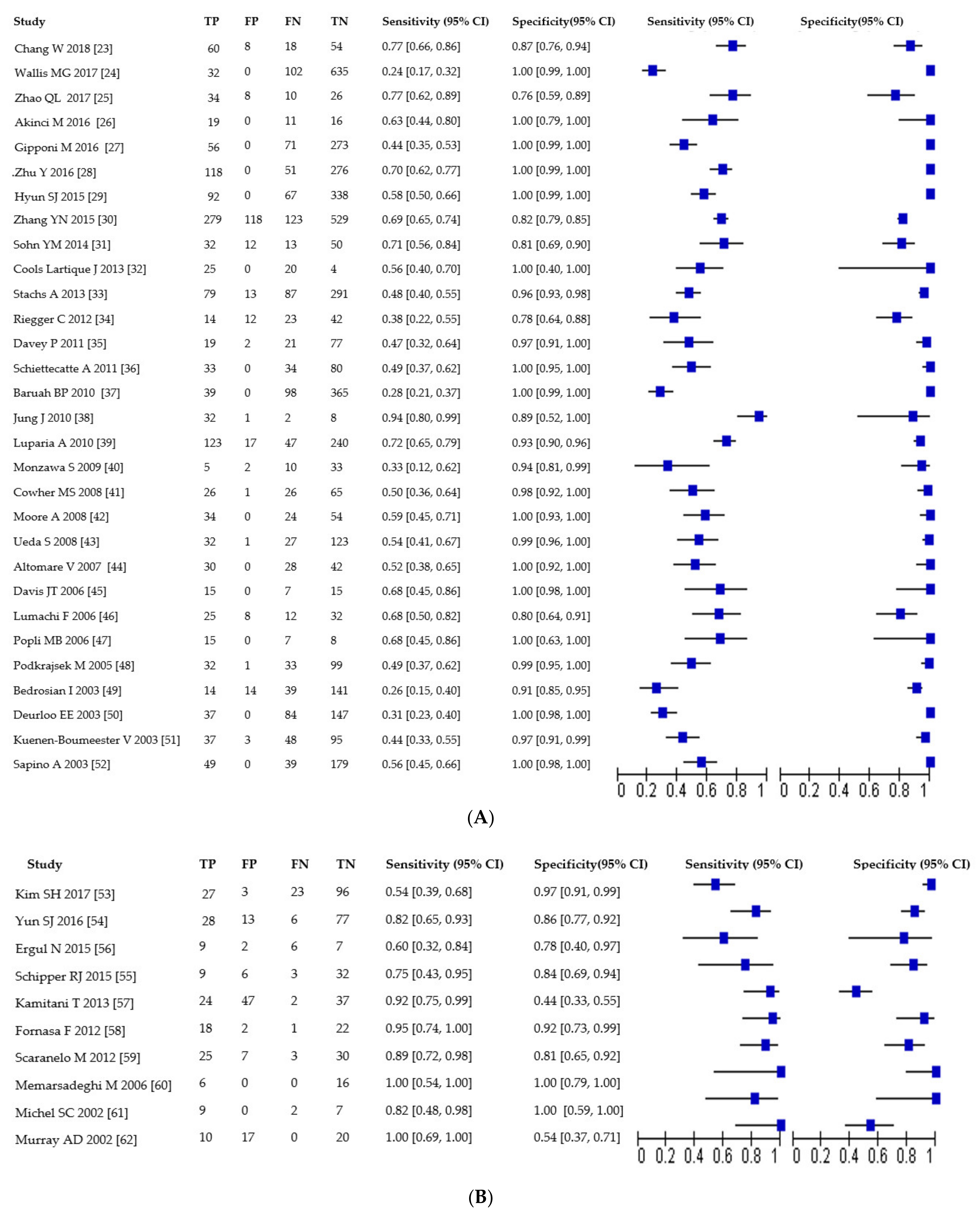
3. Results
3.1. Number and Characteristics of Included Studies
3.2. Quality of Included Studies
3.3. Sensitivity and Specificity of US, MRI and PET
3.4. US Subgroups Analysis
3.5. MRI Subgroups Analysis
3.6. PET Subgroups Analysis
3.7. Subgroup Analysis on Axillary Metastatic Burden
4. Discussion
5. Conclusions
Author Contributions
Funding
Conflicts of Interest
References
- Torre, L.A.; Islami, F.; Siegel, R.L.; Ward, E.M.; Jemal, A. Global cancer in women: Burden and trends. Cancer Epidemiol. Biomark. Prev. 2017, 26, 444–457. [Google Scholar] [CrossRef] [PubMed] [Green Version]
- Ferlay, J.; Soerjomataram, I.; Dikshit, R.; Eser, S.; Mathers, C.; Rebelo, M.; Parkin, D.M.; Forman, D.; Bray, F. Cancer incidence and mortality worldwide: Sources, methods and major patterns in GLOBOCAN 2012. Int. J. Cancer 2015, 136, E359–E386. [Google Scholar] [CrossRef]
- Houssami, N.; Ciatto, S.; Turner, R.M.; Cody, H.S.; MacAskill, P. Preoperative ultrasound-guided needle biopsy of axillary nodes in invasive breast cancer: Meta-analysis of its accuracy and utility in staging the axilla. Ann. Surg. 2011, 254, 243–251. [Google Scholar] [CrossRef] [PubMed]
- Cooper, K.L.; Harnan, S.; Meng, Y.; Ward, S.E.; Fitzergerald, P.; Papioannou, D.; Wyld, L.; Ingram, C.; Wilkinson, I.D.; Lorenz, E. Positron emission tomography (PET) for assessment of axillary lymph node status in early breast cancer: A systematic review and meta-analysis. Eur. J. Surg. Oncol. 2011, 37, 187–198. [Google Scholar] [CrossRef] [Green Version]
- Crane-Okada, R.; Wascher, R.A.; Elashoff, D.; Giuliano, A.E. Long-term morbidity of sentinel node biopsy versus complete axillary dissection for unilateral breast cancer. Ann. Surg. Oncol. 2008, 15, 1996–2005. [Google Scholar] [CrossRef] [PubMed]
- McLaughlin, S.A.; Wright, M.J.; Morris, K.T.; Giron, G.L.; Sampson, M.R.; Brockway, J.P.; Hurley, K.E.; Riedel, E.R.; van Zee, K.J. Prevalence of lymphedema in women with breast cancer 5 years after sentinel lymph node biopsy or axillary dissection: Objective measurements. J. Clin. Oncol. 2008, 26, 5213–5219. [Google Scholar] [CrossRef]
- Liu, C.; Guo, Y.; Shi, J.; Sheng, Y. Late morbidity associated with a tumour-negative sentinel lymph node biopsy in primary breast cancer patients: A systematic review. Eur. J. Cancer. 2009, 45, 1560–1568. [Google Scholar] [CrossRef]
- Kang, B.; Jun, H.; Lee, K.; Lee, K.; Kim, S. Clinical application of sentinel lymph node biopsy based on axillary anatomy in breast cancer: A single institution experience. Breast 2014, 23, 812–815. [Google Scholar] [CrossRef]
- Liang, X.; Yu, J.; Wen, B.; Xie, J.; Cai, Q.; Yang, Q. MRI and FDG-PET/CT based assessment of axillary lymph node metastasis in early breast cancer: A meta-analysis. Clin. Radiol. 2017, 72, 295–301. [Google Scholar] [CrossRef]
- Sui, W.F.; Chen, X.; Peng, Z.K.; Ye, J.; Wu, J.T. The diagnosis of metastatic axillary lymph nodes of breast cancer by diffusion weighted imaging: A meta-analysis and systematic review. World J. Surg. Oncol. 2016, 14. [Google Scholar] [CrossRef] [Green Version]
- Wang, X.-W.; Xiong, Y.-H.; Zen, X.-Q.; Lin, H.-B.; Liu, Q.-Y. Diagnostic accuracy of ultrasonograph guided fine-needle aspiration cytologic in staging of axillary lymph node metastasis in breast cancer patients: A meta-analysis. Asian Pac. J. Cancer Prev. 2012, 13, 5517–5523. [Google Scholar] [CrossRef] [PubMed] [Green Version]
- Lyman, G.H.; Giuliano, A.E.; Somerfield, M.R.; Benson, A.B.; Bodurka, D.C.; Burstein, H.J.; Cochran, A.J.; Cody, H.S.; Edge, S.B.; Galper, S.; et al. American Society of Clinical Oncology guideline recommendations for sentinel lymph node biopsy in early-stage breast cancer. J. Clin. Oncol. 2005, 23, 7703–7720. [Google Scholar] [CrossRef] [Green Version]
- Giuliano, A.E.; Ballman, K.V.; McCall, L.; Beitsch, P.D.; Bennan, M.B.; Kelemen, P.R.; Ollila, D.W.; Hansen, N.M.; Whitworth, P.W.; Blumencranz, P.W. Effect of axillary dissection vs no axillary dissection on 10-year overall survival among women with invasive breast cancer and sentinel node metastasis: The ACOSOG Z0011 (Alliance) randomized clinical trial. JAMA 2017, 318, 918–926. [Google Scholar] [CrossRef]
- Harnan, S.E.; Cooper, K.L.; Meng, Y.; Ward, S.E.; Fitzgerald, P.; Papaioannou, D.; Ingram, C.; Lorenz, E.; Wilkinson, I.D.; Wyld, L. Magnetic resonance for assessment of axillary lymph node status in early breast cancer: A systematic review and meta-analysis. Eur. J. Surg. Oncol. 2011, 37, 928–936. [Google Scholar] [CrossRef] [PubMed] [Green Version]
- Peare, R.; Staff, R.T.; Heys, S.D. The use of FDG-PET in assessing axillary lymph node status in breast cancer: A systematic review and meta-analysis of the literature. Breast Cancer Res. Treat. 2010, 123, 281–290. [Google Scholar] [CrossRef] [PubMed] [Green Version]
- Yang, W.T.; Le-Petross, H.T.; Macapinlac, H.; Carkaci, S.; Gonzalez-Angulo, A.M.; Dawood, S.; Resetkova, E.; Hortobagyi, G.N.; Cristofanilli, M. Inflammatory breast cancer: PET/CT, MRI, mammography, and sonography findings. Breast Cancer Res. Treat. 2008, 109, 417–426. [Google Scholar] [CrossRef] [PubMed]
- Moher, D.; Liberati, A.; Tetzlaff, J.; Altman, D.G. Preferred reporting items for systematic reviews and meta-analyses: The PRISMA statement. Int. J. Surg. 2010, 8, 336–341. [Google Scholar] [CrossRef] [Green Version]
- PRISMA Statement Website. 2015. Available online: www.prisma-statement.org (accessed on 15 March 2018).
- Whiting, P.; Rutjes, A.W.S.; Reitsma, J.B.; Bossuyt, P.M.M.; Kleijnen, J. The development of QUADAS: A tool for the quality assessment of studies of diagnostic accuracy included in systematic reviews. BioMed Cent. Med. Res. Methodol. 2003, 13, 1–13. [Google Scholar] [CrossRef] [Green Version]
- Glas, A.S.; Lijmer, J.G.; Prins, M.H.; Bonsel, G.J.; Bossuyt, P.M.M. The diagnostic odds ratio: A single indicator of test performance. J. Clin. Epidemiol. 2003, 56, 1129–1135. [Google Scholar] [CrossRef]
- Reitsma, J.B.; Glas, A.S.; Rutjes, A.W.S.; Scholten, R.J.P.M.; Bossuyt, P.M.; Zwinderman, A.H. Bivariate analysis of sensitivity and specificity produces informative summary measures in diagnostic reviews. J. Clin. Epidemiol. 2005, 58, 982–990. [Google Scholar] [CrossRef]
- Higgins, J.P.T.; Thompson, S.G. Quantifying heterogeneity in a meta-analysis. Stat. Med. 2002, 21, 1539–1558. [Google Scholar] [CrossRef] [PubMed]
- Chang, W.; Jia, W.; Shi, J.; Yuan, C.; Zhang, Y.; Chen, M. Role of elastography in axillary examination of patients with breast cancer. J. Ultrasound Med. 2018, 37, 699–707. [Google Scholar] [CrossRef] [Green Version]
- Wallis, M.G.; Kilburn-Toppin, F.; Taylor-Phillips, S. Does preoperative axillary staging lead to overtreatment of women with screen-detected breast cancer? Clin. Radiol. 2017, 10–15. [Google Scholar] [CrossRef] [PubMed] [Green Version]
- Zhao, Q.L.; Xia, X.N.; He, J.J.; Sheng, W.; Ruan, L.T.; Yin, Y.M.; Lou, H.L. Elastosonography and two-dimensional ultrasonography in diagnosis of axillary lymph node metastasis in breast cancer. Clin. Radiol. 2017, 5–11. [Google Scholar] [CrossRef] [Green Version]
- Akinci, M.; Bulut, S.P.; Erozgen, F.; Gurbuzel, M.; Gulsen, G.; Kocakusak, A.; Gulen, M.; Kaplan, R. Predictive value of fine needle aspiration biopsy of axillary lymph nodes in preoperative breast cancer staging. Turk. J. Surg. 2016, 32, 191–196. [Google Scholar] [CrossRef] [PubMed] [Green Version]
- Gipponi, M.; Fregatti, P.; Garlaschi, A.; Murelli, F.; Margarino, C.; Depaoli, F.; Baccini, P.; Gallo, M.; Friedman, D. Axillary ultrasound and Fine-Needle Aspiration Cytology in the preoperative staging of axillary node metastasis in breast cancer patients. Breast 2016, 30, 146–150. [Google Scholar] [CrossRef]
- Zhu, Y.; Zhou, W.; Zhou, J.Q.; Fei, X.C.; Ye, T.J.; Huang, O.; Chen, X.S.; Zhan, W.W. Axillary staging of early-stage invasive breast cancer by ultrasound-guided fine-needle aspiration cytology: Which ultrasound criteria for classifying abnormal lymph nodes should be adopted in the post-ACOSOG Z0011 trial era? J. Ultrasound Med. 2016, 35, 885–893. [Google Scholar] [CrossRef] [Green Version]
- Hyun, S.J.; Kim, E.K.; Yoon, J.H.; Moon, H.J.; Kim, M.J. Adding MRI to ultrasound and ultrasound-guided fine-needle aspiration reduces the false-negative rate of axillary lymph node metastasis diagnosis in breast cancer patients. Clin. Radiol. 2015, 70, 716–722. [Google Scholar] [CrossRef] [PubMed]
- Zhang, Y.N.; Wang, C.J.; Xu, Y.; Zhu, Q.L. Sensitivity, specificity and accuracy of ultrasound in diagnosis of breast cancer metastasis to the axillary lymph nodes in Chinese patients. Ultrasound Med. Biol. 2015, 41, 1835–1841. [Google Scholar] [CrossRef]
- Sohn, Y.-M.; Hong, I.K.; Han, K. Role of [18F] fluorodeoxyglucose positron emission tomography-computed tomography, sonography, and sonographically guided fine-needle aspiration biopsy in the diagnosis of axillary lymph nodes in patients with breast cancer. J. Ultrasound Med. 2014, 33, 1013–1021. [Google Scholar] [CrossRef]
- Cools-Lartigue, J.; Sinclair, A.; TRabulsi, N.; Meguerditchian, A.; Mesurolle, B.; Fuhrer, R.; Meterissian, S. Preoperative axillary ultrasound and fine-needle aspiration biopsy in the diagnosis of axillary metastases in patients with breast cancer: Predictors of accuracy and future implications. Ann. Surg. Oncol. 2013, 20, 819–827. [Google Scholar] [CrossRef] [PubMed]
- Stachs, A.; Gode, K.; Hartmann, S.; Stengel, B.; Nierling, U.; Dieterich, M.; Reimer, T.; Gerber, B. Accuracy of axillary ultrasound in preoperative nodal staging of breast cancer - size of metastases as limiting factor. Springerplus 2013, 2, 1–9. [Google Scholar] [CrossRef] [PubMed] [Green Version]
- Riegger, C.; Koeninger, A.; Hartung, V.; Otterbach, F.; Kimmig, R.; Forsting, M.; Bockisch, A.; Antoch, G.; Heusner, T.A. Comparison of the diagnostic value of FDG-PET / CT and axillary ultrasound for the detection of lymph node metastases in breast cancer patients. Acta Radiol. 2012, 53, 1092–1108. [Google Scholar] [CrossRef] [PubMed]
- Davey, P.; Stokes, M.; Kennedy, R.; Kirk, S.; Newell, J.; Majury, C.; McKillen, J. The value of axillary ultrasound with fine needle aspiration as a pre-operative staging procedure in breast cancer: Northern Irish experience. Ir. J. Med. Sci. 2011, 180, 509–511. [Google Scholar] [CrossRef]
- Schiettecatte, A.; Bourgain, C.; Breucq, C.; Buls, N.; De Wilde, V.; De Mey, J. Initial axillary staging of breast cancer using ultrasound-guided fine needle aspiration: A liquid-based cytology study. Cytopathology 2011, 22, 30–35. [Google Scholar] [CrossRef]
- Baruah, B.P.; Goyal, A.; Young, P.; Douglas-Jones, A.G.; Mansel, R.E. Axillary node staging by ultrasonography and fine-needle aspiration cytology in patients with breast cancer. Br. J. Surg. 2010, 97, 680–683. [Google Scholar] [CrossRef]
- Jung, J.; Park, H.; Park, J.; Kim, H. Accuracy of preoperative ultrasound and ultrasound-guided fine needle aspiration cytology for axillary staging in breast cancer. ANZ J. Surg. 2010, 80, 271–275. [Google Scholar] [CrossRef]
- Luparia, A.; Campanino, P.; Cotti, R.; Lucarelli, D.; Durando, M.; Mariscotti, G.; Gandini, G. Role of axillary ultrasound in the preoperative diagnosis of lymph node metastases in patients affected by breast carcinoma. Radiol. Med. 2010, 115, 225–237. [Google Scholar] [CrossRef]
- Monzawa, S.; Adachi, S.; Suzuki, K.; Hirokaga, H.; Takao, S.; Sakuma, T.; Hanioka, H. Diagnostic performance of fluorodeoxyglucose-positron emission tomography/computed tomography of breast cancer in detecting axillary lymph node metastasis: Comparison with ultrasonography and contrast-enhanced CT. Ann. Nucl. Med. 2009, 23, 855–861. [Google Scholar] [CrossRef]
- Cowher, M.S.; Erb, K.M.; Poller, W.; Julian, T.B. Correlation of the use of axillary ultrasound and lymph node needle biopsy with surgical lymph node pathology in patients with invasive breast cancer. Am. J. Surg. 2008, 196, 756–759. [Google Scholar] [CrossRef]
- Moore, A.; Hester, M.; Nam, M.W.; Brill, Y.M.; McGrath, P.; Wright, H.; Weisinger, K.; Romond, E.; Samayoa, L.M. Distinct lymph nodal sonographic characteristics in breast cancer patients at high risk for axillary metastases correlate with the final axillary stage. Br. J. Radiol. 2008, 81, 630–636. [Google Scholar] [CrossRef] [PubMed]
- Ueda, S.; Tsuda, H.; Asakawa, H.; Omata, J.; Fukatsu, K.; Kondo, N.; Kondo, T.; Hama, Y.; Tamura, K.; Ishida, J.; et al. Utility of 18F-fluoro-deoxyglucose emission tomography/ computed tomography fusion imaging (18F-FDG PET/CT) in combination with ultrasonography for axillary staging in primary breast cancer. BMC Cancer 2008, 8, 1–10. [Google Scholar] [CrossRef] [PubMed] [Green Version]
- Altomare, V.; Guerriero, G.; Carino, R.; Battista, C.; Primavera, A.; Altomare, A.; Vaccaro, D.; Esposito, A.; Ferri, A.M.; Rabitti, C. Axillary lymph node echo-guided fine-needle aspiration cytology enables breast cancer patients to avoid a sentinel lymph node biopsy. Preliminary experience and a review of the literature. Surg. Today 2007, 37, 735–739. [Google Scholar] [CrossRef]
- Davis, J.T.; Brill, Y.M.; Simmons, S.; Sachleben, B.C.; Cibull, M.L.; McGrath, P.; Wright, H.; Romond, E.; Hester, M.; Moore, A.; et al. Ultrasound-guided fine-needle aspiration of clinically negative lymph nodes versus sentinel node mapping in patients at high risk for axillary metastasis. Ann. Surg. Oncol. 2006, 13, 1545–1552. [Google Scholar] [CrossRef] [PubMed]
- Lumachi, F.; Tregnaghi, A.; Ferretti, G.; Povolato, M.; Marzola, M.C.; Zucchetta, P.; Checchin, D.; Bui, F. Accuracy of ultrasonography and 99mTc-sestamibi scintimammography for assessing axillary lymph node status in breast cancer patients. A prospective study. Eur. J. Surg. Oncol. 2006, 32, 933–936. [Google Scholar] [CrossRef] [PubMed]
- Popli, M.B.; Sahoo, M.; Mehrota, N.; Choudhury, M.; Kumar, A.; Pathania, O.P.; Thomas, S. Preoperative ultrasound-guided fine-needle aspiration cytology for axillary staging in breast carcinoma. Australas. Radiol. 2006, 50, 122–126. [Google Scholar] [CrossRef]
- Podkrajsek, M.; Music, M.M.; Kadivec, M.; Zgajnar, J.; Besic, N.; Pogacnik, A.; Hocevar, M. Role of ultrasound in the preoperative staging of patients with breast cancer. Eur. Radiol. 2005, 15, 1044–1050. [Google Scholar] [CrossRef]
- Bedrosian, I.; Bedi, D.; Kuerer, H.M.; Fornage, B.D.; Harker, L.; Ross, M.I.; Ames, F.C.; Krishnamurthy, S.; Edeiken-Monroe, B.S.; Meric, F.; et al. Impact of clinicopathological factors on sensitivity of axillary ultrasonography in the detection of axillary nodal metastases in patients with breast cancer. Ann. Surg. Oncol. 2003, 10, 1025–1030. [Google Scholar] [CrossRef]
- Deurloo, E.E.; Tanis, P.J.; Gilhuijs, K.G.; Muller, S.H.; Kroger, R.P.J. Reduction in the number of sentinel lymph node procedures by preoperative ultrasonography of the axila in breast cancer. Eur. J. Cancer 2003, 39, 1068–1073. [Google Scholar] [CrossRef]
- Kuenen-Boumeester, V.; Menke-Pluymers, M.; de Kanter, A.Y.; Obdeijn, I.-M.A.; Urich, D.; Van Der Kwast, T.H. Ultrasound-guided fine needle aspiration cytology of axillary lymph nodes in breast cancer patients. A preoperative staging procedure. Eur. J. Cancer 2003, 39, 170–174. [Google Scholar] [CrossRef]
- Sapino, A.; Cassoni, P.; Zanon, E.; Fraire, F.; Croce, S.; Coluccia, C.; Donadio, M.; Bussolati, G. Ultrasonographically-guided fine-needle aspiration of axillary lymph nodes: Role in breast cancer management. Br. J. Cancer 2003, 88, 702–706. [Google Scholar] [CrossRef] [Green Version]
- Kim, S.H.; Shin, H.J.; Shin, K.C.; Chae, E.Y.; Choi, W.J.; Cha, J.H.; Kim, H.H. Diagnostic performance of fused diffusion-weighted imaging using T1-weighted imaging for axillary nodal staging in patients with early breast cancer. Clin. Breast Cancer 2017, 17, 154–163. [Google Scholar] [CrossRef]
- Yun, S.J.; Sohn, Y.M.; Seo, M. Differentiation of benign and metastatic axillary lymph nodes in breast cancer: Additive value of MRI computer-aided evaluation. Clin. Radiol. 2016, 71, 403.e1–403.e7. [Google Scholar] [CrossRef]
- Schipper, R.; Paiman, M.; Beets-Tan, R.; Nelemans, P.J.; de Vries, B.; Heuts, E. Diagnostic performance of dedicated axillary T2-and diffusion-weighted MR imaging for nodal staging in breast cancer. Radiology 2015, 275, 345–355. [Google Scholar] [CrossRef] [PubMed] [Green Version]
- Ergul, N.; Kadioglu, H.; Yildiz, S.; Yucel, S.B.; Gucin, Z.; Erdogan, E.B.; Aydin, M.; Muslumanoglu, M. Assessment of multifocality and axillary nodal involvement in early-stage breast cancer patients using 18F-FDG PET/CT compared to contrast-enhanced and diffusion-weighted magnetic resonance imaging and sentinel node biopsy. Acta Radiol. 2015, 56, 917–923. [Google Scholar] [CrossRef] [PubMed]
- Kamitani, T.; Hatakenaka, M.; Yabuuchi, H.; Matsuo, Y.; Fujita, N.; Jinnouchi, M.; Nagao, M.; Shirahane, K.; Tokunaga, E.; Honda, H. Detection of axillary node metastasis using diffusion-weighted MRI in breast cancer. Clin. Imaging 2013, 37, 56–61. [Google Scholar] [CrossRef] [PubMed]
- Fornasa, F.; Nesoti, M.V.; Bovo, C.; Bonavina, M.G. Diffusion-weighted magnetic resonance imaging in the characterization of axillary lymph nodes in patients with breast cancer. J. Magn. Reson. Imaging 2012, 36, 858–864. [Google Scholar] [CrossRef]
- Scaranelo, A.M.; Eiada, R.; Jacks, L.M.; Kulkarni, S.R.; Crystal, P. Accuracy of unenhanced MR imaging in the detection of axillary lymph node metastasis: Study of reproducibility and reliability. Radiology 2012, 262, 425–434. [Google Scholar] [CrossRef] [PubMed] [Green Version]
- Memarsadeghi, M.; Riedl, C.C.; Kaneider, A.; Galid, A.; Rudas, M.; Matzek, W.; Helbich, T.H. Axillary lymph node metastases in patients with breast carcinomas: Assessment with nonenhanced versus USPIO-enhanced MR imaging. Radiology 2006, 241, 367–377. [Google Scholar] [CrossRef]
- Michel, S.C.; Keller, T.M.; Frohlich, J.M.; Fink, D.; Caduff, R.; Seifert, B.; Marincek, B.; Kubik-Huch, R.A. Preoperative breast cancer staging: MR imaging of the axilla with ultrasmall superparamagnetic iron oxide enhancement. Radiology 2002, 225, 527–536. [Google Scholar] [CrossRef] [PubMed]
- Murray, A.D.; Staff, R.T.; Redpath, T.W.; Gilbert, F.J.; Ah-See, A.K.; Brookes, J.A.; Miller, I.D.; Payne, S. Dynamic contrast enhanced MRI of the axilla in women with breast cancer: Comparison with pathology of excised nodes. Br. J. Radiol. 2002, 75, 220–228. [Google Scholar] [CrossRef] [PubMed]
- Jeong, Y.J.; Kang, D.Y.; Yoon, H.J.; Son, H.J. Additional value of F-18 FDG PET/CT for initial staging in breast cancer with clinically negative axillary nodes. Breast Cancer Res. Treat. 2014, 145, 137–142. [Google Scholar] [CrossRef] [PubMed]
- Park, J.; Byun, B.H.; Noh, W.C.; Lee, S.S.; Kim, H.-A.; Kim, E.-K.; Choi, C.W.; Lim, S.M. Lymph node to primary tumor SUV ratio by 18F-FDG PET/CT and the prediction of axillary lymph node metastases in breast cancer. Clin. Nucl. Med. 2014, 39, e249–e253. [Google Scholar] [CrossRef] [PubMed]
- Machida, Y.; Kubota, K.; Katayama, T.; Toriihara, A.; Shibuya, H. Diagnostic performance of fluorodeoxyglucose-positron emission tomography/computed tomography combined with ultrasonography-guided fine needle aspiration cytology for identifying axillary lymph node status in patients with breast cancer. Eur. J. Surg. Oncol. 2013, 39, 26–30. [Google Scholar] [CrossRef] [PubMed]
- Seok, J.W.; Kim, Y.; An, Y.-S.; Kim, B.S. The clinical value of tumor FDG uptake for predicting axillary lymph node metastasis in breast cancer with clinically negative axillary lymph nodes. Ann. Nucl. Med. 2013, 27, 546–553. [Google Scholar] [CrossRef]
- Hahn, S.; Hecktor, J.; Grabellus, F.; Hartung, V.; Poppel, T.; Kimmig, R.; Forsting, M.; Antoch, G.; Heusner, T.A. Diagnostic accuracy of dual-time-point 18F-FDG PET/CT for the detection of axillary lymph node metastases in breast cancer patients. Acta Radiol. 2012, 53, 518–523. [Google Scholar] [CrossRef] [PubMed]
- Choi, W.H.; Yoo, I.R.; O, J.H.; Kim, S.H.; Chung, S.K. The value of dual-time-point 18 F-FDG PET/CT for identifying axillary lymph node metastasis in breast cancer patients. Br. J. Radiol. 2011, 84, 593–599. [Google Scholar] [CrossRef] [Green Version]
- Heusner, T.A.; Kuemmel, S.; Hahn, S.; Koeninger, A.; Otterbach, F.; Hamami, M.E.; Kimmig, K.R.; Forsting, M.; Bockisch, A.; Antoch, G.; et al. Diagnostic value of full-dose FDG PET/CT for axillary lymph node staging in breast cancer patients. Eur. J. Nucl. Med. Mol. Imaging 2009, 36, 1543–1550. [Google Scholar] [CrossRef]
- Kim, J.; Lee, J.; Chang, E.; Kim, S.; Suh, K.; Sul, J.; Song, I.; Kim, Y.; Lee, C. Selective sentinel node plus additional non-sentinel node biopsy based on an FDG-PET/CT scan in early breast cancer patients: Single institutional experience. World J. Surg. 2009, 33, 943–949. [Google Scholar] [CrossRef]
- Taira, N.; Ohsumi, S.; Takabatake, D.; Hara, F.; Takashima, S.; Aogi, K.; Takashima, S.; Inoue, T.; Sugata, S.; Nishimura, R. Determination of indication for sentinel lymph node biopsy in clinical node-negative breast cancer using preoperative 18F-fluorodeoxyglucose positron emission tomography/computed tomography fusion imaging. Jpn. J. Clin. Oncol. 2008, 39, 16–21. [Google Scholar] [CrossRef] [Green Version]
- Veronesi, U.; De Cicco, C.; Galimberti, V.E.; Fernandez, J.R.; Rotmensz, N.; Viale, G.; Spano, G.; Luini, A.; Intra, M.; Veronesi, P.; et al. A comparative study on the value of FDG-PET and sentinel node biopsy to identify occult axillary metastases. Ann. Oncol. 2007, 18, 473–478. [Google Scholar] [CrossRef] [PubMed]
- Kumar, R.; Zhuang, H.; Schnall, M.; Conant, E.; Damia, S.; Weinstein, S.; Chandra, P.; Czerniecki, B.; Alavi, A. FDG PET positive lymph nodes are highly predictive of metastasis in breast cancer. Nucl. Med. Commun. 2006, 27, 231–236. [Google Scholar] [CrossRef]
- Weir, L.; Worsley, D.; Bernstein, V. The value of FDG positron emission tomography in the management of patients with breast cancer. Breast J. 2005, 11, 204–209. [Google Scholar] [CrossRef] [PubMed]
- Fehr, M.K.; Hornung, R.; Varga, Z.; Burger, D.; Hess, T.; Haller, U.; Fink, D.; von Schulthess, G.K.; Steinert, H.C. Axillary staging using positron emission tomography in breast cancer patients qualifying for sentinel lymph node biopsy. Breast J. 2004, 10, 89–93. [Google Scholar] [CrossRef] [PubMed]
- Zornoza, G.; Gaŕcia-Velloso, M.J.; Sola, J.; Regueira, F.M.; Pina, L.; Beorlegui, C. 18F-FDG PET complemented with sentinel lymph node biopsy in the detection of axillary involvement in breast cancer. Eur. J. Surg. Oncol. 2004, 30, 15–19. [Google Scholar] [CrossRef]
- Barranger, E.; Grahek, D.; Antoine, M.; Montravers, F.; Talbot, J.N.; Uzan, S. Evaluation of fluorodeoxyglucose positron emission tomography in the detection of axillary lymph node metastases in patients with early-stage breast cancer. Ann. Surg. Oncol. 2003, 10, 622–627. [Google Scholar] [CrossRef] [PubMed]
- Guller, U.; Nitzsche, E.U.; Schirp, U.; Viehl, C.T.; Torhorst, J.; Moch, H.; Langer, I.; Marti, W.R.; Oertli, D.; Harder, F.; et al. Selective axillary surgery in breast cancer patients based on positron emission tomography with 18F-fluoro-2-deoxy-D-glucose: Not yet! Breast Cancer Res. Treat. 2002, 71, 171–173. [Google Scholar] [CrossRef]
- Nakamoto, Y.; Chang, A.E.; Zasadny, K.R.; Wahl, R.L. Comparison of attenuation-corrected and non-corrected FDG-PET images for axillary nodal staging in newly diagnosed breast cancer. Mol. Imaging Biol. 2002, 4, 161–169. [Google Scholar] [CrossRef]
- Rieber, A.; Schirrmeister, H.; Gabelmann, A.; Nuessle, K.; Reske, S.; Kreienberg, R.; Brambs, H.J.; Kuehn, T. Pre-operative staging of invasive breast CA with MR mammo and or PET: Boon or bunk? Br. J. Radiol. 2002, 75, 789–798. [Google Scholar] [CrossRef]
- Van der Hoeven, J.J.M.; Hoekstra, O.S.; Comans, E.F.I.; Pijpers, R. Determinants of diagnostic performance of [F-18] fluorodeoxyglucose positron emission tomography for axillary staging in breast cancer. Ann. Surg. 2002, 236, 619–624. [Google Scholar] [CrossRef]
- Ganott, M.A.; Zuley, M.L.; Abrams, G.S.; Lu, A.H.; Kelly, A.E.; Sumkin, J.H.; Chivukula, M.; Carter, G.; Austin, R.M.; Bandos, A.I. Ultrasound guided core biopsy versus fine needle aspiration for evaluation of axillary lymphadenopathy in patients with breast cancer. ISRN Oncol. 2014, 2014, 703160. [Google Scholar] [CrossRef] [PubMed] [Green Version]
- Hayes, B.D.; Feeley, L.; Quinn, C.M.; Kennedy, M.M.; O’Doherty, A.; Flanagan, F.; O’Connell, A.M. Axillary fine needle aspiration cytology for pre-operative staging of patients with screen-detected invasive breast carcinoma. J. Clin. Pathol. 2011, 64, 338–342. [Google Scholar] [CrossRef] [PubMed]
- Tahir, M.; Osman, K.A.; Shabbir, J.; Rogers, C.; Suarez, R.; Reynolds, T.; Bucknall, T. Preoperative axillary staging in breast cancer–Saving time and resources. Breast J. 2008, 14, 369–371. [Google Scholar] [CrossRef] [PubMed]
- Kim, T.; Giuliano, A.E.; Lyman, G.H. Lymphatic mapping and sentinel lymph node biopsy in early-stage breast carcinoma: A metaanalysis. Cancer 2006, 106, 4–16. [Google Scholar] [CrossRef] [PubMed]
- Tanaka, K.; Yamamoto, D.; Kanematsu, S.; Okugawa, H.; Kamiyama, Y. A four node axillary sampling trial on breast cancer patients. Breast 2006, 15, 203–209. [Google Scholar] [CrossRef]
- Zhu, Y.; Zhou, J.Q.; Jia, X.H.; Zhou, W.; Zhan, W.W. Interobserver variability between experienced radiologists in evaluating the number of abnormal lymph nodes seen on preoperative axillary ultrasound. Clin. Radiol. 2020, 76, 60–66. [Google Scholar] [CrossRef]
- Lowes, S.; Leaver, A.; Cox, K.; Satchithananda, K.; Cosgrove, D.; Lim, A. Evolving imaging techniques for staging axillary lymph nodes in breast cancer. Clin. Radiol. 2018. [Google Scholar] [CrossRef] [PubMed]
- Hieken, T.J.; Jones, K.N.; Boughey, J.C.; Shah, S.; Glazebrook, K.N. Second-look axillary ultrasound after breast MRI for enhanced preoperative nodal staging in newly diagnosed breast cancer patients. J. Clin. Oncol. 2013, 31, 98. [Google Scholar] [CrossRef]
- Kajáry, K.; Lengyel, Z.; Tokes, A.M.; Kulka, J.; Dank, M.; Tímea, T. Dynamic FDG-PET/CT in the initial staging of primary breast cancer: Clinicopathological correlations. Pathol. Oncol. Res. 2019, 26, 997–1006. [Google Scholar] [CrossRef] [Green Version]
- Acar, E.; Turgut, B.; Yiğit, S.; Kaya, G. Comparison of the volumetric and radiomics findings of 18F-FDG PET/CT images with immunohistochemical prognostic factors in local/locally advanced breast cancer. Nucl. Med. Commun. 2019. [Google Scholar] [CrossRef]


| (A) Characteristics of ultrasound included studies | |||||||||||||
| Author | Year | Country | Index Test | Second Test | Reference Standard | Prospective/Retrospective | N Analysed | N with Axillary Metastases | Prevalence of Axillary Metastases | Mean Age | Years of Study | Other Criteria | |
| Chang W. [23] | 2018 | China | US | US + Elastosonography | Histology (SLNB/ALND) | Retrospective | 140 | 78 | 55.7% | 55.3 | 2013–2014 | Disjunctive method | |
| Wallis M.G. [24] | 2017 | UK | US | US + CNB | Histology (SLNB/ALND) | Retrospective | 769 | 134 | 17.4% | ND | 2008–2015 | ||
| Zhao Q.L. [25] | 2017 | China | US | US + Elastosonography | Histology (SLNB/ALND) | Prospective | 78 | 44 | 56.4% | 52.5 | 2012–2013 | Disjunctive method | |
| Akinci M. [26] | 2016 | Turkey | US | US + FNA | Histology (SLNB/ALND) | Prospective | 46 | 30 | 65.2% | ND | 2011–2013 | ||
| Gipponi M. [27] | 2016 | Italy | US | US + FNA | Histology (SLNB/ALND) | Prospective | 400 | 127 | 31.8% | 64.6 | 2013–2015 | Only T1-T2-T3 tumors | |
| Zhu Y. [28] | 2016 | China | US | US + FNA | Histology (SLNB/ALND) | Retrospective | 445 | 169 | 38.0% | 55.6 | 2013–2014 | Only T1-T2 tumors | |
| Hyun S.J. [29] | 2015 | South Korea | US | US + FNA | Histology (SLNB/ALND) | Retrospective | 497 | 159 | 32.0% | 52 | 2012–2013 | ||
| Zhang Y.N. [30] | 2015 | China | US | US grayscale | Histology (SLNB/ALND) | Retrospective | 1049 | 402 | 38.3% | 50.3 | 2010–2011 | ||
| Sohn Y.M.b[31] | 2014 | South Korea | US | US grayscale | Histology (SLNB/ALND) | Retrospective | 107 | 45 | 42.1% | 53.9 | 2009–2012 | ||
| Cools Lartique J. [32] | 2013 | Canada | US | US + FNA | Histology (SLNB/ALND) | Prospective | 234 | 90 | 38.5% | 57.8 | 2005–2007 | ||
| Stachs A. [33] | 2013 | Germany | US | US grayscale | Histology (SLNB/ALND) | Retrospective | 470 | 166 | 35.3% | ND | 2008–2010 | ||
| Riegger C. [34] | 2012 | Germany | US | US grayscale | Histology (SLNB/ALND) | Retrospective | 91 | 37 | 40.7% | 55.5 | 2007–2010 | ||
| Davey P. [35] | 2011 | Northern Ireland | US | US + FNA | Histology (SLNB/ALND) | Retrospective | 119 | 40 | 33.6% | ND | 2009 | ||
| Schiettecatte A. [36] | 2011 | Belgium | US | US + FNA | Histology (SLNB/ALND) | Retrospective | 147 | 67 | 45.6% | 56 | ND | Breast tumors < 3cm | |
| Baruah B.P. [37] | 2010 | UK | US | US + FNA | Histology (SLNB/ALND) | Retrospective | 502 | 137 | 27.3% | 61 | 2006–2009 | ||
| Jung J. [38] | 2010 | South Korea | US | US + FNA | Histology (SLNB/ALND) | Retrospective | 189 | 61 | 32.3% | ND | 2005–2006 | ||
| Luparia A. [39] | 2010 | Italy | US | US grayscale | Histology (SLNB/ALND) | Retrospective | 427 | 170 | 39.8% | 60.9 | 2005–2008 | ||
| Monzawa S. [40] | 2009 | Japan | US | US grayscale | Histology (SLNB/ALND) | Retrospective | 50 | 15 | 30.0% | 59 | 2005–2006 | ||
| Cowher M.S. [41] | 2008 | USA | US | US + FNA | Histology (SLNB/ALND) | Retrospective | 125 | 57 | 45.6% | 61.3 | 2004–2005 | ||
| Moore A. [42] | 2008 | USA | US | US + FNA | Histology (SLNB/ALND) | Retrospective | 112 | 58 | 51.8% | ND | ND | High risk of metastases | |
| Ueda S. [43] | 2008 | Japan | US | US grayscale | Histology (SLNB/ALND) | Prospective | 183 | 59 | 32.2% | 57 | 2005–2007 | ||
| Altomare V. [44] | 2007 | Italy | US | US + FNA | Histology (SLNB/ALND) | Retrospective | 100 | 30 | 30.0% | 53 | 2004–2005 | Only T1-T2-T3 tumors. FNA performed for all patients | |
| Davis J.T. [45] | 2006 | USA | US | US + FNA | Histology (SLNB/ALND) | Prospective | 37 | 22 | 59.5% | ND | 2004–2005 | High risk of metastases | |
| Lumachi F. [46] | 2006 | Italy | US | US grayscale | Histology (SLNB/ALND) | Prospective | 77 | 37 | 48.1% | 54 | ND | Only T1-T2 tumors. | |
| Popli M.B. [47] | 2006 | India | US | US + FNA | Histology (SLNB/ALND) | Prospective | 30 | 22 | 73.3% | ND | ND | ||
| Podkrajsek M. [48] | 2005 | Slovenia | US | US + FNA | Histology (SLNB/ALND) | Retrospective | 165 | 65 | 39.4% | 56 | 2001–2003 | ||
| Bedrosian I. [49] | 2003 | USA | US | US + FNA | Histology (SLNB/ALND) | Prospective | 208 | 53 | 25.5% | 55.4 | 1994–2000 | ||
| Deurloo E.E. [50] | 2003 | The Netherlands | US | US + FNA | Histology (SLNB/ALND) | Prospective | 268 | 121 | 45.1% | 56 | 1999–2001 | Only patients eligible for SLNB | |
| Kuenen-Boumeester V. [51] | 2003 | The Netherlands | US | US + FNA | Histology (SLNB/ALND) | Retrospective | 183 | 85 | 46.4% | ND | 1998–2003 | ||
| Sapino A. [52] | 2003 | Italy | US | US + FNA | Histology (SLNB/ALND) | Prospective | 298 | 88 | 29.5% | ND | 2000 | 31 in situ breast cancer | |
| TOTAL | 7546 | 2668 | 35.4% | 56 | |||||||||
| (B) Characteristics of Magnetic Resonance Imaging included studies | |||||||||||||
| Author | Year | Country | Index Test | Second Test | Number of Testla | Reference Standard | Prospective/Retrospective | N Analysed | N with Axillary Metastases | Prevalence of Axillary Metastases | MEAN AGE | Period of Study | Other Criteria |
| Kim S.H. [53] | 2017 | South Korea | MRI | With and without DWI + Gadolinium IV | 3T | Histology | Retrospective | 149 | 50 | 33.6% | 49.2 | 2014–2015 | |
| (SLNB/ALND) | |||||||||||||
| Yun S.J. [54] | 2016 | South Korea | MRI | With DWI + Gadolinium IV | 3T | Histology | Retrospective | 124 | 34 | 27.4% | 59.8 | 2011–2014 | |
| (SLNB/ALND) | |||||||||||||
| Schipper R.J. [55] | 2015 | The Netherlands | MRI | With and without DWI | 3T | Histology | Retrospective | 50 | 12 | 24.0% | 60 | 2012–2013 | Only T1-T2-T3 |
| (SLNB/ALND) | tumors | ||||||||||||
| Ergul N. [56] | 2015 | Turkey | MRI | With and without DWI | 1.5T | Histology | Prospective | 24 | 15 | 62.5% | 47 | 2012–2013 | Only T1-T2 |
| (SLNB/ALND) | tumors | ||||||||||||
| Kamitani T. [57] | 2013 | Japan | MRI | DWI alone | 1.5T | Histology | Retrospective | 110 | 26 | 23.6% | 54.9 | 2006–2007 | |
| (SLNB/ALND) | |||||||||||||
| Fornasa F. [58] | 2012 | Italy | MRI | With DWI + Gadolinium IV | 1.5T | Histology | Prospective | 43 | 19 | 44.2% | 58 | 2008–2010 | |
| (SLNB/ALND) | |||||||||||||
| Scaranelo M. [59] | 2012 | Canada | MRI | With and without DWI | 1.5T | Histology | Prospective | 65 | 28 | 43.1% | 53 | 2008–2009 | |
| (SLNB/ALND) | |||||||||||||
| Memarsadeghi M. [60] | 2006 | Austria | MRI | Without DWI + USPIO IV | 1T | Histology | Prospective | 22 | 6 | 27.3% | 62 | 5 months | |
| (SLNB/ALND) | |||||||||||||
| Michel S.C. [61] | 2002 | Switzerland | MRI | Without DWI + USPIO IV | 1.5T | Histology | Prospective | 18 | 11 | 61.1% | 53 | 2000–2001 | |
| (SLNB/ALND) | |||||||||||||
| Murray A.D. [62] | 2002 | UK | MRI | Without DWI + Gadolinium IV | 0.95T | Histology | ND | 47 | 10 | 21.3% | 63 | ND | |
| (SLNB/ALND) | |||||||||||||
| TOTAL | 652 | 211 | 32.4% | 55.4 | |||||||||
| (C) Characteristics of FDG Positron Emission Tomography included studies | |||||||||||||
| Author | Year | Country | Index Test | Second Test | Evaluation | Reference Standard | Prospective/Retrospective | N Analysed | N with Axillary Metastases | Prevalence of Axillary Metastases | Mean Age | Years of Study | Other Criteria |
| Ergul N. [56] | 2015 | Turkey | FDG PET | With CT | Visual and semi-quantitative | Histology | Prospective | 24 | 15 | 62.5% | 47 | 2012–2013 | Only T1-T2 tumors |
| (SLNB/ALND) | |||||||||||||
| Jeong Y.J. [63] | 2014 | South Korea | FDG PET | With CT | Visual and semi-quantitative | Histology | Retrospective | 178 | 48 | 27.0% | 54.9 | 2010–2013 | |
| (SLNB/ALND) | |||||||||||||
| Park J. [64] | 2014 | South Korea | FDG PET | With CT | Visual and semi-quantitative | Histology | Retrospective | 136 | 70 | 51.5% | 49.7 | 2009–2012 | 3 patients without FDG-avid breast tumors excluded |
| (SLNB/ALND) | |||||||||||||
| Sohn Y.M. [31] | 2014 | South Korea | FDG PET | With CT | Visual | Histology | Retrospective | 107 | 45 | 42.1% | 53.9 | 2009–2012 | |
| (SLNB/ALND) | |||||||||||||
| Machida Y. [65] | 2013 | Japan | FDG PET | With CT | Visual and semi-quantitative | Histology | Retrospective | 227 | 54 | 23.8% | ND | 2005–2009 | |
| (SLNB/ALND) | |||||||||||||
| Seok J.W. [66] | 2013 | South Korea | FDG PET | With CT | Visual and semi-quantitative | Histology | Retrospective | 104 | 21 | 20.2% | 49.4 | 2010–2012 | Only T1-T2 tumors |
| (SLNB/ALND) | |||||||||||||
| Hahn S. [67] | 2012 | Germany | FDG PET | With CT | Visual and semi-quantitative | Histology | Retrospective | 38 | 16 | 26.9% | 52 | 2008 | Only T1-T2 tumors |
| (SLNB/ALND) | |||||||||||||
| Riegger C. [34] | 2012 | Germany | FDG PET | With CT | Visual | Histology | Retrospective | 91 | 37 | 40.7% | 55.5 | 2007–2010 | |
| (SLNB/ALND) | |||||||||||||
| Choi W.H. [68] | 2011 | South Korea | FDG PET | With CT | Visual and semi-quantitative | Histology | Retrospective | 171 | 73 | 42.7% | 50.1 | 2003–2006 | |
| (SLNB/ALND) | |||||||||||||
| Heusner T.A. [69] | 2009 | Germany | FDG PET | With CT | Visual | Histology | Retrospective | 61 | 24 | 39.3% | 56 | 2007–2008 | |
| (SLNB/ALND) | |||||||||||||
| Kim J [70] | 2009 | South Korea | FDG PET | With CT | Visual | Histology | Prospective | 137 | 35 | 25.5% | 50.5 | 2007–2008 | Only T1-T2 tumors |
| (SLNB/ALND) | |||||||||||||
| Monzawa S. [40] | 2009 | Japan | FDG PET | With CT | Visual | Histology | Retrospective | 50 | 15 | 30.0% | 59 | 2005–2006 | |
| (SLNB/ALND) | |||||||||||||
| Taira N. [71] | 2008 | Japan | FDG PET | With CT | Visual and semi-quantitative | Histology | Retrospective | 92 | 27 | 29.3% | 54.6 | 2006–2007 | |
| (SLNB/ALND) | |||||||||||||
| Ueda S. [43] | 2008 | Japan | FDG PET | With CT | Visual | Histology | Prospective | 183 | 59 | 32.2% | 57 | 2005–2007 | |
| (SLNB/ALND) | |||||||||||||
| Veronesi U. [72] | 2007 | Italy | FDG PET | With CT | Visual and semi-quantitative | Histology | Retrospective | 236 | 103 | 43.6% | 49 | 2003–2005 | Only T1-T2-T3 tumors |
| (SLNB/ALND) | |||||||||||||
| Kumar R. [73] | 2006 | USA | FDG PET | Without CT | ND | Histology | Prospective | 80 | 36 | 45.0% | 52 | ND | |
| (SLNB/ALND) | |||||||||||||
| Weir L. [74] | 2005 | Canada | FDG PET | Without CT | Visual | Histology | Retrospective | 40 | 18 | 45.0% | 52 | 2000–2003 | |
| (SLNB/ALND) | |||||||||||||
| Fehr M.K. [75] | 2004 | Switzerland | FDG PET | Without CT | Visual | Histology | Prospective | 24 | 10 | 41.7% | 56 | ND | Tumors |
| (SLNB/ALND) | < 3 cm (clinical) | ||||||||||||
| Zornoza M.J. [76] | 2004 | Spain | FDG PET | Without CT | Visual | Histology | Prospective | 200 | 107 | 53.5% | 52.2 | ND | Tumors < 3.5 cm (ND) |
| (SLNB/ALND) | |||||||||||||
| Barranger E. [77] | 2003 | France | FDG PET | Without CT | Visual | Histology | Prospective | 32 | 15 | 46.9% | 58 | 2001 | Only T1-T2 tumors |
| (SLNB/ALND) | |||||||||||||
| Guller U. [78] | 2002 | Switzerland | FDG PET | Without CT | ND | Histology | Prospective | 31 | 14 | 45.2% | 64.8 | ND | |
| (SLNB/ALND) | |||||||||||||
| Nakamoto Y. [79] | 2002 | USA | FDG PET | Without CT | Visual | Histology | Prospective | 36 | 15 | 41.7% | 50.6 | ND | |
| (SLNB/ALND) | |||||||||||||
| Rieber A. [80] | 2002 | Germany | FDG PET | Without CT | ND | Histology | Retrospective | 40 | 20 | 50.0% | 52.9 | ND | |
| (SLNB/ALND) | |||||||||||||
| Van der Hoeven J.M. [81] | 2002 | The Netherlands | FDG PET | Without CT | Visual | Histology (SLNB/ALND) | Prospective | 70 | 32 | 45.7% | 58 | 1997–2000 | |
| TOTAL | 2388 | 909 | 38.1% | 52.9 | |||||||||
| (D) Characteristics of Fine Needle Aspiration included studies | |||||||||||||
| Author | Year | Country | Index Test | Evaluation | Prospective/Retrospective? | N Analysed | N with Axillary Metastases | Prevalence of Axillary Metastases | Mean Age | Years of Studies | Other Criteria | ||
| Zhu Y. [28] | 2016 | China | FNA | Histology | Retrospective | 445 | 169 | 38.0% | 55.6 | 2013–2014 | Only T1-T2 tumors | ||
| (SLNB/ALND) | |||||||||||||
| Sohn Y.M. [31] | 2014 | South Korea | FNA | Histology | Retrospective | 107 | 45 | 42.1% | 53.9 | 2009–2012 | |||
| (SLNB/ALND) | |||||||||||||
| Ganott M.A. [82] | 2014 | USA | FNA | Histology | Prospective | 44 | 26 | 59.1% | ND | 2008–2010 | |||
| (SLNB/ALND) | |||||||||||||
| Hayes B.D. [83] | 2011 | Ireland | FNA | Histology | Retrospective | 161 | 86 | 53.4% | ND | 2006–2009 | |||
| (SLNB/ALND) | |||||||||||||
| Schiettecatte A. [36] | 2011 | Belgium | FNA | Histology | Retrospective | 147 | 67 | 45.6% | 56 | ND | |||
| (SLNB/ALND) | |||||||||||||
| Luparia A. [39] | 2010 | Italy | FNA | Histology | Retrospective | 427 | 170 | 39.8% | 60.9 | 2005–2008 | FNA was not performed for all suspicious axillary US | ||
| (SLNB/ALND) | |||||||||||||
| Tahir M. [84] | 2008 | UK | FNA | Histology | Prospective | 38 | 17 | 44.7% | 56.7 | 2005–2006 | |||
| (SLNB/ALND) | |||||||||||||
| Cowher M.S. [41] | 2008 | USA | FNA | Histology | Retrospective | 125 | 57 | 45.6% | 61.3 | 2004–2005 | |||
| (SLNB/ALND) | |||||||||||||
| Moore A. [42] | 2008 | USA | FNA | Histology | Retrospective | 112 | 58 | 51.8% | ND | ND | Only high risk of metastases | ||
| (SLNB/ALND) | |||||||||||||
| Davis J.T. [45] | 2006 | USA | FNA | Histology | Prospective | 37 | 22 | 59.5% | ND | 2004–2005 | Only high risk of metastases | ||
| (SLNB/ALND) | |||||||||||||
| Popli M.B. [47] | 2006 | India | FNA | Histology | Prospective | 30 | 22 | 73.3% | ND | ND | |||
| (SLNB/ALND) | |||||||||||||
| Podkrajsek M. [48] | 2005 | Slovenia | FNA | Histology | Retrospective | 165 | 65 | 39.4% | 56 | 2001–2003 | |||
| (SLNB/ALND) | |||||||||||||
| Deurloo E.E. [50] | 2003 | The Netherlands | FNA | Histology | Prospective | 268 | 121 | 45.1% | 56 | 1999–2001 | |||
| (SLNB/ALND) | |||||||||||||
| Sapino A. [52] | 2003 | Italy | FNA | Histology | Prospective | 298 | 88 | 29.5% | ND | 2000 | |||
| (SLNB/ALND) | |||||||||||||
| TOTAL | 2404 | 1013 | 42.1% | 49.9 | |||||||||
| Imaging Technique | N Studies | Sensitivity | I2 | Specificity | I2 | DOR |
|---|---|---|---|---|---|---|
| US | 30 | 0.55 (0.49, 0.62) | 90.01 | 0.99 (0.97, 1.00) | 95.06 | 112 (39, 320) |
| US grayscale | 24 | 0.63 (0.56, 0.69) | 88.86 | 0.88 (0.82, 0.92) | 93.91 | 12 (8, 18) |
| US + FNA|CNB | 20 | 0.51 (0.43, 0.59) | 88.44 | 1.00 (0.99, 1.00) | 94.19 | 752 (98, 5765) |
| FNA | 14 | 0.78 (0.73, 0.83) | 55.40 | 0.99 (0.96, 1.00) | 48.73 | 560 (91, 3451) |
| MRI | 10 | 0.83 (0.72, 0.91) | 75.81 | 0.85 (0.72, 0.92) | 93.00 | 28 (16, 51) |
| MRI without DWI | 7 | 0.81 (0.49, 0.95) | 89.17 | 0.84 (0.74, 0.91) | 89.04 | 22 (7, 72) |
| MRI with DWI | 4 | 0.78 (0.60, 0.89) | 79.35 | 0.90 (0.82, 0.95) | 67.07 | 33 (17, 65) |
| DWI alone | 5 | 0.74 (0.50, 0.89) | 83.54 | 0.78 (0.51, 0.92) | 93.63 | 10 (5, 19) |
| PET FDG | 24 | 0.49 (0.39, 0.59) | 87.03 | 0.94 (0.91, 0.96) | 73.98 | 15 (8, 26) |
| PET FDG without CT | 9 | 0.44 (0.28, 0.62) | 90.90 | 0.95 (0.91, 0.97) | 0 | 14 (5, 44) |
| PET FDG with CT | 15 | 0.51 (0.40, 0.63) | 86.04 | 0.93 (0.89, 0.96) | 79.51 | 14 (8, 27) |
Publisher’s Note: MDPI stays neutral with regard to jurisdictional claims in published maps and institutional affiliations. |
© 2021 by the authors. Licensee MDPI, Basel, Switzerland. This article is an open access article distributed under the terms and conditions of the Creative Commons Attribution (CC BY) license (https://creativecommons.org/licenses/by/4.0/).
Share and Cite
Le Boulc’h, M.; Gilhodes, J.; Steinmeyer, Z.; Molière, S.; Mathelin, C. Pretherapeutic Imaging for Axillary Staging in Breast Cancer: A Systematic Review and Meta-Analysis of Ultrasound, MRI and FDG PET. J. Clin. Med. 2021, 10, 1543. https://0-doi-org.brum.beds.ac.uk/10.3390/jcm10071543
Le Boulc’h M, Gilhodes J, Steinmeyer Z, Molière S, Mathelin C. Pretherapeutic Imaging for Axillary Staging in Breast Cancer: A Systematic Review and Meta-Analysis of Ultrasound, MRI and FDG PET. Journal of Clinical Medicine. 2021; 10(7):1543. https://0-doi-org.brum.beds.ac.uk/10.3390/jcm10071543
Chicago/Turabian StyleLe Boulc’h, Morwenn, Julia Gilhodes, Zara Steinmeyer, Sébastien Molière, and Carole Mathelin. 2021. "Pretherapeutic Imaging for Axillary Staging in Breast Cancer: A Systematic Review and Meta-Analysis of Ultrasound, MRI and FDG PET" Journal of Clinical Medicine 10, no. 7: 1543. https://0-doi-org.brum.beds.ac.uk/10.3390/jcm10071543