Article

Comparison of Antero-Lateral Thigh Flap and Vastus Lateralis Muscle Flap for the Treatment of Extensive Scalp Defects—A Retrospective Cohort Study

1
Department of Oral and Cranio-Maxillofacial Surgery, University of Heidelberg, Im Neuenheimer Feld 400, D-69120 Heidelberg, Germany
2
Department of Neurosurgery, University of Heidelberg, Im Neuenheimer Feld 400, D-69120 Heidelberg, Germany
*
Author to whom correspondence should be addressed.
J. Clin. Med. 2023, 12(19), 6208; https://0-doi-org.brum.beds.ac.uk/10.3390/jcm12196208
Submission received: 8 August 2023 / Revised: 31 August 2023 / Accepted: 23 September 2023 / Published: 26 September 2023
(This article belongs to the Special Issue Innovation in Head and Neck Reconstructive Surgery—Series 2)

Abstract

:
Free flap reconstruction is the standard of care for extensive defects of the head and neck area. In this study, two types of free flaps, the antero-lateral thigh flap (ALT) and the vastus lateralis muscle flap, were compared. The primary endpoint was flap success, secondary endpoints were complication rates, hospitalization and surgery time. Cases with defect situations of the scalp and consecutive microvascular free flap reconstructions using either ALT flaps or vastus lateralis muscle flaps between 2014 and 2022 were retrospectively analyzed. Indications, perioperative handling and outcomes were compared. Twenty patients were included in the analysis. Ten patients (50%) received a free flap reconstruction using an ALT flap and ten patients (50%) received a vastus lateralis flap. A simultaneous two-team approach was possible in each case and the flap success rate was 100% with the need for one successful anastomosis revision. The mean defect size in our cohort was 147 ± 46 cm2. There were no significant differences in surgery time, duration of hospitalization or complication rate between both cohorts. Both free flaps, the ALT and the vastus lateralis flap, are suitable for the closure of large scalp defects. They provide high success rates, short surgery times without the need for patient repositioning and low donor-site morbidity. The vastus lateralis muscle flap bares the advantage of being perforator-independent and allows for the preparation of long vessels for anastomosis if needed while baring the disadvantage of a prolonged period of healing via granulation or the need for secondary surgery in terms of covering by split-thickness skin grafts which may interfere with necessary adjuvant treatment in oncological patients.

1. Introduction

Scalp defects may follow after ablative tumor surgery, trauma or as a consequence of wound healing disorders secondary to cranial surgery. Depending on their localizations, size and the medical history of the affected patients, they may impose a relevant challenge on the treating specialist. In many cases, defect situations affect soft and hard tissue and lead to the necessity to provide both, hard tissue reconstruction via cranioplasty, and the restoration of soft tissue [1]. While minor defects may be treated with primary wound closure or local flaps, free tissue transfer using microvascular free flaps offers the possibility to adequately treat extensive defects [2,3,4]. Apart from aesthetical considerations, sufficient wound closure in cranial defects is of clinical relevance to prevent the spread of infections, especially in extensive defects with intracranial communication and when cranioplasty was performed [5,6].
Several types of free flaps have been presented for defect restoration of the cranium and each type has distinct features that potentially may influence peri- and postoperative procedures and clinical outcomes [7]. Studies on clinical outcomes in free flap reconstruction in the head and neck area are numerous and several factors with potential influence on flap success have been described, including a history of radiotherapy, vascular diseases and previous surgical procedures [8,9]. Those risk factors often are present in patients after sequential surgical procedures to the cranium due to recurrent tumors or wound healing disorders after decompressive surgery.
The goal of the present study, therefore, was to investigate a cohort of patients with extensive cranial defects who were treated in an interdisciplinary approach including neurosurgeons and reconstructive oral and maxillofacial surgeons. Two different types of free flaps were used, the antero-lateral thigh (ALT) flap and the vastus-lateralis muscle flap, and compared regarding success rates and peri- and postoperative management.

2. Materials and Methods

2.1. Patients and Data Acquisition

The presented study was designed as a retrospective single-center cohort study to compare two different free flaps for the closure of extensive cranial defects (i.e., antero-lateral thigh flap—ALT and vastus lateralis muscle flap—VL). The primary endpoint was flap success, secondary endpoints were complication rates, hospitalization and surgery time. We retrospectively analyzed the medical records of patients receiving free flap reconstruction of the head and neck area in the Department of Oral and Cranio-Maxillofacial Surgery of the Heidelberg University Hospital between September 2010 and November 2021. All patients receiving a free flap reconstruction to the cranium with or without cranioplasty were included in this analysis.
The study was conducted in accordance with the principles stated in the Declaration of Helsinki. Approval of the local ethics committee was given (ethic vote: S-513/2017) and written informed consent was obtained from all patients.
Clinical data were collected focusing on perioperative data including initial diagnosis and indication for the reconstructive approach, type of flap used in each case, pedicle vessels, duration of surgical procedures and hospitalization, and peri- and postoperative morbidity and complications.

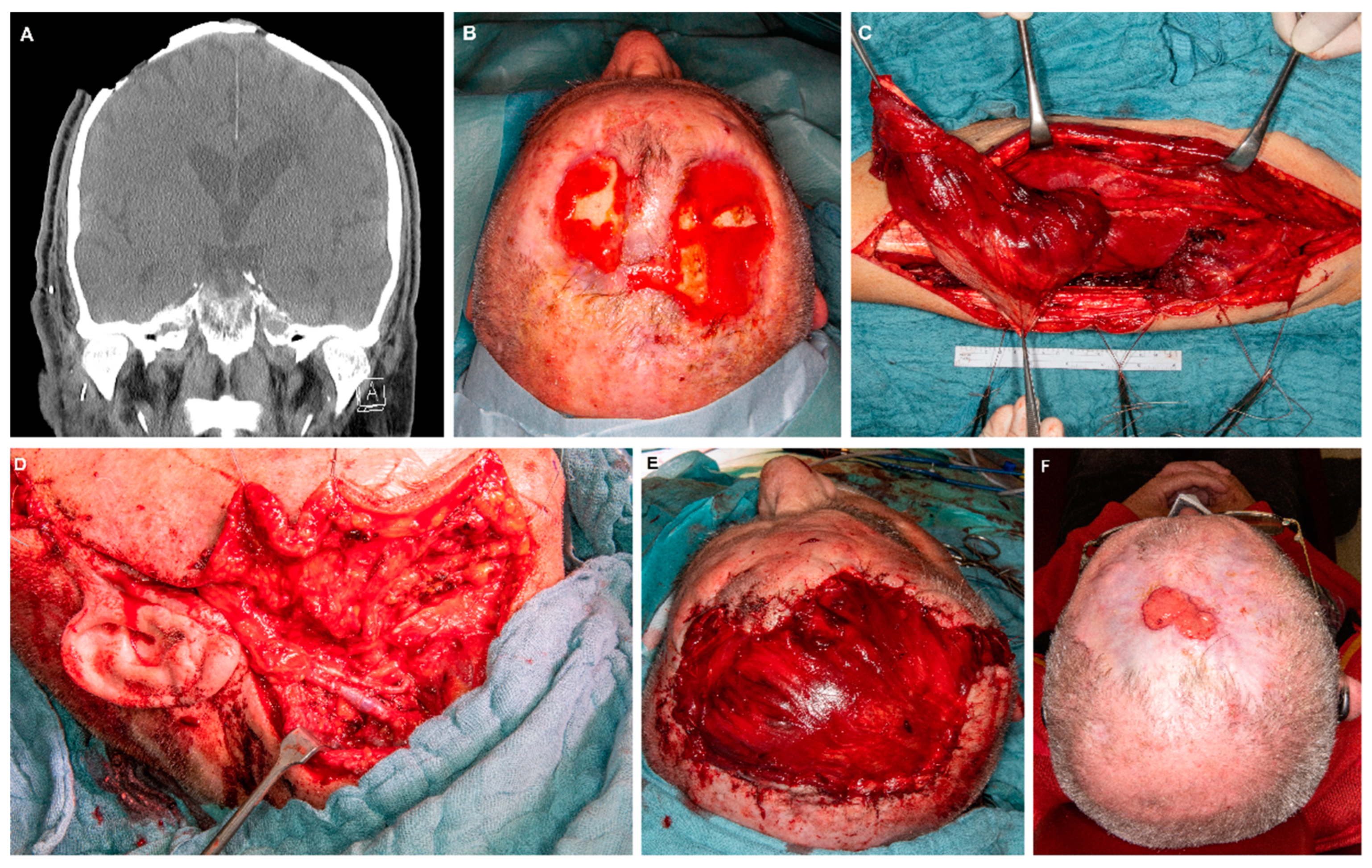
2.2. Surgical Procedures

Patients were treated surgically by the Department of Oral and Maxillofacial Surgery with or without the participation of the Department of Neurosurgery in cases where cranioplasty needed to be performed. All patients received wound closure with a free flap (either ALT or vastus lateralis flap). All surgical procedures were performed in a simultaneous two-team approach to economize surgery time. In some patients with cutaneous squamous cell carcinoma elective or therapeutic neck dissection was performed additionally. The recipient vessels were chosen intra-operatively with regard to size and localization of the defect, pedicle length of the flap and the need to perform a neck dissection. No preoperative diagnostics to determine recipient vessels were performed.

2.3. Statistical Analysis

Statistics were performed using SPSS Statistics® 25 (IBM, Armonk, NY, USA). Clinical and demographical data were analyzed and summarized with descriptive statistics. Comparison of categorical data was performed with Chi-squared testing and Student’s t-test was used for comparison of mean values of unpaired parameters. A p-value of 0.05 or less was considered to indicate statistical significance.

3. Results

3.1. Patient Cohort

An overall number of 20 patients were included in the analysis. Eight patients were female (40%) and 12 were male (60%). The mean age was 65 ± 19 years (range from 27 to 96 years). The reasons for the reconstructive approaches were wound healing disorders following cranial surgery in 10 cases (50%) and cutaneous tumors in 10 cases (50%). Further information on the patients, including medical history is provided in Table 1 and Table 2.
The affected subsites of the cranium included the parietal region in 11 cases (55%), the temporal region in 8 cases (40%), and the occipital region in 1 case (5%).

3.2. Surgical Procedures and Perioperative Management

All patients received a free flap for defect closure. Ten defects (50%) were treated with an ALT flap and ten defects (50%) were treated with a vastus lateralis muscle flap. Cranioplasty was performed in 7 patients (35%) using palacos cement in 4 patients (57%), patient-specific titanium implants in two patients (29%) and Polymethyl methacrylate (PMMA) in 1 patient (14%). The treated skin defects ranged from 10 cm to 18 cm in diameter with a mean defect size of 145 ± 47 cm2 (range: 80–252 cm2).
The facial artery and the internal jugular vein were the vessels mostly used for anastomosis in the cohort. Table 3 provides detailed information on the vessels used for anastomosis. In 1 patient (5%) a venous interposition graft was used to enhance the pedicle length in order to facilitate the anastomosis. Six patients (30%) with cutaneous squamous cell carcinoma received an ipsi- or bilateral neck dissection according to tumor localization and preoperative CT scan within the same surgery.
Primary closure of the donor site defect could be achieved in 18 patients (90%), while secondary healing/closure happened in two patients (10%) who received a cranial defect closure with an ALT flap. Supplementary Figure S1 exemplifies the donor sites of two patients following ALT harvesting with primary closure in one patient and secondary healing via granulation in another patient.
Table 4 gives an overview of perioperative management, complications and hospitalization in dependence of the used flaps. There were no significant differences regarding all investigated parameters between the two groups. The mean surgery time for the whole cohort was 273 ± 71 min and did not differ between the ALT and the vastus lateralis group (Table 4). The mean hospitalization in the ICU was 8 ± 15 days (median: 2.5 days). The mean hospitalization was 18 ± 13 days (median: 14.5 days). Figure 1 and Figure 2 illustrate the preoperative status, perioperative management and surgical results of cranial defect closures using a vastus lateralis muscle flap (Figure 1) and an ALT flap (Figure 2), respectively. Figure 3 provides pre- and postoperative impressions on four patients treated with vastus lateralis muscle flaps or ALT flaps.

3.3. Complications

The flap success rate in this cohort was 100% and revision of the anastomosis was successfully performed in one patient (5%) with an ALT flap. Major complications including postoperative cardio-pulmonary deterioration occurred in four patients (20%) and led to prolongated hospitalization (Table 4). Three patients with major complications suffered from recurrent SCC and one patient from recurrent meningioma with extensive wound healing disorders.

4. Discussion

Defect situations of the scalp mostly follow after ablative surgery of cutaneous tumors or as a consequence of wound healing disorders after cranial surgery. Especially composite defects of the skin and the calvarial bone that may result after multiple surgeries or multimodality treatment of cranial pathologies require adequate treatment to prevent secondary complications like intracranial spread of infections. Large defects and those arising after complex pre-treatment, often including multiple surgical procedures with or without radiotherapy, in most cases require the use of free flaps in order to provide sufficient tissue for a reliable reconstruction [10,11]. Several different strategies and algorithms have been described for the treatment of scalp defects and a variety of free flaps have been described as reliable options. Those include the latissimus dorsi free flap for large defects, the parascapular flap, the radial forearm flap and the anterolateral thigh flap (ALT) among others [7,12,13,14,15,16,17]. Other alternatives for extensive defects, like the omentum flap that has been described for pharyngeal reconstruction, bear the necessity for intraabdominal flap harvest, and, thus, are usually not the first choice for scalp reconstruction [18,19].
In the present study, we aimed to present our experience with the treatment of scalp defects using our in-house favorite flaps for this indication, the ALT and the vastus lateralis muscle flap. While the ALT has often been described as the standard of care for large scalp defects among others, as stated above, the vastus lateralis (VL) flap seems to be far less commonly in use. While the VL has been described as a versatile treatment option for soft tissue defects of the head and neck in some studies, up to date, there are hardly any comparative studies on its advantages and disadvantages when used for the treatment of scalp defects [20,21,22,23,24,25].
The ALT flap provides a skin paddle which makes it the first choice for extraoral defect closure. This applies even more in patients suffering from malignant tumors (e.g., cutaneous squamous cell carcinomas) with an urgent necessity for timely adjuvant radiotherapy. Correspondingly, the use of the vastus lateralis flap implies the need for primary or secondary covering of the muscle. This may be regarded as a disadvantage as secondary surgery is time-consuming for the patient and the treating specialist and carries the usual risks of surgical procedures. Furthermore, as stated above, it may delay or even impede the prompt execution of adjuvant therapy. While it may be possible to cover the muscle using a split-thickness skin graft in the primary procedure, in our department the muscle is left uncovered in order to be able to monitor the flap perfusion during the critical postoperative phase. After a period of around 4–8 weeks of granulation, the patient either will receive a secondary covering of the muscle using split-thickness skin grafts, or, depending on the patient’s choice, wound coverings until full epithelialization as a conservative alternative. A further disadvantage of the VL muscle flap is the shrinkage of the muscle during healing. This has been observed by other authors as well and requires a certain over-dimensioning of the muscle to prevent secondary soft tissue retraction with exposure of calvarial bone or materials used for its reconstruction [23,25]. This necessity of over-dimensioning is partly due to the mentioned shrinking of muscle, and partly to the nature and form of the VL muscle itself, which impedes a preparation as a flat wound covering. Although the muscle allows for a certain shaping to adapt it to the corresponding defect, the nutrient vessels necessitate a certain flap thickness, which leads to a bulky appearance during the first 4–8 weeks. In the course of granulation; however, the muscle usually shrinks and integrates into the surrounding skin level. Covering of the muscle with split-thickness skin grafts may usually be performed after 4–8 weeks. Alternatively, if the patient refuses further surgical approaches, a conservative approach with wound coverings until full epithelialization is chosen. The period until full epithelialization was highly variable between the patients and ranged from 3–12 months after surgery. An advantage of the vastus lateralis flap, on the other hand, is its independence of perforator vessels and its long pedicle [24]. Being perforator-independent, it exhibits far fewer anatomical variations of the pedicle vessels than the ALT and may be used as an alternative to the ALT in case no sufficient perforators can be found during surgery or if a long pedicle vessel is needed, e.g., if the temporal vessels are insufficient for anastomosis and the cervical vessels have to be used as a consequence. Considering the disadvantages of muscle flaps mentioned above, the standard flap for cranial defect closure in our department is the ALT. In cases of unreliable perforators or the need for very long pedicle vessels, however, the intraoperative change to harvesting a VL muscle flap, in our experience provides a safe and time economic solution without the need for the establishment of additional donor sites (e.g., ALT of the other leg, latissimus dorsi etc.), and without the risk of a compromise regarding the safety of the anastomosis, and, consequently, the flap success. Another argument in favor of the muscle flap is related to its reported tendency to shrink. While initial overdimensioning is warranted, the flap settles to the level of the surrounding tissue during healing. The ALT, however, in many cases requires thinning of the skin paddle due to its tendency towards increased bulkiness in Western countries [7,26]. An oversized skin paddle, however, may either be accepted with a risk of aesthetical disadvantages and impaired flap perfusion due to increased tissue pressure or may be corrected by time-consuming thinning of the ALT. The thinning of skin paddles is a matter of discussion with several authors describing it as a safe procedure, while other authors reported an elevated risk of damaging the perforator vessels and, thus, of a partial or complete flap loss as a consequence [27,28,29].
The pedicles in both flaps are long and of solid caliber with the perforator vessels being a weak point of the ALT.
The mean defect size in our analysis was 153 ± 50 cm2 with a range from 80 to 300 cm2. The comparison of both groups revealed no significant differences between the defect sizes of patients treated with an ALT flap and those treated with a VL flap, respectively. Weitz et al. described scalp defect sizes of >1000 cm2 in their analysis and advocated the use of latissimus dorsi flaps in those cases. While ALT and VL flaps may be limited to certain defect sizes, we did not find patients with defects exceeding 300 cm2 in our records and, thus, successfully treated all patients with cranial defects and the need for free tissue transfer with either ALT or VL flaps [7].
For anastomosis, the superficial temporal vessels have been described as a reliable and safe option for the reconstruction of cranial defects by several authors [7,30]. In our cohort, however, the temporal vessels were only chosen for anastomosis in five patients (25%). This may be explained by weak caliber temporal vessels in some patients, and the necessity to perform a neck dissection in some patients with cutaneous squamous cell carcinoma (n = 6; 30%) that granted access to the cervical vessels anyway. The low rate of revision surgeries and flap losses in our cohort, however, suggests that the intraoperative exploration and evaluation of vessels is a safe procedure and preoperative imaging that has been suggested by other authors seems unnecessary in most cases.
Harvesting ALT flaps may lead to different degrees of donor site morbidity depending on the position of the perforator vessels and the size of the required skin paddle. Harvesting VL flaps, in contrast, necessarily leads to the resection of relevant parts of the muscle with the consequence of permanent functional impairments of this motor unit. There are numerous publications indicating comparably low donor-site morbidity and functional impairment after ALT and vastus lateralis flap harvesting, with postoperative reduction of sensitivity of the upper thigh being the most commonly mentioned side effect. Nevertheless, a more detailed examination of the postoperative muscle function revealed a significant decrease in knee flexion, walking speed, and, health-related quality-of-life in patients after VL flap harvesting [20,31,32,33]. While it probably can be assumed that the functional impairment after VL flap harvesting may be more relevant than after ALT harvesting, the primary closure of the donor site that can be achieved at 100% after VL flap harvesting may be regarded as an argument in favor of muscle flaps as it spares the patient time-consuming wound treatments in cases of secondary healing, and possibly allows for an earlier mobilization. While the use of muscle flaps may be regarded as critical in general due to the mentioned aspects, especially in oncological patients with a need for adjuvant therapy, there is a relevant number of publications advocating its use in scalp reconstruction, mainly under reference to their size and reliability.
Considering the mentioned factors, in our opinion, the ALT flap is the method of choice for cranial defect closure. The VL muscle flap, however, provides a safe and reliable option in cases of weak caliber perforator vessels or the need for long pedicle vessels, especially in patients without the urgent need for timely adjuvant radiotherapy.

5. Conclusions

Both flaps, the antero-lateral thigh (ALT) flap, and the vastus lateralis muscle flap were shown to be reliable options for the reconstruction of extensive scalp defects with no relevant differences regarding hospitalization and treatment-related complications. While usage of the vastus lateralis flap leads to the need for a certain over-dimensioning and secondary coverage of the muscle, it presents a safe strategy when no or only insufficient perforator vessels prevent the use of an ALT flap.

Supplementary Materials

The following are available online at https://0-www-mdpi-com.brum.beds.ac.uk/article/10.3390/jcm12196208/s1, STROBE statement, Supplementary Figure S1. Depiction of donor sites of two patients. (A): Left upper thigh after harvesting of ALT flap and primary wound closure. (B): Right upper thigh after harvesting of ALT flap during secondary healing of the donor site defect via granulation.

Author Contributions

Conceptualization, J.M. (Julius Moratin) and M.E.; methodology, J.M. (Julius Moratin), P.D.T., S.Z. and O.R.; software, J.M. (Julius Moratin), S.Z., K.S. and M.B.; validation, O.R., C.F., J.H., S.Z., M.E. and K.S.; formal analysis, J.M. (Julius Moratin), P.D.T., S.Z, J.M. (Jan Mrosek), M.B. and O.R.; investigation, J.M. (Julius Moratin), P.D.T., S.Z. and J.M. (Jan Mrosek); resources, M.E., C.F. and J.H.; data curation, J.M. (Julius Moratin), K.S., J.M. (Jan Mrosek), S.Z. and M.B.; writing—original draft preparation, J.M. (Julius Moratin) and M.E.; writing—review and editing, O.R., P.D.T., J.M. (Jan Mrosek), K.S., M.B., S.Z. and J.H.; visualization, J.M. (Julius Moratin) and S.Z.; supervision, M.E.; project administration, C.F., M.E. and J.H. All authors have read and agreed to the published version of the manuscript.

Funding

This research received no funding.

Institutional Review Board Statement

All procedures performed in this study were conducted in accordance with the ethical standards of the local Institutional Review Board (Ethic Vote: S-513/2017), and with the 1964 Helsinki Declaration and its later amendments or comparable ethical standards.

Informed Consent Statement

Informed consent was obtained from all subjects involved in the study.

Data Availability Statement

The data presented in this study are available on request from the corresponding author.

Acknowledgments

The authors thank Tina Schneider for her help in the study-related organization and follow-up.

Conflicts of Interest

The authors declare no conflict of interest.

References

  1. Kwarcinski, J.; Boughton, P.; van Gelder, J.; Damodaran, O.; Doolan, A.; Ruys, A. Clinical evaluation of rapid 3D print-formed implants for surgical reconstruction of large cranial defects. ANZ J. Surg. 2021, 91, 1226–1232. [Google Scholar] [CrossRef] [PubMed]
  2. Levin, L.S. The reconstructive ladder. An orthoplastic approach. Orthop. Clin. N. Am. 1993, 24, 393–409. [Google Scholar] [CrossRef]
  3. Urken, M.L.; Weinberg, H.; Buchbinder, D.; Moscoso, J.F.; Lawson, W.; Catalano, P.J.; Biller, H.F. Microvascular free flaps in head and neck reconstruction. Report of 200 cases and review of complications. Arch. Otolaryngol. Head. Neck Surg. 1994, 120, 633–640. [Google Scholar] [CrossRef] [PubMed]
  4. McConnel, F.M.; Pauloski, B.R.; Logemann, J.A.; Rademaker, A.W.; Colangelo, L.; Shedd, D.; Carroll, W.; Lewin, J.; Johnson, J. Functional results of primary closure vs flaps in oropharyngeal reconstruction: A prospective study of speech and swallowing. Arch. Otolaryngol. Head. Neck Surg. 1998, 124, 625–630. [Google Scholar] [CrossRef]
  5. Tanaka, Y.; Matsumoto, K.; Song, S.; Tajima, S.; Ohmura, T. Reconstruction of a cranial bone defect with hydroxyapatite and free flap transfer. J. Craniofac. Surg. 1997, 8, 141–145. [Google Scholar] [CrossRef]
  6. Baumeister, S.; Peek, A.; Friedman, A.; Levin, L.S.; Marcus, J.R. Management of postneurosurgical bone flap loss caused by infection. Plast. Reconstr. Surg. 2008, 122, 195e–208e. [Google Scholar] [CrossRef]
  7. Weitz, J.; Spaas, C.; Wolff, K.D.; Meyer, B.; Shiban, E.; Ritschl, L.M. A Standard Algorithm for Reconstruction of Scalp Defects With Simultaneous Free Flaps in an Interdisciplinary Two-Team Approach. Front. Oncol. 2019, 9, 1130. [Google Scholar] [CrossRef]
  8. Zhou, W.; Zhang, W.B.; Yu, Y.; Wang, Y.; Mao, C.; Guo, C.B.; Yu, G.Y.; Peng, X. Risk factors for free flap failure: A retrospective analysis of 881 free flaps for head and neck defect reconstruction. Int. J. Oral. Maxillofac. Surg. 2017, 46, 941–945. [Google Scholar] [CrossRef]
  9. Mucke, T.; Rau, A.; Weitz, J.; Ljubic, A.; Rohleder, N.; Wolff, K.D.; Mitchell, D.A.; Kesting, M.R. Influence of irradiation and oncologic surgery on head and neck microsurgical reconstructions. Oral Oncol. 2012, 48, 367–371. [Google Scholar] [CrossRef]
  10. Iblher, N.; Ziegler, M.C.; Penna, V.; Eisenhardt, S.U.; Stark, G.B.; Bannasch, H. An algorithm for oncologic scalp reconstruction. Plast. Reconstr. Surg. 2010, 126, 450–459. [Google Scholar] [CrossRef]
  11. Steiner, D.; Horch, R.E.; Eyupoglu, I.; Buchfelder, M.; Arkudas, A.; Schmitz, M.; Ludolph, I.; Beier, J.P.; Boos, A.M. Reconstruction of composite defects of the scalp and neurocranium-a treatment algorithm from local flaps to combined AV loop free flap reconstruction. World J. Surg. Oncol. 2018, 16, 217. [Google Scholar] [CrossRef] [PubMed]
  12. Goertz, O.; von der Lohe, L.; Martinez-Olivera, R.; Daigeler, A.; Harati, K.; Hirsch, T.; Lehnhardt, M.; Kolbenschlag, J. Microsurgical Reconstruction of Extensive Oncological Scalp Defects. Front. Surg. 2015, 2, 44. [Google Scholar] [CrossRef] [PubMed]
  13. Lamaris, G.A.; Knackstedt, R.; Couto, R.A.; Abedi, N.; Durand, P.; Gastman, B. The Anterolateral Thigh Flap as the Flap of Choice for Scalp Reconstruction. J. Craniofac. Surg. 2017, 28, 472–476. [Google Scholar] [CrossRef] [PubMed]
  14. Simunovic, F.; Eisenhardt, S.U.; Penna, V.; Thiele, J.R.; Stark, G.B.; Bannasch, H. Microsurgical reconstruction of oncological scalp defects in the elderly. J. Plast. Reconstr. Aesthet. Surg. 2016, 69, 912–919. [Google Scholar] [CrossRef]
  15. Nassif, T.M.; Vidal, L.; Bovet, J.L.; Baudet, J. The parascapular flap: A new cutaneous microsurgical free flap. Plast. Reconstr. Surg. 1982, 69, 591–600. [Google Scholar] [CrossRef]
  16. van Driel, A.A.; Mureau, M.A.M.; Goldstein, D.P.; Gilbert, R.W.; Irish, J.C.; Gullane, P.J.; Neligan, P.C.; Hofer, S.O.P. Aesthetic and oncologic outcome after microsurgical reconstruction of complex scalp and forehead defects after malignant tumor resection: An algorithm for treatment. Plast. Reconstr. Surg. 2010, 126, 460–470. [Google Scholar] [CrossRef]
  17. Krishna, B.V.; Green, M.F. Extended role of latissimus dorsi myocutaneous flap in reconstruction of the neck. Br. J. Plast. Surg. 1980, 33, 233–236. [Google Scholar] [CrossRef]
  18. Patel, R.S.; Makitie, A.A.; Goldstein, D.P.; Gullane, P.J.; Brown, D.; Irish, J.; Gilbert, R.W. Morbidity and functional outcomes following gastro-omental free flap reconstruction of circumferential pharyngeal defects. Head Neck 2009, 31, 655–663. [Google Scholar] [CrossRef]
  19. Carlson, G.W.; Thourani, V.H.; Codner, M.A.; Grist, W.J. Free gastro-omental flap reconstruction of the complex, irradiated pharyngeal wound. Head Neck 1997, 19, 68–71. [Google Scholar] [CrossRef]
  20. Schipper, J.; Boedeker, C.C.; Horch, R.E.; Ridder, G.J.; Maier, W. The free vastus lateralis flap for reconstruction in ablative oncologic head and neck surgery. Eur. J. Surg. Oncol. 2006, 32, 103–107. [Google Scholar] [CrossRef]
  21. Sardesai, M.G.; Yoo, J.H.; Franklin, J.H.; Fung, K. Vastus lateralis muscle-only free flap: Defining its role in head and neck reconstruction. J. Otolaryngol. Head Neck Surg. 2008, 37, 230–234. [Google Scholar] [PubMed]
  22. Nelson, J.A.; Serletti, J.M.; Wu, L.C. The vastus lateralis muscle flap in head and neck reconstruction: An alternative flap for soft tissue defects. Ann. Plast. Surg. 2010, 64, 28–30. [Google Scholar] [CrossRef]
  23. Wolff, K.D. Indications for the vastus lateralis flap in oral and maxillofacial surgery. Br. J. Oral. Maxillofac. Surg. 1998, 36, 358–364. [Google Scholar] [CrossRef] [PubMed]
  24. Wolff, K.D.; Grundmann, A. The free vastus lateralis flap: An anatomic study with case reports. Plast. Reconstr. Surg. 1992, 89, 469–475, discussion 476–467. [Google Scholar] [CrossRef]
  25. Wolff, K.D.; Howaldt, H.P. Three years of experience with the free vastus lateralis flap: An analysis of 30 consecutive reconstructions in maxillofacial surgery. Ann. Plast. Surg. 1995, 34, 35–42. [Google Scholar] [CrossRef] [PubMed]
  26. Shimizu, F.; Oatari, M.; Matsuda, K.; Uehara, M.; Sato, S.; Kato, A. Algorithm for reconstruction of composite cranial defects using the fascial component of free anterolateral thigh flaps. J. Craniofac. Surg. 2013, 24, 1631–1635. [Google Scholar] [CrossRef]
  27. Sharabi, S.E.; Hatef, D.A.; Koshy, J.C.; Jain, A.; Cole, P.D.; Hollier, L.H., Jr. Is primary thinning of the anterolateral thigh flap recommended? Ann. Plast. Surg. 2010, 65, 555–559. [Google Scholar] [CrossRef]
  28. Cigna, E.; Minni, A.; Barbaro, M.; Attanasio, G.; Sorvillo, V.; Malzone, G.; Chen, H.C.; Ribuffo, D. An experience on primary thinning and secondary debulking of anterolateral thigh flap in head and neck reconstruction. Eur. Rev. Med. Pharmacol. Sci. 2012, 16, 1095–1101. [Google Scholar]
  29. Wolff, K.D.; Kesting, M.; Loffelbein, D.; Holzle, F. Perforator-based anterolateral thigh adipofascial or dermal fat flaps for facial contour augmentation. J. Reconstr. Microsurg. 2007, 23, 497–503. [Google Scholar] [CrossRef]
  30. Hansen, S.L.; Foster, R.D.; Dosanjh, A.S.; Mathes, S.J.; Hoffman, W.Y.; Leon, P. Superficial temporal artery and vein as recipient vessels for facial and scalp microsurgical reconstruction. Plast. Reconstr. Surg. 2007, 120, 1879–1884. [Google Scholar] [CrossRef]
  31. Seth, I.; Hewitt, L.; Yabe, T.; Dunn, M.; Wykes, J.; Clark, J.R.; Ashford, B. Assessment of post-surgical donor-site morbidity in vastus lateralis free flap for head and neck reconstructive surgery: An observational study. ANZ J. Surg. 2021, 91, 2738–2743. [Google Scholar] [CrossRef] [PubMed]
  32. Klinkenberg, M.; Fischer, S.; Kremer, T.; Hernekamp, F.; Lehnhardt, M.; Daigeler, A. Comparison of anterolateral thigh, lateral arm, and parascapular free flaps with regard to donor-site morbidity and aesthetic and functional outcomes. Plast. Reconstr. Surg. 2013, 131, 293–302. [Google Scholar] [CrossRef] [PubMed]
  33. Collins, J.; Ayeni, O.; Thoma, A. A systematic review of anterolateral thigh flap donor site morbidity. Can. J. Plast. Surg. 2012, 20, 17–23. [Google Scholar] [CrossRef] [PubMed]
Figure 1. Example of a cranial reconstruction using a vastus lateralis muscle flap in a patient with two cutaneous squamous cell carcinomas of the parietal region. (A): CT scan presenting the primary tumor region with infiltration of the calvarial bone. (B): Image of the preoperative situation after partial tumor resection. (C): Intraoperative situation of the harvesting procedure of the muscle flap. (D): Intraoperative situation after anastomosis of the pedicle vessels to the superior thyroid arterial and the facial artery. (E): intraoperative situation after soft tissue reconstruction with the vastus lateralis muscle flap. (F): situation after 9 months during secondary healing (i.e., conservative therapy via wound coverings until epithelialization).
Jcm 12 06208 g001
Figure 2. Example of a cranial reconstruction using an ALT flap in a patient with a cutaneous squamous cell carcinoma of the scalp. (A): CT scan presenting the primary tumor region with infiltration of the calvarial bone. (B): Image of the intraoperative situation before tumor resection. (C): Resected soft tissue. (D): Resected calvarial bone. (E): Intraoperative situation after coverage of the osseous defect with Polymethyl methacrylate (PMMA). (F): Image of the intraoperative situation after anastomosis of the pedicle vessels to the facial artery and the facial vein. (G): Intraoperative situation after soft tissue reconstruction with the ALT flap. (H): Postoperative situation two months after surgery.
Jcm 12 06208 g002
Figure 3. Pre- and postoperative situations in four patients with free flap reconstruction of the scalp. (A,B): Patient with cutaneous squamous cell carcinoma of the parietal region of the right side; (A): preoperative situation, (B): postoperative situation (3 months after reconstruction with VL flap—size: 15 × 10 cm)—muscle under granulation. (C,D): Patient with angiosarcoma of the capillitium; (C): preoperative situation, (D): postoperative situation after tumor resection and reconstruction with ALT flap (size: 20 × 15 cm). (E,F): Patient with wound healing disorder after therapy of an astrozytoma; (E): preoperative situation with wound healing disorder in the temporal region of the right side, (F): postoperative situation 1 year after reconstruction with VL muscle flap (size: 15 × 8 cm) and after full epithelialization. (G,H): Patient with cutaneous squamous cell carcinoma; (G): preoperative situation, (H): postoperative situation after tumor resection and reconstruction with ALT (size: 10 × 8 cm).
Jcm 12 06208 g003
Table 1. List of included patients with relevant demographic and clinical data (RT: radiotherapy; ALT: antero-lateral thigh flap; VL: vastus laterals muscle flap).
IDGenderAgeReason for SurgeryPrior RTUsed Flap
1female66Wound healing disorder (cerebral aneurysm)NoALT
2male96Cutaneous SCCNoALT
3male27Wound healing disorder (Glioma)YesVL
4male78Cutaneous SCCNoVL
5female54MelanomaNoALT
6female40Wound healing disorder (cerebral aneurysm)NoVL
7male78Cutaneous SCCYesALT
8female80Wound healing disorder (Osteomyelitis)NoALT
9male95Cutaneous SCCNoVL
10male78Cutaneous SCCNoALT
11male75Cutaneous pleomorphic sarcomaNoVL
12female62Wound healing disorder (Osteoradionecrosis)YesVL
13male77Cutaneous SCCNoALT
14female40Wound healing disorder (Meningioma)YesVL
15female33Wound healing disorder (Glioma)YesVL
16female65Wound healing disorder (cerebral aneurysm)NoVL
17male66MeningiomaNoALT
18male71Wound healing disorder (Osteoradionecrosis)YesVL
19male78Cutaneous SCCNoALT
20male60Wound healing disorder (Meningioma)YesALT
Table 2. Information on medical history of the patients including prior treatment, comorbidities and reasons for cranial defects.
Medical History of Patientsn (%)
Reasons for Cranial Defects
Wound healing disorder 10 (50%)
Cerebral aneurysm/SAH3 (30%)
Glioma2 (20%)
Osteomyelitis1 (10%)
Meningioma2 (20%)
Osteoradionecrosis2 (20%)
Tumors 10 (50%)
Cutaneous squamous cell carcinoma (SCC)8 (80%)
Malignant melanoma1 (10%)
Meningioma1 (10%)
Relevant comorbidities
Cardiovascular8 (40%)
Metabolic9 (45%)
Prior treatment
1–3 operations6 (30%)
>3 operations14 (70%)
Prior radiotherapy7 (35%)
Table 3. Vessels used for anastomosis in dependence of flap.
ArteriesALT FlapVL FlapTotal
Superficial temporal artery4 (80%)1 (20%)5 (25%)
Facial artery4 (50%)4 (50%)8 (40%)
Superior thyroid artery2 (40%)3 (60%)5 (25%)
Lingual artery-2 (100%)2 (10%)
VeinsALT FlapVL FlapTotal
Superficial temporal vein4 (80%)1 (20%)5 (25%)
Facial vein1 (20%)4 (80%)5 (25%)
Internal jugular vein4 (50%)4 (50%)8 (40%)
External jugular vein1 (50%)1 (50%)2 (10%)
Table 4. Comparison of categorical data and mean values of treatment in dependence of used free flap (p-Values for comparison of categorical data according to Chi-squared test; p-Values for comparison of mean values according to Student’s t-test).
Parameter ALT FlapVL Flapp-Value
Indication for surgeryTumor6 (60%)4 (40%)0.371
Wound healing disorder4 (40%)6 (60%)
Closure of donor sitePrimary closure8 (80%)10 (100%)0.136
Secondary closure2 (20%)-
Flap revisionYes1 (10%)-0.305
No9 (90%)10 (100%)
Major complicationsYes2 (20%)2 (20%)1.0
No8 (80%)8 (80%)
Hospitalization, surgery time and defect sizes p-Value
Mean surgery time (minutes)288 (±81)258 (±59)0.351
Hospitalization—ICU (days)10 (±20)5 (±7)0.476
Hospitalization (days)22 (±17)14 (±5)0.180
Mean defect sizes154 ± 61152 ± 210.962
Disclaimer/Publisher’s Note: The statements, opinions and data contained in all publications are solely those of the individual author(s) and contributor(s) and not of MDPI and/or the editor(s). MDPI and/or the editor(s) disclaim responsibility for any injury to people or property resulting from any ideas, methods, instructions or products referred to in the content.

