Article

Sodium Hypochlorite Treatment: The Impact on Bacteria and Endotoxin Concentrations in Drinking Water Pipes of A Pig Nursery

1
Animal Welfare and Farm Animal Behaviour, Institute for Animal Hygiene, University of Veterinary Medicine Hannover, Foundation, D-30173 Hannover, Germany
2
WHO Collaborating Centre for Research and Training for Health at the Human-Animal-Environment Interface, Institute of Biometry, Epidemiology and Information Processing, University of Veterinary Medicine Hannover, Foundation, D-30559 Hannover, Germany
*
Author to whom correspondence should be addressed.
Submission received: 11 February 2020 / Revised: 11 March 2020 / Accepted: 19 March 2020 / Published: 23 March 2020

Abstract

:
Poor drinking water quality can affect pigs’ health and performance. The disinfection of water may enhance microbial water quality. In this study, bacteria and endotoxins in sodium hypochlorite-treated and -untreated water from one pig nursery were analyzed. Water samples were taken from incoming water and from compartments with treated and untreated water at the beginning and end of pipes and from nipples. The farm was visited 14 times to measure total bacteria counts and concentrations of Pseudomonas spp. and endotoxins. Additionally, the occurrence of coliform bacteria was analyzed. A mixed model analysis revealed significant reductions in total bacteria counts and Pseudomonas spp. in treated water at the beginning of pipes and at nipple drinkers. The differences between bacteria concentrations at the end of pipes had no clear trend. Endotoxin concentrations were approximately equal at the beginning of pipes and at nipple drinkers but were found to have differences at the end of pipes. The occurrence of coliform bacteria was significantly reduced in treated water. The application of sodium hypochlorite can significantly reduce bacteria in water pipes. Endotoxin concentrations were mostly unaffected by water treatment. Disinfection of the dead-end pipe sections failed, and thus these parts should be regarded as potential contamination sources.

Graphical Abstract

1. Introduction

Providing drinking water in sufficient amounts and with appropriate quality is fundamental for optimal livestock performance [1,2,3]. The physical, chemical and microbiological water quality may have adverse effects on animal health and productivity [4]. For instance, water pipes in animal husbandries contain planktonic- or biofilm-forming bacteria [5], and several reports that have been summarized by Dewulf et al. [6] indicate that water provided in pig barns can be a route of pathogen transmission. These pathogens may enter the water supply system via outside sources (e.g., well water) or via routes inside the farm (dosers, nipple drinkers etc.). However, the impact of isolated pathogens from water sources on animal health is often unclear. Farm animal drinking water that is contaminated with Escherichia coli (E. coli) may lead to diarrhea [7] and could be a source of zoonotic E. coli [8]. The effects of drinking water also impact pig production. For instance, E. coli and coliforms were found in drinking water offered to growing pigs [9]. The occurrence of E. coli and coliforms, such as Citrobacter species (spp.), Enterobacter spp., Klebsiella spp. and Proteus spp. in the pipes of pig barns has been confirmed by other studies [10], however, the consequences on pigs of these contaminations is unknown. These fecal bacteria are a group of indicator bacteria that should not be present in drinking water supplied to farm animals [9,11]. To avoid the uptake of undesired microorganisms by pigs, and to enhance the microbial qualitiy of the water offered to pigs, physical and/or chemical disinfection methods can be applied [12]. Physical disinfection by ultraviolet light (UV) inactivates bacteria, viruses and protozoa effectively [13,14,15,16]. Another option is the addition of chlorine (chlordioxide, hypochlorite, chloramine) as a chemical disinfectant. Free chlorine residuals, namely hypochlorous acid (HOCl), hypochlorite ion (OCl) and elemental chlorine (Cl2), are the most widely used disinfectants of water for human consumption [17,18]. Another disinfecting compound that can be added to water is sodium hypochlorite (NaOCl), which is typically used as a disinfactant in home, health care and industrial settings. Solutions are effective for limiting the infectivity of a variety of viruses, bacteria and fungi [19,20,21]. There are also reports about disinfection efficacy against important animal pathogens, such as foot-and-mouth disease virus and African swine fever virus [22]. The biocidal effect of NaOCl is attributed to the dissociation to HOCl and OCl after contact with water. HOCl penetrates the pathogen surfaces carrying a natural negative electrical charge and kills the microorganisms through oxidation [23]. Thus, the application of NaOCl might be useful for improving the microbial quality of water pipes in pig barns. However, chlorination kills gram-negative bacteria, damaging their cell walls and increasing the activities of endotoxins [24]. This may result in an enhanced uptake of these toxins via drinking water. The oral uptake of endotoxins affects the intestinal morpholgy of young pigs and may have detrimental effects on their health [25]. However, whether endotoxin concentration is affected by adding NaOCl in water pipes of pig barns is, to date, unknown.
This study aimed to investigate the effects of NaOCl applied to a drinking system in a nursery. Untreated and treated water pipes were sampled across four batches. Water quality and the effects of treatment were assessed by measuring microbiological parameters and endotoxin concentrations.

2. Material and Methods

2.1. Animals and Housing Conditions

The examined nursery was built during 2006 and consists of six structurally identical and climatically controlled compartments. Each compartment includes eight fully slatted pens (2.5 m × 3.9 m). The farm was managed in an all-in-all-out-rhythm that is based on single compartments. Piglets were weaned in a farrowing unit at an average age of 24 ± 1 days with a body weight (bw) of 6.35 ± 0.31 kg before they were moved to the nursery barn. To rear them to an average bw of 22 kg, a maximum of 320 piglets were housed in each compartment of the barn, with 40 animals per pen. Two compartments (A and B) were chosen for the experiments. Sampling was conducted over four batches. Each batch was housed for 63 days. Compartments were cleaned and disinfected before being occupied with new batches.
All animals were housed in accordance with the EU (European Directive 2008/120/EC) and national law (Tierschutzgesetz, Tierschutz-Nutztierhaltungs-Verordnung). In compliance with European Directive 2010/63/EC Article 1 5. (f), the present study did not imply any invasive procedure or treatment to the animals. The study was reviewed and approved by the Animal Welfare Officer from the University of Veterinary Medicine Hannover, Foundation, Germany.
The air temperature in both compartments ranged between 32 and 25 °C. Table 1 lists the numbers and ages of animals, batch numbers and the treatment status during sampling. Weaners were fed ad libitum with wet/dry feeders. Water was supplied by stub pipes with nipple drinkers that ended in each compartment.
Water samples were taken simultaneously from both compartments at 14-day intervals across 14 samplings, except for one 28-day interval. Due to the farm management, compartments were occupied at different times. Therefore, the age of pigs in the chosen compartments differed at the sampling times by about 21 days. During some samplings, only one compartment was occupied. Sampling and empty compartment was conducted at a maximum of 12 days after depopulation. Before the start of this study, no water treatment was performed for at least four months. Compartment A first served as the study compartment with treated water pipes, and compartment B was used as the control compartment. After eight samplings, this scheme was switched and the water of compartment B was treated, and compartment A served as the control (Table 1). During the sampling period, no additives or antibiotic agents were supplied via the drinking system and the water lines were not cleaned between batches. The final water consumption during the entire sampling period was nearly the same in each compartment (compartment A = 115.93 m3 and compartment B = 115.19 m3).

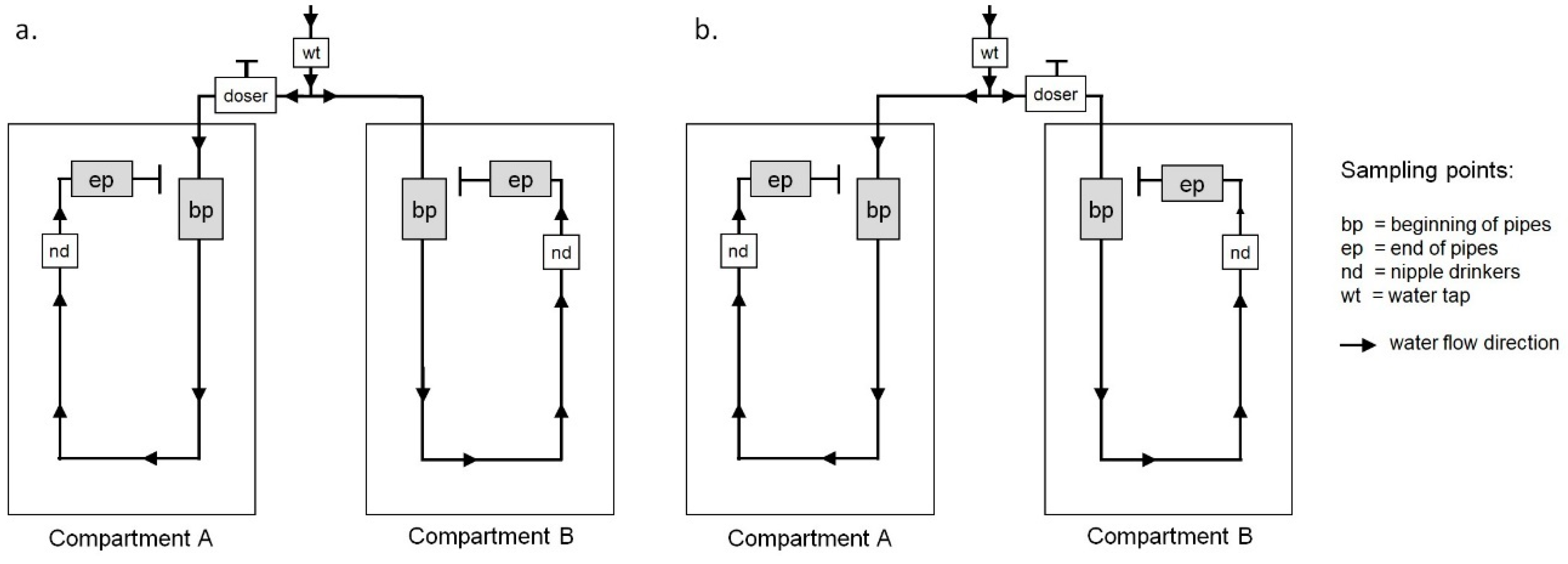
2.2. Sampling Points

A main pipe supplied all compartments of the nursery with the farm’s well water (wt). The system was operated at a constant pressure of 200 kPa. Figure 1 illustrates the structure of the pipes in the investigated compartments (A and B). Figure 1 also shows the doser positions where the NaOCl was added (a. samplings 1 to 8; b. samplings 9 to 14), and the sampling points within the compartments. At the beginning of the pipes (bp) and at the end of the pipes (ep), five exchangeable pipe segments for sampling purposes were integrated into the water pipes (Figure 2). Each pipe segment (length 250 mm) was constructed of the same material (Polyvinylchloride) and had an internal diameter of 22 mm, the same as the original pipe. However, two valves were integrated to allow an easy exchange of the pipe segments filled with drinking water. Pipe segments were individually constructed for this project by a commercial supplier (Meier-Brakenberg GmbH & Co. KG, Extertal, Germany). Prior to installation, all pipe segments and valves were washed with clear water and disinfected with ethanol (70%). Sampled nipple drinkers were located in the last pens before the dead end of the pipe. At each sampling, the same nipples were used to obtain the water samples. In the dead end of the pipes, where sampling points ep were located, water was stagnating.

2.3. Disinfection

Disinfection of drinking water was conducted by permanently dosing NaOCl (Virbac Clean Pipe VCP, Virbac GmbH, Bad Oldesloe, Germany) into the water. A dosage of 0.002% (20ml VCP/1,000l water) was adjusted via an integrated doser (Dosiersytem “Extended” V10.08, Aumann Hygienetechnik, Goldenstedt-Lutten, Germany). The distances of the dosers to the treated compartments was one meter (Compartment A) and four meters (Compartment B).

2.4. Water Sampling

Samples were taken every 14 days from October of 2014 until April of 2015. At each sampling, samples were taken from seven points (Figure 1) and transported to the laboratory in a cooling box (4 °C to 8 °C). All samples were processed within three hours after sampling. Water samplings from incoming water (well water (wt)) were collected from the same tap using sterile glass bottles (250 mL, Schott AG, Mainz, Germany) according to DIN EN ISO 19458 [26]. Within the compartments, the sampling procedure was as follows: One water sample was taken from the last nipple drinker (nd) before the end of pipe. One pipe segment was exchanged from the beginning of the pipe (bp) and one pipe segment was exchanged from the end of the pipe (ep). Sampling began with the exchange of the first pipe segments against the flow directions. After five samplings, the exchange of the pipe segments was started again from the first pipe segment against the flow direction. Samples from nipple drinkers were taken after cleaning with single-use cleaning tissues and spray disinfection (70% Ethanol). Samples from nipples were also sampled in 250 mL sterile glass bottles (Schott). To exchange pipe segments at the beginning and the end of the pipe, the valves flanking the pipe segments were closed to stop the water flow and the valves of the pipe segments were closed and exchanged by clean and disinfected pipe segments before the flanked valves were reopened.

2.5. Microbiological Analyses

In the laboratory, the exterior of each pipe segment was cleaned, dried and disinfected with ethanol (70%). One valve was opened to release the water sample (approx. 96 mL) into a sterile measuring jug. The remaining biofilm layer within the pipe segments was dissolved in 70 mL of phosphate buffered saline (PBS) by vortexing the closed pipe segments for five minutes (speed 150rpm) and placing the pipe segments (for 10 min) in an ultrasonic bath (Bandelin Sonorex Super 10 P, Bandelin Electronix GmbH & Co KG, Berlin, Germany). This suspension was merged with the water sample in the measuring jug and represented the total microbial burden of a pipe segment.
From each sample (wt, bp, ep, nd), 0.1 and 0.1 mL of a serial dilution (PBS with 0.01% Tween20) were plated as duplicates on Blood Agar Base No. 2 (Oxoid, Basingstoke, Hampshire, England) in accordance with DIN EN ISO 8199 [27]. In parallel, 0.1 mL of PBS with 0.01% Tween20 and 0.1 mL of sodium chloride were plated on Blood Agar Base No. 2 (Oxoid) as negative controls. The counting of colony-forming units (cfu) was completed after two days of incubation at 36 °C. For the detection of coliforms, including E. coli, and Pseudomonas spp., dilutions (1:2, 1:4, 1:10 to 1:10.000) of 100 mL of each of water sample (wt, nd), as well as 100 mL of the suspension from the water pipe segments (bp, ep) were filtered through a sterile cellulose–nitrate membrane filter with a pore size of 0.45 µm and a diameter of 37 mm (Sartorius 114H16-50-CAN, Sartorius AG, Göttingen, Germany). Filters were placed onto ChroMedium Coliforme Agar (SIFIN, Berlin, Germany) and onto Cetrimide Agar (Oxoid, LTD, Basingstoke, Hampshire, England). When typical colonies of E. coli or other coliform bacteria appeared after two days at 36 °C on the ChroMedium, the sample was recorded as positive. Growing conditions were controlled by growing E. coli (DSM 1103) on ChroMedium and by plating out the dilution buffer as a negative control. For the identification of presumed E. coli and coliform bacteria, isolates were plated on Columbia Agar (Oxoid, LTD, Basingstoke, Hampshire, England) and incubated at 36 °C for two days in order to access pure cultures. Matrix-assisted laser desorption-time of flight mass spectrometry (MALDI-TOF MS, microflex LT, Bruker, Billerica, USA) was used to identify the isolates. For the enumeration of Pseudomonas spp., typical colonies were counted after 48 h incubation at 36 °C according to guideline DIN EN ISO 16266 [28].
After laboratory analysis was completed, pipe segments were washed with CleanAgent solution (Mehlfeld + Göring GmbH, Kabelsketal, Germany), rinsed with clear water and disinfected with ethanol (70%) after drying.

2.6. Endotoxin Detection

Endotoxin concentration was detected via the quantitative, kinetic chromogenic limulus amebocyte lysate (LAL) method at 37 °C (Lonza, Basel, Switzerland). Therefore, one mL of each sample was stored at −20° until all samples were collected. Analyses were performed with an automated microtiter plate reader (Anthos ht III, Anthos Labtec Instruments, Wals, Austria) using WinKQCL 1.1 software (Lonza, Basel, Switzerland). A reference endotoxin of Escherichia coli-6 (American Food and Drug Administration, Silver Spring, Maryland, USA) was used as a standard control. Endotoxin concentrations were expressed as endotoxin units (EU) per mL drinking water. In addition, the mass concentration ng/mL was calculated using the internal standard with eight EU, equivalent to one ng endotoxin [29].

2.7. Statistics

All statistical analyses were performed with SAS 9.4 (SAS Institute Inc., Cary, NC, USA). The model residuals of the quantitative measurements were assessed using the Kolmogorov–Smirnov test and visual assessment of the qq-plots for normal distribution. Right-skewed (lognormal) distributed variables (numbers of colony-forming units and endotoxin units) were logarithmized prior to analysis. Geometric means and geometric confidence limits were calculated for location and scale parameter. Two-way analysis of variance with independent and repeated measurements was calculated to test for significant differences between variables at different sampling sites. Post-hoc Tukey test was used for multiple pairwise comparisons while maintaining the experiment wise error-rate.
To analyze associations between the occurrence of detected coliform bacteria in water samples and the treatment of water, the FREQ procedure was used to conduct the Fishers exact test. The threshold for statistical significance was set to P < 0.05.

3. Results

3.1. Bacterial Contents and Endotoxin Concentrations in Water Samples from Treated and Untreated Pipes

The results of the microbial analysis, the endotoxin measurements and the P-values obtained from the model analyses are summarized in Table 2 (samplings 1–8, treatment in A) and Table 3 (samplings 9–14, treatment in B). The results in both tables show clear differences between the microbiological parameters in treated and untreated pipes at the beginning of the pipes and at the nipple drinkers. Seven out of eight comparisons were found to be significantly different, indicating the efficiency of the treatment. However, at the end of the pipes, behind the last nipple drinkers (Figure 1), no effect of water treatment was found. Overall, changing the treatment from compartment A to B was found to produce nearly identical effects on the total counts of cultivable bacteria and the counts of cultivable Pseudomonades. The bacteria concentrations (geometric means) of the incoming well water were low (total count log 10 =≤ 0.60 cfu/mL, Pseudomonades log 10 = ≤ 0.48 cfu/100ml). Bacterial counts in treated pipes were, except for one sample (Pseudomonades at nipple drinkers in compartment B), always significantly lower (P < 0.0001, model data not shown) than untreated pipes. Bacteria contents from untreated water sampled at nipples were considerably higher compared to those from treated water at the same sampling point.
In contrast, the endotoxin contents revealed no clear trends. The only significant difference was detected at the end of pipes (Table 3) and, in this case, the endotoxin units were higher in the samples from the treated pipe. At the same time, the endotoxin units measured were similar to measurements from the beginning at both the treated and untreated pipes, and from nipple-derived treated and untreated water samples. Therefore, an effect on the endotoxin units due to the water disinfection was not found. However, the endotoxin units were found to reach remarkable concentrations in the investigated pipe segments. According to the transformation into mass concentrations, the endotoxin concentrations (geometric means) ranged between 75.8 (606 EU/mL) and 161.8 ng/mL (1294 EU/mL) at the beginning of pipes, and between 288.9 (2311 EU/mL) and 2205.8 ng/mL (17,646 EU/mL) at the end of the pipes. At nipple drinkers, the concentrations were lower, namely 1–3 ng/mL (9 to 21 EU/mL). With up to 0.3 ng/mL (2 EU/mL), the endotoxin content was low in the incoming water.

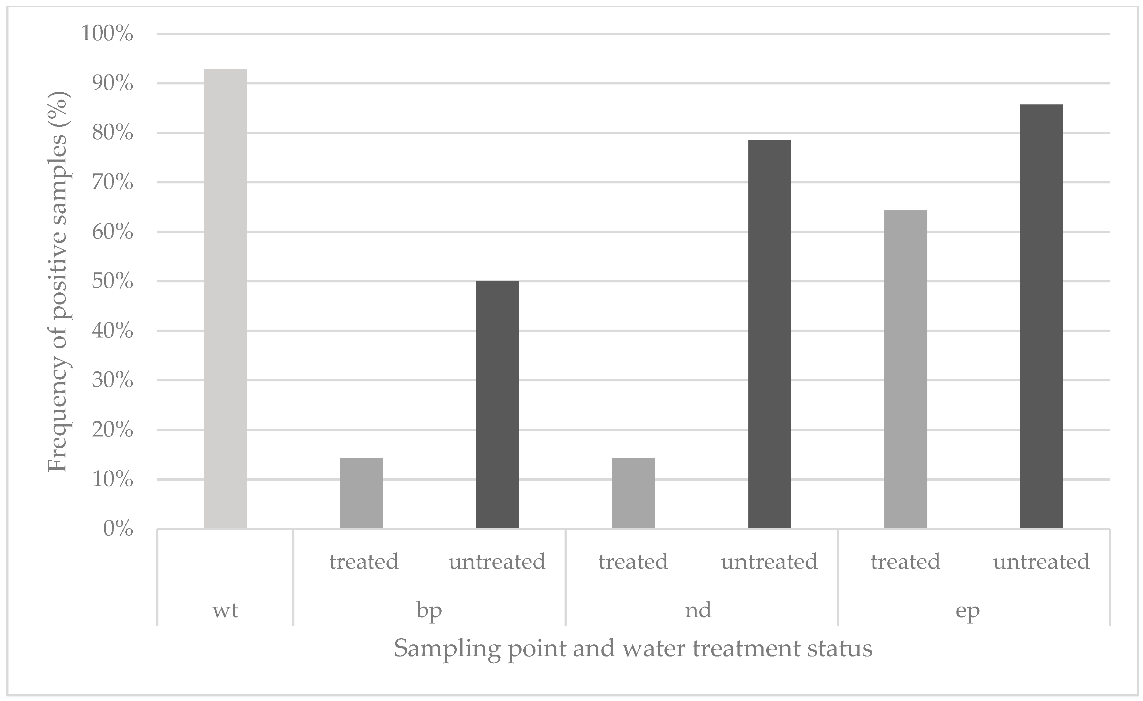
3.2. Detection of Coliform Bacteria in Treated and Untreated Pipes

Figure 3 summarizes the measurements of coliform bacteria in 14 samples at each sampling point and each water treatment status. Ninety-three percent of the samples from incoming water (wt) were already contaminated with coliform bacteria. Coliform bacteria were found in all sections of treated and untreated pipes, but at lower numbers in samples from the treated pipes. The detection rate in treated pipes was 31%, in contrast to 71% positive samples from untreated pipes.
Overall, no coliforms were detected in 29 treated samples and in 12 untreated samples. Positive samples were obtained from 30 untreated samples and from 13 treated samples. A Fishers exact test was conducted to analyze possible associations between the occurrence of coliforms and the treatment status. The calculated P-value was P = 0.0004, revealing a significant association between the treatment of water and the detection of coliforms. Overall, coliforms occurred significantly less in treated water samples. The effect seemed to be lower in samples from the end of pipes (ep), which is in accordance with results from total counts and from Pseudomonades.

3.3. Identification of Presumed Coliforms, Including E. coli

A total of 21 out of 25 isolates from different sampling points (incoming water, beginning of pipes, end of pipes, nipple drinkers) were found to belong to coliform bacteria species. These included Buttiauxella agrestis, Buttiauxella gaviniae, Citrobacter gillenii, Citrobacter freundii, Enterobacter asburiae, Kluyvera intermedia, Rahnella aquatilis and Serratia fonticola. Furthermore, seven isolates were confirmed to be E. coli. The latter were isolated from incoming water, nipple drinkers and from the end of pipes. In total, ten different species, including two species of E. coli, were found.

4. Discussion

Only limited data are available regarding the effects of drinking water treatment with NaOCl in pig barns, especially nurseries. Even fewer data are available that consider water systems with dead-end sections. To estimate the effects of disinfection on culturable bacteria and endotoxin concentration, samples from treated and untreated pipes were investigated and the results were compared. To assess possible influences from the water source (well water), samples from a tap (at the incoming main pipe) were also analyzed.
Integrating exchangeable new, clean and disinfected pipe segments in the existing pipes enabled us to measure the disinfection effects on bacteria in water and biofilms on a defined surface in treated and untreated pipe sections. This is a promising approach because the initial state of a pipe influences the growth of biofilms [29] and further information regarding the duration and speed of growth in pipes on-farm was, to the authors’ knowledge, lacking. Stagnating water flows promote microbial growth and reduces the efficiency of disinfectants [30]. Therefore, in addition to sections from the beginning of pipes, sections from pipe dead-ends were also analyzed in this study. To estimate the occurrence of selected bacteria and endotoxins in the water that is consumed by pigs, samples were also taken from nipple drinkers. In both the compartments examined here, the design of the housing and the water systems were identical. The water flow and water temperature in a pipe can affect biofilm growth [29], however, the ambient temperature and total water consumption in the chosen compartments were also identical. Flow velocities in the pipes of the two compartments were likely different because the numbers and ages of animals differed across samplings and, with that, the water withdrawal could have been affected. The possibility of an effect of these factors on microbial growth cannot be excluded. However, to consider a compartment effect, the treatment was switched between the compartments and the results were analyzed separately.
Total bacterial count and the number of Pseudomonades were significantly reduced (>log10 (3) cfu/mL) in disinfected sections compared to untreated sections at the beginning of pipes. The comparatively high bacteria concentrations in untreated pipe segments are likely the result of biofilm material. This would also explain the higher concentrations of samples from pipes compared to samples from nipple drinkers. Bacterial concentrations in flowing water are generally lower than in biofilms [31]. Significant disinfection effects were also found in our comparison of samples from nipple drinkers in the treated and untreated compartments. The threshold values of the German recommendation guidelines for water for farm animal use were never exceeded; the concentrations of the total bacteria count in the incoming well water and in the disinfected water from nipple drinkers were always below the critical value of 1,000 cfu/mL [32]. Ten of the fourteen nipple drinker samples of untreated water exceeded this critical value. These results suggest that a treatment with 0.002% NaOCl can reduce total bacteria counts and Pseudomonades at nipple drinkers and, therewith, the uptake by pigs. However, this reduction was not found to be consistently significant, which might be related to the position of the sampled nipples. Tested nipples were proximal to the dead end of pipes, in which no significant reduction in bacteria was observed. The concentration of total bacteria counts and Pseudomonades were even higher in dead-end sections of treated pipes and bacterial growth in these sections could have affected the bacteria concentrations in the nipples. It is well known that the effects of chlorination are decreased in stagnating water [33,34,35] and, as mentioned above, bacterial growth can be promoted in the dead-ends of pipes. Such constructions in nurseries that have high ambient temperatures represent a contamination source for microorganisms, especially because these sections are difficult to clean. Therefore, such dead-end water pipe constructions should be avoided in livestock buildings to promote proper water hygiene.
In contrast to the results of bacterial counts, endotoxin concentrations were found to have no clear trends. Concentrations at the beginning of pipes and at the nipple drinkers were similar, whether water was treated or not. In agreement with bacterial counts, the highest concentrations of endotoxins were measured in the dead-end sections. This indicates an accumulation of gram-negative bacteria, which is consistent with the counts of Pseudomonades. Endotoxins can be released from gram-negative cells by multiplication, death and lysis [36,37,38,39,40]. The biological activity of endotoxins measured in the EU consists of cell-bound and free forms in water [37,41]. Increases in endotoxin activity occur during the disinfection of secondary sewage [24]. We hypothesized that this might be the result of damaged cell walls and a higher release of Lipid A into drinking water [24]. However, we did not find these effects in the present study. Therefore, we assume that disinfection with NaOCl has no adverse effects concerning the endotoxin concentrations. This is in accordance with other studies that showed that chlorination has only a small effect on endotoxin activity [29,42,43]. However, the concentrations of endotoxins measured at nipple drinkers reached 28 ng/mL. Thus, an intake of one liter would result in a pig consuming 28 µg endotoxin. The oral uptake of endotoxins can contribute to adverse immunological reactions in pigs weaned at the age of 21 days [44]. Furthermore, the concentrations measured in the pipe sections indicate that concentrations could be much higher when parts of the biofilms are released into the flowing water. Because there is a lack of knowledge regarding the relationship between endotoxin uptake via drinking water and its effects on growing pigs, future studies should begin investigating these questions.
Coliform bacteria can be opportunistic pathogens and are regarded as indicator organisms for fecal contamination. This group of bacteria served as useful indicator bacteria in the present study. The incoming water from the well, or from the pipe section that originates in the well, to the compartments were found to be contaminated with coliform bacteria. Compared to the untreated pipe sections in the compartments, the incoming water was more often contaminated (Figure 3). This could have been due to a contamination of the well or within the tap. However, when treated and untreated water samples from the compartments were compared, the lower detection of coliforms was significantly associated with the treatment of the water. Species identification revealed a broad spectrum of ten different species, but, from the data and the microbiological analyses, it was not possible to determine whether the incoming water was the main or the only source of contamination. For instance, it is known that coliforms can be part of the biofilms in drinking systems [45]. To determine the origin of the isolated bacteria, a larger sample set is necessary, however, this was not the aim of the present study. Positive findings at nipple drinkers show that pigs had likely taken up coliform bacteria from the contaminated drinking system. However, as mentioned above, there are significant differences between treated and untreated pipes, which suggests that using NaOCl is sufficient to reduce coliforms in contaminated pipes. The consequence of this is probably a lower uptake of potential pathogens by the pigs. This is supported by the fact that E. coli was not detected in the 42 water samples from the treated pipes, but was detected in seven of the 42 samples from untreated pipes. The disinfection of drinking water with NaOCl has the potential to prevent the occurrence of opportunistic pathogens. Especially in drinking systems in which contamination of incoming water with fecal bacteria is unavoidable, water disinfection might be a useful preventative measure. Combining continuous disinfection measures with regular cleaning of the pipes is recommended [46]. An alternative measure to reduce bacteria and biofilms in drinking systems used for livestock buildings is regular flushing [47]. In the investigated housing system, compartments remain empty for 5–15 days before housing new pigs. Within these intervals, pipes could be flushed. This would probably be effective, because the concentrations measured in untreated pipes of unoccupied compartments were relatively high compared to the mean total counts of bacteria (bpA = log10 (4.26) cfu/mL, bpB = log10 (4.75) cfu/mL). This presents the possibility that opening the pipe at the dead end while flushing may also reduce contaminations in those sections.
To conclude, the disinfection of drinking water with NaOCl can significantly reduce bacteria, including potential pathogens and biofilm-forming bacteria, in the water pipes of nurseries. Endotoxin concentrations remained unaffected by NaOCl treatment. Constructions with dead-end sections in compartments can serve as contamination sources and should be avoided.

Author Contributions

Conceptualization: R.B., N.K., J.S.; methodology, R.B., N.K., J.S.; statistical analysis: K.R., J.S.; validation, K.R., J.S.; formal analysis, R.B., J.S.; investigation, R.B.; data curation, R.B., J.S.; writing—original draft preparation: R.B.; writing—review and editing: R.B., N.K., J.S.; visualization: R.B., N.K., J.S.; project administration: N.K. All authors read and agreed on the published version of this manuscript.

Funding

This project was partly funded by Virbac Tierarzneimittel GmbH (Virbac Tierarzneimittel GmbH, Bad Oldesloe, Germany).

Acknowledgments

The authors wish to thank the laboratory assistants of the Institute for Animal Hygiene, Animal Welfare and Farm Animal Behaviour, University of Veterinary Medicine Hannover, Foundation, Germany.

Conflicts of Interest

The authors declare no conflict of interest. The funders had no role in the design of the study; in the collection, analyses, or interpretation of data, in the writing of the manuscript, or in the decision to publish the results.

References

  1. Thacker, P.A. Water in swine nutrition. In Swine Nutrition, 2nd ed.; Lewis, A.J., Southern, L.L., Eds.; CRC Press: New York, NY, USA, 2001; pp. 381–398. [Google Scholar]
  2. Thalin, P.A.; Brumm, M.C. Water: The forgotten nutrient. In Swine Nutrition; Miller, E.R., Ullrey, D.E., Austin, A.J., Eds.; Butterworths-Heinemann: Stoneham, MA, USA, 1991; pp. 315–324. [Google Scholar]
  3. Kober, J.A. Water: The most limiting nutrient. Agri.-Pract. 1993, 14, 39–42. [Google Scholar]
  4. Patience, J.F. The importance of water in pork production. Anim. Front. 2012, 2, 28–35. [Google Scholar] [CrossRef] [Green Version]
  5. Mateus-Vargas, R.H.; Kemper, N.; Volkmann, N.; Kietzmann, M.; Meissner, J.; Schulz, J. Low-frequency electromagnetic fields as an alternative to sanitize water of drinking systems in poultry production? PLoS ONE 2019, 14, e0220302. [Google Scholar] [CrossRef]
  6. Dewulf, J.; Immerseel, F.; Postma, M.; Rooster, H.; Damiaans, B.; Sarrazin, S.; Abma, E.; Maes, D. Biosecurity in Animal Production and Veterinary Medicine; ACCO: New York, NY, USA, 2018. [Google Scholar]
  7. LeJeune, J.T.; Besser, T.E.; Merrill, N.L.; Rice, D.H.; Hancock, D.D. Livestock drinking water microbiology and the factors influencing the quality of drinking water offered to cattle. J. Dairy Sci. 2001, 84, 1856–1862. [Google Scholar] [CrossRef]
  8. Fairbrother, J.M.; Nadeau, E. Escherichia coli: On-farm contamination of animals. Rev. Sci. Technol. 2006, 25, 555–569. [Google Scholar] [CrossRef]
  9. Stull, C.L.; Farley, J.L.; Galey, F.D.; Cullor, J.S.; Wilson, R.A.; Hays, V.W.; Wilson, L.L. Assessment of Bacteria and Mycotoxins in Feed and Coliforms in Water Offered to High and Low Performing Commercial Growing Hogs in California. Prof. Anim. Sci. 1999, 15, 94–99. [Google Scholar] [CrossRef]
  10. Schulz, J.; Spindler, B.; Verspohl, J.; Sudendey, C.; Kemper, N. Tränkwasserqualität in Nutztierhaltungen: Welche Probleme gibt es und wie entstehen sie? Der Prakt. Tierarzt 2014, 95, 649–651. [Google Scholar]
  11. National Research Council. Nutrient Requirements of Swine, 11th ed.; The National Academies Press: Washington, DC, USA, 2012. [Google Scholar]
  12. Kraft, A. Electrochemical Water Disinfection: A Short Review. Platin. Metals Rev. 2008, 52, 177–785. [Google Scholar] [CrossRef]
  13. Bukhari, Z.; Clancy, J.L.; Hary, T.M.; Bolton, J.; Dussert, B.; Marshall, M.M. Medium-pressure UV for oocysts inactivation. J. Am. Water Works Assoc. 1999, 91, 86–94. [Google Scholar] [CrossRef]
  14. Clancy, J.L.; Bukhari, Z.; Hargy, T.M.; Bolton, J.R.; Dussert, B.W.; Marilyn, M. Using UV to inactivate cryptosporidium. J. Am. Water Works Assoc. 2000, 92, 97. [Google Scholar] [CrossRef]
  15. Kruithof, J.C.; Van der Leer, R.C.; Hijnen, W.A.M. Practical experiences with UV disinfection in The Netherlands. J. Water SRT-Aqua 1992, 41, 88–94. [Google Scholar]
  16. Harris, G.D.; Adams, V.D.; Sorensen, D.L.; Curtis, M.L. Ultraviolet inactivation of selected bacteria and viruses with photoreactivation of the bacteria. Water Res. 1987, 21, 687–692. [Google Scholar] [CrossRef]
  17. Shrivastava, R.; Upreti, R.K.; Jain, S.R.; Prasad, K.N.; Seth, P.K.; Chaturvedi, U.C. Suboptimal chlorine treatment of drinking water leads to selection of multidrug-resistant Pseudomonas aeruginosa. Ecotoxicol. Environ. Saf. 2004, 58, 277–283. [Google Scholar] [CrossRef]
  18. Ratnayaka, D.D.; Brandt, M.J.; Johnson, K.M. Chemistry, Microbiology and Biology of Water. Water Supply 2009, 6, 195–266. [Google Scholar]
  19. Gerba, C.P.; Kennedy, D. Enteric virus survival during household laundering and impact of disinfection with sodium hypochlorite. Appl. Environ. Microbiol. 2007, 73, 4425–4428. [Google Scholar] [CrossRef] [Green Version]
  20. Abadias, M.; Usall, J.; Oliveira, M.; Alegre, I.; Vinas, L. Efficacy of neutral electrolyzed water (NEW) for reducing microbial contamination on minimally processed vegetables. Int. J. Food Microbiol. 2008, 123, 151–158. [Google Scholar] [CrossRef]
  21. Peeters, E.; Nelis, H.J.; Coenve, T. Evaluation of the efficacy of disinfection procedures against Burkholderia cenocepacia biofilms. J. Hosp. Infect. 2008, 70, 361–368. [Google Scholar] [CrossRef]
  22. Krug, P.W.; Lee, L.J.; Eslami, A.C.; Larson, C.R.; Rodriguez, L. Chemical disinfection of high-consequence transboundary animal disease viruses on nonporous surfaces. Biologicals 2011, 39, 231–235. [Google Scholar] [CrossRef]
  23. Fukuzaki, S. Mechanisms of actions of sodium hypochlorite in cleaning and disinfection processes. Biocontrol Sci. 2006, 11, 147–157. [Google Scholar] [CrossRef]
  24. Huang, H.; Wu, Q.-Y.; Yang, Y.; Hu, H.-Y. Effect of chlorination on endotoxin activities in secondary sewage effluent and typical Gram-negative bacteria. Water Res. 2011, 45, 4751–4757. [Google Scholar] [CrossRef]
  25. Parra, J.; Agudelo, J.; Ortiz, L.; Ramírez, M.C.; Rodríguez, B.; López Herrera, A. Lipopolysaccharide (LPS) from E. coli has detrimental effects on the intestinal morphology of weaned pigs. Rev. Colom. Cienc. Pecua. 2011, 24, 598–608. [Google Scholar]
  26. Anonymous. Water Quality—Sampling for Microbiological Analysis (ISO 19458:2006); German Version EN ISO 19458:2006; Beuth Verlag GmbH: Berlin, Germany, 2006. [Google Scholar]
  27. Anonymous. Water Quality—General Guidance on the Enumeration of Micro-Organisms by Culture (ISO 8199:2017); German Version EN ISO 8199:2017 (Foreign Standard); Beuth Verlag GmbH: Berlin, Germany, 2017. [Google Scholar]
  28. Anonymous. Water Quality—Detection and Enumeration of Pseudomonas Aeruginosa—Method by Membrane Filtration (ISO 16266:2006); German Version EN ISO 16266:2008 (Foreign Standard); Beuth Verlag GmbH: Berlin, Germany, 2008. [Google Scholar]
  29. Anderson, W.B.; Mayfield, C.I.; Dixon, G.D.; Huck, P.M. Endotoxin inactivation by selected drinking water treatment oxidants. Water Res. 2003, 37, 4553–4560. [Google Scholar] [CrossRef] [PubMed]
  30. Douterelo, I.; Fish, K.E.; Boxall, J.B. Succession of bacterial and fungal communities within biofilms of a chlorinated drinking water distribution system. Water Res. 2018, 141, 74–85. [Google Scholar] [CrossRef]
  31. World Health Organization (WHO). Heterotrophic Plate Counts and Drinking-Water Safety; Bartram, J., Cotruvo, J., Exner, M., Fricker, C., Glasmacher, A., Eds.; IWA Publishing: London, UK, 2003. [Google Scholar]
  32. Flemming, H.C.; Percival, S.L.; Walker, J.T. Contamination potential of biofilms in water distribution systems. Water Sci. Technol. Water Suppl. 2002, 2, 271–280. [Google Scholar] [CrossRef]
  33. Bundesministerium für Ernährung und Landwirtschaft (BMEL). Hygienische Qualität von Tränkwasser. Orientierungsrahmen zur Futtermittelrechtlichen Beurteilung. Available online: https://www.bmel.de/DE/Tier/Tierernaehrung/_texte/Orientierungsrahmen-Traenkwasser.html (accessed on 6 February 2020).
  34. Niquette, P.; Servais, P.; Savoir, R. Bacterial dynamics in the drinking water distribution system of Brussels. Water Res. 2001, 35, 675–682. [Google Scholar] [CrossRef]
  35. Gillespie, S.; Lipphaus, P.; Green, J.; Parsons, S.; Weir, P.; Juskowiak, K.; Jefferson, B.; Jarvis, P.; Nocker, A. Assessing microbiological water quality in drinking water distribution systems with disinfectant residual using flow cytometry. Water Res. 2014, 65, 224–234. [Google Scholar] [CrossRef] [PubMed]
  36. Mao, G.; Song, Y.; Bartlam, M.; Wang, Y. Long-Term Effects of Residual Chlorine on Pseudomonas aeruginosa in Simulated Drinking Water Fed with Low AOC Medium. Front. Microbiol. 2018, 9, 879. [Google Scholar] [CrossRef]
  37. Gorbet, M.B.; Sefton, M.V. Endotoxin: The Uninvited Guest. Biomatererials 2005, 26, 6811–6817. [Google Scholar] [CrossRef]
  38. Anderson, W.B.; Slawson, R.M.; Mayfield, C.I. A review of drinking-water-associated endotoxin, including potential routes of human exposure. Can. J. Microbiol. 2002, 48, 567–587. [Google Scholar] [CrossRef]
  39. Venter, P.; Abraham, M.; Lues, J.F.R.; Ivanov, I. The influence of sanitizers on the lipopolysaccharide composition of Escherichia coli O111. Int. J. Food Microbiol. 2006, 111, 221–227. [Google Scholar] [CrossRef]
  40. Parikh, S.J.; Chorover, J. Infrared spectroscopy studies of cation effects on lipopolysaccharides in aqueous solution. Colloids Surf. B 2007, 55, 241–250. [Google Scholar] [CrossRef] [PubMed]
  41. Morrison, D.C.; Danner, R.L.; Dinarello, C.A.; Munford, R.S.; Natanson, C.; Pollack, M.; Spitzer, J.J.; Vogel, S.N.; McSweegan, E. Bacterial endotoxins and pathogenesis of gram-negative infections: Current status and future direction. J. Endotoxin Res. 1994, 1, 71–83. [Google Scholar] [CrossRef]
  42. Gehr, R.; Parent Uribe, S.; Da Silva Baptista, I.F.; Mazer, B. Concentrations of endotoxins in waters around the island of Montreal, and treatment options. Water Qual. Res. J. Can. 2008, 43, 291–303. [Google Scholar] [CrossRef]
  43. Rapala, J.; Lahti, K.; Räsänen, L.; Esala, A.; Niemelä, S.; Sivonen, K. Endotoxins associated with cyanobacteria and their removal during drinking water treatment. Water Res. 2002, 36, 2627–2635. [Google Scholar] [CrossRef]
  44. Parra, J.; Agudelo, J.; Sanín, D.; Forero, J.; Muskus, C.; López, A. Intestinal expression of pro-inflammatory cytokines induced by oral intake of lipopolysaccharide (LPS) from E. coli in weaned pigs. Rev. Colomb. Cienc. Pecu. 2013, 26, 108–118. [Google Scholar]
  45. Zacheus, O.M.; Lehtola, M.J.; Korhonen, L.K.; Martikainen, P.J. Soft deposits, the key site for microbial growth in drinking water distribution networks. Water Res. 2001, 35, 1757–1765. [Google Scholar] [CrossRef]
  46. Flemming, H.-C.; Bendinger, B.; Exner, M.; Gebel, J.; Kistemann, T.; Schaule, G.; Szewzyk, U. Erkenntnisse aus dem BMBF-Verbundprojekt: Biofilme in der Trinkwasser-Installation. Bundesministerium für Bildung und Forschung 2010. Available online: https://www.uni-due.de/imperia/md/content/water-science/ss10/1541b_03z_ss10_thesenpapier_version_1.1._f__r_pdf-versand.pdf (accessed on 6 February 2020).
  47. Lehtola, M.J.; Nissinen, T.K.; Miettinen, I.T.; Martikainen, P.J.; Vartiainen, T.; Bai, X. Removal of soft deposits from the distribution system improves the drinking water quality. Water Res. 2004, 38, 601–610. [Google Scholar] [CrossRef]
Figure 1. Sampling points and doser position at samplings 1 to 8 (a) and samplings 9 to 14 (b).
Agriculture 10 00086 g001
Figure 2. Photo of five exchangeable pipe segments integrated in the beginning of the water pipe in compartment B (Photo: R. Böger). Each pipe segment was located between two valves (red components) to enable an easy exchange. Water flow from left to right.
Agriculture 10 00086 g002
Figure 3. Detection of coliform bacteria in incoming (well) water (wt) and in samples from treated and untreated pipes with bp = beginning of the pipes, nd = nipple drinker and ep = end of the pipes. wt: = well water.
Agriculture 10 00086 g003
Table 1. Treatment status, animal numbers and batch numbers in compartments A and B.
SamplingCompartmentTreatmentAnimals [n]Age of Animals [d] at Sampling and (Batch No.)
1Ayes23931 (1)
Bno28552 (1)
2Ayes23945 (1)
Bno28566 (1)
3Ayes23959 (1)
Bno0 */
4Ayes23673 (1)
Bno20131 (2)
5Ayes23024 (2)
Bno20145 (2)
6Ayes23038 (2)
Bno20159 (2)
7Ayes23066 (2)
Bno26428 (3)
8Ayes0 */
Bno26442 (3)
9Ano27835 (3)
Byes26256 (3)
10Ano27749 (3)
Byes26270 (3)
11Ano27763 (3)
Byes0 */ (3)
12Ano0 */ (3)
Byes31935 (4)
13Ano26829 (4)
Byes30850 (4)
14Ano26442 (4)
Byes30763 (4)
* measurements after depopulation of the batch.
Table 2. Results from compartment A (treated pipe) and compartment B (control pipe).
SamplingSampling Points
Total Count Log 10
cfu/mL
P-ValueSampling Points
Pseudomonas spp. log 10
cfu/100 mL
P-ValueSampling Points
Endotoxins Log 10
EU/mL (ng/mL)
P-Value
Treated PipeUntreated PipeTreated PipeUntreated PipeTreated PipeUntreated Pipe
1–8bp(A) 0.30bp(B) 3.97<0.0001bp(A) 1.23bp(B) 5.41<0.0001bp(A) 2.93 (2.03)bp(B) 2.98 (2.08)0.9999
nd(A) 0.48nd(B) 3.36<0.0001nd(A) 0.30nd(B) 1.910.1790nd(A) 1.11 (0.21)nd(B) 1.32 (0.42)0.8407
ep(A) 4.64ep(B) 5.580.0410ep(A) 5.65ep(B) 5.940.9983ep(A) 3.65 (2.74)ep(B) 4.25 (2.31)0.0094
bp = beginning of pipe, ep = end of pipe, nd = nipple drinker, cfu = colony-forming units, EU = endotoxin units
Table 3. Results from compartment B (treated pipe) and compartment A (control pipe).
SamplingSampling Points
Total Count Log 10
cfu/mL
P-ValueSampling Points
Pseudomonas spp. Log 10
cfu/100 mL
P-ValueSampling Points
Endotoxins Log 10
EU/mL (ng/mL)
P-Value
Untreated PipeTreated PipeUntreated PipeTreated PipeUntreated PipeTreated Pipe
9–14bp(A) 3.83bp(B) 0.30<0.0001bp(A) 5.53bp(B) 0.60<0.0001bp(A) 2.78 (1.88)bp(B) 3.11 (2.21)0.4254
nd(A) 2.80nd(B) 0.00<0.0001nd(A) 1.80nd(B) 0.00<0.0001nd(A) 1.00 (0.1)nd(B) 0.95 (0.05)1.0000
ep(A) 5.49ep(B) 5.750.9640ep(A) 5.92ep(B) 6.050.9995ep(A) 3.36 (2.46)ep(B) 4.21 (0.62)0.0002
bp = beginning of pipe, ep = end of pipe, nd = nipple drinker, cfu = colony-forming units, EU = endotoxin units

