Article

Survey of Oomycetes Associated with Root and Crown Rot of Almond in Spain and Pathogenicity of Phytophthora niederhauserii and Phytopythium vexans to ‘Garnem’ Rootstock

1
Instituto Agroforestal Mediterráneo, Universitat Politècnica de València, Camino de Vera S/N, 46022 Valencia, Spain
2
Fruitcentre, Fruit Production Program, Institut de Recerca i Tecnologia Agroalimentàries (IRTA), PCiTAL, Parc de Gardeny, 25003 Lleida, Spain
*
Author to whom correspondence should be addressed.
Submission received: 26 January 2022 / Revised: 16 February 2022 / Accepted: 17 February 2022 / Published: 18 February 2022

Abstract

:
From 2018 to 2020, surveys of oomycetes associated with root and crown rot of almond (Prunus dulcis) were conducted on diseased young almond trees in commercial orchards and nurseries in six provinces of Spain. A total of 104 oomycete isolates were obtained from plant and soil samples, which h were identified by sequencing the internal transcribed spacer (ITS) region of the ribosomal DNA. Diverse species belonging to the genera Globisporangium, Phytophthora, Phytopythium and Pythium were found, Phytopythium vexans and Phytophthora niederhauserii being the most frequent. The pathogenicity of these two species to one-year-old almond seedlings of ‘Garnem’ (P. dulcis × P. persica) rootstock was studied. All seedlings inoculated with Pp. vexans and Ph. niederhauserii isolates showed severe symptoms at the late stage of the pathogenicity test (defoliation, wilting and dieback) and several plants died. Some isolates of Ph. niederhauserii significantly reduced the dry weight of the roots compared with the control, but this effect was not observed in seedlings inoculated with Pp. vexans. These results provide new information about the oomycete species present in almond crops in Spain and highlight the importance of carrying out frequent phytosanitary surveys for a better knowledge of potential risks posed by these soil-borne pathogens.

1. Introduction

In the Mediterranean area, almond crops (Prunus dulcis (Miller) D.A. Webb. syn. Prunus amygdalus Batsch) are cultivated mainly in marginal soils and rainfed conditions, quite the opposite to what happens in the USA and Australia, the two major almond producers [1], where almond crop is grown under irrigation conditions. Specifically, in Spain, this crop has been traditionally planted throughout the Mediterranean coastline, where lighter and poor soils are found and it has been cultivated mainly in dry conditions [2]. It was not until about the 2000s that almond crops began to be planted in irrigated lands in new Spanish cropping areas, with heavier and clayey soils, until then used to grow other fruit trees or extensive crops [3].
In this new Spanish almond crop paradigm, some already known diseases and new ones have emerged, mainly soil-borne diseases [4]. The incorporation of irrigation systems together with the exploration of diverse soil types (previously destined for other uses) has increased the root system problems related to waterlogging cases associated with root asphyxia and the proliferation of certain root and crown diseases. In addition, the ‘GF-677′ and ‘Garnem’ rootstocks, both hybrids of P. dulcis × P. persica, used massively up to now in almond crops mainly in irrigated orchards but also in dry crop conditions, are susceptible to soil-borne diseases [5]. New hybrid rootstocks obtained from crosses of almond or peach with plum species have appeared in recent years to avoid these problems [5,6,7]. These new materials have certain tolerance or resistance to soil-borne diseases, but it is important to note that the area planted with them is still reduced.
Almond trees can be affected by several soil-borne pathogens, such as Armillaria mellea (Vahl: Fr.) P. Kumm., Rosellinia necatrix Prill. and some oomycetes [8]. Diseases caused by oomycetes present in the soil can affect the crown and the roots of almond trees in both the nursery and established orchards. Multiple routes can spread out these diseases from the nurseries with infected plant material and infested substrate. Once established in the orchard, they can spread rapidly through cultural practices, plant debris, or through irrigation water [9]. Plant pathogenic oomycetes have evolved large arsenals of secreted proteins, termed effectors, that act as weapons in order to successfully infect plants, which respond by multiple defense actions, including the strengthening of physical barriers, production of antimicrobial molecules and programmed cell death. Oomycetes can counteract the plant’s immunity mechanisms, interfering or suppressing numerous biochemical signaling components, disabling the host’s defense actions, thus managing to invade its tissues [10].
Pinto de Torres and Mircetich [11] reported, in Chile, the presence of cankers caused by Globisporangium ultimum at the base of the trunk in 2-year-old almond trees. In Iran, Azizi et al. [12] studied the pathogenicity towards almonds of Pythium (Py.) (sensu lato) and Phytophthora (Ph.) isolates obtained from soil and roots of young almond trees. These authors identified Ph. cactorum, Py. aphanidermatum and Phytopythium (Pp.) helicoides as pathogenic species for this host, causing death in almond seedlings. Later, in this same country, Javadi and Sharifnabi [13] reported Pp. litorale as the pathogen causing root and crown rot of almond trees. This information was confirmed by Browne et al. [14], who indicated that diverse Pythium and Phytopythium species have been associated with the suppression of growth in the replanting of Prunus orchards grafted onto ‘Nemaguard’ rootstock, but their pathogenicity to almond or peach rootstocks was only established for G. ultimum var. ultimum and G. irregulare.
Many authors have reported diverse species of the genus Phytophthora causing root infection on almond rootstocks. Browne et al. [15] indicated that more than 10 species of Phytophthora attack almond trees, causing root rot and cankers on the root crown, trunk, or scaffolds. Earlier works by Wicks and Lee [16] and Wicks [17] in Australia reported the isolation of Ph. cambivora, Ph. citrophthora, Ph. cryptogea and Ph. megasperma from either crown cankers or the soil around the crown of declining almond trees. These authors were only able to test the pathogenicity of Ph. cambivora in almond seedlings grown in artificially infested soil. Later, Browne and Viveros [18] reported Ph. megasperma, Ph. cactorum and Ph. citricola associated with crown rot on dying almond trees in California, which were often of nonbearing age. Subsequent studies in Iran confirmed the pathogenicity of Ph. cactorum in a local almond rootstock [19]. In Spain, a new Phytophthora sp. associated with 2-year-old almond plants was detected in a Valencian nursery in 2007. Symptoms associated with this pathogen were chlorosis, wilting, cankers and profuse stem gumming [20]. From the infected plant material, it was possible to isolate Phytophthora taxon niederhauserii, which was confirmed as the causal agent of the disease by means of a pathogenicity test. Later, Kurbetli and Değirmenci [21] detected this pathogen for the first time, in almond orchards in Turkey. The description of this new species was completed by Abad et al. [22], who established Ph. niederhauserii as its definitive name. This oomycete is capable of infecting at least 33 species from 25 different families in both agricultural ecosystems and ornamental plant nurseries [22]. Later, in 2015, Ph. niederhauserii was first detected, causing almond crown rot in California [15]. Finally, more recent publications have reported symptoms of cankers and root and crown rot in almond trees, associated with Ph. megasperma [23] and Ph. plurivora [24] in Turkey; and wilting and decreased vigor caused by Ph. chlamydospora in Turkey [25] and California [26].
There is a lack of updated information about the diversity of oomycetes infecting almond trees in Spain. Thus, the objectives of this study are: (i) to survey, isolate and identify oomycetes from almond trees showing root and crown rot symptoms in the main producing areas of Spain; and (ii) to study the pathogenicity to ‘Garnem’ rootstock of the most frequent species found.

2. Materials and Methods

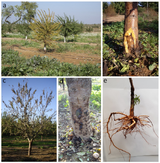
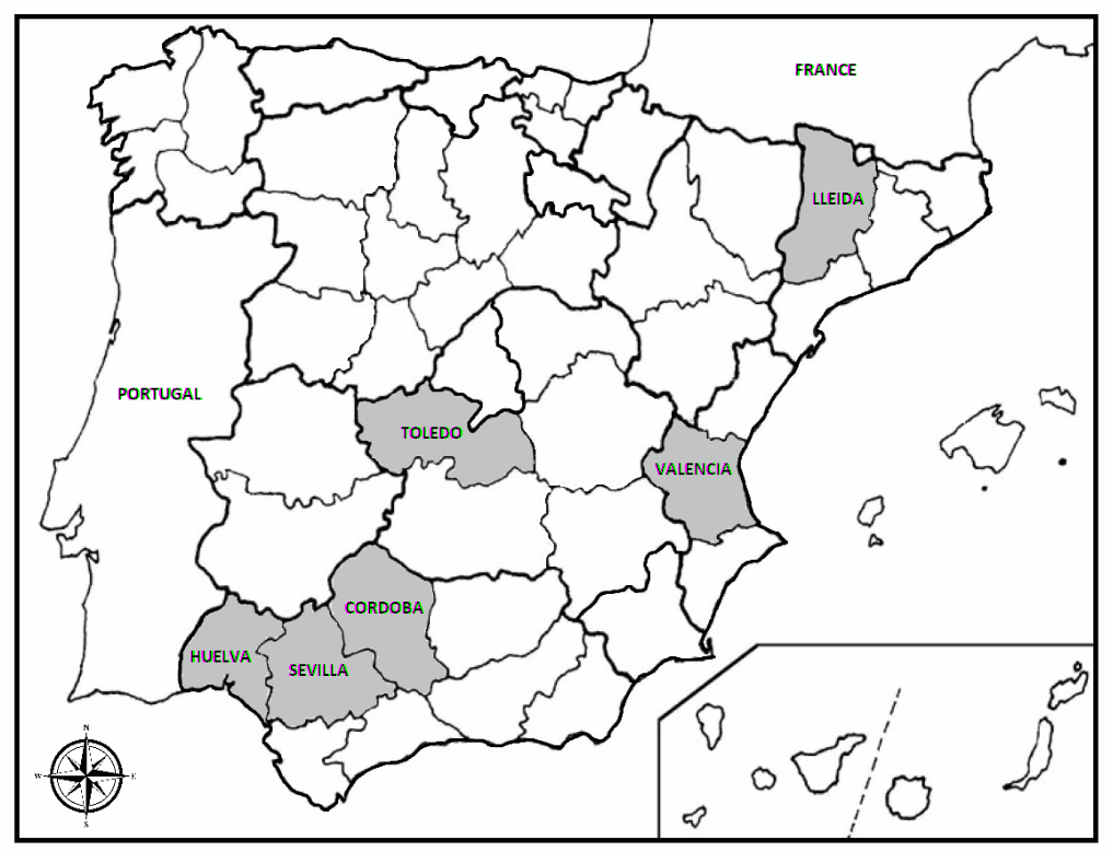
2.1. Field and Nursery Survey and Sampling

From 2018 to 2020, diseased young almond trees, of 1–3 years old, showing the symptoms root necrosis and root rot, cankers at the crown area, gum exudates at the base of the trunk and general decline (leaf yellowing, defoliation and wilting) (Figure 1), were surveyed in commercial orchards and nursery fields in Spain. The sampling sites were selected in order to represent the main almond-growing regions and different environmental conditions. A total of 149 samples was collected in six provinces, Córdoba (4 samples), Huelva (n = 5), Lleida (n = 39), Sevilla (n = 9), Toledo (n = 10) and Valencia (n = 82) (Figure 2). Each sample was composed of the base of the trunk of the symptomatic tree, the entire root system and from 2 to 4 kg of adjacent soil.

2.2. Isolation of Oomycetes from Plant and Soil Samples

Samples were brought to the laboratory for analysis. The crown area and the roots were carefully washed under running tap water to rinse the oil away. Segments from the crown and primary and secondary roots showing symptoms of browning and necrosis were cut. These plant tissue fragments were disinfected with 70% ethanol for 30 s, then rinsed with sterile distilled water and allowed to dry on absorbent paper. Pieces from 1 to 2 mm long were cut in a laminar flow chamber and seeded in Petri dishes with Corn Meal Agar with Pimaricin + Ampicillin + Rifampicin + Pentachloronitrobenzene + Benomyl (CMA-PARPB) medium and CMA-PARPBH (CMA-PARPB corrected with 0,069 g/L of Hymexazol) and then incubated at 25 °C for 72 h [27]. Pure cultures were obtained by transferring the mycelial growth of the margins of colonies to potato dextrose agar medium (PDA; Biokar-Diagnostics, Beauvais, France) for their subsequent identification [28].
For the isolation of oomycetes from the soil, two trapping modalities were applied. For the first, the soil was placed in 10 × 30 × 10 cm plastic buckets, then moistened and saturated with distilled water. Hedera helix L., Robinia pseudoacacia L. and Pittosporum tobira (Thunb.) W.T.Aiton young leaves were added to the surface of the water and incubated in a growth chamber at 23 °C for 3–7 days until points of infection appeared [29]. For the second, Granny Smith apples were used as baits. Four perpendicular perforations of 1 cm2 in diameter were made around an apple previously disinfected with 96% alcohol. In each perforation, a previously moistened soil subsample was added and covered with a plastic tape, to maintain humidity. The apples were incubated at room temperature for 3–7 days [28]. The leaves and apples that presented necrotic or watery lesions were cut into 1–2 mm pieces and seeded in Petri dishes with CMA-PARPB and CMA-PARPBH media, then incubated at 25 °C for one week, checking daily for the development of oomycete-like colonies. Pure cultures were transferred to PDA as described above. The oomycete isolates were stored in an incubator at 10 °C, in plates with oatmeal agar, in the dark, at Instituto Agroforestal Mediterráneo in Universitat Politècnica de València (IAM-UPV; Valencia, Spain).

2.3. DNA Isolation, Amplification and Sequencing of Oomycetes

Genomic DNA was obtained from a one-week-old pure culture of each sample to be analyzed, grown on PDA at 25 °C in the dark. For DNA extraction, the method of Collado-Romero et al. [30] was used, with a slight variation, whereby the mycelium was scraped with a sterile pipette tip and placed in a 0.2 mL polymerase chain reaction (PCR) tube with 20 µL of 25 mM NaOH at pH 12. The tubes with the samples were incubated in a PTC 200 thermal cycler (MJ Research Inc., Waltham, MA, USA) with a DNA denaturation program (100 °C for 15 min and 4 °C for 5 min). Subsequently, 40 μL of 40 mM Tris-HCl was added at pH 5. The ITS region of the ribosomal DNA of the isolates was amplified, using universal primers ITS-6 (5’ GAA GGT GAA GTC GTA ACA AGG 3’) [31] and ITS-4 (5’ TCC TCC GCT TAT TGA TATGC 3’) [32]. Each PCR tube contained water (13.3 μL), Canvax Buffer B (x) (2.5 μL), Canvax MgCl2 (nM) (2.5 μL), Canvax dNTPs (nM) (2.5 μL), Canvax Horse Power Taq polymerase (U/μL) (0.2 μL), primers (1 μL of each) and genomic DNA (2 μL), for a total of 25 μL. PCR amplification was performed in the same thermocycler mentioned above, using the following program: initial denaturation of 1 cycle at 94 °C for 3 min; 35 cycles of denaturation, annealing and extension at 94 °C for 30 s, 55 °C for 30 s and 72 °C for 45 s, respectively; and a final amplification cycle at 72 °C for 10 min. The PCR products were electrophoresed (140 V) on 1.5% agarose gel (1.5% agarose dissolved in TAE buffer (Tris-acetate-EDTA) 40 mM Tris-acetate, 1 mM EDTA) 1X; the staining of nucleic acids was made with RedSafe (20,000×). To identify the size of the bands, a molecular marker (GeneRuler T.M. 100 bp Plus DNA Ladder; Thermo Scientific, Waltham, MA, USA) was loaded into the first well of the gel. The presence of the bands was observed with the aid of a UV light transilluminator. The PCR products were sent to Macrogen (Madrid, Spain) for sequencing. The sequences obtained were subjected to a search in NCBI BLAST (http: //blast.ncbi.nlm.nih.gov/Blast.cgi; accessed on 15 October 2021), identifying the isolates at the species level.

2.4. Pathogenicity Tests

The inoculum was prepared in 1 L glass flasks with a mix of 200 mL of vermiculite, 20 mL of oat grains and 175 mL of eight vegetables (V8) broth (200 mL/L of V8 juice, 800 mL/L of demineralized water and 2 g/L of CaCO3) [33]. The glass flasks were sterilized three times for 20 min at 120 °C in an autoclave.
The flasks were inoculated separately with four isolates of Ph. niederhauserii (PAL-21, PAL-62, PAL74 and PAL-100) and one isolate of Pp. vexans (PAL-98), previously grown on V8 media (V8A). Inoculated flaks were incubated for six weeks in the dark at room temperature [34]. After this time, the inoculum mixture was rinsed with demineralized water to remove excess nutrients, before inoculations.
The pathogenicity tests were conducted on one-year-old almond seedlings of ‘Garnem’ rootstock. Seedlings were selected based on morphological homogeneity and healthy appearance. For inoculation, 20 g of inoculum was added to plastic pots with 200 g of potting mix that contained peat, vermiculite and sand (1:1:1, v/v/v), autoclaved three times prior to use; then, the rootstocks were planted. Control plants were inoculated with a non-infested inoculum mixture. In total, 5 plants were inoculated for each isolate and control and the experiment was repeated. All plants were randomly distributed in a growth chamber under a 12 h photoperiod at 23 °C. All seedlings were watered the day before the inoculation. Immediately after inoculation, the seedlings were flooded for 48 h and the flooding was repeated every two weeks to stimulate the formation of zoosporangia, as previously described by Pérez-Sierra et al. [34].
The experiment was completed 58 days after inoculation. Seedlings were uprooted and the root system was washed carefully under running water to remove the substrate. Re-isolations from seedlings were performed by plating symptomatic fine root fragments in CMA-PARPBH to confirm Koch’s postulates.
Severity evaluations for each plant started ten days after inoculation; then, they were performed every four days until the end of the experiment. Severity was evaluated using the following scale: 0 = symptom-free plant; 1 = foliar chlorosis; 2 = wilting, dieback and defoliation; and 3 = dead plant [35]. With the severity values of each plant over time, the area under the disease-progress curve (AUDPC) was calculated by the trapezoidal integration method [36].
The dry weight of the root biomass was measured. For this purpose, roots were separated from the main stem and shoots by cutting at the root crown, then placed into paper bags and dried for five days in an oven at 35 °C. The dry weight of the roots was recorded.

2.5. Data Analysis

An analysis of variance (ANOVA) was performed for the dry weight of the root biomass and AUDPC. The assumptions of normality and homoscedasticity of the ANOVA were previously verified. Those data that did not meet the assumptions were transformed to the reciprocal value (1/×). Mean values were compared using the least significant difference (LSD) test at the 99% confidence level. All calculations were performed using Statgraphics Centurion XVI (Statgraphics Technologies, Inc., The Plains, VA, USA).

3. Results

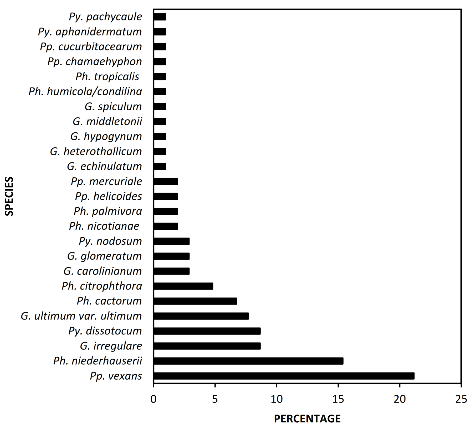
3.1. Occurrence of Oomycetes in Almond Samples

A total of 104 oomycete isolates was obtained (Table 1). Lleida was the province in which the most isolates were obtained, 41, representing 39.4% of the total, followed by Valencia, with 29 (27.8%). Fewer isolates were obtained from the Huelva, Toledo, Sevilla and Córdoba provinces, having 12 (11.5%), 11 (10.5%), 7 (6.7%) and 4 (3.8%), respectively.
The most frequent genus was Phytophthora, representing 32.7% of isolates, followed by Globisporangium and Phytopythium both with 26.9% and Pythium with 13.4% of the total isolates. Regarding the species frequency (Figure 3), the most frequent was Pp. vexans, which represented 21.2% of the isolates, followed by Ph. niederhauserii (15.4%). Less frequent species were G. irregulare and Py. dissotocum (8.7%), G. ultimum var. ultimum (7.7%), Ph. cactorum (6.7%) and Ph. citrophthora (4.8%). The remaining species had percentages below 3%. Regarding the source of isolation, 83.6% of the oomycetes were isolated from soil samples with baiting techniques and only 16.3% were isolated directly from the roots.

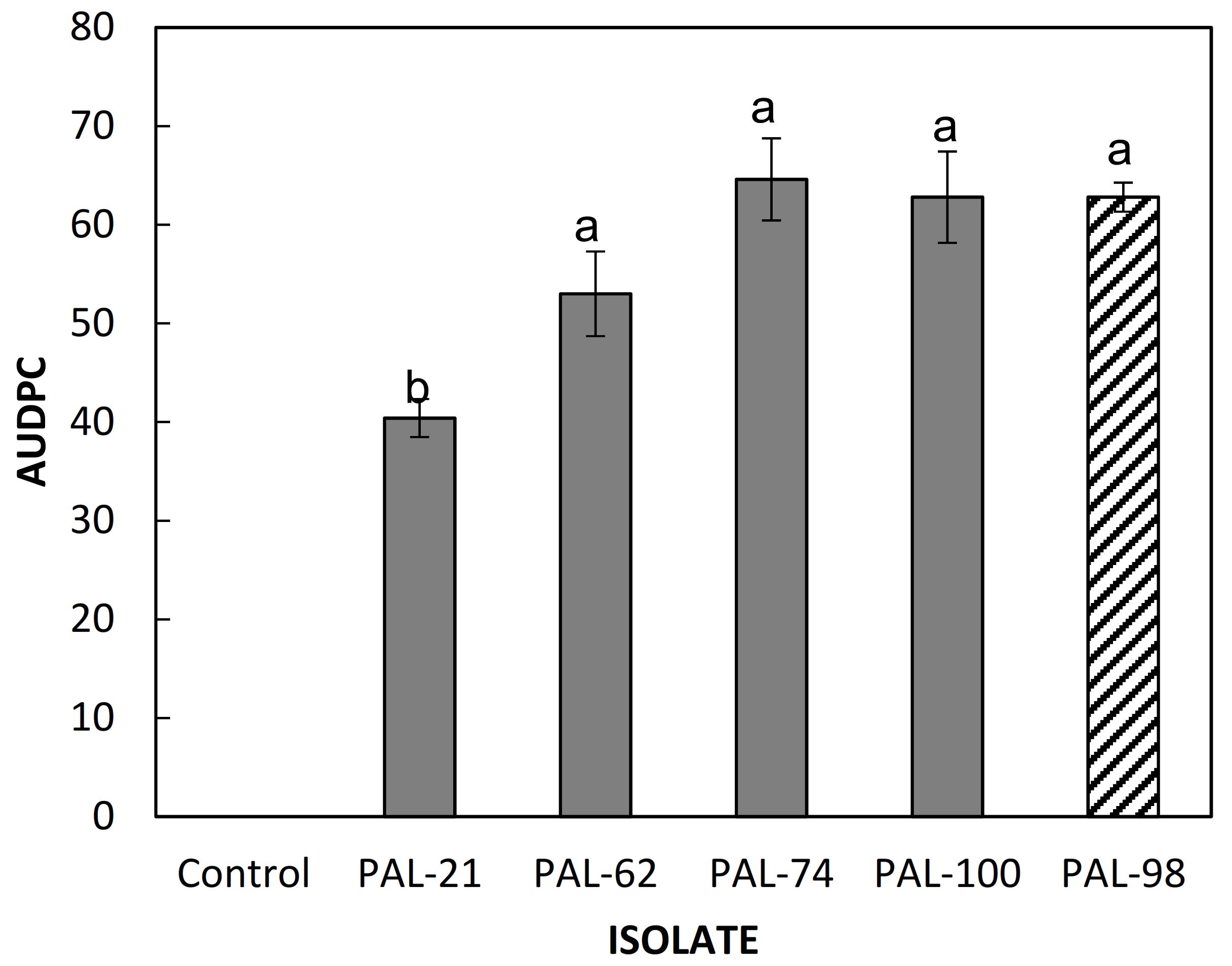
3.2. Pathogenicity Tests

All ‘Garnem’ seedlings inoculated with Ph. niederhauserii and Pp. vexans isolates showed root symptoms (small dark necrotic lesions, root cankers, loss of fine roots and tap root rot), as well as aerial symptoms (chlorosis, wilting and defoliation), and some plants died. The re-isolations from symptomatic roots confirmed Koch’s postulates. In contrast, control treatment seedlings remained healthy and it was not possible to re-isolate Ph. niederhauserii and Pp. vexans from the roots.
The severity of the infections and the AUDPC calculated for each isolate of Ph. niederhauserii and Pp. vexans are shown in Figure 4 and Figure 5. In general, the symptoms appeared earlier in the rootstocks inoculated with the isolate PAL-98 of Pp. vexans (10 days post-inoculation), where the disease progressed rapidly, reaching an AUDPC of 62.8 at day 58 after inoculation. The symptoms caused by isolates PAL-62, PAL-74 and PAL-100 of Ph. niederhauserii started on day 26 after inoculation for the three isolates, reaching an AUDPC value of 53, 64.6 and 62.8, respectively, at 58 days after inoculation. The latest isolate in the expression of symptoms was PAL-21, which started 30 days after inoculation, reaching a value of 40.4 of AUDPC, this being the lowest value obtained for all the inoculated isolates. The ANOVA analysis showed statistically significant differences in the AUDPC mean values (P ≤ 0.01) between the PAL-21 isolate and the other inoculated species. There were no statistically significant differences in the AUDPC of the isolates PAL-62, PAL-74, PAL-98 and PAL-100.
The root dry-weight results are shown in Figure 6. There were statistically significant differences (P ≤ 0.01) between the root dry weight of the control and the isolates PAL-74 and PAL-100 of Ph. niederhauserii, which caused a 33% and a 31% reduction in root dry weight when compared with the un-inoculated control, respectively. There were no statistically significant differences between the control and the isolates PAL-21 and PAL-62 of Ph. niederhauserii and the isolate PAL-98 of Pp. vexans. Although, in the isolates PAL-21 and PAL-62, there were no statistically significant differences with respect to the control, a reduction in the mean weight of 22% and 18%, respectively, was observed when they were compared with the control.

4. Discussion

This study presents the results of a three-year survey of oomycetes associated with root and crown rot of almond trees in Spain. We found a great diversity of oomycete species belonging to the genera Globisporangium, Phytophthora, Phytopythium and Pythium present in root and soil samples, as previously described in almond crops in Iran [12] and California [14,15]. Phytopythium vexans and Ph. niederhauserii were the most frequent species isolated; thus, its pathogenicity to almond rootstock ‘Garnem’, widely used in Spain due to its high vigor and high resistance to Meloidogyne spp., chlorosis and drought [37], was investigated. Moreover, most of the oomycetes were isolated from the soil samples with baiting techniques. It is well known that most oomycetes are difficult to isolate from necrotic tissues because they often harbor many secondary pathogens; therefore, performing the baiting of soil samples taken from affected trees is the best approach [28,38].
The high frequency of Pp. vexans agrees with the results from a study conducted in peach-orchard replanting soil in California by Yang et al. [39], who reported a 65% prevalence of this species in peach trees, showing a dramatic reduction in plant growth, vigor and yield, with a shortened production life. Phytopythium vexans was considered a saprophytic species, rapidly colonizing dead roots in peach trees, playing an invasive role in respect to secondary decaying rootlets [40]. However, its pathogenicity has recently been confirmed on fruit hosts such as grapevine in South Africa [41], avocado in Spain [42] and apple trees in Morocco [43]. Regarding the inoculation of Pp. vexans on the ‘Garnem’ rootstock, our results show a high severity of symptoms at the late stage of the pathogenicity test, which corresponded with advanced defoliation, wilting and dieback symptoms, leading to the plant death of some seedlings. On the contrary, the dry weight of the root did not reduce compared with the control. Similar results were obtained by Ivors et al. [44], who indicated that the root rot indices and the maximum fresh weight of Fraser fir plants (Abies fraseri (Pursh) Poir) inoculated with Pp. vexans were not significantly different from the weight of the controls. To our knowledge, there are no previous works that reported the pathogenicity of this species on almonds; thus, our study is the first report of Pp. vexans as an almond pathogen.
The second species with the highest frequency of isolation was Ph. niederhauserii. Previous studies conducted in Spain reported it for the first time to be affecting almond trees in nurseries [20], but our results confirm that this species is currently present in almond orchards in diverse Spanish provinces. Later, this oomycete was found to affect almonds in Turkey [21] and California [15]. Regarding its pathogenicity to ‘Garnem’ rootstock, the four isolates included in our study caused severe symptoms in the aerial part of the seedlings at the end of the experiment, although only the isolates PAL-74 and PAL-100 significantly reduced the dry weight of the roots compared with the control. Root dry-weight reduction could possibly be the consequence of the reduction in the lateral roots, due to the necrosis caused by Ph. niederhauserii infection. This effect was already described by Rodríguez-Padrón et al. [42] and Kurbetli et al. [45], when these authors inoculated Ph. niederhauserii to avocado and pomegranate, respectively. Furthermore, both research works evidenced that infection by this species was more virulent than that caused by other Phytophthora species evaluated. Although attacks of Ph. niederhauserii have been reported in other crops, mainly ornamental hosts [22,46,47], its worldwide geographical distribution in almond crops is still limited.
Globisporangium irregulare and G. ultimum var. ultimum presented an isolation frequency of less than 10%. Mircetich [40] indicated that G. irregulare and G. ultimum var. ultimum were isolated with high frequency in peach orchards, but there was no relationship between the decay of the trees and the frequency of appearance of these species in the roots. For this reason, this author suggested that both species may be saprophytes on the roots of the peach tree. Although the pathogenicity of G. irregulare and G. ultimum var. ultimum was not investigated in our study, it has been previously studied in America. In Chile, Pinto de Torres and Mircetich [11] reported trunk rot at the crown area of almond trees caused by G. ultimum. Later, in Michigan, USA, Smither and Jones [48] isolated G. irregulare from the ground, in sour cherry orchards, with symptoms of discolored and necrotic roots, in which Phytophthora spp. are commonly found. In greenhouse trials, increased root discoloration, necrosis and reductions in root and shoot growth occurred on sour cherry seedlings inoculated with G. irregulare. These authors concluded that, despite causing these symptoms, G. irregulare was not able to kill this host. For this reason, they suggested that this species was not the cause of the decline in and death of cherry trees in the evaluated orchards but may contribute to a reduction in the growth of cherry trees planted in heavy soils. Coinciding with our study, Yang et al. [39] reported a low isolation frequency of G. irregulare in replanting soils of peach orchards in California; around 8% of the isolates of total oomycetes recovered. Regarding the pathogenicity of these species, in California, Schmidt and Browne [49] detected the presence of Pythium sp. associated with Prunus Replant Disease (PRD), a little-known disease caused by a complex of soil microorganisms. These authors carried out pathogenicity tests in the greenhouse, with grafted peach trees on ‘Nemaguard’ rootstock, inoculating G. irregulare and G. ultimum, among others. In this study, G. irregulare and G. ultimum significantly reduced the fresh weight of the ‘Nemaguard’ rootstock and caused various levels of necrosis of the root cortex, confirming that these species contribute to the development of PRD.
The isolation frequency of Py. dissotocum was also less than 10%. This oomycete has been described mainly as a pathogen of vegetables grown in hydroponic conditions such as lettuce in the USA [50], sweet peppers in Canada [51] and spinach in China [52], but not on fruit crops.
Phytophthora cactorum and Ph. citrophthora were isolated less than 7%. These results match with previous works performed by Wicks and Lee [16] and Browne and Viveros [18], who reported the isolation of these pathogens from almond cankers and soil, without proving their pathogenicity. Later, in Iran, Sahragard and Banihashemi [19] confirmed the pathogenicity of Ph. cactorum in a local almond tree rootstock, which increased mortality rate and decreased root weight. Despite this, the literature is not conclusive in associating specific Phytophthora species with root rot and cankers on the root crown, trunk, or scaffolds, observed in almond trees [15]. Multiple species, such as Ph. cambivora, Ph. cryptogea [17], Ph. citricola [18], Ph. megasperma [23], Ph. plurivora [24] and Ph. chlamydospora [25,26], have been associated with almond crops, but they were not found in our survey.
The remaining oomycete species were found with a very low frequency and were occasionally isolated from a few samples. Most of these species are pathogens of ornamentals, cereals, vegetables and some fruit trees and some have also been described as saprophytes. However, Pp. helicoides has been reported as a pathogen in Prunus in California, causing root rot and roots of stunted seedlings of ‘Nemaguard’ peach rootstock [14].
Disease severity, AUDPC and root dry weight were useful parameters to evaluate the pathogenicity of Pp. vexans and Ph. niederhauserii to almond rootstock ‘Garnem’. The application of standard protocols for pathogenicity testing and disease assessment in plants facilitates inter-study comparisons, thus improving accuracy [53]. Moreover, further studies about pathogenicity testing of oomycetes to Prunus rootstocks would improve the understanding of their infections mechanisms and identifying methods to provide durable resistance, which are major research goals for rootstock breeding programs [54,55].
The results of our research study provide new information about the pathogenic oomycete species present in almond crops in Spain, which represent an emerging threat for almond and other Prunus spp. production. They also highlight the importance of carrying out frequent phytosanitary surveys in fruit nurseries and orchards for a better knowledge of potential risks posed by soil borne pathogens, including the pathogenicity evaluation of the species found.

Author Contributions

Conceptualization, F.B., X.M., L.T., J.A. and P.A.-C.; methodology, F.B., X.M., L.T., J.A. and P.A.-C.; software, F.B.; validation, F.B., X.M., L.T., J.A. and P.A.-C.; formal analysis, F.B., J.A. and P.A.-C.; investigation, F.B., X.M., L.T., J.A. and P.A.-C.; resources, J.A. and P.A.-C.; data curation, F.B., J.A. and P.A.-C.; writing—original draft preparation, F.B., J.A. and P.A.-C.; writing—review and editing, F.B., X.M., J.A. and P.A.-C.; visualization, F.B., X.M., L.T., J.A. and P.A.-C.; supervision, F.B., J.A. and P.A.-C.; project administration, J.A. and P.A.-C.; funding acquisition, J.A. and P.A.-C. All authors have read and agreed to the published version of the manuscript.

Funding

This research study was funded by INIA (Instituto Nacional de Investigación y Tecnología Agraria y Alimentaria), Spain, through projects RTA2017-00009-C04-01 and RTA2017-00009-C04-04, and matching funds from the ERDF (European Regional Development Fund) and Grant PID2020-114648RR-C33 funded by MCIN/AEI/ 10.13039/501100011033.

Institutional Review Board Statement

Not applicable.

Informed Consent Statement

Not applicable.

Data Availability Statement

Not applicable.

Acknowledgments

Francisco Beluzán was supported by Agencia Nacional de Investigación y Desarrollo/Subdirección de Capital Humano/Doctorado Becas Chile en el Extranjero/72200145. Xavier Miarnau was supported by the CERCA Program, Generalitat de Catalunya.

Conflicts of Interest

The authors declare no conflict of interest.

References

  1. Gradziel, T.; Curtis, R.; Socias i Company, R. Production and Growing Regions. In Almonds: Botany, Production and Uses; Socias i Company, R., Gradizel, T., Eds.; CABI: Wallingford, UK, 2017; pp. 70–86. [Google Scholar]
  2. Felipe, A. El Almendro I: El Material Vegetal, 1st ed.; Mira Editores: Zaragoza, Spain, 2000; pp. 1–461. [Google Scholar]
  3. Miarnau, X.; Torguet, L.; Batlle, I.; Alegre, S. El cultivo del almendro en alta densidad. Rev. Frutic. 2016, 49, 68–87. [Google Scholar]
  4. Torguet, L.; Maldonado, M.; Miarnau, X. Importancia y control de las enfermedades en el cultivo del almendro. Agri. Rev. Agropec. Gan. 2019, 1026, 72–77. [Google Scholar]
  5. Rubio-Cabetas, M.; Felipe, A.; Reighard., G. Rootstock Development. In Almonds: Botany, Production and Uses; Rafel Socias i Company, Gradizel, T., Eds.; CABI: Wallingford, UK, 2017; pp. 209–227. [Google Scholar]
  6. Iglesias, I.; Carbó, J. Situació actual, característiques i comportament agronòmic dels portaempelts de presseguer. Dossier Tècnic. 2006, 17, 3–18. [Google Scholar]
  7. Pinochet, J. ‘Replantpac’ (Rootpac® R), a plum–almond hybrid rootstock for replant situations. HortScience 2010, 45, 299–301. [Google Scholar] [CrossRef] [Green Version]
  8. Teviotdale, B.; Michailides, T.; Pscheidt, J. Compendium of Nut Crop Diseases in Temperate Zones; American Phytopathological Society: Portland, OR, USA, 2002; pp. 18–429. [Google Scholar]
  9. Pérez-Sierra, A.; Mora-Sala, B.; León-Santana, M.; García-Jiménez, J.; Abad-Campos, P. Enfermedades causadas por Phytophthora en viveros de plantas ornamentales. Bol. Sanid. Veg. Plagas 2012, 38, 143–156. [Google Scholar]
  10. Wang, Y.; Tyler, B.; Wang, Y. Defense and counterdefense during plant-pathogenic oomycete infection. Annu. Rev. Microbiol. 2019, 73, 667–696. [Google Scholar] [CrossRef]
  11. Pinto de Torres, A.; Mircetich, S. Pythium ultimun Trow causante de pudrición del tronco en plántulas de frutales de carozo y en almendros y damascos de dos años. Agric. Téc. 1976, 36, 171–173. [Google Scholar]
  12. Azizi, Z.; Amini, J.; Shaykh-Eslami, M.; Abasi, S. Pathogenicity of some isolates of Pythium and Phytophthora on detached shoots and seedlings of almond. Iran. J. Plant. Pathol. 2013, 49, 33–39. [Google Scholar]
  13. Javadi, N.; Sharifnabi, B. Phytopythium litorale, the Causal Agent of Almond Root and Crown rot in Iran. Proceedings of 22nd Iranian Plant Protection Congress, Karaj, Iran, 27–30 August 2016; College of Agriculture and Natural Resources: Karaj, Iran, 2016; pp. 27–30. [Google Scholar]
  14. Browne, G.; Ott, N.; Fichtner, E. First Report of Phytopythium helicoides causing root rot on peach rootstock in California. Plant. Dis. 2019, 103, 2968. [Google Scholar] [CrossRef]
  15. Browne, G.; Schmidt, L.; Brar, G. First report of Phytophthora niederhauserii causing crown rot of almond (Prunus dulcis) in California. Plant. Dis. 2015, 99, 1863. [Google Scholar] [CrossRef]
  16. Wicks, T.; Lee, T. Phytophthora crown rot of almond trees. Aust. J. Agric. Res. 1986, 37, 277–287. [Google Scholar] [CrossRef]
  17. Wicks, T. Susceptibility of almond and cherry rootstocks and scions to Phytophthora species. Aust. J. Exp. Agric. 1989, 29, 103–109. [Google Scholar] [CrossRef]
  18. Browne, G.; Viveros, M. Diverse symptoms and tree losses caused by Phytophthora spp. in California almonds. Acta Hortic. 1997, 470, 570–575. [Google Scholar] [CrossRef]
  19. Sahragard, N.; Banihashemi, Z. Evaluation of resistance of some almond genotypes and cultivars to Phytophthora cactorum. Iran. J. Plant. Pathol. 2006, 42, 97–99. [Google Scholar]
  20. Pérez-Sierra, A.; León, M.; Álvarez, L.; Alaniz, S.; Berbegal, M.; García-Jiménez, J.; Abad-Campos, P. Outbreak of a new Phytophthora sp. associated with severe decline of almond trees in eastern Spain. Plant. Dis. 2010, 94, 534–541. [Google Scholar] [CrossRef]
  21. Kurbetli, I.; Değirmenci, K. First report of Phytophthora taxon niederhauserii causing decline of almond in Turkey. New Dis. Rep. 2011, 23, 14. [Google Scholar] [CrossRef] [Green Version]
  22. Abad, G.; Abad, J.; Cacciola, S.; Pane, A.; Faedda, R.; Moralejo, E.; Pérez-Sierra, A.; Abad-Campos, P.; Álvarez-Bernaola, L.; Bakonyi, J.; et al. Phytophthora niederhauserii sp. nov., a polyphagous species associated with ornamentals, fruit trees and native plants in 13 countries. Mycologia 2014, 106, 431–447. [Google Scholar] [CrossRef]
  23. Kurbetli, İ.; Yılmaz, A. Almond decline caused by Phytophthora megasperma in southeastern Anatolian region of Turkey. J. Turk. Phytopathol. 2016, 45, 13–20. [Google Scholar]
  24. Çiftçi, O.; Türkölmez, Ş.; Derviş, S.; Serçe, Ç. First report of canker and root rot of almond caused by Phytophthora plurivora in Turkey. Plant. Dis. 2016, 100, 1507. [Google Scholar] [CrossRef]
  25. Türkölmez, Ş.; Derviş, S.; Çiftçi, O.; Ulubaş Serçe, Ç. First report of Phytophthora chlamydospora causing root and crown rot on almond (Prunus dulcis) trees in Turkey. Plant. Dis. 2016, 100, 1796. [Google Scholar] [CrossRef]
  26. Browne, G.; Ott, N.; Forbes, H.; Yaghmour, M.; Milliron, L. First Report of Phytophthora chlamydospora causing crown and root rot on almond in California. Plant. Dis. 2020, 104, 2033. [Google Scholar] [CrossRef]
  27. Jeffers, S.; Martin, S. Comparison of two media selective for Phytophthora and Pythium species. Plant. Dis. 1986, 70, 1038–1043. [Google Scholar] [CrossRef]
  28. Erwin, D.; Ribeiro, O. Phytophthora Disease Worldwide; American Phytopathology Society Press: Saint Paul, MN, USA, 1996; pp. 1–562. [Google Scholar]
  29. Linderman, R.; Zeitoun, F. Phytophthora cinnamomi causing root rot and wilt of nursery-grown native western azalea and salal. Plant. Dis. Report. 1977, 61, 1045–1048. [Google Scholar]
  30. Collado-Romero, M.; Mercado-Blanco, J.; Olivares-García, C.; Valverde-Corredor, A.; Jiménez-Díaz, R. Molecular variability within and among Verticillium dahliae vegetative compatibility groups determined by fluorescent amplified fragment length polymorphism and polymerase chain reaction markers. Phytopathology 2006, 96, 485–495. [Google Scholar] [CrossRef] [Green Version]
  31. Cooke, D.; Drenth, A.; Duncan, J.; Wagels, G.; Brasier, C. A molecular phylogeny of Phytophthora and related oomycetes. Fungal Genet. Biol. 2000, 30, 17–32. [Google Scholar] [CrossRef]
  32. White, T.; Bruns, T.; Lee, S.; Taylor, J. Amplification and direct sequencing of fungi ribosomal RNA genes for phylogenetics. In PCR Protocols. A Guide to Methods and Applications; Innis, M., Gelfand, D., Sninsky, J., White, T., Eds.; Academic Press: Millbrae, CA, USA, 1990; pp. 315–322. [Google Scholar]
  33. Jung, T.; Blaschke, H.; Neumann, P. Isolation, identification and pathogenicity of Phytophthora species from declining oak stands. Eur. J. For. Pathol. 1996, 26, 253–272. [Google Scholar] [CrossRef]
  34. Pérez-Sierra, A.; López-García, C.; León, M.; García-Jiménez, J.; Abad-Campos, P.; Jung, T. Previously unrecorded low-temperature Phytophthora species associated with Quercus decline in a Mediterranean forest in eastern Spain. For. Pathol. 2013, 43, 331–339. [Google Scholar] [CrossRef]
  35. Jönsson, U.; Jung, T.; Rosengren, U.; Nihlgård, B.; Sonesson, K. Pathogenicity of Swedish isolates of Phytophthora quercina to Quercus robur in two different soils. New Phytol. 2003, 158, 355–364. [Google Scholar] [CrossRef] [Green Version]
  36. Campbell, C.; Madden, L. Introduction to plant disease epidemiology, 1st ed; John Wiley & Sons: New York, NY, USA, 1990; pp. 192–194. [Google Scholar]
  37. Bielsa, B.; Rubio-Cabetas, M. La elección del patrón en la almendricultura moderna. Rev. Frut. 2018, 65, 92–113. [Google Scholar]
  38. Drenth, A.; Sendall, B. Practical Guide to Detection and Identification of Phytophthora; CRC for Tropical Plant Protection: Brisbane, Australia, 2001; pp. 1–41. [Google Scholar]
  39. Yang, J.; Ruegger, P.; McKenry, M.; Becker, J.; Borneman, J. Correlations between root-associated microorganisms and peach replant disease symptoms in a California Soil. PLoS ONE 2012, 7, e46420. [Google Scholar] [CrossRef]
  40. Mircetich, S. The role of Pythium in feeder roots of diseased and symptomless peach trees. Phytopathology 1971, 61, 357–360. [Google Scholar] [CrossRef]
  41. Langenhoven, S.; Halleen, F.; Spies, C.; Stempien, E.; Mostert, L. Detection and quantification of black foot and crown and root rot pathogens in grapevine nursery soils in the Western Cape of South Africa. Phytopathol. Mediter. 2018, 57, 519–537. [Google Scholar] [CrossRef]
  42. Rodríguez-Padrón, C.; Siverio, F.; Pérez-Sierra, A.; Rodríguez, A. Isolation and pathogenicity of Phytophthora species and Phytopythium vexans recovered from avocado orchards in the Canary Islands, including Phytophthora niederhauserii as a new pathogen of avocado. Phytopathol. Mediterr. 2018, 57, 89–106. [Google Scholar] [CrossRef]
  43. Jabiri, S.; Lahlali, R.; Bahra, C.; Amraoui, M.; Tahiri, A.; Amiri, S. First report of Phytopythium vexans associated with dieback disease of apple trees in Morocco. J. Plant. Pathol. 2020, 102, 1319. [Google Scholar] [CrossRef]
  44. Ivors, K.; Abad, Z.; Benson, D. Evaluating the pathogenicity of Pythium vexans isolates from Fraser fir in North Carolina. Plant. Health Prog. 2008, 9, 8. [Google Scholar] [CrossRef] [Green Version]
  45. Kurbetli, İ.; Karaca, G.; Aydoğdu, M.; Sülü, G. Phytophthora species causing root and collar rot of pomegranate in Turkey. Eur. J. Plant. Pathol. 2020, 157, 485–496. [Google Scholar] [CrossRef]
  46. Moralejo, E.; Pérez-Sierra, A.; Álvarez, L.; Belbahri, L.; Lefort, F.; Descals, E. Multiple alien Phytophthora taxa discovered on diseased ornamental plants in Spain. Plant. Pathol. 2009, 58, 100–110. [Google Scholar] [CrossRef]
  47. Prigigallo, M.; Mosca, S.; Cacciola, S.; Cooke, D.; Schena, L. Molecular analysis of Phytophthora diversity in nursery-grown ornamental and fruit plants. Plant. Pathol. 2015, 64, 1308–1319. [Google Scholar] [CrossRef] [Green Version]
  48. Smither, M.; Jones, A. Pythium species associated with sour cherry and the effect of P. irregulare on the growth of mahaleb cherry. Can. J. Plant. Pathol. 1989, 11, 1–8. [Google Scholar] [CrossRef]
  49. Schmidt, L.; Browne, G. Characterization of Pythium species associated with Prunus replant disease. In Phytopathology; The American Phytopathological Society: Saint Paul, MN, USA, 2013; Volume 103, p. 128. [Google Scholar]
  50. McGehee, C.; Raudales, R.; Elmer, W. First report of Pythium dissotocum causing Pythium root rot on hydroponically grown lettuce in Connecticut. Plant. Dis. 2018, 102, 2043. [Google Scholar] [CrossRef]
  51. Owen-Going, N.; Sutton, J.; Grodzinski, B. Relationships of Pythium isolates and sweet pepper plants in single-plant hydroponic units. Can. J. Plant. Pathol. 2003, 25, 155–167. [Google Scholar] [CrossRef]
  52. Huo, C.; Cao, J.; Wu, K.; Chen, Y.; Zhao, Z. First Report of Pythium dissotocum causing root rot on hydroponically grown spinach in China. Plant. Dis. 2020, 104, 3084. [Google Scholar] [CrossRef]
  53. Bhunjun, C.; Phillips, A.; Jayawardena, R.; Promputtha, I.; Hyde, K. Importance of molecular data to identify fungal plant pathogens and guidelines for pathogenicity testing based on Koch’s Postulates. Pathogens 2021, 10, 1096. [Google Scholar] [CrossRef]
  54. Fawke, S.; Doumane, M.; Schornack, S. Oomycete interactions with plants: Infection strategies and resistance principles. Microbiol. Mol. Biol. Rev. 2015, 79, 263–280. [Google Scholar] [CrossRef] [Green Version]
  55. El Hadrami, A.; El-Bebany, A.; Yao, Z.; Adam, L.; El Hadrami, I.; Daayf, F. Plants versus fungi and oomycetes: Pathogenesis, defense and counter-defense in the proteomics era. Int. J. Mol. Sci. 2012, 13, 7237–7259. [Google Scholar] [CrossRef] [Green Version]
Figure 1. Symptoms observed in field and nursery samples of almond trees: (a) leaf yellowing; (b) canker and crown rot; (c) general decline; (d) gum exudates at the base of the trunk; (e) root and crown rot.
Agriculture 12 00294 g001
Figure 2. Map showing the location of sampled provinces in Spain. The sampled provinces are highlighted in light gray.
Agriculture 12 00294 g002
Figure 3. Frequency of occurrence of Globisporangium, Phytophthora, Phytopythium and Pythium species recovered from almond orchards showing root and crown rot symptoms in six provinces of Spain (n = 104).
Agriculture 12 00294 g003
Figure 4. Results of severity of four isolates of Ph. niederhauserii and one of Pp. vexans to ‘Garnem’ rootstock 58 days after inoculation. Severity was evaluated using the following scale: 0 = symptom-free plant; 1 = foliar chlorosis; 2 = wilting, dieback and defoliation; and 3 = dead plant. Values are the mean of 10 almond seedlings. The vertical bars represent the standard error of the mean.
Agriculture 12 00294 g004
Figure 5. Results of area under the disease progress curve AUDPC of four isolates of Ph. niederhauserii and one of Pp. vexans to ‘Garnem’ rootstock 58 days after inoculation. The vertical bars represent the standard error of the mean. The letters indicate significant differences (LSD; P ≤ 0.01) among the means. The white column represents the control, gray columns represent the isolates of Ph. niederhauserii and the striped column represents the isolate of Pp. vexans.
Agriculture 12 00294 g005
Figure 6. Results of root dry weight of four isolates of Ph. niederhauserii and one of Pp. vexans to ‘Garnem’ rootstock 58 days after inoculation. The vertical bars represent the standard error of the mean. The letters indicate significant differences (LSD; P ≤ 0.01) among the isolates’ means. The white column represents the control, the gray columns represent the isolates of Ph. niederhauserii and the striped column represents the isolate of Pp. vexans.
Agriculture 12 00294 g006
Table 1. Origin and GenBank accession numbers of Globisporangium, Phytophthora, Phytopythium and Pythium species recovered from almond tree samples showing root and crown rot symptoms in six provinces of Spain.
SpeciesCodeGeographic OriginSourceRootstock/ScionAcc. No.
Globisporangium carolinianumPAL-106ValenciaSoil MZ921970
G. carolinianumPAL-13LleidaSoil MZ921972
G. carolinianumPAL-69ValenciaSoil MZ922023
G. echinulatumPAL-44ToledoSoil MZ921949
G. glomeratumPAL-107ValenciaSoil MZ921971
G. glomeratumPAL-14LleidaSoil MZ921973
G. glomeratumPAL-41LleidaSoil MZ922001
G. heterothallicumPAL-42ToledoSoil MZ922002
G. hypogynumPAL-68ValenciaSoil MZ921955
G. irregularePAL-45ToledoSoil MZ922003
G. irregularePAL-46ToledoSoil MZ922004
G. irregularePAL-47ToledoSoil MZ922005
G. irregularePAL-48ToledoSoil MZ922006
G. irregularePAL-49ToledoSoil MZ922007
G. irregularePAL-51ToledoSoil MZ922009
G. irregularePAL-52ToledoSoil MZ922010
G. irregularePAL-90HuelvaSoil MZ922042
G. irregularePAL-91SevillaRoot‘Rootpac-40′/ndMZ922043
G. middletoniiPAL-15LleidaSoil MZ921965
G. speculumPAL-50ToledoSoil MZ921950
G. ultimum var. ultimumPAL-17LleidaSoil MZ921974
G. ultimum var. ultimumPAL-58ValenciaRoot‘GF-677′/’Lauranne’MZ922015
G. ultimum var. ultimumPAL-61ValenciaRoot‘GF-677′/’Lauranne’MZ922017
G. ultimum var. ultimumPAL-63ValenciaSoil MZ922018
G. ultimum var. ultimumPAL-79HuelvaSoil MZ922030
G. ultimum var. ultimumPAL-84HuelvaSoil MZ922035
G. ultimum var. ultimumPAL-92SevillaSoil MZ922044
G. ultimum var. ultimumPAL-94SevillaSoil MZ922046
Phytophthora cactorumPAL-10LleidaSoil MZ921975
Ph. cactorumPAL-12LleidaSoil MZ921977
Ph. cactorumPAL-24LleidaSoil MZ921983
Ph. cactorumPAL-28LleidaSoil MZ921984
Ph. cactorumPAL-35LleidaSoil MZ921994
Ph. cactorumPAL-38LleidaSoil MZ921997
Ph. cactorumPAL-4LleidaSoil MZ921999
Ph. citrophthoraPAL-16LleidaSoil MZ921978
Ph. citrophthoraPAL-18LleidaSoil MZ921979
Ph. citrophthoraPAL-19LleidaSoil MZ921980
Ph. citrophthoraPAL-2LleidaSoil MZ921981
Ph. citrophthoraPAL-20LleidaSoil MZ921982
Ph. humicola/condilinaPAL-5LleidaSoil MZ922008
Ph. nicotianaePAL-57ValenciaSoil MZ922014
Ph. nicotianaePAL-71ValenciaSoil MZ922025
Ph. niederhauseriiPAL-11LleidaSoil MZ921964
Ph. niederhauseriiPAL-34LleidaSoil MZ921969
Ph. niederhauseriiPAL-60ValenciaRoot‘GF-677′/’Lauranne’MZ921953
Ph. niederhauseriiPAL-62ValenciaRoot‘GF-677′/’Lauranne’MZ921954
Ph. niederhauseriiPAL-1LleidaSoil MZ921961
Ph. niederhauseriiPAL-100CórdobaRoot‘GF-677′/ndMZ921962
Ph. niederhauseriiPAL-105ValenciaRoot‘Garnem/’Lauranne’MZ921963
Ph. niederhauseriiPAL-21LleidaSoil MZ921966
Ph. niederhauseriiPAL-26LleidaSoil MZ921967
Ph. niederhauseriiPAL-3LleidaSoil MZ921968
Ph. niederhauseriiPAL-6LleidaSoil MZ921952
Ph. niederhauseriiPAL-7LleidaSoil MZ921956
Ph. niederhauseriiPAL-74ValenciaSoil MZ921957
Ph. niederhauseriiPAL-77ValenciaSoil MZ921958
Ph. niederhauseriiPAL-78ValenciaRoot‘GF-677′/ndMZ921959
Ph. niederhauseriiPAL-8LleidaSoil MZ921960
Ph. palmivoraPAL-66ValenciaSoil MZ922021
Ph. palmivoraPAL-72ValenciaSoil MZ922026
Ph. tropicalisPAL-101CórdobaSoil MZ921976
Phytopythium chamaehyphonPAL-99CórdobaSoil MZ922051
Pp. cucurbitacearumPAL-53ValenciaRoot‘Garnem’/ndMZ921951
Pp. helicoidesPAL-76ValenciaRoot‘Garnem’/’Penta’MZ922029
Pp. helicoidesPAL-96SevillaRoot‘GF-677′/ndMZ922048
Pp. mercurialePAL-86HuelvaSoil MZ922037
Pp. mercurialePAL-87HuelvaSoil MZ922038
Pp. vexansPAL-22LleidaSoil MZ921985
Pp. vexansPAL-23LleidaRoot‘GF-677′/’Constantí’MZ921986
Pp. vexansPAL-25LleidaRoot‘GF-677′/’Vairo’MZ921987
Pp. vexansPAL-27LleidaRoot‘GF-677′/’Marinada’MZ921988
Pp. vexansPAL-29LleidaRoot‘Garnem’/’Soleta’MZ921989
Pp. vexansPAL-32LleidaSoil MZ921992
Pp. vexansPAL-33LleidaSoil MZ921993
Pp. vexansPAL-36LleidaSoil MZ921995
Pp. vexansPAL-37LleidaSoil MZ921996
Pp. vexansPAL-40LleidaSoil MZ922000
Pp. vexansPAL-59ValenciaRoot‘GF-677′/’Lauranne’MZ922016
Pp. vexansPAL-70ValenciaSoil MZ922024
Pp. vexansPAL-73ValenciaSoil MZ922027
Pp. vexansPAL-75ValenciaSoil MZ922028
Pp. vexansPAL-82HuelvaSoil MZ922033
Pp. vexansPAL-83HuelvaSoil MZ922034
Pp. vexansPAL-85HuelvaSoil MZ922036
Pp. vexansPAL-88HuelvaSoil MZ922039
Pp. vexansPAL-89HuelvaSoil MZ922040
Pp. vexansPAL-9LleidaSoil MZ922041
Pp. vexansPAL-93SevillaSoil MZ922045
Pp. vexansPAL-98CórdobaRoot‘GF-677′/ndMZ922050
Pythium aphanidermatumPAL-39LleidaSoil MZ921998
Py. dissotocumPAL-30LleidaSoil MZ921990
Py. dissotocumPAL-31LleidaSoil MZ921991
Py. dissotocumPAL-54ValenciaSoil MZ922011
Py. dissotocumPAL-55ValenciaSoil MZ922012
Py. dissotocumPAL-65ValenciaSoil MZ922020
Py. dissotocumPAL-80HuelvaSoil MZ922031
Py. dissotocumPAL-81HuelvaSoil MZ922032
Py. dissotocumPAL-95SevillaSoil MZ922047
Py. dissotocumPAL-97SevillaSoil MZ922049
Py. nodosumPAL-56ValenciaSoil MZ922013
Py. nodosumPAL-64ValenciaSoil MZ922019
Py. nodosumPAL-67ValenciaSoil MZ922022
Py. pachycaulePAL-43ToledoSoil MZ921948
Acc. No.: accession number; nd: not determined.
Publisher’s Note: MDPI stays neutral with regard to jurisdictional claims in published maps and institutional affiliations.

