Article

Highly Efficient Mesoporous Core-Shell Structured Ag@SiO2 Nanosphere as an Environmentally Friendly Catalyst for Hydrogenation of Nitrobenzene

Key Laboratory of Biomedical Functional Materials, School of Sciences, China Pharmaceutical University, Nanjing 211198, China
*
Author to whom correspondence should be addressed.
Submission received: 7 April 2020 / Revised: 29 April 2020 / Accepted: 1 May 2020 / Published: 3 May 2020

Abstract

:
The size-uniformed mesoporous Ag@SiO2 nanospheres’ catalysts were prepared in one-pot step via reducing AgNO3 by different types of aldehyde, which could control the size of Ag@SiO2 NPs and exhibit excellent catalytic activity for the hydrogenation of nitrobenzene. The results showed that the Ag core size, monitored by different aldehydes with different reducing abilities, together with the ideal monodisperse core-shell mesoporous structure, was quite important to affect its superior catalytic performances. Moreover, the stability of Ag fixed in the core during reaction for 6 h under 2.0 MPa, 140 °C made this type of Ag@SiO2 catalyst separable and environmentally friendly compared with those conventional homogeneous catalysts and metal NPs catalysts. The best catalyst with smaller Ag cores was prepared by strong reducing agents such as CH2O. The conversion of nitrobenzene can reach 99.9%, the selectivity was 100% and the catalyst maintained its activity after several cycles, and thus, it is a useful novel candidate for the production of aniline.

1. Introduction

Aniline (AN), one of the important intermediates for fine chemicals such as agrochemicals, pharmaceuticals and dyestuffs, is mainly produced by selective hydrogenation of the corresponding nitrobenzene (NB) using the chemical reagent reduction method and catalytic hydrogenation method [1,2]. The chemical reduction method includes an iron powder reduction method and a sulfur reduction method, which have the advantages of being selective and are easy to operate on a small scale in a lab. However, there are serious environmental problems in these methods mentioned above. The former produces a large amount of iron waste and wastewater, while the latter causes serious pollution due to the sulfur itself. In addition, the two methods can not completely convert the substrate and the product needs to lastly be purified. With more concern about environmental issues in the chemical industry process, these two methods are being phased out by the industry [3,4,5].
To improve the catalytic hydrogenation reactivity of aromatic nitro compounds, the chemical or physical modification of the catalyst has been focused on achieving the most satisfying results. At present, catalysts applied in the field of hydrogenation of aromatic nitro compounds include metal Co, Ni, Pd, Pt, Ru, Ir, Ag [6,7]. Co and Ni catalysts have advantages in terms of cost, but the catalytic activity is relatively poor. Pd catalyst has good catalytic activity, but Sn4+ is required for modification, and Pt, Ru, Ir catalysts are composed of the corresponding metal nanoparticles. With a suitable oxide carrier, they usually have superior activity. However, these four noble metals are limited from applications due to the high cost.
Nanoscience has made Ag into an effective catalyst which is especially used in the hydrogenation of nitro-substituted aromatic compounds at a relatively low cost [3]. However, normally, the highly dispersible Ag nanoparticles tend to aggregate during the catalytic process and are difficult to be separated from the reaction system, which can cause lower catalytic performance and waste. Some publications reported that it had a promising catalytic activity after coating silica, which may open a new route for the preparation of aniline with nitrobenzene [8,9]. The methods for preparing Ag@SiO2 are mainly reported as being the following [10,11,12]. The seed growth method is to fix the synthesized metal nanoparticles on the surface of the substrate as a seed. Since Ag colloid is easy to prepare to be used as the seed in the synthesis, it makes this method have a fatal disadvantage that the Ag nanoparticles eventually exist in the product as impurities, which will confuse the properties of Ag@SiO2 core-shell structure. Another ultrasonic chemistry method mainly uses the cavitation of ultrasonic waves. This method needs to be carried out under less oxygen or anaerobic conditions, which requires harsh reaction conditions. The third electroless plating method is that a metal ion is reduced by a reducing agent in a solution and deposited on a surface of a catalytically active object to form a metal or alloy plating layer. However, ordinary electroless plating is complicated and time-consuming and generally uses a toxic reducing agent. This is a fast process in the inhomogeneous coverage of the Ag@SiO2 surface, so a large amount of Ag colloids will aggregate in the solution.
For all the defects of Ag@SiO2 mentioned above, its preparation method and catalytic ability need to be much improved. Especially as an effective catalyst, we should be more concerned about its behavior to transport or diffuse the reactants. Wang et al. and Chi et al. have synthesized the micron-Ag@SiO2 catalyst in 4-nitrophenol reduction. Big core size (over 200 nm) and great shell thickness (over 50 nm) will affect the catalytic activity compared to the data in Table 1, despite the article claiming that it showed a comparable catalytic activity with spherical silver nanoparticles [13,14]. Herein, we discovered a simple one-pot method to get a size controllable mesoporous Ag@SiO2 NPs, where the silver core was a substantially fixed interior. Cetyltrimethyl ammonium bromide (CTAB)-capped silver nanoparticles were used as a template to form silver-silica nanoparticles, with tetraethyl orthosilicate (TEOS) as the silica source. Distinct from the typical Stöber method with several steps [15], the entrapped nanoparticles directly reduced by aldehydes [3] can be protected from aggregation and interact with the infiltrated nitrobenzene NB, which was potentially more effective to improve the reduction of NB. Moreover, it could be recycled easily in assembly as a potential environment-friendly catalyst.

2. Materials and Methods

2.1. Materials

NH3·H2O (25%), tetraethyl orthosilicate (TEOS), CTAB were of analytical reagent grade and purchased from Shanghai Chemical Reagent Co., Ltd (Shanghai, China); silver nitrate (AgNO3), formaldehyde(CH2O), acetaldehyde(C2H4O), propionaldehyde(C3H6O), benzaldehyde (C7H6O), cyclohexane(C6H6), and hydrazine hydrate(N2H4·H2O) were of analytical reagent grade and purchased from Sinopharm Chemical Reagent Co., Ltd., (Shanghai, China). Igepal CO-520 (Poly(oxyethylene) (5) nonylphenyl ether) was purchased from Shanghai Aladdin Biochemical Technology Co., Ltd. (Shanghai, China). All of these chemicals were used without further purification. All solutions were produced with ultrapure water, produced by Smart-S2 DS (Nanjing Yipu-Yida Technology Development Co., Ltd., Nanjing, China) in our laboratory.

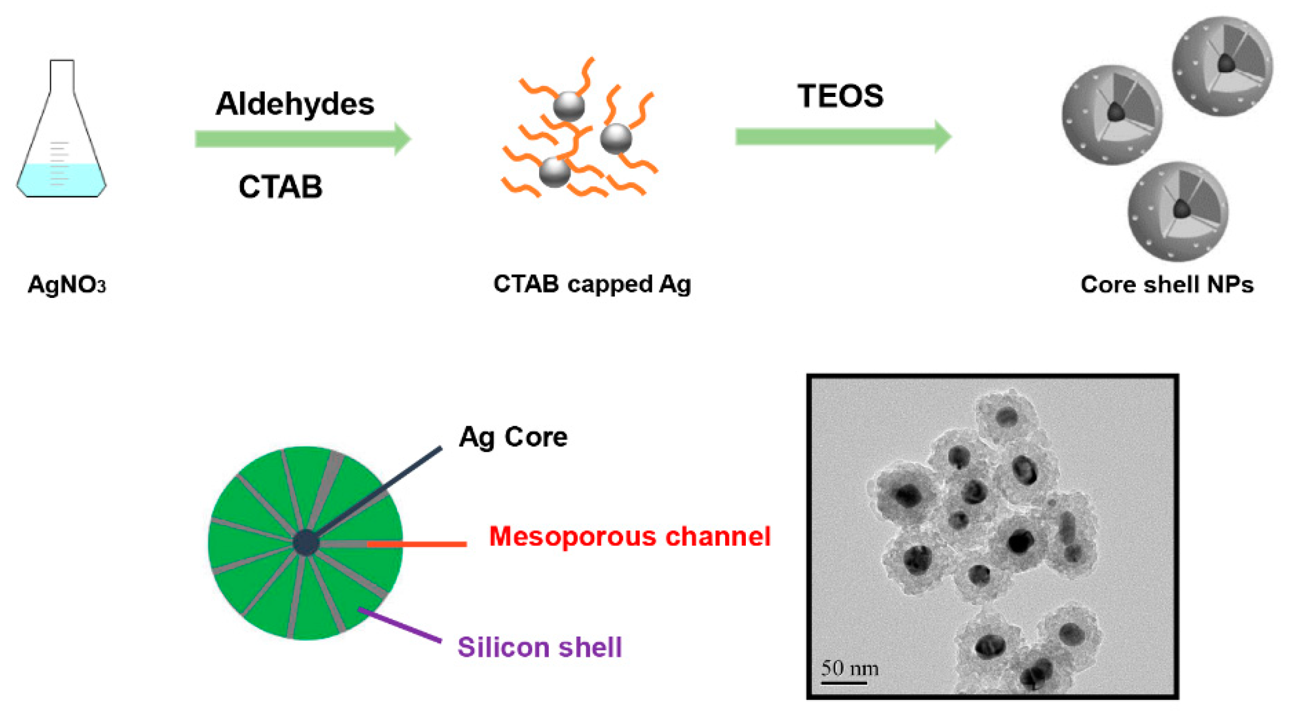
2.2. Fabrication of the Ag@SiO2 Mesoporous Nanoparticles

Silver nanoparticles were prepared by the reduction of AgNO3 by the high or low concentration of CH2O, C2H4O, C3H6O or C7H6O with the protection of CTAB. Generally, 0.4 g CTAB and 3 mL NH3·H2O (25 wt%) were dissolved in 150 mL deionized water and were heated to 60 °C for 30 min. Then, 3 mL (1.0 M) AgNO3 and 1 mL (1.0 M/0.1 M) CH2O, C2H4O, C3H6O or C7H6O were added to the solution dropwise under magnetic stirring and allowed to react for 30 min at 60 °C. Next, 2 mL silica precursor, TEOS, was added dropwise under mechanical stirring at 40 °C. After reacting for 8 h, it was washed with anhydrous ethanol (100 mL) and deionized water (100 mL), and the obtained particles were dried at 50 °C in vacuo.

2.3. Fabrication of the Ag@SiO2 Without-Mesoporous Nanoparticles

25 g Igepal CO-520 and 3 mL (1.0 M) AgNO3 were added to 200 mL C6H12. Some flocs appeared after centrifugation. 100 μL NH2NH2 was added dropwise under mechanical stirring after removing the flocs. After reacting for 0.5 h, 3 mL NH3·H2O (25 wt%) and 2 mL TEOS were added under stirring vigorously for 5 min. After reacting for 8 h, it was washed with anhydrous ethanol (100 mL) and deionized water (100 mL), and the obtained particles were vacuum dried at 50 °C.

2.4. Characterization of Ag@SiO2

Morphology of the synthesized nanoparticles were characterized by high-resolution analytical transmission electron microscopy (TEM, JEM-2100, JEOL, Tokyo, Japan) and scanning electron microscope (SEM, S-3400N II, Hitachi, Tokyo, Japan). The specific surface areas (SSA) were measured with nitrogen adsorption-desorption isotherm via the Brunauer–Emmett–Teller (BET) method. Prior to the measurements, the samples were degassed at 180 °C for 3 h. The average pore sizes, total pore volumes and pore size distributions were obtained with nitrogen desorption data, using the Barrett-Joyner-Halenda (BJH) model. Both were carried out with a Micromeritics Tristar II 3020 (Micromeritics Instrument Corp., Norcross, GA, USA), using nitrogen gas as an adsorbate at 77 K. The phase composition and crystal structure of the synthesized particles were determined by X-ray diffraction (XRD, PANalytical X’Pert Powder Pro, Malvern Panalytical, Almelo, The Netherlands) using Cu Kα1/2 radiation (λα1 = 1.5406 Å) in the range of 10°–90°(2θ). A standard silicon was measured to determine the instrumental broadening, in order to calculate the average crystallite sizes of silver based on the XRD data using Scherrer’s equation [16]. Analysis of the XRD patterns was performed using X’Pert HighScore Plus Software (Malvern Panalytical, Almelo, The Netherlands).

2.5. Catalytic Test

The hydrogenation of nitrobenzene (NB) was employed as a probe reaction to test the activity of the Ag@SiO2 catalyst that was prepared from different types and different concentrations of aldehydes. In each trial, 0.5 g nitrobenzene was used as substrate, hydrogen was used as reducing agent, 50 mg Ag@SiO2 nanoparticle was used as catalyst and stainless steel was chosen to be the reaction vessel in the catalytic studies. The reaction pressure was 2.0 MPa and the temperature was 140 °C. After reacting for 6 h, the conversion was detected by a gas chromatography-mass spectrometer (GCMS-2010, Shimadzu, Kyoto, Japan). The recycling test was also carried out 10 times.

3. Results and Discussion

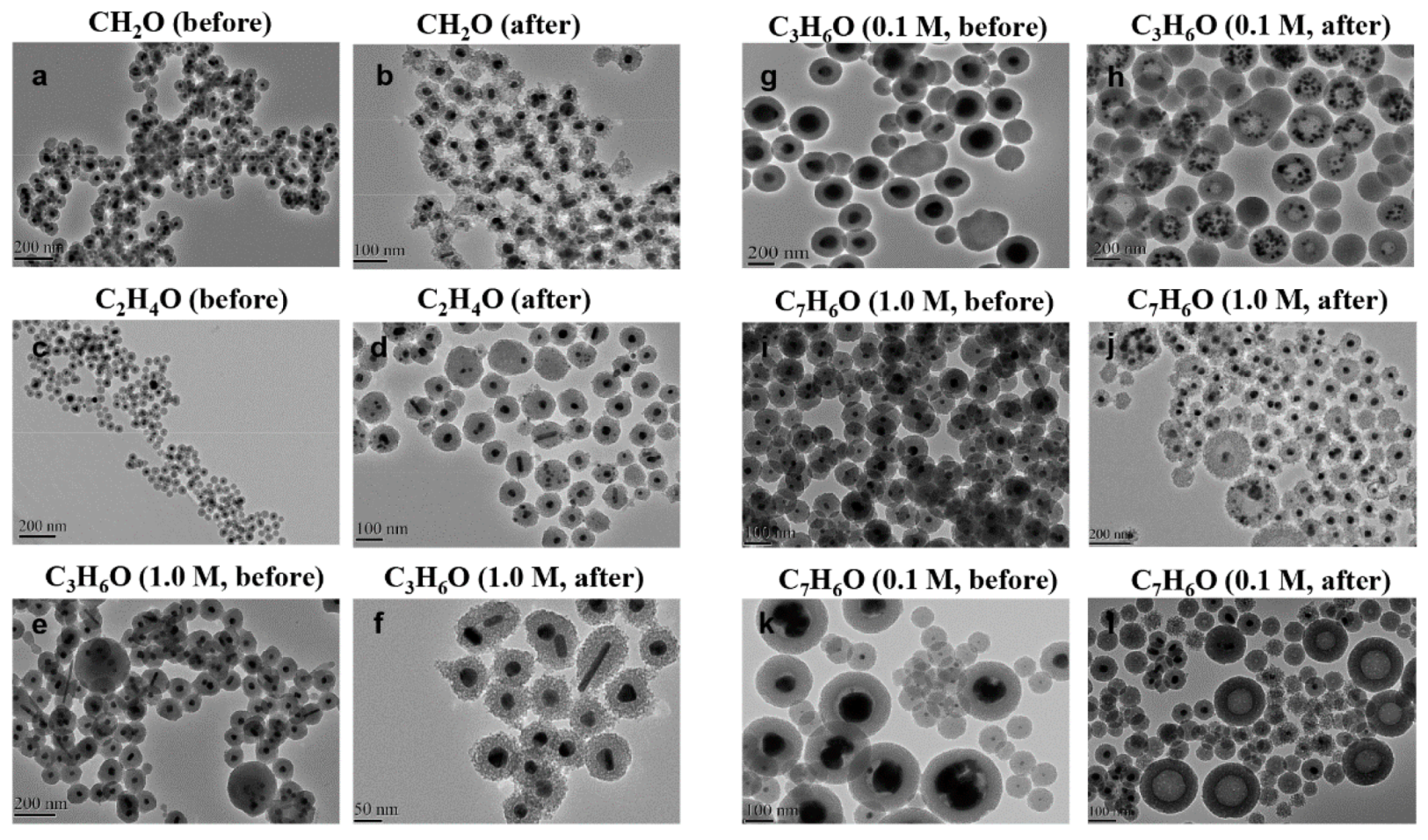
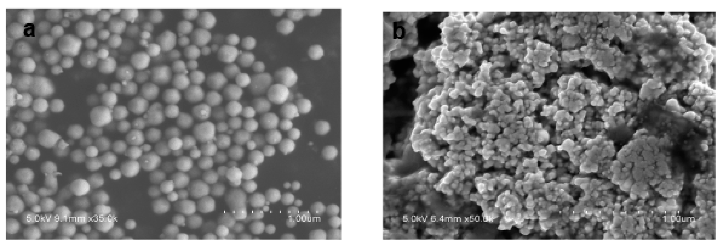
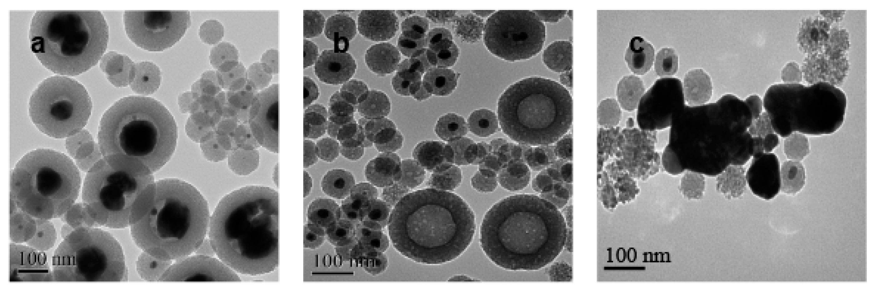
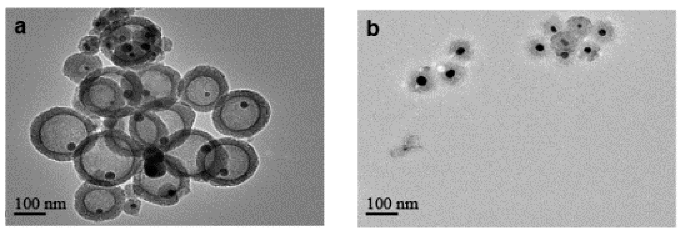
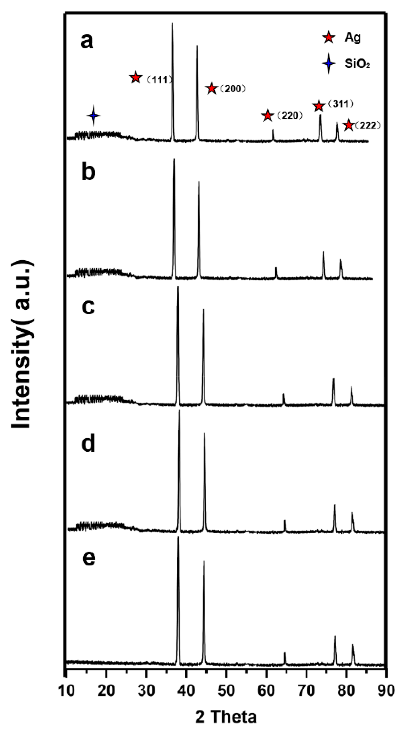
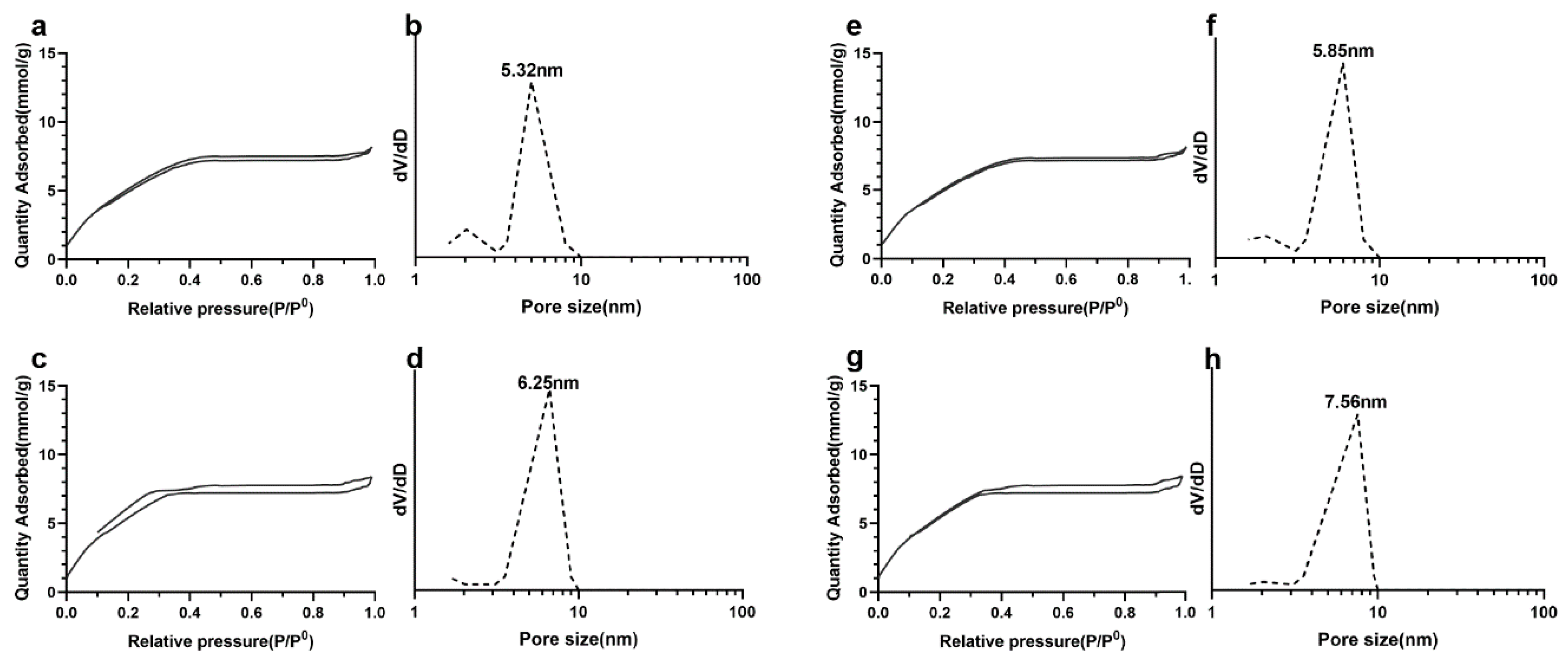
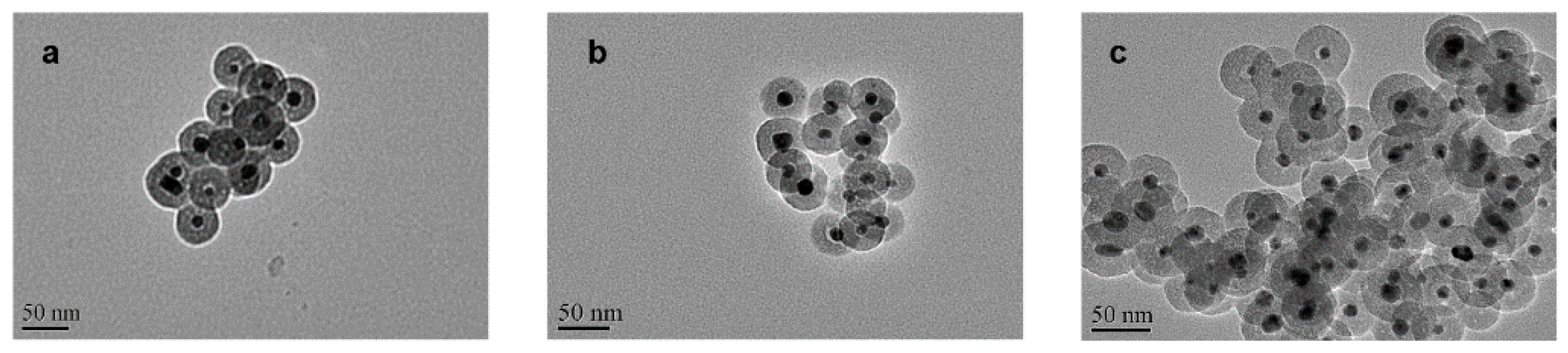
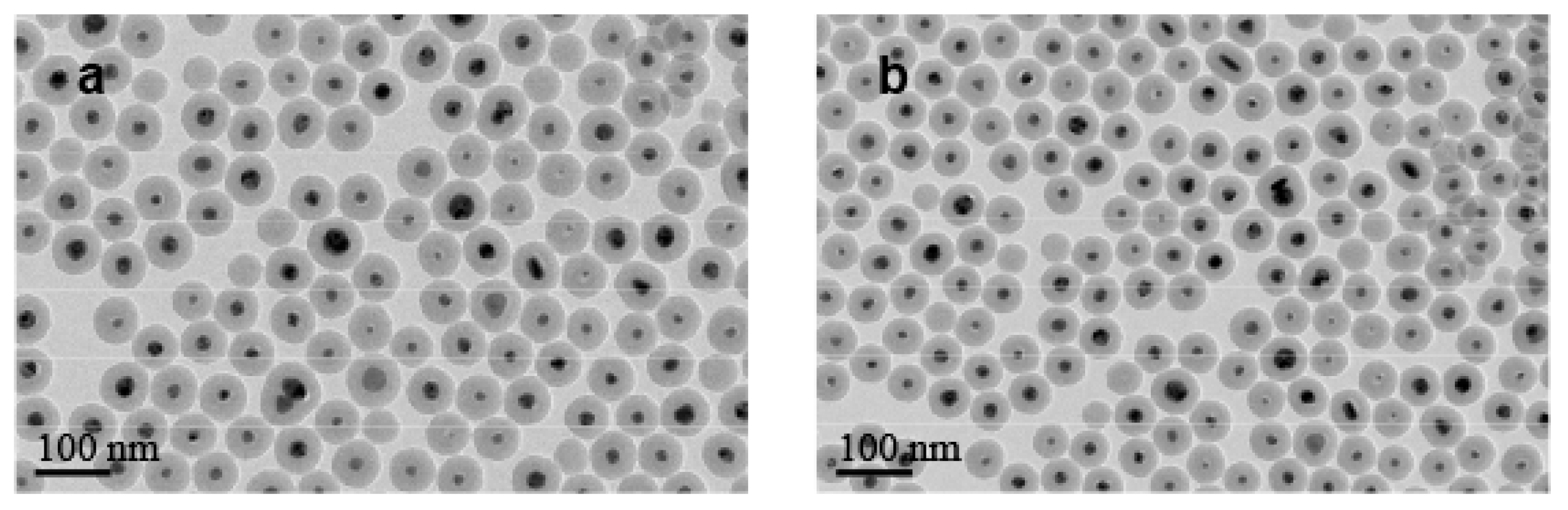
3.1. Preparation and Morphology of Catalysts

The general synthetic procedure to get silica-protected silver nanoparticles by the reduction of silver nitrate with different aldehydes [17,18,19] is shown in Figure 1. The morphology of particles prepared by different types and concentrations of aldehydes collected by TEM were shown in Figure 2. As shown in the TEM, most of the particles were monodispersed with the average core radius, which ranged from 80 nm to 150 nm, coated with the mesoporous silica. It generally had good dispersity and remarkable monodisperse core-shell structures. However, due to the different reducing ability of the aldehydes [20], the sizes of the obtained Ag core were distinct from each other. Figure 2a,c showed that the Ag core, with similar size distribution, was small and dense, which was produced by the strong reducing agent of CH2O or C2H4O at a concentration of 0.1 M. However, big and loose sliver dots were observed in Figure 2g,k, which were reduced by the weak reducing agent of C3H6O or C7H6O at the same concentration (0.1 M). We found that the weaker reducing agent might be accompanied by the slower reduction rate, which resulted in a slow nucleation rate and slow growth. Therefore, the Ag core particle size was inclined to be larger. Especially in Figure 2k, there were two size distribution patterns mixed, most of which were the bigger particles. However, with the increasing concentration of these weak reducing agents, the nucleation of Ag was promoted rapidly, due to the accelerated corresponding reducing rate. It was obvious that the weak reducing agent of C3H6O or C7H6O at a high concentration (1.0 M) was able to produce a similar morphology of Ag@SiO2 (Figure 2e,i) with Figure 2a. All in all, the reducing ability and the concentration of aldehyde are the ultimate factors to monitor the size of Ag@SiO2 NPs, thus, will exhibit distinct catalytic activities in the following study.
It can be supported by the scanning electron microscopy images (Figure 3a) of the Ag core generated in a low-concentration benzaldehyde system without TEOS. The overall particle size was relatively large; even the smallest sphere in the picture was about 100 nm with small bud-shaped Ag that emerged on the surface. These bud-shaped Ag tended to grow continuously, which might have resulted in the maximum particle size being 300 nm. Figure 3b showed the generation of Ag cores in the formaldehyde system without TEOS. The overall morphology appeared to have a much smaller particle size and a spherical shape.
We speculated that during the hydrolysis of TEOS, excessive benzaldehyde will spatially hinder the condensation between Si-OH, making the rate of SiO2 deposition slower, while the Ag core continued to grow. This leads to a larger volume of Ag core. By contrast, perhaps due to the faster growth of Ag NPS when we used a strong reducing agent such as CH2O, the rate of SiO2 deposition was faster than the former, and thus the smaller Ag cores were obtained.

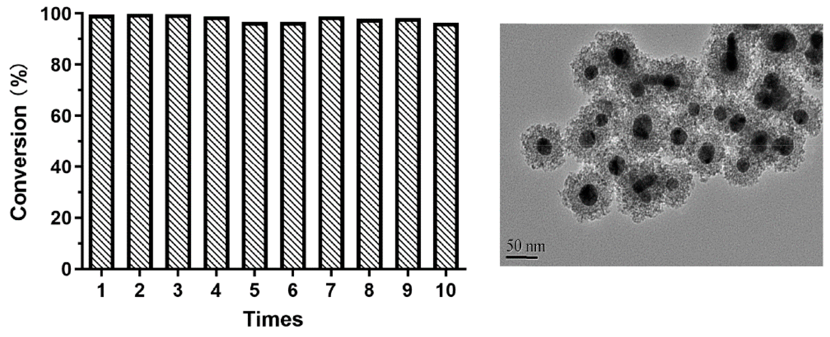
3.2. Catalytic Performances

The catalytic activities of the as-prepared nanoparticles were assessed by the reduction of 4-nitrophenol by H2 with the different catalysts in aqueous solution. Table 1 presented the results for the synthesis of aniline by different kinds and concentrations of aldehydes under high pressure. The conversion of nitrobenzene (NB) was 99.9% in the case of Ag@SiO2 prepared by the relatively strong reducing agent of CH2O, C2H4O. When using the Ag@SiO2 prepared by the weak reducing agent of C3H6O or C7H6O, the conversion of NB reached 98.6% and 97.3% at high concentration (1.0 M), but only reached 11.5% and 10.1% at low concentration (0.1 M). Figure 2 showed the characterization of the prepared nanoparticles after the nitrobenzene hydrogenation by TEM images. Compared with the morphology before the reaction, the outer layer of SiO2 in Figure 2b,d were obviously loose, but the Ag core was still kept in the interior. Similar results were obtained in Figure 2f,j. Meanwhile, Figure 2h,l depicted that almost all the big Ag cores disappeared and some of them reformed the smaller scattered Ag in the shell. Compared with the former, these bigger Ag cores had weak catalytic performance but diffused more reduced by C3H6O and C7H6O at low concentration. Some of the Ag cores were even completely removed from the SiO2 shell after hydrogenation reactions.
For the Ag@SiO2 obtained from a low concentration of benzaldehyde, Figure 4a showed the morphology of Ag@SiO2 NPs before adding H2. We found that it contained two kinds of particle diameters. One was the particle with a diameter of about 100 nm; the majority of the particles were about 250 nm bearing large Ag nuclei. Figure 4b showed the morphology of these Ag@SiO2 NPs after the nitrobenzene hydrogenation and the silver cores of large particles were almost completely diffused, while the Ag cores of the small particles still existed. The experimental results in Figure 4c showed that some Ag particles outside the shell can be found, which should be generated from the diffused bigger Ag cores and then re-aggregated after the reaction. In fact, it was true that the Ag NPs without SiO2 shell had a very low catalytic activity, which coincided with the low catalytic activity result of this Ag@SiO2 (Table 1). It revealed that the diffusion process might occur before the catalytic reaction.
We also calculated the size distribution via size deviation [15] in Table 1. The average sizes of the Ag@SiO2 particles based on the TEM images with standard deviations were found to be 85 ± 6 nm, 81 ± 7 nm, 102 ± 2 nm, 130 ± 37 nm, 105 ± 13nm and 142 ± 50 nm, which confirmed that the much narrower size distribution in case of CH2O is a further benefit of effective catalytic activity.
Moreover, the above experimental results showed an interesting phenomenon. The shells of Ag@SiO2 with high catalytic efficiency all became loose after the reaction, while the main outer layer of the particles of large-sized Ag core was still dense. However, nearly all the small Ag cores were still kept inside the looser shell after the reaction. However, most of the larger Ag cores diffused to the shell; some of them even went outside the dense shell (Table 1). This unexpected and contradictory phenomenon reminded us that the stability of the Ag core might not be related directly to the nitrobenzene hydrogenation itself. This phenomenon suggested that the diffusion of the Ag core should not be occurring after the completion of the catalytic reaction, otherwise the Ag core should overflow more easily in the loose shell particles after the reaction.
In order to prove our assumption, no substrate was added and the hydrogen gas was replaced with argon. Then, we repeated the same process (reaction pressure was 2.0 MPa, temperature was 140 °C and reaction time was 6 h) as described in Section 2.5. From the result shown in Figure 5a, we found that the bigger Ag cores which were prepared at a low concentration of C7H6O still became smaller or disappeared completely from the interior. However, small Ag cores prepared by CH2O in Figure 5b was always kept inside the core-shell structure. This result indicated that the silver core overflow was independent of the nitrobenzene hydrogenation.
In Figure 6, the XRD pattern showed that five diffraction peaks assigned to metallic Ag are found in the spectrum of Ag@SiO2. The peaks at 38.1°, 44.3°, 64.4°, 77.5° and 81.8° were ascribed to Ag (111), (200), (220), (311) and (222), which are shown apparently in the XRD pattern in good accordance with standard JCPDS cards no. 04-0783. Compared with Ag NPs in Figure 6e, the broad humps in the diffraction spectra centered at 22° are due to amorphous silica particles.
Figure 7 presented the nitrogen adsorption–desorption isotherms and pore size distributions of the dry powders. All samples exhibit an approximate type IV isotherm pattern, as defined by IUPAC for mesopore characteristics. The pore sizes were calculated as 5.32 nm, 6.25 nm, 5.85 nm and 7.56 nm by BJH methods. Other data about measuring mean pore sizes and specific surface area by BET analysis were revealed in Table 2.
So what will influence the diffusion of the Ag core under the reaction condition? Normally, the formation of Ag+ which then passes through the porous shell is a reasonable explanation. In the case of this process, the substrate or H2 is necessary to contact Ag and induce the electron donation from the Ag core. However, actually, even without these two agents the great change of bigger Ag core occurs, which means there is no way to produce Ag+. So, we suppose that the other possible mechanism of Ag atom is just diffusing at this high pressure and temperature. A similar report was published by Lei et al. in 2012 [21]. For the larger surface area of the bigger Ag core, less Ag-O-Si- occupied its surface, to make Ag atom unstable and diffuse through the porous shell easily. Another phenomenon in Figure 2h, that some Ag atoms have congregated to form a new cluster in the shell, can further prove this diffusion process. Irene et al. in 2018 [22] also mentioned that once the Ag is diffusing out of the shell, its catalytic activity will be decreased greatly because of the aggregation of the catalyst. It is coinciding with the control experiment we performed using the Ag NPs as the catalyst (about 10% conversion). In contrast, the smaller Ag core with more Ag-O-Si- cross-link occupying its surface is more stable. Then, once it contacts the reagent through the porous shell, it will activate the substrate or H2 rapidly and produce high catalytic efficiency.

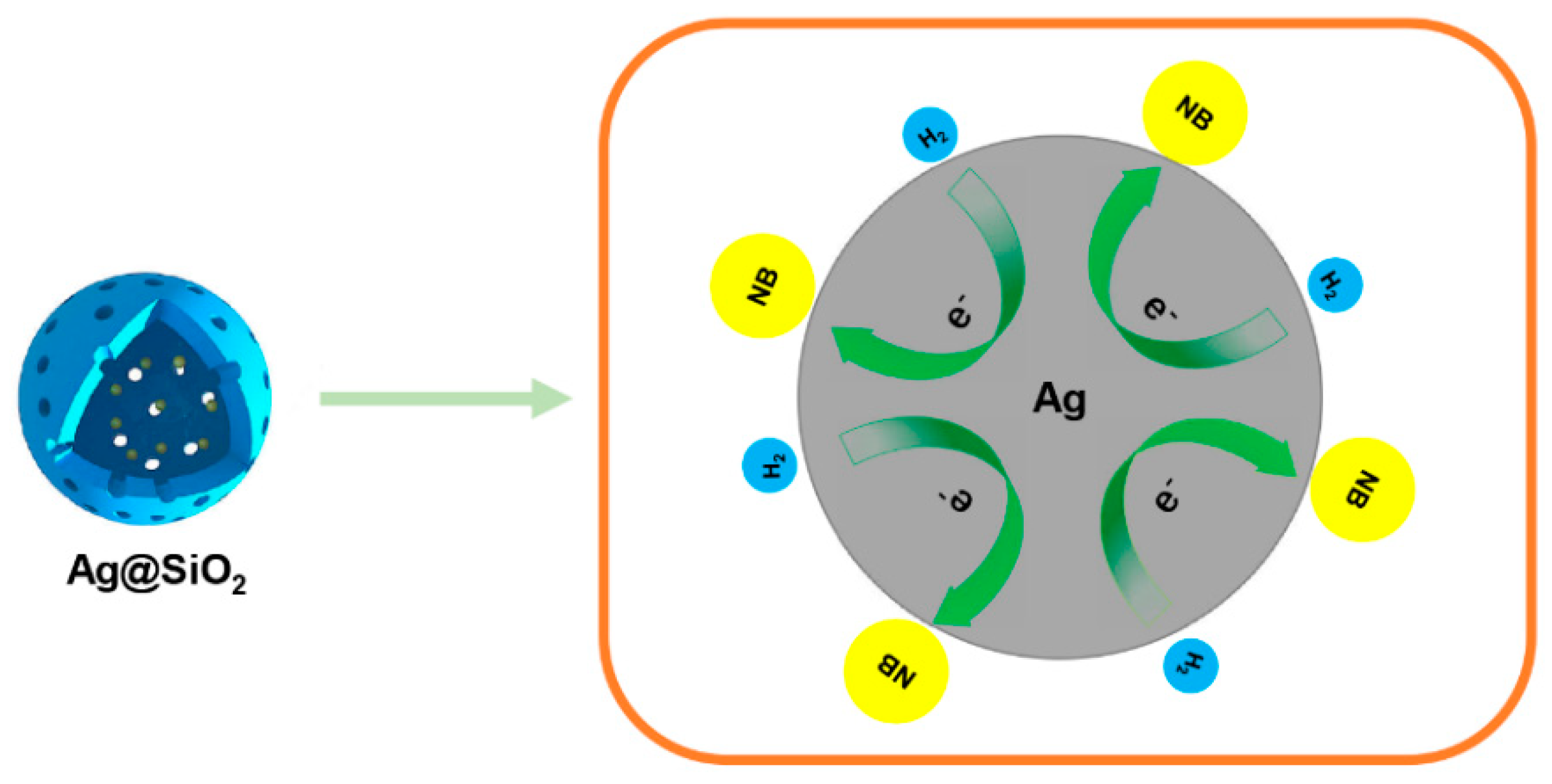
3.3. Catalytic Mechanism

In order to clarify the catalytic mechanism of Ag@SiO2 with small Ag cores, we chose those NPs prepared with CH2O for further study. Figure 8 showed that the interior Ag nearly kept unchanged after the reaction at different reaction conditions, which revealed that the state of the silver core was independent of hydrogen or substrate. These phenomena confirmed what we concluded for the stability of Ag cores. Next, we prepared some Ag@SiO2 nanoparticles with dense shell structure by reverse micelle technique [23]. In Figure 9, the shape of the nanoparticles looks like a compact sphere. After 4, 6, and 12 h of catalytic reaction, the conversion was almost 0% and the morphology of the particles had no change. These results demonstrated that the adsorption of the reactants passing through the mesoporous channel was the necessary pathway to trigger the catalytic reaction.
Thus, the model of the reaction can be classified as a Langmuir–Hinshelwood mechanism, and both reactants need to be absorbed on the surface prior to reaction. The mechanism of catalytic activities as illustrated in Figure 10 [24,25] as follows, where the NB is nitrobenzene, σ is the adsorption site on the Ag core surface, and AN is aniline.
NB + σ → NBσ
H2 + σ → H2σ
NBσ + H2σ → ANσ + H2
ANσ → AN + σ
H2Oσ → H2O + σ
Firstly, the reagents, nitrobenzene and hydrogen, passed through the silica shell via the pores and were absorbed on the surface of the Ag cores. Then, the electrons of the nucleophile (H2) transferred across the interface of Ag cores for Ag play the role of an electronic transmission medium. Then, the electrophile captured the electrons from the interface and completed conversion. Here, the rate-determining step was the reaction on the Ag core surface. The adsorption/desorption equilibrium was achieved much faster and evaluated by the Langmuir isotherm model. Silicon shell not only promoted the free diffusion of reactants towards the catalyst surface, but reduced the aggregation of the catalyst. Moreover, the attachment of SiO2 shell to the Ag surface could slow down the oxidation processes of Ag and improve the stability of Ag nanoparticles [21], which was more beneficial for the separation and possibly recycling of the catalyst.
The recycling test was carried out 10 times. As expected, the catalytic conversion rate remained higher than 95% in Figure 11 after the reaction was repeated ten consecutive times, because of the scarce leakage of silver due to the protection of the outer silica layer.

4. Conclusions

In conclusion, we have reported an Ag@SiO2 catalyst prepared by a one-pot method using different types of aldehydes, and some of them prepared by a strong reducing agent, such as CH2O, showed the best activity for hydrogenation of nitrobenzene producing the aniline. The size of the silver nanoparticles, the interaction between the silver cores and the silica support may play an important role in the catalytic reaction system. We discussed that the best catalyst may have more Ag-O-Si- cross-link occupying smaller Ag core’s surface, which was quite stable before and after the reaction. Due to the protection of the outer silica layer even after the efficient catalytic reaction, scarce leakage of silver demonstrated that this Ag@SiO2 is a promising material in the selective hydrogenation of nitrobenzene on a large scale. Therefore, this progress is valuable to enable the application of these Ag@SiO2 catalysts in energy conversion and environmental protection.

Author Contributions

Conceptualization, B.Z. and Z.D.; methodology, B.Z. and Y.W.; software, Q.W. and N.Z.; validation, B.Z. and Y.W.; formal analysis, Y.X.; investigation, B.Z.; resources, Y.W.; data curation, W.L. and F.L.; writing—original draft preparation, B.Z.; writing—review and editing, B.Z. and Y.W.; visualization, B.Z.; supervision, Y.W.; project administration, B.Z. and Y.W.; funding acquisition, Y.W. All authors have read and agreed to the published version of the manuscript.

Funding

The authors gratefully acknowledge the support of Qing Lan Project in Jiangsu Province and “Double First-Class” University project (CPU2018GY25).

Acknowledgments

We are wholeheartedly grateful to Zheng Xu at Nanjing University in China for his support and help on this work.

Conflicts of Interest

The authors declare that they have no conflicts of interest.

References

  1. Qiu, Z.; Lv, L.; Li, J.; Li, C.-C.; Li, C.-J. Direct conversion of phenols into primary anilines with hydrazine catalyzed by palladium. Chem. Sci. 2019, 10, 4775–4781. [Google Scholar] [CrossRef] [Green Version]
  2. Rauser, M.; Eckert, R.; Gerbershagen, M.; Niggemann, M. Catalyst-Free Reductive Coupling of Aromatic and Aliphatic Nitro Compounds with Organohalides. Angew. Chem. Int. Ed. 2019, 58, 6713–6717. [Google Scholar] [CrossRef]
  3. Chen, Y.; Wang, C.; Liu, H.; Qiu, J.; Bao, X. Ag/SiO2: A novel catalyst with high activity and selectivity for hydrogenation of chloronitrobenzenes. Chem. Commun. 2005, 37, 5298–5300. [Google Scholar] [CrossRef]
  4. Higaki, T.; Kitazawa, H.; Yamazoe, S.; Tsukuda, T. Partially Oxidized Iridium Clusters Within Dendrimers: Size-Controlled Synthesis and Selective Hydrogenation of 2-Nitrobenzaldehyde. Nanoscale 2016, 8, 11371–11374. [Google Scholar] [CrossRef] [PubMed] [Green Version]
  5. Kone, I.; Xie, A.; Tang, Y.; Chen, Y.; Liu, J.; Chen, Y.M.; Sun, Y.Z.; Yang, X.; Wan, P.Y. Hierarchical Porous Carbon Doped with Iron/Nitrogen/Sulfur for Efficient Oxygen Reduction Reaction. ACS Appl. Mater. Interfaces 2017, 9, 20963–20973. [Google Scholar] [CrossRef] [PubMed]
  6. Tzounis, L.; Contreras-Caceres, R.; Schellkopf, L.; Jehnichen, D.; Fischer, D.; Cai, C.; Uhlmann, P.; Stamm, M. Controlled growth of Ag nanoparticles decorated onto the surface of SiO2 spheres: A nanohybrid system with combined SERS and catalytic properties. RSC Adv. 2014, 4, 17846–17855. [Google Scholar] [CrossRef]
  7. Yin, A.; Guo, X.; Dai, W.; Fan, K. High activity and selectivity of Ag/SiO2 catalyst for hydrogenation of dimethyl oxalate. Chem. Commun. 2010, 46, 4348–4350. [Google Scholar] [CrossRef] [Green Version]
  8. Xiu, Z.; Wu, Y.; Hao, X.; Zhang, L. Fabrication of SiO2@Ag@SiO2 core–shell microspheres and thermal stability investigation. Colloids Surf. A Physicochem. Eng. Asp. 2011, 386, 135–140. [Google Scholar] [CrossRef]
  9. Ye, X.; Zhou, Y.; Chen, J.; Sun, Y. Deposition of silver nanoparticles on silica spheres via ultrasound irradiation. Appl. Surf. Sci. 2007, 253, 6264–6267. [Google Scholar] [CrossRef]
  10. Cheng, S.; Mao, D.; Guo, X.; Yu, J. Synthesis of methyl glycolate from the hydrogenation of dimethyl oxalate on Ag/SiO2 catalyst: The effects of Ag contents and promoters. React. Kinet. Mech. Catal. 2019, 126, 1067–1079. [Google Scholar] [CrossRef]
  11. Lismont, M.; Paez, C.; Dreesen, L. A one-step short-time synthesis of Ag@SiO2 core–shell nanoparticles. J. Colloid Interface Sci. 2015, 447, 40–49. [Google Scholar] [CrossRef] [PubMed]
  12. Malekzadeh, M.; Yeung, K.L.; Halali, M.; Chang, Q. Synthesis of nanostructured Ag@SiO2-Penicillin from high purity Ag NPs prepared by electromagnetic levitation melting process. Mater. Sci. Eng. C 2019, 102, 616–622. [Google Scholar] [CrossRef] [PubMed]
  13. Wang, M.; Tian, D.; Tian, P.; Yuan, L. Synthesis of micron-SiO2@nano-Ag particles and their catalytic performance in 4-nitrophenol reduction. Appl. Surf. Sci. 2013, 283, 389–395. [Google Scholar] [CrossRef]
  14. Chi, Y.; Yuan, Q.; Li, Y.; Tu, J.; Zhao, L.; Li, N.; Li, X. Synthesis of Fe3O4@SiO2–Ag magnetic nanocomposite based on small-sized and highly dispersed silver nanoparticles for catalytic reduction of 4-nitrophenol. J. Colloid Interface Sci. 2012, 383, 96–102. [Google Scholar] [CrossRef] [PubMed]
  15. Chen, Q.; Ge, Y.; Granbohm, H.; Kanerva, M. Effect of Ethanol on Ag@Mesoporous Silica Formation by In Situ Modified Stöber Method. Nanomaterials 2018, 8, 362. [Google Scholar] [CrossRef] [Green Version]
  16. Patterson, A.L. The Scherrer Formula for X-Ray Particle Size Determination. Phys. Rev. 1939, 56, 978–982. [Google Scholar] [CrossRef]
  17. Gangishetty, M.K.; Scott, R.W.J.; Kelly, T.L. Thermal degradation mechanism of triangular Ag@SiO2nanoparticles. Dalton Trans. 2016, 45, 9827–9834. [Google Scholar] [CrossRef]
  18. Hou, J.; Yu, B.; Liu, E.-G.; Dong, W.; Lu, P.-C.; Wang, Z.; Yang, V.C.; Gong, J. Highly efficient, stable and controllable multi-core, rattle-type Ag@SiO2 catalyst for the reduction of 4-nitrophenol. RSC Adv. 2016, 6, 95263–95272. [Google Scholar] [CrossRef]
  19. Suanon, F.; Sun, Q.; Li, M.; Cai, X.; Zhang, Y.; Yan, Y.; Yu, C.-P. Application of nanoscale zero valent iron and iron powder during sludge anaerobic digestion: Impact on methane yield and pharmaceutical and personal care products degradation. J. Hazard. Mater. 2017, 321, 47–53. [Google Scholar] [CrossRef]
  20. Muzamil, M.; Khalid, N.; Aziz, M.D.; Abbas, S.A. Synthesis of silver nanoparticles by silver salt reduction and its characterization. IOP Conf. Ser. Mater. Sci. Eng. 2014, 60, 012034. [Google Scholar] [CrossRef]
  21. Lei, Z.W.; Fu, Z.P.; Knize, R.J.; Lu, Y.; Liu, M.; Ge, W.; Reinhardt, K. Morphology and optical absorption change of Ag/SiO2 core-shell nanoparticles under thermal annealing. Appl. Phys. Lett. 2012, 101, 83903. [Google Scholar] [CrossRef]
  22. Vassalini, I.; Alessandri, I. Switchable Stimuli-Responsive Heterogeneous Catalysis. Catalysts 2018, 8, 569. [Google Scholar] [CrossRef] [Green Version]
  23. Li, T.; Moon, J.; Morrone, A.A.; Mecholsky, J.J.; Talham, D.R.; Adair, J. Preparation of Ag/SiO2 Nanosize Composites by a Reverse Micelle and Sol−Gel Technique. Langmuir 1999, 15, 4328–4334. [Google Scholar] [CrossRef]
  24. Fang, Z.; Wang, Y.; Zou, Y.; Hao, Z.; Dong, Q. One-pot synthesis of nickel sulfide with sulfur powder as sulfur source in solution and their electrochemical properties for hydrogen evolution reaction. Inorg. Chem. Commun. 2017, 79, 1–4. [Google Scholar] [CrossRef]
  25. Formenti, D.; Ferretti, F.; Scharnagl, F.K.; Beller, M. Reduction of Nitro Compounds Using 3d-Non-Noble Metal Catalysts. Chem. Rev. 2018, 119, 2611–2680. [Google Scholar] [CrossRef] [PubMed]
Figure 1. Graphical scheme of the fabrication of Ag@SiO2 nanoparticles with a single silver core.
Nanomaterials 10 00883 g001
Figure 2. TEM images of Ag@SiO2 nanoparticles prepared by CH2O before (a) and after (b), C2H4O before (c) and after (d), C3H6O (1.0 M) before (e) and after (f), C3H6O (0.1 M) before (g) and after (h), C7H6O (1.0 M) before (i) and after (j), and C7H6O (0.1 M) before (k) and after (l) nitrobenzene hydrogenation.
Nanomaterials 10 00883 g002
Figure 3. Characterization of the prepared Ag nanoparticles by SEM images: (a) prepared at low concentration of C7H6O, (b) prepared by CH2O.
Nanomaterials 10 00883 g003
Figure 4. Characterization of the prepared Ag@SiO2 nanoparticles by TEM images: prepared at low concentration of C7H6O (a) before and (b,c) after nitrobenzene hydrogenation.
Nanomaterials 10 00883 g004
Figure 5. Characterization of the prepared nanoparticles without substrate after reaction under argon by TEM images: (a) prepared by low concentration of C7H6O, (b) prepared by CH2O.
Nanomaterials 10 00883 g005
Figure 6. XRD patterns of a, b, c, d and e samples. (a) Ag@SiO2 prepared by CH2O before reaction, (b) Ag@SiO2 prepared by C7H6O at low concentration before reaction, (c) Ag@SiO2 prepared by CH2O after reaction, (d) Ag@SiO2 prepared by C7H6O at low concentration after reaction, (e) Ag NPs prepared by CH2O without adding tetraethyl orthosilicate (TEOS).
Nanomaterials 10 00883 g006
Figure 7. N2 adsorption–desorption isotherm and pore size distribution of Ag@SiO2. (a,b) Ag@SiO2 prepared by CH2O before reaction, (c,d) Ag@SiO2 prepared by C7H6O at low concentration before reaction, (e,f) Ag@SiO2 prepared by CH2O after reaction, (g,h) Ag@SiO2 prepared by C7H6O at low concentration after reaction.
Nanomaterials 10 00883 g007
Figure 8. Characterization of the nanoparticles prepared by CH2O after reaction by TEM images: (a)NB/under H2, (b) without NB/under H2, (c) NB/under Ar.
Nanomaterials 10 00883 g008
Figure 9. Characterization of the without-mesoporous nanoparticles prepared by reverse micelle technique by TEM images: (a) before and (b) after nitrobenzene hydrogenation.
Nanomaterials 10 00883 g009
Figure 10. Schematic explanation of the catalytic process occurring on the nanoparticles during the reaction.
Nanomaterials 10 00883 g010
Figure 11. Conversion and TEM image Ag@SiO2 (prepared by CH2O) after 10 cycles of nitrobenzene hydrogenation.
Nanomaterials 10 00883 g011
Table 1. Properties of hydrogenation reactions over fresh Ag@SiO2 catalyst.
Nanomaterials 10 00883 i001
EntryConversion (%)Selectivity (%)Ag average Size (nm) before the ReactionAg average Size (nm) after the ReactionSiO2 Thickness (nm) before the ReactionSiO2 Thickness (nm) after the ReactionParticle Size ± STDEV (nm)
CH2O99.910035.536.730.229.585 ± 6
C2H4O99.910036.237.528.727.981 ± 7
C3H6O (1.0 M)98.610051.452.937.332.4102 ± 12
C3H6O (0.1 M)11.5100103.651.238.742.7130 ± 37
C7H6O (1.0 M)97.310053.654.136.231.4105 ± 13
C7H6O (0.1 M)10.110095.725.031.442.3142 ± 50
Reaction conditions: 6 h, 413 K, 2.0 MPa.
Table 2. Pore size and specific surface area of Ag@SiO2.
Ag@
SiO2-a
Ag@
SiO2-b
Ag@
SiO2-c
Ag@
SiO2-d
Pore size (nm)5.746.936.158.12
Specific surface area (m2 g−1)375346342324
(a) prepared by CH2O, (b) prepared by C7H6O at low concentration, (c) prepared by CH2O after nitrobenzene hydrogenation, (d) prepared by C7H6O at low concentration after nitrobenzene hydrogenation.

