Communication

Nanotoxicity of ZrS3 Probed in a Bioluminescence Test on E. coli Bacteria: The Effect of Evolving H2S

by
Olga V. Zakharova
1,2,
Alexander A. Gusev
1,2,*,
Jehad Abourahma
3,
Nataliia S. Vorobeva
3,
Dmitry V. Sokolov
2,
Dmitry S. Muratov
2,
Denis V. Kuznetsov
2 and
Alexander Sinitskii
3,4,*
1
Research Institute for Environmental Science and Biotechnology, Derzhavin Tambov State University, Tambov 392000, Russia
2
Department of Functional Nanosystems and High-Temperature Materials, National University of Science and Technology “MISIS”, Moscow 119991, Russia
3
Department of Chemistry, University of Nebraska—Lincoln, Lincoln, NE 68588, USA
4
Nebraska Center for Materials and Nanoscience, University of Nebraska—Lincoln, Lincoln, NE 68588, USA
*
Authors to whom correspondence should be addressed.
Submission received: 24 June 2020 / Revised: 5 July 2020 / Accepted: 8 July 2020 / Published: 18 July 2020
(This article belongs to the Special Issue Frontiers in Nanotoxicology)

Abstract

:
Materials from a large family of transition metal trichalcogenides (TMTCs) attract considerable attention because of their potential applications in electronics, optoelectronics and energy storage, but information on their toxicity is lacking. In this study, we investigated the toxicity of ZrS3, a prominent TMTC material, toward photoluminescent E. coli bacteria in a bioluminescence test. We found that freshly prepared ZrS3 suspensions in physiological saline solution with concentrations as high as 1 g/L did not exhibit any toxic effects on the bacteria. However, ZrS3 suspensions that were stored for 24 h prior to the bioluminescence tests were very toxic to the bacteria and inhibited their emission, even at concentrations down to 0.001 g/L. We explain these observations by the aqueous hydrolysis of ZrS3, which resulted in the formation of ZrOx on the surface of ZrS3 particles and the release of toxic H2S. The formation of ZrOx was confirmed by the XPS analysis, while the characteristic H2S smell was noticeable for the 24 h suspensions. This study demonstrates that while ZrS3 appears to be intrinsically nontoxic to photoluminescent E. coli bacteria, it may exhibit high toxicity in aqueous media. The results of this study can likely be extended to other transition metal chalcogenides, as their toxicity in aqueous solutions may also increase over time due to hydrolysis and the formation of H2S. The results of this study also demonstrate that since many systems involving nanomaterials are unstable and evolve over time in various ways, their toxicity may evolve as well, which should be considered for relevant toxicity tests.

1. Introduction

In recent years, much effort has been devoted to investigating various two-dimensional materials, such as graphene, hexagonal boron nitride, transition metal dichalcogenides, MXenes and many others [1,2,3]. Many of these materials show promise for a great variety of applications in numerous fields, including electronics, optoelectronics, composites and energy storage. With the increasing prospect of incorporation of 2D materials in very diverse consumer applications, a great deal of recent research has been focused on the assessment of their potential toxicity [4]. Understanding of the potential toxic effects associated with 2D materials is also important in the view of their widely discussed biomedical applications, which include bioimaging [5], drug delivery [6], phototherapy [7], biosensors [8] and many others.
While a considerable number of in vitro and in vivo studies have focused on the assessment of toxicity of graphene-based materials [9,10], other 2D materials have received much less attention from researchers. For example, there have been no nanotoxicity studies of materials from a large family of transition metal trichalcogenides (TMTCs) with the general formula MX3 (M = Ti, Zr, Hf, Nb or Ta; X = S, Se or Te) [11,12,13,14], which have recently received much attention due to their promising electronic [15,16,17,18,19,20] and optoelectronic [21,22] and thermoelectric properties [23,24,25]. The crystal structure of TMTC materials is different from other 2D materials, which is shown in Figure 1a using ZrS3 as an example [11,12,13,14,26]. This structure can be viewed as composed of 1D chains of ZrS3 trigonal prisms, in which the Zr4+ centers are surrounded by the sulfide (S2−) and disulfide (S22−) species. The 1D chains then assemble into 2D layers through weak van der Waals-like interactions, while the layers stack into bulk crystals (Figure 1a). Because of their highly anisotropic structure, exfoliation of TMTC crystals into both 2D layers and 1D chains is possible [26].
While no TMTC has been a subject of nanotoxicity studies, this work was specifically focused on ZrS3, which was shown theoretically and experimentally to be a promising material for charge transport layers in perovskite light-emitting diodes [27], battery cathodes [28], nonlinear optics [29], photovoltaics and photocatalysis [30]. Furthermore, the results of a recent angle-resolved photoluminescence study suggested that because of its highly anisotropic band structure [31] and emission properties ZrS3 could be potentially employed in biomedical applications [32]. The breadth of possible applications of ZrS3, including those potentially involving living organisms, warrants nanotoxicity studies of this emerging 2D material. Here, we investigated the effect of ZrS3 dispersed in physiological (PS) solution on photoluminescent Escherichia coli (E. coli) bacteria using a bioluminescence test [33,34,35,36,37].
The bioluminescence test is a highly standardized method that was employed in numerous nanotoxicity studies [33,34,35,36,37,38]. It is based on monitoring the intensity of emission of photoluminescent bacteria, such as Vibrio fischeri, in the presence of toxic substances. The bioluminescence intensity decreases proportionally to the inhibition of bacteria that directly correlates to toxicity. For aqueous suspensions of ZrS3 nanoparticles we found that their toxicity to photoluminescent E. coli bacteria strongly depends on the time between suspension preparation and bioluminescence test. In freshly prepared suspensions the ZrS3 nanoparticles did not exhibit any toxicity even at concentrations up to 1 g/L. However, if suspensions were stored for 24 h prior to the tests, toxic effects were observed at concentrations down to 0.001 g/L. We explain this observation by the slow hydrolysis of ZrS3 in aqueous media that is accompanied by an evolution of highly toxic H2S.
This study demonstrates that ZrS3, while initially nontoxic to photoluminescent E. coli bacteria, starts exhibiting appreciable toxicity over time if kept in an aqueous medium. The results of this study can likely be extended to other transition metal chalcogenides, as their toxicity in aqueous solutions may also increase over time due to hydrolysis and the formation of H2S. The results of this study also demonstrate that since many systems involving nanomaterials are unstable and evolve over time in various ways, their toxicity may evolve as well, which should be considered for relevant toxicity tests.

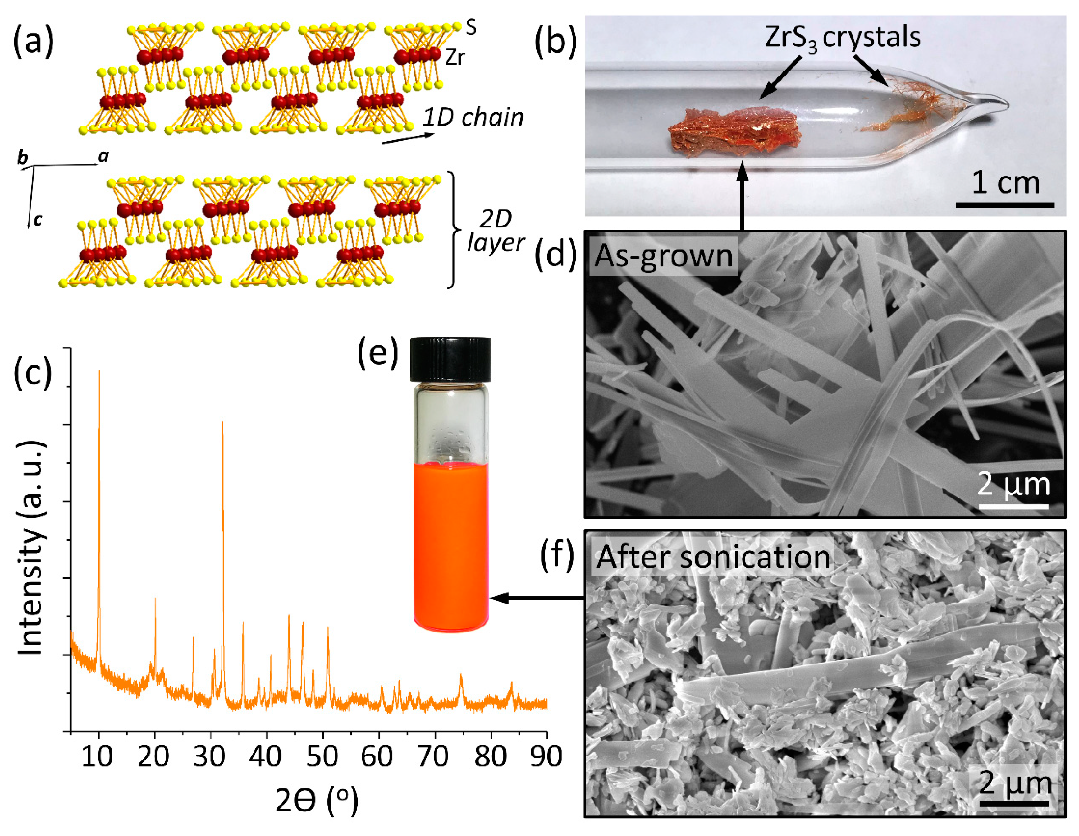
2. Materials and Methods

All chemicals were purchased from Sigma-Aldrich (USA) unless noted otherwise. ZrS3 crystals were grown by the direct reaction of stoichiometric amounts of Zr metal and S vapor in an evacuated quartz ampule at 800 °C for 48 h, as described in our previous work [39]. The crystals were characterized by X-ray diffraction (XRD) using a PANalytical Empyrean diffractometer (Netherlands) with Cu Kα radiation. Scanning electron microscopy (SEM) of ZrS3 crystals was performed using a FEI Nova NanoSEM 450 scanning electron microscope (USA) at the accelerating voltage of 5 kV. Raman spectroscopy was performed using a Thermo Scientific DXR Raman microscope (USA) with a 532 nm excitation laser and a 100× objective. XPS analysis was performed at room temperature using a Thermo Scientific K-Alpha X-ray photoelectron spectrometer (USA) with a monochromatic Al Kα (1486.6 eV) X-ray source and a low energy electron flood gun for charge compensation. High-resolution XPS spectra of Zr3d and S2p were collected using a pass energy of 20 eV and a 0.1 eV step. To collect XPS spectra of pristine ZrS3, as-grown crystals were cleaved in air directly before introduction into the loading vacuum chamber to minimize the contribution of surface contamination. XPS spectra of solution-exfoliated ZrS3 crystals were recorded on samples produced by drying droplets of the corresponding suspensions in air.
To prepare ZrS3 suspensions for nanotoxicity experiments, ZrS3 crystals were sonicated in physiological saline solution (PS; 9 g/L NaCl aqueous solution) for 20 min to produce a homogeneous orange suspension with a concentration of 1 g/L. Other suspensions were prepared by diluting this ZrS3 stock suspension to concentrations of 0.0001, 0.001, 0.01 and 0.1 g/L. The nanotoxicity experiments were performed on freshly prepared ZrS3 suspensions, which were analyzed less than 0.5 h after preparation and the suspensions that were stored for 24 h prior to the measurements.
The laboratory glassware for sample storage and biotesting was soaked in a mixture of potassium bichromate and sulfuric acid for about 3 h, then washed with deionized (DI) water, deacidified with a sodium bicarbonate solution and finally washed four times with DI water and dried in an oven.
The toxicity of ZrS3 suspensions was measured by a bioluminescence technique that is generally used for the assessment of toxic effects of nanomaterials. The procedure was similar to the widely used bioluminescence inhibition test with Vibrio fischeri [33,34,35,36,37]. We monitored changes in intensity of bioluminescence of genetically modified photoluminescent E. coli bacteria in the presence of ZrS3 nanoparticles or other tested chemicals compared to the emission of a control sample (same bacteria in a pure PS solution).
We used commercial recombinant E. coli K12 TG1 bacteria modified with luxCDABE genes of photoluminescent Photobacterium leiognathi 54D10 marine bacteria. Lyophilized E. coli K12 TG1 bacteria under the brand name Ecolum were purchased from NVO ImmunoTek (Russian Federation). Before the nanotoxicity experiments, the lyophilized bacteria were rehydrated in a PS solution at 4 °C for 30 min and then at room temperature for 1 h. The solutions used in this study had an E. coli concentration of about 2.5 × 10⁷ cells/mL. In a typical bioluminescence experiment, 0.1 mL of this E. coli solution was mixed with 0.9 mL of a ZrS3 suspension in PS. The resulting mixture was stored for 30 min prior to the measurements, which were performed using a NIKI MLT Biotox-10 luminometer (Russian Federation). The instrument detected bioluminescence of bacteria in the 300–600 nm spectral range with the maximum detector sensitivity between 380 and 490 nm. Parallel measurements of the studied and control samples were carried out. The control samples were prepared by diluting 0.1 mL of the E. coli stock solution with 0.9 mL of PS.
We compared the toxicity of ZrS3 with that of zirconyl chloride (ZrOCl2·8H2O), a common zirconium compound. Both compounds contain Zr4+, but while ZrS3 is practically insoluble in water and was present in the PS suspensions as partially exfoliated particles, zirconyl chloride is water soluble. We prepared 0.000172, 0.00172, 0.0172, 0.172 and 1.72 g/L solutions of ZrOCl2·8H2O in PS, which matched the 0.0001, 0.001, 0.01, 0.1 and 1 g/L ZrS3 suspensions, respectively, in zirconium concentrations. Then, 0.9 mL aliquots of zirconyl chloride solutions were mixed with 0.1 mL aliquots of the E. coli stock solution, and the resulting mixtures were stored for 30 min and used in bioluminescence experiments.
For the positive control we used sodium dichloroisocyanurate (C3Cl2N3NaO3, SDC), a compound with well-known toxicity that is widely used as an efficient disinfectant against Gram-positive and Gram-negative bacteria, viruses and fungi [40]. We prepared 0.0001, 0.001, 0.01, 0.1 and 1 g/L solutions of SDC in PS, and then, 0.9 mL aliquots of these solutions were mixed with 0.1 mL aliquots of the E. coli stock solution. The resulting mixtures were stored for 30 min and used in bioluminescence experiments. As in the experiments with ZrS3, the bioluminescence tests with ZrOCl2 and SDC involved control samples prepared by diluting 0.1 mL aliquots of the E. coli stock solution with 0.9 mL aliquots of PS.
Each bioluminescence experiment was performed at least 5 times, and the averaged results are presented.

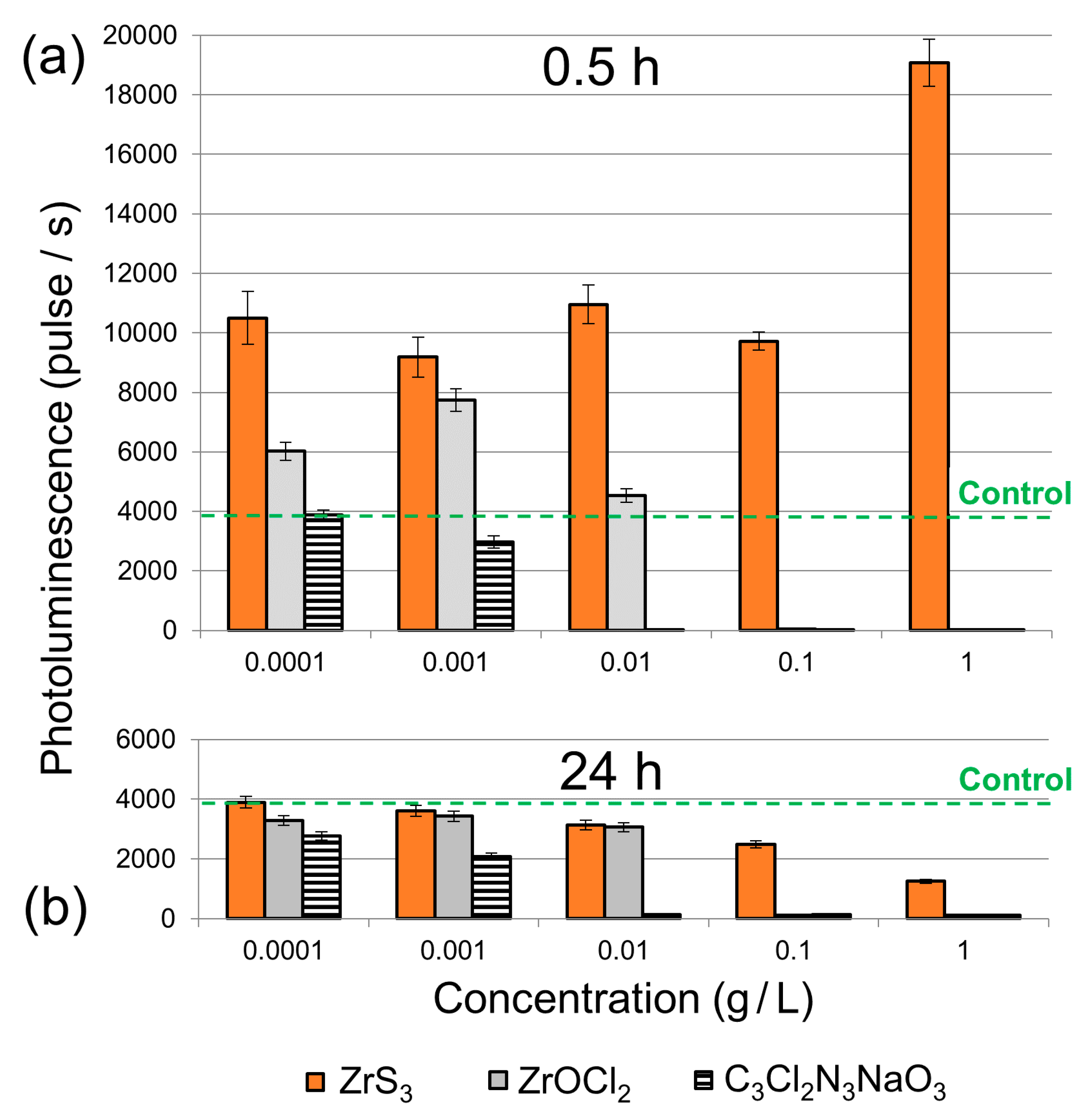
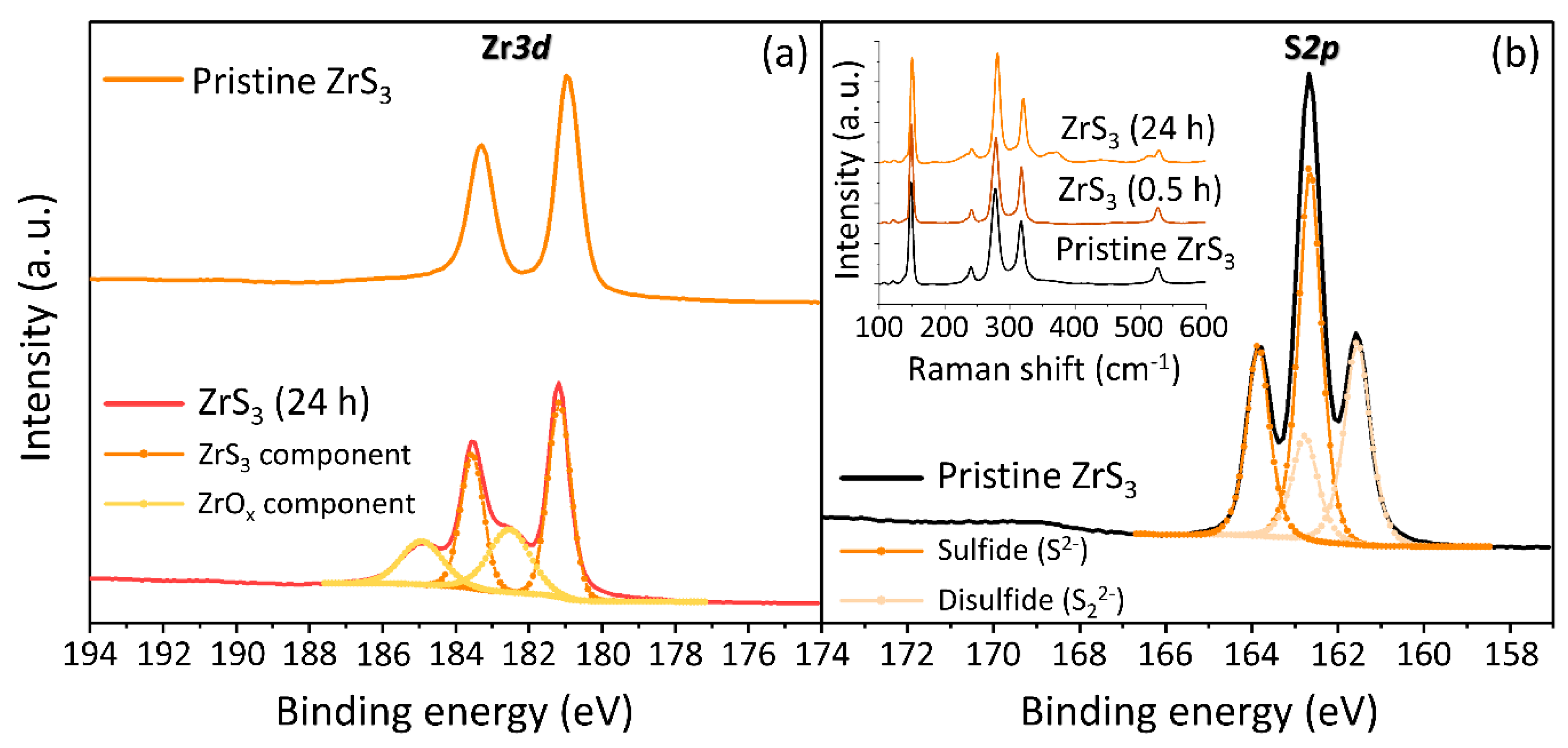
3. Results and Discussion

Figure 1b shows an optical photograph of a reaction ampule after the ZrS3 growth, which was performed through a direct reaction between a Zr foil and sulfur vapor at 800 °C. While some crystals form on the quartz surface (Figure 1b), most of the ZrS3 grows directly on a foil forming a spongy material comprising disordered ZrS3 nanobelt crystals. This material was highly crystalline, as illustrated by the XRD spectrum in Figure 1c. The peaks in the XRD spectrum are narrow and sharp, and their positions agree well with prior literature reports for ZrS3 crystals [27,31,39,41]. A representative SEM image of the ZrS3 crystals is shown in Figure 1d. The crystals had a nanobelt shape, which reflects their quasi-1D crystal structure (Figure 1a) with the b crystallographic direction of 1D chains corresponding to the long axes of the crystals. Most of the as-grown ZrS3 crystals were over 10 μm long. Figure 1e shows an optical photograph of a uniform orange suspension of exfoliated ZrS3 crystals in PS (1 g/L) that was used for the toxicity experiments. After the sonication, the average crystal size decreased, which is illustrated by the SEM image in Figure 1f (note that the SEM images of ZrS3 crystals before and after sonication are shown in panels d and f, respectively, at the same magnification). While some ZrS3 crystals retained their nanobelt shape, others were split by sonication into randomly shaped particles with sizes of about 1 μm.
The results of bioluminescence experiments are summarized in Figure 2. In both panels the emission of the control sample (E. coli in a pure PS solution) is indicated by the green dashed lines at 3754 pulses/s. As expected, SDC exhibited a strong toxic effect on E. coli bacteria, which showed decreased photoluminescence compared to the control sample for all SDC concentrations (Figure 2). The decrease in the bacterial activity was proportional to the SDC concentration.
For the zirconium compounds compared in this study, ZrS3 was generally less toxic than ZrOCl2 at all concentrations, regardless of whether the solutions were stored for 30 min or 24 h prior to their mixing with E. coli bacteria. In both cases, the ZrOCl2 solutions strongly suppressed the bacterial activities at concentrations above 0.01 g/L (Figure 2). The higher toxicity of ZrOCl2 compared to ZrS3, especially for the freshly prepared solutions (Figure 2a), can be explained by its solubility in aqueous media. Even though all these samples contained same amounts of zirconium, in the solutions of water-soluble ZrOCl2 all Zr(IV) was available for interaction with the bacteria in a form of aqueous complexes. On the contrary, since ZrS3 is insoluble in water most Zr(IV) in the suspensions was in the bulk of ZrS3 particles and thus did not directly interact with the bacteria.
The toxic effects were very different for the ZrS3 suspensions that were stored for 30 min or 24 h prior to their mixing with the bacteria. Freshly prepared ZrS3 suspensions did not exhibit any toxicity to the photoluminescent E. coli bacteria across the entire tested concentration range (Figure 2a). In fact, for all studied concentrations we observed a considerable stimulating activity of ZrS3 nanoparticles on the bioluminescence of E. coli, as all emission values were higher than for the control sample at 3754 pulses/s. However, the ZrS3 suspensions that were stored for 24 h prior to the experiment suppressed the bioluminescence of E. coli even at a concentration as low as 0.001 g/L (Figure 2b), indicating their high toxicity.
It should be noted that the ZrS3 suspensions that were stored for 24 h produced a mild but noticeable smell of H2S, which can form as a result of aqueous hydrolysis of ZrS3. Because of its well-known toxic effects, hydrogen disulfide forming in aqueous ZrS3 suspensions over time could be responsible for their increased toxicity. Since the hydrolysis of ZrS3 is accompanied by the formation of zirconium oxide (ZrOx) on the surface of nanoparticles, we investigated this process using XPS (Figure 3).
Figure 3a shows XPS Zr3d spectra of pristine ZrS3 crystals and the ZrS3 nanoparticles stored in an aqueous medium for 24 h. The top spectrum shows two peaks located at 181.16 eV and 183.55 eV, both of which can be assigned to ZrS3 [42]. The XPS Zr3d spectrum of the ZrS3 nanoparticles stored in an aqueous medium can be deconvoluted into two doublet peaks. In addition to the same doublet found in pristine ZrS3, the bottom spectrum in Figure 3a also shows the Zr3d3/2 and Zr3d5/2 components of ZrOx at 182.52 eV and 184.95 eV, respectively. The spin-orbit doublet splittings for ZrS3 and ZrOx are 2.39 eV and 2.43 eV, respectively [43]. Thus, these spectra confirm the formation of ZrOx on the surface of ZrS3 nanoparticles. The XPS S2p spectrum contains signals from sulfur located in the sulfide (S2−) and disulfide (S22−) species. The spectrum shown in Figure 3b was fitted using two doublet peaks. The binding energies of S2p1/2 and S2p3/2 for the sulfide are 161.55 eV and 162.65 eV, respectively. The disulfide group peaks are located at 162.76 eV and 163.85 eV. The intensity ratio of two sulfur components is a characteristic value for ZrS3 and should be equal to 2. The calculated ratio of the fitted S22−/S2− peaks is 1.85, which is in good agreement with literature data [42].
We also compared the as-grown ZrS3 crystals with the solution-exfoliated ZrS3 nanoparticles by Raman spectroscopy, see the inset in Figure 3b. The Raman spectrum of pristine ZrS3 crystals confirms their high quality and crystallinity, showing very sharp and well-resolved peaks at about 148, 277, 317 and 526 cm−1 in accordance with previous studies [32,44]. The spectrum of ZrS3 nanoparticles from a freshly prepared suspension is nearly identical to that of the as-grown crystals, demonstrating that while the sonication decreased the average size of particles (see Figure 1d,f), their structure and crystallinity were not much affected. The Raman spectrum of ZrS3 nanoparticles from a suspension that was stored for 24 h prior to the measurements also looks very similar to the other two spectra (see the inset in Figure 3b). While XPS, a surface-sensitive method, demonstrated the formation of ZrOx on the surface of ZrS3 nanoparticles, Raman spectroscopy shows that in bulk they retained their properties.
The XPS results confirm the formation of ZrOx due to the aqueous hydrolysis of ZrS3, which is accompanied by the evolution of H2S. It is plausible that toxic H2S is responsible for the increased toxicity of ZrS3 suspensions stored for 24 h (Figure 2b). The increased bioluminescence observed for the freshly prepared ZrS3 suspensions could also be related to the formation H2S that is initially formed in very small quantities. Other studies reported that low concentrations of hydrogen disulfide may exhibit stimulation of bacteria and protect them against oxidative stress [45,46], which is a common toxicity mechanism for nanoparticles. As the H2S concentration increases over the 24 h storage period, the ZrS3 suspensions become highly toxic to bacteria, which we observed experimentally.

4. Summary

This work reports the first nanotoxicity study of materials from a large TMTC family. More specifically, we investigated the toxicity of ZrS3, a prominent TMTC material, toward photoluminescent E. coli bacteria in a bioluminescence test. We investigated the solution exfoliation of ZrS3 and prepared aqueous suspensions of ZrS3 nanoparticles with concentrations ranging from 0.0001 to 1 g/L. We found that freshly prepared ZrS3 suspensions with concentrations as high as 1 g/L did not exhibit any toxic effects on the bacteria and, on the contrary, stimulated their activity. However, ZrS3 suspensions that were stored for 24 h prior to the bioluminescence tests were very toxic to the bacteria and inhibited their emission even at concentrations down to 0.001 g/L. We explain these observations by the aqueous hydrolysis of ZrS3, which resulted in the formation of ZrOx on the surface of nanoparticles and the release of toxic H2S. The formation of ZrOx was confirmed by the XPS analysis, while the characteristic H2S smell was noticeable for the 24 h suspensions. Hydrogen disulfide could be responsible for the bacterial stimulation in the freshly prepared ZrS3 suspensions, because at small concentrations H2S was shown to protect bacteria against oxidative stress [45,46], which is widely regarded as a common nanoparticle-induced toxicity mechanism. However, as more H2S was released during the hydrolysis of ZrS3 over a 24 h period, the suspensions became toxic to the bacteria.
This study demonstrates that ZrS3 suspensions, while initially nontoxic to photoluminescent E. coli bacteria, start exhibiting appreciable toxicity over time. The results of this study can likely be extended to other transition metal chalcogenides, as their toxicity in aqueous solutions may also increase over time due to hydrolysis and the formation of H2S. The results of this study also demonstrate that since many systems involving nanomaterials are unstable and evolve over time in various ways, their toxicity may evolve as well, which should be considered for relevant toxicity tests.

Author Contributions

Conceptualization, O.V.Z., A.A.G. and A.S.; formal analysis, O.V.Z., J.A., N.S.V., D.V.S., D.S.M., A.A.G. and A.S.; investigation, O.V.Z., J.A., N.S.V., D.V.S., D.S.M., A.A.G.; supervision, A.A.G., D.V.K. and A.S.; writing–original draft, A.A.G., O.V.Z. and A.S.; writing–review & editing, O.V.Z., J.A., N.S.V., D.S.M., D.V.K., A.A.G. and A.S. All authors have read and agreed to the published version of the manuscript.

Funding

The nanotoxicity experiments were supported by the Ministry of Education and Science of the Russian Federation through grant K2-2020-014. The research on the synthesis and characterization of ZrS3 employed the instrumentation at the Nebraska Nanoscale Facility, which is supported by Nebraska Research Initiative and the National Science Foundation (NSF) through ECCS-1542182.

Conflicts of Interest

The authors declare no conflicts of interest.

References

  1. Geim, A.K.; Grigorieva, I.V. Van der Waals heterostructures. Nature 2013, 499, 419–425. [Google Scholar] [CrossRef]
  2. Butler, S.Z.; Hollen, S.M.; Cao, L.; Cui, Y.; Gupta, J.A.; Gutiérrez, H.R.; Heinz, T.F.; Hong, S.S.; Huang, J.; Ismach, A.F.; et al. Progress, challenges, and opportunities in two-dimensional materials beyond graphene. ACS Nano 2013, 7, 2898–2926. [Google Scholar] [CrossRef]
  3. Bhimanapati, G.R.; Lin, Z.; Meunier, V.; Jung, Y.; Cha, J.; Das, S.; Xiao, D.; Son, Y.; Strano, M.S.; Cooper, V.R.; et al. Recent advances in two-dimensional materials beyond graphene. ACS Nano 2015, 9, 11509–11539. [Google Scholar] [CrossRef]
  4. Tan, E.; Li, B.L.; Ariga, K.; Lim, C.-T.; Garaj, S.; Leong, D.T. Toxicity of two-dimensional layered materials and their heterostructures. Bioconj. Chem. 2019, 30, 2287–2299. [Google Scholar] [CrossRef]
  5. Xu, S.; Li, D.; Wu, P. One-pot, facile, and versatile synthesis of monolayer MoS2/WS2 quantum dots as bioimaging probes and efficient electrocatalysts for hydrogen evolution reaction. Adv. Funct. Mater. 2015, 25, 1127–1136. [Google Scholar] [CrossRef]
  6. Yin, W.; Yan, L.; Yu, J.; Tian, G.; Zhou, L.; Zheng, X.; Zhang, X.; Yong, Y.; Li, J.; Gu, Z.; et al. High-throughput synthesis of single-layer MoS2 nanosheets as a near-infrared photothermal-triggered drug delivery for effective cancer therapy. ACS Nano 2014, 8, 6922–6933. [Google Scholar] [CrossRef] [PubMed]
  7. Chou, S.S.; Kaehr, B.; Kim, J.; Foley, B.M.; De, M.; Hopkins, P.E.; Huang, J.; Brinker, C.J.; Dravid, V.P. Chemically exfoliated MoS2 as near-infrared photothermal agents. Angew. Chem. Int. Ed. 2013, 52, 4160–4164. [Google Scholar] [CrossRef] [PubMed] [Green Version]
  8. Ha, H.D.; Han, D.J.; Choi, J.S.; Park, M.; Seo, T.S. Dual role of blue luminescent MoS2 quantum dots in fluorescence resonance energy transfer phenomenon. Small 2014, 10, 3858–3862. [Google Scholar] [CrossRef] [PubMed]
  9. Seabra, A.B.; Paula, A.J.; de Lima, R.; Alves, O.L.; Durán, N. Nanotoxicity of graphene and graphene oxide. Chem. Res. Toxicol. 2014, 27, 159–168. [Google Scholar] [CrossRef] [PubMed]
  10. Bianco, A. Graphene: Safe or toxic? The two faces of the medal. Angew. Chem. Int. Ed. 2013, 52, 4986–4997. [Google Scholar] [CrossRef] [PubMed]
  11. Furuseth, S.; Brattas, L.; Kjekshus, A. On the crystal structures of TiS3, ZrS3, ZrSe3, ZrTe3, HfS3, and HfSe3. Acta Chem. Scan. A 1975, 29, 623–631. [Google Scholar] [CrossRef] [Green Version]
  12. Srivastava, S.K.; Avasthi, B.N. Preparation, structure and properties of transition metal trichalcogenides. J. Mater. Sci. 1992, 27, 3693–3705. [Google Scholar] [CrossRef]
  13. Dai, J.; Li, M.; Zeng, X.C. Group IVB transition metal trichalcogenides: A new class of 2D layered materials beyond graphene. Wiley Interdiscip. Rev. Comput. Mol. Sci. 2016, 6, 211–222. [Google Scholar] [CrossRef]
  14. Island, J.O.; Molina-Mendoza, A.J.; Barawi, M.; Biele, R.; Flores, E.; Clamagirand, J.M.; Ares, J.R.; Sánchez, C.; van der Zant, H.S.; D’Agosta, R.; et al. Electronics and optoelectronics of quasi-1D layered transition metal trichalcogenides. 2D Mater. 2017, 4, 022003. [Google Scholar] [CrossRef] [Green Version]
  15. Island, J.O.; Barawi, M.; Biele, R.; Almazán, A.; Clamagirand, J.M.; Ares, J.R.; Sánchez, C.; van der Zant, H.S.J.; Álvarez, J.V.; D’Agosta, R.; et al. TiS3 transistors with tailored morphology and electrical properties. Adv. Mater. 2015, 27, 2595–2601. [Google Scholar] [CrossRef] [Green Version]
  16. Lipatov, A.; Wilson, P.M.; Shekhirev, M.; Teeter, J.D.; Netusil, R.; Sinitskii, A. Few-layered titanium trisulfide (TiS3) field-effect transistors. Nanoscale 2015, 7, 12291–12296. [Google Scholar] [CrossRef] [Green Version]
  17. Molina-Mendoza, A.J.; Island, J.O.; Paz, W.S.; Clamagirand, J.M.; Ares, J.R.; Flores, E.; Leardini, F.; Sánchez, C.; Agraït, N.; Rubio-Bollinger, G.; et al. High current density electrical breakdown of TiS3 nanoribbon-based field-effect transistors. Adv. Funct. Mater. 2017, 27, 1605647. [Google Scholar] [CrossRef] [Green Version]
  18. Yi, H.; Komesu, T.; Gilbert, S.; Hao, G.; Yost, A.J.; Lipatov, A.; Sinitskii, A.; Avila, J.; Chen, C.; Asensio, M.C.; et al. The band structure of the quasi-one-dimensional layered semiconductor TiS3(001). Appl. Phys. Lett. 2018, 112, 052102. [Google Scholar] [CrossRef]
  19. Gilbert, S.J.; Lipatov, A.; Yost, A.J.; Loes, M.J.; Sinitskii, A.; Dowben, P.A. The electronic properties of Au and Pt metal contacts on quasi-one-dimensional layered TiS3(001). Appl. Phys. Lett. 2019, 114, 101604. [Google Scholar] [CrossRef]
  20. Randle, M.; Lipatov, A.; Kumar, A.; Kwan, C.-P.; Nathawat, J.; Barut, B.; Yin, S.; He, K.; Arabchigavkani, N.; Dixit, R.; et al. Gate-controlled metal–insulator transition in TiS3 nanowire field-effect transistors. ACS Nano 2019, 13, 803–811. [Google Scholar] [CrossRef]
  21. Island, J.O.; Buscema, M.; Barawi, M.; Clamagirand, J.M.; Ares, J.R.; Sánchez, C.; Ferrer, I.J.; Steele, G.A.; van der Zant, H.S.J.; Castellanos-Gomez, A. Ultrahigh photoresponse of few-layer TiS3 nanoribbon transistors. Adv. Opt. Mater. 2014, 2, 641–645. [Google Scholar] [CrossRef] [Green Version]
  22. Cui, Q.; Lipatov, A.; Wilt, J.S.; Bellus, M.Z.; Zeng, X.C.; Wu, J.; Sinitskii, A.; Zhao, H. Time-resolved measurements of photocarrier dynamics in TiS3 nanoribbons. ACS Appl. Mater. Interfaces 2016, 8, 18334–18338. [Google Scholar] [CrossRef] [PubMed]
  23. Hayashi, A.; Matsuyama, T.; Sakuda, A.; Tatsumisago, M. Amorphous titanium sulfide electrode for all-solid-state rechargeable lithium batteries with high capacity. Chem. Lett. 2012, 41, 886–888. [Google Scholar] [CrossRef]
  24. Zhang, J.; Liu, X.; Wen, Y.; Shi, L.; Chen, R.; Liu, H.; Shan, B. Titanium trisulfide monolayer as a potential thermoelectric material: A first-principles-based Boltzmann transport study. ACS Appl. Mater. Interfaces 2017, 9, 2509–2515. [Google Scholar] [CrossRef]
  25. Morozova, N.V.; Korobeinikov, I.V.; Kurochka, K.V.; Titov, A.N.; Ovsyannikov, S.V. Thermoelectric properties of compressed titanium and zirconium trichalcogenides. J. Phys. Chem. C 2018, 122, 14362–14372. [Google Scholar] [CrossRef]
  26. Lipatov, A.; Loes, M.J.; Lu, H.; Dai, J.; Patoka, P.; Vorobeva, N.S.; Muratov, D.S.; Ulrich, G.; Kästner, B.; Hoehl, A.; et al. Quasi-1D TiS3 nanoribbons: Mechanical exfoliation and thickness-dependent Raman spectroscopy. ACS Nano 2018, 12, 12713–12720. [Google Scholar] [CrossRef]
  27. Muratov, D.S.; Ishteev, A.R.; Lypenko, D.A.; Vanyushin, V.O.; Gostishev, P.; Perova, S.; Saranin, D.S.; Rossi, D.; Auf der Maur, M.; Volonakis, G.; et al. Slot-die printed two-dimensional ZrS3 charge transport layer for perovskite light-emitting diodes. ACS Appl. Mater. Interfaces 2019, 11, 48021–48028. [Google Scholar] [CrossRef]
  28. Giagloglou, K.; Payne, J.L.; Crouch, C.; Gover, R.K.B.; Connor, P.A.; Irvine, J.T.S. Zirconium trisulfide as a promising cathode material for Li primary thermal batteries. J. Electrochem. Soc. 2016, 163, A3126–A3130. [Google Scholar] [CrossRef] [Green Version]
  29. Wu, J.-J.; Tao, Y.-R.; Wang, J.-N.; Wu, Z.-Y.; Fan, L.; Wu, X.-C. Reverse saturable absorption and nonlinear refraction of ultrathin ZrS3 nanobelts. Nanoscale 2016, 8, 10371–10379. [Google Scholar] [CrossRef]
  30. Abdulsalam, M.; Joubert, D.P. Electronic and optical properties of MX3 (M = Ti, Zr and Hf; X = S, Se) structures: A first principles insight. Phys. Status Solidi B 2016, 253, 868–874. [Google Scholar] [CrossRef]
  31. Yi, H.; Gilbert, S.J.; Lipatov, A.; Sinitskii, A.; Avila, J.; Abourahma, J.; Komesu, T.; Asensio, M.C.; Dowben, P.A. The electronic band structure of quasi-one-dimensional van der Waals semiconductors: The effective hole mass of ZrS3 compared to TiS3. J. Phys. Condens. Matter 2020, 32, 29LT01. [Google Scholar] [CrossRef] [PubMed]
  32. Pant, A.; Torun, E.; Chen, B.; Bhat, S.; Fan, X.; Wu, K.; Wright, D.P.; Peeters, F.M.; Soignard, E.; Sahin, H.; et al. Strong dichroic emission in the pseudo one dimensional material ZrS3. Nanoscale 2016, 8, 16259–16265. [Google Scholar] [CrossRef] [PubMed] [Green Version]
  33. Backhaus, T.; Froehner, K.; Altenburger, R.; Grimme, L.H. Toxicity testing with Vibrio fischeri: A comparison between the long term (24 h) and the short term (30 min) bioassay. Chemosphere 1997, 35, 2925–2938. [Google Scholar] [CrossRef]
  34. Lopes, I.; Ribeiro, R.; Antunes, F.E.; Rocha-Santos, T.A.P.; Rasteiro, M.G.; Soares, A.M.V.M.; Gonçalves, F.; Pereira, R. Toxicity and genotoxicity of organic and inorganic nanoparticles to the bacteria Vibrio fischeri and Salmonella typhimurium. Ecotoxicology 2012, 21, 637–648. [Google Scholar] [CrossRef]
  35. Garcia, A.; Recillas, S.; Sánchez, A.; Font, X. The luminescent bacteria test to determine the acute toxicity of nanoparticle suspensions. In Nanotoxicity: Methods and Protocols; Reineke, J., Ed.; Humana Press: Totowa, NJ, USA, 2012; pp. 255–259. [Google Scholar]
  36. Mogilnaya, O.A.; Puzyr’, A.P.; Bondar’, V.S. Growth and bioluminescence of luminous bacteria under the action of aflatoxin B1 before and after its treatment with nanodiamonds. Appl. Biochem. Microbiol. 2010, 46, 33–37. [Google Scholar] [CrossRef]
  37. Zarubina, A.P.; Lukashev, E.P.; Deev, L.I.; Parkhomenko, I.M.; Rubin, A.B. Biotesting the biological effects of single-wall carbon nanotubes using bioluminescent bacteria test-system. Nanotechnol. Russ. 2009, 4, 871–875. [Google Scholar] [CrossRef]
  38. Gusev, A.; Zakharova, O.; Muratov, D.S.; Vorobeva, N.S.; Sarker, M.; Rybkin, I.; Bratashov, D.; Kolesnikov, E.; Lapanje, A.; Kuznetsov, D.V.; et al. Medium-dependent antibacterial properties and bacterial filtration ability of reduced graphene oxide. Nanomaterials 2019, 9, 1454. [Google Scholar] [CrossRef] [Green Version]
  39. Muratov, D.S.; Vanyushin, V.O.; Vorobeva, N.S.; Jukova, P.; Lipatov, A.; Kolesnikov, E.A.; Karpenkov, D.; Kuznetsov, D.V.; Sinitskii, A. Synthesis and exfoliation of quasi-1D (Zr,Ti)S3 solid solutions for device measurements. J. Alloys Compd. 2020, 815, 152316. [Google Scholar] [CrossRef]
  40. Clasen, T.; Edmondson, P. Sodium dichloroisocyanurate (NaDCC) tablets as an alternative to sodium hypochlorite for the routine treatment of drinking water at the household level. Int. J. Hyg. Environ. Health 2006, 209, 173–181. [Google Scholar] [CrossRef]
  41. Haraldsen, H.; Rost, E.; Kjekshus, A.; Steffens, A. On properties of TiS3, ZrS3, and HfS3. Acta Chem. Scand. 1963, 17, 1283–1292. [Google Scholar] [CrossRef] [Green Version]
  42. Jellinek, F.; Pollak, R.A.; Shafer, M.W. X-ray photoelectron spectra and electronic structure of zirconium trisulfide and triselenide. Mater. Res. Bull. 1974, 9, 845–856. [Google Scholar] [CrossRef]
  43. Crist, B.V. Handbooks of Monochromatic XPS Spectra. Vol. 2. Commercially Pure Binary Oxides; XPS International LLC: Mountain View, CA,USA, 2007. [Google Scholar]
  44. Sourisseau, C.; Mathey, Y. The infrared, Raman, resonance Raman spectra and the valence force field of the ZrS3 layer-type compound. Chem. Phys. 1981, 63, 143–156. [Google Scholar] [CrossRef]
  45. Mironov, A.; Seregina, T.; Nagornykh, M.; Luhachack, L.G.; Korolkova, N.; Lopes, L.E.; Kotova, V.; Zavilgelsky, G.; Shakulov, R.; Shatalin, K.; et al. Mechanism of H2S-mediated protection against oxidative stress in Escherichia coli. Proc. Natl. Acad. Sci. USA 2017, 114, 6022–6027. [Google Scholar] [CrossRef] [PubMed] [Green Version]
  46. Kimura, H. Production and physiological effects of hydrogen sulfide. Antioxid. Redox Signal. 2014, 20, 783–793. [Google Scholar] [CrossRef] [Green Version]
Figure 1. (a) Crystal structure of ZrS3 (P 21/m space group); (b) optical photograph of an evacuated quartz ampule with orange ZrS3 crystals indicated by the arrows; (c) XRD spectrum of ZrS3 crystals; (d) SEM image of the as-grown ZrS3 crystals shown by the arrow in panel b; (e) optical photograph of a vial with 1 g/L suspension of exfoliated ZrS3 crystals, which were dispersed in PS solution by sonication; (f) SEM image of ZrS3 particles after sonication in PS solution.
Nanomaterials 10 01401 g001
Figure 2. E. coli luminescence values in (a) as-prepared (0.5 h) and (b) 24 h solutions of ZrS3, ZrOCl2 and C3Cl2N3NaO3. For ZrOCl2 solutions, the concentrations were 0.000172, 0.00172, 0.0172, 0.172 and 1.72 g/L, so that they matched the 0.0001, 0.001, 0.01, 0.1 and 1 g/L ZrS3 suspensions in the zirconium concentrations, respectively.
Nanomaterials 10 01401 g002
Figure 3. Spectroscopic characterization of ZrS3 crystals before and after sonication. (a) Comparison of XPS Zr3d spectra of pristine ZrS3 crystals (top) and ZrS3 nanoparticles stored in water for 24 h (bottom); (b) XPS S2p spectrum of pristine ZrS3. Inset shows Raman spectra of pristine ZrS3 crystals, ZrS3 nanoparticles from a freshly prepared suspension (0.5 h), and ZrS3 nanoparticles stored in an aqueous medium for 24 h.
Nanomaterials 10 01401 g003

