
Europium(III) Complex-Functionalized SiO2@mTiO2 Nanospheres for Al3+-Modulated Multicolor Emission

1
Key Laboratory of New Energy & New Functional Materials, Shaanxi Key Laboratory of Chemical Reaction Engineering, College of Chemistry and Chemical Engineering, Yan’an University, Yan’an 716000, China
2
Key Laboratory of Synthetic and Natural Functional Molecule of the Ministry of Education, College of Chemistry and Materials Science, Northwest University, Xi’an 710127, China
*
Authors to whom correspondence should be addressed.
Nanomaterials 2021, 11(11), 2886; https://0-doi-org.brum.beds.ac.uk/10.3390/nano11112886
Submission received: 9 October 2021
/
Revised: 19 October 2021
/
Accepted: 25 October 2021
/
Published: 28 October 2021
Abstract
:A europium(III) hybrid material Eu(tta)3bpdc-SiO2@mTiO2 (Htta = 2-thenoyltrifluoroacetone, H2bpdc = 2,2′-bipyridine-3,3′-dicarboxylic acid) was successfully designed and synthesized by the covalent grafting complex Eu(tta)3bpdc to SiO2@mTiO2 core–shell nanosphere. The FT-IR, PXRD, XPS, TEM, HRTEM, SAED, TGA and PL were performed to characterize these materials. The results indicate that core–shell nanosphere structure and anatase crystallites of SiO2@mTiO2 are retained well after grafting the europium complex. Hybrid material Eu(tta)3bpdc-SiO2@mTiO2 displays uniform nanosphere structure, bright red color and long lifetime, which can serve as a multicolor emission material modulated by using Al3+ ions via the cation exchange approach under a single-wavelength excitation. To the best of our knowledge, this work is the first multicolor emissive sensor for Al3+ ions based on the lanthanide hybrid material.

1. Introduction
Lanthanide-based luminescent hybrid materials have sharp emission bands, high color purity, long luminescent lifetime and high quantum efficiency. They can also act as excellent candidates for multicolor luminescent materials, and further are applied in many fields, such as bio-imaging and bioanalytical detection [1,2], luminescent probes and sensors [3,4,5], tunable luminescence [6,7,8], and optoelectronic devices and displays [9,10,11]. In the past years, color-tunable lanthanide-based materials usually control the stoichiometric ratios of doped ions in the same material to realize multicolor emission (containing white-light emission) [12,13,14,15]. Recently, Li et al. have achieved some progress in the field of single-component lanthanide-based hybrid materials with multicolor emission [16,17]. They declared that full-color emission, particularly white emission colors of europium-based hybrid materials, can be facilely tuned by using only a kind of cation or anion due to the cation exchange process or interaction between ligands and anions. However, to date, single-component lanthanide hybrid materials with multicolor emission by using the cation exchange approach remain a challenge.
Aluminum is the third most abundant metal in the Earth’s crust [18], it has been widely used in the fields of aluminum containers, cooking tools, food additives, water treatment, and aluminum-based pharmaceuticals. According to the standard of WHO, the average human intake of aluminum is about 3.0~10.0 mg/day and weekly dietary intake is 7.0 mg/kg body weight [19,20,21]. The excessive intake of aluminum can lead to the production of neurotoxic issues in humans such as Parkinson’s or Alzheimer’s disease [18,22]. Therefore, the selective detection and control of Al3+ ions in biological systems are essential. Fluorescence-based probes as a kind of simple and rapid responsive method have drawn a lot of interest for sensing applications due to these methods being low-cost, very sensitive, selective and visualizable while offering rapid response times and real-time monitoring. To date, several fluorescent Al3+ sensors such as organic compounds (Schiff base [23], phenylhydrazine carboxamide [24], coumarin-rhodamine derivative [25], amino derivative [26]), Cd-MOF [27] and europium complexes [28,29] have been reported. Noticeably, lanthanide-based hybrid materials have received considerable attention as a sensor for detecting Zn2+ [16] and Cu2+ [30], due to their novel and unique properties, such as photostability, adjustable pore size, large surface area, as well as easy recyclability.
On the basis of above consideration, we designed and fabricated a europium(III) hybrid material based on SiO2@mTiO2 core–shell nanosphere (namely, Eu(tta)3bpdc-SiO2@mTiO2) with covalent grafting complex Eu(tta)3bpdc (Htta = 2-thenoyltrifluoroacetone, H2bpdc = 2,2′-bipyridine-3,3′-dicarboxylic acid). The resulting core–shell hybrid material Eu(tta)3bpdc-SiO2@mTiO2 shows multicolor emission based on sensing of Al3+ ions by the cation exchange process. The emission color changed from red to pink and blue based on luminescence intensity changes by modulating only the concentration of Al3+ ions in an ethanol solution of hybrid material Eu(tta)3bpdc-SiO2@mTiO2. To the best of our knowledge, this work is the first multicolor emissive sensor for Al3+ ions based on the lanthanide hybrid material.
2. Experiment Section
2.1. Materials
1,10-Phenanthroline monohydrate (phen, AR, Aladdin, Shanghai, China), 2-thenoyltrifluoroacetone (Htta, AR, J&K, Beijing, China), tetraethyl orthosilicate (TEOS, Si(OC2H5)4, AR, J&K, Beijing, China), hexadecylamine (HDA, AR, J&K, Beijing, China), titanium(IV) isopropoxide (Tip, AR, J&K, Beijing, China) were purchased from a commercial source without any further purification. Hydrated EuCl3 was prepared by dissolving europium(III) oxide (99.99%) in concentrated hydrochloric acid.
2.2. Preparation of H2bpdc Ligand
To deionized water (350 mL), phen (0.04 mol, 8.00 g), NaOH (0.08 mol, 3.20 g) and KMnO4 (0.12 mol, 19.09 g) were added. The mixture was heated to 100~105 °C and stirred for 5 h, and then cooled to room temperature. The filtrate was obtained by filtration and then concentrated to 100 mL and adjusted the pH of the filtrate to 2.0 by HCl (aq, conc.). The resulting white precipitate was filtered off, recrystallized with methanol and dried in vacuo to give colorless needle-like crystals, yield: 8.2 g, 85%. Elemental Anal. Calc. for C12H8N2O4 (244.05): C, 59.02; H, 3.30; N, 11.47%. Found: C, 59.12; H, 3.15; N, 11.54%.
2.3. Preparation of Eu(tta)3(H2O)2
The complex Eu(tta)3(H2O)2 was synthesized according to the reported literature [31]. An appropriate amount of 1.0 M sodium hydroxide solution was added to an Htta ethanol solution to adjust pH = 7.0 under stirring at room temperature. Subsequently, a hydrated EuCl3 ethanol solution was dropwise added into the above mixture and kept the molar ratio of EuIII/Htta equal to 1/3. An appropriate amount of deionized water was added to the mixture and heated at 65 °C for 5 h, then cooled to room temperature. The yellow precipitates were obtained by filtration, then washed with cold ethanol and deionized water three times.
2.4. Preparation of SiO2@mTiO2 Core–Shell Nanospheres
The SiO2@mTiO2 core–shell nanospheres were synthesized according to the reported literature [32,33]. (i) SiO2 nanospheres were prepared by a modified Stöber method. Deionized water (5 mL) and 25% NH3·H2O (3 mL) were added to an ethanol solution (35 mL), and stirred continuously for 0.5 h at 25 °C. Then, TEOS (5 mL) was added dropwise to the above mixture and stirring continued for 24 h. The resultant SiO2 nanospheres were collected by centrifugation, then washed with deionized water and ethanol and dried at 60 °C for 24 h under vacuum. (ii) The SiO2@mTiO2 core–shell nanospheres were prepared by a modified cooperative assembly-directed method. Firstly, SiO2 nanospheres (150 mg) were dispersed in ethanol (15 mL) by ultrasonication, then 25% NH3·H2O (0.3 mL) and HDA (150 mg) were added under stirring at room temperature. Secondly, Tip (0.3 mL) was quickly added to the above mixture and continued to stir for 1 h. The resultant core–shell SiO2@TiO2/HDA nanospheres were collected by centrifugation, then washed with deionized water and ethanol. Thirdly, SiO2@TiO2/HDA nanospheres were dispersed in a mixture of deionized water (10 mL) and ethanol (20 mL) by ultrasonication, then transferred to a 100 mL Teflon-lined autoclave and heated to 160 °C for 36 h to completely remove the HDA. Finally, the resultant SiO2@mTiO2 core–shell nanospheres were collected by centrifugation, then washed with deionized water and ethanol, dried at 60 °C for 24 h under vacuum.
2.5. Preparation of SiO2@mTiO2-bpdc
To DMF (20 mL), SiO2@mTiO2 (0.10 g) and H2bpdc (0.30 g) were dispersed by ultrasonication, followed by refluxed at 160 °C for 6 h under stirring. The resultant SiO2@mTiO2-bpdc was collected by centrifugation, then washed with DMF and ethanol, dried at 60 °C for 24 h under vacuum.
2.6. Preparation of Hybrid Material Eu(tta)3bpdc-SiO2@mTiO2
To ethanol (20 mL), SiO2@mTiO2-bpdc (0.10 g) and Eu(tta)3(H2O)2 (0.5 mmol) were dispersed by ultrasonication, followed by reflux at 80 °C for 5 h under stirring. The resultant Eu(tta)3bpdc-SiO2@mTiO2 was collected by centrifugation and excess unbound Eu(tta)3(H2O)2 was washed with ethanol until no red emission of supernatant was observed under a 254 nm UV lamp, dried at 60 °C for 24 h under vacuum.
2.7. Luminescent Sensing Experiment
Hybrid material Eu(tta)3bpdc-SiO2@mTiO2 (3 mg) was dispersed in 3 mL 0.01 M different cations ethanol solutions (K+, Na+, Li+, Zn2+, Ca2+, Cd2+, Pb2+, Mg2+, Mn2+, Cr3+ and Al3+) at room temperature. Then, hybrid material Eu(tta)3bpdc-SiO2@mTiO2 suspensions were obtained by sonicating for 30 min for luminescent measurements.
2.8. Characterization
Fourier transform infrared (FT-IR) spectra were measured with Bruker Tensor 27 FT-IR spectrometer (Bruker physik-AG, Karlsruhe, Germany) by using KBr pellets in the range of 400–4000 cm−1. Powder X-ray diffraction (PXRD) patterns were performed with Bruker D8 ADVANCE diffractometer (Bruker physik-AG) (Cu-Kα, 1.5418 Å) at room temperature. Scanning electron microscopy (SEM) was recorded on SU8010 (Hitachi Limited, Tokyo, Japan). Transmission electron microscopy (TEM), high-resolution transmission electron microscopy (HRTEM), selected area electron diffraction (SAED) and the energy-dispersive X-ray spectroscopy (EDS) were recorded on Talos F200X (Thermo Fisher Scientific, Waltham, MA, USA). Thermogravimetric analyses (TGA) were carried out Universal V4.5A instrument (Universal Instrument, Conklin, NY, USA). Photoluminescence spectra (PL) were measured on the Hitachi F-2700 spectrophotometer (Hitachi Limited) at room temperature. The lifetimes of the samples were recorded on an Edinburgh FLSp920 fluorescence spectrometer (Edinburgh Instruments, Livingston, UK). X-ray photoelectron spectra (XPS) were measured on PHI5000 VersaProbeIII XPS (ULVAC Co. Ltd, Kanagawa, Japan) with an Al-Kα achromatic X-ray source. Inductively coupled plasma mass spectroscopy (ICP-MS) was performed on an Agilent 7900 ICP-MS (Agilent Technologies, Santa Clara, CA, USA).
3. Results and Discussion
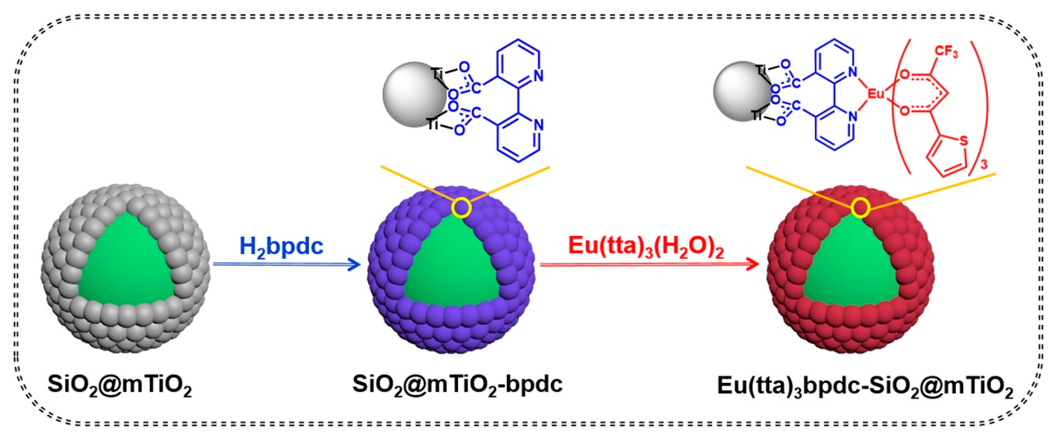
The preparation procedure and predicted structure of core–shell hybrid material Eu(tta)3bpdc-SiO2@mTiO2 are exhibited in Scheme 1. The choice of H2bpdc ligand is very important during the reaction process. The –COOH groups of H2bpdc ligand can covalently anchor onto the surface of SiO2@mTiO2 core–shell nanosphere through condensation reaction between hydroxyl groups on the surface of SiO2@mTiO2 and carboxylic groups. Meanwhile, two pyridyl N atoms of H2bpdc can coordinate to EuIII ions in the first coordination sphere of Eu(tta)3(H2O)2 complex by ligand-exchange reaction and then sensitize luminescence of EuIII ions with the primary ligand Htta via the “antenna effect”. Finally, complex Eu(tta)3bpdc can be successfully formed and covalently bonded to SiO2@mTiO2 core–shell nanospheres, then the hybrid material Eu(tta)3bpdc-SiO2@mTiO2 was successfully obtained.
3.1. FT-IR Spectra
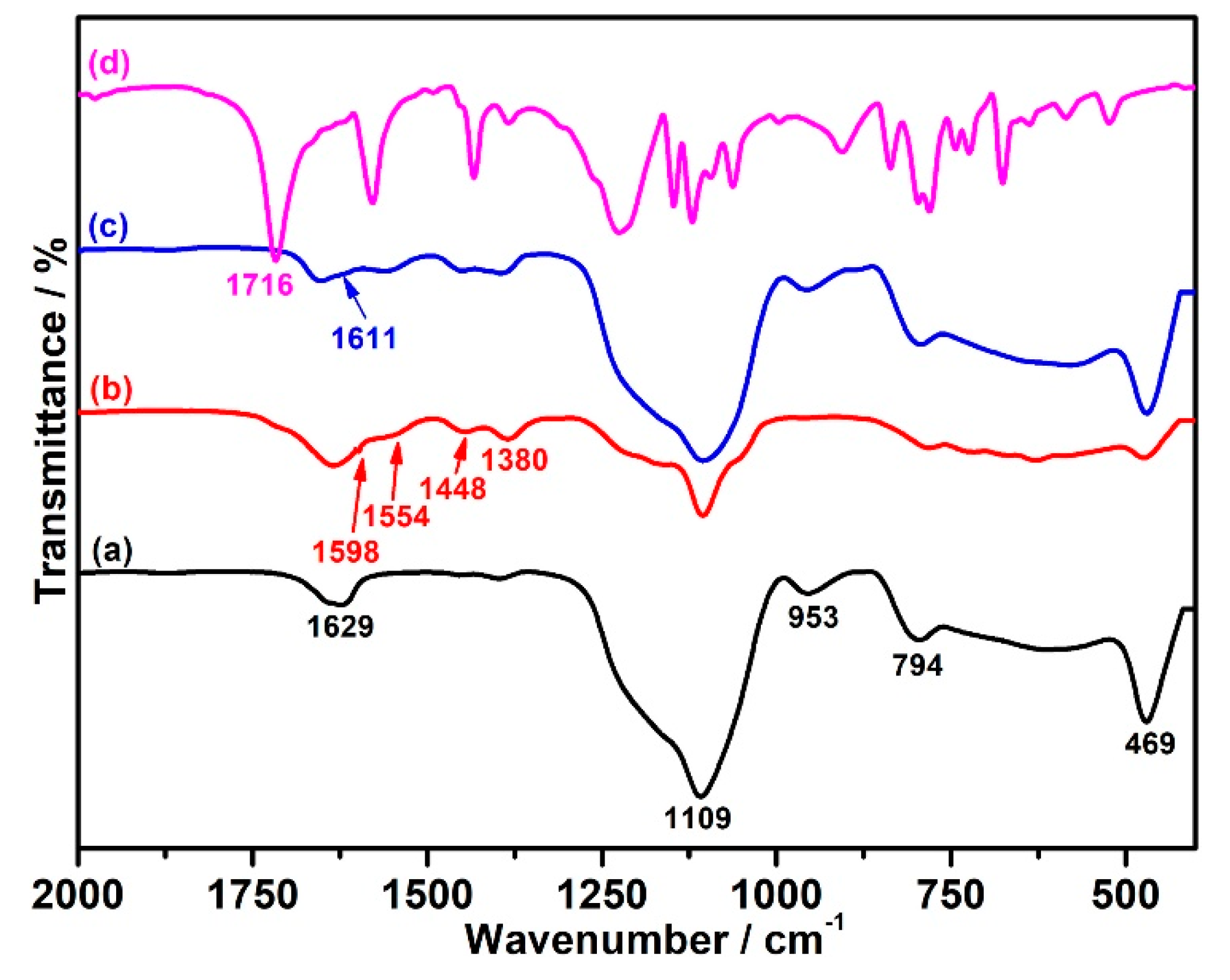
In order to confirm complex Eu(tta)3 successfully graft on the surface of SiO2@mTiO2 core–shell nanospheres, FT-IR spectra of H2bpdc ligand, SiO2@mTiO2, SiO2@mTiO2-bpdc and hybrid material Eu(tta)3bpdc-SiO2@mTiO2 were measured and shown in Figure 1 and Figure S1 and Table S1. In the spectra of SiO2@mTiO2 (Figure 1a), SiO2@mTiO2-bpdc (Figure 1b) and hybrid material Eu(tta)3bpdc-SiO2@mTiO2 (Figure 1c), the characteristic bands of Ti–O network can be observed in the range of 450~750 cm−1. In addition, the vibrational bands of Si–O asymmetric stretching vibration and symmetric stretching vibration appear at 1109 and 794 cm−1, respectively. While the peak at 953 cm−1 is ascribed to stretching vibration of the Si–OH surface groups. As shown in Figure 1d, H2bpdc ligand displays a strong band at 1716 cm−1 because of the stretching of carboxylic groups. The stretching of carboxylic groups is not found in the spectra of SiO2@mTiO2-bpdc (Figure 1b) and hybrid material Eu(tta)3bpdc-SiO2@mTiO2 (Figure 1c), indicating that the carboxylic groups of H2bpdc in SiO2@mTiO2-bpdc and Eu(tta)3bpdc-SiO2@mTiO2 were completely deprotonated. Meanwhile, the νasym(COO−) and νsym (COO−) stretching vibration of carboxylate appear at 1554 and 1380 cm−1 and the difference (Δ = νasym − νsym = 174 cm−1) of carboxylate stretching frequencies can be used to identify the bonding mode, further indicating that carboxylate groups form bidentate mode with the titania in SiO2@mTiO2 nanospheres [34,35,36,37,38]. In addition, the position of stretching vibration of C=N of H2bpdc ligand (1611 cm−1) in Eu(tta)3bpdc-SiO2@mTiO2 has shifted compared with that in SiO2@mTiO2-bpdc (1598 cm−1), indicating that a possible coordination bond of Eu–N may be formed in hybrid material Eu(tta)3bpdc-SiO2@mTiO2. The results above confirm that complex Eu(tta)3 successfully graft on the surface of SiO2@mTiO2 core–shell nanospheres by a carboxylic functionalized bipyridyl ligand.
3.2. PXRD
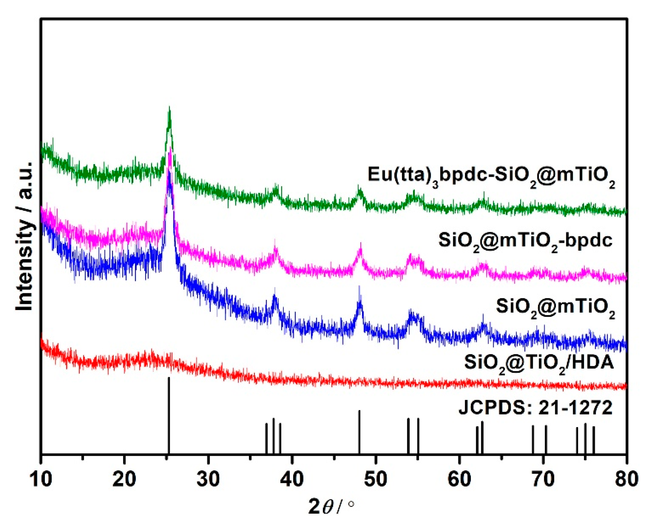
The PXRD patterns of SiO2@TiO2/HDA, SiO2@mTiO2, SiO2@mTiO2-bpdc and Eu(tta)3bpdc-SiO2@mTiO2 are shown in Figure 2. The pattern of SiO2@TiO2/HDA is completely amorphous, while that of SiO2@mTiO2 exhibits an anatase phase (JCPDS card No. 21-1272), indicating that the solvothermal method can not only remove the surfactant (HDA) in material but also change the crystal phase of TiO2 in the shell. According to the Scherrer formula, the average crystallite size of TiO2 in core–shell material can be estimated to be about 22 nm. The characteristic diffraction peaks of SiO2@mTiO2 appear at 25.27, 37.84, 48.03, 54.23, 55.04, 62.64, 69.32, 71.29 and 75.63° and correspond to the (101), (004), (200), (105), (211), (204), (116), (220) and (215) diffractions of anatase phase (JCPDS card No. 21-1272), respectively. In addition, characteristic diffraction peaks of SiO2@mTiO2-bpdc and Eu(tta)3bpdc-SiO2@mTiO2 (Table S2) are nearly unchanged compared with those of SiO2@mTiO2, indicating that the anatase phase was maintained well after complex Eu(tta)3bpdc was introduced.
3.3. SEM Images
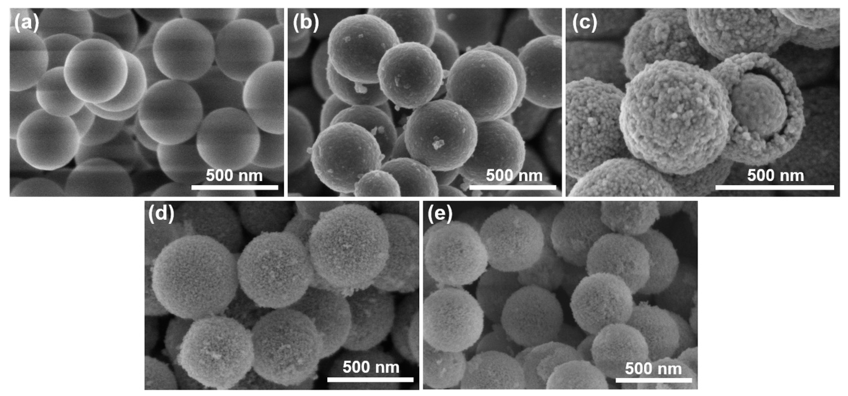
The SEM images of SiO2, SiO2@TiO2/HDA, SiO2@mTiO2, SiO2@mTiO2-bpdc and Eu(tta)3bpdc-SiO2@mTiO2 are displayed in Figure 3. It can be observed that SiO2, SiO2@TiO2/HDA, SiO2@mTiO2, SiO2@mTiO2-bpdc and Eu(tta)3bpdc-SiO2@mTiO2 show monodisperse spherical structures. The average diameter of SiO2 is about 350 nm, and that of SiO2@TiO2/HDA, SiO2@mTiO2, SiO2@mTiO2-bpdc and Eu(tta)3bpdc-SiO2@mTiO2 is about 430 nm, the thickness of the shell in the core–shell structure is about 40 nm. The surfactant (HDA) in SiO2@TiO2/HDA materials were removed by solvothermal method, then SiO2@mTiO2 core–shell nanospheres were obtained. Meanwhile, it is found that the shell of SiO2@mTiO2 becomes rough and consists of many small nanoparticles. The surface morphologies and sizes of SiO2@mTiO2-bpdc and Eu(tta)3bpdc-SiO2@mTiO2 are nearly unchanged in comparison with those of SiO2@mTiO2, suggesting that the load of the europium complex does not influence the morphology or size of SiO2@mTiO2.
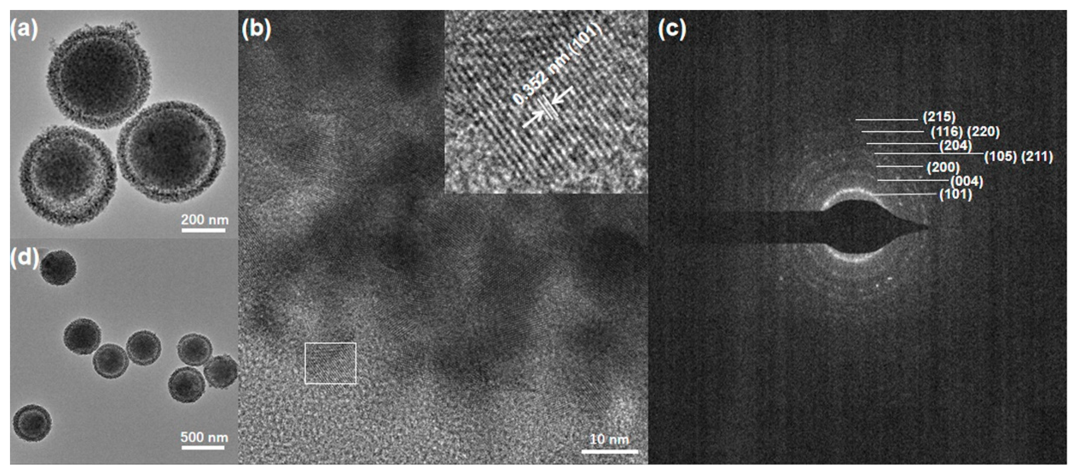
3.4. TEM Images
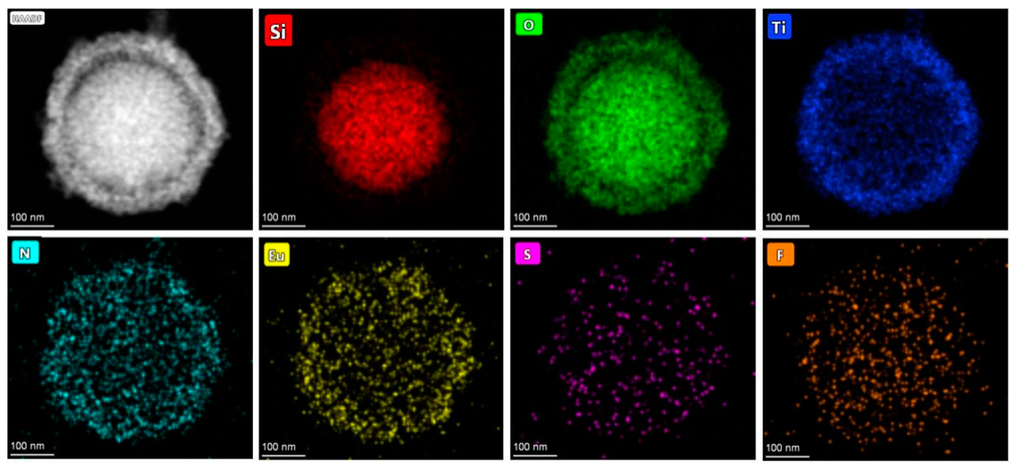
The TEM, HRTEM and SAED images of SiO2@mTiO2 and the TEM image of hybrid material Eu(tta)3bpdc-SiO2@mTiO2 are shown in Figure 4. SiO2@mTiO2 (Figure 4a) and Eu(tta)3bpdc-SiO2@mTiO2 (Figure 4d) exhibit monodisperse spherical core–shell structures with an average diameter of about 430 nm. The shell of SiO2@mTiO2 and Eu(tta)3bpdc-SiO2@mTiO2 is comprised of many small-ordered nanoparticles (Figure S2). The HRTEM can be used to identify the nature of nanocrystalline titania in SiO2@mTiO2 (Figure 4b). The fringe spacing is about 3.52 Å, corresponding to the (101) plane of anatase titania, further confirming that the shell of the nanosphere consists of single anatase nano-crystallites. As shown in Figure 4c, the SAED pattern clearly shows several continuous rings, corresponding to the (101), (004), (200), (105/211), (204), (116/220) and (215) diffractions of the anatase phase. In addition, the morphology and size of the hybrid material Eu(tta)3bpdc-SiO2@mTiO2 are nearly unchanged in comparison with those of SiO2@mTiO2, indicating that the introduction of europium complex does not influence the morphology and size of the SiO2@mTiO2 matrices. The composition and distribution of the elements in SiO2@mTiO2 and Eu(tta)3bpdc-SiO2@mTiO2 were analyzed by EDS mapping (Figure S3 and Figure 5). As displayed in Figure S3, Si and Ti elements are uniformly distributed in the core and shell, suggesting that SiO2@mTiO2 is a core–shell structure of silica coated by titania. Furthermore, for hybrid material Eu(tta)3bpdc-SiO2@mTiO2 (Figure 5), a uniform dispersity of N, Eu, S and F elements indicates the existence of complex Eu(tta)3bpdc in core–shell nanosphere, which further suggests the hybrid material was successfully obtained.
3.5. Luminescent Properties
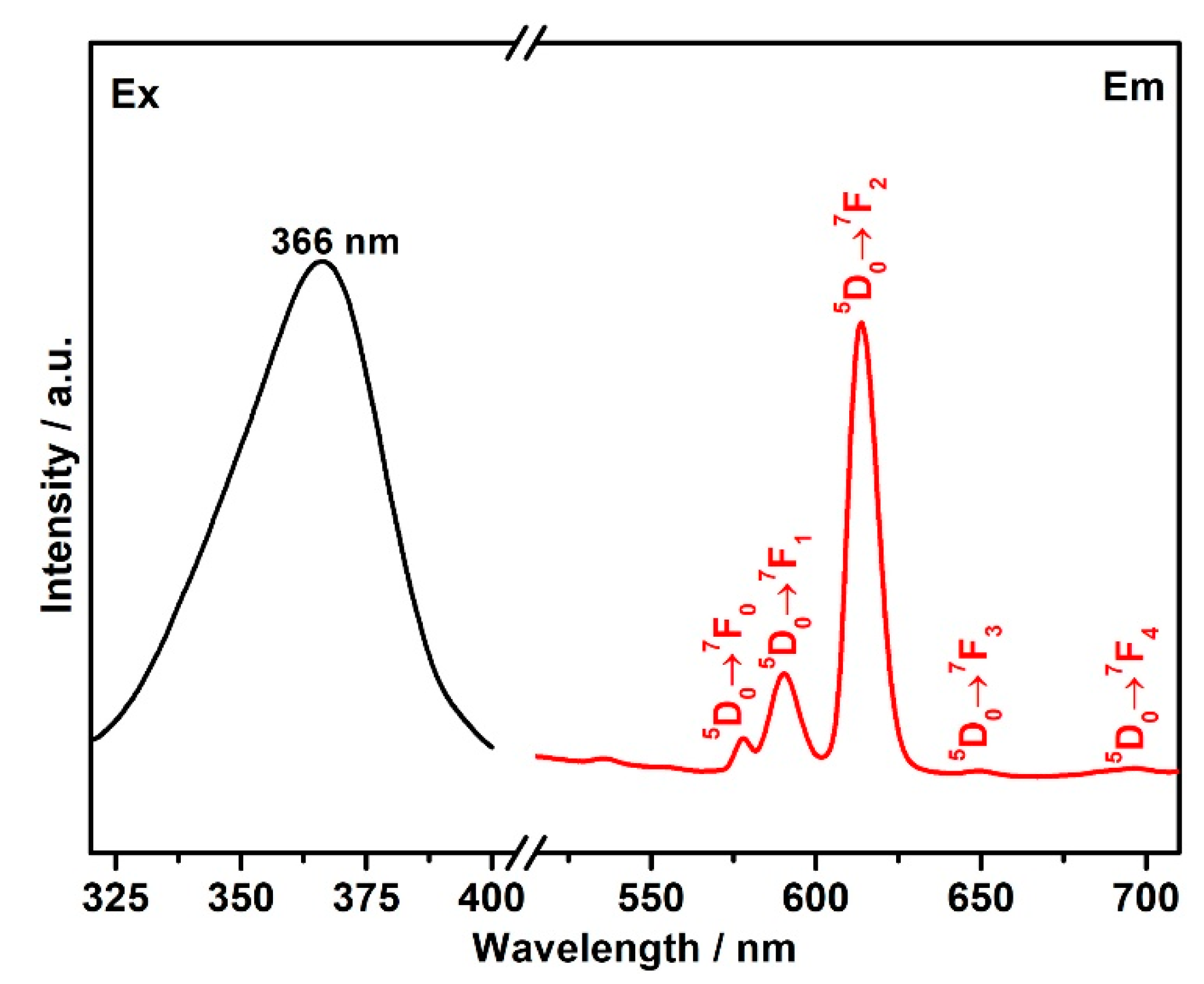
The luminescent behavior of hybrid material Eu(tta)3bpdc-SiO2@mTiO2 in solid state and ethanol solution was investigated at room temperature. The excitation spectra of hybrid material Eu(tta)3bpdc-SiO2@mTiO2 are obtained by monitoring the most intense 5D0 → 7F2 emission. As presented in Figure 6, the excitation spectrum shows a broad band in the range of 300~400 nm, which was attributed to the π → π* electron transition of H2bpdc ligand. Upon the maximum excitation (λex = 366 nm), the emission spectrum of hybrid material Eu(tta)3bpdc-SiO2@mTiO2 in a solid state presents characteristic 5D0 → 7FJ (J = 0, 1, 2, 3, 4) transitions of EuIII ions in the range of 550~710 nm, indicating that the effective energy transfer is occurred from the ligands (H2bpdc and Htta) to the central EuIII ion, further implying that core–shell SiO2@mTiO2 nanospheres can act as an efficient host to sensitize the luminescence of EuIII ion. The 5D0 → 7F2 emission intensity of EuIII is strongly dependent on the environment because of its electric-dipole character, while the intensity of the 5D0 → 7F1 transition is independent of the environment due to its magnetic dipole character. The intensity ratios I(5D0 → 7F2)/I(5D0 → 7F1) will provide valuable information about the surrounding environment changes around EuIII ion. The ratio in complex Eu(tta)3(H2O)2 is about 7.7, while that in hybrid material Eu(tta)3bpdc-SiO2@mTiO2 is about 3.9, the decrease in the ratio demonstrates that the symmetry of the coordination environment around the EuIII ions has changed during the formation of hybrid material Eu(tta)3bpdc-SiO2@mTiO2, which further indicates that the tripyridyl group replaces the coordination water molecules in the first coordination sphere of EuIII ions in a complex by ligand-exchange reaction, and the coordination bonds of Eu–N are formed. The 5D0 luminescent decay curve was recorded at 613 nm under the maximum excitation (λex = 366 nm) and is exhibited in Figure S4. The curve can be fitted by biexponential functions, the average lifetime (τ) can be calculated using the following equation:
where τi is the component decay times and Ai is the preexponential factors related to the statistical weights of each exponential. For hybrid material Eu(tta)3bpdc-SiO2@mTiO2, the obtained 5D0 lifetime value was 0.52 ms.
As shown in Figure S5, hybrid material Eu(tta)3bpdc-SiO2@mTiO2 in an ethanol solution also shows five characteristic 5D0 → 7FJ (J = 0, 1, 2, 3, 4) emission peaks of EuIII ions and shows luminous red color emission. At the same time, Eu(tta)3bpdc-SiO2@mTiO2 in an ethanol solution possesses excellent luminescent stability at room temperature. In addition, hybrid material Eu(tta)3bpdc-SiO2@mTiO2 also has a nanoscale nature, so it can act as a suitable luminescent sensor in environmental and biological systems.
3.6. Sensing of Al3+ Cations
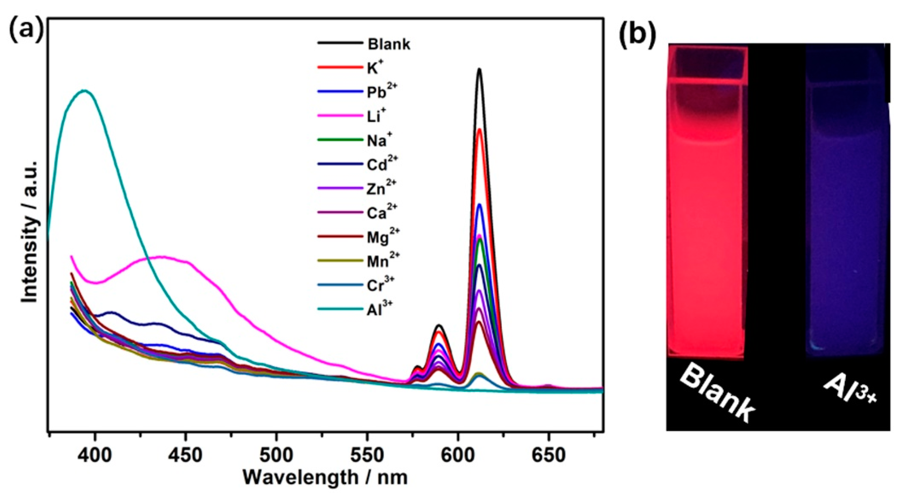
In order to investigate the potential application of core–shell hybrid material Eu(tta)3bpdc-SiO2@mTiO2 for the sensing of metal ions, Eu(tta)3bpdc-SiO2@mTiO2 powder samples were dispersed in ethanol solutions of different metal ions (KCl, NaCl, LiCl, ZnCl2, CaCl2, CdCl2, PbCl2, MgCl2, MnCl2, CrCl3 and AlCl3) for luminescent measurements. The luminescent emission of samples was recorded and presented in Figure 7a. Most of these cations influence the emission intensities of hybrid material Eu(tta)3bpdc-SiO2@mTiO2, especially for Mn2+ and Cr3+ ions show a significant quenching effect on the luminescent intensity. These phenomena are caused by the different electron configuration of metal ions: K+, Na+, Li+, Zn2+, Ca2+, Cd2+, Pb2+ and Mg2+ with a closed-shell electron configuration have much weaker effects on luminescent intensity, whereas other ions (such as Mn2+ and Cr3+) have different electron configurations and produce varying degrees of quenching of luminescent intensity [39]. However, only Al3+ ions have a significant impact on the emission spectrum of hybrid material Eu(tta)3bpdc-SiO2@mTiO2, characteristic emission of EuIII ions disappears, a broad band of H2bpdc ligand appears at 396 nm, and the luminescent color has clearly changed from red to blue under UV-light irradiation (Figure 7b). The reason for the selective recognition of hybrid material Eu(tta)3bpdc-SiO2@mTiO2 toward Al3+ ions may be that there existed stronger coordination interaction between bpdc2− ligand and Al3+ ions than bpdc2− ligand and EuIII ions [40].
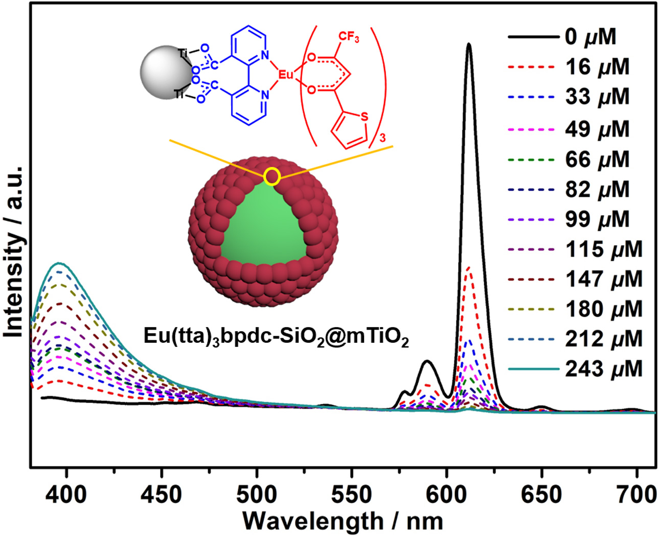
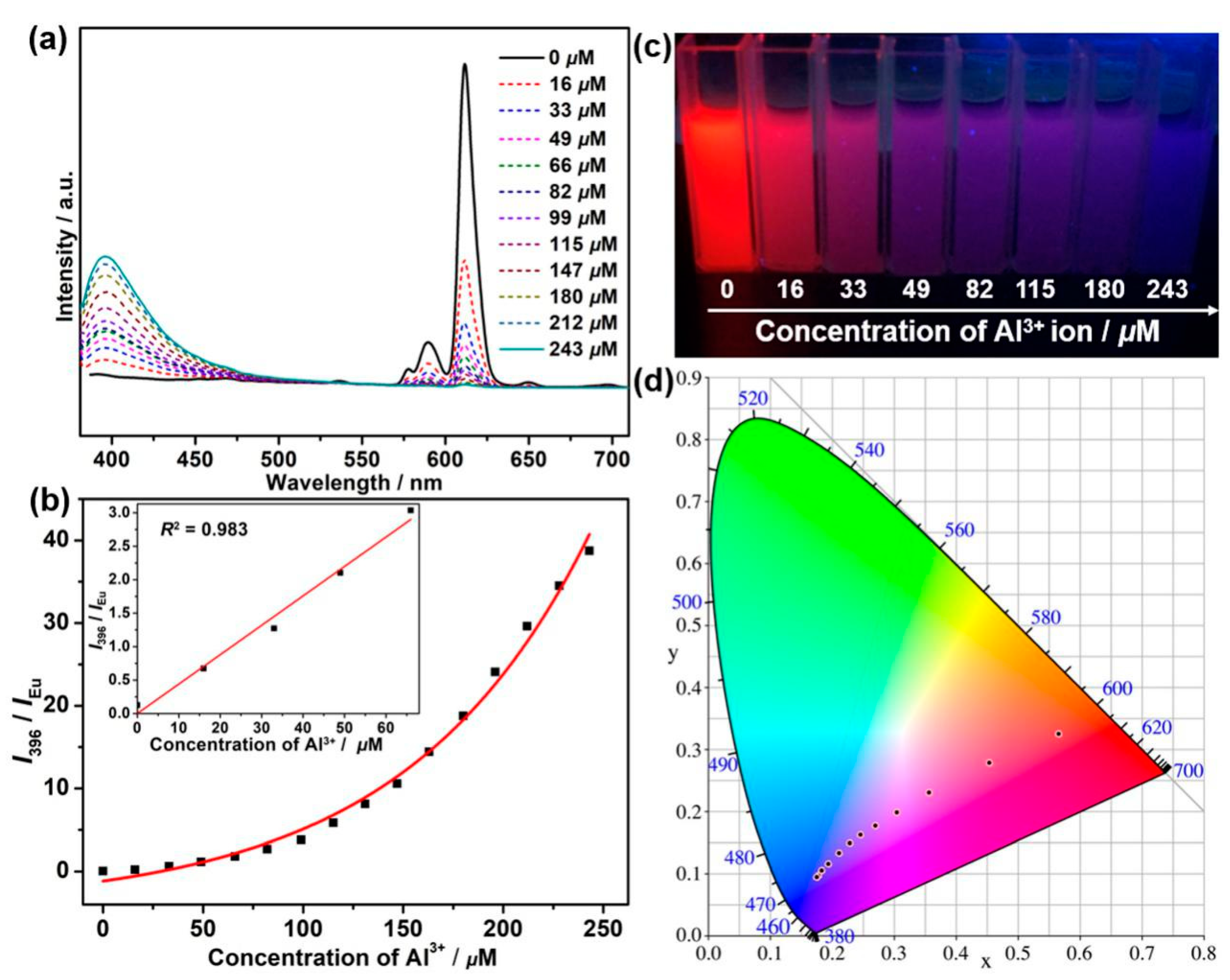
A luminescent titration experiment was carried out to evaluate the luminescent response of hybrid material Eu(tta)3bpdc-SiO2@mTiO2 to Al3+ ions by the addition of Al3+ ions to an Eu(tta)3bpdc-SiO2@mTiO2 ethanol solution. As shown in Figure 8a, with the increase in Al3+ ion concentration, the luminescent intensity of EuIII ions at 613 nm gradually weakens; meanwhile, a broad band of H2bpdc ligand at 396 nm appears and gradually enhances, leading to emission color change from red to pink, purplish pink, purplish blue, and finally blue under the 365 nm UV lamp (Figure 8c). The distinguishable colors at different concentrations of Al3+ ions are attributed to the changeable rate of the luminescent relative intensity of H2bpdc ligand-centered emissions and the characteristic emission of EuIII ions. Hence, the naked-eye recognition of Al3+ ions is feasible according to the color change of hybrid material Eu(tta)3bpdc-SiO2@mTiO2 ethanol solution. As a result, by changing the concentration of Al3+ ions in hybrid material Eu(tta)3bpdc-SiO2@mTiO2 ethanol solution, the relative emission intensities of two constituent colors (red and blue) can be precisely manipulated, resulting in a tunable multicolor output. The emission color CIE coordinates of hybrid material Eu(tta)3bpdc-SiO2@mTiO2 ethanol solution with various concentrations of Al3+ ions excited at 367 nm in the CIE 1931 chromaticity diagram (Figure 8d) and listed in Table S3. When Al3+ ion concentration increases to 243 μM, the red emission from EuIII ions almost completely quenches. Noticeably, a linear relationship is observed at low concentrations between the emission intensity ratio of 396 nm/613 nm and the concentration of Al3+ ions, followed the simple linear equation of y = 0.0439x + 0.0011 (R2 = 0.983) (Figure 8b inset). The limit of detection (LOD) for Al3+ ions was determined as 1.8 × 10⁻⁴ M according to the equation LOD = 3σ/k. The result indicates that hybrid material Eu(tta)3bpdc-SiO2@mTiO2 can be acted as a highly sensitive and selective probe for the quantitative luminescent detection of Al3+ ions in an ethanol solution.
To assess the selectivity of hybrid material Eu(tta)3bpdc-SiO2@mTiO2, the competitive tests of hybrid material Eu(tta)3bpdc-SiO2@mTiO2 with Al3+ and different metal ions were carried out in an ethanol solution. As shown in Figure S6, after adding other ions in Al3+ ethanol solution, the luminescent intensity of hybrid material Eu(tta)3bpdc-SiO2@mTiO2 at 396 nm increases compared with that in Al3+ ethanol solution. The result suggests that the detection of Al3+ ions is not affected by the other ions.
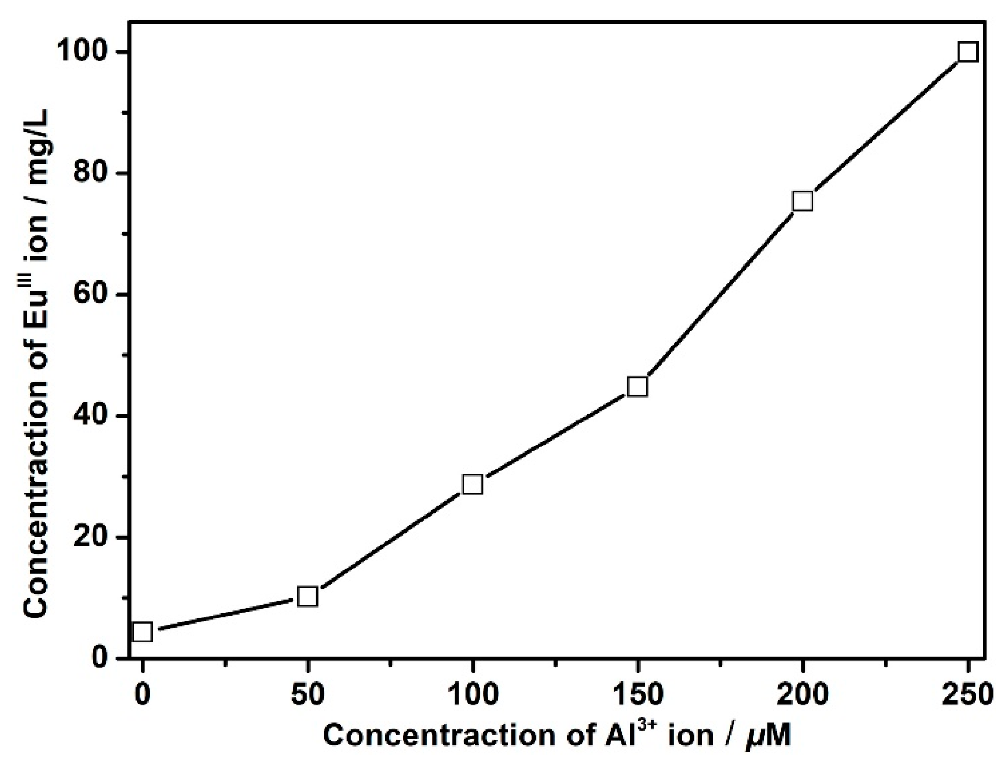
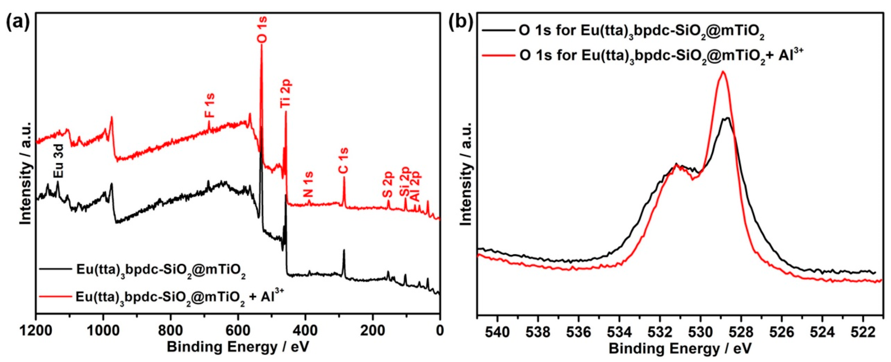
According to the reported literature [16,30], the possible luminescent sensing mechanism for the detection of Al3+ ions originates from two aspects: (i) the cation exchange of central lanthanide ions in lanthanide hybrid materials with targeted cations, (ii) the interaction between organic ligands and Al3+ ions. Therefore, in order to verify this hypothesis, ICP-MS, XPS, PXRD, TEM and EDS mapping of hybrid material Eu(tta)3bpdc-SiO2@mTiO2 after immersed in an Al3+ ion ethanol solution (namely, Eu(tta)3bpdc-SiO2@mTiO2 + Al3+) were carried out. As exhibited in Figure 9, as the content of added Al3+ ions in the filtrate increases, the concentration of EuIII ions in the filtrate gradually increases. As a result, Al3+ ions may gradually replace EuIII ions in hybrid material Eu(tta)3bpdc-SiO2@mTiO2, which corresponds to luminescence responses of hybrid material Eu(tta)3bpdc-SiO2@mTiO2 toward Al3+ ions in ethanol solutions with varying concentrations of Al3+ ions, indicating that the luminescent emission changes are attributed to the cation exchange. As shown in Figure 10 and Figure S7 and Table S4, it is clearly observed that Eu 3p peak (~1138.4 eV) disappears and Al 2p peak (~74.8 eV) appears in Eu(tta)3bpdc-SiO2@mTiO2 + Al3+ when compared with those peaks in hybrid material Eu(tta)3bpdc-SiO2@mTiO2, indicating that EuIII ions in hybrid material Eu(tta)3bpdc-SiO2@mTiO2 were gradually replaced by Al3+ ions in an ethanol solution, further suggesting that there existed a cation exchange when hybrid material Eu(tta)3bpdc-SiO2@mTiO2 was immersed in an ethanol solution of Al3+ ions. In addition, the O 1s peak [41] in the spectrum of Eu(tta)3bpdc-SiO2@mTiO2 + Al3+ almost have no shift compared with that in the spectrum of hybrid material Eu(tta)3bpdc-SiO2@mTiO2 (Figure 10b), to some content, which excludes the possibility of interaction between oxygen atoms in coordinated carboxylate group of bpdc2− ligand and Al3+ ions. As displayed in Figure S8 and Table S2, characteristic diffraction peaks of Eu(tta)3bpdc-SiO2@mTiO2 + Al3+ materials are nearly unchanged compared with those of hybrid material Eu(tta)3bpdc-SiO2@mTiO2, indicating that the anatase phase was maintained well after hybrid material Eu(tta)3bpdc-SiO2@mTiO2 was immersed in an ethanol solution of Al3+ ions. Additionally, it can be observed that Eu(tta)3bpdc-SiO2@mTiO2 + Al3+ shows the core–shell structure (Figure S9) and the size of the core–shell nanosphere of Eu(tta)3bpdc-SiO2@mTiO2 + Al3+ almost have no change compared with that of Eu(tta)3bpdc-SiO2@mTiO2. This suggests that the subsequent addition of Al3+ does not nearly change the morphology or size of the hybrid material Eu(tta)3bpdc-SiO2@mTiO2. As shown in Table S5, the Al element could be seen besides Si, O, Ti, N, F, S, and mass ratio of Eu element decreases and approaches zero compared with the mass ratio of the Eu element in hybrid material Eu(tta)3bpdc-SiO2@mTiO2, which suggests that EuIII ions in Eu(tta)3bpdc-SiO2@mTiO2 were replaced by Al3+ ions, and then excludes interaction between the ligands of Eu(tta)3bpdc-SiO2@mTiO2 and Al3+ ions. Furthermore, Si, O, Ti, N, F, S and Al elements are homogeneously distributed in the hybrid material Eu(tta)3bpdc-SiO2@mTiO2 + Al3+ (Figure S10). Based on the above results, the possible luminescence sensing mechanism for the detection of Al3+ ions originates from the cation exchange between EuIII ions in hybrid material Eu(tta)3bpdc-SiO2@mTiO2 and Al3+ ions.
4. Conclusions
A europium(III) hybrid material based on a SiO2@mTiO2 core–shell nanosphere by covalent grafting complex Eu(tta)3bpdc was successfully designed and synthesized. H2bpdc ligand can act as a bifunctional linker to graft Eu(tta)3 to the surface of the core–shell SiO2@mTiO2 nanosphere. The results indicate that core–shell structure and anatase crystallites are retained after grafting to the europium complex. Hybrid material Eu(tta)3bpdc-SiO2@mTiO2 displays uniform nanosphere structure, bright red color emission and long lifetime, which can sever as a multicolor emission material to modulate by using Al3+ ions via an ion exchange approach under a single-wavelength excitation.
Supplementary Materials
The following are available online at www.mdpi.com/article/10.3390/nano11112886/s1, Figure S1: FT-IR spectra of SiO2@mTiO2 (a), SiO2@mTiO2-bpdc (b), Eu(tta)3bpdc-SiO2@mTiO2 (c) and H2bpdc ligand (d); Figure S2: TEM image of SiO2@mTiO2; Figure S3: EDS mapping for each element in SiO2@mTiO2; Figure S4: Luminescent lifetime of hybrid material Eu(tta)3bpdc-SiO2@mTiO2 in state solid; Figure S5: Excitation and emission spectra of hybrid material Eu(tta)3bpdc-SiO2@mTiO2 in ethanol solution at room temperature; Figure S6: Luminescent intensities of hybrid material Eu(tta)3bpdc-SiO2@mTiO2 at 396 nm in presence of other metal ions and Al3+ ion in ethanol solution; Figure S7: C 1s spectra for hybrid materials Eu(tta)3bpdc-SiO2@mTiO2 and Eu(tta)3bpdc-SiO2@mTiO2 + Al3+; Figure S8: The PXRD patterns of hybrid materials Eu(tta)3bpdc-SiO2@mTiO2 and Eu(tta)3bpdc-SiO2@mTiO2 + Al3+; Figure S9: TEM image of Eu(tta)3bpdc-SiO2@mTiO2 + Al3+; Figure S10: EDS mapping for each element in Eu(tta)3bpdc-SiO2@mTiO2 + Al3+. Table S1: The FT-IR characteristic bands of SiO2@mTiO2, SiO2@mTiO2-bpdc, Eu(tta)3bpdc-SiO2@mTiO2 and H2bpdc ligand; Table S2: The characteristic diffraction peaks for PXRD in SiO2@mTiO2, SiO2@mTiO2-bpdc, Eu(tta)3bpdc-SiO2@mTiO2 and Eu(tta)3bpdc-SiO2@mTiO2 + Al3+; Table S3: CIE coordinates and emission colors of hybrid material Eu(tta)3bpdc-SiO2@mTiO2 in different concentrations of Al3+ ion; Table S4: Binding energies for XPS in hybrid materials Eu(tta)3bpdc-SiO2@mTiO2 and Eu(tta)3bpdc-SiO2@mTiO2 + Al3+; Table S5: The mass ratio of each element in Eu(tta)3bpdc-SiO2@mTiO2 and Eu(tta)3bpdc-SiO2@mTiO2 + Al3+. Reference [42] is cited in the supplementary materials.
Author Contributions
Conceptualization, C.B. and H.-M.H.; methodology, C.B., S.H., H.Z. and F.Z.; software, C.B. and H.Z.; validation, H.-M.H. and J.-J.W.; formal analysis, C.B. and S.H.; investigation, C.B., S.H. and H.Z.; resources, H.-M.H.; data curation, S.H.; writing–original draft preparation, C.B.; writing–review and editing, C.B. and H.-M.H.; visualization, H.Z. and F.Z.; supervision, J.-J.W.; project administration, J.-J.W.; funding acquisition, H.-M.H. All authors have read and agreed to the published version of the manuscript.
Funding
This work was supported by the National Natural Science Foundation of China (Grant Nos. 21473133 and 21173164).
Conflicts of Interest
The authors declare no conflict of interest.
References
- Ning, Y.; Zhu, M.; Zhang, J.-L. Near-infrared (NIR) lanthanide molecular probes for bioimaging and biosensing. Coord. Chem. Rev. 2019, 399, 213028. [Google Scholar] [CrossRef]
- Chen, H.; Cao, J.; Zhou, P.; Li, X.; Xie, Y.; Liu, W.; Tang, Y. Multiplex recognition and logic devices for molecular robot prototype based on an europium(iii)-cyclen system. Biosens. Bioelectron. 2018, 122, 1–7. [Google Scholar] [CrossRef] [PubMed]
- Dou, Z.-S.; Yu, J.-C.; Cui, Y.-J.; Yang, Y.; Wang, Z.-Y.; Yang, D.-R.; Qian, G.-D. Luminescent Metal-Organic Framework Films As Highly Sensitive and Fast-Response Oxygen Sensors. J. Am. Chem. Soc. 2014, 136, 5527–5530. [Google Scholar] [CrossRef] [PubMed]
- Chen, L.; Liu, D.; Peng, J.; Du, Q.; He, H. Ratiometric fluorescence sensing of metal-organic frameworks: Tactics and perspectives. Coord. Chem. Rev. 2020, 404, 213113. [Google Scholar] [CrossRef]
- Cui, Y.; Chen, B.; Qian, G. Lanthanide metal-organic frameworks for luminescent sensing and light-emitting applications. Coord. Chem. Rev. 2014, 273, 76–86. [Google Scholar] [CrossRef]
- SeethaLekshmi, S.; Ramya, A.R.; Reddy, M.L.P.; Varughesea, S. Lanthanide complex-derived white-light emitting solids: A survey on design strategies. J. Photoch. Photobiol. C 2017, 33, 109–131. [Google Scholar] [CrossRef]
- Bünzli, J.-C.G.; Piguet, C. Taking advantage of luminescent lanthanide ions. Chem. Soc. Rev. 2005, 34, 1048–1077. [Google Scholar] [CrossRef] [PubMed]
- Ou, Y.; Zhou, W.; Zhu, Z.; Ma, F.; Zhou, R.; Su, F.; Zheng, L.; Ma, L.; Liang, H. Host Differential Sensitization toward Color/Lifetime-Tuned Lanthanide Coordination Polymers for Optical Multiplexin. Angew. Chem. Int. Ed. 2020, 59, 2–9. [Google Scholar] [CrossRef]
- Zhang, W.; Zhang, Y.-M.; Xie, F.; Jin, X.; Li, J.; Yang, G.; Gu, C.; Wang, Y.; Zhang, S.X.-A. A Single-Pixel RGB Device in a Colorful Alphanumeric Electrofluorochromic Display. Adv. Mater. 2020, 32, 2003121. [Google Scholar] [CrossRef] [PubMed]
- Gao, W.; Wang, R.; Han, Q.; Dong, J.; Yan, L.; Zheng, H. Tuning Red Upconversion Emission in Single LiYF4:Yb3+/Ho3+ Microparticle. J. Phys. Chem. C 2015, 119, 2349–2355. [Google Scholar] [CrossRef]
- Jose, B.A.S.; Matsushita, S.; Akagi, K. Lyotropic Chiral Nematic Liquid Crystalline Aliphatic Conjugated Polymers Based on Disubstituted Polyacetylene Derivatives That Exhibit High Dissymmetry Factors in Circularly Polarized Luminescence. J. Am. Chem. Soc. 2012, 134, 19795–19807. [Google Scholar] [CrossRef]
- Li, G.G.; Hou, Z.Y.; Peng, C.; Wang, W.X.; Cheng, Z.Y.; Li, C.X.; Lian, H.Z.; Lin, J. Electrospinning Derived One-Dimensional LaOCl: Ln3+ (Ln = Eu/Sm, Tb, Tm) Nanofibers, Nanotubes and Microbelts with Multicolor-Tunable Emission Properties. Adv. Funct. Mater. 2010, 20, 3446–3456. [Google Scholar] [CrossRef]
- He, G.J.; Guo, D.; He, C.; Zhang, X.L.; Zhao, X.W.; Duan, C.Y. A color-tunable europium complex emitting three primary colors and white light. Angew. Chem. Int. Ed. 2009, 48, 6132–6135. [Google Scholar] [CrossRef] [PubMed]
- Cui, Y.J.; Xu, H.; Yue, Y.F.; Guo, Z.Y.; Yu, J.C.; Chen, Z.X.; Gao, J.K.; Yang, Y.; Qian, G.D.; Chen, B.L. A luminescent mixed-lanthanide metal-organic framework thermometer. J. Am. Chem. Soc. 2012, 134, 3979–3982. [Google Scholar] [CrossRef] [PubMed]
- Dang, S.; Zhang, J.H.; Sun, Z.M. Tunable emission based on lanthanide(III) metal-organic frameworks: An alternative approach to white light. J. Mater. Chem. 2012, 22, 8868–8873. [Google Scholar] [CrossRef]
- Zhang, Z.; Li, H.; Li, Y.; Yu, X. Full-color emission of a Eu3+-based mesoporous hybrid material modulated by Zn2+ ions: Emission color changes for Zn2+ sensing via an ion exchange approach. Dalton Trans. 2019, 48, 10547–10556. [Google Scholar] [CrossRef]
- Li, Y.; Yu, X.; Yu, T. Eu3+ based mesoporous hybrid material with tunable multicolor emission modulated by fluoride ion: Application for selective sensing toward fluoride ion. J. Mater. Chem. C 2017, 5, 5411–5419. [Google Scholar] [CrossRef]
- Perl, D.P.; Brody, A.R. Alzheimer’s disease: X-ray spectrometric evidence of aluminum accumulation in neurofibrillary tangle-bearing neurons. Science 1980, 208, 297–299. [Google Scholar] [CrossRef]
- Barcelo, J.; Poschenrieder, C. Fast root growth responses, root exudates, and internal detoxification as clues to the mechanisms of aluminum toxicity and resistance. Exp. Bot. 2002, 48, 75–92. [Google Scholar] [CrossRef]
- Valeur, B.; Leray, I. Design principles of fluorescent molecular sensors for cation recognition. Coord. Chem. Rev. 2000, 3, 205–340. [Google Scholar] [CrossRef]
- Krejpcio, Z.; Wojciak, R.W.P.J. The Influence of Al3+ Ions on Pepsin and Trypsin Activity in Vitro. Environ. Stud. 2002, 11, 251–254. [Google Scholar]
- Goswami, S.; Paul, S.; Manna, A. Selective “naked eye” detection of Al(iii) and PPi in aqueous media on a rhodamine-isatin hybrid moiety. RSC Adv. 2013, 3, 10639–10643. [Google Scholar] [CrossRef]
- Kashyap, K.S.; Kumar, A.; Hira, S.K.; Dey, S. Recognition of Al3+ through the off-on mechanism as a proficient driving force for the hydrolysis of BODIPY conjugated Schiff base and its application in bio-imaging. Inorg. Chim. Acta 2019, 498, 119157. [Google Scholar] [CrossRef]
- Kim, H.; Manivannan, R.; Son, Y.-A. A Chromone Based Fluorescent Probe for the Effective Detection of Aluminium Ion. J. Nanosci. Nanotechnol. 2020, 20, 2840–2846. [Google Scholar] [CrossRef]
- Park, J.; Angupillai, S.; Son, Y.-A. A Highly Sensitive Fluorescent Probe for Selective Detection of Al3+ Cation by Switching the Solvent from Aprotic to Protic Environment. Mol. Cryst. Liq. Cryst. 2015, 622, 103–113. [Google Scholar] [CrossRef]
- Thangaraja, S.E.; Antonya, E.J.; Selvanb, G.T.; Selvakumarb, P.M.; Enocha, I.V.M.V. A New Fluorenone-Based Turn-on Fluorescent Al3+ Ion Sensor. J. Anal. Chem. 2019, 74, 87–92. [Google Scholar] [CrossRef]
- Ma, D.; Chen, C.; Chen, M.; Zhu, S.; Wu, Y.; Li, Z.; Li, Y.; Zhou, L. A hydrostable Cadmium-Organic Framework for Highly Selective and Sensitive Luminescence Sensing of Al3+ Ion. J. Inorg. Organomet. Polym. Mater. 2019, 29, 1829–1837. [Google Scholar] [CrossRef]
- Xu, W.; Zhou, Y.; Huang, D.; Su, M.; Wang, K.; Hong, M. A highly sensitive and selective fluorescent sensor for detection of Al3+ using a Europium(III) quinolinecarboxylate. Inorg Chem. 2014, 53, 6497–6499. [Google Scholar] [CrossRef]
- Song, H.; Liu, G.; Fan, C.; Pu, S. A novel fluorescent sensor for Al3+ and Zn2+ based on a new europium complex with a 1,10-phenanthroline ligand. J. Rare Earth 2021, 39, 460–468. [Google Scholar] [CrossRef]
- Li, H.; Li, Y.; Zhang, Z.; Pang, X.; Yu, X. Highly selective luminescent sensing of Cu2+ in aqueous solution based on a Eu(III)-centered periodic mesoporous organosilicas hybrid. Mater. Design 2019, 172, 107712. [Google Scholar] [CrossRef]
- Liu, Y.; Sun, L.; Liu, J.; Peng, Y.-X.; Ge, X.; Shi, L.; Huang, W. Multicolor (Vis-NIR) mesoporous silica nanospheres linked with lanthanide complexes using 2-(5-bromothiophen)imidazo[4,5-f][1,10]phenanthroline for in vitro bioimaging. Dalton Trans. 2015, 44, 237–246. [Google Scholar] [CrossRef]
- Guan, B.Y.; Yu, L.; Li, J.; Lou, X.W. A universal cooperative assembly-directed method for coating of mesoporous TiO2 nanoshells with enhanced lithium storage properties. Sci. Adv. 2016, 2, 1501554. [Google Scholar] [CrossRef] [PubMed] [Green Version]
- Xia, Q.; Huang, Y.; Xiao, J.; Wang, L.; Lin, Z.; Li, W.; Liu, H.; Gu, Q.; Liu, H.K.; Chou, S.-L. Phosphorus-Modulation-Triggered Surface Disorder in Titanium Dioxide Nanocrystals Enables Exceptional Sodium-Storage Performance. Angew. Chem. Int. Ed. 2019, 58, 4022–4026. [Google Scholar] [CrossRef]
- Liu, P.; Li, H.R.; Wang, Y.G.; Liu, B.Y.; Zhang, W.J.; Wang, Y.J.; Yan, W.D.; Zhang, H.J.; Schubert, U. Europium complexes immobilization on titania via chemical modification of titanium alkoxide. J. Mater. Chem. 2008, 18, 735–737. [Google Scholar] [CrossRef]
- Nazeeruddin, M.K.; Humphry-Baker, R.; Liska, P.; Grätzel, M. Investigation of Sensitizer Adsorption and the Influence of Protons on Current and Voltage of a Dye-Sensitized Nanocrystalline TiO2 Solar Cell. J. Phys. Chem. B 2003, 107, 8981–8987. [Google Scholar] [CrossRef]
- Nakamoto, K. Infrared and Raman Spectra of Inorganic and Coordination Compounds; John Wiley and Sons: Hoboken, NJ, USA, 1978. [Google Scholar]
- Bai, C.; Wei, F.-H.; Hu, H.-M.; Yan, L.; Wang, X.; Xue, G.-L. New highly luminescent europium (III) complex covalently bonded with titania-based host via using a terpyridine carboxylate derivative linker for fluorescence sensing. J. Lumin. 2020, 227, 117545. [Google Scholar] [CrossRef]
- Wei, F.; Bai, C.; Hu, H.-M.; He, S.; Wang, X.; Xue, G. Novel luminescent europium-centered hybrid material covalently grafted with organically modified titania via 2-substituted imidazophenanthroline for fluorescence sensing. J. Rare Earth 2021, 39, 666–673. [Google Scholar] [CrossRef]
- Aleem, A.R.; Liu, J.; Wang, J.; Wang, J.; Zhao, Y.; Wang, Y.; Wang, Y.; Wang, W.; Rehman, F.; Kipper, M.J.; et al. Selective Sensing of Cu2+ and Fe3+ Ions with Vis-Excitation using Fluorescent Eu3+-Induced Aggregates of Polysaccharides (EIAP) in Mammalian Cells and Aqueous Systems. J. Hazard. Mater. 2020, 399, 122991. [Google Scholar] [CrossRef]
- Yu, X.D.; Wang, Z.Y.; Li, Y.J.; Geng, L.J.; Ren, J.J.; Feng, G.L. Fluorescent and Electrochemical Supramolecular Coordination Polymer Hydrogels Formed from Ion-Tuned Self-Assembly of Small Bis-Terpyridine Monomer. Inorg. Chem. 2017, 56, 7512–7518. [Google Scholar] [CrossRef]
- Orudzhev, F.; Ramazanov, S.; Sobola, D.; Isaev, A.; Wang, C.; Magomedova, A.; Kadiev, M.; Kaviyarasu, K. Atomic Layer Deposition of Mixed-Layered Aurivillius Phase on TiO2 Nanotubes: Synthesis, Characterization and Photoelectrocatalytic Properties. Nanomaterials 2020, 10, 2183. [Google Scholar] [CrossRef]
- Sun, L.; Wang, Z.; Zhang, J.Z.; Feng, J.; Liu, J.; Zhao, Y.; Shi, L. Visible and near-infrared luminescent mesoporous titania mi-crospheres functionalized with lanthanide complexes: Microstructure and luminescence with visible excitation. RSC Adv. 2014, 4, 28481–28489. [Google Scholar] [CrossRef]
Scheme 1.
The preparation procedure and predicted structure of core–shell hybrid material Eu(tta)3bpdc-SiO2@mTiO2.
Scheme 1.
The preparation procedure and predicted structure of core–shell hybrid material Eu(tta)3bpdc-SiO2@mTiO2.

Figure 1.
FT-IR spectra of (a) SiO2@mTiO2, (b) SiO2@mTiO2-bpdc, (c) Eu(tta)3bpdc-SiO2@mTiO2 and (d) H2bpdc ligand.
Figure 1.
FT-IR spectra of (a) SiO2@mTiO2, (b) SiO2@mTiO2-bpdc, (c) Eu(tta)3bpdc-SiO2@mTiO2 and (d) H2bpdc ligand.

Figure 2.
The PXRD patterns of SiO2@TiO2/HDA, SiO2@mTiO2, SiO2@mTiO2-bpdc and Eu(tta)3bpdc-SiO2@mTiO2.
Figure 2.
The PXRD patterns of SiO2@TiO2/HDA, SiO2@mTiO2, SiO2@mTiO2-bpdc and Eu(tta)3bpdc-SiO2@mTiO2.

Figure 3.
SEM images of (a) SiO2, (b) SiO2@TiO2/HDA, (c) SiO2@mTiO2, (d) SiO2@mTiO2-bpdc and (e) Eu(tta)3bpdc-SiO2@mTiO2.
Figure 3.
SEM images of (a) SiO2, (b) SiO2@TiO2/HDA, (c) SiO2@mTiO2, (d) SiO2@mTiO2-bpdc and (e) Eu(tta)3bpdc-SiO2@mTiO2.

Figure 4.
(a) TEM image of SiO2@mTiO2, (b) HRTEM image of the anatase crystals of shell in SiO2@mTiO2, (c) the corresponding SAED pattern of SiO2@mTiO2 and (d) TEM image of Eu(tta)3bpdc-SiO2@mTiO2.
Figure 4.
(a) TEM image of SiO2@mTiO2, (b) HRTEM image of the anatase crystals of shell in SiO2@mTiO2, (c) the corresponding SAED pattern of SiO2@mTiO2 and (d) TEM image of Eu(tta)3bpdc-SiO2@mTiO2.

Figure 5.
EDS mapping for each element in Eu(tta)3bpdc-SiO2@mTiO2.

Figure 6.
Excitation and emission spectra of hybrid material Eu(tta)3bpdc-SiO2@mTiO2 in solid state at room temperature.
Figure 6.
Excitation and emission spectra of hybrid material Eu(tta)3bpdc-SiO2@mTiO2 in solid state at room temperature.

Figure 7.
(a) Photoluminescence spectra (λex = 367 nm) of hybrid material Eu(tta)3bpdc-SiO2@mTiO2 after dispersed in ethanol solutions of different metal ions and (b) pictures of hybrid material Eu(tta)3bpdc-SiO2@mTiO2 under the 365 nm UV lamp after dispersed in ethanol solutions without and with Al3+ ions.

Figure 8.
(a) Photoluminescence spectra; (b) the plot of emission intensity of 396 nm/613 nm vs. concentration of Al3+ ions (Inset: the linear correlation for the plot of emission intensity of 396 nm/613 nm vs. concentration of Al3+ ions); (c) the pictures of hybrid material Eu(tta)3bpdc-SiO2@mTiO2 in different concentrations of Al3+ ions under the 365 nm UV lamp; (d) the corresponding CIE chromaticity diagram of hybrid material Eu(tta)3bpdc-SiO2@mTiO2 in an ethanol solution with different concentrations of Al3+ ions (λex = 367 nm).

Figure 9.
The concentrations of EuIII ions in the filtrate of Eu(tta)3bpdc-SiO2@mTiO2 upon the addition of different contents of Al3+ ions.
Figure 9.
The concentrations of EuIII ions in the filtrate of Eu(tta)3bpdc-SiO2@mTiO2 upon the addition of different contents of Al3+ ions.

Figure 10.
(a) XPS survey spectra for hybrid materials Eu(tta)3bpdc-SiO2@mTiO2 and Eu(tta)3bpdc-SiO2@mTiO2 + Al3+, (b) O 1s spectra for hybrid materials Eu(tta)3bpdc-SiO2@mTiO2 and Eu(tta)3bpdc-SiO2@mTiO2 + Al3+.
Figure 10.
(a) XPS survey spectra for hybrid materials Eu(tta)3bpdc-SiO2@mTiO2 and Eu(tta)3bpdc-SiO2@mTiO2 + Al3+, (b) O 1s spectra for hybrid materials Eu(tta)3bpdc-SiO2@mTiO2 and Eu(tta)3bpdc-SiO2@mTiO2 + Al3+.

Publisher’s Note: MDPI stays neutral with regard to jurisdictional claims in published maps and institutional affiliations. |
© 2021 by the authors. Licensee MDPI, Basel, Switzerland. This article is an open access article distributed under the terms and conditions of the Creative Commons Attribution (CC BY) license (https://creativecommons.org/licenses/by/4.0/).
Share and Cite
MDPI and ACS Style
Bai, C.; He, S.; Hu, H.-M.; Zeng, H.; Zou, F.; Wang, J.-J. Europium(III) Complex-Functionalized SiO2@mTiO2 Nanospheres for Al3+-Modulated Multicolor Emission. Nanomaterials 2021, 11, 2886. https://0-doi-org.brum.beds.ac.uk/10.3390/nano11112886
AMA Style
Bai C, He S, Hu H-M, Zeng H, Zou F, Wang J-J. Europium(III) Complex-Functionalized SiO2@mTiO2 Nanospheres for Al3+-Modulated Multicolor Emission. Nanomaterials. 2021; 11(11):2886. https://0-doi-org.brum.beds.ac.uk/10.3390/nano11112886
Chicago/Turabian StyleBai, Chao, Shi He, Huai-Ming Hu, Hui Zeng, Feng Zou, and Ji-Jiang Wang. 2021. "Europium(III) Complex-Functionalized SiO2@mTiO2 Nanospheres for Al3+-Modulated Multicolor Emission" Nanomaterials 11, no. 11: 2886. https://0-doi-org.brum.beds.ac.uk/10.3390/nano11112886
Note that from the first issue of 2016, this journal uses article numbers instead of page numbers. See further details here.