
Living Bacteria Directly Embedded into Electrospun Nanofibers: Design of New Anode for Bio-Electrochemical Systems

 ,
,  , , and
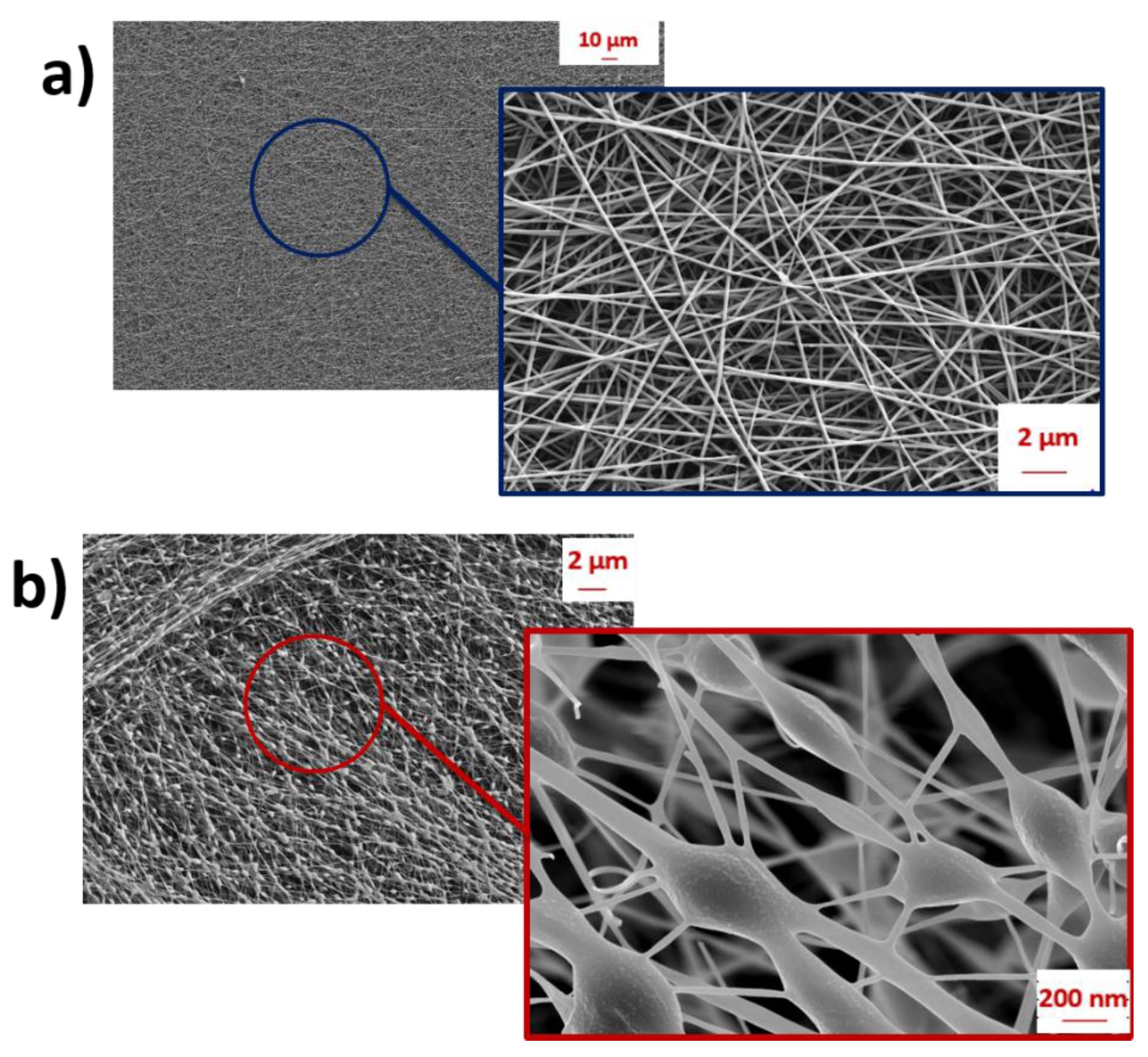
Abstract
:1. Introduction
2. Materials and Methods
2.1. Materials and Nanofibers Synthesis
2.2. Characterizations and Measurements
2.3. MFCs Architecture and Configuration
3. Results and Discussion
3.1. Morphological and Biological Characterizations
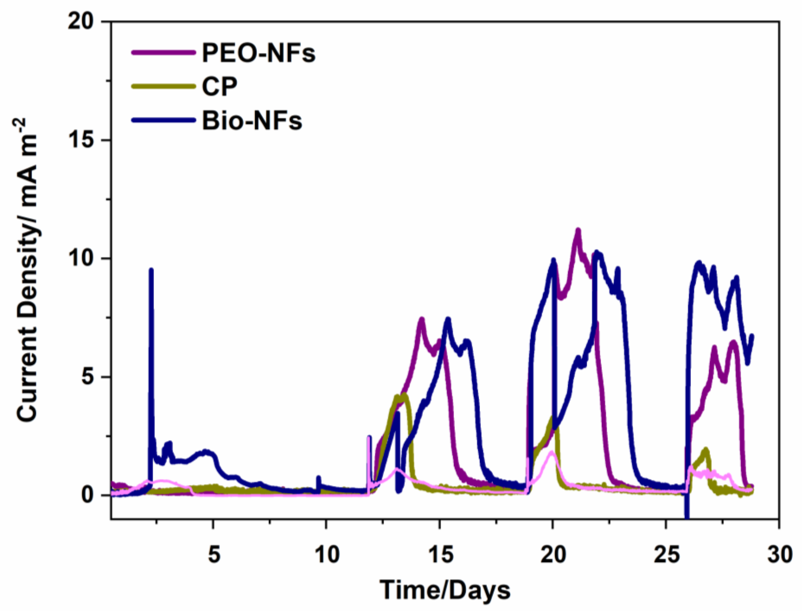
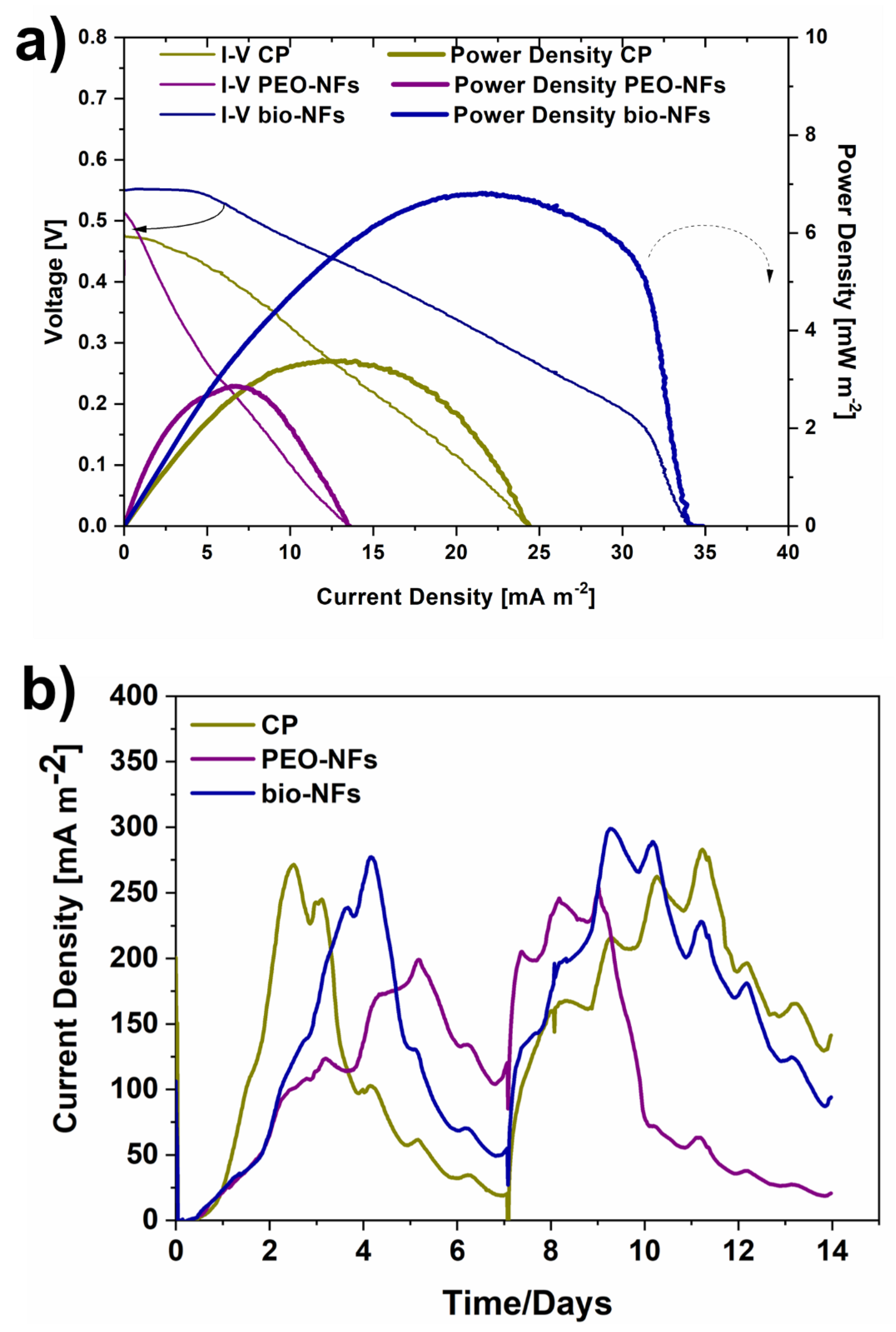
3.2. SCMFCs Performance
3.3. Electrochemical Impedance Spectroscopy Results
4. Conclusions
Author Contributions
Funding
Institutional Review Board Statement
Informed Consent Statement
Data Availability Statement
Conflicts of Interest
References
- Green Deal Europeo. Available online: https://ec.europa.eu/info/strategy/priorities-2019-2024/european-green-deal_it (accessed on 11 December 2019).
- European Green Deal Communication. Bruxelles. Available online: https://ec.europa.eu/info/sites/default/files/european-green-deal-communication_en.pdf (accessed on 11 December 2019).
- Walsh, B.; Ciais, P.; Janssens, I.; Peñuelas, J.; Riahi, K.; Rydzak, F.; Van Vuuren, D.P.; Obersteiner, M. Pathways for balancing CO2 emissions and sinks. Nat. Commun. 2017, 8, 1–12. [Google Scholar] [CrossRef] [PubMed] [Green Version]
- Sun, M.; Zhai, L.-F.; Li, W.-W.; Yu, H.-Q. ChemInform Abstract: Harvest and Utilization of Chemical Energy in Wastes by Microbial Fuel Cells. Chem. Soc. Rev. 2016, 45, 2847–2870. [Google Scholar] [CrossRef]
- Tommasi, T.; Salvador, G.P.; Quaglio, M. New insights in Microbial Fuel Cells: Novel solid phase anolyte. Sci. Rep. 2016, 6, 29091. [Google Scholar] [CrossRef] [PubMed] [Green Version]
- Agostino, V.; Massaglia, G.; Gerosa, M.; Sacco, A.; Saracco, G.; Margaria, V.; Quaglio, M. Environmental electroactive consortia as reusable biosensing element for freshwater toxicity monitoring. New Biotechnol. 2020, 55, 36–45. [Google Scholar] [CrossRef] [PubMed]
- Logan, B.E.; Rabaey, K. Conversion of Wastes into Bioelectricity and Chemicals by Using Microbial Electrochemical Technologies. Science 2012, 337, 686–690. [Google Scholar] [CrossRef] [Green Version]
- Logan, B.E. Microbial Fuel Cells; John Wiley & Sons: New York, NY, USA, 2008. [Google Scholar]
- Chiodoni, A.; Salvador, G.P.; Massaglia, G.; Delmondo, L.; Munoz-Tabares, J.A.; Garino, N.; Castellino, M.; Margaria, V.; Ahmed, D.; Pirri, C.F.; et al. MnxOy-based cathodes for oxygen reduction reaction catalysis in microbial fuel cells. Int. J. Hydrogen Energy 2019, 44, 4432–4441. [Google Scholar] [CrossRef]
- Massaglia, G.; Fiorello, I.; Sacco, A.; Margaria, V.; Pirri, C.F.; Quaglio, M. Biohybrid Cathode in Single Chamber Microbial Fuel Cell. Nanomaterials 2018, 9, 36. [Google Scholar] [CrossRef] [Green Version]
- Cecconet, D.; Molognoni, D.; Callegari, A.; Capodaglio, A.G. Agro-food industry wastewater treatment with microbial fuel cells: Energetic recovery issues. Int. J. Hydrogen Energy 2018, 43, 500–511. [Google Scholar] [CrossRef]
- Mashkour, M.; Rahimnejad, M.; Bakeri, G.; Luque, R.; Oh, S.E. Application of Wet Nanostructured Bacterial Cellulose as a Novel Hydrogel Bioanode for Microbial Fuel Cells. ChemElectroChem 2017, 4, 648–654. [Google Scholar] [CrossRef]
- Massaglia, G.; Margaria, V.; Fiorentin, M.R.; Pasha, K.; Sacco, A.; Castellino, M.; Chiodoni, A.; Bianco, S.; Pirri, F.C.; Quaglio, M. Nonwoven mats of N-doped carbon nanofibers as high-performing anodes in microbial fuel cells. Mater. Today Energy 2020, 16, 100385. [Google Scholar] [CrossRef]
- Zhang, L.S.; Wu, W.; Wang, J. Immobilization of activated sludge using improved polyvinyl alcohol (PVA) gel. J. Environ. Sci. 2007, 19, 1293–1297. [Google Scholar] [CrossRef]
- Jiang, D.; Li, B. Novel electrode materials to enhance the bacterial adhesion and in-crease the power generation in microbial fuel cells (MFCs). Water Sci. Technol. 2009, 59, 557–563. [Google Scholar] [CrossRef] [PubMed]
- Bai, X.; Ye, Z.F.; Li, Y.F.; Zhou, L.C.; Yang, L.Q. Preparation of crosslinked macroporous PVA foam carrier for immobilization of microorganisms. Process. Biochem. 2010, 45, 60–66. [Google Scholar] [CrossRef]
- Massaglia, G.; Frascella, F.; Chiadò, A.; Sacco, A.; Marasso, S.L.; Cocuzza, M.; Pirri, C.F.; Quaglio, M. Electrospun Nanofibers: From Food to Energy by Engineered Electrodes in Microbial Fuel Cells. Nanomaterials 2020, 10, 523. [Google Scholar] [CrossRef] [Green Version]
- Crespo, J.G.; Velizarov, S.; Reis, M.A. Membrane bioreactors for the removal of anionic micropollutants from drinking water. Curr. Opin. Biotechnol. 2004, 15, 463–468. [Google Scholar] [CrossRef] [PubMed]
- Singh, R.; Paul, D.; Jain, R.K. Biofilms: Implications in bioremediation. Trends Microbiol. 2006, 14, 389–397. [Google Scholar] [CrossRef] [PubMed]
- Jarvis, L.M. Exploiting biofilms: BASF explores using bacterial surfaces to its advantage. Chem. Eng. News 2008, 86, 18. [Google Scholar]
- Nicolella, C.; van Loosdrecht, M.C.M.; Heijnen, S.J. Particle-based biofilm reac-tor technology. Trends Biotechnol. 2000, 18, 312–320. [Google Scholar] [CrossRef]
- Lovley, D.R. The microbe electric: Conversion of organic matter to electricity. Curr. Opin. Biotechnol. 2008, 19, 564–571. [Google Scholar] [CrossRef]
- Liu, Y.; Rafailovich, M.H.; Malam, R.; Cohn, D.; Chidambaram, D. Engineering of bio-hybrid materials by electrospinning polymer-microbe fibers. Proc. Natl. Acad. Sci. USA 2009, 16, 14201–14206. [Google Scholar] [CrossRef] [Green Version]
- Salalha, W.; Kuhn, J.; Zussman, E. Encapsulation of bacteria and viruses in electro-spun nanofibres. Nanotechnology 2006, 17, 4675–4681. [Google Scholar] [CrossRef]
- Xiao, Y.; Zhang, E.; Zhang, J.; Dai, Y.; Yang, Z.; Christensen, H.E.M.; Ustrup, J.; Zhao, F. Extracellular polymeric substances are transient media for microbial extracellular electron transfer. Appl. Sci. Eng. 2017, 3, e1700623. [Google Scholar] [CrossRef] [Green Version]
- Chen, S.; Yang, F.; Li, C.; Zheng, S.; Zhang, H.; Li, M.; Yao, H.; Zhao, F.; Hou, H. Encapsulation of a living bioelectrode by a hydrogel for bioelectrochemical systems in alkaline media. J. Mater. Chem. B 2015, 3, 4641–4646. [Google Scholar] [CrossRef]
- Sanchez, J.-L.; Laberty-Robert, C. A novel microbial fuel cell electrode design: Prototyping a self-standing one-step bacteria encapsulating bioanode with electrospinning. J. Mater. Chem. B 2021, 9, 4309. [Google Scholar] [CrossRef] [PubMed]
- Wang, M.; Hou, J.; Yu, D.-G.; Li, S.; Zhu, J.; Chen, Z. Electrospun tri-layer nanodepots for sustained release of acyclovir. J. Alloys Compd. 2020, 846, 156471. [Google Scholar] [CrossRef]
- Wang, M.; Li, D.; Li, J.; Li, S.; Chen, Z.; Yu, D.-G.; Liu, Z.; Guo, J.Z. Electrospun Janus zein–PVP nanofibers provide a two-stage controlled release of poorly water-soluble drugs. Mater. Des. 2020, 196, 109075. [Google Scholar] [CrossRef]
- Xie, J.; Duan, R.G.; Han, Y.; Kerr, J.B. Morphological, rheological and electrochemical studies of Poly(ethylene oxide) electrolytes containing fumed silica nanoparticles. Solid State Ion. 2004, 175, 755–758. [Google Scholar] [CrossRef]
- Quaglio, M.; Chiodoni, A.; Massaglia, G.; Delmondo, L.; Sacco, A.; Garino, N.; Castellino, M.; Bianco, S.; Margaria, V.; Salvador, G.P.; et al. Electrospinning-on-Electrode Assembly for Air-Cathodes in Microbial Fuel Cells. Adv. Mater. Interfaces 2018, 5, 1801107. [Google Scholar] [CrossRef]
- Margaria, V.; Tommasi, T.; Pentassuglia, S.; Agostino, V.; Sacco, A.; Armato, C.; Chiodoni, A.; Schilirò, T.; Quaglio, M. Effects of pH variations on anodic marine consortia in a dual chamber microbial fuel cell. Int. J. Hydrogen Energy 2017, 42, 1820–1829. [Google Scholar] [CrossRef]
- Massaglia, G.; Sacco, A.; Castellino, M.; Chiodoni, A.; Frascella, F.; Bianco, S.; Pirri, C.F.; Quaglio, M. N-doping modification by plasma treatment in polyacrylonitrile derived carbon-based nanofibers for Oxygen Reduction Reaction. Int. J. Hydrogen Energy 2021, 46, 13845–13854. [Google Scholar] [CrossRef]
- Salvador, G.P.; Gerosa, M.; Sacco, A.; Garino, N.; Castellino, M.; Massaglia, G.; Delmondo, L.; Agostino, V.; Margaria, V.; Chiodoni, A.; et al. Green-Synthesized Nitrogen-Doped Carbon-Based Aerogels as Environmentally Friendly Catalysts for Oxygen Reduction in Microbial Fuel Cells. Energy Technol. 2018, 6, 1052–1059. [Google Scholar] [CrossRef]
- Wang, X.; Cheng, S.; Feng, Y.; Merrill, M.; Saito, T.; Logan, B.E. Use of carbon mesh anodes and the effect of different pretreatment methods on power production in mi-crobial fuel cells. Environ. Sci. Technol. 2009, 43, 6870–6874. [Google Scholar] [CrossRef] [PubMed]
- Quaglio, M.; Massaglia, G.; Vasile, N.; Margaria, V.; Chiodoni, A.; Salvador, G.P.; Marasso, S.L.; Cocuzza, M.; Saracco, G.; Pirri, C.F. A fluid dynamics perspective on mate-rial selection in microbial fuel cell-based biosensors. Int. J. Hydrogen Energy 2019, 44, 4533–4542. [Google Scholar] [CrossRef]
- Logan, B.E.; Hamelers, H.V.M.; Rozendal, R.A.; Schroder, U. Microbial Fuel Cells: Methodology and Technology. Environ. Sci. Technol. 2006, 40, 5181–5192. [Google Scholar] [CrossRef]
- You, J.; Walter, X.A.; Greenman, J.; Melhuish, C.; Ieropoulos, I. Stability and reliability of anodic biofilms under different feedstock conditions: Towards microbial fuel cell sensors. Sens. Bio-Sens. Res. 2015, 6, 43–50. [Google Scholar] [CrossRef] [Green Version]
- Song, C.; Zhang, J. Pem Fuel Cell Electrocatalysts and Catalyst Layers; Springer: London, UK, 2008. [Google Scholar]
- Orazem, M.; Tribollet, B. Electrochemical Impedance Spectroscopy; John Wiley & Sons: Hoboken, NJ, USA, 2008. [Google Scholar]
- Wu, Q.; Jiang, L.; Qi, L.; Wang, E.; Sun, G. Electrocatalytic performance of Ni modified MnOx/C composites toward oxygen reduction reaction and their application in Zn–air battery. Int. J. Hydrogen Energy 2014, 39, 3423–3432. [Google Scholar] [CrossRef]


Publisher’s Note: MDPI stays neutral with regard to jurisdictional claims in published maps and institutional affiliations. |
© 2021 by the authors. Licensee MDPI, Basel, Switzerland. This article is an open access article distributed under the terms and conditions of the Creative Commons Attribution (CC BY) license (https://creativecommons.org/licenses/by/4.0/).
Share and Cite
Massaglia, G.; Sacco, A.; Chiodoni, A.; Pirri, C.F.; Quaglio, M. Living Bacteria Directly Embedded into Electrospun Nanofibers: Design of New Anode for Bio-Electrochemical Systems. Nanomaterials 2021, 11, 3088. https://0-doi-org.brum.beds.ac.uk/10.3390/nano11113088
Massaglia G, Sacco A, Chiodoni A, Pirri CF, Quaglio M. Living Bacteria Directly Embedded into Electrospun Nanofibers: Design of New Anode for Bio-Electrochemical Systems. Nanomaterials. 2021; 11(11):3088. https://0-doi-org.brum.beds.ac.uk/10.3390/nano11113088
Chicago/Turabian StyleMassaglia, Giulia, Adriano Sacco, Angelica Chiodoni, Candido Fabrizio Pirri, and Marzia Quaglio. 2021. "Living Bacteria Directly Embedded into Electrospun Nanofibers: Design of New Anode for Bio-Electrochemical Systems" Nanomaterials 11, no. 11: 3088. https://0-doi-org.brum.beds.ac.uk/10.3390/nano11113088