
Microwave-Assisted Sol–Gel Preparation of the Nanostructured Magnetic System for Solid-Phase Synthesis

 , , ,
, , ,  ,
,  , and
, and
Abstract
:1. Introduction
2. Materials and Methods
2.1. Chemicals and Reagents
2.2. Preparation
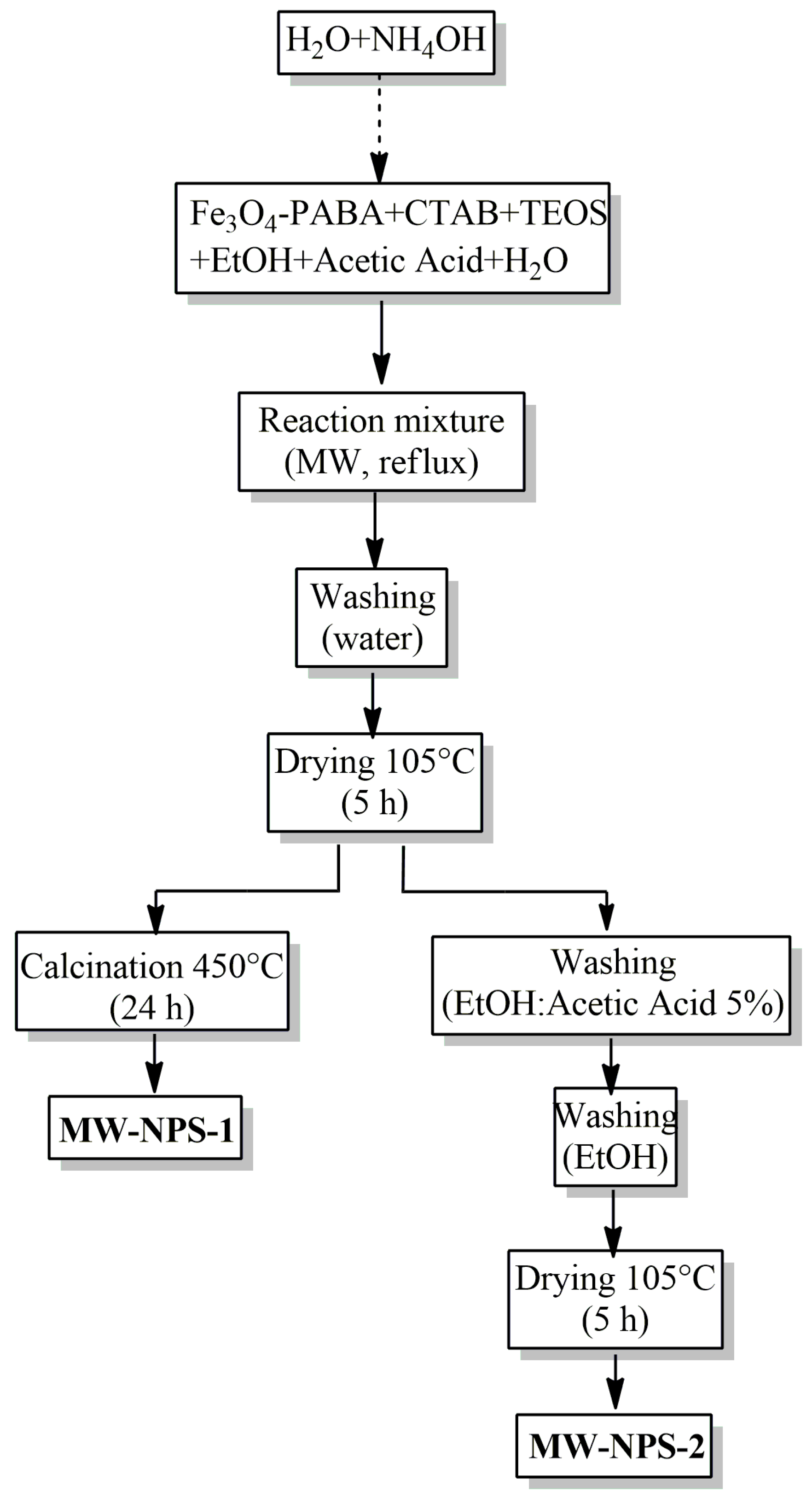
2.2.1. Synthesis of Nanostructured System
- a.
- Synthesis of magnetic core–shell nanoparticles (Fe3O4-PABA)
- b.
- Core-shell Fe3O4-PABA-SiO2 nanoparticle synthesis
- c.
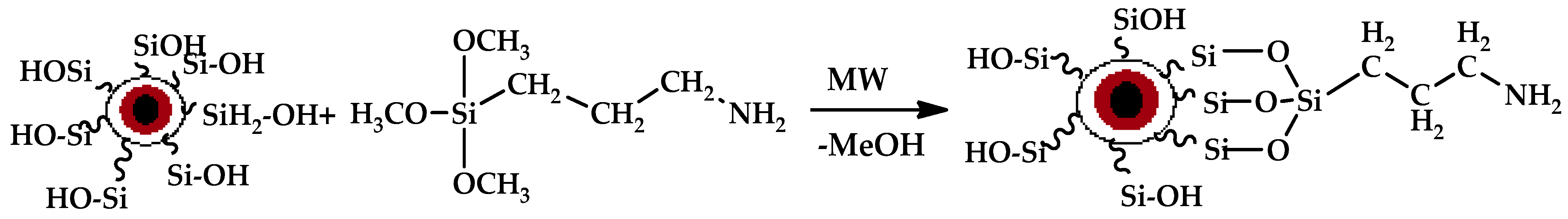
- Synthesis of Fe3O4-PABA-SiO2-APS (MW-NPS-APS)
- d.
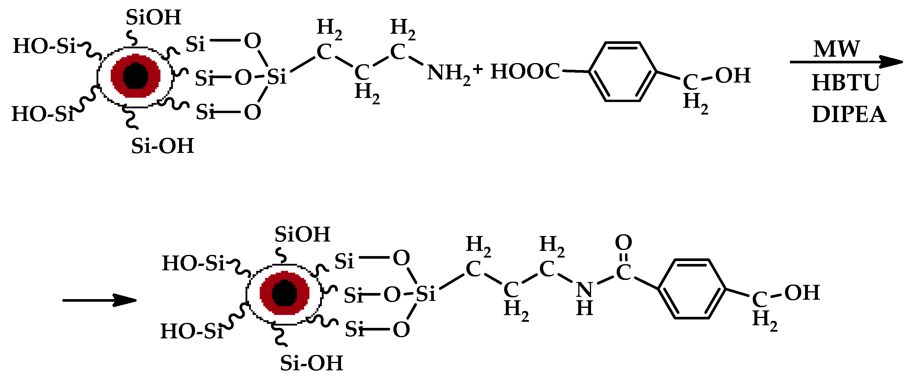
- Synthesis of Fe3O4-PABA-SiO2-APS-HMBA (MW-NPS-APS-HMBA)
2.2.2. The Peptide Synthesis
3. Results and Discussion
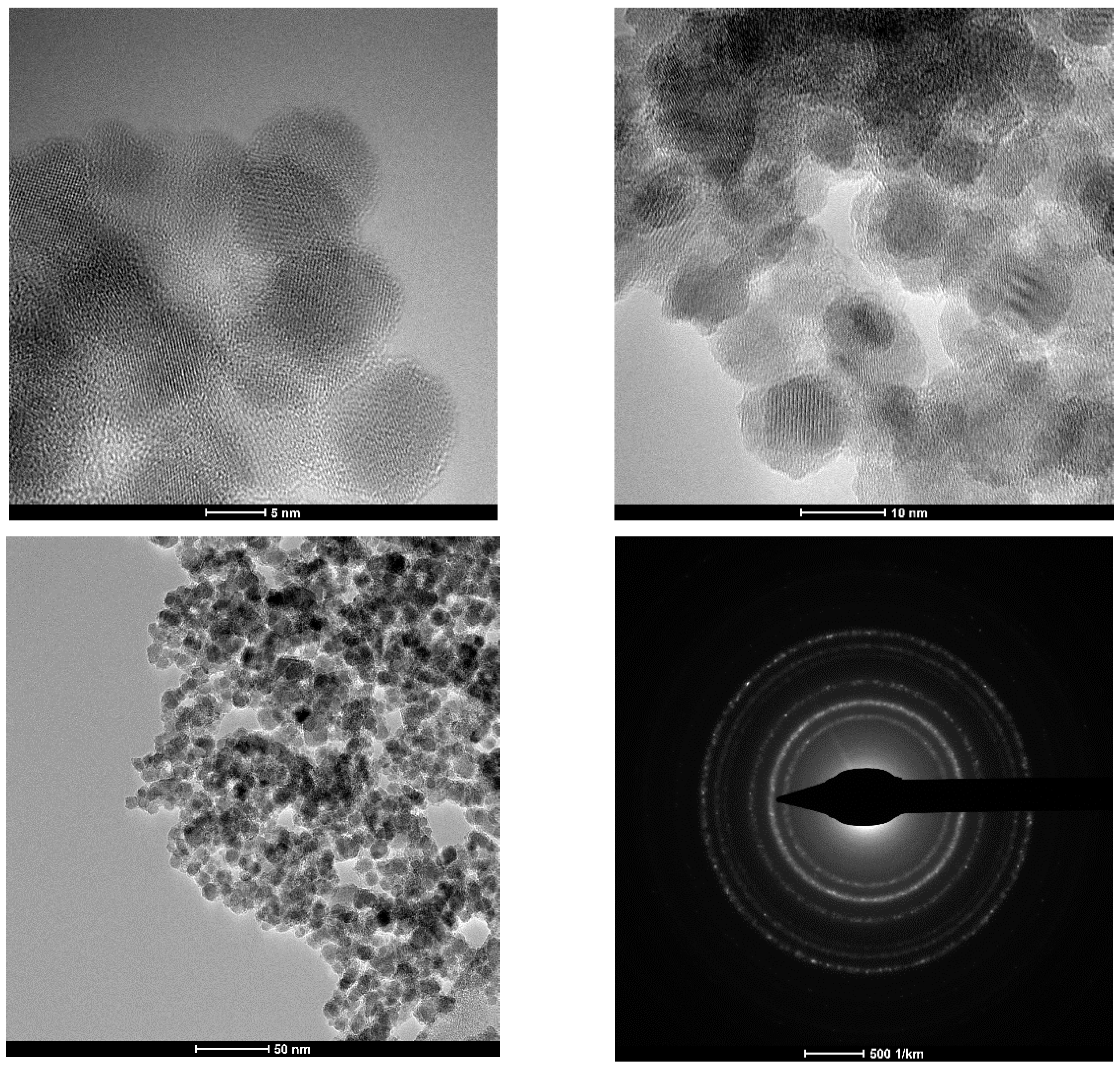
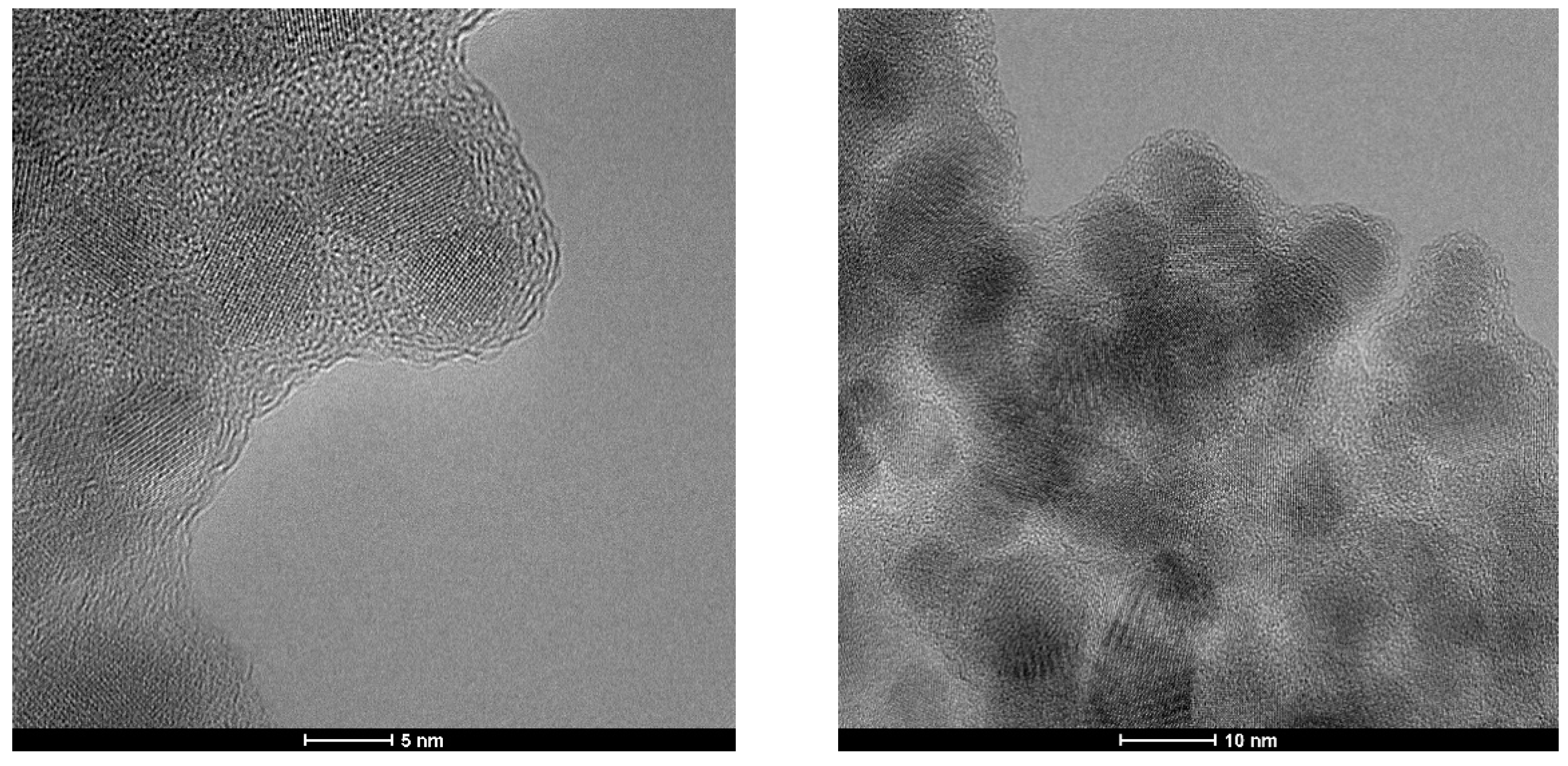
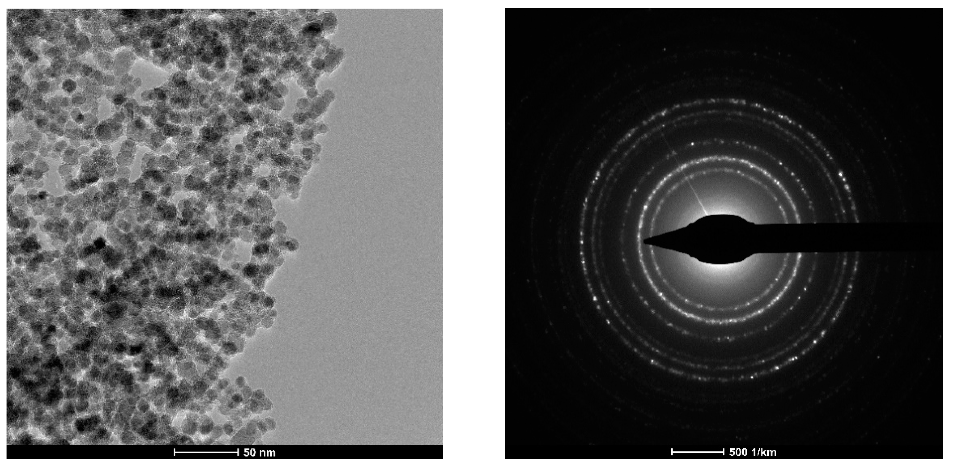
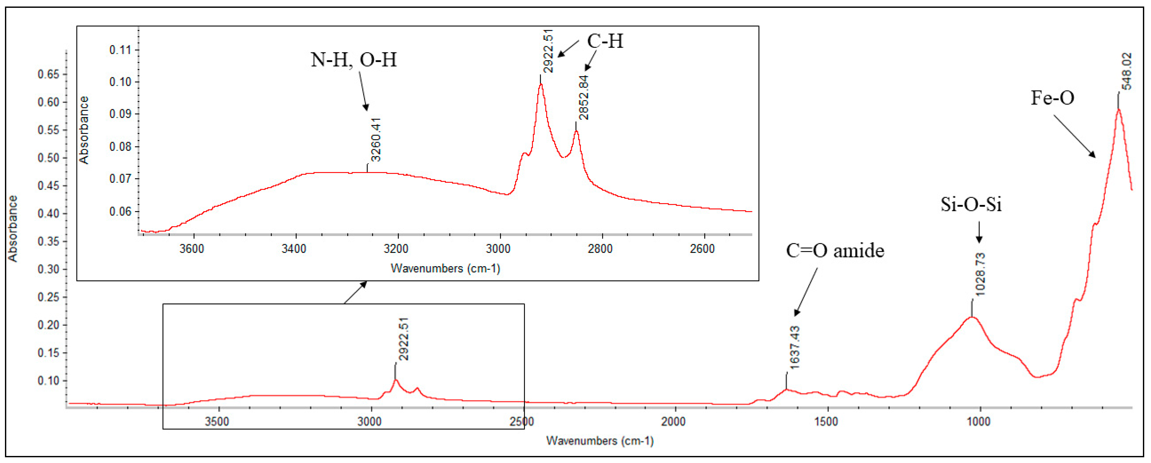
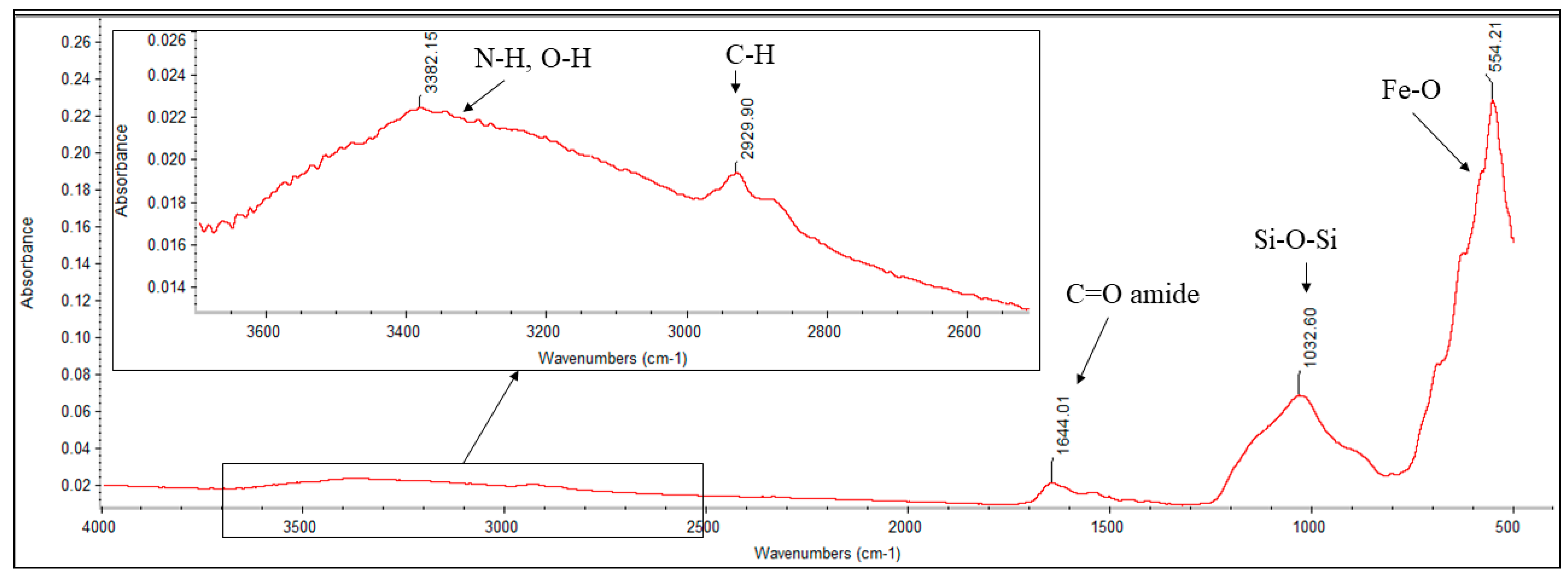
3.1. Nanostructured System
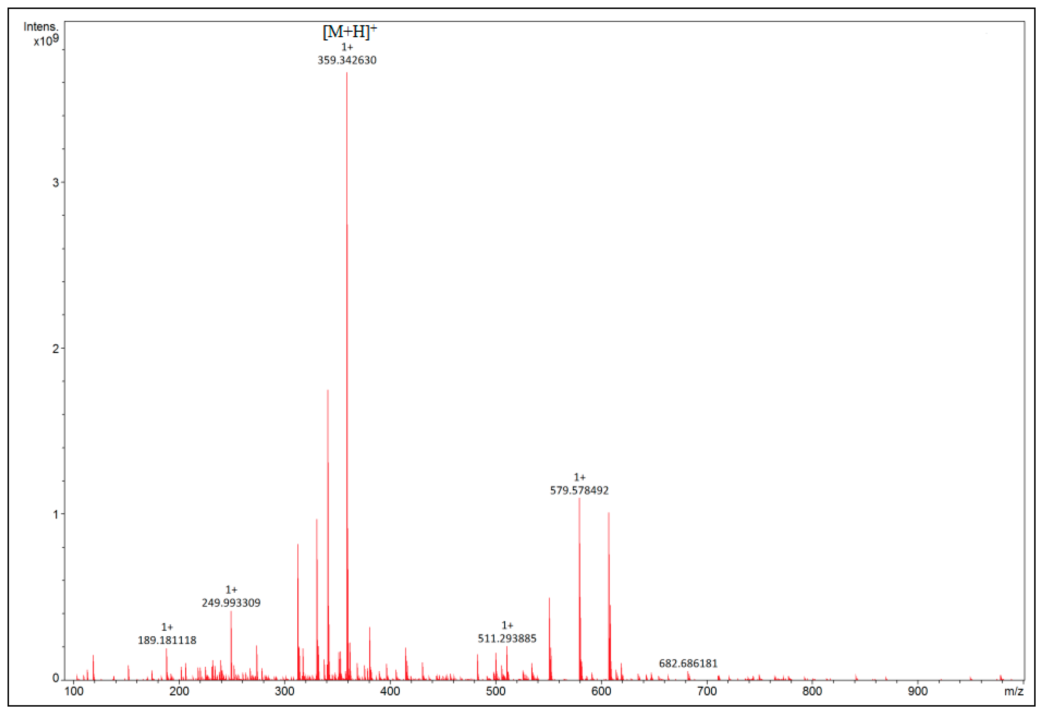
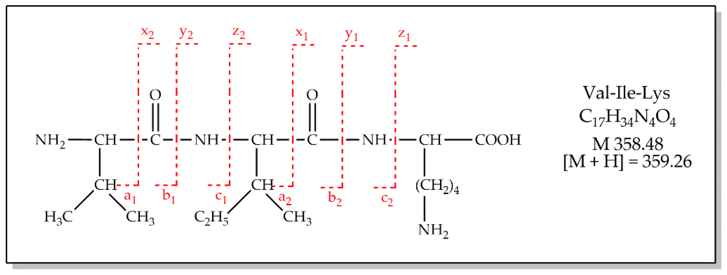
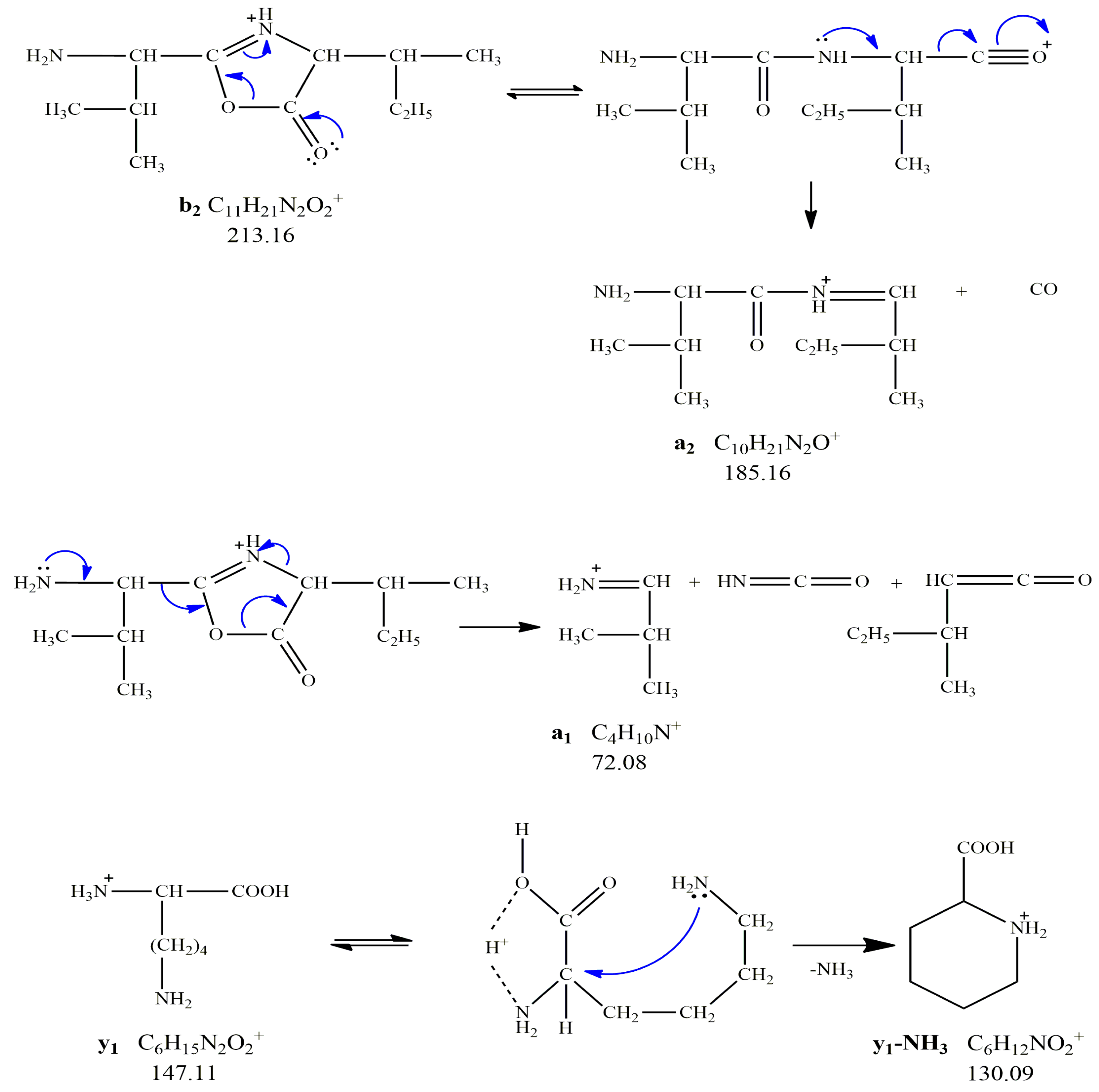
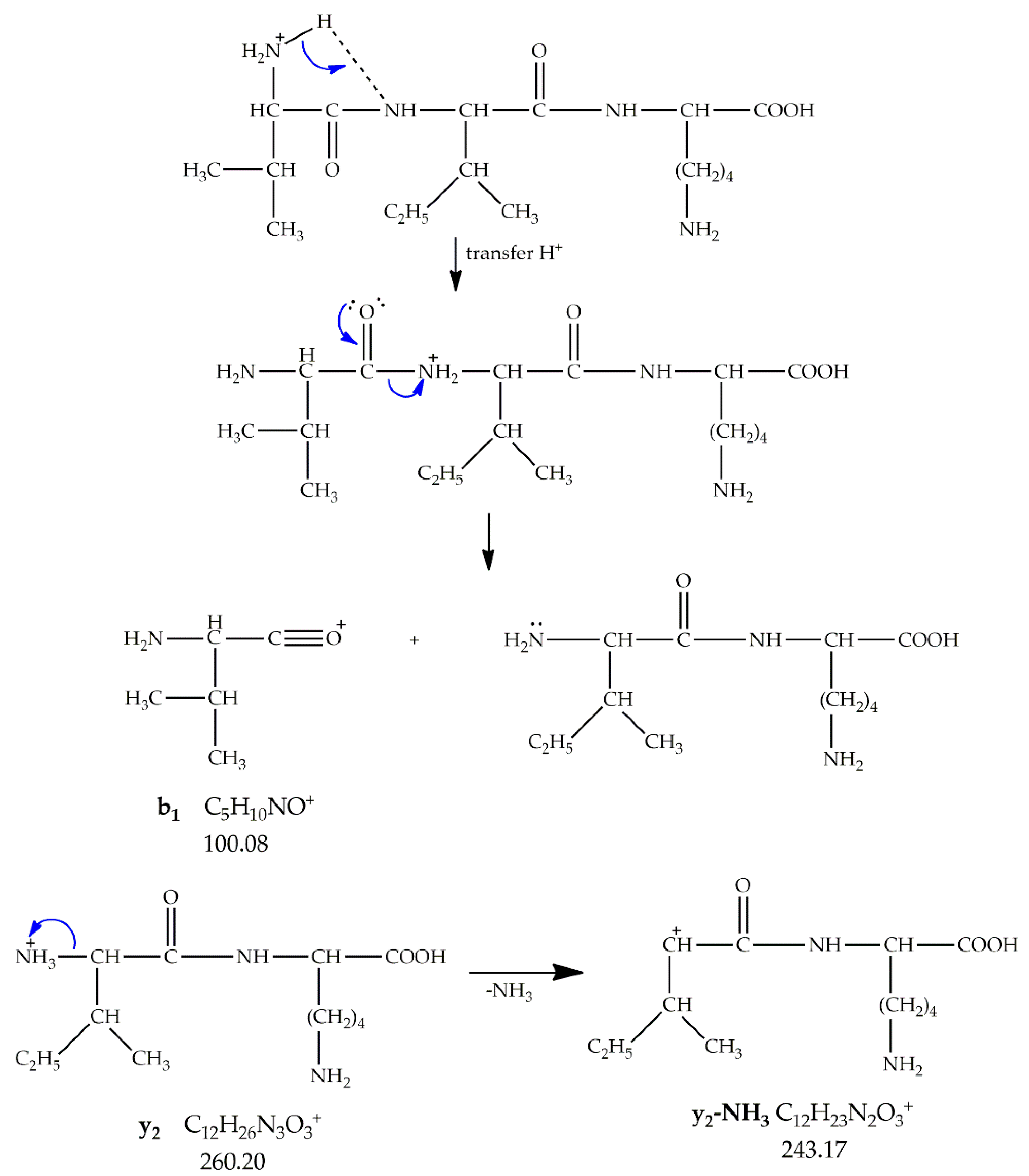
3.2. Peptide Synthesis
4. Conclusions
Supplementary Materials
Author Contributions
Funding
Institutional Review Board Statement
Informed Consent Statement
Conflicts of Interest
References
- Liu, F.; Niu, F.; Peng, N.; Su, Y.; Yang, Y. Synthesis, characterization, and application of Fe3O4@SiO2–NH2 nanoparticles. RSC Adv. 2015, 5, 18128–18136. [Google Scholar] [CrossRef]
- Zhu, N.; Ji, H.; Yu, P.; Niu, J.; Farooq, M.; Akram, M.W.; Udego, I.O.; Li, H.; Niu, X. Surface Modification of Magnetic Iron Oxide Nanoparticles. Nanomaterials 2018, 8, 810. [Google Scholar] [CrossRef] [PubMed] [Green Version]
- Gdula, K.; Dąbrowski, A.; Skwarek, E. Synthesis, surface characterization and electrokinetic properties of colloidal silica nanoparticles with magnetic core. Adsorption 2016, 22, 681–688. [Google Scholar] [CrossRef] [Green Version]
- Owens, G.J.; Singh, R.K.; Foroutan, F.; Alqaysi, M.; Han, C.-M.; Mahapatra, C.; Kim, H.-W.; Knowles, J.C. Sol-gel based materials for biomedical applications. Prog. Mater. Sci. 2016, 77, 1–79. [Google Scholar] [CrossRef] [Green Version]
- Sun, X.; Bai, X.; Liu, X. Preparation and characterization of magnetic mesoporous silica nanoparticles with a core-shell structure. IFOST 2013, 1, 56–60. [Google Scholar] [CrossRef]
- Sharaf, Z.; Bakhshi, B.; Javidi, J.; Adrangi, S. Synthesis of Silica-coated Iron Oxide Nanoparticles: Preventing Aggregation without Using Additives or Seed Pretreatment. Iran J. Pharm. Res. 2018, 17, 386–395. [Google Scholar]
- Chellappa, M.; Vijayalakshmi, U. Fabrication of Fe3O4-silica core-shell magnetic nano-particles and its characterization for biomedical applications. Mater. Today Proc. 2019, 9, 371–379. [Google Scholar] [CrossRef]
- Andrade, A.L.; Souza, D.M.; Pereira, M.C.; Fabris, J.D.; Domingues, R.Z. Synthesis and characterization of magnetic nanoparticles coated with silica through a sol-gel approach. Cerâmica 2009, 55, 420–424. [Google Scholar] [CrossRef] [Green Version]
- Moroşan, A.; Mihaiescu, D.E.; Istrati, D.; Voicu, G.; Radu, M.; Hanganu, A.; Stan, R. Functionalized silica shell magnetic nanoparticles for nanophase peptide synthesis applications. Micropor. Mesopor. Mater. 2019, 286, 45–56. [Google Scholar] [CrossRef]
- Norén, K.; Kempe, M. Multilayered Magnetic Nanoparticles as a Support in Solid-Phase Peptide Synthesis. Int. J. Pept. Res. Ther. 2009, 15, 287–292. [Google Scholar] [CrossRef]
- Stutz, C.; Bilecka, I.; Thünemann, A.F.; Niederberger, M.; Börner, H.G. Superparamagnetic core–shell nanoparticles as solid supports for peptide synthesis. ChemComm 2012, 48, 7176–7178. [Google Scholar] [CrossRef] [PubMed]
- Hansen, J.; Diness, F.; Meldal, M. C-Terminally modified peptides via cleavage of the HMBA linker by O-, N- or S-nucleophiles. Org. Biomol. Chem. 2016, 14, 3238–3245. [Google Scholar] [CrossRef] [PubMed] [Green Version]
- Riaz, S.; Ashraf, R.; Akbar, A.; Naseem, S. Microwave Assisted Iron Oxide Nanoparticles—Structural and Magnetic Properties. IEEE Trans. Magn. 2014, 50, 1–4. [Google Scholar] [CrossRef]
- Fernández-Barahona, I.; Muñoz-Hernando, M.; Herranz, F. Microwave-Driven Synthesis of Iron-Oxide Nanoparticles for Molecular Imaging. Molecules 2019, 24, 1224. [Google Scholar] [CrossRef] [Green Version]
- Díaz de Greñu, B.; de los Reyes, R.; Costero, A.M.; Amorós, P.; Ros-Lis, J.V. Recent Progress of Microwave-Assisted Synthesis of Silica Materials. Nanomaterials 2020, 10, 1092. [Google Scholar] [CrossRef]
- Roto, R. Surface Modification of Fe3O4 as Magnetic Adsorbents for Recovery of Precious Metals. In Advanced Surface Engineering Research; Chowdhury, M.A., Ed.; IntechOpen: London, Uk, 2018; pp. 128–139. [Google Scholar] [CrossRef] [Green Version]
- Moroșan, A.; Mihaiescu, D.E.; Istrati, D.; Voicu, G.; Fudulu, A.; Stan, R. Polar shell magnetic nanostructured systems for heterogeneous nanophase reactions. Sci. Bull. B Chem. Mater. Sci. UPB 2018, 80, 53–64. [Google Scholar]
- Chen, H.; Deng, C.; Zhang, X. Synthesis of Fe3O4@SiO2@PMMA Core–Shell–Shell Magnetic Microspheres for Highly Efficient Enrichment of Peptides and Proteinsfor MALDI-ToF MS Analysis. Angew. Chem. Int. Ed. 2010, 49, 607–611. [Google Scholar] [CrossRef]
- Saha, R.; Uppaluri, R.V.S.; Tiwari, P. Effects of interfacial tension, oil layer break time, emulsification and wettability alteration on oil recovery for carbonate reservoirs. Colloids Surf. A Physicochem. Eng. Asp. 2018, 559, 92–103. [Google Scholar] [CrossRef]
- Patole, A.S.; Hyeon, J.-M.; Hyun, J.-M.; Kim, T.-H.; Patole, S.P.; Hong, D.-J.; Lee, C.-B.; Choi, C.-H. Synthesis and Characterization of a Novel Laser Ablation Sensitive Triazene Incorporated Epoxy Resin. Electron. Mater. Lett. 2014, 10, 173–182. [Google Scholar] [CrossRef]
- Nonaka, A.G.; Batista, M.A.; da Costa, A.C.S.; Inoue, T.T.; Bonadio, T.G.M.; de Souza Junior, I.G. Kinetics of Thermal Transformation of Synthetic Al-Maghemites into Al-Hematites. Rev. Bras. Ciênc. Solo. 2017, 41, e0160384. [Google Scholar] [CrossRef] [Green Version]
- Blokhin, D.S.; Efimov, S.V.; Klochkov, A.V.; Yulmetov, A.R.; Filippov, A.V.; Antzutkin, O.N.; Aganov, A.V.; Klochkov, V.V. Spatial Structure of the Decapeptide Val-Ile-Lys-LysSer-Thr-Ala-Leu-Leu-Gly in Water and in a Complex with Sodium Dodecyl Sulfate Micelles. Appl. Magn. Reson. 2011, 41, 267–282. [Google Scholar] [CrossRef]
- Afonso, C.; Cole, R.B.; Tabet, J.-C. Dissociation of Even-Electron Ions. In Electrospray and MALDI Mass Spectrometry Fundamentals, Instrumentation, Practicalities, and Biological Applications; Cole, R.B., Ed.; John Wiley & Sons, Inc.: Hoboken, NJ, USA, 2010; pp. 632–659. [Google Scholar]
- Demarque, D.P.; Crotti, A.E.M.; Vessecchi, R.; Lopes, J.L.C.; Lopes, N.P. Fragmentation reactions using electrospray ionization mass spectrometry: An important tool for the structural elucidation and characterization of synthetic and natural products. Nat. Prod. Rep. 2016, 33, 432–455. [Google Scholar] [CrossRef] [PubMed] [Green Version]


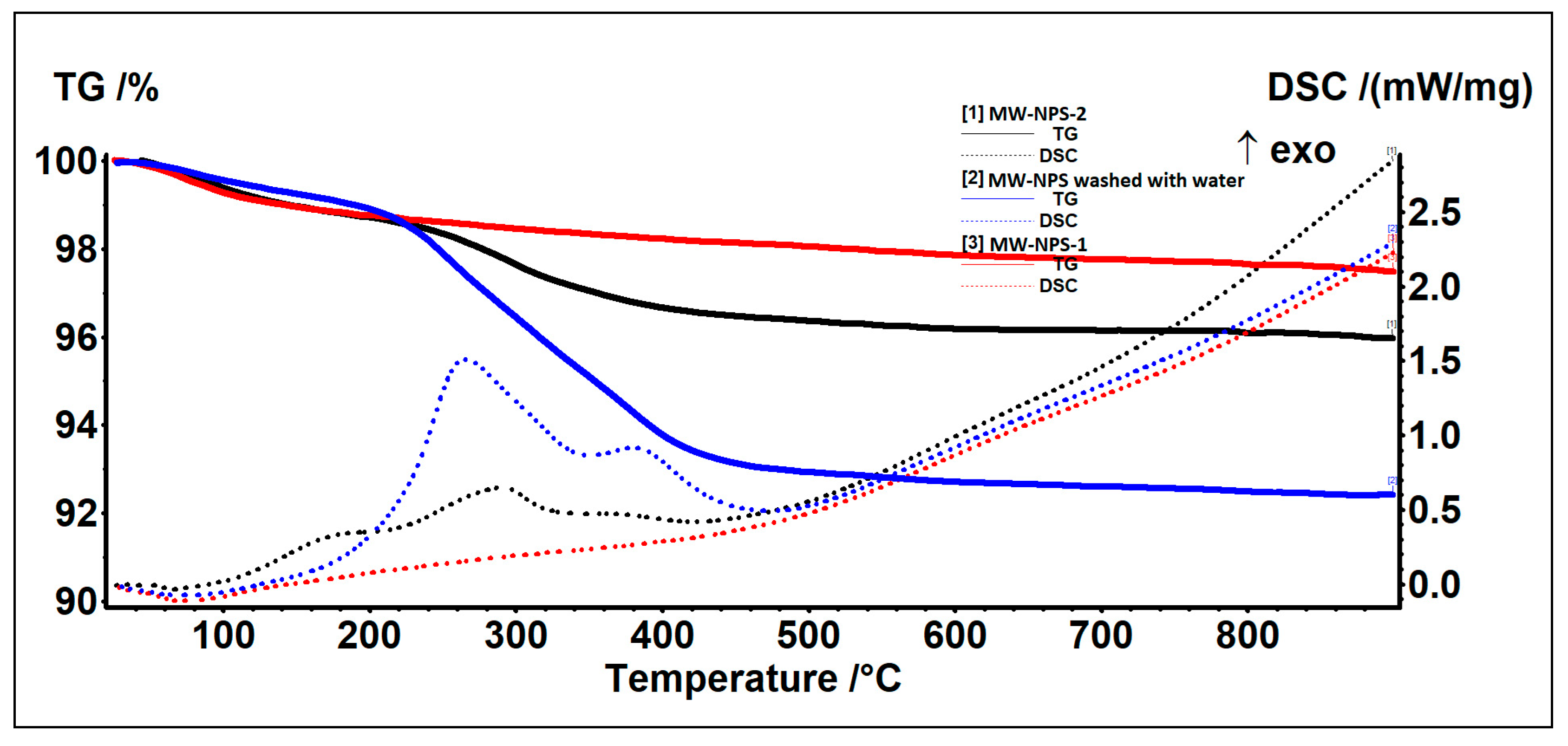
| Samples | RT | Residue | Endo | Exo I | Exo II | Exo III | ||
|---|---|---|---|---|---|---|---|---|
| RT-190 | 190–500 | 500–900 | ||||||
| MW-NPS washed with water | 1.01% | 6.06% | 0.51% | 92.41% | 74.2 °C | - | 265.8 °C | 379.5 °C |
| MW-NPS-1 | 1.23% | 0.73% | 0.56% | 97.48% | 73.2 °C | - | - | - |
| MW-NPS-2 | 1.25% | 2.39% | 0.39% | 95.97% | 65.2 °C | 170.0 °C | 290.4 °C | 373.6 °C |
| Samples | BET Analysis | ||
|---|---|---|---|
| SBET | dBJH | Vp | |
| Fe3O4-PABA | 124 m2 g−1 | 12.019 nm | 0.295 cm3 g−1 |
| MW-NPS-1 | 127 m2 g−1 | 33.223 nm | 0.690 cm3 g−1 |
| MW-NPS-2 | 122 m2 g−1 | 19.497 nm | 0.595 cm3 g−1 |
| Magnetic Nanostructured Support | Peptide Sequence | Chemical Formula | MW (g mol−1) |
|---|---|---|---|
| MW-NPS-2-APS-HMBA | Val-Ile-Lys (VIK) | C17H34N4O4 | 358.46 |
| Fragment Type | Chemical Formula | m/z Theoretical | m/z Experimental |
|---|---|---|---|
| a1 | C4H10N+ | 72.08 | - |
| a2 | C10H21N2O+ | 185.16 | 185.92 |
| b1 | C5H10NO+ | 100.08 | - |
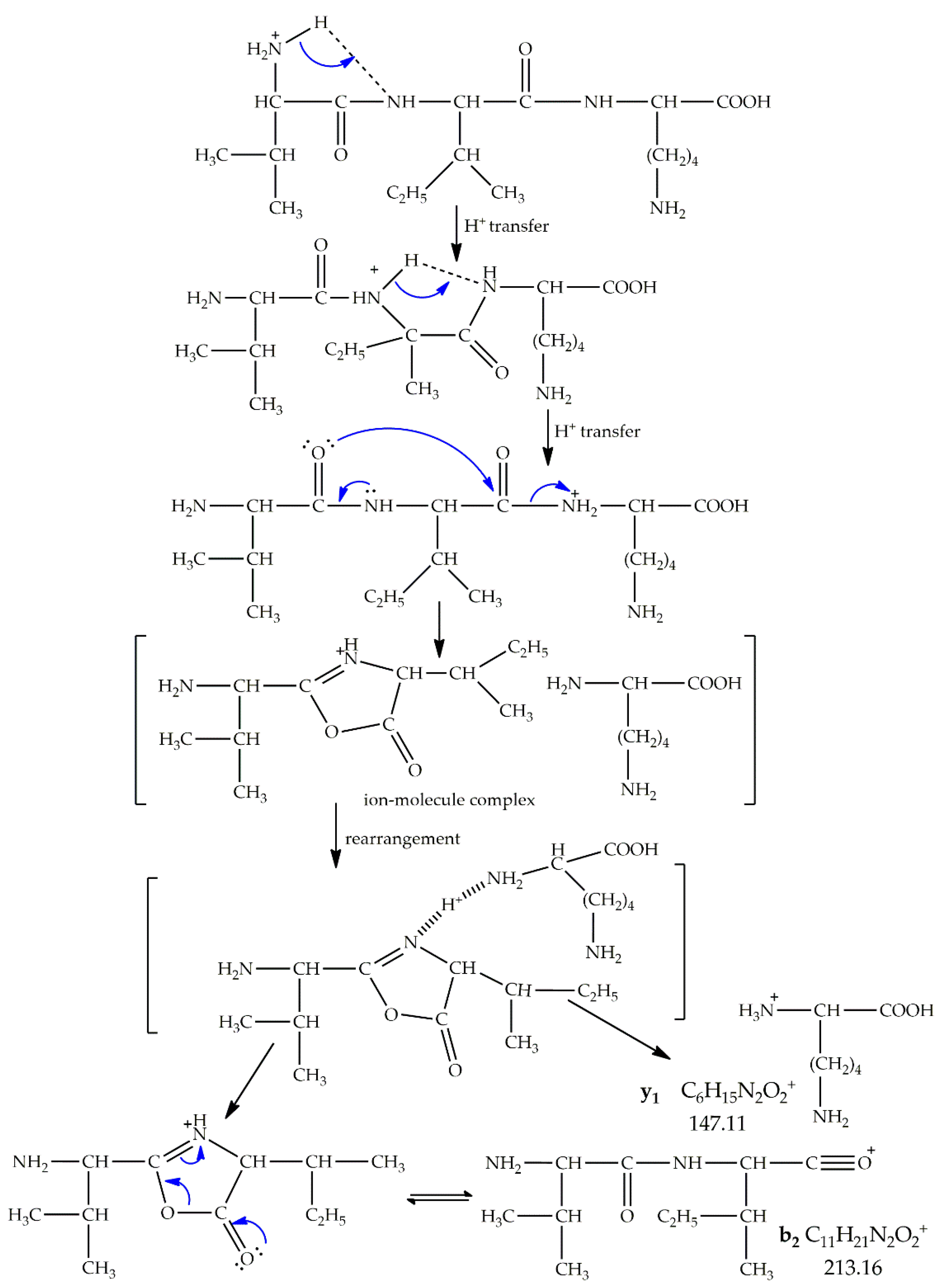
| b2 | C11H21N2O2+ | 213.16 | 213.90 |
| c1 | C5H13N2O+ | 117.10 | 117.43 |
| c2 | C11H24N3O2+ | 230.19 | 230.99 |
| x1 | C7H13N2O3+ | 173.09 | 173.00 |
| x2 | C13H24N3O4+ | 286.18 | 286.82 |
| y1 | C6H15N2O2+ | 147.11 | 147.99 |
| y2 | C12H26N3O3+ | 260.20 | 260.88 |
| z1 | C6H12NO2+ | 130.09 | 130.16 |
| z2 | C12H23N2O3+ | 243.17 | 243.80 |
| Fragment Type | Chemical Formula | m/z Theoretical | m/z Experimental |
|---|---|---|---|
| [M+H-H2O]+ | C17H33N4O3+ | 341.25 | 341.26 |
| [M+H-CO-H2O]+ | C16H33N4O2+ | 313.26 | 313.22 |
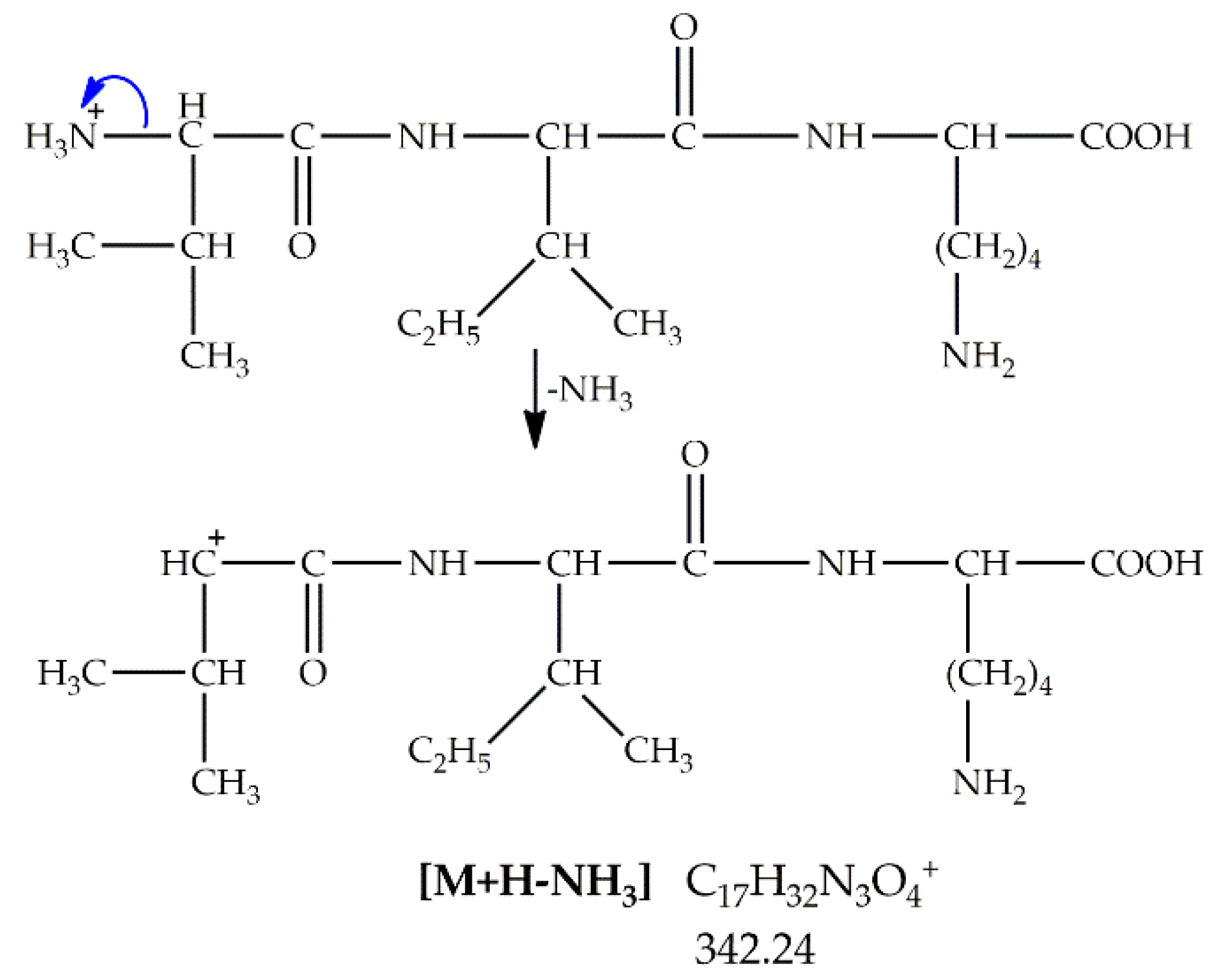
| [M+H-NH3]+ | C17H32N3O4+ | 342.23 | 342.26 |
| [M-H2O+Na]+ | C17H32N4O3Na+ | 363.23 | 363.30 |
| [M+H-2H2O]+ | C17H31N4O2+ | 323.24 | 323.24 |
Publisher’s Note: MDPI stays neutral with regard to jurisdictional claims in published maps and institutional affiliations. |
© 2021 by the authors. Licensee MDPI, Basel, Switzerland. This article is an open access article distributed under the terms and conditions of the Creative Commons Attribution (CC BY) license (https://creativecommons.org/licenses/by/4.0/).
Share and Cite
Istrati, D.; Moroșan, A.; Stan, R.; Vasile, B.Ș.; Vasilievici, G.; Oprea, O.; Dolete, G.; Purcăreanu, B.; Mihaiescu, D.E. Microwave-Assisted Sol–Gel Preparation of the Nanostructured Magnetic System for Solid-Phase Synthesis. Nanomaterials 2021, 11, 3176. https://0-doi-org.brum.beds.ac.uk/10.3390/nano11123176
Istrati D, Moroșan A, Stan R, Vasile BȘ, Vasilievici G, Oprea O, Dolete G, Purcăreanu B, Mihaiescu DE. Microwave-Assisted Sol–Gel Preparation of the Nanostructured Magnetic System for Solid-Phase Synthesis. Nanomaterials. 2021; 11(12):3176. https://0-doi-org.brum.beds.ac.uk/10.3390/nano11123176
Chicago/Turabian StyleIstrati, Daniela, Alina Moroșan, Raluca Stan, Bogdan Ștefan Vasile, Gabriel Vasilievici, Ovidiu Oprea, Georgiana Dolete, Bogdan Purcăreanu, and Dan Eduard Mihaiescu. 2021. "Microwave-Assisted Sol–Gel Preparation of the Nanostructured Magnetic System for Solid-Phase Synthesis" Nanomaterials 11, no. 12: 3176. https://0-doi-org.brum.beds.ac.uk/10.3390/nano11123176