
Application of Nanotechnology in Analysis and Removal of Heavy Metals in Food and Water Resources


1
School of Chinese Medicine, Hong Kong Baptist University, Hong Kong 999077, China
2
Institute of Basic Research in Clinical Medicine, China Academy of Chinese Medical Sciences, Beijing 100700, China
*
Authors to whom correspondence should be addressed.
Nanomaterials 2021, 11(7), 1792; https://0-doi-org.brum.beds.ac.uk/10.3390/nano11071792
Submission received: 6 June 2021
/
Revised: 2 July 2021
/
Accepted: 5 July 2021
/
Published: 9 July 2021
(This article belongs to the Special Issue Advances in Food Nanotechnology)
Abstract
:Toxic heavy metal contamination in food and water from environmental pollution is a significant public health issue. Heavy metals do not biodegrade easily yet can be enriched hundreds of times by biological magnification, where toxic substances move up the food chain and eventually enter the human body. Nanotechnology as an emerging field has provided significant improvement in heavy metal analysis and removal from complex matrices. Various techniques have been adapted based on nanomaterials for heavy metal analysis, such as electrochemical, colorimetric, fluorescent, and biosensing technology. Multiple categories of nanomaterials have been utilized for heavy metal removal, such as metal oxide nanoparticles, magnetic nanoparticles, graphene and derivatives, and carbon nanotubes. Nanotechnology-based heavy metal analysis and removal from food and water resources has the advantages of wide linear range, low detection and quantification limits, high sensitivity, and good selectivity. There is a need for easy and safe field application of nanomaterial-based approaches.
1. Introduction
Heavy metals generally have densities of more than 5 g/cm3. Most heavy metals, such as arsenic, lead, mercury, chromium, zinc, cadmium, copper, and nickel, are regarded as environmental pollutants [1]. Since heavy metals are not biodegradable, heavy metal ions in waters and in soil may be biologically accumulated via the food chain towards the human body [2]. Other than the essential elements needed for human body, such as sodium, calcium, and zinc, some heavy metals exert serious toxicities even at low intake levels (Table 1). However, heavy metals such as copper, ion, cobalt, and chromium in small concentrations are essential elements in the human body. Therefore, it is necessary to detect and remove heavy metals from our food and water resources.
Conventional approaches for heavy metal detection from complex matrices include atomic absorption spectroscopy [3], neutron activation analysis [4], X-ray fluorescence spectrometry [5], energy dispersive X-ray fluorescence [6], inductively coupled plasma mass spectroscopy [7,8], inductively coupled plasma atomic/optical emission spectrometry [9], and flame atomic absorption spectrometry [10]. Although they can generally achieve satisfactory detection sensitivity, they are inevitably challenged with heavy equipment, high cost, and lab-only and complicated operation. Classical methods for heavy metal removal from wastewater include precipitation, ion exchange [11], reverse osmosis [12], membrane filtration [13] and oxidation [14]. However, their effectiveness is limited due to disadvantages of sludge contamination, high pH sensitivity, and corrosiveness [15,16,17].
Nanotechnology refers to employing multidisciplinary techniques integrating principles of physics, chemistry, engineering, and biology to build nanoscale materials or devices [18]. With the upsurge of nanotechnology, nanomaterials have been delicately designed and fabricated for heavy metal detection and removal, exerting numerous advantages as compared to the previous methods.

Table 1.
Representative toxic heavy metals and their adverse effects on human health.
| Element | Adverse Effects on Human Health | Ref. |
|---|---|---|
| Lead (Pb) | Kidney damage, cognitive disorders, hypertension. | [19] |
| Mercury (Hg) | Maternal and fetal toxicity; mental retardation, cerebral palsy, deafness, blindness, dysarthria; gastrointestinal tract toxicity: necrotic intestinal mucosa, abdominal pain, vomiting, bloody diarrhea. | [20,21] |
| Arsenic (As) | Skin lesions, blackfoot disease, peripheral neuropathy, encephalopathy, hepatomegaly, liver cirrhosis, altered heme metabolism, myeloid depression, diabetes, proximal tubule degeneration, papillary and cortical necrosis. | [22,23] |
| Chromium (Cr) | Skin rashes, stomach ulcers, weakened immune systems, kidney and liver damage, genetic material change, lung cancer; respiratory system issues: rhinitis, pharyngitis, laryngitis, bronchitis. | [24] |
| Copper (Cu) | Wilsons’s disease, upper respiratory tract irritation, nasal mucosa, hemolytic anemia, epigastric pain, nausea, dizziness. | [25] |
| Nickel (Ni) | Pneumoconiosis, asthma, lung and laryngeal cancer. | [26] |
| Zinc (Zn) | Tachycardia, vascular shock, dyspepsia, nausea, headache, nasal and lung cancer, asthma, vomiting, diarrhea, hypoglycemia, pancreatitis and liver parenchyma damage, growth and reproduction disorders. | [27] |
| Cadmium (Cd) | Kidney (proximal renal tubule cells), lung, liver, and vascular system damage; bone demineralization. | [28] |
2. Nanotechnology-Based Heavy Metal Analysis in Food and Water
2.1. Electrochemical Analysis by Nanosensors
2.1.1. Carbon-Based Nanomaterials
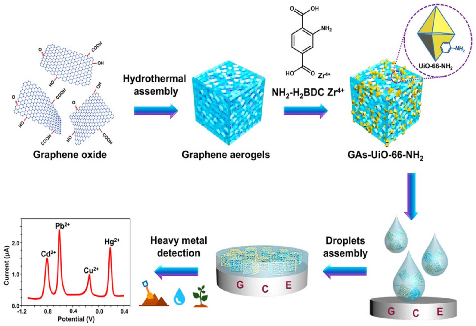
Graphene, graphene oxides (GO), and reduced graphene oxide (rGO), all with heavy metal ion adsorption capability, have been developed as advanced nano-electrocatalyst for constructing electrochemical sensors (Table 2). Graphene-based electrodes coated with in-situ bismuth film [29] and electrostatically attracted phytic acid [30], respectively, were reported with boosted sensitivity towards Pb2+ and Cd2+. Polypyrrole (PPy), a conductive polymer, can be physically adsorbed on GO with good dispersion [31] and subsequently enhance electrode conductivity [32]. Nanocomposite of GO linked with a bipyridine ligand, [Ru(bpy)3]2+, was used to modify a gold (Au) electrode with enhanced mass transport efficiency and charge transfer, which favored the sensitive and selective detection of metal ions [33]. Moreover, as a novel porous derivative of GO, graphene aerogel (GA), has unique porous structure which provides abundant coupling locations with metal–organic frameworks (MOFs). A GA-MOF composite-modified glass carbon electrode (GCE) showed enhanced conductivity and high affinity to heavy metals ions via hydrophilic groups from MOF; the GA-MOF-based GCE was demonstrated with simultaneous detection of various heavy metal ions in river water, soil, and spinach (Figure 1) [34].
Various rGO-based electrodes have been applied in food and water samples. rGO has strong hydrophilicity and strong adsorption capacity for metal ions because of the functional groups on its surface [35]. An Au electrode covered by micro-patterned rGO and electrodeposited bismuth was applied in trace analysis of Cd2+ and Pb2+ in drinking water [36]. Green hybrid electrochemical nanosensors constructed with rGO, cellulose nano whiskers (promoted green reduction of GO resulting in rGO), and polyamide 6 electrospun nanofibers (increased electrode surface area and amplified signal) were applied in detecting heavy metals in water samples [37,38].
Carbon nanotubes (CNTs), cylindrical macromolecules with carbon atoms arranged in hexagonal rings, have significant advantages of ordered structure, high ground object ratio, good mechanical strength, and high electrical conductivity [39]. Single-walled CNTs (SWCNTs) have high sensitivity to local dielectric or redox environment due to their band gap; they also specially undergo strong redox reaction with Hg2+. SWCNT-based nanosensors with good sensitivity and selectivity were successfully prepared, for instance, to analyze drinking water samples [40]. Multi-walled CNTs (MWCNTs) have become one of the most active materials in carbon paste electrodes due to their unique structure. A super selectivity sensor was reported comprising MWCNTs (as a carbon paste electrode), 1-n-butyl-3-methylimidazolium tetrafluoroborate (BMIM·BF4; as a super conductive binder), and 1-(2-ethoxyphenyl)-3-(3-nitrophenyl) triazene (ENTZ; as an ionophore); the sensor showed greatly enhanced potentiometric sensitivity, good reproducibility, low response time and long-term stability in quantification of Hg2+ in water samples [41].
When designing carbon-based sensing electrodes for detecting heavy metal ions, the combination of CNTs and graphene derivatives works better than either alone. A study used graphene-MWCNTs to prepare nanocomposites with excellent conductivity were obtained conveniently by the direct electrochemical reduction of GO–MWCNTs nanocomposites. The graphene-MWCNTs-modified electrode was successfully applied to the simultaneous detection of real electroplating effluent samples containing a large number of surface-active impurities [42]. In another study, 2-aminothiophene (L)-grafted carboxylate-functionalized MWCNTs (L-g-MWCNT-COOH) were synthesized as a sensor with hydrophobic environment and high complex stability, due to the presence of hard donor atoms (N, O, and S) and aromatic rings. The sensor was applied to various environmental samples [43].
Ordered mesoporous carbon nitride (MCN) as the platform of electrochemical sensor has great detection sensitivity of phenols and heavy metals [44]; self-doped polyanilines (SPANs) have strong conductivity and electrochemical activity. Therefore, SPAN/MCN-modified GCE, combining advantages of SPAN and MCN, has been reported as a novel electrode for heavy metal analysis in river water samples [45]. Graphitized carbon nitride (g-C3N4), a 2D structure composed of tri-s-triazine units connected with planar amino groups, with high affinity to metal ions due to its electrostatic bonding or coordination with several N atoms [46], is widely applied as green material for its simple preparation, good chemical stability and high catalytic activity [47]. An electrode modified by poly (2,5-bis(3,4-ethylenedioxythienyl) pyridine) (poly (BPE)) and g-C3N4 demonstrated to enhance adsorption of metal ions from tap water; the binding of poly(BPE) to g-C3N4 not only improved the surface conduction pathway of the electrode, but also produced a strong conjugation effect [48]. Colloidal hollow carbon nanospheres (HCNs) as sensing materials have distinctive features of large void and porous shell configuration, special spherical morphology, high specific surface area, and low density. A series of carbon electrodes with good microstructure were prepared by calculating contact points between HCNs and GCE, to bridge the electrode structure with sensing performance; such sensors were proved successful in analyzing heavy metals in drinking water [49].
2.1.2. Magnetic Nanoparticles
Magnetic nanoparticles (MNPs), such as superparamagnetic metal oxides, are commonly used as electrode modifiers, because they can significantly enhance electron strength transfer between the electrode and the analyte, leading to a high charge transfer capability [50]. Chitosan (CHT) is a natural polymer with abundant primary amino and hydroxyl groups; it has excellent adhesion, hydrophilicity, doping feasibility, and chemical stability. A novel electrode was coated with a mixture of Fe3O4 and CHT solution on an ionic liquid solid phase extraction, followed by a bismuth film deposited in situ; such electrode combined the advantages of Fe3O4, CHT, and ionic liquid screen-printed electrode (ILSPE), and accurately detected the content of Cd2+ in soil samples thus reducing the contamination of cultivated rice [51].
2.1.3. Semiconducting Nanomaterials
Tin oxide (SnO2) has well-known semi conductivity; however, SnO2 nanoparticles tend to aggregate easily, hindering their optimal performance. rGO can serve as a template to prevent the accumulation of SnO2 nanoparticles. rGO/SnO2 electrodes were reported with applications of synchronous and selective electrochemical detection of trace Cd2+, Pb2+, Cu2+, and Hg2+ in drinking water [52].
Porous silicon has been reported to be a nano scavenger for determining trace level of heavy metals in water. Thiol- and amino-grafted Si nanowire-modified electrodes exhibit poor electrochemical stability and sensitivity [53], such defects can be overcome by combining Si with a conductive polymer, e.g. poly (3,4-ethyldioxythiophene) (PEDOT). PEDOT and its derivatives are typical electrode sensor materials, with low oxidation potential and high thermal and chemical stability. The polymers also contained a variety of special functional groups and donor atoms that enhanced their coordination ability with metal ions [54]. Similarly, poly(3,4-proplenedioxythiophene) (PProDOT) and its derivatives showed capability in detecting heavy metal ions [55] that thiol-grafted (3,4-proplenedioxythiophene) (PProDOT(MeSH)2)/Si composites significantly enhanced heavy metal adsorption and improved the performance of catalytic electrode materials in analyzing water samples (Figure 2) [56].
2.1.4. Noble Metal Nanoparticles
Gold nanoparticles (AuNPs) have been demonstrated to be appropriate to modify electrodes due to their small size, good electrical conductivity and excellent catalytic activity, based on which they can “guide” electroactive substances towards the electrodes. AuNP-modified GCE with chloride desorption can provide a three-times higher sensitivity in trace detection of Hg2+ than that with no desorption step. Such detection was proven successful in analyzing groundwater samples; the results were compared well with those obtained by conventional cold vapor atomic fluorescence spectroscopy [57].
Table 2.
Representative studies of electrochemical analysis of heavy metals from food and water resources by nanosensors.
Table 2.
Representative studies of electrochemical analysis of heavy metals from food and water resources by nanosensors.
| Electrode | Analytical Technique | Sample | Linear Range | Limit of Detection | Ref. |
|---|---|---|---|---|---|
| Graphene/Bi/PGE | SWASV | Canadian-certified reference water sample and tap water sample | 5–100 μg/L Cd2+; Pb2+ | 0.12 μg/L Cd2+ 0.29 μg/L Pb2+ | [29] |
| PA/PPy/GO | DPV | Tap water samples | 5–150 μg/L Cd2+; Pb2+ | 2.13 μg/L Cd2+ 0.41 μg/L Pb2+ | [30] |
| [Ru(bpy)3]2+/GO/AuNPs | DPV | Cauvery river water | 0.02–3 μM Cd2+; Pb2+, 0.02–2 μM As3+; Hg2+ | 2.8 nM Cd2+ 1.41 nM Pb2+ 2.3 nM As3+ 1.6 nM Hg2+ | [33] |
| MOF/GA/GCE | DPV | River water soil and vegetable sample (spinach) | 0.01−1.5 μM Cd2+, 0.001−2 μM Pb2+, 0.01−1.6 μM Cu2+, 0.001−2.2 μM Hg2+ | 0.02 μM Cd2+ 1.5 nM Pb2+ 7 nM Cu2+ 2 nM Hg2+ | [34] |
| Bi/rGO/Au electrode | SWASV | Drinking water sample | 1.0–120.0 μg/L Cd2+; Pb2+ | 0.4 μg/L Pb2+ 1.0 μg/L Cd2+ | [36] |
| PA6/CNW/rGO | DPV | Water sample | 2.5–200 μM Hg2+ | 5.2 nM Hg2+ | [38] |
| SWCNTs/SiO2 | ISE | Drinking water sample | 10 nM−1 mM Hg2+ | 10 nM Hg2+ | [40] |
| BMIM·BF4/MWCNTs/graphite powder | ISE | Water sample | 5.0 nM−1.0mM Hg2+ | 2.5 nM Hg2+ | [41] |
| Graphene/MWCNTs/Bi | SWASV | Real electroplating effluent samples | 0.5–30 mg/L Pb2+; Cd2+ | 0.2 μg/L Pb2+ 0.1 μg/L Cd2+ | [42] |
| L-g-MWCNTs/CPE | ISE | Soils, waste waters, lead accumulator waste and black tea | 5.9 nM − 1.0 mM Pb2+ | 3.2 nM Pb2+ | [43] |
| SPANs/MCN/GCE | SWASV | River water sample | 5–80 nM Cd2+; Pb2+ | 0.7 nM Cd2+ 0.2 nM Pb2+ | [45] |
| Poly (BPE)/g-C3N4 | DPV | Tap water samples | 0.12–7.2 μM Cd2+, 0.08–7.2 μM Pb2+, | 0.018 μM Cd2+ 0.00324 μM Pb2+ | [48] |
| HCNs/GCE | SWASV | Drinking water sample | / | 0.6 nM Pb2+ | [49] |
| Fe3O4/Bi/ILSPE | DPV | Soil sample | 0.5−40 μg/L Cd2+ | 0.05 μg/L Cd2+ | [51] |
| SnO2/rGO/GCE | SWASV | Drinking water sample | 0.3–1.2 μM Hg2+; Cu2+; Cd2+; Pb2+ | 0.3 nM Hg2+ 0.2 nM Cu2+ 0.1 nM Cd2+ 0.2 nM Pb2+ | [52] |
| PProDOT (MeSH)2/Si/GCE | DPV | Tap water sample | 0.04−2.8 μM Cd2+, 0.024−2.8 μM Pb2+, 0.16−3.2 μM Hg2+ | 0.00575 μM Cd2+ 0.0027 μM Pb2+ 0.0017 μM Hg2+ | [56] |
| AuNPs/GCE | SWASV | Natural groundwater sample | 0.3–10 nM Hg2+ | 80 pM Hg2+ | [57] |
Abbreviations: Square wave anodic stripping voltammetry (SWASV), differential pulse voltammetry (DPV), and ion-selective electrode (ISE), pencil graphite electrode (PGE), phytic acid (PA), polypyrrole (PPy), graphene oxide (GO), Au nanoparticles (AuNPs), metal–organic frameworks (MOFs), graphene aerogel (GA), glassy carbon electrode (GCE), reduced graphene oxide (rGO), polyamide 6 (PA6), cellulose nano whiskers (CNW), single-walled CNTs (SWCNTs), 1-n-butyl-3-methylimidazolium tetrafluoroborate (BMIM·BF4), multi-walled CNTs (MWCNTs), carbon paste electrode (CPE), self-doped polyanilines (SPANs), mesoporous carbon nitride (MCN), poly (2,5-bis(3,4-ethylenedioxythienyl)pyridine) (poly (BPE)), hollow carbon nanospheres (HCNs), ionic liquid screen-printed electrode (ILSPE), tin oxide (SnO2), thiol (–SH) grafted poly(3,4-proplenedioxythiophene) (PProDOT(MeSH)2), silicon spheres (Si).
2.2. Colorimetric Analysis Based on Gold and Silver Nanoparticles
2.2.1. Gold Nanoparticles
Since AuNPs have localized surface plasmon resonances (LSPR), some heavy metal ions can be analyzed based on dispersion of AuNPs [58] (Table 3). An Hg2+ sensor was reported constructed with 4-mercaptophenylboronic acid (MPBA) and AuNPs. The AuNPs can be initially stabilized with a coating of negatively charged citrate ions, which provide electrostatic repulsion preventing particle aggregation [59]. MPBA, an aggregation agent, exhibits a high affinity for AuNPs via Au–S crosslinking, leading to a visible color change of the AuNP solution from red to blue. With increasing concentration of Hg2+, the thiol group of MPBA would prefer binding with Hg2+ than polymerizing AuNPs, revealing the solution from blue to red [60]. Such Hg2+ nanosensors with high detection specificity were successfully applied in testing water samples [61] (Figure 3).
Besides particle dispersion, thiol-dependent aggregation of AuNPs can be utilized for detecting heavy metal ions, e.g. Co2+, Pb2+, Hg+, and Ag+. Co2+ level in aqueous solution can be measured based on aggregation of thiosulfate (S2O32−)-stabilized AuNPs with the addition of ethylenediamine (en), a chelating agent that forms complexes with heavy metal ions. In the presence of S2O32− coating on AuNP surface, the Co2+/en complexes can be oxidized to Co(en)33+ and in turn form (en)2CoS2O3+. The AuNPs will then aggregate due to insufficient surface charges, changing the solution from red to blue. Such label-free colorimetric sensing was adopted in rapid detection of Co2+ in drinking water and development of Co2+ test papers [62]. Similarly, in the case of measuring Pb2+ in environmental samples, the Pb/Au alloy formed on the surface of AuNPs would weaken the stability of Au+/S2O32− and thus enhance the access of 4-mercaptobutanol (4-MB) to AuNP surface, inducing AuNP aggregation [63]. When it comes to measuring aqueous Hg2+ and Ag+, AuNPs simultaneously coated with citric acid and Tween 20 can be used. The modified AuNPs are initially stable against high ionic strength; the citrate ions would induce bond formation of Hg–Au and Hg–Ag on AuNP surface, causing Tween 20 removal and consequently AuNP aggregation [64]. Furthermore, other thiol-containing substances, such as mercaptan carboxylic acid [65], quaternary ammonium compound, cysteine [66], dithioerythritol [67], and alkanethiols [68] have been reported in colorimetric detection for Hg2+.
Additionally, AuNPs can be capped by graphite-like nitride doped carbon quantum dots (AuNPs/g-CQDs) as a sensitive color-tuning indicator to measure the typical weak catalytic heavy metal ions of Cd2+ ions. Due to the large number of functional groups on the AuNPs/g-CQDs surface, the nanocomposite aggregates upon contacting Cd2+ ions. In addition, the biodistribution and aggregation of Cd2+ ions in mouse organ tissues have also been effectively detected, indicating that it has great potential in practical application [69].
Furthermore, leaching of AuNPs can be accelerated by GO in the presence of Pb2+, resulting in a color change. Such principle has been adopted in detecting Pb2+ in drinking water and river water samples [70].
2.2.2. Silver Nanoparticles
Modified AgNPs have been reported as heavy metal sensors based on different detection principles, including color changes due to (1) particle aggregation, (2) increased fluorescence intensity, and (3) silver chloride precipitate formation.
The detection of Cu2+ can be performed based on induced aggregation of thiomalic acid (TMA)-functionalized AgNPs. In the presence of Cu2+, carboxylic acid groups in TMA preferentially interact with Cu2+ to form stable complexes, inducing aggregation of the AgNPs to generate a red shift in LSPR band. Such sensors have been applied in real-time monitoring of Cu2+ in various environmental water samples [71]. Similarly, 5-sulfosalicylic acid -functionalized AgNPs (SAA/AgNPs) is widely used as a low-cost ($0.10 per sample) sensor in detecting Cd2+ ions, for example, in tap water, milk, urine, and serum samples [72]. Xylan-capped AgNPs can easily react with Hg2+; formation of Hg2+/xylan complex results in nanoparticle aggregation. Therefore, xylan-AgNPs composites can detect Hg2+ with high sensitivity and selectivity, as a simple and fast approach for detection of Hg2+ in environmental water [73].
Poly(acrylic acid)-templated silver nanoclusters (PAA/AgNCs) can measure Hg2+ ions via selective fluorescence initiation. In the presence of Hg2+ ions, the fluorescence intensity of AgNCs would raise dramatically, causing the solution to change color from purple to light red, which can be observed by naked eye. The method has been proved practical in analyzing samples of river water, tap water, and mineral water [74].
An AgCl/Ag sensor modified by natural seaweed polysaccharide carrageenan was reported exhibiting sensitive and robust detection of Hg2+ ions from environmental or biological samples, where AgCl/Ag interacted with Hg2+ ions to form an Hg–Ag alloy which reduced surface plasmon resonance (SPR) intensity, and the formation of silver chloride caused a visible change in solution from dark brown to white [75]. AgNPs can also be synthesized using Cordia myxa leaf extract for determination of Hg2+, where Ag atoms can be selectively oxidized by Hg2+, leading to AgNPs dissolution in the presence of Cl− ions [76].
Table 3.
Representative studies of colorimetric analysis of heavy metals from food and water resources based on nanoparticle aggregation.
Table 3.
Representative studies of colorimetric analysis of heavy metals from food and water resources based on nanoparticle aggregation.
| Electrode | Sample | Linear Range | Limits of Detection | Ref. |
|---|---|---|---|---|
| MPBA/AuNPs | Water samples | 0.01–5 μM Hg2+ | 8 nM Hg2+ | [61] |
| (en)/S2O32−/AuNPs | Drinking water | 0.1–0.7 μM Co2+ | 0.04 μM (2.36 ppb) Co2+ | [62] |
| 4-MB/AuNPs (S2O32−) | River water, Montana soil, and urine samples | 0.5–10 nM Pb2+ | 0.2 nM Pb2+ | [63] |
| Tween 20/Citrate ions/AuNPs | Drinking water and seawater sample | / | 0.1 μM Hg2+; Ag+ | [64] |
| DTETAuNPs | Water sample | 1–6 μM Hg2+ | 100 nM Hg2+ | [67] |
| AuNPs/g-CQDs | Mice organ tissues sample | 0.01–3.0 μM Cd2+ | 10 nM Cd2+ | [69] |
| AuNPs/GO/S2O32− | Pure drinking water and river water samples | 0.1–20 μM Pb2+ | 0.05 μM Pb2+ | [70] |
| TMA/AgNPs | Tap, lake, and river water sample | / | 0.5 nM Cu2+ | [71] |
| SAA/AgNPs | Tap water, lake water, milk, urine, and serum sample | 0.05–1.1 μM Cd2+ | 3.0 nM Cd2+ | [72] |
| AgNPs/biopolymer xylan | River water samples | 10–500 μM Hg2+ | 4.6 nM Hg2+ | [73] |
| PAA/AgNCs | Tap water and mineral water samples | 0–20 μM Hg2+ | 2 nM Hg2+ | [74] |
| Carr/Ag/AgCl NPs | Environmental or biological samples | 1–100 μM Hg2+ | 1 μM Hg2+ | [75] |
| AgNPs | Metal ions sample | 3–30 nM Hg2+ | 0.037 nM Hg2+ | [76] |
Abbreviations: 4-mercaptophenylboronic acid (MPBA), Au nanoparticles (AuNPs), ethylenediamine (en), thiosulfate (S2O32−), 4-mercaptobutanol (4-MB), dithioerythritol-modified AuNPs (DTET-AuNPs), graphite-like nitride doped carbon quantum dots (g-CQDs), graphene oxide (GO), thiomalic acid functionalized silver nanoparticles (TMA/AgNPs), sulfosalicylic acid functionalized silver nanoparticles (SAA/AgNPs), poly(acrylic acid)-templated silver nanoclusters (PAA/AgNCs), carrageenan (Carr), Ag nanoparticles (AgNPs).
2.3. Fluorescent Analysis by Nanofluorophores and Nanoquenchers
2.3.1. Quantum Dots
Fluorescence resonance energy transfer (FRET) is an optical phenomenon in which energy is transferred from the donor fluorophore to the recipient fluorophore through a non-radiation path. In recent years, efforts have been made to develop nanomaterials into FRET sensors (Table 4). Traditional semiconductor quantum dots (QDs) are particles usually below 10 nm in size. The exceptionally high surface-to-volume ratio brings excellent optical properties. QDs far outweigh organic fluorophores in fluorescence intensity, excitation and emission ranges, and photostability [77].
The fluorescence of cadmium telluride (CdTe) QDs capped by mercaptopropionic acid (MPA) can be quenched in EDTA system and then selectively restored by Zn2+. The “turned on” QD sensor has been applied to the detection of trace Zn2+ in zinc-fortified salt, energy drinks, and different foods [78]. Fluorescence of QDs can also be “turned off” in the presence of heavy metal ions. Citric acid-capped cadmium sulfide (CdS) QDs can selectively interact with Cu2+ and thus reducing the fluorescence intensity; such property of CdS/QDs has been applied in analyzing environmental samples [79]. The fluorescence emission of (E)-2,2′-(4,4′-dioxo-2,2′-dithioxo-2H,2′H-[5,5′-bithiazolylidene]-3,3′(4H,4′H)-diyl) bis(3-mercaptopropanoic acid) (DTM)-capped CdTe/CdS QDs can be reduced by the strong and stable Hg2+/DTM complex; the selective sensor has been reported for rapid detection of Hg2+ in tap water [80]. Compared to intrinsic QDs, transition metal ion-modified QDs are equipped with enhanced thermal stability, reduced chemical sensitivity, and eliminated self-quenching and re-absorption due to large Stokes displacement [81]. For example, Mn-modified ZnSe/ZnS QDs were used for Hg2+ detection, respectively, based on selective fluorescence quenching of heavy metal ions [82].
Carbon quantum dots (CQDs) exhibit stronger fluorescence properties and lower toxicity than traditional QDs. Nitrogen and sulfur co-doped CQDs (N, S-CDs) exhibits an improved fluorescence quantum yield (QY) [83] and excitation-independent emission [84]. Fluorescence of N, S-CQDs can be “turned off” by Cr(VI) and Hg2+, respectively. Because of higher affinity between Hg2+ and I−, the Hg2+-CQDs system can be further “turned on” by I−. Therefore, such CQDs have been successfully used to detect Cr(VI) [85], Hg2+, and I− [86] in food and water samples. However, the application of CQDs-based fluorescence sensor in field detection has been limited, since CQDs usually need to be mixed with water samples. To overcome such drawbacks, much effort has been devoted to immobilizing CQDs in a hydrogel matrix with integrated photonic functions. Photonic devices composed of CQDs-doped hydrogel waveguides can be directly used to detect Hg2+ in water samples [87]. Furthermore, graphene quantum dots can remain fluorescence for 4–5 months; they have been successfully used for selective recognition of Pb2+ and Cr3+ in aqueous media [88].
2.3.2. Gold Nanoclusters
Metal nanoclusters (NCs) possess several advantages as promising fluorescent probes: low toxicity, high photoluminescence, and good biocompatibility [89]. Noble metal NCs usually compose of several to dozens of atoms; their size are less than 1 nm which is comparable to the Fermi wavelength of conducting electrons. The spatial limitation of free electrons in metal NCs results in discrete and size adjustable electronic transitions, contributing to molecular-like properties, such as luminescence and unique charging characteristics [90].
Fluorescent AuNCs usually consists of an inorganic gold core and an organic ligand shell [91]; the core–shell structure can be modified in multiple ways in order to construct multi-functional optical chemical/biological sensors [92]. Since CQDs and AuNCs can be excited by a single wavelength simultaneously, a dual-emission nano system of CDs and AuNCs was constructed for real-time visual detection of Ag+ in the presence of other environmentally relevant metal ions (Figure 4) [93]. A double-emission ratio fluorescent nanoprobe (RFN) was synthesized in three steps: (1) doping SiO2 with fluorescein isothiocyanate (FITC), (2) coating the FITC-SiO2 microspheres with amino groups, and (3) combining AuNCs on the surface via interaction with amino groups. The RFN successfully determined trace Hg2+ in environmental water samples, and showed prospect in monitoring heavy metal ions, pollutants and environmental protection [94]. Another RFN was synthesized by covalently linking 11-mercaptoundecanoic acid-stabilized AuNCs to the surface of amino-functionalized CD/SiO2 nanoparticles. The sensor has successfully achieved sensitive and accurate visual detection of Cu2+ in water samples and human serum samples, showing promises in environmental monitoring and medical diagnosis [95].
2.3.3. Graphene and Its Derivatives
Graphene and its derivatives can not only act as energy receptors to quench various fluorophores but also as fluorescence donors to produce fluorescence signals under certain circumstances. AuNPs-functionalized graphene can be used as an effective fluorescent sensing platform for Pb2+ in environmental water samples. The sensing platform shows minimal background fluorescence because AuNPs have extremely high quenching capacity. With the addition of Pb2+, the fluorescence restoration was enhanced significantly by accelerating the extraction rate of AuNPs in the presence of thiosulphates and 2-mercaptoethanol on graphene surface [96].
GO has a large number of carboxyl groups, hydroxyl groups, and epoxy groups, which usually lead to non-emission and non-radiative recombination of local electron hole pairs. After modification of these oxygen-containing groups, the non-radiative composite sites can be removed, and the photoluminescence properties of the modified GO can be exhibited [97,98]. A GO-based fluorescence nano sensor could be designed by covalent grafting of allylamine on the surface of GO. In aqueous media, the sensor displays a highly selective and sensitive discrimination of Fe3+ from Fe2+ and other metal ions through electron transfer-induced fluorescence quenching [99]. 8-hydroxyquinoline (8-HQ) has good binding ability to many divalent and trivalent metal ions [100].8-HQ-functionalized GO has been developed for the fluorescence detection of Zn2+: fluorescent emission of the nanosensor can be “turned on” at 366 nm, which could be attributed to chelating mode II of 8-HQ binding to Zn2+, forming complex near the GO sheet. GO-8-HQ exhibits satisfactory selectivity and sensitivity toward Zn2+ ions in aqueous media [101].
In addition, graphitic carbon nitride nanosheets (GCNNs) is a 2D material with unique and excellent physical and chemical properties [102]. High fluorescence GCNNs were reported prepared via an environmentally friendly one-step synthetic pathway, using sodium citrate and melamine as carbon and nitrogen sources. Fluorescence of such sensor can be selectively quenched by Hg2+; it has been successfully used for the detection of Hg2+ in water and milk samples [103].
Table 4.
Representative studies of fluorescent analysis of heavy metals from food and water resources by nanofluorophores and nanoquenchers.
Table 4.
Representative studies of fluorescent analysis of heavy metals from food and water resources by nanofluorophores and nanoquenchers.
| Electrode | Sample | Linear Range | Limits of Detectio | Ref. |
|---|---|---|---|---|
| EDTA/(CdTe) QDs/MPA | Zinc fortified table salts and energy drinks sample | 9.1–109.1 μM Zn2+ | 2 μM Zn2+ | [78] |
| CA-CdS QDs | Real water sample | 10–5000 nM Cu2+ | 9.2 nM Cu2+ | [79] |
| DTM/(CdTe/CdS) QDs | Tap water sample | 0.3–21 nM Hg2+ | 0.08 nM Hg2+ | [80] |
| Mn/QDs (ZnSe/ZnS)/MPA | Real drinking water | 0–20 nM Hg2+ | 0.1 nM Hg2+ | [82] |
| S, N-CQDs | Tap water sample | 0.065–198 μmol/L Cr(VI) | 0.56 nmol/L Cr(VI) | [85] |
| N, S-CQDs | Tap, river and mineral waters and fish samples. | 0.1–20 μM Hg2+ 0.1–10 μM I− | 62 nM Hg2+ 72 nM I− | [86] |
| hydrogel/CQDs | Water samples | 0–5 μM Hg2+ | 4 nM Hg2+ | [87] |
| GQD | Water sample | / | 100 nM Cr3+ 100 nm Pb2+ | [88] |
| CQDs/AuNCs | Aqueous solution | 10–1000 nM Ag+ | 2 nM Ag+ | [93] |
| FITC–SiO2-amino groups -AuNCs | Water samples | 0.1–10 nM Hg2+ | 0.1 nM Hg2+ | [94] |
| 11-MUA-AuNCs-CQDs-SiO2 | Real water samples and human serum samples | 0.025–4 μM Cu2+ | 0.013 μM Cu2+ | [95] |
| S2O32−/2-ME/AuNPs/G | Tap water and mineral water samples | 50–1000 nM Pb2+ | 10 nM Pb2+ | [96] |
| GO | Aqueous solution | 0–120 μM Fe3+ | 4.6 μM Fe3+ | [99] |
| GO/8-HQ | Aqueous solution | 0.05-1.5 μM (Zn2+) | / | [101] |
| GCNNs | Water and milk samples | 0.001–1.0 μM Hg2+ | 0.3 nM Hg2+ | [103] |
Abbreviations: Cadmium telluride (CdTe), quantum dots (QDs), mercaptopropionic acid (MPA), citric acid-capped cadmium sulfide quantum dots (CA-CdS QDs), (E)-2,2′-(4,4′-dioxo-2,2′-dithioxo-2H,2′H-[5,5′-bithiazolylidene]-3,3′(4H,4′H)-diyl) bis(3-mercaptopropanoic acid) (DTM), mercaptopropionic acid (MPA), sulfur and nitrogen dual-doped carbon quantum dots (S, N-CQDs), carbon quantum dots (CQDs), graphene quantum dots (GQD), fluoresce isothiocyanate (FITC), 11-mercaptoundecanoic acid (11-MUA), AuNPs on graphene surfaces in the presence of both thiosulfate (S2O32−) and 2-mercaptoethanol (2-ME) (S2O32−/2-ME/AuNPs/G), graphene oxide (GO), 8-hydroxyquinoline (8-HQ), graphitic carbon nitrides nanosheets (GCNNs).
2.4. Nanomaterial-Improved Biosensing
2.4.1. Gold and Silver Nanoparticles
In a biosensor, an incorporated biological component (e.g. nucleotides, enzymes, antibodies) specifically interacts with its target analytes; the generated biochemical changes are usually transformed into electrochemical or colorimetric signals, which can be detected by a nanomaterial-based sensing component [104,105] (Table 5). Among various type of biological components used in biosensors, aptamers are single stranded oligonucleotides, which can be synthesized to bind target molecules with high affinity and specificity [106,107]. Compared with other biological materials, aptamers exhibit outstanding resistance of high temperature, extreme pH values, and harsh chemical conditions [108]; they have a wide range of targets including inorganic/small organic molecules, peptides/proteins, carbohydrates, antibiotics, as well as cells and organisms [109]. Synthesized aptamers can be selected via a process called Systematic Evolution of Ligands by Exponential Enrichment (SELEX) [110,111], where the G-quartets and G-quadruplexes of the selected nucleotide chain intrinsically have specific interactions with multiple metal ions, including K+, Hg2+, and Pb2+ [112].
AuNPs have excellent catalytic properties, electrical conductivity, and biocompatibility with enzymes. They can be widely used in electrochemical biosensors to improve their response, stability, and sensitivity. T–Hg2+–T coordination technology can be used to detect Hg2+ [113]. Aptamer and target binding results in the desorption of aptamer from the surface of AuNPs, while the residual aptamer adsorbed on the surface of AuNPs triggers the growth of AuNPs with different morphologies and nanostructures, forming solutions in different colors. Bare AuNPs interact with short single-stranded DNA (ssDNA) and double-stranded DNA (dsDNA) in different ways. In the presence of Hg2+, two single-stranded oligonucleotides can easily form DNA double stranded structure through T–Hg2+–T complex. Double- and single-stranded oligonucleotides have different electrostatic properties. The essential difference is that ssDNA can dissociate sufficiently to expose their bases, while dsDNA have stable double helix geometry and always present a negatively charged phosphoric acid backbone. Therefore, a short ssDNA (usually shorter than 30 bases) can be quickly adsorbed on the bare colloidal gold nanoparticles, while a long ssDNA and dsDNA cannot [114], which indicates that only ssDNA can be used. 8-mer was used to detect Hg2+ with improved sensitivity. The sensor has been successfully applied in the determination of Hg2+ in tap water and lake water samples [115] (Figure 5).
AuNPs are also commonly used as a tracer in lateral flow strip (LFS). However, most of the LFSs based on AuNPs are not sensitive to heavy metals. In contrast, fluorescent LFS has a higher sensitivity and accuracy. Latex fluorescent microspheres (FMs) are a type of tracers used in fluorescent LFSs [116]. FMs coupled antigen was coated on test-line to provide a fluorescence signal. With the use of AuNPs coupling antibody as quenching agent, detection of Hg2+ can be carried out based on T–Hg2+–T principle, which is mentioned above. This method can detect Hg2+ in river water samples sensitively and quickly [117].
AuNPs can be widely used in electrochemical biosensors, AuNPs enhance the electron transfer reactions and improve the analytical performance of the biosensors [118]. Based on chitosan (CS), AuNPs modified GCE and aptamer/(AuNPs/CS)2/GCE, an aptamer electrochemical sensor with high sensitivity and selectivity was developed. CS was used to immobilize AuNPs on electrodes, while AuNPs were used to immobilize aptamers and enhance electron transfer to improve analytical response. The result shows that the aptamer biosensor is a promising method for trace Hg2+ detection in real samples and it has a good practical application prospect. [119]
Similar to AuNPs, AgNPs can also be linked to biomolecules. Based on reduced glutathione (GSH)-stabilized AgNPs in the presence of L-cysteine (Cys), a biosensor for the detection of Al3+ was developed via induced aggregation. GSH molecules is a stabilizer which can be adsorbed on AgNPs surface by thiol group (-SH). However, Cys (another small molecule containing thiol group) may attack the surface of AgNPs to some extent and replace GSH, resulting in destabilization and aggregation of AgNPs. In addition, due to the unique and strong interaction between thiol group and AgNPs, Cys can etch AgNPs [120], thus increasing the size of nanoparticles. Since Al3+ can form a complex with GSH [121], the Cys aggregates etch the smaller GSH-AgNPs to a large extent after incubation with Al3+. The serious aggregation of the GSH-AgNPs results in the obvious color change from yellow to red brown, as well as the red shift and a reduced intensity in LSPRs absorption. It shows that the method can be used for the detection of real water samples (tap water sample) [122]. In addition, sodium salt of N-cholyl-L-cysteine (NaCysC) can be used as reducing agent and stabilizer to synthesize AgNPs through size and shape control. Results reported in the literature show that only Hg2+ can trigger the aggregation of smaller corrosion AgNPs, while the other metal ions or anions have no obvious influence on the color and UV-Vis spectra of GSH-AgNPs dispersions. Therefore, the sensor is suitable for the detection of Hg2+ in real environment samples (tap water and drinking water) [123,124].
2.4.2. Quantum Dots
QDs have been widely explored in biolabeling. Among all semiconductors, CdS QDs provide a wide spectral band gap tunability. They has been modified with various surfactants, biomolecules, and organic molecules that act as capping agents and fluorescence tools for detecting discrete metal ions in aqueous media [125,126]. GSH is a thiol containing important biological oligopeptide, which composed of active carboxyl and amino groups. It can be chelated with Cd ions to generate QDs. The synthetic CdS QDs are environmentally friendly and biocompatibility that can be used in sensing applications. The fluorescent probe is suitable for the detection in real samples (tap water and lake water sample) [127].
Luminescent CdSe semiconductor QDs clad denatured bovine serum albumin (dBSA) shells were used for copper (II) ion detection. Cu2+ ions were reduced to Cu+ through the dBSA shell, and then Cu+ and Cd2+ were chemically replaced on the surface in the ultra-micro-small metal core, thus affecting the fluorescence performance. Furthermore, the dBSA capping can avail a numerous functional biomass or biomolecules to facilitate further modifications for biocompatible QD synthesis. QDs have the potential to be developed into a fluorescent probe for the detection of heavy metals in aqueous solution [128].
In recent years, a lot of effort has been put into the biosynthesis of QDs for developing environment-friendly process, mild reaction conditions, and good biocompatibility [129,130]. Biosynthesis is achieved by simply adding the appropriate enzymes and/or reaction precursors, rather than using entire microorganisms based on a full understanding of the enzymes involved in some substrates. A common sulfur dual element CdSe QDs was synthesized in Bacillus licheniformis (B. licheniformis) and used as an integral biological probe for detection without further separation. As a nanomaterial for intracellular biosynthesis, QDs are naturally protected by bacterial semi-permeable membrane, which can selectively transport molecules. Cu2+ can be detected without additional purification and modification. It is a selective, reliable, and simple method for monitoring and screening of Cu2+ in complex environmental samples [131].
2.4.3. Magnetic Nanoparticles
Sensors that combine magnetic nanoparticles and biomolecules can be used to detect heavy metals. Most of them consist of electrochemical detectors. Fe3O4 NPs is one of the most widely used magnetic nanomaterials with high surface volume ratio and magnetic properties. Fe3O4 has attracted much attention due to its novel magnetic and catalytic properties. Fe3O4-based composites can be used as a platform for DNA biosensors. DNA modified Fe3O4/AuNPs, and magnetic glassy carbon electrode were used to detect Ag+ and Hg2+ with high sensitivity and selectivity [132]. DNA probe (DNA1) was thiolated on the Fe3O4/AuNPs surface. The DNA probes (DNA 2 and 3), labeled with two electrochemical substances consisting of ferrocene and methylene blue, could characterize the levels of different types of heavy metal ions simultaneously. The target was identified by using stable metal ions coordination DNA base pairs on the surface Fe3O4/AuNPs. The sensing strategy designs specific interactions between metal ions and mismatched base pairs (C-C and T-T). Specifically, the stable structure of cytosine-Ag+-cytosine and thymine-Hg2+-thymine formed in the presence of Ag+ and Hg2+ can assist the hybridization of DNA1/DNA2 and DNA1/DNA3, and corresponding electrochemical substances can be found on the magnetic Fe3O4/AuNPs surface. The method has been successfully applied to the analysis of Ag+ and Hg2+ in lake water, drinking water, orange juice, and red wine, and it has a potential application for monitoring various water samples [133].
Besides, the MOFs can be used as a biosensor for the identification of small heavy metal ions or nucleic acid molecules. It is because the special functional groups of organic ligands in MOF provide the source of stacking, hydrogen bonding, and electrostatic interactions with negatively charged nucleic acid sequences [134,135]. Among all kinds of MOF materials, as an electrode material, Fe3+ based MOF (Fe-MOF) has a good stability and redox activity. A new core–shell nanostructured composite of Fe-MOF and m Fe3O4/mC (denoted as Fe-MOF/mFe3O4/mC) possesses the inner cavity and orderly mesoporous opening structure. Then the aptamer strands were fixed and the heavy metal ions (Pb2+ and As3+) were detected through the biometric interaction between the aptamer strands and heavy metal ions. Due to supramolecular stacking and hydrogen bond interaction, the tight binding between Fe-MOF and aptamer chain can produce a higher fixation force for aptamer chain. The nanocomposite material can be applied to detect Pb2+ and As3+ in the river water and the human blood serum samples [136].
Porphyrin ligands are a class of ligands with wide applications. These porphyrin derivatives play a key role in many chemical and biological processes. When MOF is used as a platform for immobilizing porphyrin groups, its rigid structure with high specific surface area and porosity can not only make each porphyrin contact with the substrate, but also prevent the dimerization of reaction center, thus blocking the catalytic pathway [137]. Therefore, DNA (GR) functionalized iron porphyrin metal organic framework (Fe-P)n-MOF-Au-GR) was used as a signal probe, while a novel gold nanoparticle modified paper electrode (Au-PWE) was used as a high sensitivity electrochemical biosensor. In the presence of Pb2+, GR could cleave specifically at the ribonucleotide (rA) site to produce a short (Fe-P)n-MOF linked oligonucleotide fragment and hybridize with the hairpin DNA fixed on the surface of Au-PWE. As (Fe-P)n-MOF has properties similar to peroxidase, the electrochemical signal is amplified by the enzyme, which allows the sensitive detection of Pb2+. In addition, Pb2+ dependent GR can selectively detect Pb2+ in the presence of other metal ions. The method can be applied to water samples (industrial wastewater samples and river water sample), fruit juice samples (orange juice and apple juice), solid samples and other real samples (real sample preparation in supporting data) [138].
2.4.4. Graphene and Its Derivatives
Graphene DNA biosensors can establish a platform for biological cognition. Most graphene DNA biosensors are developed based on the adsorption between nucleotides and graphene surface, or condensation reaction between the amino group of amino modified DNA and the carboxyl group of GO [139,140]. However, there are some problems in these methods, we should develop new binding methods of DNA and graphene. Dopamine was selected as reducing agent to prepare dopamine-functionalized rGO, and NH2-ssDNA was grafted onto its surface. In the absence of Hg2+, the target DNA of four thymine–thymine (T-T) mismatches could not be hybridized with probe DNA on the glassy carbon electrode, but they can be hybridized by T-Hg2+-T coordination chemistry in the presence of Hg2+. This method has been successfully applied to the quick, convenient, and effective detection of Hg2+ in environmental samples [141].
GO are coupled with the aptamer functionalized CdSe/ZnS QDs to form a “turn-on” fluorescent sensor for detecting Pb2+. QDs (CdSe/ZnS) acts as the energy donor and GO serves as the energy acceptor. In the presence of Pb2+, the interaction between Pb2+ and the aptamer resulted in the conformational change of the aptamer, forming the G-quadruplex/Pb2+ complex. As a result, the QDs linked to the G-quadruplex/Pb2+ complex was separated from the GO sheet, which “turn-on” the fluorescence of QDs. The sensor can be applied to the detection of Pb2+ in river water samples [142]. Since GO can act as an indicator of electrochemical signals, Hg2+ ions can be detected by measuring the current of GO during electrochemical reduction. Thiol-functionalized poly-T-oligonucleotides were used as a platform for sensing Hg2+ ions on the gold electrode surface. T-Hg2+-T bonds were formed by inserting Hg2+ ions between T-T mismatches, and the π-π interaction between ssDNA and GO was stronger than that of dsDNA [143,144], therefore, the structure of the DNA transitioned from a single-stranded to a double-stranded form. The current required for electrochemical reduction of GO was monitored on the generated surface, showing a linear response in the range of 1-300 nm Hg2+ ion concentration, and has high selectivity for Hg2+ ions [145].
2.4.5. Other Materials
Up conversion nanoparticles (UCNPs) as fluorescent biomarkers have attracted great attentions from researchers due to their attractive optical and chemical properties, including low toxicity, large Stokes’s shift, high resistance to photobleaching, scintillation, and photochemical stability [146]. Lanthanide-based UCNPs can emit short wavelength in different colors under the excitation of near infrared light [147]. The sensitivity can be improved to a certain extent due to the absence of autofluorescence. A turn-on nano sensor was developed based on FRET between long-strand aptamers functionalized UCNPs and short-strand aptamers functionalized AuNPs. In the absence of Hg2+, FRET between UCNPs and AuNPs occurred owing to the specific matching between two aptamers, resulting in the fluorescence quenching of UCNPs. The method can be used for the determination of Hg2+ in tap water and milk samples with good accuracy and selectivity [148].
CQDs contain a variety of functional groups, including carboxyl and amino groups. Obviously, CQDs, as a polydentate ligand, has a good application prospect in the construction of new optical sensors using AuNPs. Based on CQDs induced aggregation of AuNPs assisted by GSH, Hg2+ ions were detected by colorimetry. In this sensor system, AuNPs/CQDs composite is formed by Au-N bond. At the same time, the color of the solution changed from wine red to blue. When GSH is added, the competitive binding of GSH and CQDs on the surface of AuNP can protect AuNP from aggregation, thus the well dispersed AuNPs can be recovered. In the presence of Hg2+ ions, GSH can chelate with Hg2+ to form a complex, which leading to CQDs induced aggregation, thus achieving the detection of Hg2+ [149].
Table 5.
Representative studies of probed-based detection of heavy metals from food and water resources by nanosensors.
Table 5.
Representative studies of probed-based detection of heavy metals from food and water resources by nanosensors.
| Electrode | Analytical Technique | Sample | Linear Range | Limits of Detection | Aptamer Sequence | Ref. |
|---|---|---|---|---|---|---|
| AuNPs-aptamer | Colorimetry | Water samples | /1 nM–0.1 mM Hg2+ | 0.6 nM Hg2+ | 5′-TTTTTTTTTT-3′(Hg2+); 5′-CCAACCACAC-3′ (Control random oligonucleotide sequence) | [115] |
| FMs/AuNPs-aptamer | Fluorometry | River water samples | 0.13–4 ng/mL Hg2+ | 0.13 ng/mL Hg2+ | MRP: 5′-biotin-AAA AAA AAA ATT CTT TCT TCC CCT TGT TTG TT-3′; T-line Probe1: 5′-biotin-AAA AAA AAA ACA CAA ACA AGG CCA ACA-3′ | [117] |
| (AuNPs/CS)2-GCE- aptamer | Electrochemical | Tap water samples | 0.01–500 nM Hg2+ | 0.005 nM Hg2+ | 5′-HS-(CH2)6-TCA TGT TTG TTT GTT GGC CCC CCT TCT TTC TTA-Fc-3′ | [119] |
| GSH/AgNPs (Cys) | Colorimetry | Water samples | 0.4–4.0 μM Al3+ | 1.2 μM (eyes) Al3+ 0.16 μM (UV) Al3+ | / | [122] |
| AgNPs/NaCysC | Colorimetry | Tap water and drinking water | 5–50 nM Hg2+ | 8 nM Hg2+ | / | [124] |
| CdS (QDs)/GSH | Fluorometry | Industrial wastewater sample | 10 nM–20 μM Cd2+ | 0.54 nM Cd2+ | / | [127] |
| CdSe (QDs) Bacillus licheniformis cells | Fluorometry | Complex solution environment | 0–20 μM Cu2+ | 0.91 μM Cu2+ | / | [130] |
| DNA/Fe3O4/AuNPs/MGCE | Electrochemical | Taihu Lake water, drinking water, orange juice and red wine sample | 10–150 nM Ag+ 10–100 nM Hg2+ | 3.4 nM Ag+ 1.7 nM Hg2+ | DNA1 (thiolate), DNA2 (Fc and MB) | [133] |
| Fe-MOF/mFe3O4mC-aptamer | Electrochemical | River water samples | 0.01–10.0 nM Pb2+ | 2.2 pM Pb2+ | 5′-CAA-CGG-TGG-GTG-TGG-TTGG-3′ | [136] |
| Fe-MOF/mFe3O4mC-aptamer | Electrochemical | River water samples | 0.01–10.0 nM As3+ | 6.73 pM As3+ | 5′-GGT-AAT-ACG-ACT-CAC-TAT-AGG-GAG-ATA-CCA-GCT-TAT-TCAATT-TTA-CAG-AAC -AAC-CAA-CGT-CGC-TCC-GGG-TAC-TTC-TTC-ATC-GAG-ATA-GTAAGT-GCA-ATCT-3′ | [136] |
| Au-PWE/((Fe-P)n-MOF-Au-GR) | Electrochemical | Water, fruit juice soil sample | 0.03–1000 Pb2+ | 0.02 Pb2+ | / | [138] |
| PDA/rGO/DNA/GCE | Electrochemical | Kunyu River sample | 8–100 nM Hg2+ | 5 nM Hg2+ | Probe DNA (NH2−ssDNA: 5′-NH2−(CH2)6-GAT-TCC-GTG-CAT-GAC-TCA-G-3′) Target DNA (4-Mis DNA:5′-C-TGT-GTC-TTG-CTC-GGT-ATC-3′) Control DNA (5′-GAT-TCC-GTG-CAT-GAC-TCA-G-3′) | [141] |
| GO/aptamer- CdSe/ZnS (QDs) | Fluorometry | River water samples | 0.1–10 nM Pb2+ | 90 pM Pb2+ | 5′-NH2−(CH2)6–GGGTGGGTGGGTGGGT–3′ | [142] |
| GO/Au electrode (Thiol-PTO) | Electrochemical | Drinking water | 1–300 nM Hg2+ | 1 nM Hg2+ | (Thiol-PTO) (SH-C6-5′TTT-TTT-TTT-TTT-TTT-TTT-TTT-TTT-TTT-TTT-3′) | [145] |
| Long strand aptamer UCNPs/AuNPs-Short strand aptamer | Fluorometry | Tap water and milk samples | 0.2–20 μM Hg2+ | 60 nM Hg2+ | 5’NH2C6-CTA CAG TTT CAC CTT TTC CCC CGT TTT GGT GTT T-3′ (Long stranded aptamer), (short-stranded aptamer) 5’SHC6-GAA ACT GTA G-3’ | [148] |
| CQDs/AuNPs/GSH | Colorimetry | Environmental Water samples | 10–300 nM Hg2+ | 7.5 nM Hg2+ | / | [149] |
Abbreviations: Au nanoparticles (AuNPs), latex fluorescent microspheres (FMs), chitosan (CS), glassy carbon electrode (GCE), glutathione (GSH), L-cysteine (Cys), Sodium salt of N-cholyl-L-cysteine (NaCysC), cadmium sulfide (CdS), magnetic glassy carbon electrode (MGCE), Fe3+-based metal–organic framework (Fe-MOF), DNA (GR) functionalized iron porphyrin metal organic framework (Fe-P)n-MOF-Au-GR), gold nanoparticle modified paper electrode (Au-PWE), polydopamine (PDA), quantum dots (QDs), poly-T-oligonucleotides (PTO), up-conversion nanoparticles (UCNPs).
3. Nanotechnology-Based Heavy Metal Removal in Food and Water
Although the use of commercially available activated carbon and different grades of zeolite is still popular, these processes are very expensive [150]. In recent years, green chemical methods including natural biopolymers and biological wastes have been developed to synthesize magnetic nano adsorbents due to low cost, high availability, biodegradability and strong affinity for metal capping. As a kind of nano material, magnetic nanoparticles have a large surface area and high adsorption capacity, thus improving the purification efficiency. Unfortunately, they are unstable in acid or alkaline conditions, resulting in reduced magnetic force and shorter life. To overcome the drawbacks, chemical modification on the surface of adsorbent is needed for the effective removal of heavy metal ions [151]. The removal of heavy metals by metal oxide nanoparticles, MNPs, graphene, its oxides and its derivatives, CNTs, and other nanomaterial will be discussed in the following sections (Table 6).
3.1. Metal Oxide Nanoparticles
Due to their unique physical and chemical properties, metal oxide nanoparticles have been employed in the removal of toxic heavy metal ions from polluted water. In recent years, green chemical methods including natural biopolymers and biological wastes have been developed to synthesize magnetic nano adsorbents due to low cost, high availability, biodegradability, and strong affinity for metal capping.
The removal of chromium was carried out by adsorption, with the use of Mo(IV) as the active site and accompanied with redox reaction in the removal process [152]. Molybdenum disulfide (MoS2) is a typical layered transition metal dichloride [153]. According to the spatial arrangement between Mo and S atoms, MoS2 can be divided into two different crystal phases: semiconductor 2H and metastable 1T [154]. Metalic1T phase has a completely different electronic structure. Compared with the semiconductor 2H phase, MoS2 shows higher chemical activity because of the lower charge transfer resistance, different electronic properties and larger interlayer spacing in 1T phase [155,156]. The ultrasTtable 1T-MoS2 with wide interlayer space was synthesized in the presence of water and ethanol. The formation of sulfur vacancies on M-MoS2 by ethanol induced solvothermal reaction accelerated the conversion of 2H to 1T phase in MoS2. Meantime, the strong interaction between ethanol and MoS2 surface reduces the total energy of MoS2 nanosheets, which improves the stability of 1T-MoS2. The ultrastable 1T- MoS2 is an ideal material for heavy metal ion purification; however, it is difficult to synthesize high purity 1T-MoS2 directly because of its high formation energy. MoS2 can be used as a new adsorption material for Cr(VI) removal, with good adsorption capacity, stability and recyclability.
In addition, CuO with different structures was synthesized at the nano scale, which showed good adsorption properties for As3+, As(V), Pb2+, and Cr(VI). CuO nanoparticles with high specific surface area and uniform particle size distribution can be prepared by cold finger assisted magnetron sputtering technique, which has an important adsorption effect on the removal of heavy metal ions [157]. The adsorption ability can be improved by increasing the adsorption site and surface area of CuO. The structure modification of CuO at nanoscale enhances the number of active centers for metal ions, which plays a key role in removing heavy metal ions.
The preparation of SiO2 mesoporous nanoparticles with organic surfactants is a new way to solve the environmental problems caused by Cr3+ ion. Organo-silica mesoporous materials with cyano functional groups were prepared by a one pot co-condensation of 2-cyanoethyltriethoxysilane and tetraethoxysilane at ratios of 1:4 and 1:9 using either sunflower oil or n-dodecyl amine as templating agents. Cyano group was used as adsorption site or hydrolyzed to carboxyl surface functional group. The removal rate of Cr3+ ion is 48–83%, which depends on the functional groups of adsorbents and the ratio of silicone to silica. The results indicated that the prepared material is a good adsorbent [158].
3.2. Magnetic Nanoparticles
Magnetic Fe3O4 nanoparticles are often modified with a surface coating to (1) avoid aggregation and oxidation and (2) increase selectivity to specific targets. For example, a core–shell structure was reported consisting of a core of Fe3O4, SiO2, and polythiophene. It can be used for rapid separation and enrichment of Hg2+ ions in various matrices [159]. Another example, Fe3O4 hybridized with polyaniline and MnO2 (Fe3O4/PANI/MnO2), can be synthesized economically and in a green way [160,161], and exhibit the strong adsorption capacity for heavy metal ions (including Cd2+, Zn2+, Pb2+, and Cu2+) [162]. As a typical amino conjugated polymer, poly-(m-phenylenediamine) (PmPD) is densely packed with amine and imine functional groups, which provide it with excellent redox performance, chelating capacity and a large number of adsorption sites for pollutants [163]. Since PmPD has a similar structure and property to PANI. Therefore, it is feasible to modify PmPD by MnO2 in theory. MnO2/Fe3O4/PmPD core–shell hybrid has a high adsorption ability and inherent paramagnetic property, which can adsorb metal ions through electrostatic attraction, ion exchange, and coordination interaction, facilitating easy separation and removal of heavy metal pollutants at large scale [164].
PPy, as previously described, is a conductive polymer for the removal of hexavalent chromium from water by reducing toxic Cr(VI) to less toxic Cr3+. Since amine functional groups can be used as the adsorption sites of Cr(VI), promoting the chelation or adsorption of Cr(VI) ions, many studies have reported that the functionalization of PPy can be done by increasing the number of amine functional groups [165,166]. The addition of m-phenyl diamine (mPD) enhanced the absorption of Cr(VI) by increasing the active sites of amines and the surface area of PPy. Then Fe3O4 nanoparticles were added into PPy-mPD composite, which can improve the adsorption capacity of Cr(VI), simplify the separation of adsorbent and aqueous solution, as well as prevent the oxidation of Fe3O4 nanoparticles. PPy-mPD/Fe3O4 nanocomposite is a potential adsorbent for Cr(VI) removal due to its high adsorption capacity and selectivity. It is also a very low-cost super-adsorption material for heavy metal ions [167].
Nano adsorbents were synthesized from rice husk, pine wood, wood, sawdust, orange peel, plant leaves, and other biological wastes. Sawdust readily available, low cost, and which contains lignocellulose, for reduction of Fe3+ to Fe2+ ions [168]. Sawdust containing phenols and lignocellulose can be used for metal reduction. Fe3O4/sawdust composites were prepared utilizing single iron source as a precursor and sawdust as a green reducing agent. In addition, EDTA can be used to modify Fe3O4/sawdust and increase the adsorption capacity of Cd2+. The adsorption mechanism includes electron attraction and chemical adsorption (Figure 6) [169].
In addition, nano zero valent iron (NZVI) has good repair ability and low production cost. The ability to adsorb and purify toxic pollutants, especially inorganic ions and heavy metal ions, has been proved [170,171,172]. However, NZVI is oxidized under the exposure of oxygen, air, and water, leading to the reduction in reactivity. Nickel ions are more stable than iron ions which can be employed to reduce the oxidation rate. Using NaBH4 as a reducing agent, nanoscale zerovalent nickel was synthesized by a direct and simple grinding reduction method. It can be used as an adsorbent for Cr(VI) [173].
3.3. Graphene and Its Derivatives
Graphene has a large theoretical specific surface area (∼2630 m2/g), while GO has an extended layered structure and abundant hydrophilic polar groups, such as hydroxyl (–OH) groups, epoxy resin, and carboxyl (–COOH) groups, indicating its potential to remove heavy metal ions and organic pollutants [174,175]. However, due to the small particle size of graphene, it is difficult to separate graphene from aqueous solution by the traditional centrifugal filtration method [176]. Therefore, graphene-based magnetic adsorbents have been applied in the field of environmental remediation because they can be separated easily under the action of magnetic field. A ternary magnetic composite was prepared by rGO, PPy, and Fe3O4 nanoparticles (PPy/Fe3O4/rGO). PPy has a positively charged nitrogen atom in the polymer chain, which can adsorb and combine with heavy metals easily. For PPy/Fe3O4/rGO, it has been demonstrated that the Cr(VI) removal is mainly conducted through electrostatic attraction and ion exchange process. The presence of other ions had little effect on the removal of Cr(VI), but the coexisting SO42− ligands alone inhibited the adsorption of Cr(VI). It could be a promising material for the removal of Cr(VI) from wastewater [177]. PANI-based adsorbents with low cost and high stability can be used for mercury removal. However, PANI have a limited surface area and active sites, which resulted in a poor adsorption capacity and recovery. In order to improve these problems aniline was polymerized in the presence of rGO/Fe3O4 nanocomposites [178,179]. The removal of Hg2+ is mainly due to the complexation of the nitrogen-containing group in PANI with Hg2+. The adsorbent can be regenerated well in hydrochloric acid solution at pH = 2, and PANI in PANI/rGO/Fe3O4 can inhibit iron leaching during acid regeneration [180].
In addition, GO was magnetized by mixing FeCl3·6H2O, FeCl2·4H2O, and GO with distilled water. In this way, magnetic graphene oxide can be obtained [181]. Then, magnetic nanoparticles and additional carbon supports were grafted onto a novel cyanogen containing porous silica matrix (MgO/SiO2−CN). The results show that the overall adsorption performance of composite is good, and its magnetic separation performance is significantly enhanced. The adsorption of lead is achieved by the complexation of Pb2+ with grafted -C≡N bond. The synergistic effect of the three components (i.e. magnetic graphene oxide matrix, three bonds containing organic parts and inorganic porous silica framework) greatly improves the adsorption performance, making it an ideal adsorbent for removing Pb2+ [182].
3.4. Carbon Nanotubes
CNTs are proven to be superior adsorbents for several divalent metal ions in water because of their large surface areas and the capability to establish (π-π) electrostatic interactions [183,184]. However, the poor interaction between carbon nanotubes molecules hinders the dispersion of carbon nanotubes in solvents to a great extent. Chemical functionalization is a common method to improve the dispersion of CNTs and realize their huge properties. The formation of functional groups on the surface of CNTs improves the reaction activity and provides active sites for further chemical modification. Generally speaking, the methods include: (i) amidation or esterification of carboxylated CNTs; and (ii) covalent attachment of functional groups at side wall or original carbon nanotubes.
The adsorption of Hg2+ in aqueous solution by unmodified MWCNTs is effective. Under the optimum conditions, the removal rate can reach 100% [185]. The adsorption properties of MWCNTs are mainly related to the functional groups attached on the surface of MWCNTs. In order to improve the adsorption capacity, selectivity and removal efficiency of carbon-based adsorbents and CNTs for heavy metals and organic compounds, CNTs were functionalized with amino and thiol groups. Among these functional groups, thiol have an excellent binding ability with some metals such as Ag+, Hg2+, Cu2+, Ni2+, and Zn2+. The functionalization of MWCNTs was achieved by reacting ethylenediamine, cyanuric chloride and sodium 2−mercaptoethanol in sequence as efficient ways to introduce amine and thiol functional groups onto the nanotube sidewalls. Amino and thiol functionalized MWCNTs were successfully prepared for the removal of Hg2+ ions from synthetic and chlor-alkali wastewater [186].
In recent years, some studies on biocompatibility and cytotoxicity of CNTs have been carried out [187,188,189], but it is unclear whether the cytotoxicity is caused by CNT itself or its pollutants, such as metal catalysts or amorphous carbon structure. Since the purification or functionalization of carbon nanotubes can significantly reduce their cytotoxicity and improve their biocompatibility, amino functionalized MWCNTs, rather than the unmodified MWCNTs, were used as an adsorbent to remove Cd2+ ions from aqueous solutions. 1,6-hexanediamine, diethylenetriamine, triethylenetetramine, and 1,4-phenylenediamine was applied to functionalize the amino groups of MWCNTs, and the direct coupling of ethylenediamine with carboxylic groups to introduce amino groups via amide formation using O-(7-azabenzotriazol-1-yl)-N,N,N’,N’-tetramethyluronium hexafluorophosphate was performed. The biocompatibility of ethylenediamine (EDA)-functionalized, oxidized, and control original MWCNT were compared on L929 mouse fibroblast cell line. EDA-functionalized MWCNTs have acceptable biocompatibility in vitro because they do not produce cytotoxicity even at a high concentration. The adsorption capacity and affinity of MWCNTs (untreated and modified) for metal ions mainly depend on the surface groups, pH value, and temperature, rather than the specific surface area, pore volume, and pore diameter [190].
3.5. Nanocomposite Films
As the main separation element of membrane-based processes, membranes have attracted great attention from researchers in many water treatment applications such as wastewater treatment, water purification, water disinfection, toxic and non-toxic chemical molecules, and heavy metals [191]. Compared with the original ultrafiltration membrane, nanocomposite materials can increase the surface roughness and the number of pores with decreased pore size, improving the ultrafiltration performance by increasing the selectivity and permeability of the membrane [192].
A new type of GO impregnated with mixed matrix membrane (MMM) and prepared by using polysulfone (PSF) in N, N-dimethylformamide (DMF) for the adsorption of heavy metal ions, such as Cr(VI), Cu2+, Pb2+, and Cd2+. The enhanced solvent-non-solvent porous membranes are more layered, resulting in a higher permeation flux. This membrane can adsorb different heavy metals at natural pH [193]. Similarly, compared with sulfonated pentablock copolymer (s-PBC), the s-PBC/GO nanocomposite membrane has a higher adsorption efficiency as an adsorbent to remove heavy metal ions from aqueous solutions. GO is dispersed in the polymer matrix to maintain the layered superlattice structure and increase the spacing between layers (up to 51 nm); it forms a porous structure, which has a superior adsorption capacity than the polymer itself. The composite membrane can be recovered from the metal ions adsorbed in the membrane after the water purification process [194].
3.6. Other Nanomaterials
The MOF is a highly crystalline and porous material with a wide range of applications and can also be used as an adsorption material [195]. An exemplary case of tunable MOFs is the water-stable zirconium-based UiO-66 family which can be functionalized with the strong binding part of heavy metal ions at their inorganic nodes or organic connectors. Inorganic nanoparticles (iNPs), as an adsorbent with high activity and specificity, have been widely studied for heavy metal ion removal [196,197]. It is found that the aggregation of iNPs reduces its adsorption efficiency, but it can be addressed by incorporating the iNPs into the porous matrix of other materials, such as MOF. MOF is an attractive partner of iNPs; it not only prevents the accumulation of iNPs and promotes its recovery after adsorption, but also facilitates its repair ability. In the field of pollutant removal, iNPs have been combined with MOF to produce MOF/iNPs adsorbents with additional functions. MOF/iNPs composite microspheres can remove all kinds of metal ions in water simultaneously, including As3+, As(V), Cd2+, Cr3+, Cr(VI), Cu2+, Pb2+, and Hg2+. It is stable for water purification and can be regenerated by mild acid treatment [198].
Cellulose is the most extensive and renewable biopolymer in nature, and it is a very promising raw material [199]. Banana fiber, which consists of cellulose, hemicellulose, and lignin, is one of the natural phloem fibers which is biodegradable and environmentally friendly that is commonly used in wastewater treatment [200]. Natural cellulose can be classified as a semi crystalline fiber material, which is composed of an amorphous zone and a crystalline zone. As a structural defect, the amorphous region is easily eroded by acid, and then cellulose nanocrystals (CNCs), a single short single crystal nanoparticle, are released. Because of the high specific surface area and many active groups, the modified CNCs exhibit an excellent adsorption property. Nano crystals (CNCs)-graft-n butyl acrylate (CNCs-g-nBA) grafted copolymer composite was synthesized and evaluated for the removal of Pb2+ from aqueous solution. The results showed that the cellulose CNCs-g-nBA-grafted copolymer has an effective adsorption capacity for the removal of Pb2+ ions from aqueous solutions under optimum conditions [201].
In addition, nanofibers are very suitable for adsorption of heavy metals due to their high porosity, and appropriate mechanical properties [202,203]. The ability of microalgae and macroalgae to adsorb or accumulate heavy metals (such as cadmium, chromium, nickel, lead, zinc, and mercury) from aqueous solutions has been fully demonstrated [204]. Sodium alginate (SA) is a non-toxic, biocompatible, and biodegradable biopolymer extracted from brown algae. It can be used to prepare nanofibers, but modification is needed due to its hard structure, limited solubility and high viscosity. Polyvinyl alcohol (PVA) is a kind of polymer material with high flexibility which can be applied in practical research because of its high film-forming ability and fiber properties [205]. This is due to their water solubility and the formation of stable compounds by strong polymer–polymer and hydrogen bonds [206]. PVA can combine with SA to form SA/PVA nanofibers [207]. Nanocomposite nanofibers were successfully prepared by electrospinning technology to remove Cd2+ ions from aqueous solutions [208].
By adding nano adsorbent into polymer matrix in the form of nanocomposite, such as SA, the difficulty of separation of nano adsorbent can be avoided. As mentioned above, SA biopolymers, which consists of blocks of 1–4 linked α-guluronic and β-d-mannuronic acids, can be used to remove toxic metal ions [209]. In the presence of two valent cations, especially Ca2+ ions, SA easily forms a cross-linked gel matrix. Alginate (G) consists of carboxyl groups which enable complex formations with metal ions in an aqueous solution, making it an efficient adsorbent [210,211]. Titanate (T) nanotubes have a large surface area and an excellent adsorption capacity. However, they are difficult to separate and recover from the treated solution. This can be solved by adding a nano composite into polymer matrix, such as G, in the form of nanocomposites. Titanate nanotubes were the most promising adsorbents of As3+. Compared with T, although the adsorption performance of T/G nanocomposites is weaker, they have an enhanced separation and recovery capacity [212].
Table 6.
Representative studies of heavy metal removal from food and water resources by nanomaterials.
Table 6.
Representative studies of heavy metal removal from food and water resources by nanomaterials.
| Nanomaterial | Contact Time | Adsorbed Metal | Removal Efficiency (mg/g) | Ref. |
|---|---|---|---|---|
| 1T-MoS2; M-MoS2; 2H-MoS2 | 24 h | Cr(VI) 1000 mg/L | 200.3 1T-MoS2; 82.8 M-MoS2; 70.7 2H-MoS2 | [152] |
| CuO | 2 h | Cr(VI) 20 mg/L | 15.625 Cr(VI) | [157] |
| Organo-modified MTS | 30 min | Cr3+ 0.2g | 13.81 Cr3+ | [158] |
| Fe3O4/SiO2/polythiophene | 6.8 min | Hg2+ 40 μg/L | 59 Hg2+ (sorption capacity) | [159] |
| Fe3O4/PANI/MnO2 | 30 min | Cd2+ | 99.1% (154) | [162] |
| MnO2/ Fe3O4/PmPD | 24 h | Pb2+ 1.2 ppm, Cd2+ | 438.6 Pb2+;121.5 Cd2+ | [164] |
| PPy–mPD/Fe3O4 | 24 h | Cr(VI) 100-600mg/L | 99.6% (25 °C; 100mg/L Cr(VI)); 555.6 (maximum adsorption capacity) | [167] |
| EDTA/ Fe3O4/SC | 2 h | Cd2+ 30 mg/L | 63.3 Cd2+ | [169] |
| nZVN | 5 h | Cr(VI) | Cr(VI) (98%), 60.0 (adsorption capacity); 0.6699/min (26 °C); 0.7956/min (30 °C); 1.0251/min (36 °C) | [173] |
| PPy/Fe3O4/rGO | 12 h | Cr(VI) | 293.3 Cr(VI) | [177] |
| rGO/Fe3O4 (PANI) | 5 h | Hg2+ 125.45mg/L | 375 Hg2+ | [180] |
| MGO/SiO2/CN | 40 min | Pb2+ | 111.11 Pb2+ | [182] |
| MWCNTs | 2 h | Hg2+ 0.1 mg/L | 0.486 Hg2+ | [185] |
| MWCNTs (amine and -SH) | 5 min | Hg2+ | 84.66 (88.7%) | [186] |
| e-MWCNTs | 30 min | Cd2+ 5 mg/L | 25.7 Cd2+ | [190] |
| PSF-GO | 10.5 h Cr6+ 12 h Cd2+ 13 h Cu2+ 15 h Pb2+ | Pb2+, Cu2+, Cd2+, Cr(VI) 500 mg/L | 78.50 Pb2+, 68.30 Cu2+, 75.60 Cd2+, 159.50 Cr(VI) | [193] |
| s-PBC/GO | 180 min | (1000mg/L) | 74.2 Co2+, 93 Ni2+, 229.4 Pb2+, | [194] |
| MOF/iNPs | 3 h | As3+, As(V), Cd2+, Cr3+, Cu2+, Pb2+ and Hg 2+ | Cu2+ 65%; Cr3+ 99% ; Cu2+ 99% ;Pb2+ 99%; Hg2+ 99% ; As(V) 86% ; | [198] |
| CNCs-g-nBA | 360 min | Pb2+ 125 mg/L | 140.95 Pb2+ | [201] |
| Nanofiber (PVA/SA ratios (40/60)) | 100 min | Cd2+ 40 ppm | Cd2+ 67.05 (max) | [208] |
| T/G | 30 min | As3+ | As3+ 98%(T) | [212] |
Abbreviations: Metallic 1T phase of MoS2 (1T- MoS2), 1T/2H mixed phase MoS2 (M- MoS2), micelle templated silicas (MTS), polyaniline (PANI), polypyrrole/m-phenyl diamine (PPy–mPD), sawdust composites (SC), nano zerovalent nickel (nZVN), reduced graphene oxide (rGO), polypyrrole (PPy), cyanogen (CN), ethylenediamine-functionalized MWCNTs (e-MWCNT), polysulfone (PSF), sulfonated pentablock copolymer (s-PBC), metal–organic framework (MOF), inorganic nanoparticles (iNPs), cellulose nano crystals (CNCs), butyl acrylate (BA), nano crystals (CNCs)-graft-n butyl acrylate (CNCs-g-nBA), titanate (T), alginate (G).
4. Conclusions and Perspectives
Representative progress in the nanotechnology-based analysis and removal of heavy metals has been summarized in this review. For heavy metal analysis, nanomaterials can be incorporated as different roles of (1) adsorbent, (2) filter membrane, (3) reducing agent, (4) peroxide catalyst, and (5) conjugator. The detection can be based on electrochemical, colorimetrical, fluorescent, and biosensing technologies. Various nanomaterials, such as carbon-based, magnetic, semiconducting, and noble metal nanoparticles, have been introduced and discussed. For heavy metal removal, magnetic nanoparticles are ideal adsorbents since they can be easily recovered and collected by an external magnet. On the contrary, using other nanomaterials (that with no magnetism) to separate targets from complex matrices involves cumbersome procedures, such as filtration or centrifuge, which inevitably increase the separation costs.
Nanocomposite materials exhibit outstanding performance in both analysis and removal of heavy metals. As heavy metal nanosensors, such materials have advantages of wide linear range, low detection limit, high sensitivity, and good selectivity. However, since they are in the early stages of development, they currently have defects of (1) fluctuated stability, (2) low practicability in field, (3) high synthetic price, and (4) complex synthetic procedures. As heavy metal nanoremovers, nanocomposites are drawing concerns about the toxicity of some interface materials (including mercury and QDs), especially when they are designed for food, water, and other environmental samples. Current safety standards of nanomaterials are suggested to be reviewed on a relatively frequent basis, in order to keep up with the emerging field. More studies on the biocompatibility of nanomaterials with human health as well as the environment are needed.
In summary, nanotechnology has been evolving into a significant approach for heavy metal analysis and removal from food and water. It is expected that nanosensors and nanoremovers can be evolved for easy and accurate field operation, with balanced properties of high performance, low cost, and great portability.
Author Contributions
Conceptualization: Q.C. and H.C.; original draft preparation, Z.G.; manuscript review and editing, Q.C. and H.T.C.; funding acquisition, H.C. All authors have read and agreed to the published version of the manuscript.
Funding
This work was supported by the National Natural Science Foundation of China (82074123), Health and Medical Research Fund in Hong Kong (16170251), and Innovation and Technology Fund in Hong Kong (PRP/036/20FX; MHP/023/20).
Acknowledgments
The authors thank BQ He for polishing the manuscript.
Conflicts of Interest
The authors declare no conflict of interest.
References
- Antoun, H.; Kloepper, J. Plant Growth Promoting Rhizobacteria (PGPR). Encycl. Genet. 2001, 1477–1480. [Google Scholar] [CrossRef]
- Malik, L.A.; Bashir, A.; Qureashi, A.; Pandith, A.H. Detection and removal of heavy metal ions: A review. Environ. Chem. Lett. 2019, 17, 1495–1521. [Google Scholar] [CrossRef]
- Tuzen, M.; Soylak, M. Multi-element coprecipitation for separation and enrichment of heavy metal ions for their flame atomic absorption spectrometric determinations. J. Hazard. Mater. 2009, 162, 724–729. [Google Scholar] [CrossRef] [PubMed]
- El-Araby, E.H.; Abd El-Wahab, M.; Diab, H.M.; El-Desouky, T.M.; Mohsen, M. Assessment of Atmospheric heavy metal deposition in North Egypt aerosols using neutron activation analysis and optical emission inductively coupled plasma. Appl. Radiat. Isot. 2011, 69, 1506–1511. [Google Scholar] [CrossRef] [PubMed]
- O’Neil, G.D.; Newton, M.E.; Macpherson, J.V. Direct identification and analysis of heavy metals in solution (Hg, Cu, Pb, Zn, Ni) by use of in situ electrochemical X-ray fluorescence. Anal. Chem. 2015, 87, 4933–4940. [Google Scholar] [CrossRef] [PubMed]
- Obiajunwa, E.I.; Pelemo, D.A.; Owolabi, S.A.; Fasasi, M.K.; Johnson-Fatokun, F.O. Characterisation of heavy metal pollutants of soils and sediments around a crude-oil production terminal using EDXRF. Nucl. Instrum. Methods Phys. Res. Sect. B Beam Interact. Mater. Atoms 2002, 194, 61–64. [Google Scholar] [CrossRef]
- Zhou, L.; Wang, S.; Hao, Q.; Kang, L.; Kang, C.; Yang, J.; Yang, W.; Jiang, J.; Huang, L.Q.; Guo, L. Bioaccessibility and risk assessment of heavy metals, and analysis of arsenic speciation in Cordyceps sinensis. Chin. Med. 2018, 13, 1–8. [Google Scholar] [CrossRef] [Green Version]
- Djedjibegovic, J.; Larssen, T.; Skrbo, A.; Marjanović, A.; Sober, M. Contents of cadmium, copper, mercury and lead in fish from the Neretva river (Bosnia and Herzegovina) determined by inductively coupled plasma mass spectrometry (ICP-MS). Food Chem. 2012, 131, 469–476. [Google Scholar] [CrossRef]
- Moor, C.; Lymberopoulou, T.; Dietrich, V.J. Determination of heavy metals in soils, sediments and geological materials by ICP-AES and ICP-MS. Mikrochim. Acta 2001, 136, 123–128. [Google Scholar] [CrossRef] [Green Version]
- Sohrabi, M.R.; Matbouie, Z.; Asgharinezhad, A.A.; Dehghani, A. Solid phase extraction of Cd(II) and Pb(II) using a magnetic metal-organic framework, and their determination by FAAS. Microchim. Acta 2013, 180, 589–597. [Google Scholar] [CrossRef]
- Dabrowski, A.; Hubicki, Z.; Podkościelny, P.; Robens, E. Selective removal of the heavy metal ions from waters and industrial wastewaters by ion-exchange method. Chemosphere 2004, 56, 91–106. [Google Scholar] [CrossRef]
- Kheriji, J.; Tabassi, D.; Hamrouni, B. Removal of Cd(II) ions from aqueous solution and industrial effluent using reverse osmosis and nanofiltration membranes. Water Sci. Technol. 2015, 72, 1206–1216. [Google Scholar] [CrossRef]
- Efome, J.E.; Rana, D.; Matsuura, T.; Lan, C.Q. Effects of operating parameters and coexisting ions on the efficiency of heavy metal ions removal by nano-fibrous metal-organic framework membrane filtration process. Sci. Total Environ. 2019, 674, 355–362. [Google Scholar] [CrossRef]
- Ates, N.; Uzal, N. Removal of heavy metals from aluminum anodic oxidation wastewaters by membrane filtration. Environ. Sci. Pollut. Res. 2018, 25, 22259–22272. [Google Scholar] [CrossRef]
- Fu, F.; Wang, Q. Removal of heavy metal ions from wastewaters: A review. J. Environ. Manag. 2011, 92, 407–418. [Google Scholar] [CrossRef]
- Hua, M.; Zhang, S.; Pan, B.; Zhang, W.; Lv, L.; Zhang, Q. Heavy metal removal from water/wastewater by nanosized metal oxides: A review. J. Hazard. Mater. 2012, 211–212, 317–331. [Google Scholar] [CrossRef]
- Badruddoza, A.Z.M.; Shawon, Z.B.Z.; Tay, W.J.D.; Hidajat, K.; Uddin, M.S. Fe3O4/cyclodextrin polymer nanocomposites for selective heavy metals removal from industrial wastewater. Carbohydr. Polym. 2013, 91, 322–332. [Google Scholar] [CrossRef]
- He, X.; Deng, H.; Hwang, H. ScienceDirect The current application of nanotechnology in food and agriculture. J. Food Drug Anal. 2018, 27, 1–21. [Google Scholar] [CrossRef] [Green Version]
- Zhang, J.; Cao, H.; Zhang, Y.; Zhang, Y.; Ma, J.; Wang, J.; Gao, Y.; Zhang, X.; Zhang, F.; Chu, L. Nephroprotective effect of calcium channel blockers against toxicity of lead exposure in mice. Toxicol. Lett. 2013, 218, 273–280. [Google Scholar] [CrossRef]
- Boucher, O.; Muckle, G.; Jacobson, J.L.; Carter, R.C.; Kaplan-Estrin, M.; Ayotte, P.; Dewailly, É.; Jacobson, S.W. Domain-specific effects of prenatal exposure to PCBs, mercury, and lead on infant cognition: Results from the environmental contaminants and child development study in nunavik. Environ. Health Perspect. 2014, 122, 310–316. [Google Scholar] [CrossRef] [Green Version]
- Bernhoft, R.A. Mercury toxicity and treatment: A review of the literature. J. Environ. Public Health 2012, 2012, 460508. [Google Scholar] [CrossRef]
- Hughes, M.F. Arsenic toxicity and potential mechanisms of action. Toxicol. Lett. 2002, 133, 1–16. [Google Scholar] [CrossRef] [Green Version]
- Jomova, K.; Jenisova, Z.; Feszterova, M.; Baros, S.; Liska, J.; Hudecova, D.; Rhodes, C.J.; Valko, M. Arsenic: Toxicity, oxidative stress and human disease. J. Appl. Toxicol. 2011, 31, 95–107. [Google Scholar] [CrossRef]
- Bagchi, D.; Stohs, S.J.; Downs, B.W.; Bagchi, M.; Preuss, H.G. Cytotoxicity and oxidative mechanisms of different forms of chromium. Toxicology 2002, 180, 5–22. [Google Scholar] [CrossRef]
- Martínez, C.E.; Motto, H.L. Solubility of lead, zinc and copper added to mineral soils. Environ. Pollut. 2000, 107, 153–158. [Google Scholar] [CrossRef]
- Rehman, K.; Fatima, F.; Waheed, I.; Akash, M.S.H. Prevalence of exposure of heavy metals and their impact on health consequences. J. Cell. Biochem. 2018, 119, 157–184. [Google Scholar] [CrossRef]
- Rattan, R.K.; Datta, S.P.; Chhonkar, P.K.; Suribabu, K.; Singh, A.K. Long-term impact of irrigation with sewage effluents on heavy metal content in soils, crops and groundwater-A case study. Agric. Ecosyst. Environ. 2005, 109, 310–322. [Google Scholar] [CrossRef]
- Kumar, S.; Sharma, A. Cadmium toxicity: Effects on human reproduction and fertility. Rev. Environ. Health 2019, 34, 327–338. [Google Scholar] [CrossRef]
- Kiliç, H.D.; Kizil, H. Simultaneous analysis of Pb2+ and Cd2+ at graphene/bismuth nanocomposite film-modified pencil graphite electrode using square wave anodic stripping voltammetry. Anal. Bioanal. Chem. 2019, 411, 8113–8121. [Google Scholar] [CrossRef]
- Dai, H.; Wang, N.; Wang, D.; Ma, H.; Lin, M. An electrochemical sensor based on phytic acid functionalized polypyrrole/graphene oxide nanocomposites for simultaneous determination of Cd(II) and Pb(II). Chem. Eng. J. 2016, 299, 150–155. [Google Scholar] [CrossRef]
- Li, J.; Xie, H. Synthesis of graphene oxide/polypyrrole nanowire composites for supercapacitors. Mater. Lett. 2012, 78, 106–109. [Google Scholar] [CrossRef]
- Bora, C.; Dolui, S.K. Fabrication of polypyrrole/graphene oxide nanocomposites by liquid/liquid interfacial polymerization and evaluation of their optical, electrical and electrochemical properties. Polymer 2012, 53, 923–932. [Google Scholar] [CrossRef]
- Gumpu, M.B.; Veerapandian, M.; Krishnan, U.M.; Rayappan, J.B.B. Simultaneous electrochemical detection of Cd(II), Pb(II), As(III) and Hg(II) ions using ruthenium(II)-textured graphene oxide nanocomposite. Talanta 2017, 162, 574–582. [Google Scholar] [CrossRef] [PubMed]
- Lu, M.; Deng, Y.; Luo, Y.; Lv, J.; Li, T.; Xu, J.; Chen, S.W.; Wang, J. Graphene Aerogel-Metal-Organic Framework-Based Electrochemical Method for Simultaneous Detection of Multiple Heavy-Metal Ions. Anal. Chem. 2019, 91, 888–895. [Google Scholar] [CrossRef]
- Madadrang, C.J.; Kim, H.Y.; Gao, G.; Wang, N.; Zhu, J.; Feng, H.; Gorring, M.; Kasner, M.L.; Hou, S. Adsorption behavior of EDTA-graphene oxide for Pb (II) removal. ACS Appl. Mater. Interfaces 2012, 4, 1186–1193. [Google Scholar] [CrossRef]
- Xuan, X.; Hossain, M.F.; Park, J.Y. A Fully Integrated and Miniaturized Heavy-metal-detection Sensor Based on Micro-patterned Reduced Graphene Oxide. Sci. Rep. 2016, 6, 1–8. [Google Scholar] [CrossRef] [Green Version]
- Chinnappan, A.; Baskar, C.; Baskar, S.; Ratheesh, G.; Ramakrishna, S. An overview of electrospun nanofibers and their application in energy storage, sensors and wearable/flexible electronics. J. Mater. Chem. C 2017, 5, 12657–12673. [Google Scholar] [CrossRef]
- Teodoro, K.B.R.; Migliorini, F.L.; Facure, M.H.M.; Correa, D.S. Conductive electrospun nanofibers containing cellulose nanowhiskers and reduced graphene oxide for the electrochemical detection of mercury(II). Carbohydr. Polym. 2019, 207, 747–754. [Google Scholar] [CrossRef]
- Lau, K.K.S.; Bico, J.; Teo, K.B.K.; Chhowalla, M.; Amaratunga, G.A.J.; Milne, W.I.; McKinley, G.H.; Gleason, K.K. Superhydrophobic carbon nanotube forests. Nano Lett. 2003, 3, 1701–1705. [Google Scholar] [CrossRef] [Green Version]
- Kim, T.H.; Lee, J.; Hong, S. Highly selective environmental nanosensors based on anomalous response of carbon nanotube conductance to mercury ions. J. Phys. Chem. C 2009, 113, 19393–19396. [Google Scholar] [CrossRef]
- Khani, H.; Rofouei, M.K.; Arab, P.; Gupta, V.K.; Vafaei, Z. Multi-walled carbon nanotubes-ionic liquid-carbon paste electrode as a super selectivity sensor: Application to potentiometric monitoring of mercury ion(II). J. Hazard. Mater. 2010, 183, 402–409. [Google Scholar] [CrossRef]
- Huang, H.; Chen, T.; Liu, X.; Ma, H. Ultrasensitive and simultaneous detection of heavy metal ions based on three-dimensional graphene-carbon nanotubes hybrid electrode materials. Anal. Chim. Acta 2014, 852, 45–54. [Google Scholar] [CrossRef]
- Guo, J.; Chai, Y.; Yuan, R.; Song, Z.; Zou, Z. Lead (II) carbon paste electrode based on derivatized multi-walled carbon nanotubes: Application to lead content determination in environmental samples. Sens. Actuators B Chem. 2011, 155, 639–645. [Google Scholar] [CrossRef]
- Zhou, Y.; Tang, L.; Xie, X.; Zeng, G.; Wang, J.; Deng, Y.; Yang, G.; Zhang, C.; Chen, J. Sensitive impedimetric biosensor based on duplex- like DNA scaffolds and ordered mesoporous carbon nitride for silver (I) ion detection. Analyst 2014, 139, 6529–6535. [Google Scholar] [CrossRef]
- Zhang, C.; Zhou, Y.; Tang, L.; Zeng, G.; Zhang, J.; Peng, B.; Xie, X.; Lai, C.; Long, B.; Zhu, J. Determination of Cd2+ and Pb2+ based on mesoporous carbon nitride/self-doped polyaniline nanofibers and squarewave anodic stripping voltammetry. Nanomaterials 2016, 6, 7. [Google Scholar] [CrossRef] [Green Version]
- Amiri, M.; Salehniya, H.; Habibi-Yangjeh, A. Graphitic carbon nitride/chitosan composite for adsorption and electrochemical determination of mercury in Real Samples. Ind. Eng. Chem. Res. 2016, 55, 8114–8122. [Google Scholar] [CrossRef]
- Zhou, Y.; Tang, L.; Zeng, G.; Chen, J.; Cai, Y.; Zhang, Y.; Yang, G.; Liu, Y.; Zhang, C.; Tang, W. Mesoporous carbon nitride based biosensor for highly sensitive and selective analysis of phenol and catechol in compost bioremediation. Biosens. Bioelectron. 2014, 61, 519–525. [Google Scholar] [CrossRef]
- Ding, S.; Ali, A.; Jamal, R.; Xiang, L.; Zhong, Z.; Abdiryim, T. An electrochemical sensor of poly(EDOT-pyridine- EDOT)/graphitic carbon nitride composite for simultaneous detection of Cd2+ and Pb2+. Materials 2018, 11, 702. [Google Scholar] [CrossRef] [Green Version]
- Zhang, L.H.; Li, W.C.; Yan, D.; Wang, H.; Lu, A.H. Size dependent electrochemical detection of trace heavy metal ions based on nano-patterned carbon sphere electrodes. Nanoscale 2016, 8, 13695–13700. [Google Scholar] [CrossRef]
- Widder, K.J.; Senyei, A.E.; Ovadia, H.; Paterson, P.Y. Magnetic protein A microspheres: A rapid method for cell separation. Clin. Immunol. Immunopathol. 1979, 14, 395–400. [Google Scholar] [CrossRef]
- Wang, H.; Zhao, G.; Yin, Y.; Wang, Z.; Liu, G. Screen-printed electrode modified by bismuth /Fe3O4 nanoparticle/ionic liquid composite using internal standard normalization for accurate determination of Cd(II) in soil. Sensors 2018, 18, 6. [Google Scholar] [CrossRef] [Green Version]
- Wei, Y.; Gao, C.; Meng, F.L.; Li, H.H.; Wang, L.; Liu, J.H.; Huang, X.J. SnO2/reduced graphene oxide nanocomposite for the simultaneous electrochemical detection of cadmium(II), lead(II), copper(II), and mercury(II): An interesting favorable mutual interference. J. Phys. Chem. C 2012, 116, 1034–1041. [Google Scholar] [CrossRef]
- Guo, Z.; Seol, M.L.; Gao, C.; Kim, M.S.; Ahn, J.H.; Choi, Y.K.; Huang, X.J. Functionalized porous Si nanowires for selective and simultaneous electrochemical detection of Cd(II) and Pb(II) ions. Electrochim. Acta 2016, 211, 998–1005. [Google Scholar] [CrossRef]
- Abdiryim, T.; Ubul, A.; Jamal, R.; Xu, F.; Rahman, A. Electrochemical properties of the carbon nanotubes composite synthesized by solid-state heating method. Synth. Met. 2012, 162, 1604–1608. [Google Scholar] [CrossRef]
- Lamy, M.; Darmanin, T.; Guittard, F. Highly hydrophobic films with high water adhesion by electrodeposition of poly(3,4-propylenedioxythiophene) containing two alkoxy groups. Colloid Polym. Sci. 2015, 293, 933–940. [Google Scholar] [CrossRef]
- Abdulla, M.; Ali, A.; Jamal, R.; Bakri, T.; Wu, W.; Abdiryim, T. Electrochemical sensor of double-thiol linked PProDOT@Si composite for simultaneous detection of Cd(II), Pb(II), and Hg(II). Polymers 2019, 11, 815. [Google Scholar] [CrossRef] [Green Version]
- Laffont, L.; Hezard, T.; Gros, P.; Heimbürger, L.E.; Sonke, J.E.; Behra, P.; Evrard, D. Mercury(II) trace detection by a gold nanoparticle-modified glassy carbon electrode using square-wave anodic stripping voltammetry including a chloride desorption step. Talanta 2015, 141, 26–32. [Google Scholar] [CrossRef] [Green Version]
- Ghosh, S.K.; Pal, T. Interparticle coupling effect on the surface plasmon resonance of gold nanoparticles: From theory to applications. Chem. Rev. 2007, 107, 4797–4862. [Google Scholar] [CrossRef]
- Liu, S.; Du, Z.; Li, P.; Li, F. Sensitive colorimetric visualization of dihydronicotinamide adenine dinucleotide based on anti-aggregation of gold nanoparticles via boronic acid-diol binding. Biosens. Bioelectron. 2012, 35, 443–446. [Google Scholar] [CrossRef]
- Liu, D.; Qu, W.; Chen, W.; Zhang, W.; Wang, Z.; Jiang, X. Highly sensitive, colorimetric detection of mercury (II) in aqueous media by quaternary ammonium group-capped gold nanoparticles at room temperature. Anal. Chem. 2010, 82, 9606–9610. [Google Scholar] [CrossRef]
- Zhou, Y.; Dong, H.; Liu, L.; Li, M.; Xiao, K.; Xu, M. Selective and sensitive colorimetric sensor of mercury (II) based on gold nanoparticles and 4-mercaptophenylboronic acid. Sens. Actuators B Chem. 2014, 196, 106–111. [Google Scholar] [CrossRef]
- Zhang, Z.; Zhang, J.; Lou, T.; Pan, D.; Chen, L.; Qu, C.; Chen, Z. Label-free colorimetric sensing of cobalt(ii) based on inducing aggregation of thiosulfate stabilized gold nanoparticles in the presence of ethylenediamine. Analyst 2012, 137, 400–405. [Google Scholar] [CrossRef] [PubMed]
- Hung, Y.L.; Hsiung, T.M.; Chen, Y.Y.; Huang, C.C. A label-free colorimetric detection of lead ions by controlling the ligand shells of gold nanoparticles. Talanta 2010, 82, 516–522. [Google Scholar] [CrossRef] [PubMed]
- Lin, C.Y.; Yu, C.J.; Lin, Y.H.; Tseng, W.L. Colorimetric sensing of silver(I) and mercury(II) ions based on an assembly of tween 20-stabilized gold nanoparticles. Anal. Chem. 2010, 82, 6830–6837. [Google Scholar] [CrossRef]
- Huang, C.C.; Chang, H.T. Parameters for selective colorimetric sensing of mercury(II) in aqueous solutions using mercaptopropionic acid-modified gold nanoparticles. Chem. Commun. 2007, 12, 1215–1217. [Google Scholar] [CrossRef]
- You, J.; Hu, H.; Zhou, J.; Zhang, L.; Zhang, Y.; Kondo, T. Novel cellulose polyampholyte-gold nanoparticle-based colorimetric competition assay for the detection of cysteine and mercury(II). Langmuir 2013, 29, 5085–5092. [Google Scholar] [CrossRef]
- Kim, Y.R.; Mahajan, R.K.; Kim, J.S.; Kim, H. Highly sensitive gold nanoparticle-based colorimetric sensing of mercury(II) through simple ligand exchange reaction in aqueous Media. ACS Appl. Mater. Interfaces 2010, 2, 292–295. [Google Scholar] [CrossRef]
- Hirayama, T.; Taki, M.; Kashiwagi, Y.; Nakamoto, M.; Kunishita, A.; Itoh, S.; Yamamoto, Y. Colorimetric response to mercury-induced abstraction of triethylene glycol ligands from a gold nanoparticle surface. Dalt. Trans. 2008, 4705–4707. [Google Scholar] [CrossRef]
- Zhang, Z.; Zhang, Z.; Liu, H.; Mao, X.; Liu, W.; Zhang, S.; Nie, Z.; Lu, X. Ultratrace and robust visual sensor of Cd2+ ions based on the size-dependent optical properties of Au@g-CNQDs nanoparticles in mice models. Biosens. Bioelectron. 2018, 103, 87–93. [Google Scholar] [CrossRef]
- Shi, X.; Gu, W.; Zhang, C.; Zhao, L.; Peng, W.; Xian, Y. A label-free colorimetric sensor for Pb2+ detection based on the acceleration of gold leaching by graphene oxide. Dalt. Trans. 2015, 44, 4623–4629. [Google Scholar] [CrossRef]
- Tharmaraj, V.; Yang, J. Sensitive and selective colorimetric detection of Cu2+ in aqueous medium via aggregation of thiomalic acid functionalized Ag nanoparticles. Analyst 2014, 139, 6304–6309. [Google Scholar] [CrossRef]
- Jin, W.; Huang, P.; Wu, F.; Ma, L.H. Ultrasensitive colorimetric assay of cadmium ion based on silver nanoparticles functionalized with 5-sulfosalicylic acid for wide practical applications. Analyst 2015, 140, 3507–3513. [Google Scholar] [CrossRef]
- Luo, Y.; Shen, S.; Luo, J.; Wang, X.; Sun, R. Green synthesis of silver nanoparticles in xylan solution via Tollens reaction and their detection for Hg2+. Nanoscale 2015, 7, 690–700. [Google Scholar] [CrossRef]
- Tao, Y.; Lin, Y.; Huang, Z.; Ren, J.; Qu, X. Poly(acrylic acid)-templated silver nanoclusters as a platform for dual fluorometric turn-on and colorimetric detection of mercury (II) ions. Talanta 2012, 88, 290–294. [Google Scholar] [CrossRef]
- Narayanan, K.B.; Han, S.S. Highly selective and quantitative colorimetric detection of mercury(II) ions by carrageenan-functionalized Ag/AgCl nanoparticles. Carbohydr. Polym. 2017, 160, 90–96. [Google Scholar] [CrossRef]
- Azimpanah, R.; Solati, Z.; Hashemi, M. Green synthesis of silver nanoparticles and their applications as colorimetric probe for determination of Fe3+ and Hg2+ ions. IET Nanobiotechnol. 2018, 12, 673–677. [Google Scholar] [CrossRef]
- Lesnyak, V.; Gaponik, N.; Eychmüller, A. Colloidal semiconductor nanocrystals: The aqueous approach. Chem. Soc. Rev. 2013, 42, 2905–2929. [Google Scholar] [CrossRef]
- Liu, W.; Wei, F.; Xu, G.; Wu, Y.; Hu, C.; Song, Q.; Yang, J.; Hu, Q. Ethylene diamine tetraacetic acid etched quantum dots as a “turn-on” fluorescence probe for detection of trace zinc in food. J. Nanosci. Nanotechnol. 2016, 16, 6511–6519. [Google Scholar] [CrossRef]
- Wang, Z.; Xiao, X.; Zou, T.; Yang, Y.; Xing, X.; Zhao, R.; Wang, Z.; Wang, Y. Citric acid capped CdS quantum dots for fluorescence detection of copper ions (II) in aqueous solution. Nanomaterials 2019, 9, 32. [Google Scholar] [CrossRef] [Green Version]
- Hallaj, R.; Hosseinchi, Z.; Babamiri, B.; Zandi, S. Synthesis and characterization of novel bithiazolidine derivatives-capped CdTe/CdS quantum dots used as a novel Hg2+ fluorescence sensor. Spectrochim. Acta Part A Mol. Biomol. Spectrosc. 2019, 216, 418–423. [Google Scholar] [CrossRef]
- Zhao, B.; Yao, Y.; Yang, K.; Rong, P.; Huang, P.; Sun, K.; An, X.; Li, Z.; Chen, X.; Li, W. Mercaptopropionic acid-capped Mn2+:ZnSe/ZnO quantum dots with both downconversion and upconversion emissions for bioimaging applications. Nanoscale 2014, 6, 12345–12349. [Google Scholar] [CrossRef] [Green Version]
- Ke, J.; Li, X.; Zhao, Q.; Hou, Y.; Chen, J. Ultrasensitive quantum dot fluorescence quenching assay for selective detection of mercury ions in drinking water. Sci. Rep. 2014, 4, 4–9. [Google Scholar] [CrossRef] [Green Version]
- Ding, H.; Wei, J.S.; Xiong, H.M. Nitrogen and sulfur co-doped carbon dots with strong blue luminescence. Nanoscale 2014, 6, 13817–13823. [Google Scholar] [CrossRef]
- Dong, Y.; Pang, H.; Yang, H.B.; Guo, C.; Shao, J.; Chi, Y.; Li, C.M.; Yu, T. Carbon-based dots co-doped with nitrogen and sulfur for high quantum yield and excitation-independent emission. Angew. Chem. Int. Ed. Engl. 2013, 52, 7800–7804. [Google Scholar] [CrossRef]
- Song, S.; Liang, F.; Li, M.; Du, F.; Dong, W.; Gong, X.; Shuang, S.; Dong, C. A label-free nano-probe for sequential and quantitative determination of Cr(VI) and ascorbic acid in real samples based on S and N dual-doped carbon dots. Spectrochim. Acta A Mol. Biomol. Spectrosc. 2019, 215, 58–68. [Google Scholar] [CrossRef]
- Tabaraki, R.; Sadeghinejad, N. Microwave assisted synthesis of doped carbon dots and their application as green and simple turn off–on fluorescent sensor for mercury (II) and iodide in environmental samples. Ecotoxicol. Environ. Saf. 2018, 153, 101–106. [Google Scholar] [CrossRef]
- Guo, J.; Zhou, M.; Yang, C. Fluorescent hydrogel waveguide for on-site detection of heavy metal ions. Sci. Rep. 2017, 7, 1–8. [Google Scholar] [CrossRef] [Green Version]
- Raj, S.K.; Yadav, V.; Bhadu, G.R.; Patidar, R.; Kumar, M.; Kulshrestha, V. Synthesis of highly fluorescent and water soluble graphene quantum dots for detection of heavy metal ions in aqueous media. Environ. Sci. Pollut. Res. 2020. [Google Scholar] [CrossRef]
- Lu, Y.; Chen, W. Sub-nanometre sized metal clusters: From synthetic challenges to the unique property discoveries. Chem. Soc. Rev. 2012, 41, 3594–3623. [Google Scholar] [CrossRef]
- Xie, J.; Zheng, Y.; Ying, J.Y. Protein-directed synthesis of highly fluorescent gold nanoclusters. J. Am. Chem. Soc. 2009, 131, 888–889. [Google Scholar] [CrossRef]
- Yuan, X.; Luo, Z.; Yu, Y.; Yao, Q.; Xie, J. Luminescent noble metal nanoclusters as an emerging optical probe for sensor development. Chem. Asian J. 2013, 8, 858–871. [Google Scholar] [CrossRef] [PubMed]
- Zhang, J.; Yuan, Y.; Liang, G.; Arshad, M.N.; Albar, H.A.; Sobahi, T.R.; Yu, S.H. A microwave-facilitated rapid synthesis of gold nanoclusters with tunable optical properties for sensing ions and fluorescent ink. Chem. Commun. 2015, 51, 10539–10542. [Google Scholar] [CrossRef] [PubMed]
- Han, B.; Li, Y.; Hu, X.; Yan, Q.; Jiang, J.; Yu, M.; Peng, T.; He, G. Paper-based visual detection of silver ions and l-cysteine with a dual-emissive nanosystem of carbon quantum dots and gold nanoclusters. Anal. Methods 2018, 10, 3945–3950. [Google Scholar] [CrossRef]
- Luo, Z.; Xu, H.; Ning, B.; Guo, Z.B.; Li, N.; Chen, L.; Huang, G.; Li, C.; Zheng, B. Ratiometric fluorescent nanoprobe for highly sensitive determination of mercury ions. Molecules 2019, 24, 2278. [Google Scholar] [CrossRef] [PubMed] [Green Version]
- Liu, H.; Jia, L.; Wang, Y.; Wang, M.; Gao, Z.; Ren, X. Ratiometric fluorescent sensor for visual determination of copper ions and alkaline phosphatase based on carbon quantum dots and gold nanoclusters. Anal. Bioanal. Chem. 2019, 411, 2531–2543. [Google Scholar] [CrossRef]
- Fu, X.; Lou, T.; Chen, Z.; Lin, M.; Feng, W.; Chen, L. “Turn-on” Fluorescence Detection of Lead Ions Based on Accelerated Leaching of Gold Nanoparticles on the Surface of Graphene. ACS Appl. Mater. Interfaces 2012, 4, 1080–1086. [Google Scholar] [CrossRef]
- Mei, Q.; Zhang, K.; Guan, G.; Liu, B.; Wang, S.; Zhang, Z. Highly efficient photoluminescent graphene oxide with tunable surface properties. Chem. Commun. 2010, 46, 7319–7321. [Google Scholar] [CrossRef]
- Dreyer, D.R.; Park, S.; Bielawski, C.W.; Ruoff, R.S. The chemistry of graphene oxide. Chem. Soc. Rev. 2010, 39, 228–240. [Google Scholar] [CrossRef]
- He, L.; Li, J.; Xin, J.H. A novel graphene oxide-based fluorescent nanosensor for selective detection of Fe3+ with a wide linear concentration and its application in logic gate. Biosens. Bioelectron. 2015, 70, 69–73. [Google Scholar] [CrossRef]
- Gurnani, V.; Singh, A.K.; Venkataramani, B. Cellulose functionalized with 8-hydroxyquinoline: New method of synthesis and applications as a solid phase extractant in the determination of metal ions by flame atomic absorption spectrometry. Anal. Chim. Acta 2003, 485, 221–232. [Google Scholar] [CrossRef]
- Eftekhari-Sis, B.; Rezazadeh, Z.; Akbari, A.; Amini, M. 8-Hydroxyquinoline Functionalized Graphene Oxide: An Efficient Fluorescent Nanosensor for Zn2+ in Aqueous Media. J. Fluoresc. 2018, 28, 1173–1180. [Google Scholar] [CrossRef]
- Zhang, J.; Chen, Y.; Wang, X. Two-dimensional covalent carbon nitride nanosheets: Synthesis, functionalization, and applications. Energy Environ. Sci. 2015, 8, 3092–3108. [Google Scholar] [CrossRef]
- Zhuang, Q.; Sun, L.; Ni, Y. One-step synthesis of graphitic carbon nitride nanosheets with the help of melamine and its application for fluorescence detection of mercuric ions. Talanta 2017, 164, 458–462. [Google Scholar] [CrossRef]
- Ghindilis, A.L.; Atanasov, P.; Wilkins, M.; Wilkins, E. Immunosensors: Electrochemical sensing and other engineering approaches. Biosens. Bioelectron. 1998, 13, 113–131. [Google Scholar] [CrossRef]
- Ragavan, K.V.; Rastogi, N.K. Graphene-copper oxide nanocomposite with intrinsic peroxidase activity for enhancement of chemiluminescence signals and its application for detection of Bisphenol-A. Sens. Actuators B Chem. 2016, 229, 570–580. [Google Scholar] [CrossRef]
- Salehnia, F.; Hosseini, M.; Ganjali, M.R. A fluorometric aptamer based assay for cytochrome C using fluorescent graphitic carbon nitride nanosheets. Microchim. Acta 2017, 184, 2157–2163. [Google Scholar] [CrossRef]
- Sun, H.; Zhu, X.; Lu, P.Y.; Rosato, R.R.; Tan, W.; Zu, Y. Oligonucleotide aptamers: New tools for targeted cancer therapy. Mol. Ther. Nucleic Acids 2014, 3, e182. [Google Scholar] [CrossRef]
- Kubo, I.; Eguchi, T. Study on electrochemical insulin sensing utilizing a DNA aptamer-immobilized gold electrode. Materials 2015, 8, 4710–4719. [Google Scholar] [CrossRef] [Green Version]
- Reinemann, C.; Freiin von Fritsch, U.; Rudolph, S.; Strehlitz, B. Generation and characterization of quinolone-specific DNA aptamers suitable for water monitoring. Biosens. Bioelectron. 2016, 77, 1039–1047. [Google Scholar] [CrossRef]
- Zhang, J.; Liu, B.; Liu, H.; Zhang, X.; Tan, W. Aptamer-conjugated gold nanoparticles for bioanalysis. Nanomedicine 2013, 8, 983–993. [Google Scholar] [CrossRef]
- Navani, N.K.; Li, Y. Nucleic acid aptamers and enzymes as sensors. Curr. Opin. Chem. Biol. 2006, 10, 272–281. [Google Scholar] [CrossRef]
- Ou, T.M.; Lu, Y.J.; Tan, J.H.; Huang, Z.S.; Wong, K.Y.; Gu, L.Q. G-quadruplexes: Targets in anticancer drug design. ChemMedChem 2008, 3, 690–713. [Google Scholar] [CrossRef]
- Ren, W.; Zhang, Y.; Chen, H.G.; Gao, Z.F.; Li, N.B.; Luo, H.Q. Ultrasensitive label-free resonance rayleigh scattering aptasensor for Hg2+ using Hg2+-triggered exonuclease III-assisted target recycling andgrowth of G-wires for signal amplification. Anal. Chem. 2016, 88, 1385–1390. [Google Scholar] [CrossRef]
- Li, H.; Rothberg, L. Colorimetric detection of DNA sequences based on electrostatic interactions with unmodified gold nanoparticles. Proc. Natl. Acad. Sci. USA 2004, 101, 14036–14039. [Google Scholar] [CrossRef] [Green Version]
- Li, L.; Li, B.; Qi, Y.; Jin, Y. Label-free aptamer-based colorimetric detection of mercury ions in aqueous media using unmodified gold nanoparticles as colorimetric probe. Anal. Bioanal. Chem. 2009, 393, 2051–2057. [Google Scholar] [CrossRef]
- Khreich, N.; Lamourette, P.; Lagoutte, B.; Ronco, C.; Franck, X.; Créminon, C.; Volland, H. A fluorescent immunochromatographic test using immunoliposomes for detecting microcystins and nodularins. Anal. Bioanal. Chem. 2010, 397, 1733–1742. [Google Scholar] [CrossRef]
- Wu, Z.; Shen, H.; Hu, J.; Fu, Q.; Yao, C.; Yu, S.; Xiao, W.; Tang, Y. Aptamer-based fluorescence-quenching lateral flow strip for rapid detection of mercury (II) ion in water samples. Anal. Bioanal. Chem. 2017, 409, 5209–5216. [Google Scholar] [CrossRef] [PubMed]
- Taei, M.; Hasanpour, F.; Dinari, M.; Sohrabi, N.; Jamshidi, M.S. Synthesis of 5-[(2-hydroxynaphthalen-1-yl)diazenyl]isophthalic acid and its application to electrocatalytic oxidation and determination of adrenaline, paracetamol, and tryptophan. Chin. Chem. Lett. 2017, 28, 240–247. [Google Scholar] [CrossRef]
- Liu, Y.; Deng, Y.; Li, T.; Chen, Z.; Chen, H.; Li, S.; Liu, H. Aptamer-based electrochemical biosensor for mercury ions detection using aunps-modified glass carbon electrode. J. Biomed. Nanotechnol. 2018, 14, 2156–2161. [Google Scholar] [CrossRef] [PubMed]
- Yuan, X.; Tay, Y.; Dou, X.; Luo, Z.; Leong, D.T.; Xie, J. Glutathione-protected silver nanoclusters as cysteine-selective fluorometric and colorimetric probe. Anal. Chem. 2013, 85, 1913–1919. [Google Scholar] [CrossRef] [PubMed]
- Wang, X.; Li, K.; Yang, X.D.; Wang, L.L.; Shen, R.F. Complexation of Al(III) with reduced glutathione in acidic aqueous solutions. J. Inorg. Biochem. 2009, 103, 657–665. [Google Scholar] [CrossRef]
- Yang, N.; Gao, Y.; Zhang, Y.; Shen, Z.; Wu, A. A new rapid colorimetric detection method of Al3+ with high sensitivity and excellent selectivity based on a new mechanism of aggregation of smaller etched silver nanoparticles. Talanta 2014, 122, 272–277. [Google Scholar] [CrossRef]
- Annadhasan, M.; SankarBabu, V.R.; Naresh, R.; Umamaheswari, K.; Rajendiran, N. A sunlight-induced rapid synthesis of silver nanoparticles using sodium salt of N-cholyl amino acids and its antimicrobial applications. Colloids Surf. B Biointerfaces 2012, 96, 14–21. [Google Scholar] [CrossRef]
- Annadhasan, M.; Rajendiran, N. Highly selective and sensitive colorimetric detection of Hg(II) ions using green synthesized silver nanoparticles. RSC Adv. 2015, 5, 94513–94518. [Google Scholar] [CrossRef]
- Wang, Z.; Xing, X.; Yang, Y.; Zhao, R.; Zou, T.; Wang, Z.; Wang, Y. One-step hydrothermal synthesis of thioglycolic acid capped CdS quantum dots as fluorescence determination of cobalt ion. Sci. Rep. 2018, 8, 1–12. [Google Scholar] [CrossRef]
- Singh, A.; Guleria, A.; Kunwar, A.; Neogy, S.; Rath, M.C. Saccharide capped CdSe quantum dots grown via electron beam irradiation. Mater. Chem. Phys. 2017, 199, 609–615. [Google Scholar] [CrossRef]
- Kaur, J.; Komal, F.; Renu, K.; Kumar, V.; Tikoo, K.B.; Bansal, S.; Kaushik, A.; Singhal, S. Glutathione modified fluorescent CdS QDs synthesized using environmentally Benign pathway for detection of mercury ions in aqueous phase. J. Fluoresc. 2020, 30, 773–785. [Google Scholar] [CrossRef]
- Bu, X.; Zhou, Y.; He, M.; Chen, Z.; Zhang, T. Bioinspired, direct synthesis of aqueous CdSe quantum dots for high-sensitive copper(ii) ion detection. Dalt. Trans. 2013, 42, 15411–15420. [Google Scholar] [CrossRef]
- Jacob, J.M.; Sharma, S.; Balakrishnan, R.M. Exploring the fungal protein cadre in the biosynthesis of PbSe quantum dots. J. Hazard. Mater. 2017, 324, 54–61. [Google Scholar] [CrossRef]
- Kominkova, M.; Milosavljevic, V.; Vitek, P.; Polanska, H.; Cihalova, K.; Dostalova, S.; Hynstova, V.; Guran, R.; Kopel, P.; Richtera, L.; et al. Comparative study on toxicity of extracellularly biosynthesized and laboratory synthesized CdTe quantum dots. J. Biotechnol. 2017, 241, 193–200. [Google Scholar] [CrossRef]
- Yan, Z.Y.; Du, Q.Q.; Wan, D.Y.; Lv, H.; Cao, Z.R.; Wu, S.M. Fluorescent CdSe QDs containing Bacillus licheniformis bioprobes for Copper (II) detection in water. Enzyme Microb. Technol. 2017, 107, 41–48. [Google Scholar] [CrossRef] [PubMed]
- Zhu, J.; Lu, Y.; Li, Y.; Jiang, J.; Cheng, L.; Liu, Z.; Guo, L.; Pan, Y.; Gu, H. Synthesis of Au-Fe3O4 heterostructured nanoparticles for in vivo computed tomography and magnetic resonance dual model imaging. Nanoscale 2014, 6, 199–202. [Google Scholar] [CrossRef]
- Miao, P.; Tang, Y.; Wang, L. DNA modified Fe3O4@Au magnetic nanoparticles as selective probes for Simultaneous detection of heavy metal ions. ACS Appl. Mater. Interfaces 2017, 9, 3940–3947. [Google Scholar] [CrossRef]
- Chen, A.; Chatterjee, S. Nanomaterials based electrochemical sensors for biomedical applications. Chem. Soc. Rev. 2013, 42, 5425–5438. [Google Scholar] [CrossRef]
- Chen, L.; Zheng, H.; Zhu, X.; Lin, Z.; Guo, L.; Qiu, B.; Chen, G.; Chen, Z.N. Metal-organic frameworks-based biosensor for sequence-specific recognition of double-stranded DNA. Analyst 2013, 138, 3490–3493. [Google Scholar] [CrossRef]
- Zhang, Z.; Ji, H.; Song, Y.; Zhang, S.; Wang, M.; Jia, C.; Tian, J.Y.; He, L.; Zhang, X.; Liu, C. Sen Fe(III)-based metal–organic framework-derived core–shell nanostructure: Sensitive electrochemical platform for high trace determination of heavy metal ions. Biosens. Bioelectron. 2017, 94, 358–364. [Google Scholar] [CrossRef]
- McKinlay, A.C.; Morris, R.E.; Horcajada, P.; Férey, G.; Gref, R.; Couvreur, P.; Serre, C. BioMOFs: Metal-organic frameworks for biological and medical applications. Angew. Chem. Int. Ed. 2010, 49, 6260–6266. [Google Scholar] [CrossRef]
- Wang, X.; Yang, C.; Zhu, S.; Yan, M.; Ge, S.; Yu, J. 3D origami electrochemical device for sensitive Pb2+ testing based on DNA functionalized iron-porphyrinic metal-organic framework. Biosens. Bioelectron. 2017, 87, 108–115. [Google Scholar] [CrossRef]
- Bonanni, A.; Ambrosi, A.; Pumera, M. Nucleic acid functionalized graphene for biosensing. Chem. Eur. J. 2012, 18, 1668–1673. [Google Scholar] [CrossRef] [PubMed]
- Bonanni, A.; Pumera, M. Graphene platform for hairpin-DNA-based impedimetric genosensing. ACS Nano 2011, 5, 2356–2361. [Google Scholar] [CrossRef] [PubMed]
- Zhang, Y.; Zhao, H.; Wu, Z.; Xue, Y.; Zhang, X.; He, Y.; Li, X.; Yuan, Z. A novel graphene-DNA biosensor for selective detection of mercury ions. Biosens. Bioelectron. 2013, 48, 180–187. [Google Scholar] [CrossRef]
- Li, M.; Zhou, X.; Guo, S.; Wu, N. Detection of lead (II) with a “turn-on” fluorescent biosensor based on energy transfer from CdSe/ZnS quantum dots to graphene oxide. Biosens. Bioelectron. 2013, 43, 69–74. [Google Scholar] [CrossRef]
- Wu, M.; Kempaiah, R.; Huang, P.J.J.; Maheshwari, V.; Liu, J. Adsorption and desorption of DNA on graphene oxide studied by fluorescently labeled oligonucleotides. Langmuir 2011, 27, 2731–2738. [Google Scholar] [CrossRef] [Green Version]
- Tang, Z.; Wu, H.; Cort, J.R.; Buchko, G.W.; Zhang, Y.; Shao, Y.; Aksay, I.A.; Liu, J.; Lin, Y. Constraint of DNA on functionalized graphene improves its biostability and specificity. Small 2010, 6, 1205–1209. [Google Scholar] [CrossRef]
- Park, H.; Hwang, S.J.; Kim, K. An electrochemical detection of Hg2+ ion using graphene oxide as an electrochemically active indicator. Electrochem. Commun. 2012, 24, 100–103. [Google Scholar] [CrossRef]
- Lim, S.F.; Riehn, R.; Ryu, W.S.; Khanarian, N.; Tung, C.K.; Tank, D.; Austin, R.H. In vivo and scanning electron microscopy imaging of upconverting nanophosphors in Caenorhabditis elegans. Nano Lett. 2006, 6, 169–174. [Google Scholar] [CrossRef]
- Wang, M.; Abbineni, G.; Clevenger, A.; Mao, C.; Xu, S. Upconversion nanoparticles: Synthesis, surface modification and biological applications. Nanomed. Nanotechnol. Biol. Med. 2011, 7, 710–729. [Google Scholar] [CrossRef] [Green Version]
- Liu, Y.; Ouyang, Q.; Li, H.; Chen, M.; Zhang, Z.; Chen, Q. Turn-On fluoresence sensor for Hg2+ in food based on FRET between aptamers-functionalized upconversion nanoparticles and gold nanoparticles. J. Agric. Food Chem. 2018, 66, 6188–6195. [Google Scholar] [CrossRef]
- Wang, F.; Sun, J.; Lu, Y.; Zhang, X.; Song, P.; Liu, Y. Dispersion-aggregation-dispersion colorimetric detection for mercury ions based on an assembly of gold nanoparticles and carbon nanodots. Analyst 2018, 143, 4741–4746. [Google Scholar] [CrossRef]
- Lavecchia, R.; Medici, F.; Patterer, M.S.; Zuorro, A. Lead removal from water by adsorption on spent coffee grounds. Chem. Eng. Trans. 2016, 47, 295–300. [Google Scholar] [CrossRef]
- Guo, B.; Ji, S.; Zhang, F.; Yang, B.; Gu, J.; Liang, X. Preparation of C18-functionalized Fe3O4@SiO2 core-shell magnetic nanoparticles for extraction and determination of phthalic acid esters in Chinese herb preparations. J. Pharm. Biomed. Anal. 2014, 100, 365–368. [Google Scholar] [CrossRef] [PubMed]
- Li, Z.; Fan, R.; Hu, Z.; Li, W.; Zhou, H.; Kang, S.; Zhang, Y.; Zhang, H.; Wang, G. Ethanol introduced synthesis of ultrastable 1T-MoS2 for removal of Cr(VI). J. Hazard. Mater. 2020, 394, 122525. [Google Scholar] [CrossRef] [PubMed]
- Chhowalla, M.; Shin, H.S.; Eda, G.; Li, L.J.; Loh, K.P.; Zhang, H. The chemistry of two-dimensional layered transition metal dichalcogenide nanosheets. Nat. Chem. 2013, 5, 263–275. [Google Scholar] [CrossRef] [PubMed]
- Wang, D.; Zhang, X.; Bao, S.; Zhang, Z.; Fei, H.; Wu, Z. Phase engineering of a multiphasic 1T/2H MoS2 catalyst for highly efficient hydrogen evolution. J. Mater. Chem. A 2017, 5, 2681–2688. [Google Scholar] [CrossRef]
- Zhu, J.; Wang, Z.; Yu, H.; Li, N.; Zhang, J.; Meng, J.; Liao, M.; Zhao, J.; Lu, X.; Du, L.; et al. Argon plasma induced phase transition in monolayer MoS2. J. Am. Chem. Soc. 2017, 139, 10216–10219. [Google Scholar] [CrossRef]
- Bai, J.; Zhao, B.; Zhou, J.; Si, J.; Fang, Z.; Li, K.; Ma, H.; Dai, J.; Zhu, X.; Sun, Y. Glucose-induced synthesis of 1T-MoS2 /C hybrid for high-rate lithium-ion batteries. Small 2019, 15, e1805420. [Google Scholar] [CrossRef]
- Gupta, V.K.; Chandra, R.; Tyagi, I.; Verma, M. Removal of hexavalent chromium ions using CuO nanoparticles for water purification applications. J. Colloid Interface Sci. 2016, 478, 54–62. [Google Scholar] [CrossRef]
- Gervas, C.; Mubofu, E.B.; Mdoe, J.E.G.; Revaprasadu, N. Functionalized mesoporous organo-silica nanosorbents for removal of chromium (III) ions from tanneries wastewater. J. Porous Mater. 2016, 23, 83–93. [Google Scholar] [CrossRef]
- Abolhasani, J.; Hosseinzadeh Khanmiri, R.; Babazadeh, M.; Ghorbani-Kalhor, E.; Edjlali, L.; Hassanpour, A. Determination of Hg(II) ions in sea food samples after extraction and preconcentration by novel Fe3O4@SiO2@polythiophene magnetic nanocomposite. Environ. Monit. Assess. 2015, 187, 554. [Google Scholar] [CrossRef]
- Ma, Z.; Zhao, D.; Chang, Y.; Xing, S.; Wu, Y.; Gao, Y. Synthesis of MnFe2O4@Mn-Co oxide core-shell nanoparticles and their excellent performance for heavy metal removal. Dalt. Trans. 2013, 42, 14261–14267. [Google Scholar] [CrossRef]
- Cao, S.; Han, N.; Han, J.; Hu, Y.; Fan, L.; Zhou, C.; Guo, R. Mesoporous hybrid shells of carbonized polyaniline/Mn2O3 as non-precious efficient oxygen reduction reaction catalyst. ACS Appl. Mater. Interfaces 2016, 8, 6040–6050. [Google Scholar] [CrossRef]
- Zhang, J.; Han, J.; Wang, M.; Guo, R. Fe3O4/PANI/MnO2 core-shell hybrids as advanced adsorbents for heavy metal ions. J. Mater. Chem. A 2017, 5, 4058–4066. [Google Scholar] [CrossRef]
- Wang, T.; Zhang, L.; Li, C.; Yang, W.; Song, T.; Tang, C.; Meng, Y.; Dai, S.; Wang, H.; Chai, L.; et al. Synthesis of core-shell magnetic Fe3O4@poly(m-phenylenediamine) particles for chromium reduction and adsorption. Environ. Sci. Technol. 2015, 49, 5654–5662. [Google Scholar] [CrossRef]
- Xiong, T.; Yuan, X.; Cao, X.; Wang, H.; Jiang, L.; Wu, Z.; Liu, Y. Mechanistic insights into heavy metals affinity in magnetic MnO2@Fe3O4/poly(m-phenylenediamine) core−shell adsorbent. Ecotoxicol. Environ. Saf. 2020, 192, 110326. [Google Scholar] [CrossRef]
- Yuan, Z.; Cheng, X.; Zhong, L.; Wu, R.; Zheng, Y. Preparation, characterization and performance of an electrospun carbon nanofiber mat applied in hexavalent chromium removal from aqueous solution. J. Environ. Sci. 2019, 77, 75–84. [Google Scholar] [CrossRef]
- Wang, J.; Pan, K.; He, Q.; Cao, B. Polyacrylonitrile/polypyrrole core/shell nanofiber mat for the removal of hexavalent chromium from aqueous solution. J. Hazard. Mater. 2013, 244–245, 121–129. [Google Scholar] [CrossRef]
- Maponya, T.C.; Ramohlola, K.E.; Kera, N.H.; Modibane, K.D.; Maity, A.; Katata-Seru, L.M.; Hato, M.J. Influence of magnetic nanoparticles on modified polypyrrole/m-phenylediamine for adsorption of Cr(VI) from aqueous solution. Polymers 2020, 12, 1–17. [Google Scholar] [CrossRef] [Green Version]
- Yu, X.; Tong, S.; Ge, M.; Zuo, J.; Cao, C.; Song, W. One-step synthesis of magnetic composites of cellulose@iron oxide nanoparticles for arsenic removal. J. Mater. Chem. A 2013, 1, 959–965. [Google Scholar] [CrossRef]
- Kataria, N.; Garg, V.K. Green Synthesis of Fe3O4 Nanoparticles Loaded Sawdust Carbon for Cadmium (II) Removal from Water: Regeneration and mechanism; Elsevier: Amsterdam, The Netherlands, 2018; Volume 208, ISBN 9198120581. [Google Scholar]
- Sohn, K.; Kang, S.W.; Ahn, S.; Woo, M.; Yang, S.K. Fe(0) nanoparticles for nitrate reduction: Stability, reactivity, and transformation. Environ. Sci. Technol. 2006, 40, 5514–5519. [Google Scholar] [CrossRef]
- Dong, H.; He, Q.; Zeng, G.; Tang, L.; Zhang, C.; Xie, Y.; Zeng, Y.; Zhao, F.; Wu, Y. Chromate Removal by Surface-Modified Nanoscale Zero-Valent Iron: Effect of Different Surface Coatings and Water Chemistry. J. Colloid Interface Sci. 2016, 471, 7–13. [Google Scholar] [CrossRef]
- Shi, L.; Lin, Y.-M.; Zhang, X.; Chen, Z. Synthesis, characterization and kinetics of bentonite supported nZVI for the removal of Cr(VI) from aqueous solution. Chem. Eng. J. 2011, 171, 612–617. [Google Scholar] [CrossRef]
- Camps, I.; Maldonado-Castillo, A.; Kesarla, M.K.; Godavarthi, S.; Casales-Díaz, M.; Martínez-Gómez, L. Zerovalent nickel nanoparticles performance towards Cr(VI) adsorption in polluted water. Nanotechnology 2020, 31, 195708. [Google Scholar] [CrossRef]
- Wang, S.; Sun, H.; Ang, H.M.; Tadé, M.O. Adsorptive remediation of environmental pollutants using novel graphene-based nanomaterials. Chem. Eng. J. 2013, 226, 336–347. [Google Scholar] [CrossRef]
- Zhu, Y.; Murali, S.; Cai, W.; Li, X.; Suk, J.W.; Potts, J.R.; Ruoff, R.S. Graphene and graphene oxide: Synthesis, properties, and applications. Adv. Mater. 2010, 22, 3906–3924. [Google Scholar] [CrossRef]
- Liu, M.; Wen, T.; Wu, X.; Chen, C.; Hu, J.; Li, J.; Wang, X. Synthesis of porous Fe3O4 hollow microspheres/ graphene oxide composite for Cr(vi) removal. Dalt. Trans. 2013, 42, 14710–14717. [Google Scholar] [CrossRef]
- Wang, H.; Yuan, X.; Wu, Y.; Chen, X.; Leng, L.; Wang, H.; Li, H.; Zeng, G. Facile synthesis of polypyrrole decorated reduced graphene oxide-Fe3O4 magnetic composites and its application for the Cr(VI) removal. Chem. Eng. J. 2015, 262, 597–606. [Google Scholar] [CrossRef]
- Li, R.; Liu, L.; Yang, F. Preparation of polyaniline/reduced graphene oxide nanocomposite and its application in adsorption of aqueous Hg(II). Chem. Eng. J. 2013, 229, 460–468. [Google Scholar] [CrossRef]
- Li, R.; Liu, L.; Yang, F. Removal of aqueous Hg(II) and Cr(VI) using phytic acid doped polyaniline/cellulose acetate composite membrane. J. Hazard. Mater. 2014, 280, 20–30. [Google Scholar] [CrossRef]
- Li, R.; Liu, L.; Yang, F. Polyaniline/reduced graphene oxide/Fe3O4 nano-composite for aqueous Hg(II) removal. Water Sci. Technol. 2015, 72, 2062–2070. [Google Scholar] [CrossRef]
- Mohammadi Nodeh, M.K.; Soltani, S.; Shahabuddin, S.; Rashidi Nodeh, H.; Sereshti, H. Equilibrium, kinetic and thermodynamic study of magnetic polyaniline/graphene oxide based nanocomposites for ciprofloxacin removal from water. J. Inorg. Organomet. Polym. Mater. 2018, 28, 1226–1234. [Google Scholar] [CrossRef]
- Gabris, M.A.; Jume, B.H.; Rezaali, M.; Shahabuddin, S.; Nodeh, H.R.; Saidur, R. Novel magnetic graphene oxide functionalized cyanopropyl nanocomposite as an adsorbent for the removal of Pb(II) ions from aqueous media: Equilibrium and kinetic studies. Environ. Sci. Pollut. Res. 2018, 25, 27122–27132. [Google Scholar] [CrossRef] [PubMed]
- Taleshi, F.; Hosseini, A.A. Synthesis of uniform MgO/CNT nanorods by precipitation method. J. Nanostruct. Chem. 2012, 3, 3–7. [Google Scholar] [CrossRef] [Green Version]
- Moradi, O.; Zare, K.; Monajjemi, M.; Yari, M.; Aghaie, H. The studies of equilibrium and thermodynamic adsorption of Pb(II), Cd(II) and Cu(II) ions from aqueous solution onto SWCNTs and SWCNT-COOH surfaces. Fuller. Nanotub. Carbon Nanostruct. 2010, 18, 285–302. [Google Scholar] [CrossRef]
- Tawabini, B.; Al-Khaldi, S.; Atieh, M.; Khaled, M. Removal of mercury from water by multi-walled carbon nanotubes. Water Sci. Technol. 2010, 61, 591–598. [Google Scholar] [CrossRef]
- Hadavifar, M.; Bahramifar, N.; Younesi, H.; Li, Q. Adsorption of mercury ions from synthetic and real wastewater aqueous solution by functionalized multi-walled carbon nanotube with both amino and thiolated groups. Chem. Eng. J. 2014, 237, 217–228. [Google Scholar] [CrossRef] [Green Version]
- Nicoletti, I.; Migliorati, G.; Pagliacci, M.C.; Grignani, F.; Riccardi, C. A rapid and simple method for measuring thymocyte apoptosis by propidium iodide staining and flow cytometry. J. Immunol. Methods 1991, 139, 271–279. [Google Scholar] [CrossRef]
- Zhu, L.; Chang, D.W.; Dai, L.; Hong, Y. DNA damage induced by multiwalled carbon nanotubes in mouse embryonic stem cells. Nano Lett. 2007, 7, 3592–3597. [Google Scholar] [CrossRef]
- Dumortier, H.; Lacotte, S.; Pastorin, G.; Marega, R.; Wu, W.; Bonifazi, D.; Briand, J.P.; Prato, M.; Muller, S.; Bianco, A. Functionalized carbon nanotubes are non-cytotoxic and preserve the functionality of primary immune cells. Nano Lett. 2006, 6, 1522–1528. [Google Scholar] [CrossRef]
- Vuković, G.D.; Marinković, A.D.; Čolić, M.; Ristić, M.D.; Aleksić, R.; Perić-Grujić, A.A.; Uskoković, P.S. Removal of cadmium from aqueous solutions by oxidized and ethylenediamine-functionalized multi-walled carbon nanotubes. Chem. Eng. J. 2010, 157, 238–248. [Google Scholar] [CrossRef]
- Castro-Muñoz, R.; González-Melgoza, L.L.; García-Depraect, O. Ongoing progress on novel nanocomposite membranes for the separation of heavy metals from contaminated water. Chemosphere 2021, 270, 129421. [Google Scholar] [CrossRef]
- Raviya, M.R.; Gauswami, M.V.; Raval, H.D. A novel Polysulfone/Iron-Nickel oxide nanocomposite membrane for removal of heavy metal and protein from water. Water Environ. Res. 2020, 92, 1990–1998. [Google Scholar] [CrossRef]
- Mukherjee, R.; Bhunia, P.; De, S. Impact of graphene oxide on removal of heavy metals using mixed matrix membrane. Chem. Eng. J. 2016, 292, 284–297. [Google Scholar] [CrossRef]
- Filice, S.; Mazurkiewicz-Pawlicka, M.; Malolepszy, A.; Stobinski, L.; Kwiatkowski, R.; Boczkowska, A.; Gradon, L.; Scalese, S. Sulfonated pentablock copolymer membranes and graphene oxide addition for efficient removal of metal ions from water. Nanomaterials 2020, 10, 1157. [Google Scholar] [CrossRef]
- Tran, T.V.; Nguyen, D.T.C.; Le, H.T.N.; Tu, T.T.K.; Le, N.D.; Lim, K.T.; Bach, L.G.; Nguyen, T.D. MIL-53 (Fe)-directed synthesis of hierarchically mesoporous carbon and its utilization for ciprofloxacin antibiotic remediation. J. Environ. Chem. Eng. 2019, 7, 102881. [Google Scholar] [CrossRef]
- Simeonidis, K.; Mourdikoudis, S.; Kaprara, E.; Mitrakas, M.; Polavarapu, L. Inorganic engineered nanoparticles in drinking water treatment: A critical review. Environ. Sci. Water Res. Technol. 2016, 2, 43–70. [Google Scholar] [CrossRef] [Green Version]
- Frimmel, F.H.; Niessner, R. Nanoparticles in the water cycle: Properties, analysis and environmental relevance. Nanopart. Water Cycle Prop. Anal. Environ. Relev. 2010, 1–239. [Google Scholar] [CrossRef]
- Boix, G.; Troyano, J.; Garzón-Tovar, L.; Camur, C.; Bermejo, N.; Yazdi, A.; Piella, J.; Bastus, N.G.; Puntes, V.F.; Imaz, I.; et al. MOF-Beads Containing Inorganic Nanoparticles for the Simultaneous Removal of Multiple Heavy Metals from Water. ACS Appl. Mater. Interfaces 2020, 12, 10554–10562. [Google Scholar] [CrossRef] [PubMed]
- Lin, N.; Dufresne, A. Nanocellulose in biomedicine: Current status and future prospect. Eur. Polym. J. 2014, 59, 302–325. [Google Scholar] [CrossRef] [Green Version]
- Preethi, P.; Balakrishna, G. Physical and Chemical Properties of Banana Fibre Extracted from Commercial Banana Cultivars Grown in Tamilnadu State. Agrotechnology 2013, 1, 10–12. [Google Scholar] [CrossRef] [Green Version]
- Rani, K.; Gomathi, T.; Vijayalakshmi, K.; Saranya, M.; Sudha, P.N. Banana fiber Cellulose Nano Crystals grafted with butyl acrylate for heavy metal lead (II) removal. Int. J. Biol. Macromol. 2019, 131, 461–472. [Google Scholar] [CrossRef]
- Rad, L.R.; Momeni, A.; Ghazani, B.F.; Irani, M.; Mahmoudi, M.; Noghreh, B. Removal of Ni2+ and Cd2+ ions from aqueous solutions using electrospun PVA/zeolite nanofibrous adsorbent. Chem. Eng. J. 2014, 256, 119–127. [Google Scholar] [CrossRef]
- Xu, R.; Zhou, Q.; Li, F.; Zhang, B. Laccase immobilization on chitosan/poly(vinyl alcohol) composite nanofibrous membranes for 2,4-dichlorophenol removal. Chem. Eng. J. 2013, 222, 321–329. [Google Scholar] [CrossRef]
- Zuorro, A.; Maffei, G.; Lavecchia, R. Kinetic modeling of azo dye adsorption on non-living cells of Nannochloropsis oceanica. J. Environ. Chem. Eng. 2017, 5, 4121–4127. [Google Scholar] [CrossRef]
- Islam, M.S.; Karim, M.R. Fabrication and characterization of poly(vinyl alcohol)/alginate blend nanofibers by electrospinning method. Colloids Surf. A Physicochem. Eng. Asp. 2010, 366, 135–140. [Google Scholar] [CrossRef]
- Kuila, S.B.; Ray, S.K. Dehydration of dioxane by pervaporation using filled blend membranes of polyvinyl alcohol and sodium alginate. Carbohydr. Polym. 2014, 101, 1154–1165. [Google Scholar] [CrossRef]
- Shalumon, K.T.; Anulekha, K.H.; Nair, S.V.; Nair, S.V.; Chennazhi, K.P.; Jayakumar, R. Sodium alginate/poly(vinyl alcohol)/nano ZnO composite nanofibers for antibacterial wound dressings. Int. J. Biol. Macromol. 2011, 49, 247–254. [Google Scholar] [CrossRef]
- Ebrahimi, F.; Sadeghizadeh, A.; Neysan, F.; Heydari, M. Fabrication of nanofibers using sodium alginate and Poly(Vinyl alcohol) for the removal of Cd2+ ions from aqueous solutions: Adsorption mechanism, kinetics and thermodynamics. Heliyon 2019, 5, e02941. [Google Scholar] [CrossRef] [Green Version]
- Davis, T.A.; Llanes, F.; Volesky, B.; Diaz-Pulido, G.; McCook, L.; Mucci, A. 1H-NMR study of Na alginates extracted from sargassum spp. in Relation to metal biosorption. Appl. Biochem. Biotechnol. Part A Enzym. Eng. Biotechnol. 2003, 110, 75–90. [Google Scholar] [CrossRef]
- Cataldo, S.; Muratore, N.; Orecchio, S.; Pettignano, A. Enhancement of adsorption ability of calcium alginate gel beads towards Pd(II) ion. A kinetic and equilibrium study on hybrid Laponite and Montmorillonite-alginate gel beads. Appl. Clay Sci. 2015, 118, 162–170. [Google Scholar] [CrossRef]
- Crist, R.H.; Oberholser, K.; Shank, N.; Nguyen, M. Nature of Bonding between Metallic Ions and Algal Cell Walls. Environ. Sci. Technol. 1981, 15, 1212–1217. [Google Scholar] [CrossRef]
- Esmat, M.; Farghali, A.A.; Khedr, M.H.; El-Sherbiny, I.M. Alginate-based nanocomposites for efficient removal of heavy metal ions. Int. J. Biol. Macromol. 2017, 102, 272–283. [Google Scholar] [CrossRef] [PubMed]
Figure 1.
Schematic Illustration of GAs-UiO-66-NH2 Composite Synthesized via the In Situ Growth of the UiO-66-NH2 Crystal on GAs Matrix and the Detection of Heavy-Metal Ions by GAs-UiO-66-NH2 Modified Electrode [34]. Reproduced with permission from Lu et al., Anal. Chem.; published by the American Chemical Society, 2018.

Figure 2.
The synthesis route of PProDOT(MeSH)2@Si composite [56]. Reproduced with permission from Abdulla et al., Polymers (Basel); published by the MDPI, 2019.
Figure 2.
The synthesis route of PProDOT(MeSH)2@Si composite [56]. Reproduced with permission from Abdulla et al., Polymers (Basel); published by the MDPI, 2019.

Figure 3.
Schematic presentation of the MPBA/AuNPs colorimetric mechanism for Hg2+ detection [61]. Reproduced with permission from Zhou et al., Sensors Actuators, B Chem. published by the Elsevier B.V., 2014.
Figure 3.
Schematic presentation of the MPBA/AuNPs colorimetric mechanism for Hg2+ detection [61]. Reproduced with permission from Zhou et al., Sensors Actuators, B Chem. published by the Elsevier B.V., 2014.

Figure 4.
Schematic illustration of the detection of Ag+ and L-cysteine with dual emissive nano system of CQDs and AuNCs [93]. Reproduced with permission from Han et al., Anal. Method.; published by The Royal Society of Chemistry, 2014.
Figure 4.
Schematic illustration of the detection of Ag+ and L-cysteine with dual emissive nano system of CQDs and AuNCs [93]. Reproduced with permission from Han et al., Anal. Method.; published by The Royal Society of Chemistry, 2014.

Figure 5.
Schematic illustration of the AuNP colorimetric strategy for Hg2+ detection [115]. Reproduced with permission from Li et al., Anal. Bioanal. Chem. published by the Springer-Verlag 2009.
Figure 5.
Schematic illustration of the AuNP colorimetric strategy for Hg2+ detection [115]. Reproduced with permission from Li et al., Anal. Bioanal. Chem. published by the Springer-Verlag 2009.

Figure 6.
Scheme for synthesis of Fe3O4/SC and EDTA@Fe3O4/SC [169]. Reproduced with permission from Kataria et al., Elsevier B.V. Eng.; published by the chemosphere 2018.
Figure 6.
Scheme for synthesis of Fe3O4/SC and EDTA@Fe3O4/SC [169]. Reproduced with permission from Kataria et al., Elsevier B.V. Eng.; published by the chemosphere 2018.

Publisher’s Note: MDPI stays neutral with regard to jurisdictional claims in published maps and institutional affiliations. |
© 2021 by the authors. Licensee MDPI, Basel, Switzerland. This article is an open access article distributed under the terms and conditions of the Creative Commons Attribution (CC BY) license (https://creativecommons.org/licenses/by/4.0/).
Share and Cite
MDPI and ACS Style
Gong, Z.; Chan, H.T.; Chen, Q.; Chen, H. Application of Nanotechnology in Analysis and Removal of Heavy Metals in Food and Water Resources. Nanomaterials 2021, 11, 1792. https://0-doi-org.brum.beds.ac.uk/10.3390/nano11071792
AMA Style
Gong Z, Chan HT, Chen Q, Chen H. Application of Nanotechnology in Analysis and Removal of Heavy Metals in Food and Water Resources. Nanomaterials. 2021; 11(7):1792. https://0-doi-org.brum.beds.ac.uk/10.3390/nano11071792
Chicago/Turabian StyleGong, Zhaoyuan, Hiu Ting Chan, Qilei Chen, and Hubiao Chen. 2021. "Application of Nanotechnology in Analysis and Removal of Heavy Metals in Food and Water Resources" Nanomaterials 11, no. 7: 1792. https://0-doi-org.brum.beds.ac.uk/10.3390/nano11071792
Note that from the first issue of 2016, this journal uses article numbers instead of page numbers. See further details here.