
Tailoring Interfacial Exchange Anisotropy in Hard–Soft Core-Shell Ferrite Nanoparticles for Magnetic Hyperthermia Applications

 , , , and
Abstract
:1. Introduction
2. Materials and Methods
2.1. Synthesis of CoFe2O4 Nanoparticles
2.2. Synthesis of Core-Shell Nanoparticles
2.3. Characterization of the Nanoparticles
2.4. Magneto Thermal Measurements
3. Results and Discussion
3.1. Structural and Magnetic Characterization of CoFe2O4 Nanoparticle Seeds
3.2. Structural and Magnetic Characterization of Core-Shell Nanoparticles
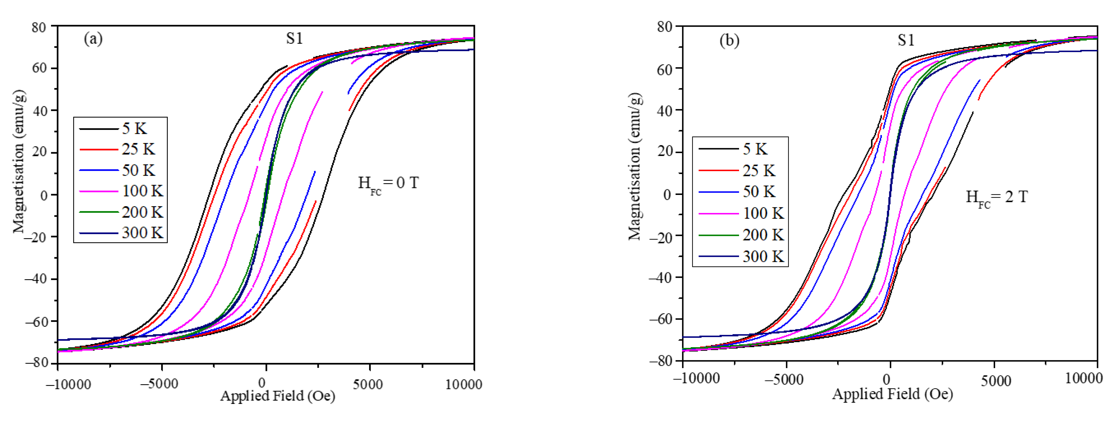
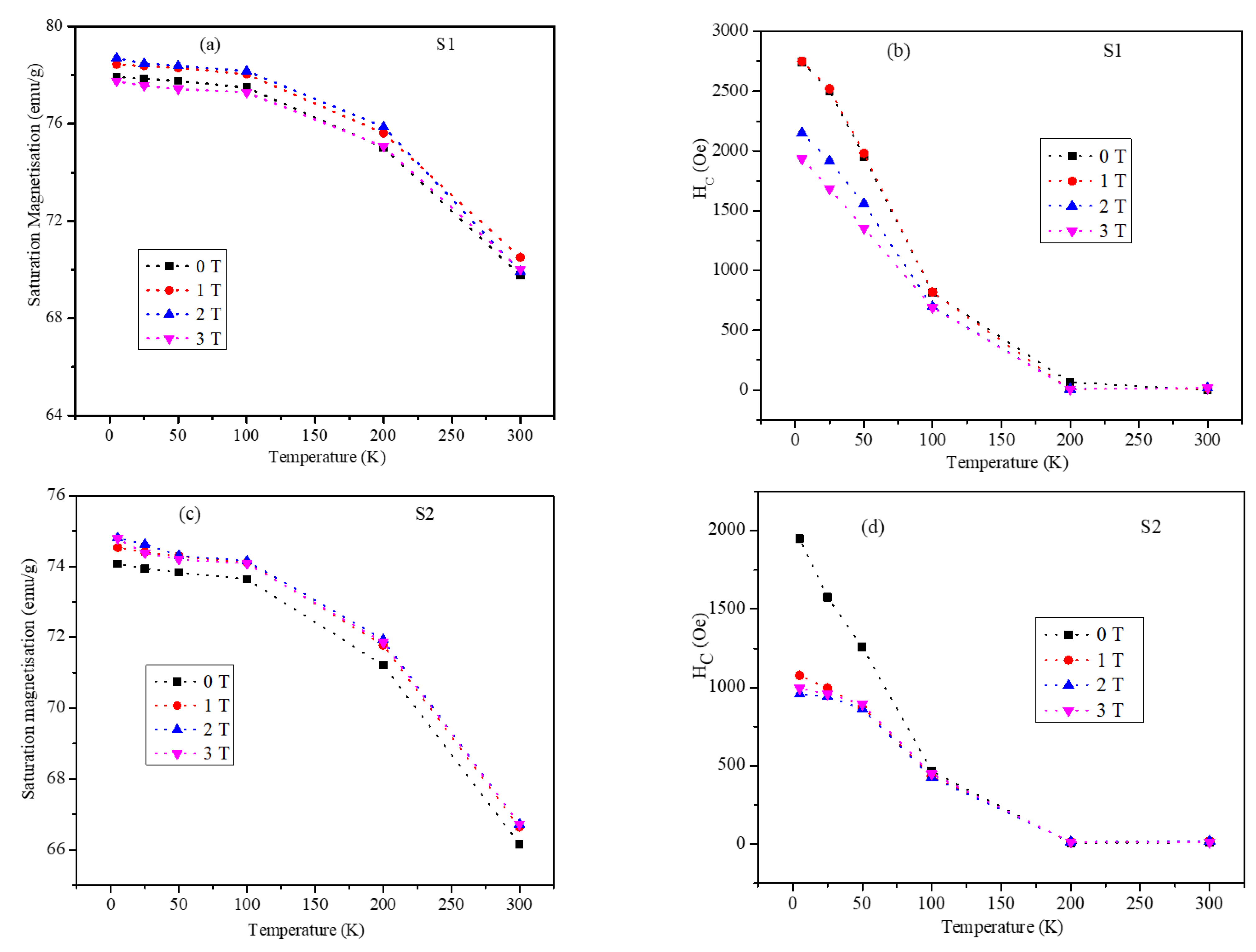
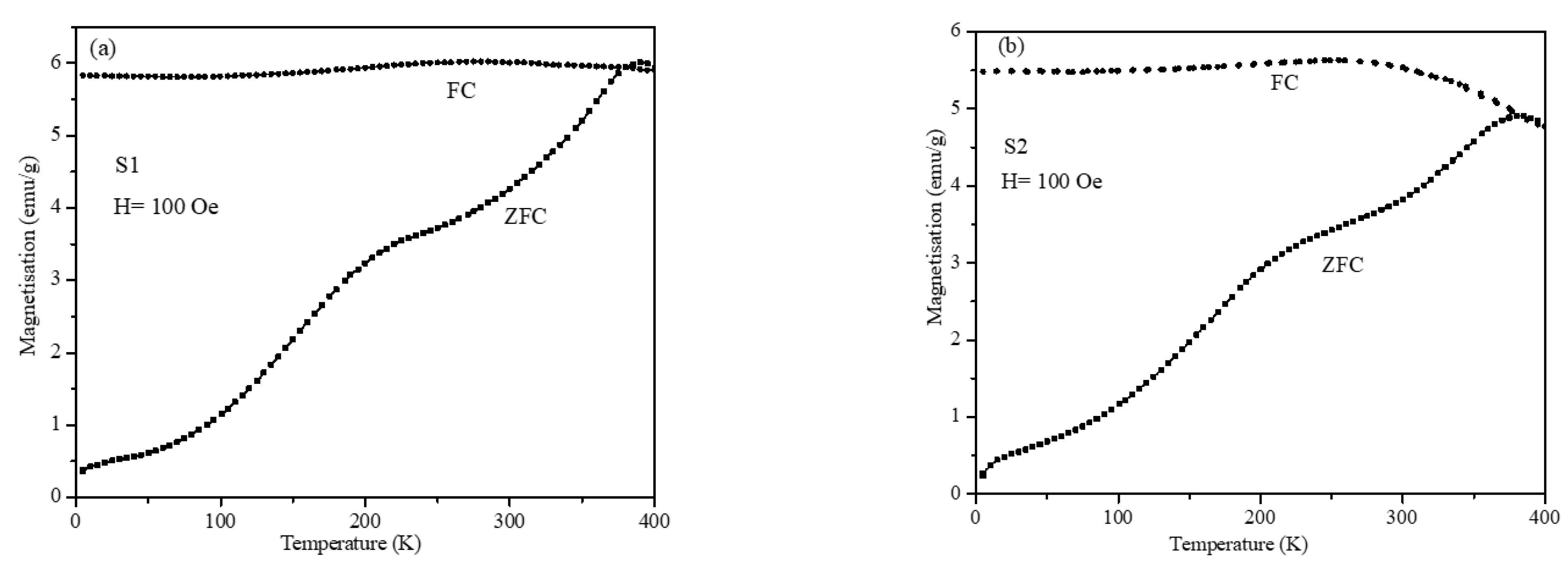
3.3. Magnetic Measurements of the S1 and S2 Core-Shell Nanoparticles
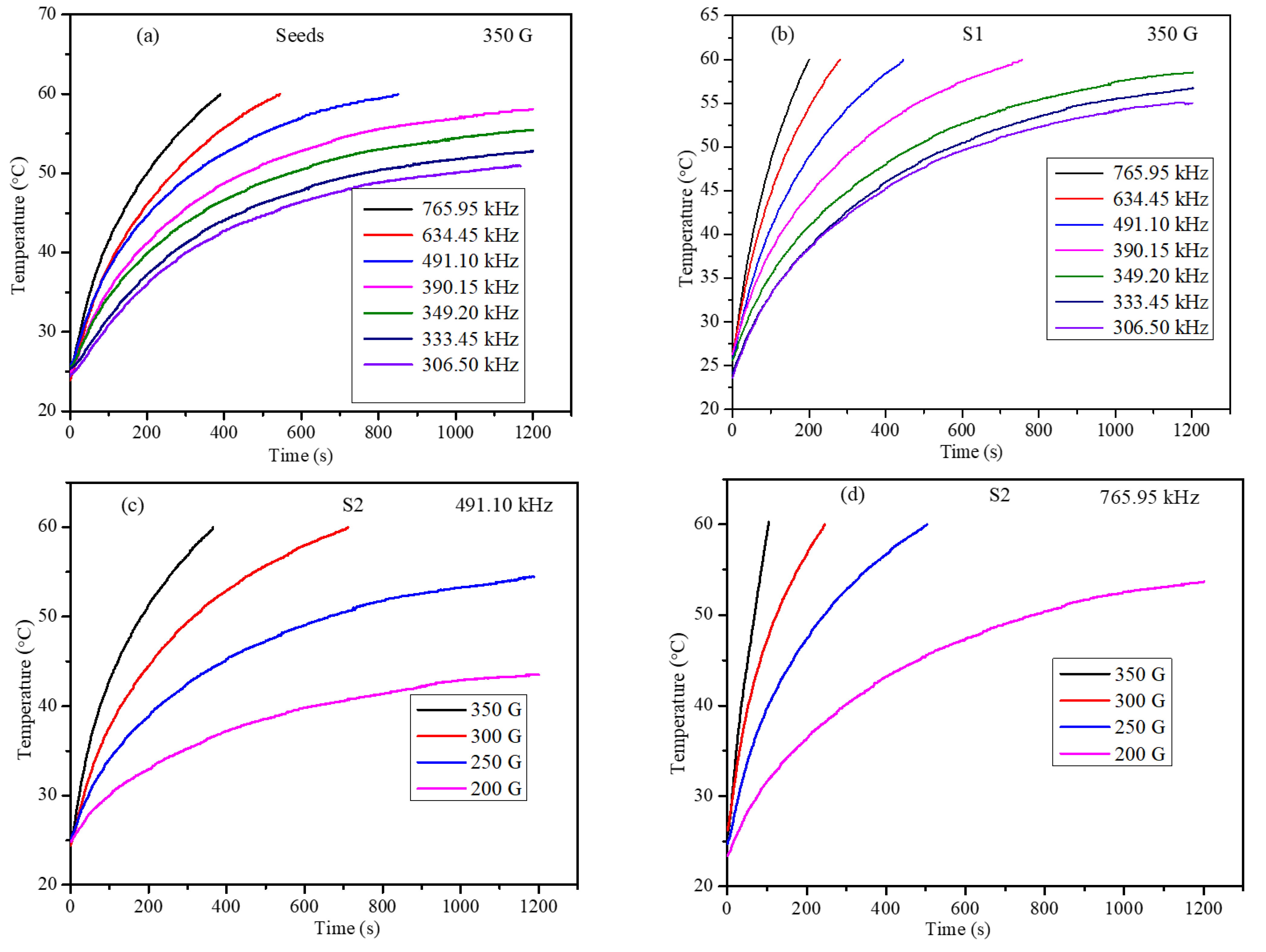
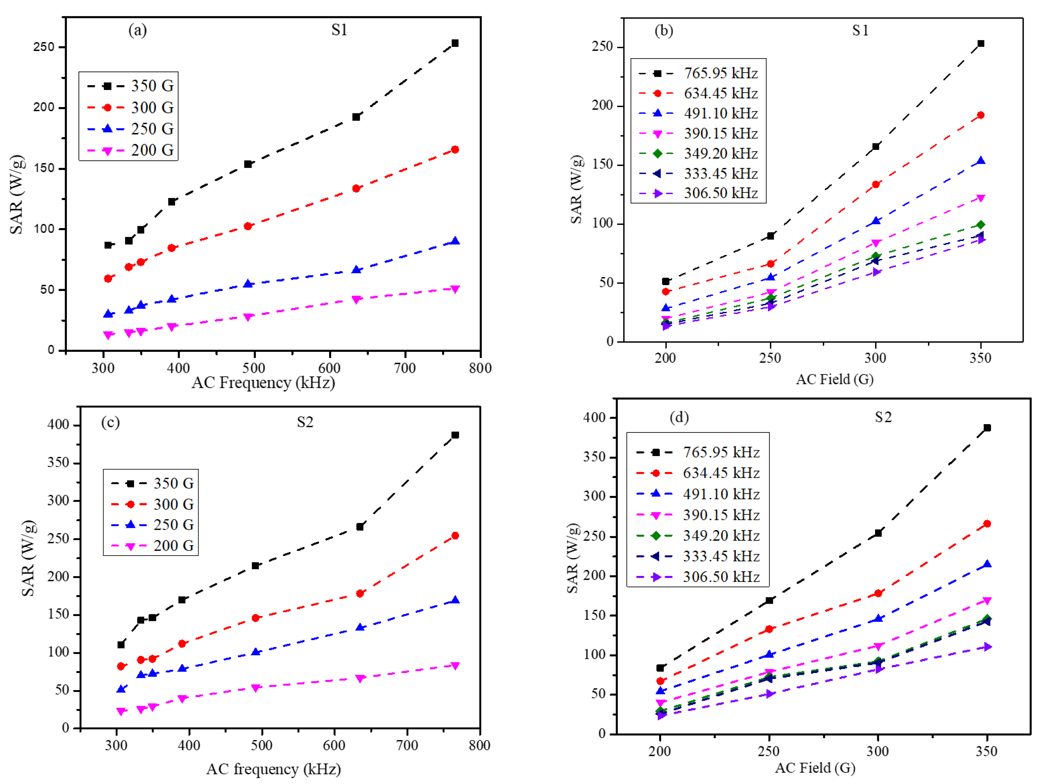
3.4. Magnetic Hyperthermia Studies of Core-Shell Nanoparticles in Agar Gel Phantom
4. Conclusions
Author Contributions
Funding
Institutional Review Board Statement
Informed Consent Statement
Data Availability Statement
Acknowledgments
Conflicts of Interest
References
- Kostevšek, N. A Review on the Optimal Design of Magnetic Nanoparticle-Based T2 MRI Contrast Agents. Magnetochemistry 2020, 6, 11. [Google Scholar] [CrossRef] [Green Version]
- Obaidat, I.M.; Narayanaswamy, V.; Alaabed, S.; Sambasivam, S.; Muralee Gopi, C.V.V. Principles of Magnetic Hyperthermia: A Focus on Using Multifunctional Hybrid Magnetic Nanoparticles. Magnetochemistry 2019, 5, 67. [Google Scholar] [CrossRef] [Green Version]
- Cardoso, V.F.; Francesko, A.; Ribeiro, C.; Bañobre-López, M.; Martins, P.; Lanceros-Mendez, S. Advances in Magnetic Nanoparticles for Biomedical Applications. Advanced Healthcare Materials 2018, 7, 1700845. [Google Scholar] [CrossRef] [PubMed]
- Rajan, A.; Sahu, N.K. Review on Magnetic Nanoparticle-Mediated Hyperthermia for Cancer Therapy. J. Nanopart. Res. 2020, 22, 319. [Google Scholar] [CrossRef]
- Issa, B.; Qadri, S.; Obaidat, I.M.; Bowtell, R.W.; Haik, Y. PEG Coating Reduces NMR Relaxivity of Mn0.5Zn0.5Gd0.02Fe1.98O4 Hyperthermia Nanoparticles. J. Magn. Reason. Imaging 2011, 34, 1192–1198. [Google Scholar] [CrossRef] [Green Version]
- Lee, J.-H.; Jang, J.; Choi, J.; Moon, S.H.; Noh, S.; Kim, J.; Kim, J.-G.; Kim, I.-S.; Park, K.I.; Cheon, J. Exchange-Coupled Magnetic Nanoparticles for Efficient Heat Induction. Nat. Nanotech. 2011, 6, 418–422. [Google Scholar] [CrossRef]
- Obaidat, I.M.; Issa, B.; Haik, Y. The Role of Aggregation of Ferrite Nanoparticles on Their Magnetic Properties. J. Nanosci. Nanotechnol. 2011, 11, 3882–3888. [Google Scholar] [CrossRef]
- He, X.; Zhong, W.; Au, C.-T.; Du, Y. Size Dependence of the Magnetic Properties of Ni Nanoparticles Prepared by Thermal Decomposition Method. Nanoscale Res. Lett. 2013, 8, 446. [Google Scholar] [CrossRef] [Green Version]
- DeFotis, G.C.; Molloy, J.C.; Komatsu, C.H.; Van Dongen, M.J.; Davis, C.M.; DeSanto, C.L.; May, W.M.; Owens, T.M. Composition Dependence of Magnetic Properties in the Mixed Magnet Co1−xNixCl2·H2O. J. Magn. Magn. Mater. 2019, 484, 478–483. [Google Scholar] [CrossRef]
- Gawande, M.B.; Goswami, A.; Asefa, T.; Guo, H.; Biradar, A.V.; Peng, D.-L.; Zboril, R.; Varma, R.S. Core–Shell Nanoparticles: Synthesis and Applications in Catalysis and Electrocatalysis. Chem. Soc. Rev. 2015, 44, 7540–7590. [Google Scholar] [CrossRef]
- Freire, T.M.; Galvão, W.S.; Freire, R.M.; Fechine, P.B.A. Bimagnetic Core/Shell Nanoparticles: Current Status and Future Possibilities; Springer: Berlin/Heidelberg, Germany, 2017; Available online: https://www.springerprofessional.de/en/bimagnetic-core-shell-nanoparticles-current-status-and-future-po/12200914 (accessed on 28 December 2021).
- Pardo, A.; Yáñez, S.; Piñeiro, Y.; Iglesias-Rey, R.; Al-Modlej, A.; Barbosa, S.; Rivas, J.; Taboada, P. Cubic Anisotropic Co- and Zn-Substituted Ferrite Nanoparticles as Multimodal Magnetic Agents. ACS Appl. Mater. Interfaces 2020, 12, 9017–9031. [Google Scholar] [CrossRef]
- Chung, H.; Bae, H.; Kim, C.; Rhee, I. Cube-Shaped Triethylene Glycol-Coated Ni−Mn Ferrite Nanoparticles for Use as T2 Contrast Agents in Magnetic Resonance Imaging. J. Korean Phys. Soc. 2019, 74, 48–52. [Google Scholar] [CrossRef]
- De Queiroz, D.F.; de Camargo, E.R.; Martines, M.U. Synthesis and Characterization of Magnetic Nanoparticles of Cobalt Ferrite Coated with Silica. Biointerface Res. Appl. Chem. 2020, 10, 4908–4913. [Google Scholar] [CrossRef]
- Phan, M.-H.; Alonso, J.; Khurshid, H.; Lampen-Kelley, P.; Chandra, S.; Stojak Repa, K.; Nemati, Z.; Das, R.; Iglesias, Ó.; Srikanth, H. Exchange Bias Effects in Iron Oxide-Based Nanoparticle Systems. Nanomaterials 2016, 6, 221. [Google Scholar] [CrossRef] [PubMed]
- López-Ortega, A.; Tobia, D.; Winkler, E.; Golosovsky, I.V.; Salazar-Alvarez, G.; Estradé, S.; Estrader, M.; Sort, J.; González, M.A.; Suriñach, S.; et al. Size-Dependent Passivation Shell and Magnetic Properties in Antiferromagnetic/Ferrimagnetic Core/Shell MnO Nanoparticles. J. Am. Chem. Soc. 2010, 132, 9398–9407. [Google Scholar] [CrossRef] [PubMed]
- Simeonidis, K.; Martinez-Boubeta, C.; Serantes, D.; Ruta, S.; Chubykalo-Fesenko, O.; Chantrell, R.; Oró-Solé, J.; Balcells, L.; Kamzin, A.S.; Nazipov, R.A.; et al. Controlling Magnetization Reversal and Hyperthermia Efficiency in Core–Shell Iron–Iron Oxide Magnetic Nanoparticles by Tuning the Interphase Coupling. ACS Appl. Nano Mater. 2020, 3, 4465–4476. [Google Scholar] [CrossRef]
- Hu, H. Recent Advances of Bioresponsive Nano-Sized Contrast Agents for Ultra-High-Field Magnetic Resonance Imaging. Front. Chem. 2020, 8, 203. [Google Scholar] [CrossRef] [PubMed] [Green Version]
- Anik, M.I.; Hossain, M.K.; Hossain, I.; Mahfuz, A.M.U.B.; Rahman, M.T.; Ahmed, I. Recent Progress of Magnetic Nanoparticles in Biomedical Applications: A Review. Nano Select 2021, 2, 1146–1186. [Google Scholar] [CrossRef]
- Dobson, J. Remote Control of Cellular Behaviour with Magnetic Nanoparticles. Nat. Nanotech. 2008, 3, 139–143. [Google Scholar] [CrossRef]
- Price, P.M.; Mahmoud, W.E.; Al-Ghamdi, A.A.; Bronstein, L.M. Magnetic Drug Delivery: Where the Field Is Going. Front. Chem. 2018, 6, 619. [Google Scholar] [CrossRef] [PubMed] [Green Version]
- Dennis, C.L.; Ivkov, R. Physics of Heat Generation Using Magnetic Nanoparticles for Hyperthermia. Int. J. Hyperth. 2013, 29, 715–729. [Google Scholar] [CrossRef]
- Usov, N.A.; Rytov, R.A.; Bautin, V.A. Properties of Assembly of Superparamagnetic Nanoparticles in Viscous Liquid. Sci. Rep. 2021, 11, 6999. [Google Scholar] [CrossRef]
- Fabris, F.; Lima, E.; Biasi, E.D.; Troiani, H.E.; Mansilla, M.V.; Torres, T.E.; Pacheco, R.F.; Ibarra, M.R.; Goya, G.F.; Zysler, R.D.; et al. Controlling the Dominant Magnetic Relaxation Mechanisms for Magnetic Hyperthermia in Bimagnetic Core–Shell Nanoparticles. Nanoscale 2019, 11, 3164–3172. [Google Scholar] [CrossRef] [PubMed]
- Nica, V.; Caro, C.; Páez-Muñoz, J.M.; Leal, M.P.; Garcia-Martin, M.L. Bi-Magnetic Core-Shell CoFe2O4@MnFe2O4 Nanoparticles for In Vivo Theranostics. Nanomaterials 2020, 10, 907. [Google Scholar] [CrossRef]
- Moon, S.H.; Noh, S.; Lee, J.-H.; Shin, T.-H.; Lim, Y.; Cheon, J. Ultrathin Interface Regime of Core–Shell Magnetic Nanoparticles for Effective Magnetism Tailoring. Nano Lett. 2017, 17, 800–804. [Google Scholar] [CrossRef] [PubMed]
- Tsopoe, S.P.; Borgohain, C.; Fopase, R.; Pandey, L.M.; Borah, J.P. A Comparative Investigation of Normal and Inverted Exchange Bias Effect for Magnetic Fluid Hyperthermia Applications. Sci. Rep. 2020, 10, 18666. [Google Scholar] [CrossRef]
- Venkatesha, N.; Pudakalakatti, S.M.; Qurishi, Y.; Atreya, H.S.; Srivastava, C. MnFe2O4–Fe3O4 Core–Shell Nanoparticles as a Potential Contrast Agent for Magnetic Resonance Imaging. RSC Adv. 2015, 5, 97807–97815. [Google Scholar] [CrossRef]
- Andreu, I.; Natividad, E. Accuracy of Available Methods for Quantifying the Heat Power Generation of Nanoparticles for Magnetic Hyperthermia. Int. J. Hyperth. 2013, 29, 739–751. [Google Scholar] [CrossRef] [Green Version]
- Su, K.P.; Zhao, C.Y.; Wang, H.O.; Huang, S.; Liu, Z.W.; Huo, D.X. Synthesis, Structure and Magnetic Properties of CoFe2O4 Ferrite Nanoparticles. Mater. Res. Express 2018, 5, 056102. [Google Scholar] [CrossRef]
- Boda, N.; Naidu, K.C.B.; Batoo, K.M.; Joice, G.H.R.; Naik, J.L.; Ravinder, D. Structural, Morphological and Electronic Properties of Cadmium Cobalt Ferrite Nanoparticles. Biointerface Res. Appl. Chem. 2020, 10, 4752–4763. [Google Scholar] [CrossRef]
- Patterson, A.L. The Scherrer Formula for X-Ray Particle Size Determination. Phys. Rev. 1939, 56, 978–982. [Google Scholar] [CrossRef]
- Holland, T.; Redfern, S. UNITCELL: A Nonlinear Least-Squares Program for Cell-Parameter Refinement Implementing Regression and Deletion Diagnostics. J. Appl. Crystallogr. 1997, 30, 84. [Google Scholar] [CrossRef] [Green Version]
- Sanpo, N.; Wang, J.; Berndt, C. Sol-Gel Synthesized Copper-Substituted Cobalt Ferrite Nanoparticles for Biomedical Applications. J. Nano Res. 2013, 22, 95–106. [Google Scholar] [CrossRef]
- Yan, H.; Zhang, J.; You, C.; Song, Z.; Yu, B.; Shen, Y. Influences of Different Synthesis Conditions on Properties of Fe3O4 Nanoparticles. Mater. Chem. Phys. 2009, 113, 46–52. [Google Scholar] [CrossRef]
- Robles, J.; Das, R.; Glassell, M.; Phan, M.H.; Srikanth, H. Exchange-Coupled Fe3O4/CoFe2O4 Nanoparticles for Advanced Magnetic Hyperthermia. AIP Adv. 2018, 8, 056719. [Google Scholar] [CrossRef] [Green Version]
- Yelenich, O.V.; Solopan, S.; Greneche, J.-M.; Belous, A. Synthesis and Properties MFe2O4 (M = Fe, Co) Nanoparticles and Core-Shell Structures. Solid State Sci. 2015, 46, 19–26. [Google Scholar] [CrossRef]
- Alves, C.R.; Aquino, R.; Depeyrot, J.; Cotta, T.A.P.; Sousa, M.H.; Tourinho, F.A.; Rechenberg, H.R.; Goya, G.F. Surface Spin Freezing of Ferrite Nanoparticles Evidenced by Magnetization Measurements. J. Appl. Phys. 2006, 99, 08M905. [Google Scholar] [CrossRef]
- Labarta, A.; Batlle, X.; Iglesias, O. From Finite-Size and Surface Effects to Glassy Behaviour in Ferrimagnetic Nanoparticles. In Surface Effects in Magnetic Nanoparticles; Springer: Boston, MA, USA, 2005. [Google Scholar]
- Obaidat, I.M.; Nayek, C.; Manna, K.; Bhattacharjee, G.; Al-Omari, I.A.; Gismelseed, A. Investigating Exchange Bias and Coercivity in Fe3O4–γ-Fe2O3 Core–Shell Nanoparticles of Fixed Core Diameter and Variable Shell Thicknesses. Nanomaterials 2017, 7, 415. [Google Scholar] [CrossRef] [Green Version]
- Cai, J.W.; Liu, K.; Chien, C.L. Exchange Coupling in the Paramagnetic State. Phys. Rev. B 1999, 60, 72–75. [Google Scholar] [CrossRef] [Green Version]
- Guo, S.; Liu, X.H.; Cui, W.B.; Liu, W.; Zhao, X.G.; Li, D.; Zhang, Z.D. Unconventional Exchange Bias in CoCr2O4/Cr2O3 Nanocomposites. J. Appl. Phys. 2009, 105, 064702. [Google Scholar] [CrossRef]
- Behera, B.C.; Jana, S.; Bhat, S.G.; Gauquelin, N.; Tripathy, G.; Anil Kumar, P.S.; Samal, D. Evidence for Exchange Bias Coupling at the Perovskite/Brownmillerite Interface in Spontaneously Stabilized SrCoO3−δ/SrCoO2.5 Bilayers. Phys. Rev. B 2019, 99, 024425. [Google Scholar] [CrossRef] [Green Version]
- Sossmeier, K.D.; Schafer, D.; Bastos, A.P.O.; Schmidt, J.E.; Geshev, J. Tailoring Coercivity of Unbiased Exchange-Coupled Ferromagnet/Antiferromagnet Bilayers. J. Appl. Phys. 2012, 112, 013904. [Google Scholar] [CrossRef] [Green Version]
- O’Grady, K.; Fernandez-Outon, L.E.; Vallejo-Fernandez, G. A New Paradigm for Exchange Bias in Polycrystalline Thin Films. J. Magn. Magn. Mater. 2010, 322, 883–899. [Google Scholar] [CrossRef]
- Coutrim, L.T.; Bittar, E.M.; Garcia, F.; Bufaiçal, L. Influence of Spin Glass-like Magnetic Relaxation on the Zero-Field-Cooled Exchange Bias Effect. Phys. Rev. B 2018, 98, 064426. [Google Scholar] [CrossRef] [Green Version]
- Ali, M.; Adie, P.; Marrows, C.H.; Greig, D.; Hickey, B.J.; Stamps, R.L. Exchange Bias Using a Spin Glass. Nat. Mater. 2007, 6, 70–75. [Google Scholar] [CrossRef] [PubMed]
- Wang, B.M.; Liu, Y.; Ren, P.; Xia, B.; Ruan, K.B.; Yi, J.B.; Ding, J.; Li, X.G.; Wang, L. Large Exchange Bias after Zero-Field Cooling from an Unmagnetized State. Phys. Rev. Lett. 2011, 106, 077203. [Google Scholar] [CrossRef] [PubMed]
- Rojas Sánchez, J.C.; Nelson-Cheeseman, B.; Granada, M.; Arenholz, E.; Steren, L.B. Exchange-Bias Effect at La0.75Sr0.25MnO3/LaNiO3 Interfaces. Phys. Rev. B Condens. Matter Mater. Phys. 2012, 85, 094427. [Google Scholar] [CrossRef]
- Zhou, G.; Guan, X.; Bai, Y.; Quan, Z.; Jiang, F.; Xu, X. Interfacial Spin Glass State and Exchange Bias in the Epitaxial La0.7Sr0.3MnO3/LaNiO3 Bilayer. Nanoscale Res. Lett. 2017, 12, 330. [Google Scholar] [CrossRef]
- Miltényi, P.; Gierlings, M.; Keller, J.; Beschoten, B.; Güntherodt, G.; Nowak, U.; Usadel, K.D. Diluted Antiferromagnets in Exchange Bias: Proof of the Domain State Model. Phys. Rev. Lett. 2000, 84, 4224–4227. [Google Scholar] [CrossRef]
- Giri, S.; Patra, M.; Majumdar, S. Exchange Bias Effect in Alloys and Compounds. J. Phys. Condens. Matter 2011, 23, 073201. [Google Scholar] [CrossRef]
- Doyle, S.; John, C.; Maniv, E.; Murphy, R.A.; Maniv, A.; Ramakrishna, S.K.; Tang, Y.-L.; Ramesh, R.; Long, J.R.; Reyes, A.P.; et al. Tunable Giant Exchange Bias in an Intercalated Transition Metal Dichalcogenide. Nat. Phys. 2021, 17, 525–530. [Google Scholar] [CrossRef]
- Hudl, M.; Mathieu, R.; Nordblad, P. Tunable Exchange Bias in Dilute Magnetic Alloys—Chiral Spin Glasses. Sci. Rep. 2016, 6, 19964. [Google Scholar] [CrossRef] [Green Version]
- Spizzo, F.; Tamisari, M.; Chinni, F.; Bonfiglioli, E.; Del Bianco, L. Interface Adjustment and Exchange Coupling in the IrMn/NiFe System. J. Magn. Magn. Mater. 2017, 421, 234–240. [Google Scholar] [CrossRef]
- Obaidat, I.; Mohite, V.; Issa, B.; Tit, N.; Haik, Y. Predicting a Major Role of Surface Spins in the Magnetic Properties of Ferrite Nanoparticles. Cryst. Res. Technol. 2009, 44, 489–494. [Google Scholar] [CrossRef]
- Nogués, J.; Sort, J.; Langlais, V.; Skumryev, V.; Suriñach, S.; Muñoz, J.S.; Baró, M.D. Exchange Bias in Nanostructures. Phys. Rep. 2005, 422, 65–117. [Google Scholar] [CrossRef]
- Estrader, M.; López-Ortega, A.; Estradé, S.; Golosovsky, I.V.; Salazar-Alvarez, G.; Vasilakaki, M.; Trohidou, K.N.; Varela, M.; Stanley, D.C.; Sinko, M.; et al. Robust Antiferromagnetic Coupling in Hard-Soft Bi-Magnetic Core/Shell Nanoparticles. Nat. Commun. 2013, 4, 2960. [Google Scholar] [CrossRef] [PubMed]
- Vasilakaki, M.; Trohidou, K.N.; Nogués, J. Enhanced Magnetic Properties in Antiferromagnetic-Core/Ferrimagnetic-Shell Nanoparticles. Sci. Rep. 2015, 5, 9609. [Google Scholar] [CrossRef] [Green Version]
- Egea-Benavente, D.; Ovejero, J.G.; Morales, M.d.P.; Barber, D.F. Understanding MNPs Behaviour in Response to AMF in Biological Milieus and the Effects at the Cellular Level: Implications for a Rational Design That Drives Magnetic Hyperthermia Therapy toward Clinical Implementation. Cancers 2021, 13, 4583. [Google Scholar] [CrossRef]


| Nanoparticles | Lattice Parameter (Å) | |
|---|---|---|
| CoFe2O4 | 8.387 | Ref [34] |
| Fe3O4 | 8.40 | Ref [35] |
| CoFe2O4-seeds | 8.376 | This work |
| S1-Core-shell | 8.420 | This work |
| S2-Core-shell | 8.390 | This work |
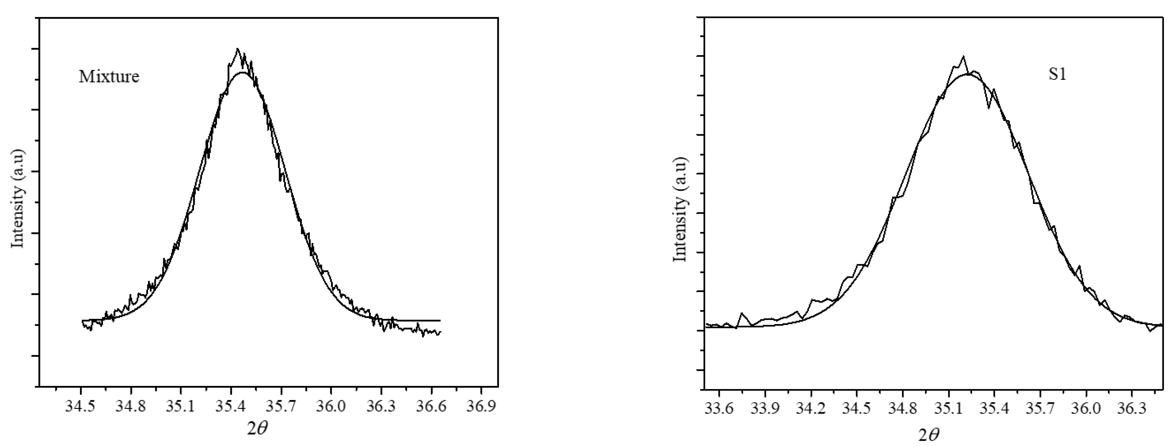
| Particles | (311) Peak Position |
|---|---|
| Fe3O4 | 35.51 |
| CoFe2O4 | 35.45 |
| Mixture | 35.48 |
| S1-Core shell | 35.30 |
| Compositions | Average Sizes from XRD (nm) | Average TEM Sizes (nm) | Shell Thickness (nm) | MS (emu/g) | Keff 104 J/m3 | SAR (W/g) at 765.97 kHz and 350 G |
|---|---|---|---|---|---|---|
| CoFe2O4 seeds | 6.9 | 6.0 ± 1.0 | 39.07 | 5.55 | 170.2 | |
| S1 [Fe3O4(CoFe2O4) | 10.5 | 9.5 ± 1.1 | 3.5 | 69.97 | 1.23 | 253.2 |
| S2 [Fe3O4(CoFe2O4) | 13.5 | 12.1 ± 1.7 | 6.1 | 66.16 | 2.16 | 387.6 |
Publisher’s Note: MDPI stays neutral with regard to jurisdictional claims in published maps and institutional affiliations. |
© 2022 by the authors. Licensee MDPI, Basel, Switzerland. This article is an open access article distributed under the terms and conditions of the Creative Commons Attribution (CC BY) license (https://creativecommons.org/licenses/by/4.0/).
Share and Cite
Narayanaswamy, V.; Al-Omari, I.A.; Kamzin, A.S.; Issa, B.; Obaidat, I.M. Tailoring Interfacial Exchange Anisotropy in Hard–Soft Core-Shell Ferrite Nanoparticles for Magnetic Hyperthermia Applications. Nanomaterials 2022, 12, 262. https://0-doi-org.brum.beds.ac.uk/10.3390/nano12020262
Narayanaswamy V, Al-Omari IA, Kamzin AS, Issa B, Obaidat IM. Tailoring Interfacial Exchange Anisotropy in Hard–Soft Core-Shell Ferrite Nanoparticles for Magnetic Hyperthermia Applications. Nanomaterials. 2022; 12(2):262. https://0-doi-org.brum.beds.ac.uk/10.3390/nano12020262
Chicago/Turabian StyleNarayanaswamy, Venkatesha, Imaddin A. Al-Omari, Aleksandr S. Kamzin, Bashar Issa, and Ihab M. Obaidat. 2022. "Tailoring Interfacial Exchange Anisotropy in Hard–Soft Core-Shell Ferrite Nanoparticles for Magnetic Hyperthermia Applications" Nanomaterials 12, no. 2: 262. https://0-doi-org.brum.beds.ac.uk/10.3390/nano12020262