
Recent Research Progress of Mn4+-Doped A2MF6 (A = Li, Na, K, Cs, or Rb; M = Si, Ti, Ge, or Sn) Red Phosphors Based on a Core–Shell Structure
 ,
,
Abstract
:1. Introduction
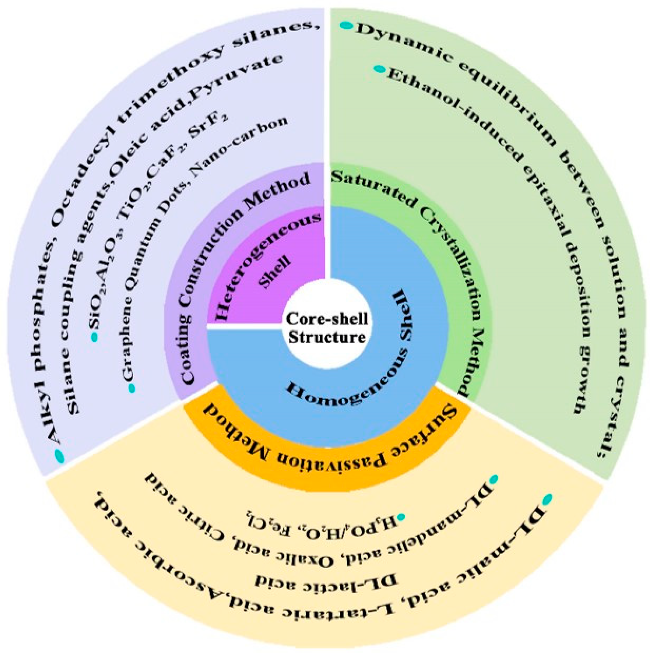
2. Classification of the Shell Layer in A2MF6: Mn4+
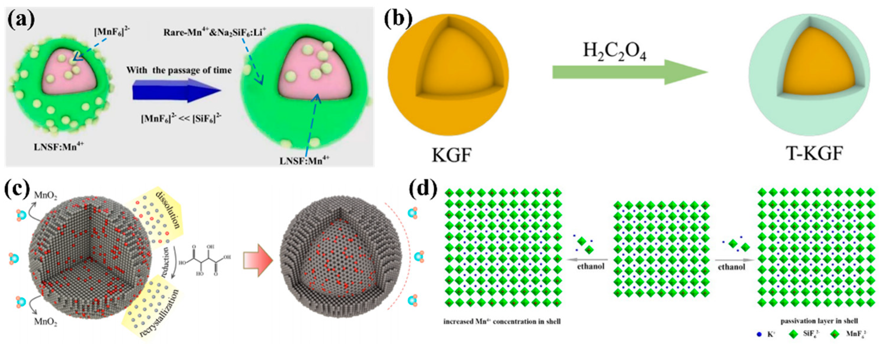
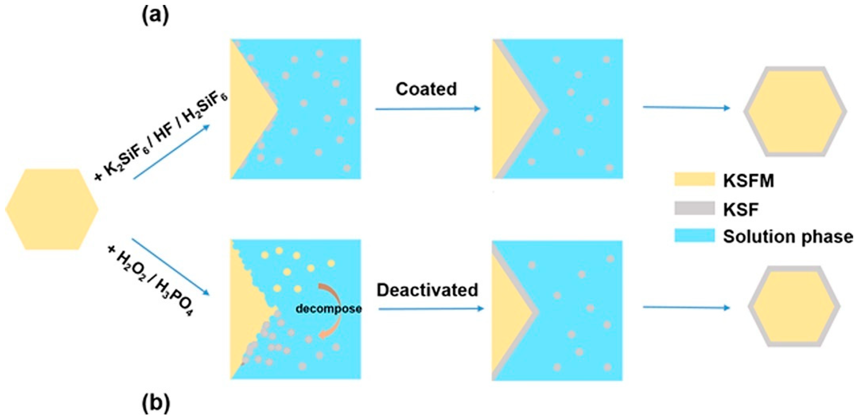
2.1. Heterogeneous Shell Layer
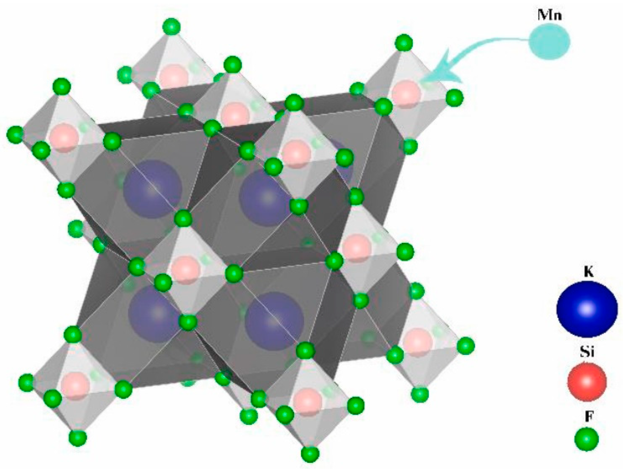
2.2. Homogeneous Shell Layer
3. Preparation Methods of Shell Layer in A2MF6: Mn4+
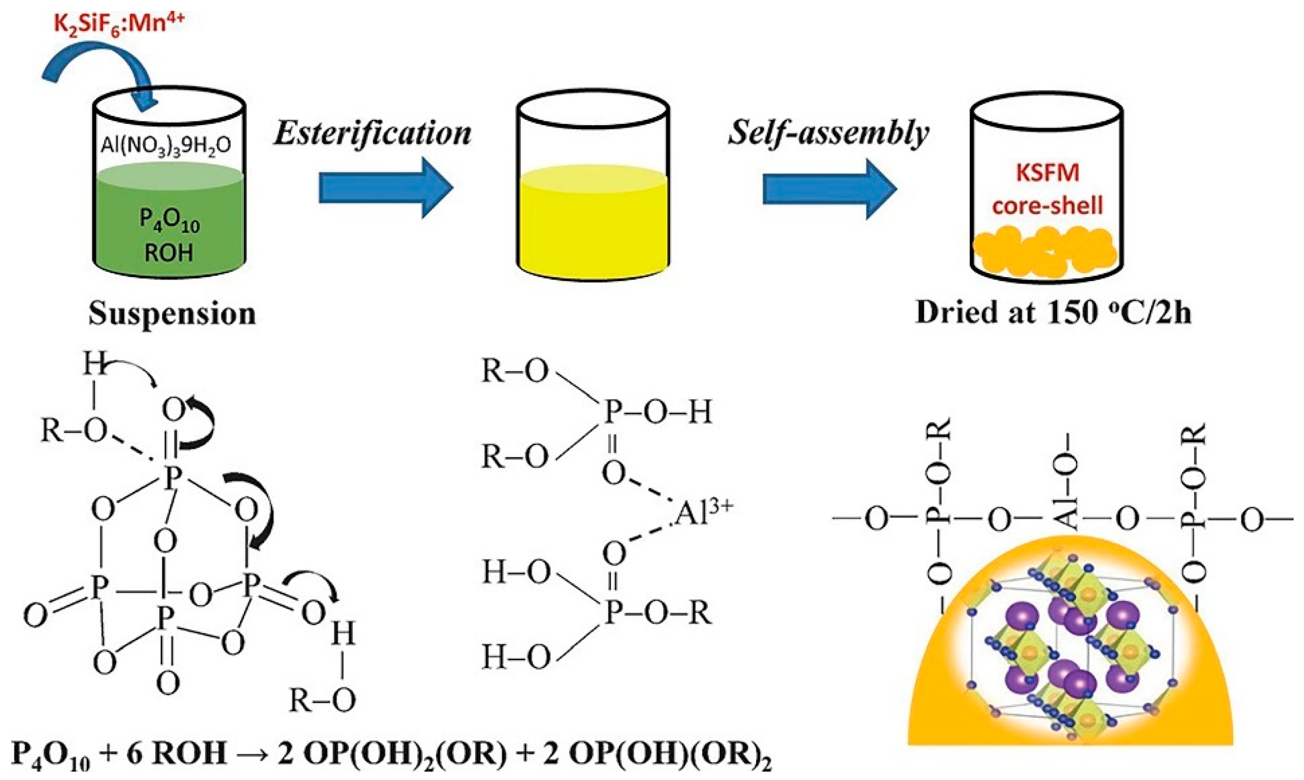
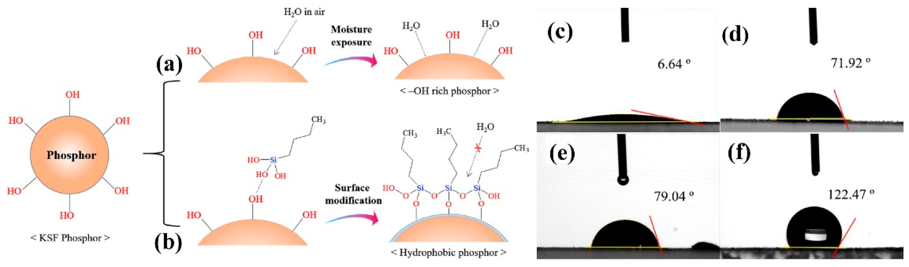
3.1. Coating Construction Method
3.2. Surface Passivation Method
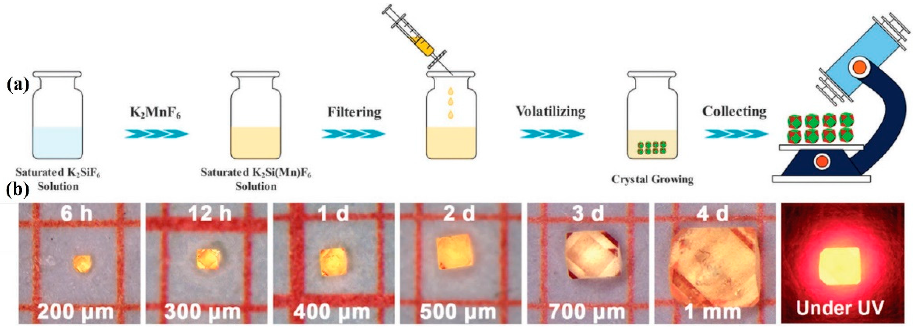
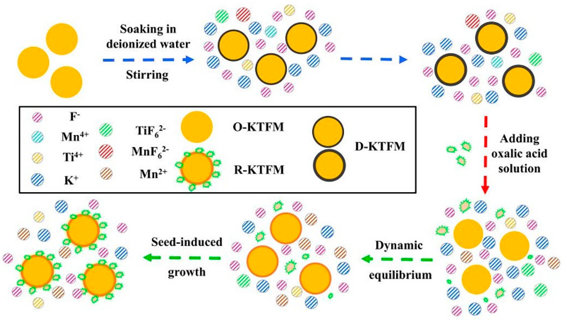
3.3. Saturated Crystallization Method
4. Luminescent Mechanism of A2MF6: Mn4+
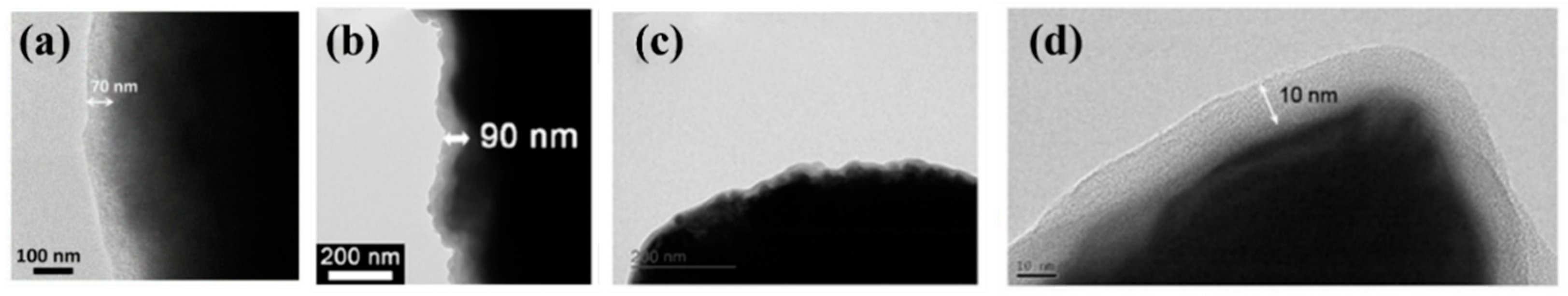
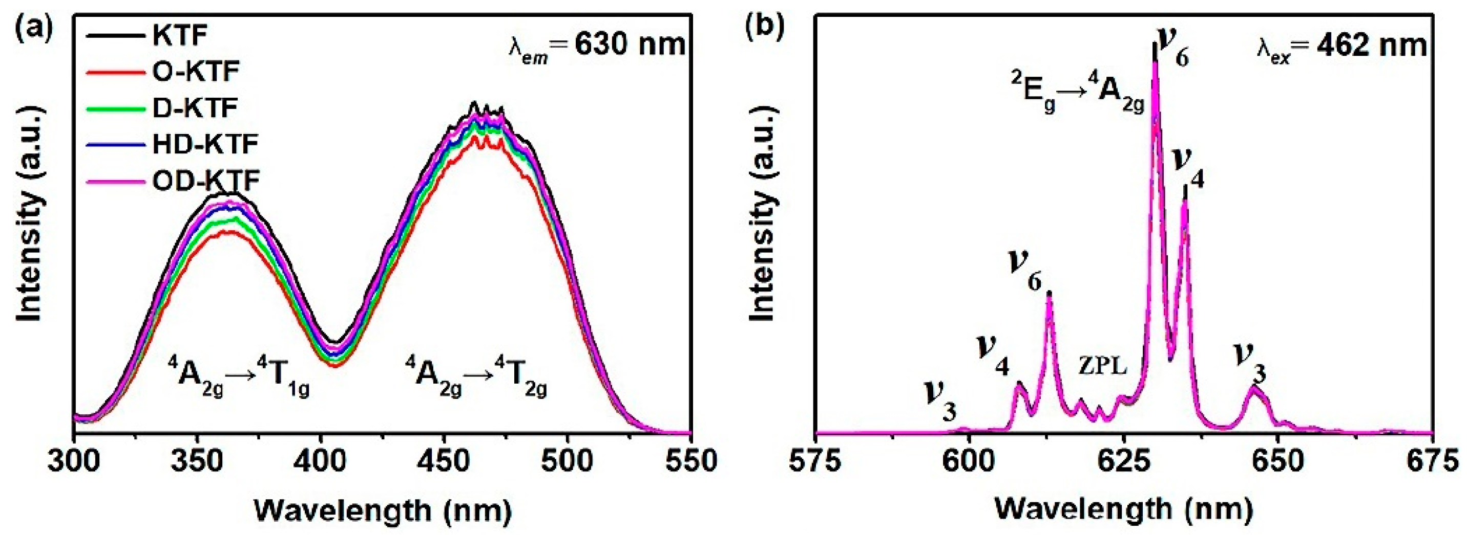
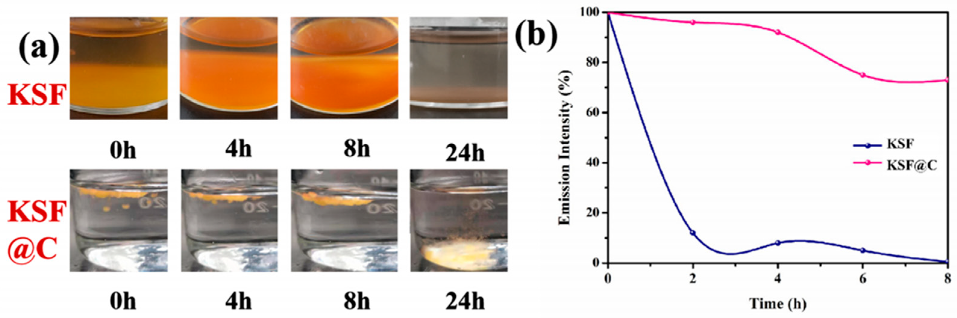
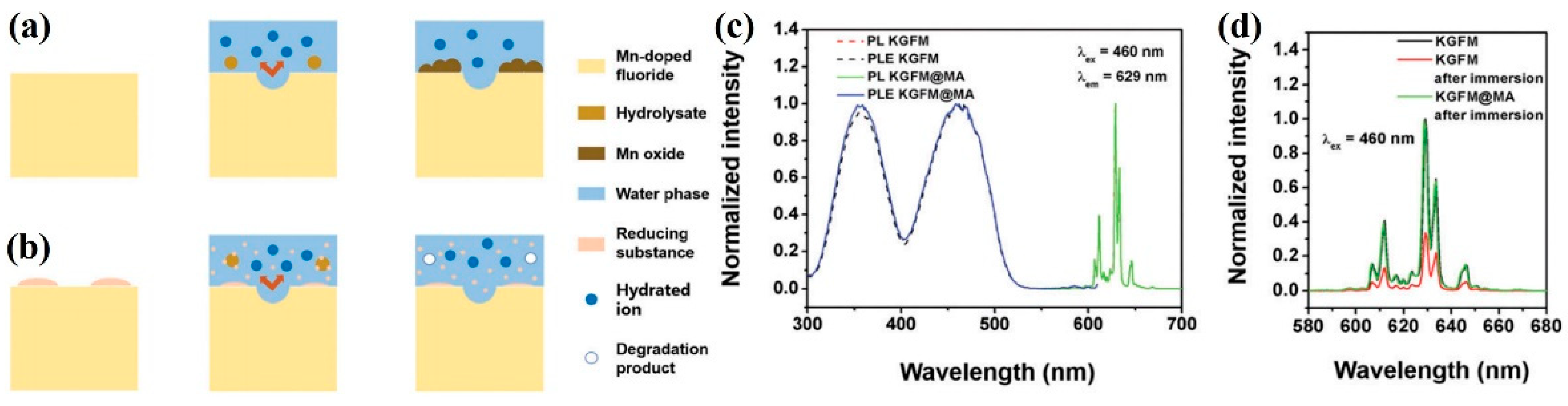
5. Fluorescent Properties and Water Resistance
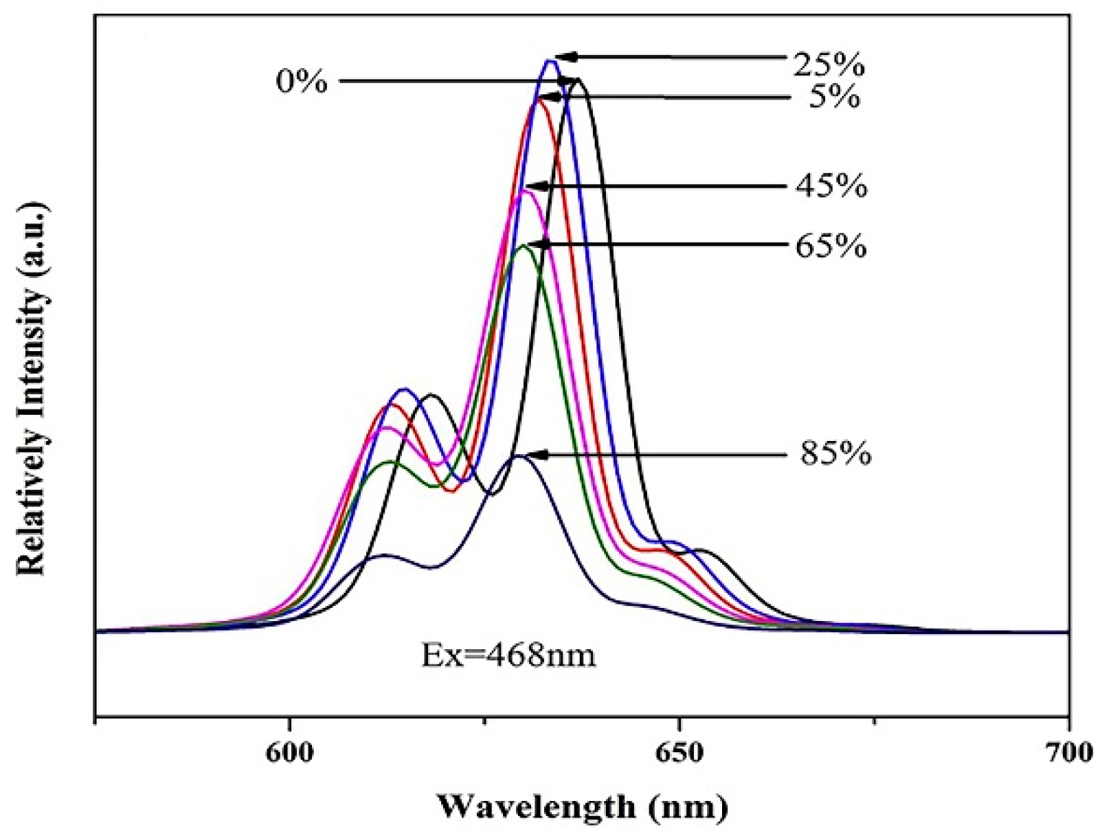
5.1. A2MF6: Mn4+ with Heterogeneous Shell Layer
5.2. A2MF6: Mn4+ with Homogeneous Shell Layer
6. Application of A2MF6: Mn4+ in WLED
7. Conclusions
Author Contributions
Funding
Data Availability Statement
Conflicts of Interest
References
- Li, G.G.; Tian, Y.; Zhao, Y.; Lin, J. Recent progress in luminescence tuning of Ce3+ and Eu2+-activated phosphors for pc-WLEDs. Chem. Soc. Rev. 2015, 44, 8688–8713. [Google Scholar] [CrossRef] [PubMed]
- Li, J.H.; Zhang, Z.H.; Li, X.H.; Xu, Y.Q.; Ai, Y.Y.; Yan, J.; Shi, J.X.; Wu, M.M. Luminescence properties and energy transfer of YGa1.5Al1.5(BO3)4: Tb3+, Eu3+ as a multi-colour emitting phosphor for WLEDs. J. Mater. Chem. C 2017, 5, 6294–6299. [Google Scholar] [CrossRef]
- Chen, Y.J.; Xing, W.S.; Liu, Y.X.; Zhang, X.S.; Xie, Y.Y.; Shen, C.Y.; Liu, J.G.X.; Geng, C.; Xu, S. Efficient and Stable CdSe/CdS/ZnS Quantum Rods-in-Matrix Assembly for White LED Application. Nanomaterials 2020, 10, 317. [Google Scholar] [CrossRef] [PubMed]
- Oh, J.H.; Eo, Y.J.; Yoon, H.C.; Huh, Y.-D.; Do, Y.R. Evaluation of new color metrics: Guidelines for developing narrow-band red phosphors for WLEDs. J. Mater. Chem. C 2016, 4, 8326–8348. [Google Scholar] [CrossRef]
- Wang, L.; Wang, X.; Kohsei, T.; Yoshimura, K.; Izumi, M.; Hirosaki, N.; Xie, R.J. Highly efficient narrow-band green and red phosphors enabling wider color-gamut LED backlight for more brilliant displays. Opt. Express 2015, 23, 28707–28717. [Google Scholar] [CrossRef]
- Feng, X.Y.; Jiang, K.; Zeng, H.B.; Lin, H.W. A Facile Approach to Solid-State White Emissive Carbon Dots and Their Application in UV-Excitable and Single-Component-Based White LEDs. Nanomaterials 2019, 9, 725. [Google Scholar] [CrossRef]
- Zhou, Z.; Zhou, N.; Xia, M.; Yokoyama, M.; Hintzen, H.T. Research progress and application prospects of transition metal Mn4+-activated luminescent materials. J. Mater. Chem. C 2016, 4, 9143–9161. [Google Scholar] [CrossRef]
- Zhu, H.; Lin, C.C.; Luo, W.; Shu, S.; Liu, Z.; Liu, Y.; Kong, J.; Ma, E.; Cao, Y.; Liu, R.S.; et al. Highly efficient non-rare-earth red emitting phosphor for warm white light-emitting diodes. Nat. Commun. 2014, 5, 4312. [Google Scholar] [CrossRef]
- Liang, S.S.; Shang, M.M.; Lian, H.Z.; Li, K.; Zhang, Y.; Lin, J. An efficient rare-earth free deep red emitting phosphor for improving the color rendering of white light-emitting diodes. J. Mater. Chem. C 2017, 5, 2927–2935. [Google Scholar] [CrossRef]
- Lin, C.C.; Liu, R.S. Advances in Phosphors for Light-emitting Diodes. J. Phys. Chem. Lett. 2011, 2, 1268–1277. [Google Scholar] [CrossRef]
- Liu, X.F.; Qiu, J.R. Recent advances in energy transfer in bulk and nanoscale luminescent materials: From spectroscopy to appli cations. Chem. Soc. Rev. 2015, 44, 8714–8746. [Google Scholar] [CrossRef]
- Lu, W.; Lv, W.Z.; Zhao, Q.; Jiao, M.M.; Shao, B.Q.; You, H.P. A novel efficient Mn4+ activated Ca14Al10Zn6O35 phosphor: Applica tion in red-emitting and white LEDs. Inorg. Chem. 2014, 53, 11985–11990. [Google Scholar] [CrossRef] [PubMed]
- Blum, O.; Shaked, N.T. Prediction of photothermal phase signatures from arbitrary plasmonic nanoparticles and experimental verification. Light Sci. Appl. 2015, 4, e322. [Google Scholar] [CrossRef]
- Vanetsev, A.; Põdder, P.; Oja, M.; Khaidukov, N.M.; Makhov, V.N.; Nagirnyi, V.; Romet, I.; Vielhauer, S.; Mändar, H. Microwave-hydrothermal synthesis and investigation of Mn-doped K2SiF6 microsize powder as a red phosphor for warm white LEDs. J. Lumin. 2021, 239, 118389. [Google Scholar] [CrossRef]
- Du, J.; Poelman, D. Facile Synthesis of Mn4+-Activated Double Perovskite Germanate Phosphors with Near-Infrared Persistent Luminescence. Nanomaterials 2019, 9, 1759. [Google Scholar] [CrossRef]
- Hong, F.; Yang, L.; Xu, H.P.; Chen, Z.; Liu, Q.X.; Liu, G.X.; Dong, X.G.; Yu, W.S. A red-emitting Mn4+ activated phosphor with controlled morphology and two-dimensional luminescence nanofiber film: Synthesis and application for high-performance warm white light-emitting diodes (WLEDs). J. Alloys Compd. 2019, 808, 151551. [Google Scholar] [CrossRef]
- Adachi, S. Photoluminescence properties of Mn4+-activated oxide phosphors for use in white-LED applications: A review. J. Lumin. 2018, 202, 263–281. [Google Scholar] [CrossRef]
- Adachi, S. Photoluminescence spectra and modeling analyses of Mn4+-activated fluoride phosphors: A review. J. Lumin. 2018, 197, 119–130. [Google Scholar] [CrossRef]
- Lang, T.C.; Han, T.; Fang, S.Q.; Wang, J.Y.; Cao, S.X.; Peng, L.L.; Liu, B.T.; Korepanov, V.I.; Yakovlev, A.N. Improved phase stability of the metastable K2GeF6:Mn4+ phosphors with high thermal stability and water-proof property by cation substitution. Chem. Eng. J. 2020, 380, 122429. [Google Scholar] [CrossRef]
- Hong, F.; Cheng, H.; Song, C.; Liu, G.X.; Yu, W.S.; Wang, J.; Dong, X.T. Novel polygonal structure Mn4+ activated In3+-based Elpasolite-type hexafluorides red phosphor for warm white light-emitting diodes (WLEDs). Dalton Trans. 2019, 48, 1376–1385. [Google Scholar] [CrossRef]
- Adachi, S.; Takahashi, T. Direct synthesis and properties of K2SiF6:Mn4+ phosphor by wet chemical etching of Si wafer. J. Appl. Phys. 2008, 104, 023512. [Google Scholar] [CrossRef]
- Jansen, T.; Baur, F.; Jüstel, T. Red emitting K2NbF7: Mn4+ and K2TaF7: Mn4+ for warm-white LED applications. J. Lumin. 2017, 192, 644–652. [Google Scholar] [CrossRef]
- Jin, Y.; Fang, M.H.; Grinberg, M.; Mahlik, S.; Lesniewski, T.; Brik, M.G.; Luo, G.Y.; Lin, J.G.; Liu, R.S. Narrow Red Emission Band Fluoride Phosphor KNaSiF6:Mn4+ for Warm White Light-Emitting Diodes. ACS Appl. Mater. Interfaces 2016, 8, 11194–11203. [Google Scholar] [CrossRef]
- Liao, J.S.; Nie, L.L.; Zhong, L.F.; Gu, Q.J.; Wang, Q. Co-precipitation synthesis and luminescence properties of K2TiF6:Mn4+ red phosphors for warm white light-emitting diodes. Luminescence 2016, 31, 802–807. [Google Scholar] [CrossRef] [PubMed]
- Tang, F.; Su, Z.C.; Ye, H.G.; Wang, M.Z.; Lan, X.; Phillips, D.L.; Cao, Y.G.; Xu, S.J. A set of manganese ion activated fluoride phosphors (A2BF6:Mn4+, A = K, Na, B = Si, Ge, Ti): Synthesis below 0 °C and efficient room-temperature photoluminescence. J. Mater. Chem. C 2016, 4, 9561–9568. [Google Scholar] [CrossRef]
- Zhu, M.M.; Pan, Y.X.; Xi, L.Q.; Lian, H.Z.; Lin, J. Design, preparation, and optimized luminescence of a dodec-fluoride phosphor Li3Na3Al2F12:Mn4+ for warm WLED applications. J. Mater. Chem. C. 2017, 5, 10241–10250. [Google Scholar] [CrossRef]
- Zhu, Y.W.; Cao, L.Y.; Brik, M.G.; Zhang, X.J.; Huang, L.; Xuan, T.T.; Wang, J. Facile synthesis, morphology and photolumines cence of a novel red fluoride nanophosphor K2NaAlF6:Mn4+. J. Mater. Chem. C 2017, 5, 6420–6426. [Google Scholar] [CrossRef]
- Huang, D.C.; Zhu, H.M.; Deng, Z.H.; Zou, Q.L.; Lu, H.Y.; Yi, X.D.; Guo, W.; Lu, C.D.; Chen, X.Y. Moisture-Resistant Mn4+-Doped Core-Shell-Structured Fluoride Red Phosphor Exhibiting High Luminous Efficacy for Warm White Light-Emitting Diodes. Angew. Chem. Int. Ed. 2019, 58, 3843–3847. [Google Scholar] [CrossRef] [PubMed]
- Wan, P.P.; Liang, Z.J.; Luo, P.L.; Lian, S.X.; Zhou, W.L.; Liu, R.-S. Reconstruction of Mn4+-free shell achieving highly stable red-emitting fluoride phosphors for light-emitting diodes. Chem. Eng. J. 2021, 426, 131350. [Google Scholar] [CrossRef]
- Zhou, Y.Y.; Yu, C.K.; Song, E.H.; Wang, Y.J.; Ming, H.; Xia, Z.G.; Zhang, Q.Y. Three Birds with One Stone: K2SiF6:Mn4+ Single Crystal Phosphors for High-Power and Laser-Driven Lighting. Adv. Opt. Mater. 2020, 8, 2000976. [Google Scholar] [CrossRef]
- Noh, M.; Yoon, D.H.; Kim, C.H.; Lee, S.J. Organic solvent-assisted synthesis of the K3SiF7:Mn4+ red phosphor with improved morphology and stability. J. Mater. Chem. C 2019, 7, 15014–15020. [Google Scholar] [CrossRef]
- Adachi, S. Review-Negative Thermal Quenching of Mn4+ Luminescence in Fluoride Phosphors: Effects of the 4A2g → 4T2g Excitation Transitions and Normal Thermal Quenching. ECS J. Solid State Sci. Technol. 2022, 11, 036001. [Google Scholar] [CrossRef]
- Kim, Y.H.; Ha, J.; Im, W.B. Towards green synthesis of Mn4+-doped fluoride phosphors: A review. J. Mater. Res. Technol. 2021, 11, 181–195. [Google Scholar] [CrossRef]
- Yan, S. Critical Review—On the Anomalous Thermal Quenching of Mn4+ Luminescence in A2XF6:Mn4+ (A = K, Na, Rb or Cs; X = Si, Ti, Ge, Sn, Zr or Hf). ECS J. Solid State Sci. Technol. 2020, 9, 106004. [Google Scholar] [CrossRef]
- Nguyen, H.D.; Lin, C.C.; Liu, R.S. Waterproof Alkyl Phosphate Coated Fluoride Phosphors for Optoelectronic Materials. Angew. Chem. 2015, 54, 10862–10866. [Google Scholar] [CrossRef] [PubMed]
- Zhou, Y.Y.; Song, E.H.; Deng, T.T.; Zhang, Q.Y. Waterproof Narrow-Band Fluoride Red Phosphor K2TiF6:Mn4+ via Facile Superhydrophobic Surface Modification. ACS Appl. Mater. Interfaces 2018, 10, 880–889. [Google Scholar] [CrossRef]
- Kim, J.; Jang, I.; Song, G.Y.; Kim, W.-H.; Jeon, S.-W.; Kim, J.-P. Controlling surface property of K2SiF6:Mn4+ for improvement of lighting-emitting diode reliability. J. Phys. Chem. Solids 2018, 116, 118–125. [Google Scholar] [CrossRef]
- Arunkumar, P.; Kim, Y.H.; Kim, H.J.; Unithrattil, S.; Im, W.B. Hydrophobic Organic Skin as a Protective Shield for Moisture-Sensitive Phosphor-Based Optoelectronic Devices. ACS Appl. Mater. Interfaces 2017, 9, 7232–7240. [Google Scholar] [CrossRef] [PubMed]
- Fang, M.H.; Hsu, C.S.; Su, C.; Liu, W.; Wang, Y.H.; Liu, R.S. Integrated Surface Modification to Enhance the Luminescence Properties of K2TiF6:Mn4+ Phosphor and Its Application in White-Light-Emitting Diodes. ACS Appl. Mater. Interfaces 2018, 10, 29233–29237. [Google Scholar] [CrossRef]
- Luo, P.L.; Ye, M.L.; Zhou, W.L.; Wan, P.P.; Ma, Z.Y.; Qiu, Z.X.; Zhang, J.L.; Liu, R.-S.; Lian, S.X. Simultaneous construction of impermeable dual-shell stabilizing fluoride phosphors for white light-emitting diodes. Chem. Eng. J. 2022, 435, 134951. [Google Scholar] [CrossRef]
- Dong, Q.Z.; Guo, C.J.; He, L.; Lu, X.F.; Yin, J.B. Improving the moisture resistance and luminescent properties of K2TiF6:Mn4+ by coating with CaF2. Mater. Res. Bull. 2019, 115, 98–104. [Google Scholar] [CrossRef]
- Li, Y.L.; Zhong, X.; Yu, Y.; Liu, Y.M.; Liao, S.; Huang, Y.H.; Zhang, H.X. H2O2-free preparation of K2SiF6:Mn4+ and remarkable high luminescent thermal stability induced by coating with graphene quantum dots. Mater. Chem. Phys. 2021, 260, 124149. [Google Scholar] [CrossRef]
- Fang, Z.Y.; Lai, X.H.; Zhang, J.; Zhang, R. Surface modification of K2TiF6:Mn4+ phosphor with SrF2 coating to enhance water resistance. Int. J. Appl. Ceram. Technol. 2021, 18, 1106–1113. [Google Scholar] [CrossRef]
- Verstraete, R.; Rampelberg, G.; Rijckaert, H.; Van Driessche, I.; Coetsee, E.; Duvenhage, M.-M.; Smet, P.F.; Detavernier, C.; Swart, H.; Poelman, D. Stabilizing Fluoride Phosphors: Surface Modification by Atomic Layer Deposition. Chem. Mater. 2019, 31, 7192–7202. [Google Scholar] [CrossRef]
- Ten Kate, O.M.; Zhao, Y.J.; Jansen, K.M.B.; Ruud van Ommen, J.; Hintzen, H.T. Effects of Surface Modification on Optical Properties and Thermal Stability of K2SiF6:Mn4+ Red Phosphors by Deposition of an Ultrathin Al2O3 Layer Using Gas-Phase Deposition in a Fluidized Bed Reactor. ECS J. Solid State Sci. Technol. 2019, 8, R88. [Google Scholar] [CrossRef]
- Quan, V.T.H.; Tuyet, D.T.; Dereń, P.J.; Hieu, N.P.T.; Duy, N.H. Feasible preparation of red-phosphor K2SiF6:Mn4+ coated with SiO2 for white light emitting diodes application. Vietnam J. Chem. 2019, 57, 384–388. [Google Scholar] [CrossRef]
- Yu, Y.; Wang, T.M.; Deng, D.S.; Zhong, X.; Li, Y.L.; Wang, L.; Liao, S.; Huang, Y.H.; Long, J.Q. Enhancement of the luminescent thermal stability and water resistance of K2SiF6: Mn4+, Na+ by double coating of GQDs and K2SiF6. J. Alloys Compd. 2022, 898, 162819. [Google Scholar] [CrossRef]
- Liu, Y.X.; Hu, J.X.; Ju, L.C.; Cai, C.; Hao, V.B.; Zhang, S.H.; Zhang, Z.W.; Xu, X.; Jian, X.; Yin, L.J. Hydrophobic surface modification toward highly stable K2SiF6:Mn4+ phosphor for white light-emitting diodes. Ceram. Int. 2020, 46, 8811–8818. [Google Scholar] [CrossRef]
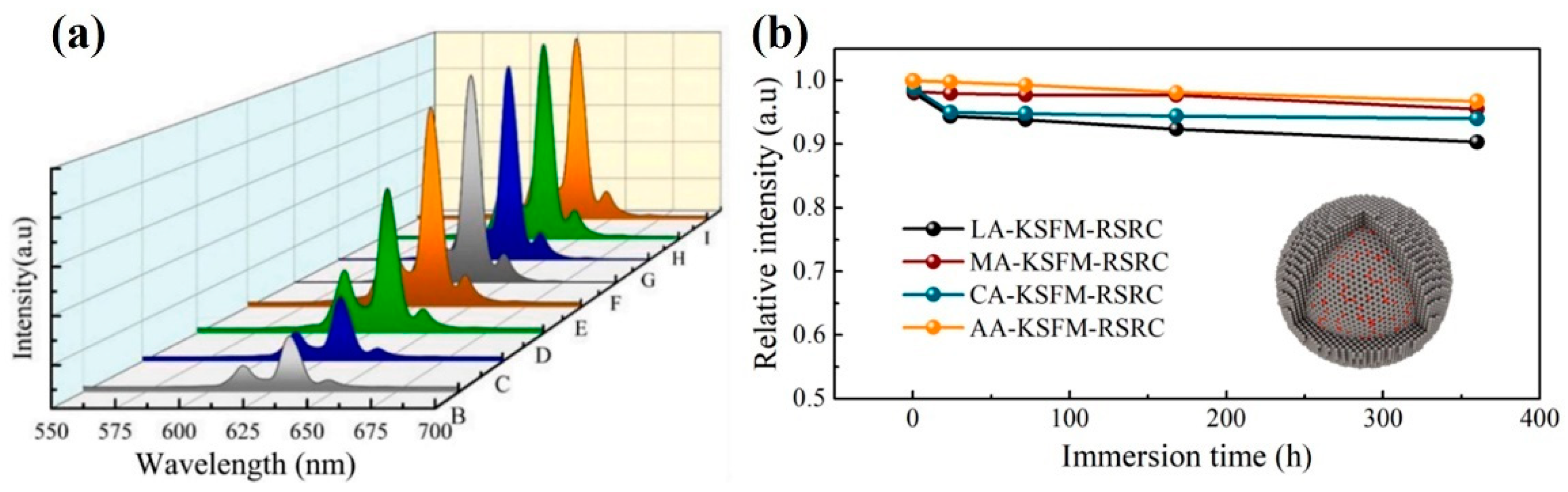
- Huang, L.; Liu, Y.; Si, S.C.; Brik, M.G.; Wang, C.X.; Wang, J. A new reductive dl-mandelic acid loading approach for moisture-stable Mn4+ doped fluorides. Chem. Commun. 2018, 54, 11857–11860. [Google Scholar] [CrossRef] [PubMed]
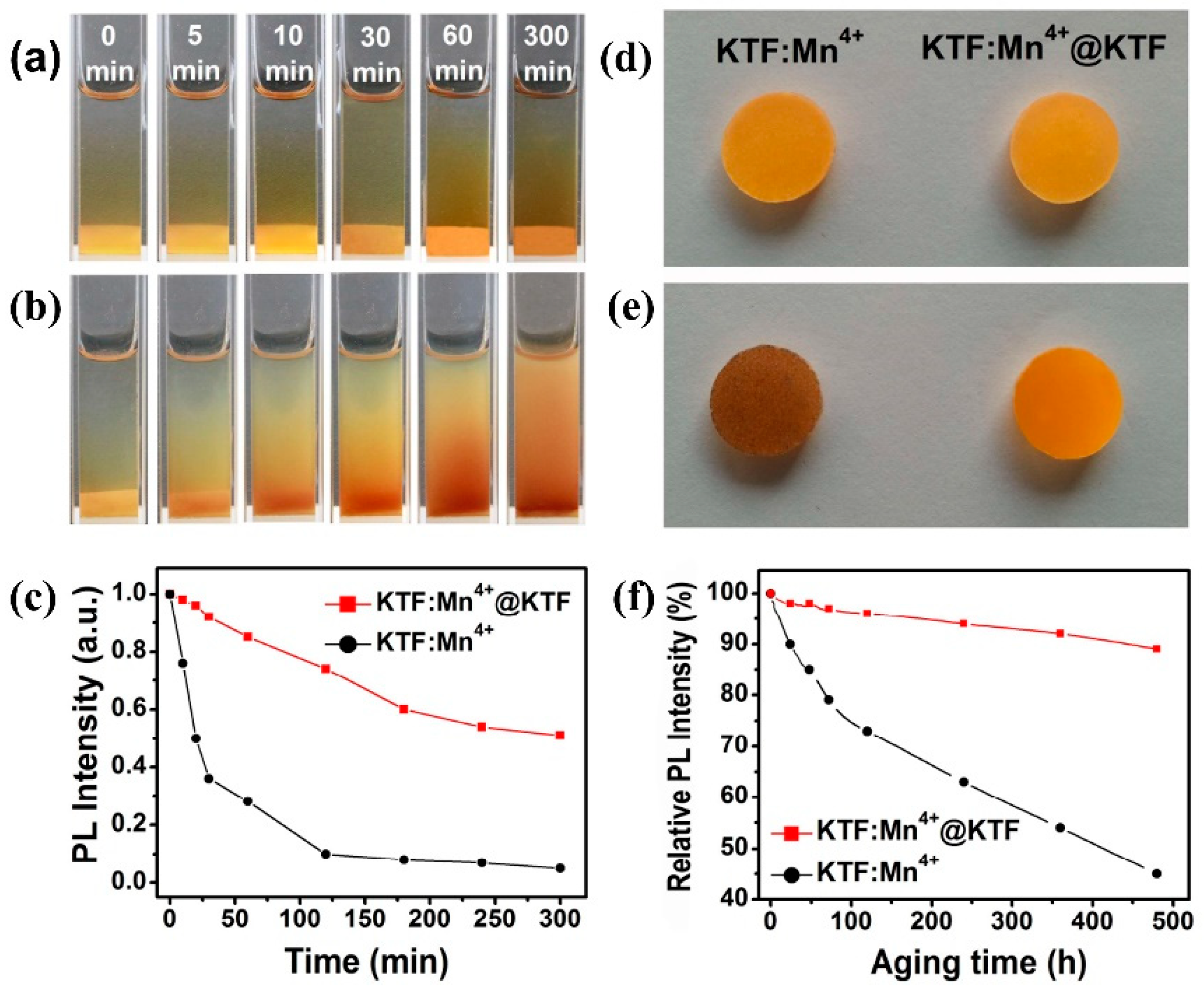
- Huang, L.; Liu, Y.; Yu, J.B.; Zhu, Y.W.; Pan, F.J.; Xuan, T.T.; Brik, M.G.; Wang, C.X.; Wang, J. Highly Stable K2SiF6:Mn4+@K2SiF6 Composite Phosphor with Narrow Red Emission for White LEDs. ACS Appl. Mater. Interfaces 2018, 10, 18082–18092. [Google Scholar] [CrossRef]
- Zhou, Y.Y.; Song, E.H.; Deng, T.T.; Wang, Y.J.; Xia, Z.G.; Zhang, Q.Y. Surface Passivation toward Highly Stable Mn4+-Activated Red-Emitting Fluoride Phosphors and Enhanced Photostability for White LEDs. Adv. Mater. Interfaces 2019, 6, 1802006. [Google Scholar] [CrossRef]
- Liu, Y.; Zhou, Z.; Huang, L.; Brik, M.G.; Si, S.C.; Lin, L.T.; Xuan, T.T.; Liang, H.B.; Qiu, J.B.; Wang, J. High-performance and moisture-resistant red-emitting Cs2SiF6:Mn4+ for high-brightness LED backlighting. J. Mater. Chem. C 2019, 7, 2401–2407. [Google Scholar] [CrossRef]
- Yu, H.J.; Wang, B.C.; Bu, X.Y.; Liu, Y.-g.; Chen, J.; Huang, Z.H.; Fang, M.H. A facile in situ surface-coating passivation strategy for improving the moisture resistance of Mn4+-activated fluoride red phosphor. Ceram. Int. 2020, 46, 18281–18286. [Google Scholar] [CrossRef]
- Jiang, C.Y.; Brik, M.G.; Srivastava, A.M.; Li, L.H.; Peng, M.Y. Significantly conquering moisture-induced luminescence quench ing of red line-emitting phosphor Rb2SnF6:Mn4+ through H2C2O4 triggered particle surface reduction for blue converted warm white light-emitting diodes. J. Mater. Chem. C 2019, 7, 247–255. [Google Scholar] [CrossRef]
- Liu, L.; Wu, D.; He, S.G.; Ouyang, Z.; Zhang, J.F.; Du, F.; Peng, J.Q.; Yang, F.L.; Ye, X.L. A Reverse Strategy to Restore the Mois ture-deteriorated Luminescence Properties and Improve the Humidity Resistance of Mn4+-doped Fluoride Phosphors. Chem. Asian. J. 2020, 15, 3326–3337. [Google Scholar] [CrossRef] [PubMed]
- Li, D.; Pan, Y.X.; Wei, X.N.; Lin, J. Significantly enhanced the humidity resistance of a novel red phosphor CsNaGe0.5Sn0.5F6:Mn4+ through surface modification. Chem. Eng. J. 2021, 420, 127673. [Google Scholar] [CrossRef]
- Zhong, X.; Deng, D.S.; Wang, T.M.; Li, Y.L.; Yu, Y.; Qiang, J.W.; Liao, S.; Huang, Y.H.; Long, J.Q. A facile surface passivation strategy for Na2SiF6:Mn4+, Li+ phosphors to achieve high moisture resistance and luminescent thermal stability. J. Lumin. 2022, 243, 118643. [Google Scholar] [CrossRef]
- Cai, W.T. Highly Stable Mn4+-Activated Red-Emitting Fluoride Phosphors and Enhanced moisture stability for White LEDs. E3S Web Conf. 2021, 271, 04016. [Google Scholar] [CrossRef]
- Li, Y.L.; Yu, Y.; Zhong, X.; Liu, Y.M.; Chen, L.; Liao, S.; Huang, Y.H.; Zhang, H.X. K2SiF6:Mn4+@K2SiF6 phosphor with remarkable negative thermal quenching and high water resistance for warm white LEDs. J. Lumin. 2021, 234, 117968. [Google Scholar] [CrossRef]
- Zhong, X.; Deng, D.S.; Wang, T.M.; Li, Y.L.; Yu, Y.; Qiang, J.W.; Liao, S.; Huang, Y.H.; Long, J.Q. High Water Resistance and Luminescent Thermal Stability of LiyNa2-ySiF6:Mn4+ Red-Emitting Phosphor Induced by Codoping of Li. Inorg. Chem. 2022, 61, 5484–5494. [Google Scholar] [CrossRef] [PubMed]
- Jiang, C.Y.; Li, L.H.; Brik, M.G.; Lin, L.T.; Peng, M.Y. Epitaxial growth via anti-solvent-induced deposition towards a highly efficient and stable Mn4+ doped fluoride red phosphor for application in warm WLEDs. J. Mater. Chem. C 2019, 7, 6077–6084. [Google Scholar] [CrossRef]
- Wang, Z.-L.; Guo, R.; Li, G.-R.; Ding, L.-X.; Ou, Y.-N.; Tong, Y.-X. Controllable synthesis of ZnO-based core/shell nanorods and core/shell nanotubes. RSC Adv. 2011, 1, 48–51. [Google Scholar] [CrossRef]
- Yu, Y.; Wang, T.M.; Zhong, X.; Li, Y.L.; Wang, L.; Liao, S.; Huang, Y.H.; Long, J.Q. High luminescent thermal stability and water resistance of K2SiF6:Mn4+@CaF2 red emitting phosphor. Ceram. Int. 2021, 47, 33172–33179. [Google Scholar] [CrossRef]
- Xu, H.P.; Hong, F.; Pang, G.; Liu, G.X.; Dong, X.T.; Wang, J.X.; Yu, W.S. Co-precipitation synthesis, luminescent properties and application in warm WLEDs of Na3GaF6:Mn4+ red phosphor. J. Lumin. 2020, 219, 116960. [Google Scholar] [CrossRef]
- Subhoni, M.; Zafari, U.; Srivastava, A.M.; Beers, W.W.; Cohen, W.; Brik, M.G.; Yamamoto, T. First-principles investigations of geometrical and electronic structures of Mn4+ doped A2SiF6 (A = K, Rb, Cs) red phosphors. Opt. Mater. 2021, 115, 110986. [Google Scholar] [CrossRef]


| Phosphor | Shell | Water Resistance | Thermal Stability | Ref. | ||||
|---|---|---|---|---|---|---|---|---|
| HT HH Storage Time/h | PL Intensity Relative to Room Temperature/% | Soaking Time in Water/h | PL Intensity Relative to Room Temperature/% | Temperature/°C | PL Intensity Relative to Room Temperature/% | |||
| K2SiF6: Mn4+@OA K2SiF6: Mn4+ | Oleic acid None | 450 | 85% 77% | - | - | - | - | [38] |
| K2SiF6: Mn4+-98PA K2SiF6: Mn4+ | Pyruvate None | 360 | 88.5% 51.6% | - | - | - | - | [40] |
| K2SiF6: Mn4+@CaF2 K2SiF6: Mn4+ | CaF2 None | - | - | 6 | 88.24 41.68 | 210 | 2.07 1.93 | [63] |
| K2SiF6: Mn4+@SiO2 K2SiF6: Mn4+ | SiO2 None | - | - | 1 | 43 7 | 250 | 100 82 | [46] |
| K2SiF6:Mn4+, Na+@GQDs@K2SiF6 K2SiF6: Mn4+, Na+@GQDs | GQDs@K2SiF6 GQDs | - | - | 6 | 91.63 70.57 | 180 | 298 217 | [47] |
| K2SiF6: Mn4+@C K2SiF6: Mn4+ | C None | - | - | 8 | 73 0.7 | - | - | [48] |
| WR-K2SiF6: Mn4+-8 IE-K2SiF6: Mn4+ | K2SiF6 K2SiF6 | - | - | 6 | 76 11 | - | - | [50] |
| R-K2SiF6: Mn4+ K2SiF6: Mn4+ | K2SiF6 None | - | - | 5 | 62.3 | 150 | 111.9 106.7 | [55] |
| LA-K2SiF6: Mn4+-RSRC MA-K2SiF6: Mn4+-RSRC CA-K2SiF6: Mn4+-RSRC AA-K2SiF6: Mn4+-RSRC | K2SiF6 K2SiF6 K2SiF6 K2SiF6 | - | - | 360 | 90 96 94 97 | - | - | [29] |
| T-K2SiF6: Mn4+ K2SiF6: Mn4+ | K2SiF6 None | - | - | 5.3 | 80.3 63.4 | - | - | [58] |
| K2SiF6: Mn4+@K2SiF6 K2SiF6: Mn4+ | K2SiF6 None | - | - | 5 | 88 1 | 120 | 213 | [59] |
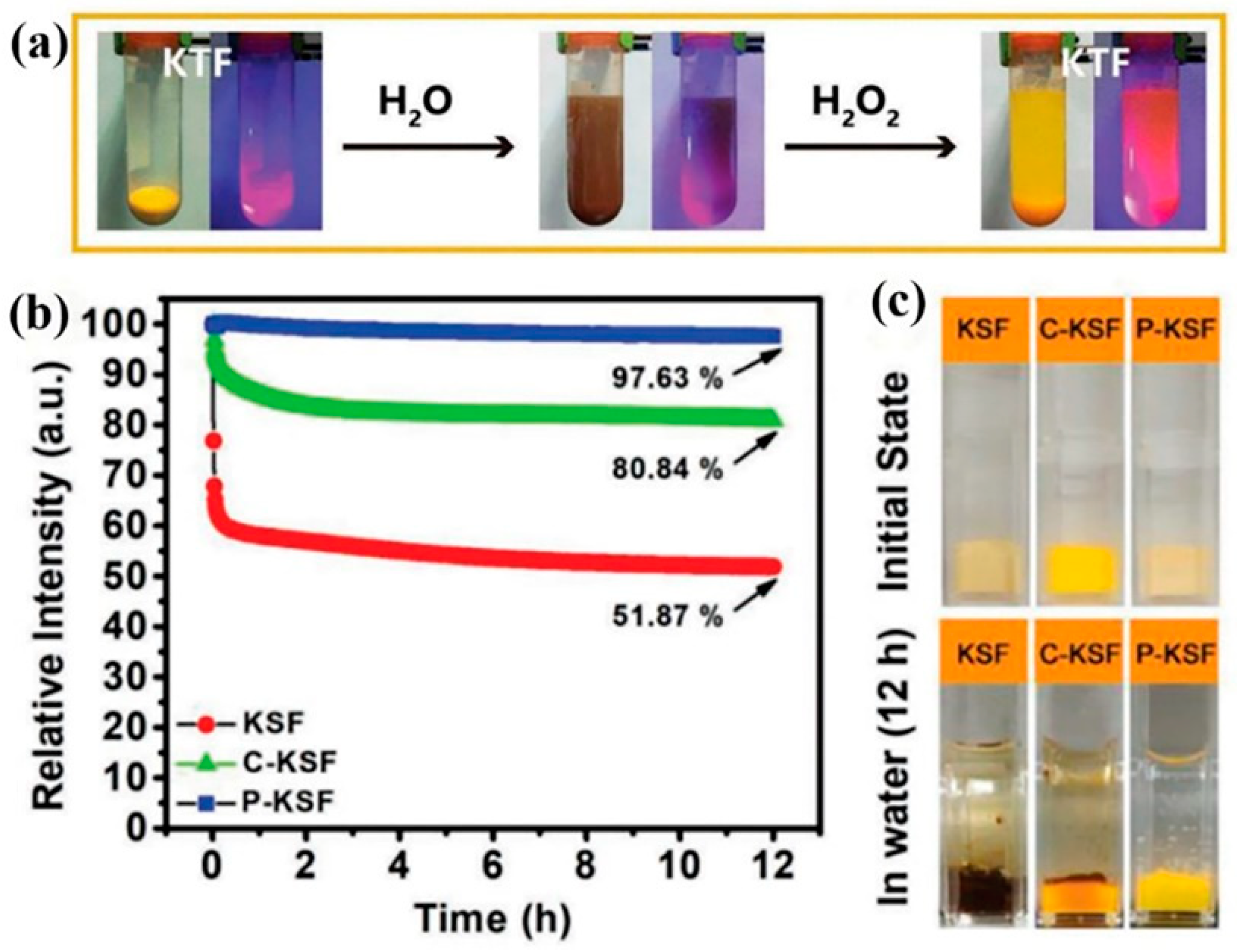
| K2SiF6: Mn4+-CP K2SiF6: Mn4+ | K2SiF6 None | - | - | 12 | 97.6 80.8 | 200 | 100.8 87.1 | [30] |
| K2SiF6: Mn4+@K2SiF6 K2SiF6: Mn4+ | K2SiF6 None | 240 | 90 - | 4 | 82 38 | - | - | [61] |
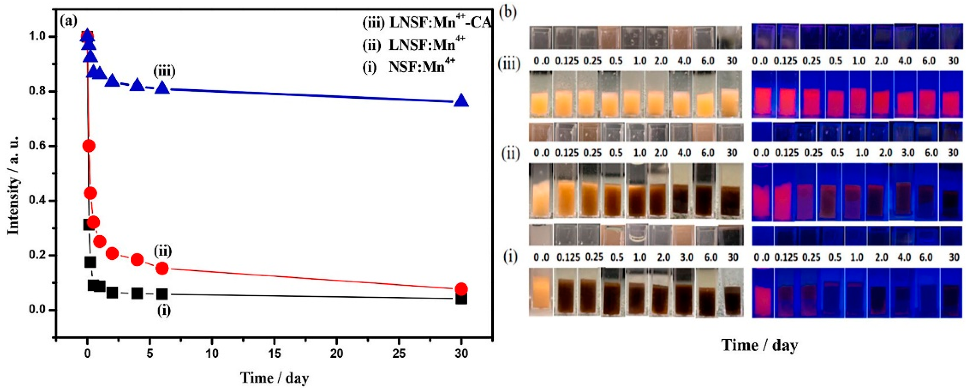
| LiNaSiF6: Mn4+-CA LiNaSiF6: Mn4+ | LiNaSiF6 None | - | - | 6 | 92.33 42.73 | 150 | 118 | [57] |
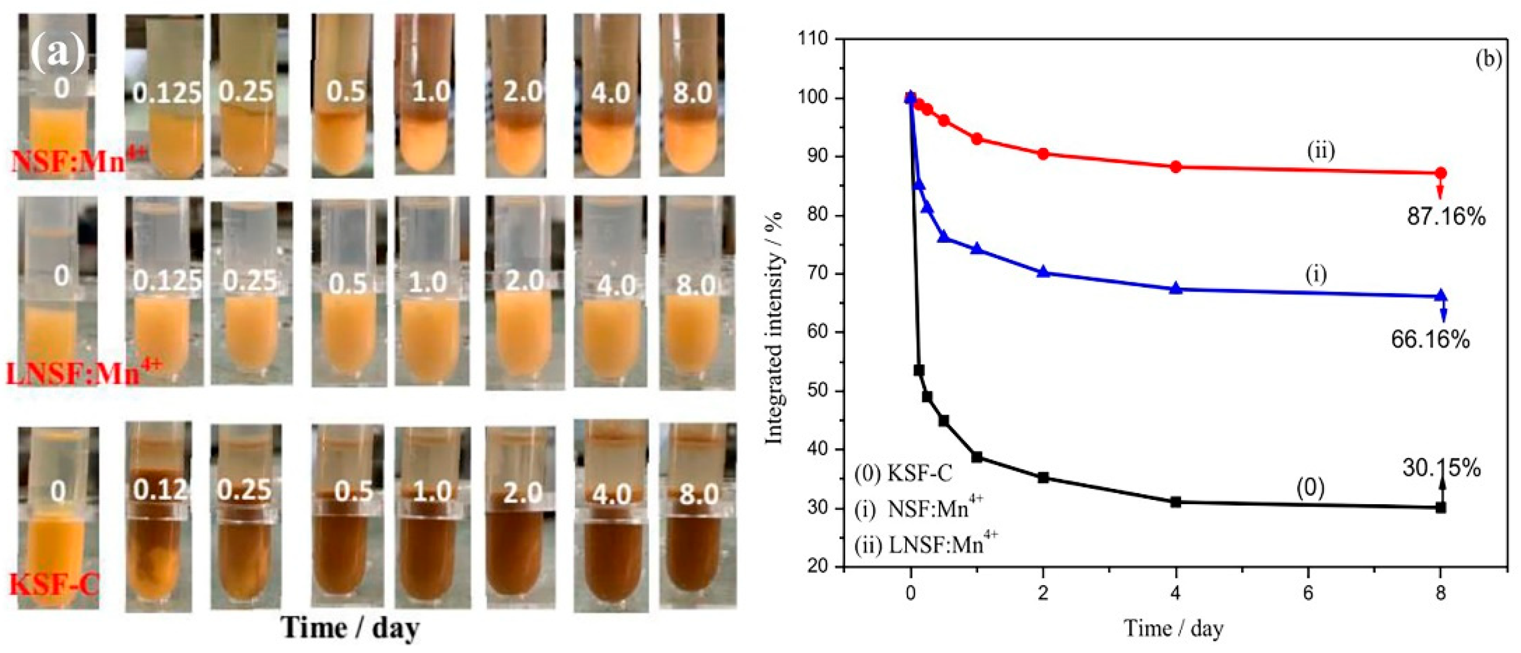
| LiNaSiF6: Mn4+ Na2SiF6: Mn4+ | LiNaSiF6 | - | - | 182 | 87.16 66.16 | - | - | |
| Cs2SiF6: Mn4+-P Cs2SiF6: Mn4+ | Cs2SiF6 None | - | - | 168 | 74 13.6 | 152 | 101 | [52] |
| K2TiF6: Mn4+@CaF2 K2TiF6: Mn4+ | CaF2 None | - | - | 2 | 86.4 6.8 | - | - | [41] |
| K2TiF6: Mn4+@SrF2 K2TiF6: Mn4+ | SrF2 None | - | - | 2 | 80.3 63.4 | - | - | [43] |
| P-K2TiF6: Mn4+ C-K2TiF6: Mn4+ K2TiF6: Mn4+ | K2TiF6 K2TiF6 None | - | - | 12 | 97.63 80.84 51.87 | - | - | [50] |
| K2TiF6: Mn4+@K2TiF6 | K2TiF6 | 480 | 89 | - | - | - | - | [28] |
| K2GeF6: Mn4+@MA K2GeF6: Mn4+ | K2GeF6 None | - | - | 168 | 98 33 | - | - | [49] |
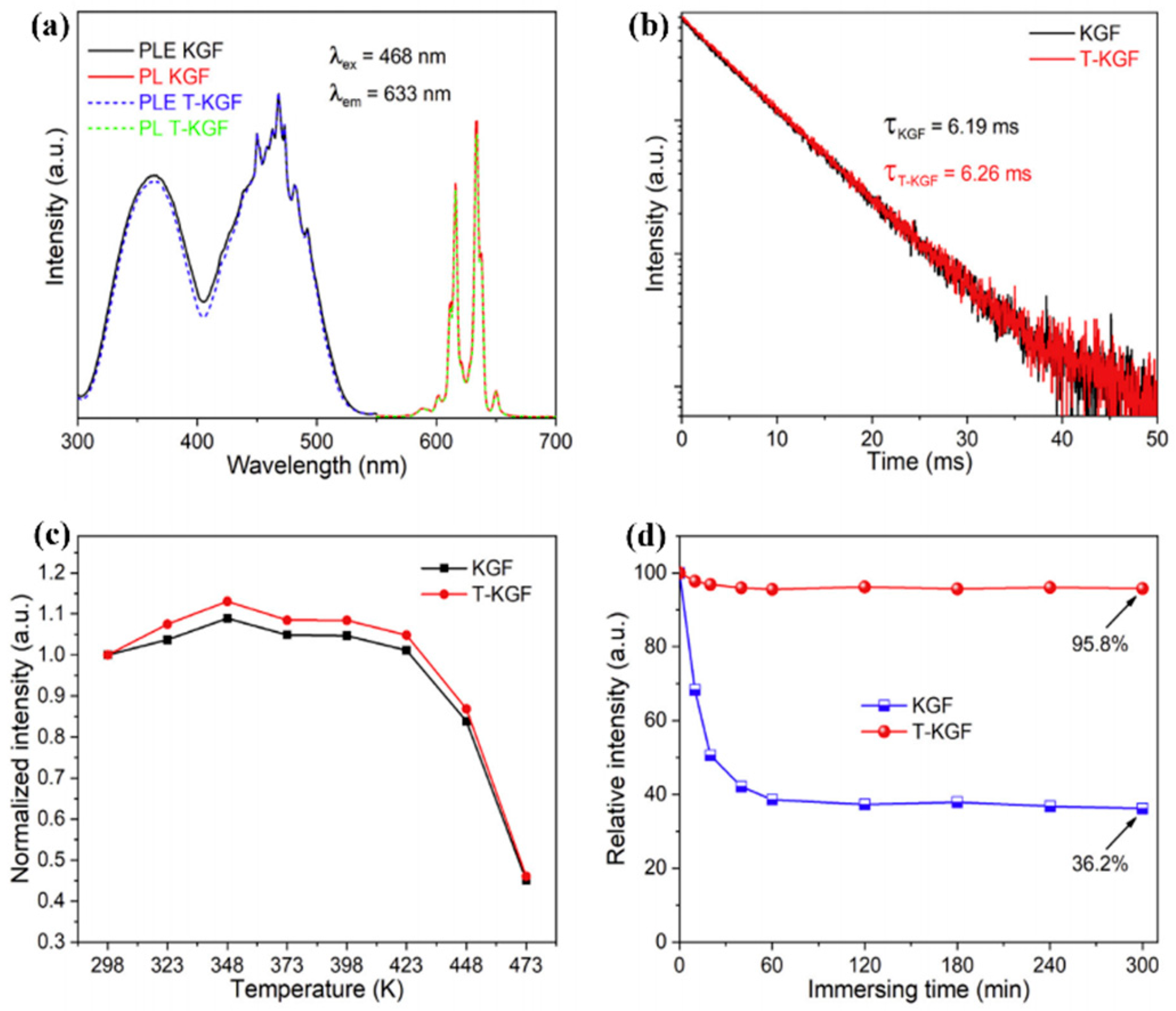
| T-K2GeF6:Mn4+ K2GeF6:Mn4+ | K2GeF6 None | - | - | 5 | 95.8 36.2 | - | - | [53] |
| Rb2SnF6:Mn4+ | Rb2SnF6 | - | - | 3 (Boiling water) | 95 | - | - | [54] |
| Phosphor | Shell | Current/mA | Ra | R9 | CCT/K | LE | Ref |
|---|---|---|---|---|---|---|---|
| K2SiF6: Mn4+@CaF2 | CaF2 | 20 | 89.3 | 3956 | - | [63] | |
| K2SiF6: Mn4+, Na+@GQDs@ K2SiF6 | GQDs@ K2SiF6 | 20 | 91.3 | - | 4546 | - | [47] |
| K2SiF6: Mn4+@K2SiF6 | K2SiF6 | 20 | 80.5 | 63.8 | 5398 | 96 | [50] |
| R-K2SiF6: Mn4+ | K2SiF6 | 20 | 90.4 | 94.2 | 2892 | 183.31 | [55] |
| T-K2SiF6: Mn4+ | K2SiF6 | 20 | - | - | 3500 | 81.6 | [58] |
| K2SiF6: Mn4+@K2SiF6 | K2SiF6 | 20 | 91.3 | - | 3326 | 100.5 | [59] |
| R-K2SiF6: Mn4+ | K2SiF6 | 20 | 90.4 | 94.2 | 2892 | 183.31 | [55] |
| K2SiF6: Mn4+@K2SiF6 | K2SiF6 | 20 | 91.3 | - | 3326 | 100.5 | [59] |
| K2SiF6: Mn4+@K2SiF6 | K2SiF6 | 20 | - | - | 2929 | 119.74 | [61] |
| LiNaSiF6: Mn4+-CA LNSF: Mn4+ | LiNaSiF6 None | 20 | 89.6 74.6 | - | 3916 3939 | 109.6 107.8 | [57] |
| LiNaSiF6: Mn4+ | LiNaSiF6 | 20 | 90.4 | 89 | 3173 | 122 | [60] |
| CsSiF6: Mn4+-P | CsSiF6 | 20 | - | - | 6880 | 133 | [52] |
| K2GeF6: Mn4+@K2GeF6 | K2GeF6 | 20 | 86.3 | - | 3824 | 152.37 | [53] |
Disclaimer/Publisher’s Note: The statements, opinions and data contained in all publications are solely those of the individual author(s) and contributor(s) and not of MDPI and/or the editor(s). MDPI and/or the editor(s) disclaim responsibility for any injury to people or property resulting from any ideas, methods, instructions or products referred to in the content. |
© 2023 by the authors. Licensee MDPI, Basel, Switzerland. This article is an open access article distributed under the terms and conditions of the Creative Commons Attribution (CC BY) license (https://creativecommons.org/licenses/by/4.0/).
Share and Cite
Xie, Y.; Tian, T.; Mao, C.; Wang, Z.; Shi, J.; Yang, L.; Wang, C. Recent Research Progress of Mn4+-Doped A2MF6 (A = Li, Na, K, Cs, or Rb; M = Si, Ti, Ge, or Sn) Red Phosphors Based on a Core–Shell Structure. Nanomaterials 2023, 13, 599. https://0-doi-org.brum.beds.ac.uk/10.3390/nano13030599
Xie Y, Tian T, Mao C, Wang Z, Shi J, Yang L, Wang C. Recent Research Progress of Mn4+-Doped A2MF6 (A = Li, Na, K, Cs, or Rb; M = Si, Ti, Ge, or Sn) Red Phosphors Based on a Core–Shell Structure. Nanomaterials. 2023; 13(3):599. https://0-doi-org.brum.beds.ac.uk/10.3390/nano13030599
Chicago/Turabian StyleXie, Yueping, Tian Tian, Chengling Mao, Zhenyun Wang, Jingjia Shi, Li Yang, and Cencen Wang. 2023. "Recent Research Progress of Mn4+-Doped A2MF6 (A = Li, Na, K, Cs, or Rb; M = Si, Ti, Ge, or Sn) Red Phosphors Based on a Core–Shell Structure" Nanomaterials 13, no. 3: 599. https://0-doi-org.brum.beds.ac.uk/10.3390/nano13030599