Article

In Vitro Toxicity of TiO2:SiO2 Nanocomposites with Different Photocatalytic Properties

1
POLARIS Research Centre, Department of Earth and Environmental Sciences, University of Milano—Bicocca, Piazza della Scienza 1, 20126 Milano, MI, Italy
2
Institute of Science and Technology for Ceramics (CNR-ISTEC), National Research Council of Italy, Via Granarolo 64, 48018 Faenza, RA, Italy
*
Author to whom correspondence should be addressed.
Submission received: 18 June 2019 / Revised: 13 July 2019 / Accepted: 17 July 2019 / Published: 21 July 2019
(This article belongs to the Special Issue Toxicity and Ecotoxicity of Nanomaterials)

Abstract

:
The enormous technological relevance of titanium dioxide (TiO2) nanoparticles (NPs) and the consequent concerns regarding potentially hazardous effects that exposure during production, use, and disposal can generate, encourage material scientists to develop and validate intrinsically safe design solution (safe-by-design). Under this perspective, the encapsulation in a silica dioxide (SiO2) matrix could be an effective strategy to improve TiO2 NPs safety, preserving photocatalytic and antibacterial properties. In this work, A549 cells were used to investigate the toxic effects of silica-encapsulated TiO2 having different ratios of TiO2 and SiO2 (1:1, 1:3, and 3:1). NPs were characterized by electron microscopy and dynamic light scattering, and cell viability, oxidative stress, morphological changes, and cell cycle alteration were evaluated. Resulting data demonstrated that NPs with lower content of SiO2 are able to induce cytotoxic effects, triggered by oxidative stress and resulting in cell necrosis and cell cycle alteration. The physicochemical properties of NPs are responsible for their toxicity. Particles with small size and high stability interact with pulmonary cells more effectively, and the different ratio among silica and titania plays a crucial role in the induced cytotoxicity. These results strengthen the need to take into account a safe(r)-by-design approach in the development of new nanomaterials for research and manufacturing.

1. Introduction

Nanotechnology is growing tremendously fast, and it has led to the creation of a new class of materials, called nanomaterials (NMs), which are nowadays present in everyday life goods. In particular, metal oxide nanoparticles (NPs), thanks to their several properties (antibacterial, photocatalytic, etc.) [1,2] are gaining great attention. Nevertheless, there is a growing concern about the safety of these NMs, due to the increasing release in the market and their consequent intentional and unintentional emission into the environment. In this perspective, to guarantee a sustainable development of these new technologies, the environmental and health safety issues should be addressed in parallel.
Titanium dioxide (TiO2) is largely used as a white pigment in the production of several manufacturings (paint, ceramics, textiles, dental care products, etc.) thanks to its ability to strongly adsorb ultraviolet (UV) lights. Under UV light irradiation, the physical properties of TiO2 change, promoting the decomposition of organic and inorganic compounds. For these properties, TiO2 is highly employed as a photocatalytic agent in a variety of applications, such as pollution remediation in air and water, sterilization, and production of self-cleaning surfaces. Photocatalyzed TiO2 nanoparticles (NPs) have been thoroughly examined given their potential application in cancer therapies and their ability in eradicating cancer cells depends on particle concentrations, cell types, and surface chemistry [3]. Recently, TiO2 NPs have been identified as efficient sensitizers for photodynamic and sonodynamic cancer therapy, especially upon functionalization with antibodies aimed to optimize the selective distribution to target cells [4].
TiO2 nanoparticles (NPs), which are also commonly used in food coloring and packaging, cosmetic and oral care products have recently been investigated as efficient antimicrobial agents. TiO2 NPs have long been known as efficient antimicrobial agents [5]. The photocatalytic properties of TiO2 NPs depend on its crystalline structure, with the anatase crystalline structure having the highest photocatalytic activity [6,7], and on particle size and specific surface area (the smaller the particle size, the greater the number of active surface sites, and the surface charge-carrier-transfer rate increases in photocatalysis). It has been previously demonstrated that TiO2 nanoparticles’ complexation with silica (SiO2) increases TiO2 photocatalytic activity on self-cleaning textiles, proportionally related to SiO2 content. The higher photocatalytic effect has been ascribed to an increase of titanium surface acidity that, in turn, affected its hydrophilicity [8].
The issue of TiO2 toxicity is a crucial aspect to be considered for its application as a photocatalytic agent in self-cleaning surfaces. Even if over the last decades the research activity on the toxicity of TiO2 NPs has been strongly improved, questions about the safe use of these nanoparticles are still open. As photocatalytic activation of TiO2 induces a greater generation of reactive oxygen species (ROS), so it is likely that this also improves its toxicological potential. Some reports from the literature have shown that TiO2 is toxic in the presence of UV irradiation due to ROS generation [9]. UV irradiation of TiO2 suspensions can also change the state of agglomeration of the nanoparticles affecting their photocatalytic activities [10] with an indirect impact on cell viability. Several in vitro studies evidenced that a certain toxicity and genotoxicity is associated with the exposure to TiO2 NPs even in the absence of photoactivation. Oxidative stress is the main toxicity mechanism for non-photoactivated NPs also, with alteration of mitochondrial functionality and induction of DNA damage in different human and non-human cell lines [11,12,13].
The surface functionalization of TiO2 NPs with SiO2, resulting in an increase of hydrophilicity, not only was demonstrated to be able to improve their photocatalytic potential on textile surfaces [8] but also to affect their biocompatibility. TiO2 NPs encapsulation in a SiO2 matrix was demonstrated to be an effective solution to control the redox reactivity of TiO2 NPs by reducing ROS production [14]. Nevertheless, the toxic effects on live cells, as a function of encapsulating SiO2 content, have limited investigations. NPs photocatalyst performance is related to the ratio TiO2:SiO2 in the nanocomposites, which can also be responsible for different cellular consequences upon direct exposure to these new nanomaterials (NMs).
Recently, studies and development of hybrid and core@shell NPs, nanocomposites, and layer-by-layer NPs have arisen [15,16,17,18]. Since these new NMs have different properties from their single NPs constituents, concern and research about their safety are increasing in parallel, pointing out the importance of investigating NPs toxicity in a perspective of sustainable nanotechnology. This study aims to compare the different biological effects induced by silica-encapsulated TiO2 [19], comparing different SiO2 content (TiO2/SiO2 ratio). The effects of the different nanocomposites were tested against the human lung epithelial cells A549, that have been demonstrated as being a good target for the safety assessment of titania NPs [13,20]. Furthermore, A549 cells, derived from a human adenocarcinoma of the lung, are the most often used cell line for toxicity testing. The cells show properties such as surfactant production and transport=like AT-II cells in vivo, secrete cytokines, and perform phase I and phase II xenobiotic biotransformation similar to lung tissue [21,22]. Furthermore, it is important to select the cell line which best represents the intended exposure route, and in this perspective, A549 cells are used for the screening of nanoparticles in inhalation toxicology. Finally, in the context of the NANoREG framework for the safety assessment of NMs [23], A549 cells were used as the main model common for several biological endpoints, making this cell line a promising candidate as a cellular model for in vitro testing of NMs at the regulatory level.
The obtained results are of great importance in the selection of the best SiO2-modified TiO2 photocatalysts, matching safe-by-design requirements.

2. Materials and Methods

2.1. Materials

The following commercial materials were used to prepare the TiO2:SiO2 samples: TiO2 colloidal nanosuspension (nanosol) containing 6 wt % titania (Colorobbia Italia SpA, Sovigliana Vinci, Italy) and SiO2 colloidal nanosol, Ludox HS-40® containing 40 wt % silica (Grace Davison, Columbia, MD, USA). Dowex® 66 anionic exchange resin was purchased from Sigma-Aldrich (Saint Louis, MO, USA).

2.2. TiO2:SiO2 Nanosuspensions Preparation

The commercial TiO2 and SiO2 nanosols, after appropriate dilution with distilled water, were mixed in well-defined ratios and ball milled for 24 h (zirconia balls with 5 mm diameter). Weight ratios of 1:1, 1:3, and 3:1 were investigated for TiO2:SiO2 NPs. The TiO2:SiO2 samples were mixed at opposite surface charge conditions to promote colloidal heterocoagulation [24]. The obtained samples were basified by anionic exchange resin until a pH of 5 to be more compatible with biological targets. The nanocomposites obtained by the colloidal approach were named TiO2:SiO2 1:1, TiO2:SiO2 1:3, and TiO2:SiO2 3:1 and characterized by a TiO2 content equal to 0.75 wt %.

2.3. TiO2:SiO2 Nanoparticles Characterization

The TiO2:SiO2 nanosols underwent morphological analysis using the FEI Titan transmission electron microscope (TEM) operating at an acceleration voltage of 300 kV (FEI Company, Hillsboro, OR, USA). One drop of the nanoparticle suspension diluted in deionized water (75 μg/mL) was deposited on a film-coated copper grid and characterized.
To perform the identification of crystalline phases, TiO2:SiO2 1:1, TiO2:SiO2 1:3, and TiO2:SiO2 3:1 samples were dried at T° = 100 °C for 3 h in an oven. The obtained powders were characterized by X-ray diffraction (XRD) using a Bragg–Brentano diffractometer (Bruker D8 Advance, Karlsruhe, Germany) operating in a θ/2θ configuration, with an X’Celeretor detector LynkEye (10°–80° 2θ range, 0.02 step size, 0.5 s per step).
To investigate the colloidal behavior of TiO2:SiO2 NPs in water, hydrodynamic diameter (z-average) and zeta potential (ζ–potential) were measured by dynamic light scattering (DLS) and electrophoretic light scattering (ELS) measurements, respectively, using Zetasizer Nanoseries apparatus (Malvern Instruments, Malvern, UK).

2.4. Cells Culture Maintenance and Treatments

The A549 cell line (ATCC® CCL-185™) was routinely maintained as previously reported in Gualtieri and colleagues [25]. Briefly, cells were maintained in OptiMEM medium (Gibco, Life Technologies) supplemented with fetal bovine serum (Gibco, Life Technologies, Monza, Italy) 10% and Pen/Strep 1% (Euroclone, Pero, Italy) at 37 °C, 5% CO2.
For the treatments with the different TiO2:SiO2 NPs, cells were seeded in 6 MW (Corning) at the density of 1.5*105 cells/well and incubated until they attached and reached to the confluence of 80% (about 24 h). Cells were treated with NPs for 24 h in all experiments, except for cytofluorimetric analysis (ROS and uptake) in which they were exposed for 180 min (3 h). Each experiment included negative control (untreated cells) and positive control (1 mM H2O2 for ROS detection). For the cell viability experiments, cells were treated with different doses of TiO2 (7.5, 75, and 750 μg TiO2/mL), while for the other experiment, a dose of 75 µg TiO2/mL was used.

2.5. Cell Viability

The cytotoxicity of TiO2:SiO2 was evaluated through two different analysis: by the 3-(4,5-dimethylthiazol-2-yl)-2,5-diphenyltetrazolium bromide (MTT) test and Hoechst 33342/propidium iodide (H/PI) staining.
MTT (Sigma Aldrich, Milano, Italy) assay was performed according to previous works [26]. Briefly, after 24 h of exposure to NPs, supernatants were collected, centrifuged at 1200 rpm for 6 min, and stored at −80 °C. Cells were rinsed with phosphate buffered saline (PBS), and MTT solution was added to the medium (final concentration 0.3 mg/mL) for 2 h. After the formazan crystal, which are the MTT reduction products, were dissolved in 1 mL Dimethyl sulfoxide (DMSO, Sigma Aldrich), absorbance was analyzed at 570 nm by a multi-plate reader (Infinite 200 Pro, TECAN, Männedorf, Switzerland).
The H/PI nuclear staining allowed to count the number of viable, necrotic, apoptotic, and mitotic A549 cells after NPs exposure. Cells were detached by trypsinization and then re-suspended in a complete medium; 10 μL of 1:1 solution of Hoechst 33342/PI (Sigma Aldrich) was added to cell suspensions and stored in the dark for 15 to 30 min at room temperature. Cells were then centrifuged for 6 min at 1200 rpm, and re-suspended in 20 μL of FBS. Three drops (about 4 μL) of cell suspensions were smeared on a coverslip and observed under a fluorescence microscope (Zeiss-Axioplan, Carl Zeiss Microscopy GmbH, Jena, Germany) with a UV filter (365 nm). At least 300 cells per sample were scored according to nuclei staining and plasma membrane integrity as viable normal cells (H positive and PI negative, without special nuclear characteristic and an intact plasma membrane), necrotic cells (non-apoptotic and PI positive), apoptotic cells (bright H or PI positive stained with condensed or fragmented nuclei), mitotic cells (H positive with chromosome condensation).

2.6. Cytological Observation

For morphological analysis, cells were seeded on a cover slide at a concentration of 1.5*105 cells/well, cultured for 24 h and then exposed to NPs for further 24 h. At the end of the treatment, cells were processed for hematoxylin-eosin (HE) staining, as previously described in [27]. The slides were observed on an optical microscope (Zeiss-Axioplan, Carl Zeiss Microscopy GmbH, Jena, Germany), and pictures were acquired using an AxioCam MRc5 digital camera and processed using AxioVision Real 4.8 software (Carl Zeiss Solutions, Jena, Germany).
Cytoskeleton organization was instead analyzed by staining actin microfilaments with rhodamin phalloidine (1:40 dilution, Cytoskeleton Inc., Denver, CO, USA). Cells, seeded on a cover slide, after exposure to 75 µg/mL of NPs, were fixed in 4% paraformaldehyde for 20 min, washed and permeabilized with 0.1% Triton-100X and then incubated with rhodamin phalloidine for 30 min. After PBS washing, slides were mounted with ProLong™ Gold Antifade Mountant with DAPI (Molecular Probe, Life Technologies, Monza, Italy), dried overnight and then observed under the inverted microscope AxioObserver Z1 Cell Imaging station (Carl-ZEISS Spa, Milano, Italy) and images were acquired by an MRc5 digital camera and elaborated with the dedicated software ZEN 2.3 Blue edition.

2.7. Cytofluorimetric Analysis

Cytofluorimetric analyses were performed: (i) to evaluate TiO2:SiO2 NPs capability to induce ROS formation, (ii) to investigate the interaction between different NPs with A549 cells by the side scatter analysis, and (iii) to analyze cell cycle alterations.
For ROS detection, A549 cells were pre-incubated for 20 min with the probe Carboxy-2′,7′-Dichlorofluorescein Diacetate (carboxy-DCFDA, 5 μM, Life Technologies) and incubated with NPs for 180 min. After incubation, cells were detached, centrifuged at 1200 rpm for 6 min, re-suspended in 500 μL of PBS and analyzed in the FITC channel by flow cytometry (CytoFLEX, Beckman Coulter GmbH, Krefeld, Germany) with the software CytoExpert. The signal from unloaded samples (cells without the probe carboxy-DCFDA) was evaluated as reference to assess cells and NPs’ autofluorescence. These values were then subtracted from the values to DCFDA stained samples.
To investigate the effects on cell cycle, untreated and cells treated for 24 h were harvested, fixed in ethanol 90% and stored at −20 °C until analysis. Cells were then centrifuged at 1600 rpm for 6 min, ethanol was discharged, and cells suspended in PBS containing RNAsi-DNasi free (10 µL) for 30 min at 37 °C. Then 5 µL of Propidium Iodide (PI) was added for 7 min and finally, cells analyzed by the Cytoflex in the ECD-channel (λEcc 488 nm, λEm 610/20 nm). The percentage of cells in the different cell cycle phase (sugG0, G1, S, and G2/M) was displayed. The side scatter (SSC) was also analyzed to assess cell–NP interaction.
Furthermore, the capability of the different NPs to induce autophagy was assessed through the analysis of LC3B II expression by cytofluorimeter. Cells, after 24 h of exposure to 75 µg/mL of NPs, were harvested, fixed with paraformaldehyde 4% for 15 min, permeabilized with methanol 90%, and then stained with the antibody rabbit anti-human LC3BII (1:400 dilution, Cell Signaling). After 1 h of incubation in the appropriate buffer (0.5% bovine serum albumin in PBS 1X), cells were washed twice in buffer and then incubated for 30 min with secondary antibody AlexaFluor anti-rabbit 488 (1:200 dilution, Life Technologies). After washing, cells were resuspended in PBS and read by the CytoFLEX in the FITC channel.

2.8. Statistical Analysis

The data represent the mean ± standard error of the mean (SEM) of at least three independent experiments. Statistical analyses were performed using Sigma Stat 3.2 software, using unpaired t-test or one-way ANOVA and Bonferroni’s post hoc analysis. Values of p < 0.05 were considered statistically significant.

3. Results

3.1. TiO2:SiO2 Nanocomposites Characterization

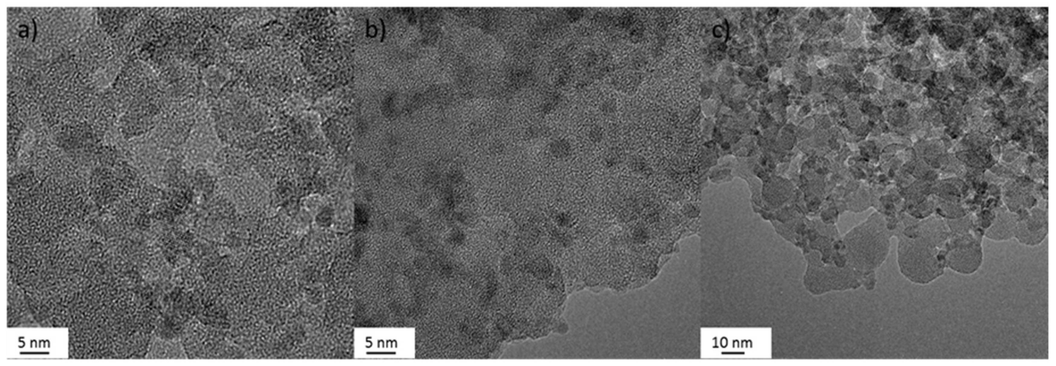
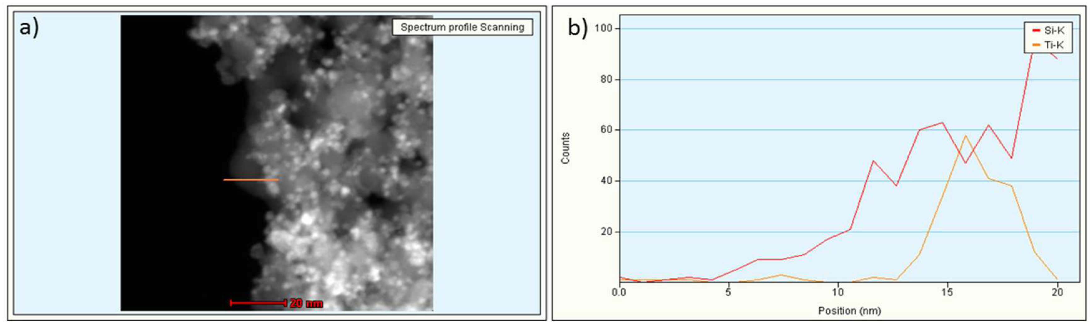
3.1.1. Transmission Electron Microscopy (TEM)

The TEM micrographs (Figure 1) showed aggregated samples with TiO2 randomly distributed within a silica matrix. This matrix encapsulation structure is typical of the colloidal heterocoagulation method used to prepare TiO2:SiO2 samples [19,28]. The energy-dispersive X-ray (EDX) line scan analysis of the elemental distribution, on TiO2:SiO2 1:1 sample, confirmed the presence of a matrix encapsulation structure, where the TiO2 NPs were surrounded by SiO2 NPs (Figure 2).
Data on TiO2 and SiO2 NPs morphology are shown in Supplementary materials (Figure S1).

3.1.2. Colloidal Properties
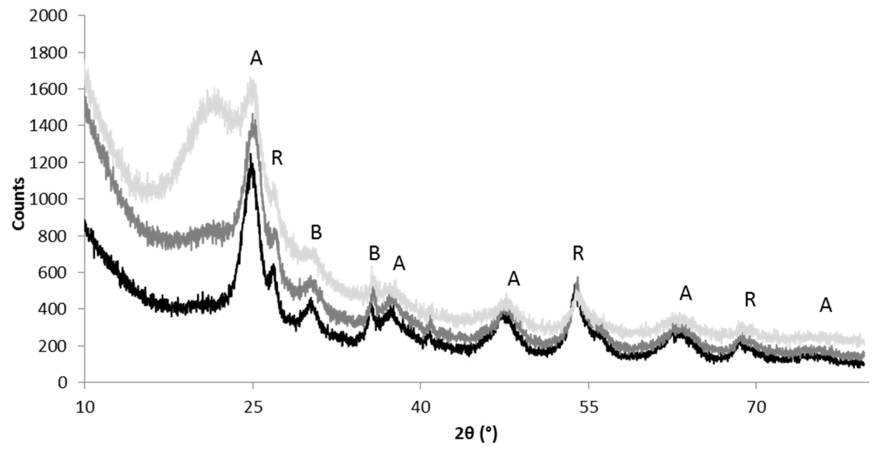
Data about the hydrodynamic behavior, PdI and ζ-potential values of TiO2:SiO2 NPs are reported in Table 1. As expected, TiO2 (+38 mV) and SiO2 (−43 mV) NPs were positively and negatively charged, respectively, at natural pH in water. Their opposite surface charge triggers the heterocoagulation phenomenon, which, by means of electrostatic interactions, promotes the encapsulation of TiO2 NPs into the SiO2 matrix. The small size, low polydispersity index (PdI) and high values of zeta-potential demonstrated the good colloidal stability of both starting NPs suspensions. The increase of hydrodynamic diameter as a function of the TiO2:SiO2 ratio is caused by both the steric hindrance of SiO2 heterocoagulated on the TiO2 surface and the consequent electrostatic destabilization due to the progressive neutralization of the TiO2 surface charge with the increase of SiO2 content [29,30]. The hydrodynamic diameters showed that TiO2:SiO2 NPs 1:1 and 3:1 have similar behavior, with average values of 126 and 148 nm, respectively. On the other hand, the hydrodynamic diameter of the sample TiO2:SiO2 1:3 was much larger than the other NPs (1490 nm) evidencing its tendency to aggregate in an aqueous medium. Moreover, TiO2:SiO2 1:3 also showed the highest PdI, consistent with a very broad size distribution. Such a different behavior among the samples was well expressed by the zeta potential values. A positively charged surface of the NPs was measured for all the heterocoagulated suspensions. The zeta potentials of TiO2, TiO2:SiO2 1:1 and TiO2:SiO2 3:1, higher than +35 mV, pointed out that these suspensions are stable. On the contrary TiO2:SiO2 1:3 exhibited a low ζ-potential value (<+10 mV), meaning that the electrostatic repulsion barrier cannot prevent flocculation and coagulation phenomena.

3.1.3. XRD-Analysis

Figure 3 shows the XRD patterns for TiO2SiO2 3:1, TiO2SiO2 1:1 and TiO2SiO2 1:3 powder samples. The samples show a broad band centered at 2θ = 22.0, the characteristic peak for amorphous SiO2 (JCPDS 29–0085) with an intensity proportional to the amount of silica added. Anatase (JCPDS 21–1272) is the predominant crystalline phase of TiO2 component, then there are small amounts of brookite (JCPDS 29–1360) and rutile (JCPDS 65–0190). In general, broad peaks are present, they are typical of nano-sized crystallites.

3.2. Cytotoxicity

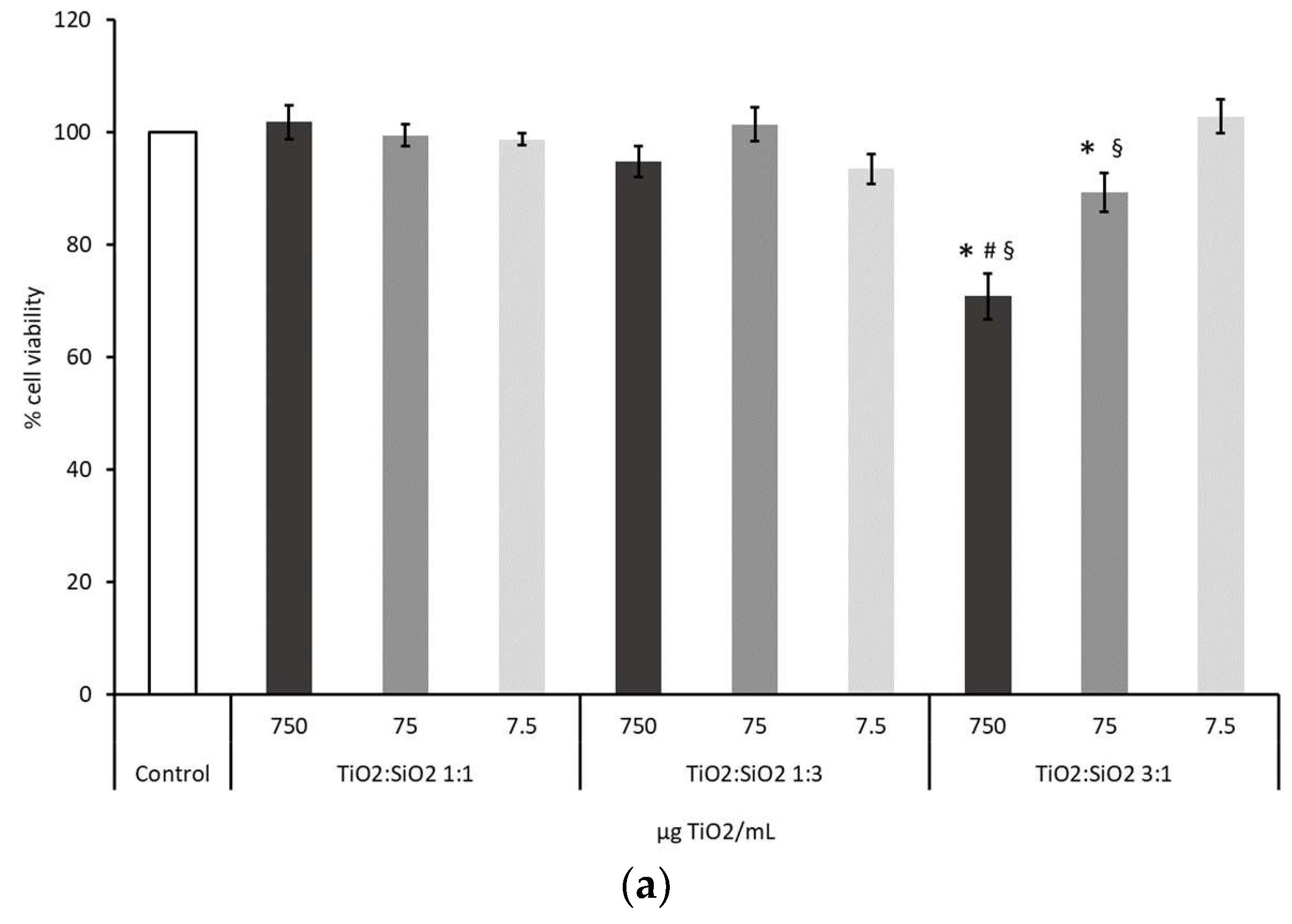
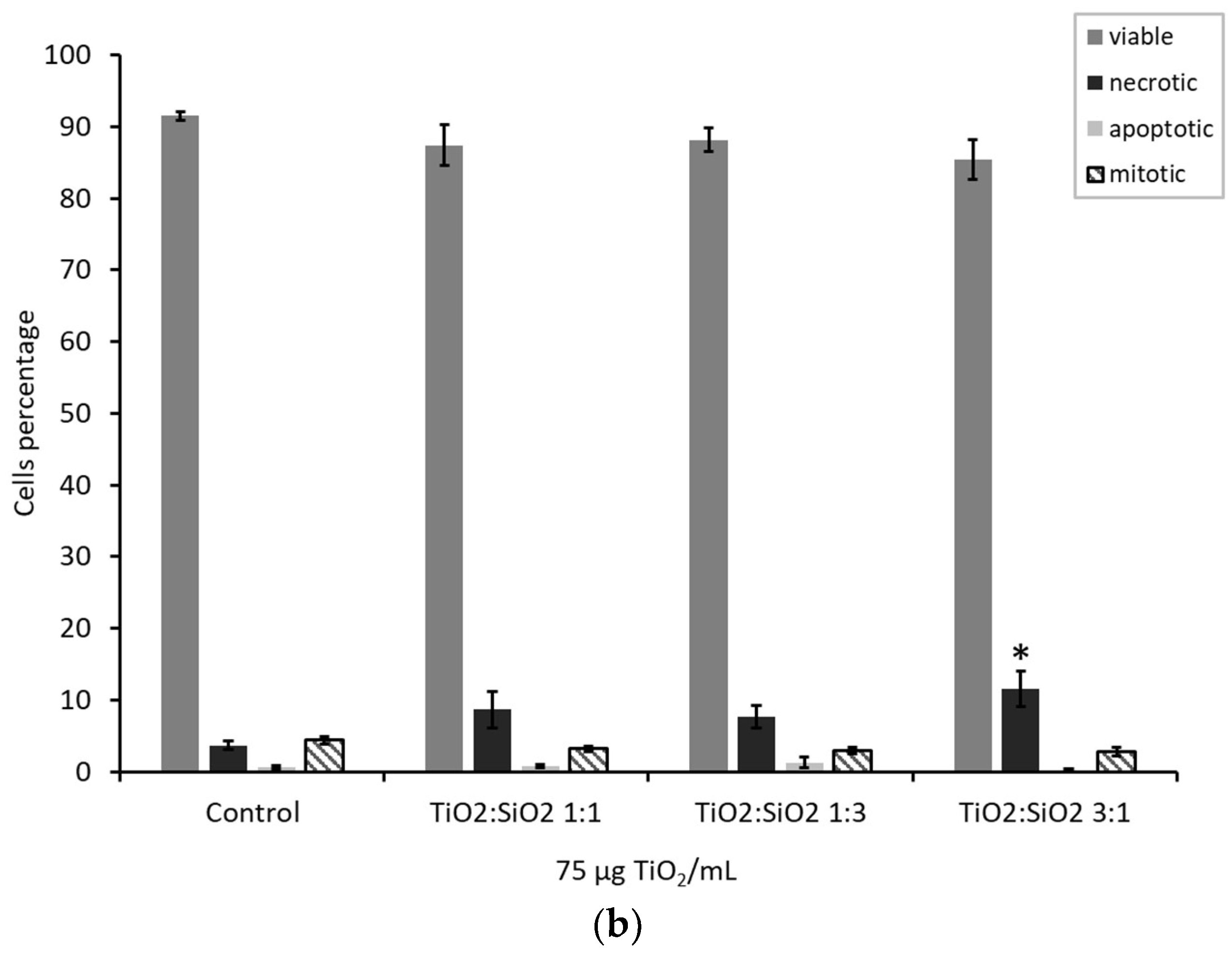
3.2.1. Cell Viability: MTT Test and H/PI

Cells viability was assessed to evaluate the toxic potential of TiO2 based nanocomposites. Data from MTT test showed that particles TiO2:SiO2 3:1, with a lower amount of SiO2 vs. TiO2, have affected the viability of the cells at the doses 750 and 75 µg/mL with a mortality of the 30% and 10%, respectively (Figure 4a). Cell viability data obtained from the exposure to TiO2 and SiO2 NPs alone are reported in Supplementary Materials (Figure S2). To investigate the type of cell death induced by these NPs, H/PI staining has been performed at the dose 75 µg TiO2/mL, which is the dose used for the other endpoints. Furthermore, this effective concentration falls in the range (from 1 µg/mL until 200 µg/mL) recommended for the in vitro testing of NMs aiming to investigate biological responses and in mechanistic studies [31,32]. Results from this test showed that TiO2:SiO2 3:1 induced a reduction of cell viability, as demonstrated by the increased number of necrotic cells (Figure 4b). Results on TiO2 NPs alone are reported in the Supplementary Materials (Figure S3a).
Data were also confirmed by the Annexin V/PI test, which evidenced that the exposure to TiO2:SiO2 3:1, as well as TiO2 NPs alone, increased the percentage of early necrotic cells (Annexin V+/PI+). Thanks to the Annexin V test, a slight increase of apoptotic (Annexin V+) cells, after exposure to TiO2:SiO2 1:1, 3:1, and TiO2 NPs alone, was appreciated (Supplementary Materials S3b,c).

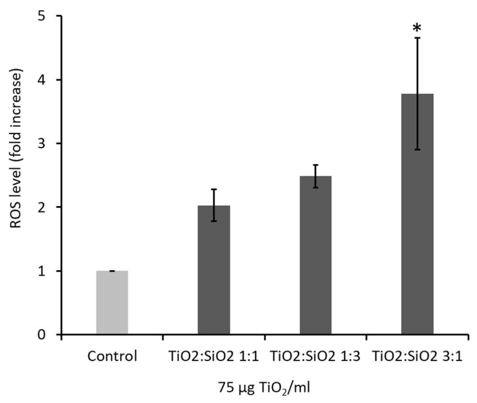
3.2.2. Oxidative stress: Reactive Oxygen Species (ROS) Formation

The induction of oxidative stress was evaluated by measuring the intracellular levels of ROS at early time points (180 min). Data showed that only TiO2:SiO2 3:1 NPs at the dose of 75 µg/mL was able to induce a significant increase of ROS levels (3.5-fold) with respect to control (Figure 5). Data on TiO2 and SiO2 NPs alone are reported in the Supplementary Materials (Figure S4).

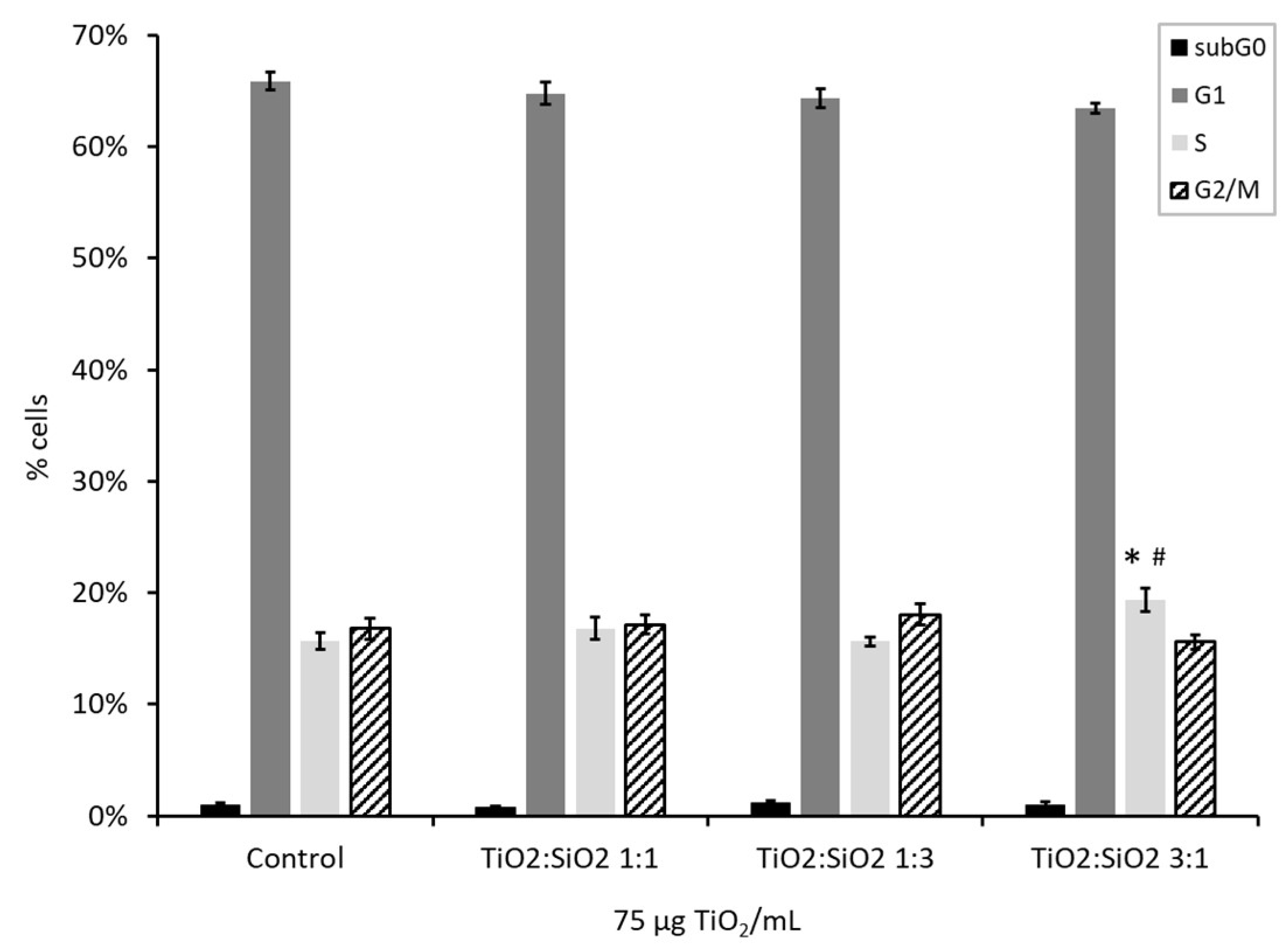
3.2.3. Cell Cycle Analysis

The analysis of the cell cycle allows us to understand the potential of NPs to induce DNA damage. Data showed that TiO2:SiO2 3:1 NPs at the dose of 75 µg/mL induced a slight but significant arrest of the cell cycle in the S-phase (Figure 6). In fact, in control sample, 15.6% of cells were in the S-phase, while this percentage of cells is increased to 19.3% when treated with TiO2:SiO2 3:1 NPs.

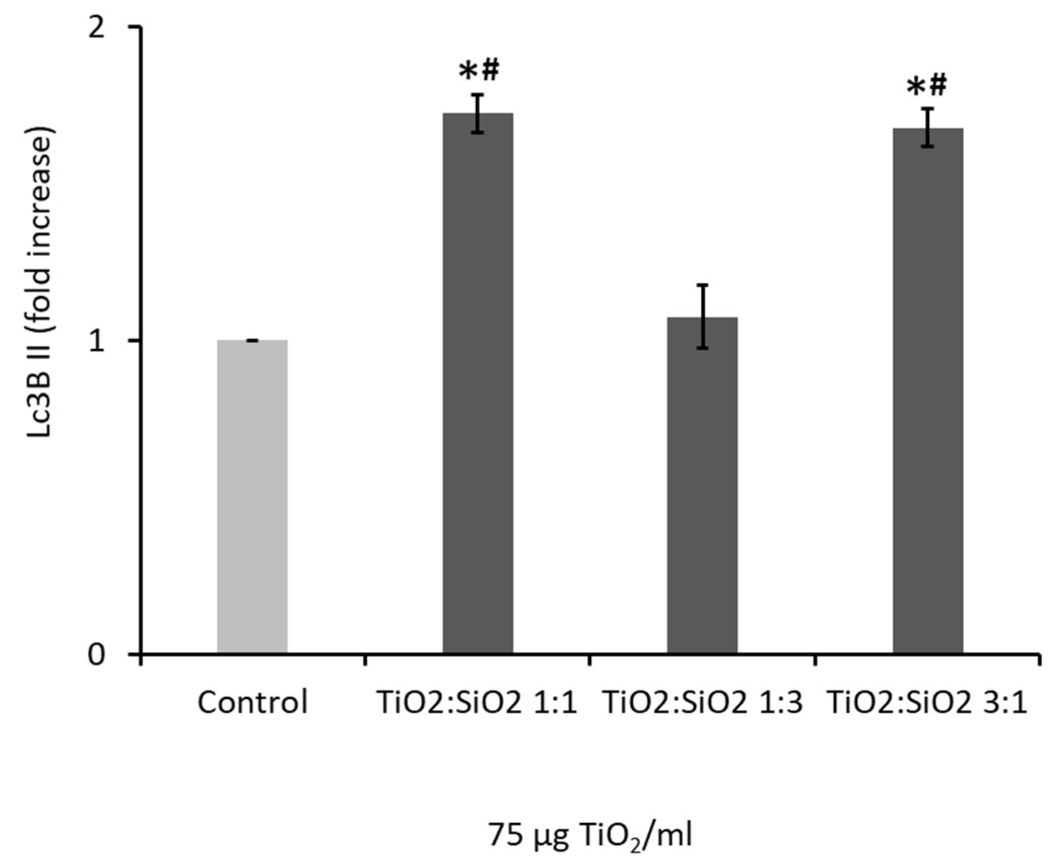
3.2.4. NPs Induced Autophagy: LC3B II Expression

The analysis of the LC3BII protein allows us to understand the potential of NPs to induce cell autophagy. Data showed that TiO2:SiO2 1:1 and 3:1 NPs at the dose of 75 µg/mL induced a significant increase of LC3BII expression, while 1:3 NPs values are similar to the control one (Figure 7). Data on TiO2 and SiO2 NPs alone are reported in the Supplementary Materials (Figure S5).

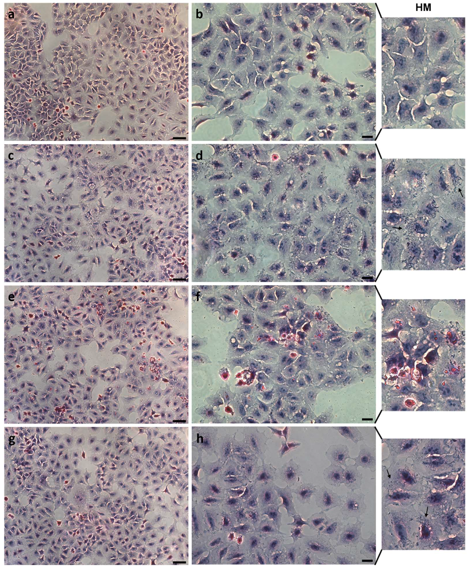
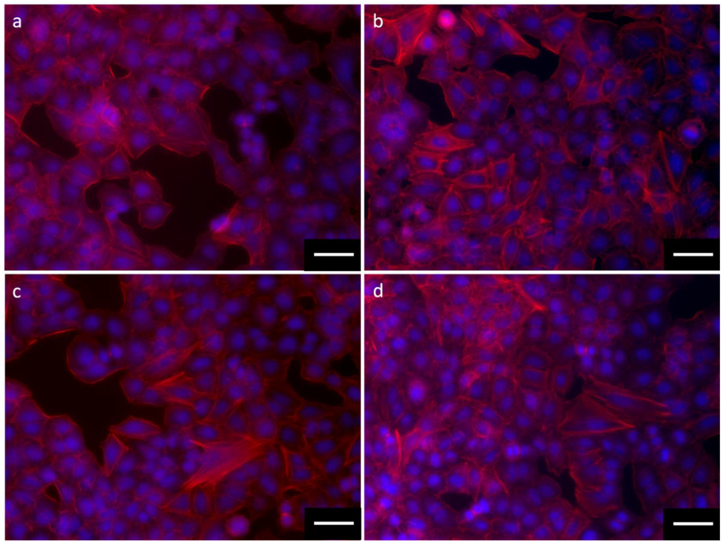
3.3. Cell Morphology and NPs Interaction

3.3.1. Morphological Analysis

Data from HE staining showed that the exposure to the different NPs does not induce any significant morphological change (Figure 8). High magnification images (HM) clearly show the interaction of TiO2:SiO2 NPs 1:1 and 3:1 with A549 cells, while an analog localization of NPs is not detectable in the cells exposed to TiO2:SiO2 1:3. On the contrary, high dimension aggregates are visible on these cells. Data on TiO2 NPs alone are reported in the Supplementary Materials (Figure S6).
Furthermore, data from rhodamine phalloidin staining showed that the exposure to the different NPs seems to induce actin network morphological change (Figure 9). The interaction of TiO2:SiO2 NPs 1:1 (Figure 9b) and 3:1 (Figure 9d) with A549 cells, increased cytoskeleton disassembly and the presence of cellular filopodia, while an analog localization of actin is not detectable in the cells exposed to TiO2:SiO2 1:3 (Figure 9c). Data on TiO2 NPs alone are reported in the Supplementary Materials (Figure S7).

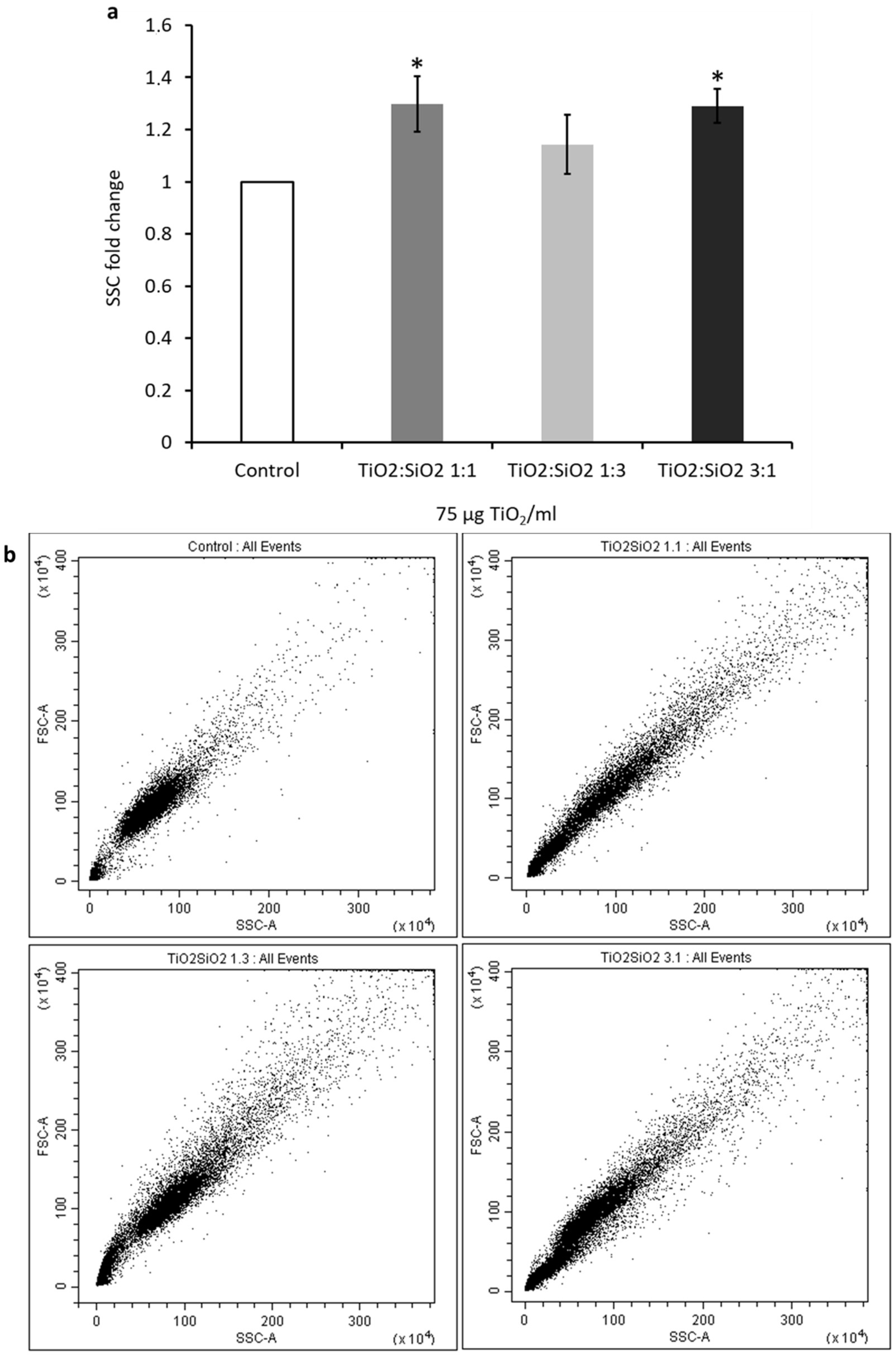
3.3.2. Cell–NP Interactions

The interaction among cells and NPs was investigated by analyzing the side scatter (SSC) of cells through cytofluorimetric analysis. Data showed that there is an increase of 1.3-fold in the SSC with TiO2:SiO2 1:1 and 3:1 NPs (Figure 10). This result confirms the higher interaction with A549 cells of TiO2:SiO2 1:1 and 3:1 NPs, as suggested by optical microscopy images, in line with the smaller size and more positive surface charge of these nanocomposites with respect to TiO2:SiO2 1:3 (Table 1). Data on TiO2 NPs alone are reported in the Supplementary Materials (Figure S8).

4. Discussion

Crystalline TiO2 recently has gained great attention thanks to its special role in a wide range of applications in areas of photovoltaics, photocatalysis, photochromics, and sensors [33]. The employment of TiO2 in industry has been a reality for almost 20 years [34], and TiO2-based nanomaterials are currently applied in a wide range of manufacturing fields (i.e., personal care, inks, plastics, food products, textiles, and medical devices) [35] and in the development of new anticancer therapies [36,37].
Materials coated with TiO2 have long been described for their anti-bacterial properties [38]. Furthermore, TiO2 nanotube coated surfaces can enhance osteoblast adhesion and proliferation [39]. Data on murine osteogenic cells demonstrated that the TiO2 nanotube arrays can reduce Staphylococcus epidermidis colonization and enhance C3H10T1/2 cell adhesion and this dual effect is due to the multiple physical and chemical properties of the TiO2 nanotube surface [40]. In 2016, Hashizume et al. demonstrated that after TiO2 exposure alteration of the pulmonary inflammatory response may occur and may depend on the surface coating material. Therefore, the pulmonary toxicities of coated TiO2 need to be further evaluated, and the physicochemical properties may be useful for predicting the pulmonary risk posed by new nano-TiO2 materials [41].
It is likely that the use of TiO2 and titania-based nanoparticles as antimicrobial devices is still limited by possible adverse effect on human health, especially upon the skin and pulmonary exposure [42]. Inhalation of engineered NPs, including TiO2 NPs, mainly occurs during their production and use in laboratories, industries, and factories, but it is important to consider the whole nanomaterial lifecycle from the perspective of possible releases into the environment. TiO2 NPs have been widely investigated for their cytotoxic effects, mainly due to their capability to induce oxidative stress under UV-irradiation [9], but also when not photoactivated [11,12,13]. A recent study has reported how airborne TiO2 NPs can induce oxidative stress under outdoor conditions, including UV-irradiation and relative humidity [43], corroborating the crucial issue of TiO2 NPs risk assessment, due to the widespread distribution of nanomaterials in the environment [44,45].
In the last decade, different TiO2:SiO2 nanostructures were designed to enhance TiO2 photocatalytic effect [8,46], but TiO2 encapsulation in SiO2 matrix was also demonstrated effective in reducing ROS production. The protective role of SiO2 on TiO2-induced oxidative stress was demonstrated by spectroscopic quantification of free radicals production by spin trap molecules [14], suggesting a promising safety outcome in live cells.
In the current study, a comparison of cytotoxicity of TiO2:SiO2 NPs having different ratios of TiO2 and SiO2 was performed by analyzing their effects on A549 viability, cell cycle, morphology, and oxidative stress. Cells viability was first assessed after exposure to TiO2:SiO2 1:1, TiO2:SiO2 1:3, and TiO2:SiO2 3:1 with an NP concentration ranging from 7.5 to 750 µg TiO2/mL, which is representative of realistic human exposure [47].
A significant impact on cell viability was observed only with TiO2:SiO2 3:1 at the higher concentrations and necrosis, and slight apoptosis, seems to be the main pathways of cell death. A549 necrosis induced by exposure to 75 µg TiO2/mL TiO2:SiO2 3:1 is mediated by increasing reactive oxygen species, according to mechanisms widely described in literature concerning NPs cytotoxicity [48,49].
The comparison with the biological effects induced by the exposure TiO2 and SiO2 NPs alone is quite controversial, but to have a complete overview of the differences between diverse NMs properties, several endpoints have also been investigated after TiO2 and SiO2 treatment.
In particular, cytotoxicity was investigated for both SiO2 and TiO2 NPs alone and data revealed that SiO2 NPs are highly toxic even at the intermediate dose of exposure, while TiO2 induced cell reduction only at the dose of 750 µg/mL. Compared to cytotoxicity TiO2 NPs values, TiO2:SiO2 3:1 NPs resulted more toxic. These could be explained by the fact that the nanocomposites are physically different from TiO2 and SiO2 single NPs nanoparticles. This evidence is confirmed by TEM and DLS results and from previous data from Ortelli et al. [8], in which authors have demonstrated that in an aqueous medium TiO2/SiO2 samples have a different behavior/reactivity towards water compared to titania and silica alone. The presence of SiO2 in the TiO2 matrix increased the number of adsorbed molecules without modifying adsorption energy. The addition of SiO2 caused an increase in the surface hydrophilicity of titania. Even if the TiO2 NPs used in these works are not highly cytotoxic, there are evidences of induced biological effects, such as necrosis, autophagy, ROS induction, and morphological changes (Supplementary Materials). Furthermore, SiO2 NPs, which have a very low size (20 nm), are even more toxic of TiO2 NPs alone and TiO2:SiO2 NPs. Nevertheless, TiO2 and SiO2 NPs alone are not good photocatalysts, and for these reasons, these NPs were combined to improve TiO2 effectiveness in a safe manner.
The lack of any significant effect of colloidal TiO2:SiO2 1:3 NPs on the oxidative state of living A549 cells is in line with previous results obtained by Ortelli et al. using a comparable concentration of NPs for a longer exposure time [24]. Moreover, differently from the previous work of Ortelli and colleagues, in the present work, the effect on cells viability, cells cycle, and cell–NP interaction was investigated and nanocomposites with different of TiO2:SiO2 ratio compared, pointing out that the modification of physicochemical properties, by safe-by-design approach, is crucial in estimating potential in vitro biological responses [50].
Data about the cytotoxic effects of silica-coated-TiO2 are in line with in vivo findings on mice, in which these NMs induced pulmonary and sensory irritation after single and repeated exposure and airflow limitation and pulmonary inflammation after repeated exposure [51].
DNA damage is one of the major effects of ROS production [52], and analysis of cell cycle phases is a fast screening for the evaluation of possible alteration at the DNA level after exposure to NPs. Alteration of the cell cycle is, therefore, a marker of genotoxicity. We observed that TiO2:SiO2 3:1 was able to induce a slight but significant arrest of the cell cycle in the S-phase. Previous data have reported that TiO2 NPs cause arrest in the G2/M-phase [53], even if other evidences have reported a TiO2 NPs-induced G1/S-phase arrest due the presence of DNA strand breaks as a consequence of oxidative stress. The arrest in the G1/S and S-phase has also been previously demonstrated in response to other nanoparticles (e.g., ZnO or AuNPs) and with different cell targets [54].
In 2015, Wang et al. demonstrated that TiO2 NPs can inhibit A549 cell proliferation, cause DNA damage, and induce apoptosis via a mechanism primarily involving the activation of the intrinsic mitochondrial pathway, suggesting that exposure to TiO2 NPs could cause cell injury and be hazardous to health [55]. Furthermore, in vivo experiments showed that exposure in mice to different ratios of SiO2:TiO2 NPs, induced different effects on DNA damage evidencing that SiO2:TiO2 composite induced in vivo toxicity, oxidative DNA damage, bargain of the antioxidant enzymes, while SiO2:ZrO2 composites revealed a lower toxicity in mice compared with that of TiO2 [56]. These results confirmed that TiO2 NPs can potentially cause adverse effects on organ, tissue, cellular, subcellular, and protein levels due to their unusual physicochemical properties [57].
Another molecular mechanism activated by metal oxide NPs exposure, including titanium dioxide TiO2 NPs, is the autophagic pathway associated with toxic effects [38,58,59]. Dysfunction in autophagy could be due to the metal oxide NPs’ ability to increase oxidative stress and cationic damage. Autophagy is an evolutionarily conserved cellular quality control process, and in the case of toxicant and/or metal-induced toxicity, autophagy may act as a survival mechanism by targeting novice components to the lysosomes for degradation [60]. However, excess autophagy may also lead to cell death. Lopes and colleagues [61] showed that the uptake of TiO2 NPs leads to a dose-dependent increase in autophagic effect under non-cytotoxic conditions as a cellular response to TiO2 NPs and they suggested that simple toxicity data are not enough to understand the full impact of TiO NPs and their effects on cellular pathways or function.
Microtubule-associated protein light-chain protein-3 (LC3) is a key protein in autophagosome formation, and it is converted from its cytosolic form (LC3 I) into an active membrane-bound form (LC3 II) by sequential proteolysis and lipidation during autophagosome assembly [54]. Our data on LC3B II expression confirm that autophagy occurs with TiO2:SiO2 1:1, 3:1, and TiO2 NPs, confirming that autophagy is one of the main pathways involved in the response to metal oxide NPs.
Therefore, while TiO2:SiO2 1:1 and TiO2:SiO2 1:3 are totally ineffective on A549, TiO2:SiO2 3:1 is able to exert a cytotoxic effect, based on ROS-dependent necrosis, autophagy, and cell cycle arrest.
Although TiO2 NPs exposure can induce limited cytotoxicity, they can affect/disassembly actin and tubulin networks with consequent alterations in the cytoskeleton. In addition, proteomic analyses have evidenced that some alterations in proteins related to cytoskeleton disturbances may occur after TiO2 NPs exposure [62].
No significant alterations have been observed in the morphology of cells exposed to the three types of silica-modified TiO2 nano formulation with hematoxylin and eosin (HE/E) staining. Nevertheless, high magnification images show that the interaction between cells and TiO2:SiO2 1:1 and 3:1 are more similar to each other and to TiO2 NPs compared to NPs 1:3, as also shown by side scattering analyses. Moreover, further investigations on the actin microfilaments showed that the exposure to TiO2:SiO2 1:1, 3:1, and TiO2 induced modification of cellular cytoskeleton. In particular, the increased localization of actin protein at the cellular cortex, loss of stress fibers, and increased filopodia could be due to the more evident interaction between cells and NPs.
Cytoskeleton alterations may be related to oxidative stress and inflammation even under low concentrations of NP exposure, as well as direct effects on cytoskeleton components.
The different distribution of NPs within A549 cells is in line with their physicochemical parameters: TiO2:SiO2 1:1 and 3:1 have dimensions in the nanometric range (126 and 148 nm, respectively), highly positive surface charge and low polydispersity index are favorable to efficient interaction with cells. The lower positive ζ-potential and the higher PdI of TiO2:SiO2 1:3 indicates that these NPs are less stable in water suspension and tend to form big agglomerates, as also proved by the large hydrodynamic size and confirmed by microscopy observation.
Previous works have reported that the silica coating reduces the toxic effects of other antimicrobial NPs, such as zinc oxide (ZnO) and iron oxide (Fe3O4) nanoparticles [17,18]. Silica prevents zinc and iron toxicity because it improves their stability in the biological fluids, reducing oxidative stress and their overall cytotoxicity and genotoxicity. Moreover, bare NPs release more zinc and iron ions in the intracellular space, with a stronger in situ degradation. In our case, encapsulation is likely not working by preventing ions dissolution from the NP surface, even considering that TiO2 is poorly solubilized in an aqueous cellular medium [63]. It is more likely that SiO2 encapsulation is able to mask the titania crystal surface, which in turn may produce local ROS once in contact with intracellular molecules.

5. Conclusions

In recent studies, SiO2-modified TiO2 NPs have been proposed as photocatalytic and anti-bacterial devices for surface coatings of different materials (e.g., textiles). Silica coating increases the photocatalytic activity of titania NPs and at the same time reduces the potential toxic effects induced by TiO2. The current application of photoactive TiO2 nanoparticles in many industrial fields raises the crucial issue of their safety both for workers, in the production sites, and end-users. In this scenario, our study not only confirms the relevance of silica coating as a powerful strategy to prevent the adverse impact of TiO2 NPs but also provides an exact indication on the most effective SiO2 vs. TiO2 ratio into the nanocomposite. Higher protection is obtained in recent studies. SiO2-modified TiO2 NPs have been proposed as photocatalytic and anti-bacterial devices for surface coatings of different materials (e.g., textiles). Silica coating increases the photocatalytic activity of titania NPs and at the same time, reduces the potential toxic effects induced by TiO2. The major toxic effects induced by TiO2 are triggered by oxidative stress, which leads to cellular necrosis and DNA damage. The size and physical properties of NPs are partly responsible for the NPs toxicity, with particles with smaller size and higher stability are more interactive with pulmonary cells. The encapsulation of TiO2 NPs in a SiO2 matrix with the higher content of SiO2 vs. TiO2 decreases the cytotoxic effects of TiO2.
The major toxic effects induced by TiO2 are triggered by oxidative stress, which leads to cellular necrosis and DNA damage. The size and physical properties of NPs are partly responsible for the NPs toxicity, with particles with smaller size and higher stability being more interactive with pulmonary cells. The chemical composition of NPs and the ratio among silica and titania plays a crucial role in the induction of the observed biological effects posing great attention in the design of these nanomaterials for their safe application in several industrial and research fields.

Supplementary Materials

The following are available online at https://0-www-mdpi-com.brum.beds.ac.uk/2079-4991/9/7/1041/s1, Figure S1: TEM images of a) TiO2 and b) SiO2 nanoparticles. Figure S2: Cell viability was assessed by MTT test after 24h of exposure to increasing doses of TiO2 (dark grey bars) and SiO2 (grey bars) NPs. Figure S3: Hoechst/PI test and Annexin V/PI assay for the evaluation of necrotic and apoptotic cells. Figure S4: Oxidative stress was evaluating by detecting ROS in A549 after 180 min of exposure to TiO2 and SiO2 NPs (75 µg/mL) and positive control H2O2 (100 µM) by using the fluorescent probe DCFDA. Figure S5: Autophagy was investigated through the analysis of the expression of LC3B II protein by cytofluorimeter. Figure S6: Morphology of cells after exposure to 75 µg /mL of TiO2. Figure S7: Morphology of cells after exposure to 75 µg/mL of TiO2. Figure S8: Cells and NPs interactions after exposure for 24h to TiO2 NPs.

Author Contributions

Conceptualization, P.M. and L.F.; Methodology, R.B. and S.O.; Investigation, R.B. and S.O.; Data Curation, R.B. and S.O.; Writing—Original Draft Preparation, R.B. and L.F.; Writing—Review and Editing, R.B., S.O., M.B., A.C., P.M., and L.F.; Supervision, L.F. and P.M.; Funding Acquisition, P.M.

Funding

This work was funded by the European Community’s Horizon 2020 Framework Program H2020 (H2020-720851 project PROTECT—Pre-commercial lines for production of surface nanostructured antimicrobial and anti-biofilm textiles, medical devices, and water treatment membranes) (www.protect-h2020.eu).

Conflicts of Interest

The authors declare no conflict of interest.

References

  1. Hajipour, M.J.; Fromm, K.M.; Akbar Ashkarran, A.; Jimenez de Aberasturi, D.; de Larramendi, I.R.; Rojo, T.; Serpooshan, V.; Parak, W.J.; Mahmoudi, M. Antibacterial properties of nanoparticles. Trends Biotechnol. 2012, 30, 499–511. [Google Scholar] [CrossRef] [PubMed] [Green Version]
  2. Jin, S.-E.; Jin, J.E.; Hwang, W.; Hong, S.W. Photocatalytic antibacterial application of zinc oxide nanoparticles and self-assembled networks under dual UV irradiation for enhanced disinfection. Int. J. Nanomed. 2019, 14, 1737–1751. [Google Scholar] [CrossRef] [PubMed]
  3. Thevenot, P.; Cho, J.; Wavhal, D.; Timmons, R.B.; Tang, L. Surface chemistry influences cancer killing effect of TiO2 nanoparticles. Nanomedicine 2008, 4, 226–236. [Google Scholar] [CrossRef] [PubMed] [Green Version]
  4. Bogdan, J.; Pławińska-Czarnak, J.; Zarzyńska, J. Nanoparticles of Titanium and Zinc Oxides as Novel Agents in Tumor Treatment: A Review. Nanoscale Res. Lett. 2017, 12, 225. [Google Scholar] [CrossRef] [PubMed]
  5. Matsunaga, T.; Tomoda, R.; Nakajima, T.; Wake, H. Photoelectrochemical sterilization of microbial cells by semiconductor powders. FEMS Microbiol. Lett. 1985, 29, 211–214. [Google Scholar] [CrossRef]
  6. Nakata, K.; Fujishima, A. TiO2 photocatalysis: Design and applications. J. Photochem. Photobiol. C Photochem. Rev. 2012, 13, 169–189. [Google Scholar] [CrossRef]
  7. Luttrell, T.; Halpegamage, S.; Tao, J.; Kramer, A.; Sutter, E.; Batzill, M. Why is anatase a better photocatalyst than rutile?–Model studies on epitaxial TiO2 films. Sci. Rep. 2015, 4, 4043. [Google Scholar] [CrossRef]
  8. Ortelli, S.; Blosi, M.; Delpivo, C.; Gardini, D.; Dondi, M.; Gualandi, I.; Tonelli, D.; Aina, V.; Fenoglio, I.; Gandhi, A.A.; et al. Multiple approach to test nano TiO2 photo-activity. J. Photochem. Photobiol. A Chem. 2014, 292, 26–33. [Google Scholar] [CrossRef]
  9. Yin, J.-J.; Liu, J.; Ehrenshaft, M.; Roberts, J.E.; Fu, P.P.; Mason, R.P.; Zhao, B. Phototoxicity of nano titanium dioxides in HaCaT keratinocytes—Generation of reactive oxygen species and cell damage. Toxicol. Appl. Pharmacol. 2012, 263, 81–88. [Google Scholar] [CrossRef]
  10. Sun, J.; Guo, L.-H.; Zhang, H.; Zhao, L. UV Irradiation Induced Transformation of TiO2 Nanoparticles in Water: Aggregation and Photoreactivity. Environ. Sci. Technol. 2014, 48, 11962–11968. [Google Scholar] [CrossRef]
  11. Huerta-García, E.; Pérez-Arizti, J.A.; Márquez-Ramírez, S.G.; Delgado-Buenrostro, N.L.; Chirino, Y.I.; Iglesias, G.G.; López-Marure, R. Titanium dioxide nanoparticles induce strong oxidative stress and mitochondrial damage in glial cells. Free Radic. Biol. Med. 2014, 73, 84–94. [Google Scholar] [CrossRef]
  12. Dubey, A.; Goswami, M.; Yadav, K.; Chaudhary, D. Oxidative Stress and Nano-Toxicity Induced by TiO2 and ZnO on WAG Cell Line. PLoS ONE 2015, 10, e0127493. [Google Scholar] [CrossRef]
  13. Hanot-Roy, M.; Tubeuf, E.; Guilbert, A.; Bado-Nilles, A.; Vigneron, P.; Trouiller, B.; Braun, A.; Lacroix, G. Oxidative stress pathways involved in cytotoxicity and genotoxicity of titanium dioxide (TiO2) nanoparticles on cells constitutive of alveolo-capillary barrier in vitro. Toxicol. Vitr. 2016, 33, 125–135. [Google Scholar] [CrossRef]
  14. Ortelli, S.; Poland, C.A.; Baldi, G.; Costa, A.L. Silica matrix encapsulation as a strategy to control ROS production while preserving photoreactivity in nano-TiO2. Environ. Sci. Nano 2016, 3, 602–610. [Google Scholar] [CrossRef]
  15. Gawande, M.B.; Goswami, A.; Asefa, T.; Guo, H.; Biradar, A.V.; Peng, D.-L.; Zboril, R.; Varma, R.S. Core–shell nanoparticles: Synthesis and applications in catalysis and electrocatalysis. Chem. Soc. Rev. 2015, 44, 7540–7590. [Google Scholar] [CrossRef]
  16. Ivanova, A.; Ivanova, K.; Hoyo, J.; Heinze, T.; Sanchez-Gomez, S.; Tzanov, T. Layer-By-Layer Decorated Nanoparticles with Tunable Antibacterial and Antibiofilm Properties against Both Gram-Positive and Gram-Negative Bacteria. ACS Appl. Mater. Interface 2018, 10, 3314–3323. [Google Scholar] [CrossRef]
  17. Chia, S.L.; Leong, D.T. Reducing ZnO nanoparticles toxicity through silica coating. Heliyon 2016, 2, e00177. [Google Scholar] [CrossRef] [Green Version]
  18. Malvindi, M.A.; De Matteis, V.; Galeone, A.; Brunetti, V.; Anyfantis, G.C.; Athanassiou, A.; Cingolani, R.; Pompa, P.P. Toxicity Assessment of Silica Coated Iron Oxide Nanoparticles and Biocompatibility Improvement by Surface Engineering. PLoS ONE 2014, 9, e85835. [Google Scholar] [CrossRef]
  19. Ortelli, S.; Costa, A.L. Nanoencapsulation techniques as a “safer by (molecular) design” tool. Nano-Struct. Nano-Objects 2018, 13, 155–162. [Google Scholar] [CrossRef]
  20. Srivastava, R.; Rahman, Q.; Kashyap, M.; Singh, A.; Jain, G.; Jahan, S.; Lohani, M.; Lantow, M.; Pant, A. Nano-titanium dioxide induces genotoxicity and apoptosis in human lung cancer cell line, A549. Hum. Exp. Toxicol. 2013, 32, 153–166. [Google Scholar] [CrossRef]
  21. Fröhlich, E.; Salar-Behzadi, S. Toxicological Assessment of Inhaled Nanoparticles: Role of in Vivo, ex Vivo, in Vitro, and in Silico Studies. Int. J. Mol. Sci. 2014, 15, 4795–4822. [Google Scholar] [CrossRef]
  22. Castell, J.V.; Donato, M.T.; Gómez-Lechón, M.J. Metabolism and bioactivation of toxicants in the lung. The in vitro cellular approach. Exp. Toxicol. Pathol. 2005, 57 (Suppl. 1), 189–204. [Google Scholar] [CrossRef]
  23. Van Teunenbroek, T. NANoREG, a Common European Approach to the Regulatory Testing of Nanomaterials Final Report (Part 1); Ministry of Infrastructure and the Environment: The Hague, The Netherlands, 2016. [Google Scholar]
  24. Ortelli, S.; Costa, A.L.; Matteucci, P.; Miller, M.R.; Blosi, M.; Gardini, D.; Tofail, S.A.M.; Tran, L.; Tonelli, D.; Poland, C.A. Silica modification of titania nanoparticles enhances photocatalytic production of reactive oxygen species without increasing toxicity potential in vitro. RSC Adv. 2018, 8, 40369–40377. [Google Scholar] [CrossRef] [Green Version]
  25. Gualtieri, M.; Rigamonti, L.; Galeotti, V.; Camatini, M. Toxicity of tire debris extracts on human lung cell line A549. Toxicol. Vitr. 2005, 19, 1001–1008. [Google Scholar] [CrossRef]
  26. Mosmann, T. Rapid colorimetric assay for cellular growth and survival: Application to proliferation and cytotoxicity assays. J. Immunol. Methods 1983, 65, 55–63. [Google Scholar] [CrossRef]
  27. Bengalli, R.; Zerboni, A.; Marchetti, S.; Longhin, E.; Priola, M.; Camatini, M.; Mantecca, P. In vitro pulmonary and vascular effects induced by different diesel exhaust particles. Toxicol. Lett. 2019, 306, 13–24. [Google Scholar] [CrossRef]
  28. Zuidam, N.J.; Nedovic, V. Encapsulation Technologies for Active Food Ingredients; Springer: Berlin, Germany, 2012; ISBN 9781441910073. [Google Scholar]
  29. Wu, Q.; Wang, Z.; Kong, X.; Gu, X.; Xue, G. A Facile Strategy for Controlling the Self-Assembly of Nanocomposite Particles Based on Colloidal Steric Stabilization Theory. Langmuir 2008, 24, 7778–7784. [Google Scholar] [CrossRef]
  30. Sun, J.; Gao, L. Development of a dispersion process for carbon nanotubes in ceramic matrix by heterocoagulation. Carbon 2003, 41, 1063–1068. [Google Scholar] [CrossRef]
  31. Drasler, B.; Sayre, P.; Steinhäuser, K.G.; Petri-Fink, A.; Rothen-Rutishauser, B. In vitro approaches to assess the hazard of nanomaterials. Nano Impact 2017, 8, 99–116. [Google Scholar] [CrossRef]
  32. Farcal, L.; Torres Andón, F.; Di Cristo, L.; Rotoli, B.M.; Bussolati, O.; Bergamaschi, E.; Mech, A.; Hartmann, N.B.; Rasmussen, K.; Riego-Sintes, J.; et al. Comprehensive In Vitro Toxicity Testing of a Panel of Representative Oxide Nanomaterials: First Steps towards an Intelligent Testing Strategy. PLoS ONE 2015, 10, e0127174. [Google Scholar] [CrossRef]
  33. Gupta, S.; Tripathi, M. A review on the synthesis of TiO2 nanoparticles by solution route. Open Chem. 2012, 10, 279–294. [Google Scholar] [CrossRef]
  34. Hashimoto, K.; Irie, H.; Fujishima, A. TiO2 Photocatalysis: A Historical Overview and Future Prospects. Jpn. J. Appl. Phys. 2005, 44, 8269–8285. [Google Scholar] [CrossRef]
  35. Waghmode, M.S.; Gunjal, A.B.; Mulla, J.A.; Patil, N.N.; Nawani, N.N. Studies on the titanium dioxide nanoparticles: Biosynthesis, applications and remediation. SN Appl. Sci. 2019, 1, 310. [Google Scholar] [CrossRef]
  36. Çeşmeli, S.; Biray Avci, C. Application of titanium dioxide (TiO2 ) nanoparticles in cancer therapies. J. Drug Target. 2019, 27, 762–766. [Google Scholar] [CrossRef]
  37. Oh, W.-K.; Kim, S.; Choi, M.; Kim, C.; Jeong, Y.S.; Cho, B.-R.; Hahn, J.-S.; Jang, J. Cellular Uptake, Cytotoxicity, and Innate Immune Response of Silica−Titania Hollow Nanoparticles Based on Size and Surface Functionality. ACS Nano 2010, 4, 5301–5313. [Google Scholar] [CrossRef]
  38. Muranyi, P.; Schraml, C.; Wunderlich, J. Antimicrobial efficiency of titanium dioxide-coated surfaces. J. Appl. Microbiol. 2009, 108, 1966–1973. [Google Scholar] [CrossRef]
  39. Eaninwene, G.; Yao, C.; Webster, T.J. Enhanced osteoblast adhesion to drug-coated anodized nanotubular titanium surfaces. Int. J. Nanomed. 2008, 3, 257–264. [Google Scholar] [Green Version]
  40. Tang, T.; Peng, J.; Ni, K.; Zheng, Y.; Shen, X.; Wang, G.; He, S.; Jin, T. Dual effects and mechanism of TiO2 nanotube arrays in reducing bacterial colonization and enhancing C3H10T1/2 cell adhesion. Int. J. Nanomed. 2013, 8, 3093. [Google Scholar] [CrossRef]
  41. Hashizume, N.; Oshima, Y.; Nakai, M.; Kobayashi, T.; Sasaki, T.; Kawaguchi, K.; Honda, K.; Gamo, M.; Yamamoto, K.; Tsubokura, Y.; et al. Categorization of nano-structured titanium dioxide according to physicochemical characteristics and pulmonary toxicity. Toxicol. Rep. 2016, 3, 490–500. [Google Scholar] [CrossRef] [Green Version]
  42. Shakeel, M.; Jabeen, F.; Shabbir, S.; Asghar, M.S.; Khan, M.S.; Chaudhry, A.S. Toxicity of Nano-Titanium Dioxide (TiO2-NP) Through Various Routes of Exposure: A Review. Biol. Trace Elem. Res. 2016, 172, 1–36. [Google Scholar] [CrossRef]
  43. Vernez, D.; Sauvain, J.-J.; Laulagnet, A.; Otaño, A.P.; Hopf, N.B.; Batsungnoen, K.; Suárez, G. Airborne nano-TiO2 particles: An innate or environmentally-induced toxicity? J. Photochem. Photobiol. A Chem. 2017, 343, 119–125. [Google Scholar] [CrossRef]
  44. Shah, S.N.A.; Shah, Z.; Hussain, M.; Khan, M. Hazardous Effects of Titanium Dioxide Nanoparticles in Ecosystem. Bioinorg. Chem. Appl. 2017, 2017, 4101735. [Google Scholar] [CrossRef]
  45. John, A.; Küpper, M.; Manders-Groot, A.; Debray, B.; Lacome, J.-M.; Kuhlbusch, T.; John, A.C.; Küpper, M.; Manders-Groot, A.M.M.; Debray, B.; et al. Emissions and Possible Environmental Implication of Engineered Nanomaterials (ENMs) in the Atmosphere. Atmosphere 2017, 8, 84. [Google Scholar] [CrossRef]
  46. Hu, S.; Li, F.; Fan, Z. Preparation of SiO2-Coated TiO2 Composite Materials with Enhanced Photocatalytic Activity Under UV Light. Bull. Korean Chem. Soc. 2012, 33, 1895–1899. [Google Scholar] [CrossRef]
  47. Gangwal, S.; Brown, J.S.; Wang, A.; Houck, K.A.; Dix, D.J.; Kavlock, R.J.; Hubal, E.A.C. Informing Selection of Nanomaterial Concentrations for ToxCast in Vitro Testing Based on Occupational Exposure Potential. Environ. Health Perspect. 2011, 119, 1539–1546. [Google Scholar] [CrossRef]
  48. Abdal Dayem, A.; Hossain, M.K.; Lee, S.B.; Kim, K.; Saha, S.K.; Yang, G.-M.; Choi, H.Y.; Cho, S.-G. The Role of Reactive Oxygen Species (ROS) in the Biological Activities of Metallic Nanoparticles. Int. J. Mol. Sci. 2017, 18. [Google Scholar] [CrossRef]
  49. Khanna, P.; Ong, C.; Bay, B.H.; Baeg, G.H. Nanotoxicity: An Interplay of Oxidative Stress, Inflammation and Cell Death. Nanomaterials 2015, 5, 1163–1180. [Google Scholar] [CrossRef] [Green Version]
  50. Lu, S.; Duffin, R.; Poland, C.; Daly, P.; Murphy, F.; Drost, E.; Macnee, W.; Stone, V.; Donaldson, K. Efficacy of simple short-term in vitro assays for predicting the potential of metal oxide nanoparticles to cause pulmonary inflammation. Environ. Health Perspect. 2009, 117, 241–247. [Google Scholar] [CrossRef]
  51. Leppänen, M.; Korpi, A.; Mikkonen, S.; Yli-Pirilä, P.; Lehto, M.; Pylkkänen, L.; Wolff, H.; Kosma, V.-M.; Alenius, H.; Joutsensaari, J.; et al. Inhaled silica-coated TiO2 nanoparticles induced airway irritation, airflow limitation and inflammation in mice. Nanotoxicology 2015, 9, 210–218. [Google Scholar] [CrossRef]
  52. Stoccoro, A.; Di Bucchianico, S.; Uboldi, C.; Coppedè, F.; Ponti, J.; Placidi, C.; Blosi, M.; Ortelli, S.; Costa, A.L.; Migliore, L. A panel of in vitro tests to evaluate genotoxic and morphological neoplastic transformation potential on Balb/3T3 cells by pristine and remediated titania and zirconia nanoparticles. Mutagenesis 2016, 31, 511–529. [Google Scholar] [CrossRef]
  53. Kansara, K.; Patel, P.; Shah, D.; Shukla, R.K.; Singh, S.; Kumar, A.; Dhawan, A. TiO2 nanoparticles induce DNA double strand breaks and cell cycle arrest in human alveolar cells. Environ. Mol. Mutagen. 2015, 56, 204–217. [Google Scholar] [CrossRef]
  54. Patel, P.; Kansara, K.; Senapati, V.A.; Shanker, R.; Dhawan, A.; Kumar, A. Cell cycle dependent cellular uptake of zinc oxide nanoparticles in human epidermal cells. Mutagenesis 2016, 31, 481–490. [Google Scholar] [CrossRef]
  55. Wang, Y.; Cui, H.; Zhou, J.; Li, F.; Wang, J.; Chen, M.; Liu, Q. Cytotoxicity, DNA damage, and apoptosis induced by titanium dioxide nanoparticles in human non-small cell lung cancer A549 cells. Environ. Sci. Pollut. Res. 2015, 22, 5519–5530. [Google Scholar] [CrossRef]
  56. El-Bassyouni, G.T.; Eshak, M.G.; Barakat, I.A.H.; Khalil, W.K.B. Immunotoxicity evaluation of novel bioactive composites in male mice as promising orthopaedic implants. Cent. Eur. J. Immunol. 2017, 1, 54–67. [Google Scholar] [CrossRef] [Green Version]
  57. Schrand, A.M.; Rahman, M.F.; Hussain, S.M.; Schlager, J.J.; Smith, D.A.; Syed, A.F. Metal-based nanoparticles and their toxicity assessment. Wiley Interdiscip. Rev. Nanomed. Nanobiotechnol. 2010, 2, 544–568. [Google Scholar] [CrossRef]
  58. Halamoda Kenzaoui, B.; Chapuis Bernasconi, C.; Guney-Ayra, S.; Juillerat-Jeanneret, L. Induction of oxidative stress, lysosome activation and autophagy by nanoparticles in human brain-derived endothelial cells. Biochem. J. 2012, 441, 813–821. [Google Scholar] [CrossRef] [Green Version]
  59. Kermanizadeh, A.; Jantzen, K.; Ward, M.B.; Durhuus, J.A.; Juel Rasmussen, L.; Loft, S.; Møller, P. Nanomaterial-induced cell death in pulmonary and hepatic cells following exposure to three different metallic materials: The role of autophagy and apoptosis. Nanotoxicology 2017, 11, 184–200. [Google Scholar] [CrossRef]
  60. Klionsky, D.J. Autophagy: From phenomenology to molecular understanding in less than a decade. Nat. Rev. Mol. Cell Biol. 2007, 8, 931–937. [Google Scholar] [CrossRef]
  61. Lopes, V.R.; Loitto, V.; Audinot, J.-N.; Bayat, N.; Gutleb, A.C.; Cristobal, S. Dose-dependent autophagic effect of titanium dioxide nanoparticles in human HaCaT cells at non-cytotoxic levels. J. Nanobiotechnol. 2016, 14, 22. [Google Scholar] [CrossRef]
  62. Vuong, N.Q.; Goegan, P.; Mohottalage, S.; Breznan, D.; Ariganello, M.; Williams, A.; Elisma, F.; Karthikeyan, S.; Vincent, R.; Kumarathasan, P. Proteomic changes in human lung epithelial cells (A549) in response to carbon black and titanium dioxide exposures. J. Proteomics 2016, 149, 53–63. [Google Scholar] [CrossRef] [Green Version]
  63. Soto-Alvaredo, J.; Blanco, E.; Bettmer, J.; Hevia, D.; Sainz, R.M.; López Cháves, C.; Sánchez, C.; Llopis, J.; Sanz-Medel, A.; Montes-Bayón, M. Evaluation of the biological effect of Ti generated debris from metal implants: Ions and nanoparticles. Metallomics 2014, 6, 1702–1708. [Google Scholar] [CrossRef]
Figure 1. TEM images of TiO2:SiO2 sample: (a) TiO2:SiO2 1:1, (b) TiO2:SiO2 1:3, (c) TiO2:SiO2 3:1.
Nanomaterials 09 01041 g001
Figure 2. EDX-STEM analysis on the TiO2:SiO2 1:1 sample: (a) sample area analyzed; (b) Si and Ti distribution within the selected area confirm the phase gradient represented in the sketch.
Nanomaterials 09 01041 g002
Figure 3. X-ray diffractograms of TiO2SiO2 3:1 (black), TiO2SiO2 1:1 (medium gray) and TiO2SiO2 1:3 (light gray). (A = anatase; B = brookite; R = rutile).
Nanomaterials 09 01041 g003
Figure 4. (a) Cell viability was assessed by MTT test after 24 h of exposure to increasing doses of different TiO2:SiO2 NPs. The white histogram represents control sample, dark grey bars represent dose 750 µg TiO2/mL, grey bar dose 75 µg TiO2/mL, and light grey bars dose 7.5 µg TiO2/mL. Data show the mean ± SE of at least three independent experiments. * Statistically significant with respect to control, p < 0.001; # Statistically different with respect to TiO2:SiO2 1:1 at dose 750 µg TiO2/mL, p < 0.001; § Statistically different with respect to TiO2:SiO2 1:3 at dose 750 µg TiO2/mL, p = 0.006. One-way ANOVA + Bonferroni’s post hoc test. (b) Hoechst 33342/propidium iodide (H/PI) staining of A549 treated for 24 h with different TiO2:SiO2 NPs (75 µg TiO2/mL). The histograms represent the percentage of viable (grey bars), necrotic (black bars), apoptotic (light grey bars), and mitotic (dashed bars) cells. Data show the mean ± SE of at least three independent experiments. * Statistically significant with respect to control according to one-way ANOVA + Bonferroni’s post hoc test; p < 0.05.
Nanomaterials 09 01041 g004aNanomaterials 09 01041 g004b
Figure 5. Oxidative stress was evaluated by detecting ROS in A549 after 180 min of exposure to TiO2:SiO2 NPs (75 µg TiO2/mL) by using the fluorescent probe DCFDA. The histograms represent the mean ± SE of at least three independent experiments. * Statistically significant with respect to control; One-way ANOVA + Bonferroni’s post hoc test; p < 0.05.
Nanomaterials 09 01041 g005
Figure 6. Different phases of the cell cycle (subG0, G1, S, and G2/M) were evaluated through Propidium Iodide content by cytofluorimetric analysis. The histograms represent the mean ± SE of at least three independent experiments. * Statistically significant with respect to control sample; # Statistically significant with respect to the sample TiO2:SiO2 1:3. Unpaired t-test, p < 0.05.
Nanomaterials 09 01041 g006
Figure 7. Autophagy was investigated through the analysis of the expression of LC3B II protein by cytofluorimeter. Cells were stained with LC3B II antibody after 24 h exposure to TiO2:SiO2 NPs (75 µg TiO2/mL). The histograms represent the fold change of LC3B II expression over control, and they are the mean ± SE of at least three independent experiments. * Statistically significant with respect to control; # Statistically different from sample TiO2:SiO2 1:3; t-test + Bonferroni’s post hoc test; p < 0.05.
Nanomaterials 09 01041 g007
Figure 8. Morphology of cells after exposure to 75 µg TiO2/mL of TiO2:SiO2 NPs 1:1 (c,d), 1:3 (e,f), 3:1 (g,h). Control cells are shown in (a,b). Cells were fixed and stained with Hematoxylin/Eosin. Scale bars: 50 µm (a,c,e,g); 20 µm (b,d,f,h). HM: high magnification of (b,d,f,h). Black arrows: nanoparticles interacting with cells.
Nanomaterials 09 01041 g008
Figure 9. Morphology of cells after exposure to 75 µg TiO2/mL of TiO2:SiO2 NPs 1:1 (b), 1:3 (c), 3:1 (d). Control cells are shown in (a). Cells were fixed and stained with DAPI (blue) and Rhodamine Phalloidin (red). Scale bars: 50 µm.
Nanomaterials 09 01041 g009
Figure 10. Cell and NP interactions after exposure for 24 h to TiO2:SiO2 NPs. (a) The histograms show the fold change of side scatter (SSC), and data represent the mean ± SE of at least three independent experiments. (b) Representative dot-plots of forward scatter (FCS, y-axis) and side scatter (SSC, x-axis) of untreated cells (Control) and cells exposed for 24 h with TiO2:SiO2 1:1 (1.1), 1:3 (1.3), and 3:1 (3.1) NPs. * Statistically significant with respect to control sample; unpaired t-test; p < 0.05.
Nanomaterials 09 01041 g010
Table 1. Colloidal properties of NPs. Hydrodynamic diameter (Z-average), polydispersity index (PdI) and zeta potential (ζ-potential) of TiO2:SiO2 nanoparticles (NPs) in water.
SamplepHdDLS (nm)PdIZeta-potELS (mV)
SiO29.020.4 ± 0.30.2−42.9 ± 1.1
TiO21.553.0 ± 0.90.2+38.3 ± 1.8
TiO2:SiO2 1:15.0125.7 ± 1.260.4+40.5 ± 0.3
TiO2:SiO2 1:35.01490.3 ± 8290.8+7.08 ± 0.4
TiO2:SiO2 3:15.0147.8 ± 2.30.3+46.1 ± 0.3

