Olfactory Optogenetics: Light Illuminates the Chemical Sensing Mechanisms of Biological Olfactory Systems


Abstract
:1. Introduction

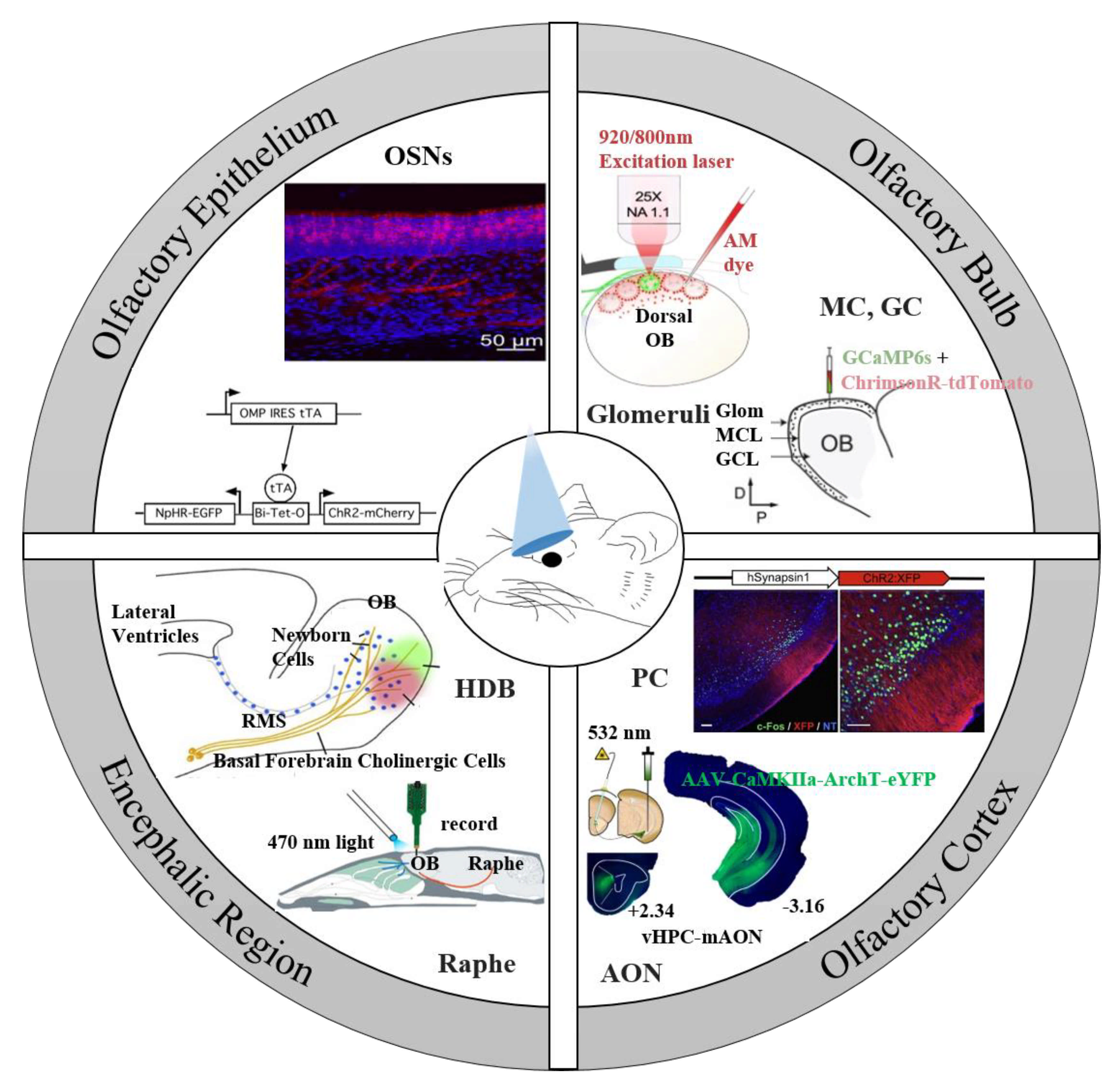
2. Optogenetic Tools for the Olfactory System

| Level | Expression Target | Model Animal | Expression Approach | Light Delivery | Electrophysiology Recordings | Behavior | Ref. | ||||
|---|---|---|---|---|---|---|---|---|---|---|---|
| Tool | Wavelength (nm) | Duration (ms) | Frequency (Hz) | Power (mW) | |||||||
| GL | Glomeruli | OMP-ChR2-YFP transgenic mice | Transgenic animal model | A 470 nm LED coupled with an objective | 470 | 10 | - | - | M/T cells: patch-clamp | - | [6] |
| SACs | TH-Cre mice | Injection of AAV-ChR2 into the GL | A solid-state laser coupled with an optical fiber | 473 | - | - | 100 | M/T cells or ETCs: whole-cell patch-clamp or cell-attached and tungsten microelectrodes | - | [39] | |
| EPL | EPL-INs | Crh-Cre mice | Injection of AAV-ChR2 into the OB | A BLM-Series 473 nm blue laser system coupled with an objective | 473 | 20–40 | EPL INs: whole-cell patch-clamp | Olfactory associative learning training | [40] | ||
| EPL-INs | CRH-Cre mice | Injection of AAV-ChR2 into the OB | A blue laser system guided by implanted fiber optics | 473 | 10 | - | 30 | MCs: whole-cell patch-clamp and extracellular recording electrodes | Olfactory associative learning training | [59] | |
| IPL | dSACs | Chrna2-Cre mice | Injection of AAV-ChR2 into the IPL | A 75 W xenon arc lamp coupled with an objective | - | - | - | - | TCs: whole cell patch clamp | - | [35] |
| GCL | GCs | Dlx5/6-Cre mice | Injection of AAV-ChR2 into the OB | A BLM-Series 473 nm blue laser system coupled with an objective | 473 | 20–40 | GCs: whole-cell patch-clamp | Olfactory associative learning training | [40] | ||
| GCs | OMP-Cre mice | Injection of AAV-ChR2 into the GCL | An implanted LEDs driven with a high-power LED driver | 470 | 5 | 40 | 23 | M/T cells: a silicon-based recording electrode and 32 channels optrode | Habituation task; Olfactory discrimination task | [60] | |
| Encephalic Region | Expression Target | Model Animal | Expression Approach | Light Delivery | Electrophysiology Recordings | Ref. | ||||
|---|---|---|---|---|---|---|---|---|---|---|
| Tool | Wavelength (nm) | Duration (ms) | Frequency (Hz) | Power (mW) | ||||||
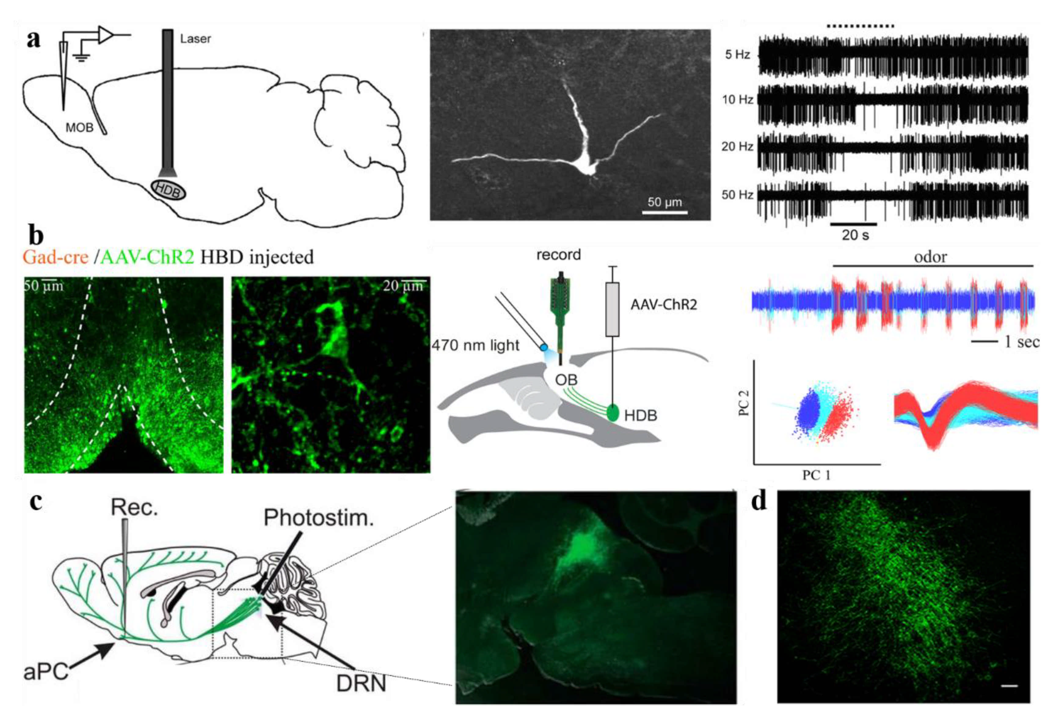
| Basal forebrain | HDB cholinergic neurons | VGLUT3-Cre mice | Injection of AAV-ChR2 into the HDB | A 75W xenon arc lamp coupled with an objective | - | 10–20 | - | - | OB cells: patch clamp | [61] |
| ChAT-ChR2-EYFP transgenic mice | Transgenic animal model | A diode-pumped solid-state 473 nm laser coupled with an optical fiber target the HDB | 473 | 15 | 5–50 | - | M/T cells and brain slices: patch clamp | [41] | ||
| ChAT-ChR2-EYFP mice transgenic | Transgenic animal model | A blue light diode laser and a blue LED coupled with implanted fiber | 473 | 15 | 5–50 | - | - | [32] | ||
| HDB GABAergic neurons | DLX5/6-Cre mice | Injection of AAV-ChR2 into the HDB | A blueCoolLED pE 100 coupled with an objective | 490 | - | - | - | OB cells: whole-cell | [62] | |
| ChAT/GAD2-Cre mice | Injection of AAV-ChR2/eNpHR into the HDB | A 470 or 565 nm LED coupled with an optical fiber positioned in the OB | 470; 565 | 10000 | - | 10;3 | M/T cells: sixteen channel electrodes | [63] | ||
| Raphe nuclei | 5-HT axons | TPH2-ChR2-YFP transgenic mice | Transgenic animal model | A bright light-emitting diode (LED) array coupled with a microscope | 473 | 10 | 10 | 15 | M/T cells: tungsten electrodes and whole-cell | [64] |
| serotonergic cells | Slc6a4-Cre mice | Injection of AAV-ChR2 into DRN | A 470 nm LED coupled with a glass fiber positioned close to the OB | 470 | 10000 | 1–10 | OB cells: 16-channel electrode | [31] | ||
| serotonergic cells | SERT-Cre mice | Injection of AAV-ChR2 in the DRN | A 470 nm laser coupled with an optrode lowered into the DRN | 470 | 10 | 1–30 | - | APC neurons: microelectrodes; | [65] | |
| locus coreuleus | noradrenergic neurons | DBH-Cre-NpHR transgenic mice | Transgenic animal model | A solid-state laser coupled with an optical fiber implanted in the OB | 532 | - | - | 2–10 | MCs: tetrodes | [66] |
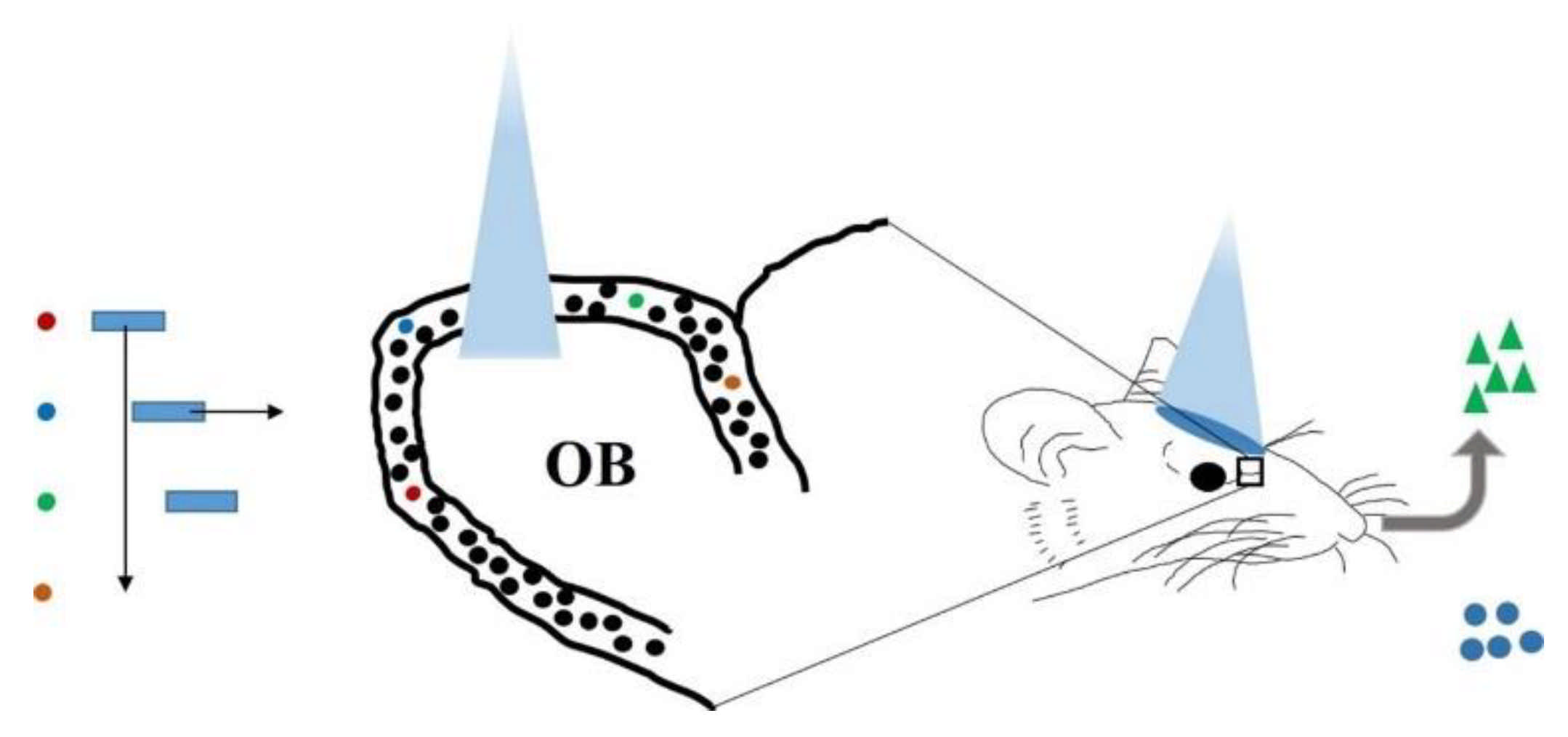
3. The Sensory Input from OSNs to the OB
4. How Do Activities of the Olfactory Bulb Neurons Affect Perception?
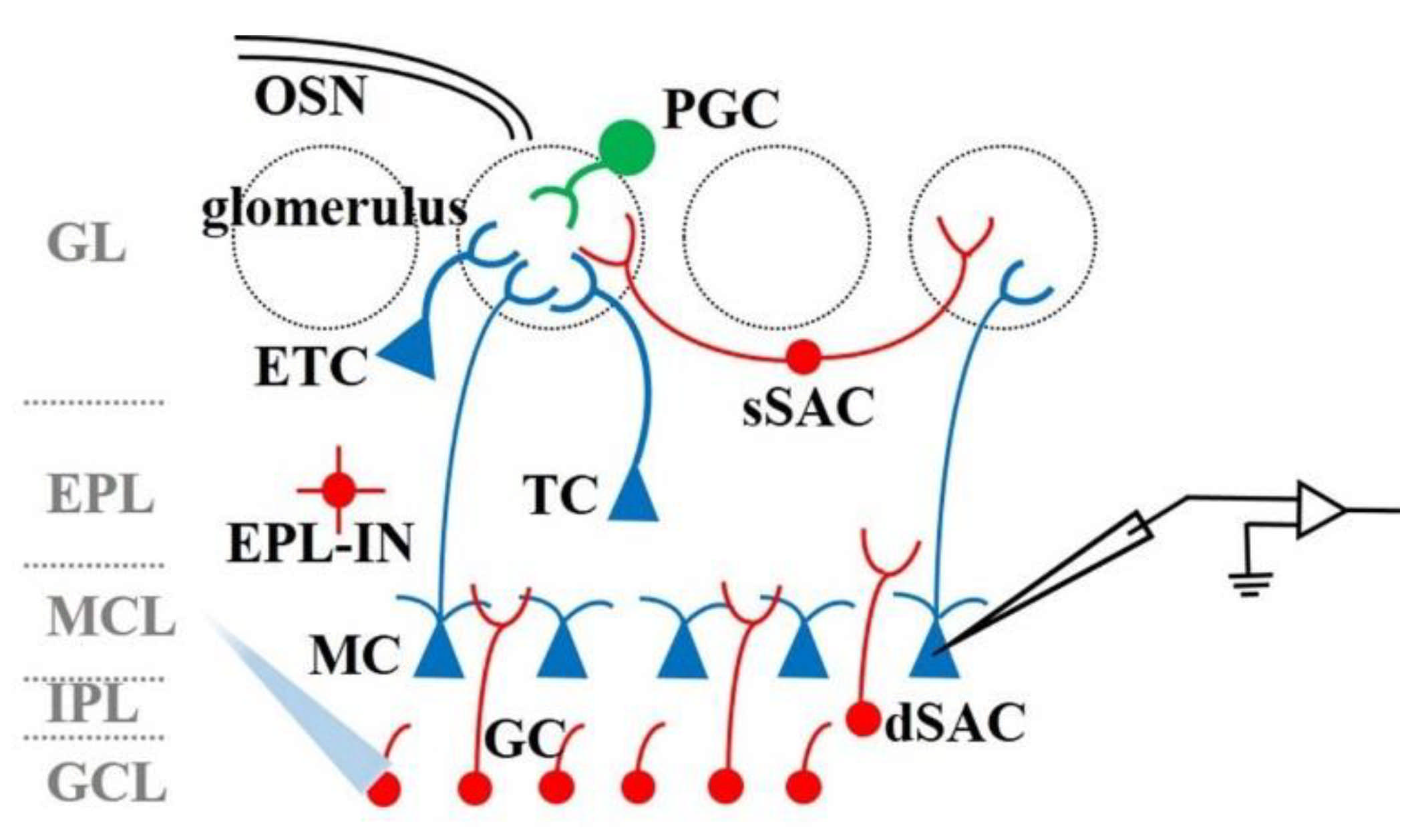
5. The Function of OB Interneurons in Odor Information Processing
5.1. The Glomerular Layer
5.2. The External Plexiform Layer
5.3. The Internal Plexiform Layer
5.4. The Granule Cells Layer
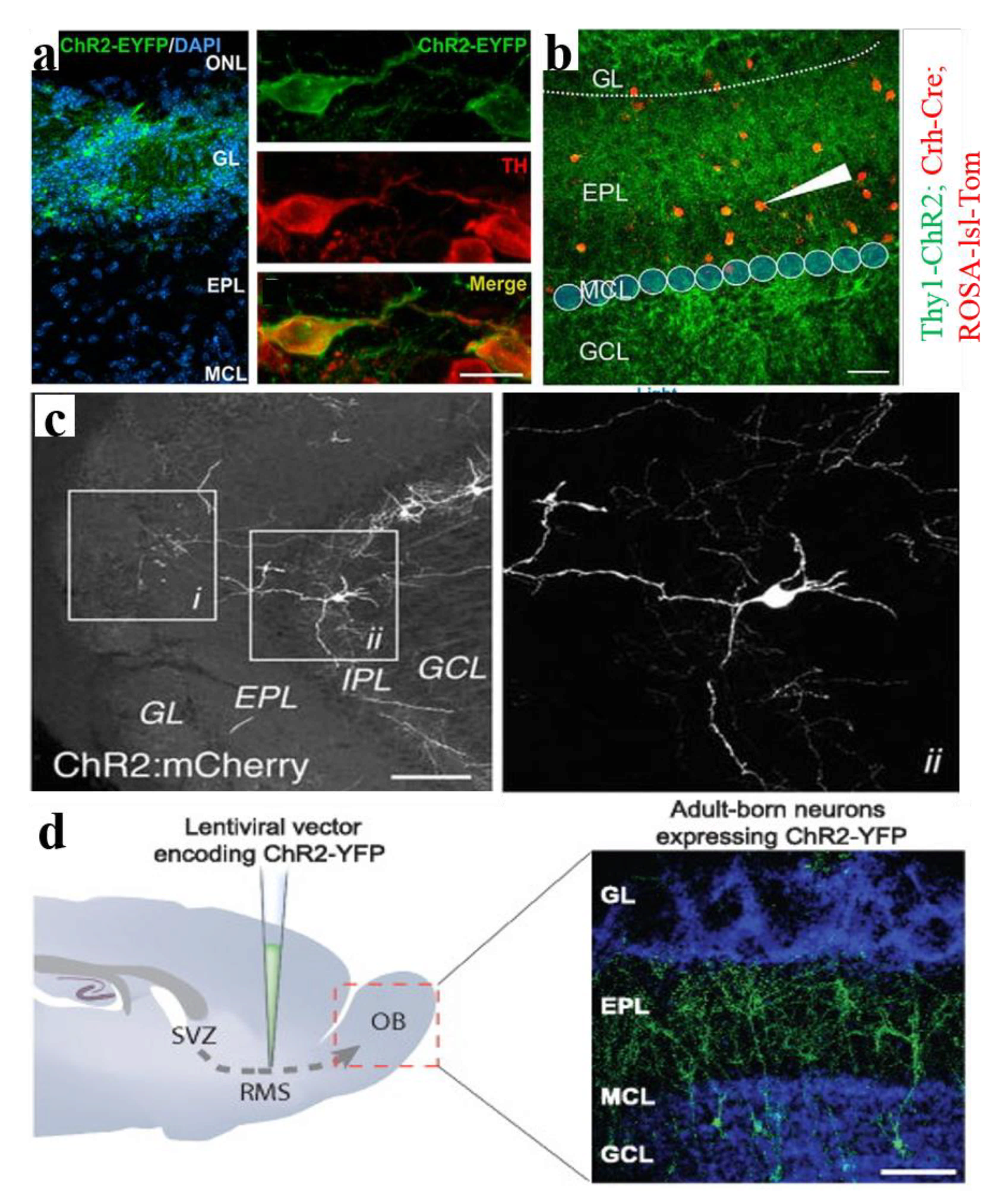
5.5. The Rostral Migratory Stream

6. Cortical Projection and Centrifugal Regulation

7. The Neuromodulation in Odor Coding by Dominating the OB
7.1. The Horizontal Diagonal Band of Broca
7.2. 5-Hydroxytriptamine
7.3. Noradrenergic (NA) Neurons in the Locus Coeruleus
8. Conclusions and Perspectives
Author Contributions
Funding
Institutional Review Board Statement
Informed Consent Statement
Conflicts of Interest
References
- Giessel, A.J.; Datta, S.R. Olfactory maps, circuits and computations. Curr. Opin. Neurobiol. 2014, 24, 120–132. [Google Scholar] [CrossRef] [Green Version]
- Genva, M.; Kenne Kemene, T.; Deleu, M.; Lins, L.; Fauconnier, M.-L. Is It Possible to Predict the Odor of a Molecule on the Basis of its Structure? Int. J. Mol. Sci. 2019, 20, 3018. [Google Scholar] [CrossRef] [Green Version]
- Mainland, J.D.; Keller, A.; Li, Y.R.; Zhou, T.; Trimmer, C.; Snyder, L.L.; Moberly, A.H.; Adipietro, K.A.; Liu, W.L.L.; Zhuang, H.; et al. The missense of smell: Functional variability in the human odorant receptor repertoire. Nat. Neurosci. 2014, 17, 114–120. [Google Scholar] [CrossRef] [PubMed] [Green Version]
- Xu, L.; Li, W.; Voleti, V.; Zou, D.-J.; Hillman, E.M.C.; Firestein, S. Widespread receptor-driven modulation in peripheral olfactory coding. Science 2020, 368, eaaz5390. [Google Scholar] [CrossRef]
- Cavarretta, F.; Burton, S.D.; Igarashi, K.M.; Shepherd, G.M.; Hines, M.L.; Migliore, M. Parallel odor processing by mitral and middle tufted cells in the olfactory bulb. Sci. Rep. 2018, 8, 7625. [Google Scholar] [CrossRef] [Green Version]
- Geramita, M.; Urban, N.N. Differences in Glomerular-Layer-Mediated Feedforward Inhibition onto Mitral and Tufted Cells Lead to Distinct Modes of Intensity Coding. J. Neurosci. 2017, 37, 1428–1438. [Google Scholar] [CrossRef]
- Liu, G.; Froudarakis, E.; Patel, J.M.; Kochukov, M.Y.; Pekarek, B.; Hunt, P.J.; Patel, M.; Ung, K.; Fu, C.-H.; Jo, J.; et al. Target specific functions of EPL interneurons in olfactory circuits. Nat. Commun. 2019, 10, 3369. [Google Scholar] [CrossRef] [PubMed] [Green Version]
- Bolding, K.A.; Franks, K.M. Recurrent cortical circuits implement concentration-invariant odor coding. Science 2018, 361. [Google Scholar] [CrossRef] [PubMed]
- Otazu, G.H.; Chae, H.; Davis, M.B.; Albeanu, D.F. Cortical Feedback Decorrelates Olfactory Bulb Output in Awake Mice. Neuron 2015, 86, 1461–1477. [Google Scholar] [CrossRef] [PubMed] [Green Version]
- Wang, C.Y.; Liu, Z.; Ng, Y.H.; Südhof, T.C. A Synaptic Circuit Required for Acquisition but Not Recall of Social Transmission of Food Preference. Neuron 2020, 107, 144–157. [Google Scholar] [CrossRef]
- Uchida, N.; Poo, C.; Haddad, R. Coding and Transformations in the Olfactory System. Annu. Rev. Neurosci. 2014, 37, 363–385. [Google Scholar] [CrossRef] [PubMed]
- Wilson, D.A.; Sullivan, R.M. Cortical processing of odor objects. Neuron 2011, 72, 506–519. [Google Scholar] [CrossRef] [Green Version]
- Sohal, V.S.; Zhang, F.; Yizhar, O.; Deisseroth, K.J.N. Parvalbumin neurons and gamma rhythms enhance cortical circuit performance. Nature 2009, 459, 698–702. [Google Scholar] [CrossRef] [PubMed] [Green Version]
- Boyden, E.S.; Zhang, F.; Bamberg, E.; Nagel, G.; Deisseroth, K. Millisecond-timescale, genetically targeted optical control of neural activity. Nat. Neurosci. 2005, 8, 1263–1268. [Google Scholar] [CrossRef]
- Zhang, F.; Wang, L.; Brauner, M.; Liewald, J.F.; Kay, K.; Watzke, N.; Wood, P.G.; Bamberg, E.; Nagel, G.; Gottschalk, A.J.N. Multimodal fast optical interrogation of neural circuitry. Nature 2007, 446, 633–639. [Google Scholar] [CrossRef] [PubMed]
- Nagel, G.; Szellas, T.; Huhn, W.; Kateriya, S.; Adeishvili, N.; Berthold, P.; Ollig, D.; Hegemann, P.; Bamberg, E. Channelrhodopsin-2, a directly light-gated cation-selective membrane channel. Proc. Natl. Acad. Sci. USA 2003, 100, 13940–13945. [Google Scholar] [CrossRef] [PubMed] [Green Version]
- Klapoetke, N.C.; Murata, Y.; Kim, S.S.; Pulver, S.R.; Birdsey-Benson, A.; Cho, Y.K.; Morimoto, T.K.; Chuong, A.S.; Carpenter, E.J.; Tian, Z.; et al. Independent optical excitation of distinct neural populations. Nat. Methods 2014, 11, 338–346. [Google Scholar] [CrossRef] [Green Version]
- Chaigneau, E.; Ronzitti, E.; Gajowa, M.A.; Soler-Llavina, G.J.; Tanese, D.; Brureau, A.Y.B.; Papagiakoumou, E.; Zeng, H.; Emiliani, V. Two-Photon Holographic Stimulation of ReaChR. Front. Cell. Neurosci. 2016, 10, 234. [Google Scholar] [CrossRef]
- Watanabe, H.; Sano, H.; Chiken, S.; Kobayashi, K.; Fukata, Y.; Fukata, M.; Mushiake, H.; Nambu, A. Forelimb movements evoked by optogenetic stimulation of the macaque motor cortex. Nat. Commun 2020, 11, 3253. [Google Scholar] [CrossRef]
- Tamura, K.; Takeda, M.; Setsuie, R.; Tsubota, T.; Hirabayashi, T.; Miyamoto, K.; Miyashita, Y. Conversion of object identity to object-general semantic value in the primate temporal cortex. Science 2017, 357, 687–692. [Google Scholar] [CrossRef] [Green Version]
- Hardt, O.; Nadel, L. Systems consolidation revisited, but not revised: The promise and limits of optogenetics in the study of memory. Neurosci. Lett. 2018, 680, 54–59. [Google Scholar] [CrossRef]
- Parnaudeau, S.; Bolkan, S.S.; Kellendonk, C.J.B.P. The Mediodorsal Thalamus: An Essential Partner of the Prefrontal Cortex for Cognition. Biol. Psychiatry 2017, 83, 648–656. [Google Scholar] [CrossRef]
- Cheng, Z.; Cui, R.; Ge, T.; Yang, W.; Li, B. Optogenetics: What it has uncovered in potential pathways of depression. Pharmacol. Res. 2020, 152, 104596. [Google Scholar] [CrossRef]
- Jarrin, S.; Finn, D.P.J.N.; Reviews, B. Optogenetics and its application in pain and anxiety research. Neurosci. Biobehav. Rev. 2019, 105, 200–211. [Google Scholar] [CrossRef]
- Grimaud, J.; Lledo, P.M. Illuminating odors: When optogenetics brings to light unexpected olfactory abilities. Learn. Mem. 2016, 23, 249–254. [Google Scholar] [CrossRef] [PubMed] [Green Version]
- Gire, D.H.; Franks, K.M.; Zak, J.D.; Tanaka, K.F.; Whitesell, J.D.; Mulligan, A.A.; Hen, R.; Schoppa, N.E. Mitral cells in the olfactory bulb are mainly excited through a multistep signaling path. J. Neurosci. 2012, 32, 2964–2975. [Google Scholar] [CrossRef] [PubMed]
- Braubach, O.A.-O.; Tombaz, T.; Geiller, T.; Homma, R.; Bozza, T.A.-O.; Cohen, L.B.; Choi, Y. Sparsened neuronal activity in an optogenetically activated olfactory glomerulus. Sci. Rep. 2018, 8, 1–17. [Google Scholar] [CrossRef] [PubMed] [Green Version]
- Gill, J.V.; Lerman, G.M.; Zhao, H.; Stetler, B.J.; Rinberg, D.; Shoham, S. Precise Holographic Manipulation of Olfactory Circuits Reveals Coding Features Determining Perceptual Detection. Neuron 2020, 108, 382–393. [Google Scholar] [CrossRef]
- Choi, G.B.; Stettler, D.D.; Kallman, B.R.; Bhaskar, S.T.; Fleischmann, A.; Axel, R. Driving Opposing Behaviors with Ensembles of Piriform Neurons. Cell 2011, 146, 1004–1015. [Google Scholar] [CrossRef] [PubMed] [Green Version]
- Aqrabawi, A.J.; Kim, J.C. Hippocampal projections to the anterior olfactory nucleus differentially convey spatiotemporal information during episodic odour memory. Nat. Commun. 2018, 9, 2735. [Google Scholar] [CrossRef] [PubMed] [Green Version]
- Brunert, D.; Tsuno, Y.; Rothermel, M.; Shipley, M.T.; Wachowiak, M. Cell-Type-Specific Modulation of Sensory Responses in Olfactory Bulb Circuits by Serotonergic Projections from the Raphe Nuclei. J. Neurosci. 2016, 36, 6820. [Google Scholar] [CrossRef] [PubMed]
- Nitenson, A.S.; Nieves, G.M.; Poeta, D.L.; Bahar, R.; Rachofsky, C.; Mandairon, N.; Bath, K.G. Acetylcholine Regulates Olfactory Perceptual Learning through Effects on Adult Neurogenesis. iScience 2019, 22, 544–556. [Google Scholar] [CrossRef] [Green Version]
- Rost, B.R.; Schneider-Warme, F.; Schmitz, D.; Hegemann, P. Optogenetic Tools for Subcellular Applications in Neuroscience. Neuron 2017, 96, 572–603. [Google Scholar] [CrossRef] [Green Version]
- McCarthy, E.A.; Kunkhyen, T.; Korzan, W.J.; Naik, A.; Maqsudlu, A.; Cherry, J.A.; Baum, M.J. A comparison of the effects of male pheromone priming and optogenetic inhibition of accessory olfactory bulb forebrain inputs on the sexual behavior of estrous female mice. Horm. Behav. 2017, 89, 104–112. [Google Scholar] [CrossRef] [PubMed] [Green Version]
- Burton, S.D.; Larocca, G.; Liu, A.; Cheetham, C.E.J.; Urban, N.N. Olfactory Bulb Deep Short-Axon Cells Mediate Widespread Inhibition of Tufted Cell Apical Dendrites. J. Neurosci. 2017, 37, 1117–1138. [Google Scholar] [CrossRef] [Green Version]
- Choy, J.M.C.; Suzuki, N.; Shima, Y.; Budisantoso, T.; Nelson, S.B.; Bekkers, J.M. Optogenetic Mapping of Intracortical Circuits Originating from Semilunar Cells in the Piriform Cortex. Cereb. Cortex 2015, 27, 589–601. [Google Scholar] [CrossRef] [Green Version]
- Markopoulos, F.; Rokni, D.; Gire, D.H.; Murthy, V.N. Functional Properties of Cortical Feedback Projections to the Olfactory Bulb. Neuron 2012, 76, 1175–1188. [Google Scholar] [CrossRef] [PubMed] [Green Version]
- Alonso, M.; Lepousez, G.; Wagner, S.; Bardy, C.; Gabellec, M.M.; Torquet, N.; Lledo, P.M. Activation of adult-born neurons facilitates learning and memory. Nat. Neurosci. 2012, 15, 897–904. [Google Scholar] [CrossRef]
- Liu, S.; Puche, A.C.; Shipley, M.T. The Interglomerular Circuit Potently Inhibits Olfactory Bulb Output Neurons by Both Direct and Indirect Pathways. J. Neurosci. 2016, 36, 9604–9617. [Google Scholar] [CrossRef] [Green Version]
- Huang, L.; Ung, K.; Garcia, I.; Quast, K.B.; Cordiner, K.; Saggau, P.; Arenkiel, B.R. Task Learning Promotes Plasticity of Interneuron Connectivity Maps in the Olfactory Bulb. J. Neurosci. 2016, 36, 8856–8871. [Google Scholar] [CrossRef] [Green Version]
- Ma, M.; Luo, M. Optogenetic Activation of Basal Forebrain Cholinergic Neurons Modulates Neuronal Excitability and Sensory Responses in the Main Olfactory Bulb. J. Neurosci. 2012, 32, 10105–10116. [Google Scholar] [CrossRef] [Green Version]
- Arenkiel, B.R.; Peca, J.; Davison, I.G.; Feliciano, C.; Deisseroth, K.; Augustine, G.J.; Ehlers, M.D.; Feng, G. In vivo light-induced activation of neural circuitry in transgenic mice expressing channelrhodopsin-2. Neuron 2007, 54, 205–218. [Google Scholar] [CrossRef] [Green Version]
- Zhao, S.; Ting, J.T.; Atallah, H.E.; Qiu, L.; Tan, J.; Gloss, B.; Augustine, G.J.; Deisseroth, K.; Luo, M.; Graybiel, A.M.; et al. Cell type–specific channelrhodopsin-2 transgenic mice for optogenetic dissection of neural circuitry function. Nat. Methods 2011, 8, 745–752. [Google Scholar] [CrossRef]
- Dhawale, A.K.; Hagiwara, A.; Bhalla, U.S.; Murthy, V.N.; Albeanu, D.F. Non-redundant odor coding by sister mitral cells revealed by light addressable glomeruli in the mouse. Nat. Neurosci. 2010, 13, 1404–1412. [Google Scholar] [CrossRef] [Green Version]
- Smear, M.C.; Shusterman, R.; Oconnor, R.P.; Bozza, T.C.; Rinberg, D. Perception of sniff phase in mouse olfaction. Nature 2011, 479, 397–400. [Google Scholar] [CrossRef] [PubMed]
- Smear, M.C.; Resulaj, A.; Zhang, J.; Bozza, T.C.; Rinberg, D. Multiple perceptible signals from a single olfactory glomerulus. Nat. Neurosci. 2013, 16, 1687–1691. [Google Scholar] [CrossRef] [PubMed]
- Luna, V.M.; Morozov, A. Input-specific excitation of olfactory cortex microcircuits. Front. Neural Circuits 2012, 6, 69. [Google Scholar] [CrossRef] [PubMed] [Green Version]
- Gunaydin, L.A.; Yizhar, O.; Berndt, A.; Sohal, V.S.; Deisseroth, K.; Hegemann, P. Ultrafast optogenetic control. Nat. Neurosci. 2010, 13, 387–392. [Google Scholar] [CrossRef]
- Murata, K.; Kinoshita, T.; Fukazawa, Y.; Kobayashi, K.; Yamanaka, A.; Hikida, T.; Manabe, H.; Yamaguchi, M. Opposing Roles of Dopamine Receptor D1- and D2-Expressing Neurons in the Anteromedial Olfactory Tubercle in Acquisition of Place Preference in Mice. Front. Behav. Neurosci. 2019, 13, 50. [Google Scholar] [CrossRef] [PubMed] [Green Version]
- Aqrabawi, A.J.; Kim, J.C. Olfactory memory representations are stored in the anterior olfactory nucleus. Nat. Commun. 2020, 11, 1246. [Google Scholar] [CrossRef] [PubMed]
- Lin, J.Y.; Knutsen, P.M.; Muller, A.; Kleinfeld, D.; Tsien, R.Y. ReaChR: A red-shifted variant of channelrhodopsin enables deep transcranial optogenetic excitation. Nat. Neurosci. 2013, 16, 1499–1508. [Google Scholar] [CrossRef] [Green Version]
- Inagaki, H.K.; Jung, Y.; Hoopfer, E.D.; Wong, A.M.; Mishra, N.; Lin, J.Y.; Tsien, R.Y.; Anderson, D.J. Optogenetic control of Drosophila using a red-shifted channelrhodopsin reveals experience-dependent influences on courtship. Nat. Methods 2014, 11, 325–332. [Google Scholar] [CrossRef] [Green Version]
- Inada, K.; Tsuchimoto, Y.; Kazama, H. Origins of Cell-Type-Specific Olfactory Processing in the Drosophila Mushroom Body Circuit. Neuron 2017, 95, 357–367.e354. [Google Scholar] [CrossRef] [PubMed] [Green Version]
- Guo, H.; Kunwar, K.; Smith, D. Odorant Receptor Sensitivity Modulation in Drosophila. J. Neurosci. 2017, 37, 9465. [Google Scholar] [CrossRef] [PubMed] [Green Version]
- Fukunaga, I.; Herb, J.T.; Kollo, M.; Boyden, E.S.; Schaefer, A.T. Independent control of gamma and theta activity by distinct interneuron networks in the olfactory bulb. Nat. Neurosci. 2014, 17, 1208–1216. [Google Scholar] [CrossRef] [Green Version]
- Gradinaru, V.; Zhang, F.; Ramakrishnan, C.; Mattis, J.; Prakash, R.; Diester, I.; Goshen, I.; Thompson, K.R.; Deisseroth, K. Molecular and Cellular Approaches for Diversifying and Extending Optogenetics. Cell 2010, 141, 154–165. [Google Scholar] [CrossRef] [Green Version]
- Midroit, M.; Chalençon, L.; Renier, N.; Milton, A.; Thevenet, M.; Sacquet, J.; Breton, M.; Forest, J.; Noury, N.; Richard, M.; et al. Neural processing of the reward value of pleasant odorants. Curr. Biol. 2021, 31, 1592–1605.e1599. [Google Scholar] [CrossRef] [PubMed]
- Kermen, F.; Midroit, M.; Kuczewski, N.; Forest, J.; Thévenet, M.; Sacquet, J.; Benetollo, C.; Richard, M.; Didier, A.; Mandairon, N. Topographical representation of odor hedonics in the olfactory bulb. Nat. Neurosci. 2016, 19, 876–878. [Google Scholar] [CrossRef] [PubMed]
- Huang, L.; Garcia, I.; Jen, H.; Arenkiel, B.R. Reciprocal connectivity between mitral cells and external plexiform layer interneurons in the mouse olfactory bulb. Front. Neural Circuits 2013, 7, 32. [Google Scholar] [CrossRef] [Green Version]
- Gschwend, O.; Abraham, N.M.; Lagier, S.; Begnaud, F.; Rodriguez, I.; Carleton, A. Neuronal pattern separation in the olfactory bulb improves odor discrimination learning. Nat. Neurosci. 2015, 18, 1474–1482. [Google Scholar] [CrossRef] [Green Version]
- Case, D.T.; Burton, S.D.; Gedeon, J.Y.; Williams, S.P.G.; Urban, N.N.; Seal, R.P. Layer- and cell type-selective co-transmission by a basal forebrain cholinergic projection to the olfactory bulb. Nat. Commun. 2017, 8, 652. [Google Scholar] [CrossRef] [PubMed]
- Diez, A.S.; Najac, M.; De Saint Jan, D. Basal forebrain GABAergic innervation of olfactory bulb periglomerular interneurons. J. Physiol. 2019, 597, 2547–2563. [Google Scholar] [CrossRef] [PubMed]
- Böhm, E.; Brunert, D.; Rothermel, M. Input dependent modulation of olfactory bulb activity by HDB GABAergic projections. Sci. Rep. 2020, 10, 10696. [Google Scholar] [CrossRef] [PubMed]
- Kapoor, V.; Provost, A.C.; Agarwal, P.; Murthy, V.N. Activation of raphe nuclei triggers rapid and distinct effects on parallel olfactory bulb output channels. Nat. Neurosci. 2016, 19, 271–282. [Google Scholar] [CrossRef] [Green Version]
- Lottem, E.; Lorincz, M.L.; Mainen, Z.F. Optogenetic Activation of Dorsal Raphe Serotonin Neurons Rapidly Inhibits Spontaneous but Not Odor-Evoked Activity in Olfactory Cortex. J. Neurosci. 2016, 36, 7–18. [Google Scholar] [CrossRef] [Green Version]
- Ramirezgordillo, D.; Ma, M.; Restrepo, D. Precision of Classification of Odorant Value by the Power of Olfactory Bulb Oscillations Is Altered by Optogenetic Silencing of Local Adrenergic Innervation. Front. Cell. Neurosci. 2018, 12, 48. [Google Scholar] [CrossRef] [PubMed] [Green Version]
- Mori, K.; Sakano, H. How is the olfactory map formed and interpreted in the mammalian brain? Annu Rev Neurosci. 2011, 34, 467–499. [Google Scholar] [CrossRef]
- Murthy, V.N. Olfactory maps in the brain. Annu Rev Neurosci. 2011, 34, 233–258. [Google Scholar] [CrossRef] [PubMed] [Green Version]
- Najac, M.; De Saint Jan, D.; Reguero, L.; Grandes, P.; Charpak, S. Monosynaptic and polysynaptic feed-forward inputs to mitral cells from olfactory sensory neurons. J. Neurosci. 2011, 31, 8722–8729. [Google Scholar] [CrossRef]
- Shmuel, R.; Secundo, L.; Haddad, R. Strong, weak and neuron type dependent lateral inhibition in the olfactory bulb. Sci Rep. 2019, 9, 1602. [Google Scholar] [CrossRef]
- Vaaga, C.E.; Westbrook, G.L. Parallel processing of afferent olfactory sensory information. J. Physiol. 2016, 594, 6715–6732. [Google Scholar] [CrossRef] [PubMed] [Green Version]
- Genovese, F.; Thews, M.; Mohrlen, F.; Frings, S. Properties of an optogenetic model for olfactory stimulation. J. Physiol. 2016, 594, 3501–3516. [Google Scholar] [CrossRef] [Green Version]
- Mombaerts, P. Axonal wiring in the mouse olfactory system. Annu. Rev. Cell Dev. Biol. 2006, 22, 713–737. [Google Scholar] [CrossRef] [Green Version]
- Vassar, R.; Chao, S.K.; Sitcheran, R.; Nuñez, J.M.; Vosshall, L.B.; Axel, R. Topographic organization of sensory projections to the olfactory bulb. Cell 1994, 79, 981–991. [Google Scholar] [CrossRef]
- Iwata, R.; Kiyonari, H.; Imai, T. Mechanosensory-Based Phase Coding of Odor Identity in the Olfactory Bulb. Neuron 2017, 96, 1139–1152.e1137. [Google Scholar] [CrossRef] [PubMed]
- Malnic, B.; Hirono, J.; Sato, T.; Buck, L.B. Combinatorial receptor codes for odors. Cell 1999, 96, 713–723. [Google Scholar] [CrossRef] [Green Version]
- Zhou, Z.; Belluscio, L. Coding odorant concentration through activation timing between the medial and lateral olfactory bulb. Cell Rep. 2012, 2, 1143–1150. [Google Scholar] [CrossRef] [Green Version]
- Grabe, V.; Sachse, S. Fundamental principles of the olfactory code. Biosystems 2018, 164, 94–101. [Google Scholar] [CrossRef] [PubMed]
- Renou, M.; Party, V.; Rouyar, A.; Anton, S. Olfactory signal coding in an odor background. Biosystems 2015, 136, 35–45. [Google Scholar] [CrossRef] [Green Version]
- Rebello, M.R.; McTavish, T.S.; Willhite, D.C.; Short, S.M.; Shepherd, G.M.; Verhagen, J.V. Perception of odors linked to precise timing in the olfactory system. PLoS Biol. 2014, 12, e1002021. [Google Scholar] [CrossRef] [Green Version]
- Li, A.; Gire, D.H.; Bozza, T.C.; Restrepo, D. Precise Detection of Direct Glomerular Input Duration by the Olfactory Bulb. J. Neurosci. 2014, 34, 16058–16064. [Google Scholar] [CrossRef]
- Wilson, C.D.; Serrano, G.O.; Koulakov, A.A.; Rinberg, D. A primacy code for odor identity. Nat. Commun. 2017, 8, 1477. [Google Scholar] [CrossRef] [PubMed] [Green Version]
- Chong, E.; Moroni, M.; Wilson, C.; Shoham, S.; Panzeri, S.; Rinberg, D. Manipulating synthetic optogenetic odors reveals the coding logic of olfactory perception. Science 2020, 368. [Google Scholar] [CrossRef] [PubMed]
- Mardinly, A.R.; Oldenburg, I.A.; Pégard, N.C.; Sridharan, S.; Lyall, E.H.; Chesnov, K.; Brohawn, S.G.; Waller, L.; Adesnik, H. Precise multimodal optical control of neural ensemble activity. Nat. Neurosci. 2018, 21, 881–893. [Google Scholar] [CrossRef] [PubMed]
- Chen, I.W.; Ronzitti, E.; Lee, B.R.; Daigle, T.L.; Dalkara, D.; Zeng, H.; Emiliani, V.; Papagiakoumou, E. In Vivo Submillisecond Two-Photon Optogenetics with Temporally Focused Patterned Light. J. Neurosci. Off. J. Soc. Neurosci. 2019, 39, 3484–3497. [Google Scholar] [CrossRef] [Green Version]
- Carey, R.M.; Verhagen, J.V.; Wesson, D.W.; Pírez, N.; Wachowiak, M. Temporal structure of receptor neuron input to the olfactory bulb imaged in behaving rats. J. Neurophysiol. 2009, 101, 1073–1088. [Google Scholar] [CrossRef]
- Miyamichi, K.; Shlomaifuchs, Y.; Shu, M.; Weissbourd, B.; Luo, L.; Mizrahi, A.J.N. Dissecting Local Circuits: Parvalbumin Interneurons Underlie Broad Feedback Control of Olfactory Bulb Output. Neuron 2013, 80, 1232–1245. [Google Scholar] [CrossRef] [Green Version]
- Eyre, M.D.; Antal, M.; Nusser, Z. Distinct Deep Short-Axon Cell Subtypes of the Main Olfactory Bulb Provide Novel Intrabulbar and Extrabulbar GABAergic Connections. J. Neurosci. 2008, 28, 8217–8229. [Google Scholar] [CrossRef]
- Burton, S.D.; Urban, N.N. Greater excitability and firing irregularity of tufted cells underlies distinct afferent-evoked activity of olfactory bulb mitral and tufted cells. J. Physiol. 2014, 592, 2097–2118. [Google Scholar] [CrossRef]
- Burton, S.D.; Urban, N.N. Rapid Feedforward Inhibition and Asynchronous Excitation Regulate Granule Cell Activity in the Mammalian Main Olfactory Bulb. J. Neurosci. 2015, 35, 14103–14122. [Google Scholar] [CrossRef]
- Stoufflet, J.; Chaulet, M.; Doulazmi, M.; Fouquet, C.; Dubacq, C.; Métin, C.; Schneider-Maunoury, S.; Trembleau, A.; Vincent, P.; Caillé, I. Primary cilium-dependent cAMP/PKA signaling at the centrosome regulates neuronal migration. Sci. Adv. 2020, 6, eaba3992. [Google Scholar] [CrossRef]
- Bardy, C.; Alonso, M.; Bouthour, W.; Lledo, P.M. How, When, and Where New Inhibitory Neurons Release Neurotransmitters in the Adult Olfactory Bulb. J. Neurosci. 2010, 30, 17023–17034. [Google Scholar] [CrossRef] [PubMed]
- Valley, M.T.; Henderson, L.G.; Inverso, S.A.; Lledo, P.M. Adult neurogenesis produces neurons with unique GABAergic synapses in the olfactory bulb. J. Neurosci. Off. J. Soc. Neurosci. 2013, 33, 14660–14665. [Google Scholar] [CrossRef] [PubMed] [Green Version]
- Grelat, A.; Benoit, L.; Wagner, S.; Moigneu, C.; Lledo, P.M.; Alonso, M. Adult-born neurons boost odor-reward association. Proc. Natl. Acad. Sci. USA 2018, 115, 2514–2519. [Google Scholar] [CrossRef] [PubMed] [Green Version]
- McGinn, M.J.; Sun, D.; Colello, R.J. Utilizing X-irradiation to selectively eliminate neural stem/progenitor cells from neurogenic regions of the mammalian brain. J. Neurosci. Methods 2008, 170, 9–15. [Google Scholar] [CrossRef] [Green Version]
- Singer, B.H.; Jutkiewicz, E.M.; Fuller, C.L.; Lichtenwalner, R.J.; Zhang, H.; Velander, A.J.; Li, X.; Gnegy, M.E.; Burant, C.F.; Parent, J.M. Conditional ablation and recovery of forebrain neurogenesis in the mouse. J. Comp. Neurol. 2009, 514, 567–582. [Google Scholar] [CrossRef] [PubMed] [Green Version]
- Bekkers, J.M.; Suzuki, N. Neurons and circuits for odor processing in the piriform cortex. Trends Neurosci. 2013, 36, 429–438. [Google Scholar] [CrossRef] [PubMed] [Green Version]
- Haddad, R.; Lanjuin, A.; Madisen, L.; Zeng, H.; Murthy, V.N.; Uchida, N. Olfactory cortical neurons read out a relative time code in the olfactory bulb. Nat. Neurosci. 2013, 16, 949–957. [Google Scholar] [CrossRef] [Green Version]
- Bolding, K.A.; Franks, K.M. Complementary codes for odor identity and intensity in olfactory cortex. Elife 2017, 6, e22630. [Google Scholar] [CrossRef]
- Stern, M.; Bolding, K.A.; Abbott, L.F.; Franks, K.M. A transformation from temporal to ensemble coding in a model of piriform cortex. Elife 2018, 7, e34831. [Google Scholar] [CrossRef]
- Boyd, A.M.; Sturgill, J.F.; Poo, C.; Isaacson, J.S. Cortical feedback control of olfactory bulb circuits. Neuron 2012, 76, 1161–1174. [Google Scholar] [CrossRef] [Green Version]
- Miyamichi, K.; Amat, F.; Moussavi, F.; Wang, C.; Wickersham, I.; Wall, N.R.; Taniguchi, H.; Tasic, B.; Huang, Z.J.; He, Z.; et al. Cortical representations of olfactory input by trans-synaptic tracing. Nature 2011, 472, 191–196. [Google Scholar] [CrossRef]
- Franks, K.M.; Russo, M.J.; Sosulski, D.L.; Mulligan, A.A.; Siegelbaum, S.A.; Axel, R. Recurrent circuitry dynamically shapes the activation of piriform cortex. Neuron 2011, 72, 49–56. [Google Scholar] [CrossRef] [Green Version]
- Blazing, R.M.; Franks, K.M. Odor coding in piriform cortex: Mechanistic insights into distributed coding. Curr. Opin. Neurobiol. 2020, 64, 96–102. [Google Scholar] [CrossRef] [PubMed]
- Balu, R.; Pressler, R.T.; Strowbridge, B.W. Multiple Modes of Synaptic Excitation of Olfactory Bulb Granule Cells. J. Neurosci. 2007, 27, 5621–5632. [Google Scholar] [CrossRef] [PubMed]
- Shipley, M.T.; Adamek, G.D. The connections of the mouse olfactory bulb: A study using orthograde and retrograde transport of wheat germ agglutinin conjugated to horseradish peroxidase. Brain Res. Bull. 1984, 12, 669–688. [Google Scholar] [CrossRef]
- Yan, Z.; Tan, J.; Qin, C.; Lu, Y.; Ding, C.; Luo, M. Precise circuitry links bilaterally symmetric olfactory maps. Neuron 2008, 58, 613–624. [Google Scholar] [CrossRef] [Green Version]
- Mazo, C.; Lepousez, G.; Nissant, A.; Valley, M.T.; Lledo, P.-M. GABAB Receptors Tune Cortical Feedback to the Olfactory Bulb. J. Neurosci. Off. J. Soc. Neurosci. 2016, 36, 8289–8304. [Google Scholar] [CrossRef] [PubMed] [Green Version]
- Boyd, A.M.; Kato, H.K.; Komiyama, T.; Isaacson, J.S. Broadcasting of cortical activity to the olfactory bulb. Cell Rep. 2015, 10, 1032–1039. [Google Scholar] [CrossRef] [Green Version]
- Lepousez, G.; Nissant, A.; Bryant, A.K.; Gheusi, G.; Greer, C.A.; Lledo, P.M. Olfactory learning promotes input-specific synaptic plasticity in adult-born neurons. Proc. Natl. Acad. Sci. USA 2014, 111, 13984–13989. [Google Scholar] [CrossRef] [Green Version]
- Shipley, M.; Reyes, P. Anatomy of the Human Olfactory Bulb and Central Olfactory Pathways. In The Human Sense of Smell; Laing, D.G., Doty, R.L., Breipohl, W., Eds.; Springer: Berlin/Heidelberg, Germany, 1991; pp. 29–60. [Google Scholar]
- Gretenkord, S.; Kostka, J.K.; Hartung, H.; Watznauer, K.; Fleck, D.; Minier-Toribio, A.; Spehr, M.; Hanganu-Opatz, I.L. Coordinated electrical activity in the olfactory bulb gates the oscillatory entrainment of entorhinal networks in neonatal mice. PLoS Biol. 2019, 17, e2006994. [Google Scholar] [CrossRef] [Green Version]
- Zhang, X.; Yan, W.; Wang, W.; Fan, H.; Hou, R.; Chen, Y.; Chen, Z.; Ge, C.; Duan, S.; Compte, A. Active information maintenance in working memory by a sensory cortex. Elife 2019, 8, e43191. [Google Scholar] [CrossRef] [PubMed]
- Hagiwara, A.; Pal, S.K.; Sato, T.F.; Wienisch, M.; Murthy, V.N. Optophysiological analysis of associational circuits in the olfactory cortex. Front. Neural Circuits 2012, 6, 18. [Google Scholar] [CrossRef] [Green Version]
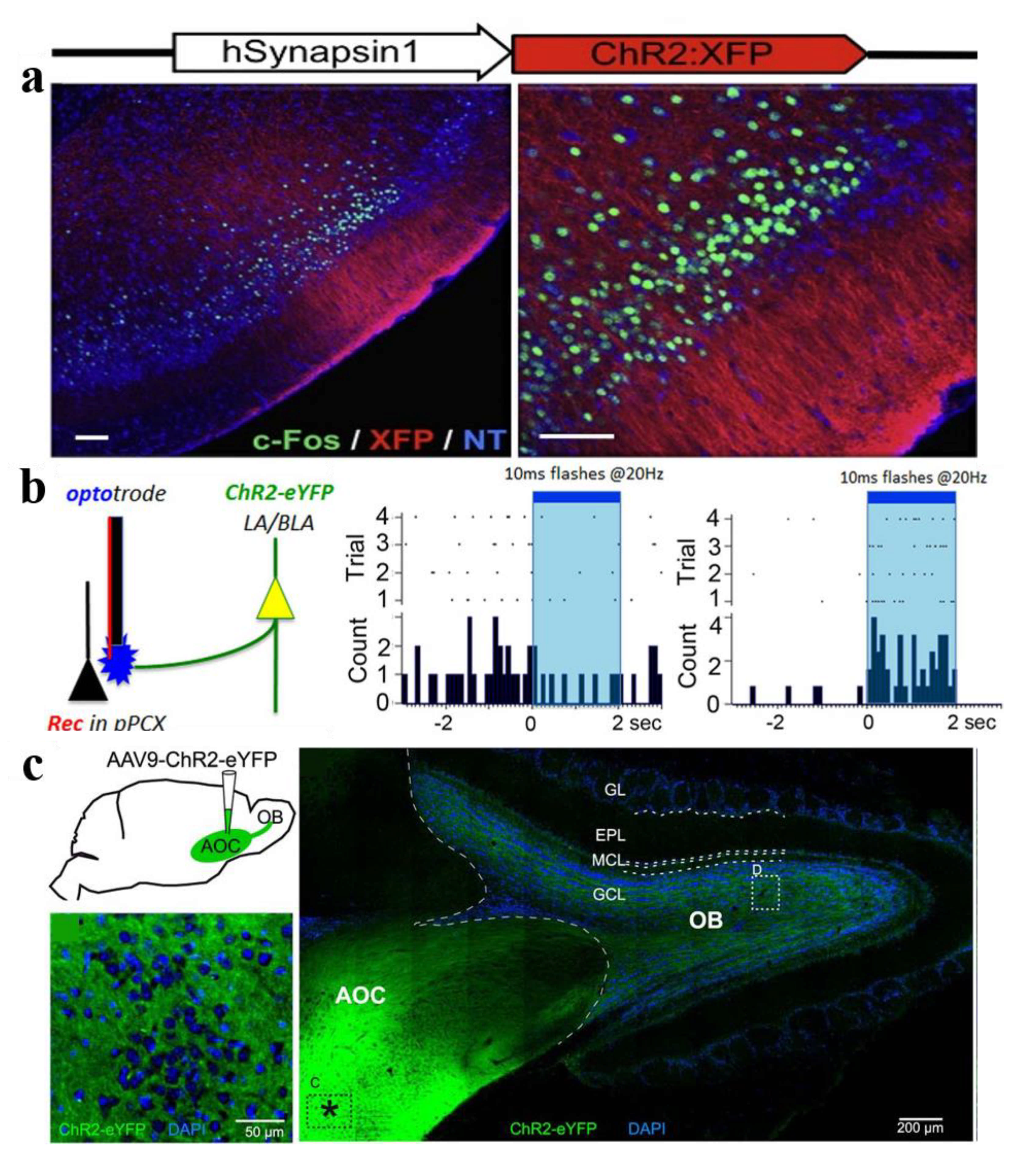
- Sadrian, B.; Wilson, D.A. Optogenetic Stimulation of Lateral Amygdala Input to Posterior Piriform Cortex Modulates Single-Unit and Ensemble Odor Processing. Front. Neural Circuits 2015, 9, 81. [Google Scholar] [CrossRef] [Green Version]
- Moberly, A.H.; Schreck, M.; Bhattarai, J.P.; Zweifel, L.S.; Luo, W.; Ma, M. Olfactory inputs modulate respiration-related rhythmic activity in the prefrontal cortex and freezing behavior. Nat. Commun. 2018, 9, 1528. [Google Scholar] [CrossRef] [PubMed]
- Zhu, J.; Cheng, Q.; Chen, Y.; Fan, H.; Han, Z.; Hou, R.; Chen, Z.; Li, C.T. Transient Delay-Period Activity of Agranular Insular Cortex Controls Working Memory Maintenance in Learning Novel Tasks. Neuron 2020, 105, 934. [Google Scholar] [CrossRef]
- Celada, P.; Puig, M.V.; Artigas, F. Serotonin modulation of cortical neurons and networks. Front. Integr. Neurosci. 2013, 7, 25. [Google Scholar] [CrossRef] [Green Version]
- Macrides, F.; Davis, B.J.; Youngs, W.M.; Nadi, N.S.; Margolis, F.L. Cholinergic and catecholaminergic afferents to the olfactory bulb in the hamster: A neuroanatomical, biochemical, and histochemical investigation. J. Comp. Neurol. 1981, 203, 495–514. [Google Scholar] [CrossRef] [PubMed]
- Salcedo, E.E.; Tran, T.; Ly, X.; Lopez, R.; Barbica, C.; Restrepo, D.; Vijayaraghavan, S. Activity-dependent changes in cholinergic innervation of the mouse olfactory bulb. PLoS ONE 2011, 6, e25441. [Google Scholar] [CrossRef]
- Niedworok, C.J.; Schwarz, I.; Ledderose, J.; Giese, G.; Conzelmann, K.; Schwarz, M.K. Charting Monosynaptic Connectivity Maps by Two-Color Light-Sheet Fluorescence Microscopy. Cell Rep. 2012, 2, 1375–1386. [Google Scholar] [CrossRef] [PubMed] [Green Version]
- Liu, S.; Aungst, J.L.; Puche, A.C.; Shipley, M.T. Serotonin modulates the population activity profile of olfactory bulb external tufted cells. J. Neurophysiol. 2012, 107, 473–483. [Google Scholar] [CrossRef] [Green Version]
- Schwarz, L.A.; Miyamichi, K.; Gao, X.J.; Beier, K.T.; Weissbourd, B.; Deloach, K.E.; Ren, J.; Ibanes, S.; Malenka, R.C.; Kremer, E.J. Viral-genetic tracing of the input–output organization of a central noradrenaline circuit. Nature 2015, 524, 88–92. [Google Scholar] [CrossRef] [Green Version]
- Gire, D.H.; Restrepo, D.; Sejnowski, T.J.; Greer, C.; De Carlos, J.A.; Lopez-Mascaraque, L. Temporal processing in the olfactory system: Can we see a smell? Neuron 2013, 78, 416–432. [Google Scholar] [CrossRef] [PubMed] [Green Version]
- Chuong, A.S.; Miri, M.L.; Busskamp, V.; Matthews, G.A.; Acker, L.C.; Sørensen, A.T.; Young, A.; Klapoetke, N.C.; Henninger, M.A.; Kodandaramaiah, S.B.; et al. Noninvasive optical inhibition with a red-shifted microbial rhodopsin. Nat. Neurosci. 2014, 17, 1123–1129. [Google Scholar] [CrossRef] [PubMed] [Green Version]
- Mahn, M.; Prigge, M.; Ron, S.; Levy, R.; Yizhar, O. Biophysical constraints of optogenetic inhibition at presynaptic terminals. Nat. Neurosci. 2016, 19, 554–556. [Google Scholar] [CrossRef]
- Gao, S.; Nagpal, J.; Schneider, M.W.; Kozjak-Pavlovic, V.; Nagel, G.; Gottschalk, A. Optogenetic manipulation of cGMP in cells and animals by the tightly light-regulated guanylyl-cyclase opsin CyclOp. Nat. Commun. 2015, 6, 8046. [Google Scholar] [CrossRef] [PubMed] [Green Version]


Publisher’s Note: MDPI stays neutral with regard to jurisdictional claims in published maps and institutional affiliations. |
© 2021 by the authors. Licensee MDPI, Basel, Switzerland. This article is an open access article distributed under the terms and conditions of the Creative Commons Attribution (CC BY) license (https://creativecommons.org/licenses/by/4.0/).
Share and Cite
Zhu, P.; Tian, Y.; Chen, Y.; Chen, W.; Wang, P.; Du, L.; Wu, C. Olfactory Optogenetics: Light Illuminates the Chemical Sensing Mechanisms of Biological Olfactory Systems. Biosensors 2021, 11, 309. https://0-doi-org.brum.beds.ac.uk/10.3390/bios11090309
Zhu P, Tian Y, Chen Y, Chen W, Wang P, Du L, Wu C. Olfactory Optogenetics: Light Illuminates the Chemical Sensing Mechanisms of Biological Olfactory Systems. Biosensors. 2021; 11(9):309. https://0-doi-org.brum.beds.ac.uk/10.3390/bios11090309
Chicago/Turabian StyleZhu, Ping, Yulan Tian, Yating Chen, Wei Chen, Ping Wang, Liping Du, and Chunsheng Wu. 2021. "Olfactory Optogenetics: Light Illuminates the Chemical Sensing Mechanisms of Biological Olfactory Systems" Biosensors 11, no. 9: 309. https://0-doi-org.brum.beds.ac.uk/10.3390/bios11090309