Body Size, Cerebral Blood Flow, Ambient Temperature, and Relative Brain Temperatures in Newborn Infants under Incubator Care

 and
and
Abstract
:1. Introduction
2. Materials and Methods
2.1. Study Population
2.2. Data Collection
2.2.1. Temperature Measurements
2.2.2. Echocardiographic Measurements
2.2.3. NIRS Data Acquisition
2.3. Data Analysis
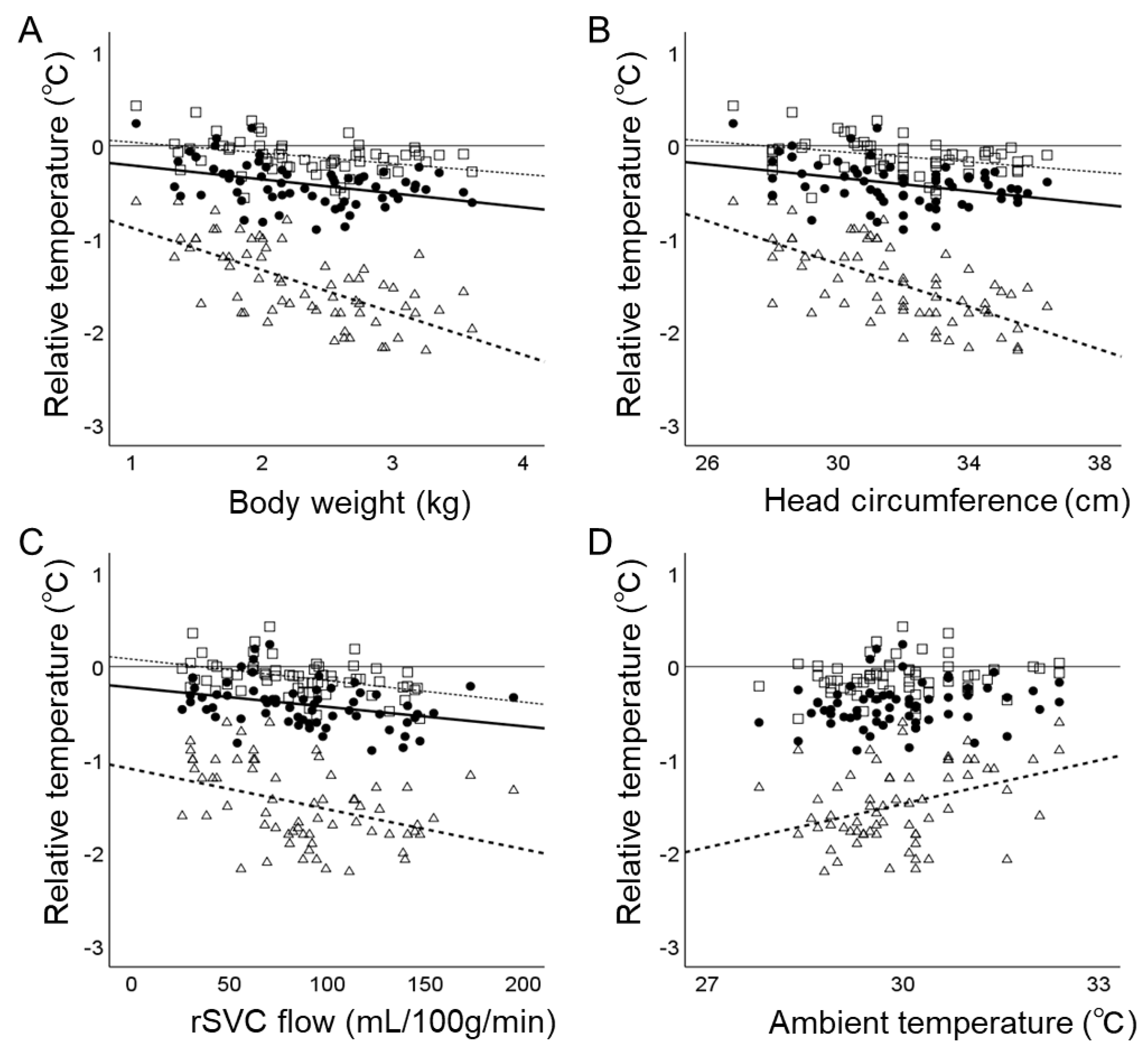
3. Results
3.1. General Findings
3.2. Determinants of Trectal
3.3. Determinants of Scalp and Brain Temperatures Adjusted for Trectal
4. Discussion
4.1. Ambient Temperature and Brain Temperature
4.2. Body, Head Size, and Brain Temperature
4.3. Cerebral Blood Flow and Brain Temperature
4.4. TOI and Brain Temperature
4.5. Limitations
5. Conclusions
Author Contributions
Funding
Institutional Review Board Statement
Informed Consent Statement
Data Availability Statement
Acknowledgments
Conflicts of Interest
References
- Wood, T.; Johnson, M.; Temples, T.; Bordelon, C. Thermoneutral Environment for Neonates: Back to the Basics. Neonatal Netw. 2022, 41, 289–296. [Google Scholar] [CrossRef] [PubMed]
- Baumgart, S. Iatrogenic hyperthermia and hypothermia in the neonate. Clin. Perinatol. 2008, 35, 183–197. [Google Scholar] [CrossRef] [PubMed]
- Tsuda, K.; Shibasaki, J.; Isayama, T.; Takeuchi, A.; Mukai, T.; Ioroi, T.; Takahashi, A.; Sano, H.; Yutaka, N.; Iwata, S.; et al. Body temperature, heart rate and long-term outcome of cooled infants: An observational study. Pediatr. Res. 2022, 91, 921–928. [Google Scholar] [CrossRef] [PubMed]
- Laptook, A.R.; Watkinson, M. Temperature management in the delivery room. Semin. Fetal Neonatal Med. 2008, 13, 383–391. [Google Scholar] [CrossRef] [PubMed]
- Wyatt, J.S.; Gluckman, P.D.; Liu, P.Y.; Azzopardi, D.; Ballard, R.; Edwards, A.D.; Ferriero, D.M.; Polin, R.A.; Robertson, C.M.; Thoresen, M.; et al. Determinants of outcomes after head cooling for neonatal encephalopathy. Pediatrics 2007, 119, 912–921. [Google Scholar] [CrossRef] [PubMed]
- Edwards, A.D.; Brocklehurst, P.; Gunn, A.J.; Halliday, H.; Juszczak, E.; Levene, M.; Strohm, B.; Thoresen, M.; Whitelaw, A.; Azzopardi, D. Neurological outcomes at 18 months of age after moderate hypothermia for perinatal hypoxic ischaemic encephalopathy: Synthesis and meta-analysis of trial data. BMJ 2010, 340, c363. [Google Scholar] [CrossRef] [PubMed]
- Kato, S.; Iwata, O.; Iwata, S.; Yamada, T.; Tsuda, K.; Tanaka, T.; Saitoh, S. Admission temperature of very low birth weight infants and outcomes at three years old. Sci. Rep. 2022, 12, 11912. [Google Scholar] [CrossRef] [PubMed]
- Miller, S.S.; Lee, H.C.; Gould, J.B. Hypothermia in very low birth weight infants: Distribution, risk factors and outcomes. J. Perinatol. 2011, 31 (Suppl. S1), S49–S56. [Google Scholar] [CrossRef] [PubMed]
- Kidokoro, H.; Anderson, P.J.; Doyle, L.W.; Woodward, L.J.; Neil, J.J.; Inder, T.E. Brain injury and altered brain growth in preterm infants: Predictors and prognosis. Pediatrics 2014, 134, e444–e453. [Google Scholar] [CrossRef] [PubMed]
- du Plessis, A.J.; Volpe, J.J. Perinatal brain injury in the preterm and term newborn. Curr. Opin. Neurol. 2002, 15, 151–157. [Google Scholar] [CrossRef] [PubMed]
- Novak, C.M.; Ozen, M.; Burd, I. Perinatal Brain Injury: Mechanisms, Prevention, and Outcomes. Clin. Perinatol. 2018, 45, 357–375. [Google Scholar] [CrossRef] [PubMed]
- Okken, A.; Blijham, C.; Franz, W.; Bohn, E. Effects of forced convection of heated air on insensible water loss and heat loss in preterm infants in incubators. J. Pediatr. 1982, 101, 108–112. [Google Scholar] [CrossRef] [PubMed]
- Iwata, S.; Iwata, O.; Thornton, J.S.; Shanmugalingam, S.; Bainbridge, A.; Peebles, D.; Wyatt, J.S.; Cady, E.B.; Robertson, N.J. Superficial brain is cooler in small piglets: Neonatal hypothermia implications. Ann. Neurol. 2006, 60, 578–585. [Google Scholar] [CrossRef] [PubMed]
- Fenton, T.R.; Kim, J.H. A systematic review and meta-analysis to revise the Fenton growth chart for preterm infants. BMC Pediatr. 2013, 13, 59. [Google Scholar] [CrossRef] [PubMed]
- Annink, K.V.; Groenendaal, F.; Cohen, D.; van der Aa, N.E.; Alderliesten, T.; Dudink, J.; Benders, M.; Wijnen, J.P. Brain temperature of infants with neonatal encephalopathy following perinatal asphyxia calculated using magnetic resonance spectroscopy. Pediatr. Res. 2020, 88, 279–284. [Google Scholar] [CrossRef] [PubMed]
- Iwata, S.; Tachtsidis, I.; Takashima, S.; Matsuishi, T.; Robertson, N.J.; Iwata, O. Dual role of cerebral blood flow in regional brain temperature control in the healthy newborn infant. Int. J. Dev. Neurosci. 2014, 37, 1–7. [Google Scholar] [CrossRef] [PubMed]
- Sinclair, J.C. Servo-control for maintaining abdominal skin temperature at 36C in low birth weight infants. Cochrane Database Syst. Rev. 2002, Cd001074. [Google Scholar] [CrossRef] [PubMed]
- Yamakage, M.; Namiki, A. Deep temperature monitoring using a zero-heat-flow method. J. Anesth. 2003, 17, 108–115. [Google Scholar] [CrossRef] [PubMed]
- Evans, N.; Kluckow, M.; Simmons, M.; Osborn, D. Which to measure, systemic or organ blood flow? Middle cerebral artery and superior vena cava flow in very preterm infants. Arch. Dis. Child. Fetal Neonatal Ed. 2002, 87, F181–F184. [Google Scholar] [CrossRef]
- Dobbing, J.; Sands, J. Head circumference, biparietal diameter and brain growth in fetal and postnatal life. Early Hum. Dev. 1978, 2, 81–87. [Google Scholar] [CrossRef] [PubMed]
- Perlman, J.; Kjaer, K. Neonatal and Maternal Temperature Regulation During and After Delivery. Anesth. Analg. 2016, 123, 168–172. [Google Scholar] [CrossRef] [PubMed]
- Duryea, E.L.; Nelson, D.B.; Wyckoff, M.H.; Grant, E.N.; Tao, W.; Sadana, N.; Chalak, L.F.; McIntire, D.D.; Leveno, K.J. The impact of ambient operating room temperature on neonatal and maternal hypothermia and associated morbidities: A randomized controlled trial. Am. J. Obstet. Gynecol. 2016, 214, 505.e501–505.e507. [Google Scholar] [CrossRef] [PubMed]
- Miller, D.L.; Oliver, T.K., Jr. Body temperature in the immediate neonatal period: The effect of reducing thermal losses. Am. J. Obstet. Gynecol. 1966, 94, 964–969. [Google Scholar] [CrossRef] [PubMed]
- Hammarlund, K.; Sedin, G.; Strömberg, B. Transepidermal water loss in newborn infants. VII. Relation to post-natal age in very pre-term and full-term appropriate for gestational age infants. Acta Paediatr. Scand. 1982, 71, 369–374. [Google Scholar] [CrossRef] [PubMed]
- Fujimoto, S.; Watanabe, T.; Yukawa, K.; Sakamoto, A. Studies on the physical surface area of Japanese. 17. Regional rates according to sex, age and body shape. Nihon Eiseigaku Zasshi 1968, 23, 437–442. [Google Scholar] [CrossRef] [PubMed]
- Harpin, V.A.; Rutter, N. Humidification of incubators. Arch. Dis. Child. 1985, 60, 219–224. [Google Scholar] [CrossRef] [PubMed]
- Sherman, T.I.; Greenspan, J.S.; St Clair, N.; Touch, S.M.; Shaffer, T.H. Optimizing the neonatal thermal environment. Neonatal Netw. 2006, 25, 251–260. [Google Scholar] [CrossRef] [PubMed]
- Merklin, R.J. Growth and distribution of human fetal brown fat. Anat. Rec. 1974, 178, 637–645. [Google Scholar] [CrossRef]
- Lidell, M.E. Brown Adipose Tissue in Human Infants. In Brown Adipose Tissue; Handbook of Experimental Pharmacology; Springer: Cham, Switzerland, 2019; Volume 251, pp. 107–123. [Google Scholar] [CrossRef]
- Li, M.X.; Sun, G.; Neubauer, H. Change in the body temperature of healthy term infant over the first 72 h of life. J. Zhejiang Univ. Sci. 2004, 5, 486–493. [Google Scholar] [CrossRef] [PubMed]
- Eichna, L.W.; Berger, A.R.; Rader, B.; Becker, W.H. Comparison of intracardiac and intravascular temperatures with rectal temperatures in man. J. Clin. Investig. 1951, 30, 353–359. [Google Scholar] [CrossRef]
- Nunneley, S.A.; Nelson, D.A. Limitations on arteriovenous cooling of the blood supply to the human brain. Eur. J. Appl. Physiol. Occup. Physiol. 1994, 69, 474–479. [Google Scholar] [CrossRef] [PubMed]
- Zhu, M.; Ackerman, J.J.; Yablonskiy, D.A. Body and brain temperature coupling: The critical role of cerebral blood flow. J. Comp. Physiol. B 2009, 179, 701–710. [Google Scholar] [CrossRef] [PubMed]
- Kondo, Y.; Hirose, N.; Maeda, T.; Yoshino, A.; Suzuki, T. Relationship between changes in regional cerebral blood volume and oxygenation and changes in cardiac output and systemic vascular resistance during spinal anesthesia in women undergoing cesarean section. J. Anesth. 2019, 33, 579–586. [Google Scholar] [CrossRef] [PubMed]
- Vanderhaegen, J.; Naulaers, G.; Vanhole, C.; De Smet, D.; Van Huffel, S.; Vanhaesebrouck, S.; Devlieger, H. The effect of changes in tPCO2 on the fractional tissue oxygen extraction—As measured by near-infrared spectroscopy—In neonates during the first days of life. Eur. J. Paediatr. Neurol. 2009, 13, 128–134. [Google Scholar] [CrossRef] [PubMed]
- Donadello, K.; Su, F.; Annoni, F.; Scolletta, S.; He, X.; Peluso, L.; Gottin, L.; Polati, E.; Creteur, J.; De Witte, O.; et al. The Effects of Temperature Management on Brain Microcirculation, Oxygenation and Metabolism. Brain Sci. 2022, 12, 1422. [Google Scholar] [CrossRef] [PubMed]
- Tong, Y.; Frederick, B.D. Time lag dependent multimodal processing of concurrent fMRI and near-infrared spectroscopy (NIRS) data suggests a global circulatory origin for low-frequency oscillation signals in human brain. Neuroimage 2010, 53, 553–564. [Google Scholar] [CrossRef] [PubMed]
- Zohdi, H.; Scholkmann, F.; Wolf, U. Frontal cerebral oxygenation asymmetry: Intersubject variability and dependence on systemic physiology, season, and time of day. Neurophotonics 2020, 7, 025006. [Google Scholar] [CrossRef] [PubMed]
- Atallah, L.; Bongers, E.; Lamichhane, B.; Bambang-Oetomo, S. Unobtrusive Monitoring of Neonatal Brain Temperature Using a Zero-Heat-Flux Sensor Matrix. IEEE J. Biomed. Health Inform. 2016, 20, 100–107. [Google Scholar] [CrossRef] [PubMed]
- Simbruner, G.; Nanz, S.; Fleischhacker, E.; Derganc, M. Brain temperature discriminates between neonates with damaged, hypoperfused, and normal brains. Am. J. Perinatol. 1994, 11, 137–143. [Google Scholar] [CrossRef] [PubMed]
- Simbruner, G.; Ruttner, E.M.; Schulze, A.; Perzlmaier, K. Premature infants are less capable of maintaining thermal balance of head and body with increases of thermal environment than with decreases. Am. J. Perinatol. 2005, 22, 25–33. [Google Scholar] [CrossRef] [PubMed]
- van der Spek, R.D.; van Lingen, R.A.; van Zoeren-Grobben, D. Body temperature measurement in VLBW infants by continuous skin measurement is a good or even better alternative than continuous rectal measurement. Acta Paediatr. 2009, 98, 282–285. [Google Scholar] [CrossRef] [PubMed]


| Variables | Mean | Standard Deviation | ||
|---|---|---|---|---|
| Clinical variables at birth | ||||
| Gestational age (week) | 35.6 | 3.8 | ||
| Body weight (g) | 2333 | 794 | ||
| Apgar scores | 1 min | 7.5 | 1.6 | |
| 5 min | 8.6 | 1.1 | ||
| Clinical variables at the time of the study | ||||
| Postnatal age (d) | 9.2 | 14.8 | ||
| Postconceptional age (week) | 36.9 | 2.2 | ||
| Body weight (g) | 2348 | 609 | ||
| Head circumference (cm) | 31.9 | 2.3 | ||
| Incubator environment | ||||
| Ambient temperature (°C) | 30.0 | 1.0 | ||
| Ambient humidity (%) | 47.3 | 8.2 | ||
| Temperature measures (°C) | ||||
| Trectal | 37.0 | 0.2 | ||
| Tbrain-25 | 36.8 | 0.2 | ||
| Tbrain-15 | 36.5 | 0.2 | ||
| Tscalp | 35.5 | 0.4 | ||
| Tbrain25 − Trectal | −0.1 | 0.2 | ||
| Tbrain15 − Trectal | −0.4 | 0.2 | ||
| Tscalp − Trectal | −1.5 | 0.4 | ||
| Haemoglobin, blood flow, and tissue oxygenation | ||||
| Blood Haemoglobin (g/dL) | 17.2 | 2.4 | ||
| rSVC flow (mL/100 g/min) | 85.5 | 34.2 | ||
| Tissue oxygenation index (%) | 64.9 | 3.5 | ||
| Variables | Trectal | Tbrain25 − Trectal | Tbrain15 − Trectal | Tscalp − Trectal | |||||
|---|---|---|---|---|---|---|---|---|---|
| Coefficient × 102 | p | Coefficient × 102 | p | Coefficient × 102 | p | Coefficient × 102 | p | ||
| Univariable analysis | |||||||||
| Clinical variables at the time of the study | |||||||||
| Female sex | −13.7 | 0.017 | 1.4 | 0.778 | −1.7 | 0.777 | 8.8 | 0.406 | |
| (−25.1 to −2.4) | (−8.2 to 10.9) | (−13.4 to 10.0) | (−12.0 to 29.6) | ||||||
| Postnatal age (d) | −0.3 | 0.113 | 0.0 | 0.771 | 0.3 | 0.151 | 0.6 | 0.087 | |
| (−0.7 to 0.1) | (−0.3 to 0.4) | (−0.1 to 0.7) | (−0.1 to 1.3) | ||||||
| Postconceptional age (week) | 4.0 | 0.002 | −1.6 | 0.141 | −3.2 | 0.016 | −11.7 | <0.001 | |
| (1.5 to 6.6) | (−3.8 to 0.5) | (−5.8 to −0.6) | (−15.6 to −7.9) | ||||||
| Body weight (kg) | 19.3 | <0.001 | −11.5 | 0.002 | −13.8 | 0.003 | −45.0 | <0.001 | |
| (10.8 to 27.8) | (−18.8 to −4.2) | (−22.8 to −4.8) | (−58.0 to −32.0) | ||||||
| Head circumference (cm) | 5.0 | <0.001 | −2.8 | 0.005 | −3.1 | 0.010 | −11.2 | <0.001 | |
| (2.8 to 7.2) | (−4.7 to −0.8) | (−5.5 to −0.8) | (−14.7 to −7.8) | ||||||
| Incubator environment | |||||||||
| Ambient temperature (°C) | 1.0 | 0.735 | 3.4 | 0.153 | 4.2 | 0.152 | 16.0 | 0.001 | |
| (−4.9 to 7.0) | (−1.3 to 8.2) | (−1.6 to 10.1) | (6.3 to 25.7) | ||||||
| Ambient humidity (%) | 0.6 | 0.115 | −0.6 | 0.037 | −0.9 | 0.012 | −1.2 | 0.051 | |
| (−0.1 to 1.3) | (−1.2 to 0.0) | (−1.6 to −0.2) | (−2.5 to 0.0) | ||||||
| Haemoglobin, blood flow, and tissue oxygenation | |||||||||
| Blood haemoglobin (g/dL) | 1.5 | 0.264 | 0.5 | 0.623 | −1.4 | 0.244 | −3.8 | 0.084 | |
| (−1.1 to 4.1) | (−1.4 to 2.4) | (−3.8 to 1.0) | (−8.0 to 0.5) | ||||||
| rSVC (mL/100 g/min) | 0.1 | 0.244 | −0.2 | <0.001 | −0.3 | <0.001 | −0.6 | <0.001 | |
| (−0.1 to 0.3) | (−0.4 to −0.1) | (−0.4 to −0.1) | (−0.8 to −0.3) | ||||||
| Tissue oxygenation index (%) | −0.1 | 0.949 | 0.7 | 0.315 | 2.0 | 0.008 | 2.9 | 0.040 | |
| (−1.8 to 1.7) | (−0.6 to 1.9) | (0.5 to 3.5) | (8.4 to 18.6) | ||||||
| Multivariable analysis | |||||||||
| Head circumference (cm) | 5.0 | <0.001 | −2.3 | 0.015 | −2.5 | 0.030 | −9.8 | <0.001 | |
| (2.8 to 7.2) | (−4.1 to −0.4) | (−4.8 to −0.2) | (−12.9 to −6.8) | ||||||
| Ambient temperature (°C) | 4.6 | 0.119 | −0.8 | 0.742 | −0.8 | 0.781 | 6.4 | 0.116 | |
| (−1.2 to 10.3) | (−5.5 to 3.9) | (−6.7 to 5.0) | (−1.6 to 14.3) | ||||||
| rSVC (mL/100 g/min) | 0.1 | 0.240 | −0.2 | 0.003 | −0.3 | 0.003 | −0.4 | 0.002 | |
| (−0.1 to 0.3) | (−0.4 to −0.1) | (−0.4 to −0.1) | (−0.6 to −0.1) | ||||||
Disclaimer/Publisher’s Note: The statements, opinions and data contained in all publications are solely those of the individual author(s) and contributor(s) and not of MDPI and/or the editor(s). MDPI and/or the editor(s) disclaim responsibility for any injury to people or property resulting from any ideas, methods, instructions or products referred to in the content. |
© 2024 by the authors. Licensee MDPI, Basel, Switzerland. This article is an open access article distributed under the terms and conditions of the Creative Commons Attribution (CC BY) license (https://creativecommons.org/licenses/by/4.0/).
Share and Cite
Fukaya, S.; Iwata, S.; Tsuda, K.; Hirose, A.; Kinoshita, M.; Saitoh, S.; Iwata, O. Body Size, Cerebral Blood Flow, Ambient Temperature, and Relative Brain Temperatures in Newborn Infants under Incubator Care. Biosensors 2024, 14, 209. https://0-doi-org.brum.beds.ac.uk/10.3390/bios14040209
Fukaya S, Iwata S, Tsuda K, Hirose A, Kinoshita M, Saitoh S, Iwata O. Body Size, Cerebral Blood Flow, Ambient Temperature, and Relative Brain Temperatures in Newborn Infants under Incubator Care. Biosensors. 2024; 14(4):209. https://0-doi-org.brum.beds.ac.uk/10.3390/bios14040209
Chicago/Turabian StyleFukaya, Satoko, Sachiko Iwata, Kennosuke Tsuda, Akiko Hirose, Masahiro Kinoshita, Shinji Saitoh, and Osuke Iwata. 2024. "Body Size, Cerebral Blood Flow, Ambient Temperature, and Relative Brain Temperatures in Newborn Infants under Incubator Care" Biosensors 14, no. 4: 209. https://0-doi-org.brum.beds.ac.uk/10.3390/bios14040209