
Antibacterial Property of Cellulose Acetate Composite Materials Reinforced with Aluminum Nitride


 , , , , , and
Abstract
:1. Introduction
2. Materials and Methods
2.1. Samples Preparation
2.2. Sample Characterization
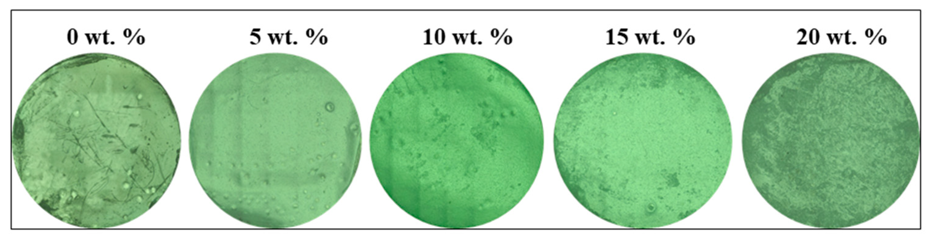
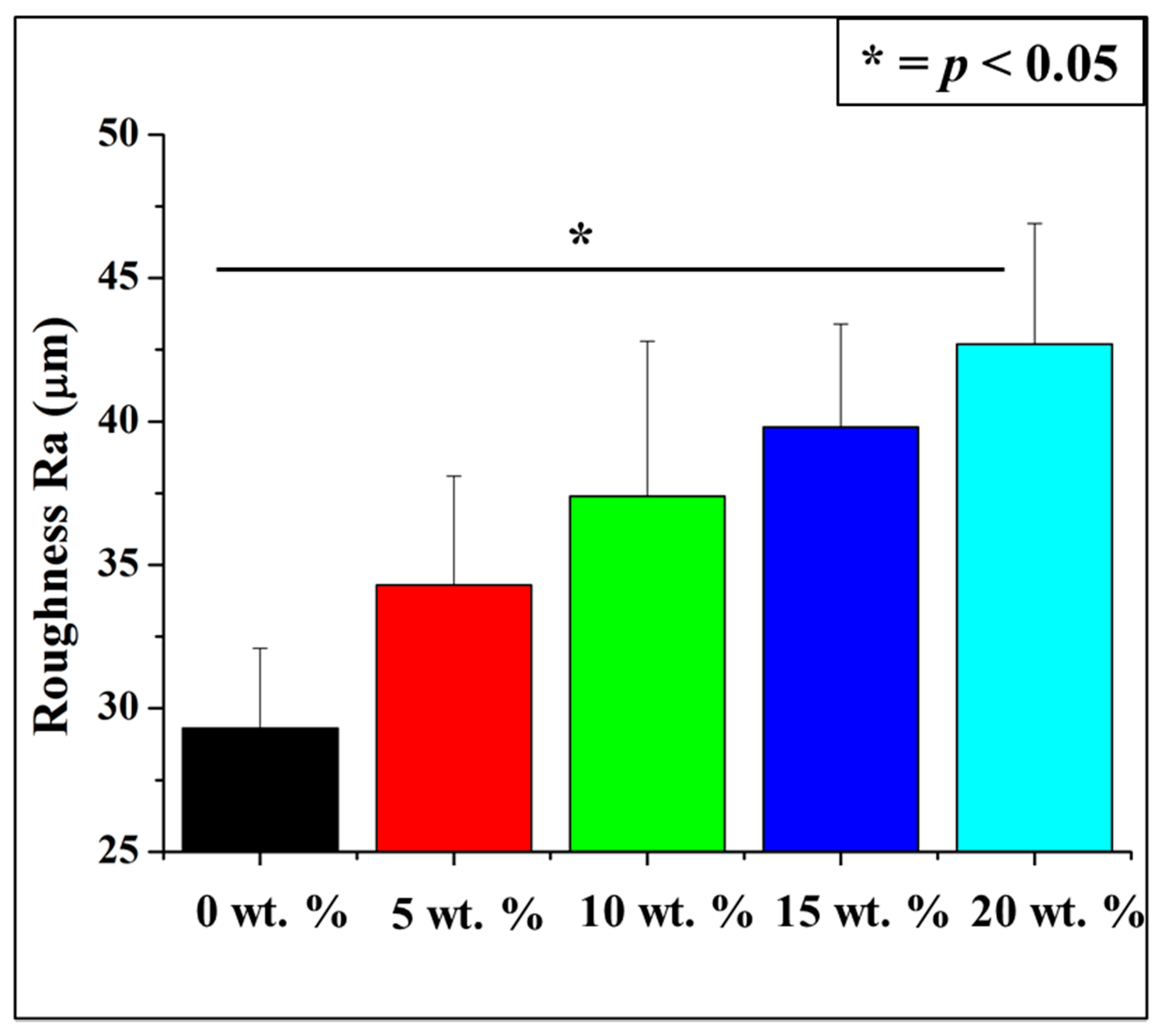
2.2.1. Laser Microscopy
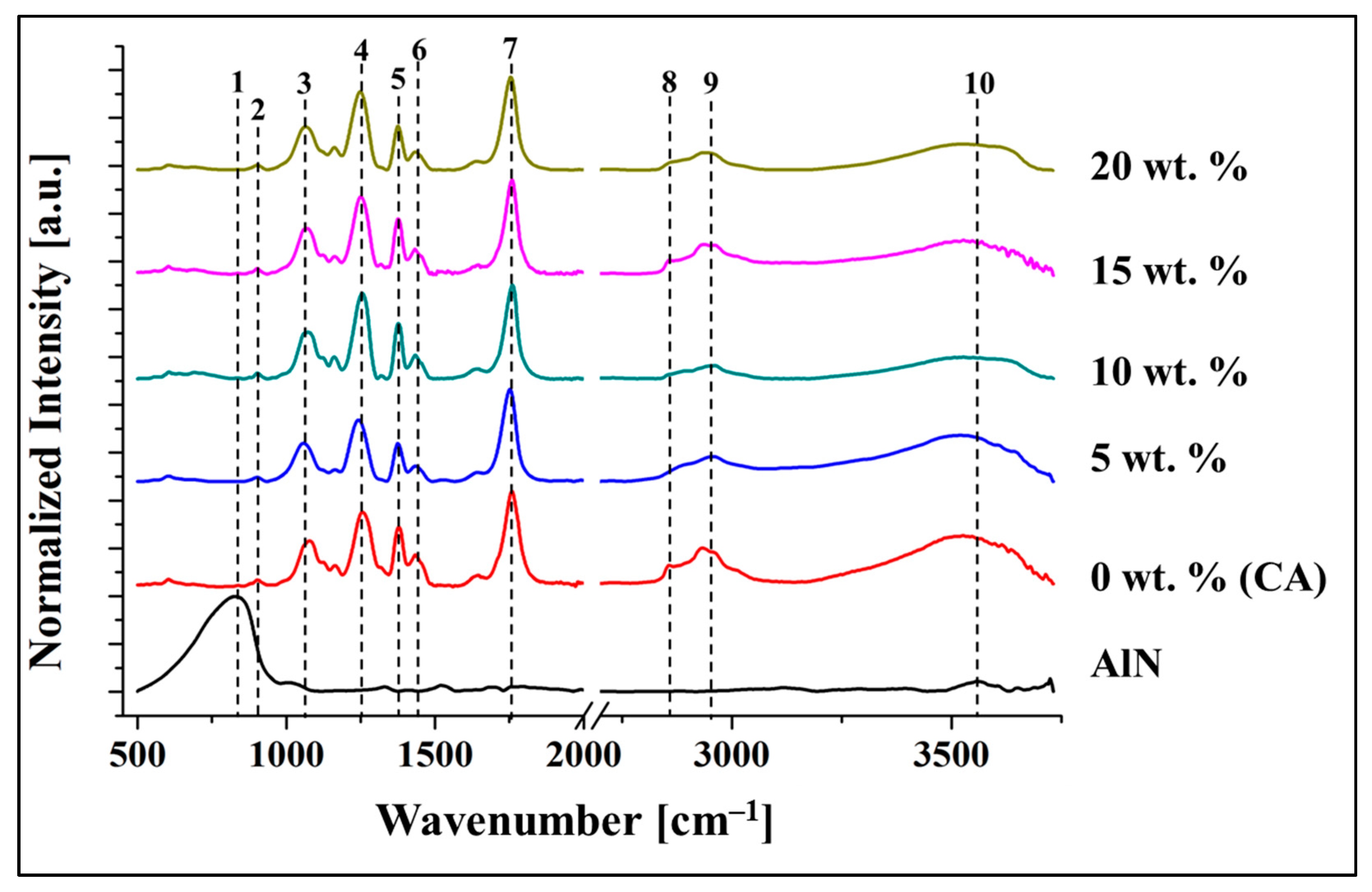
2.2.2. Fourier Transformed Infrared Spectroscopy
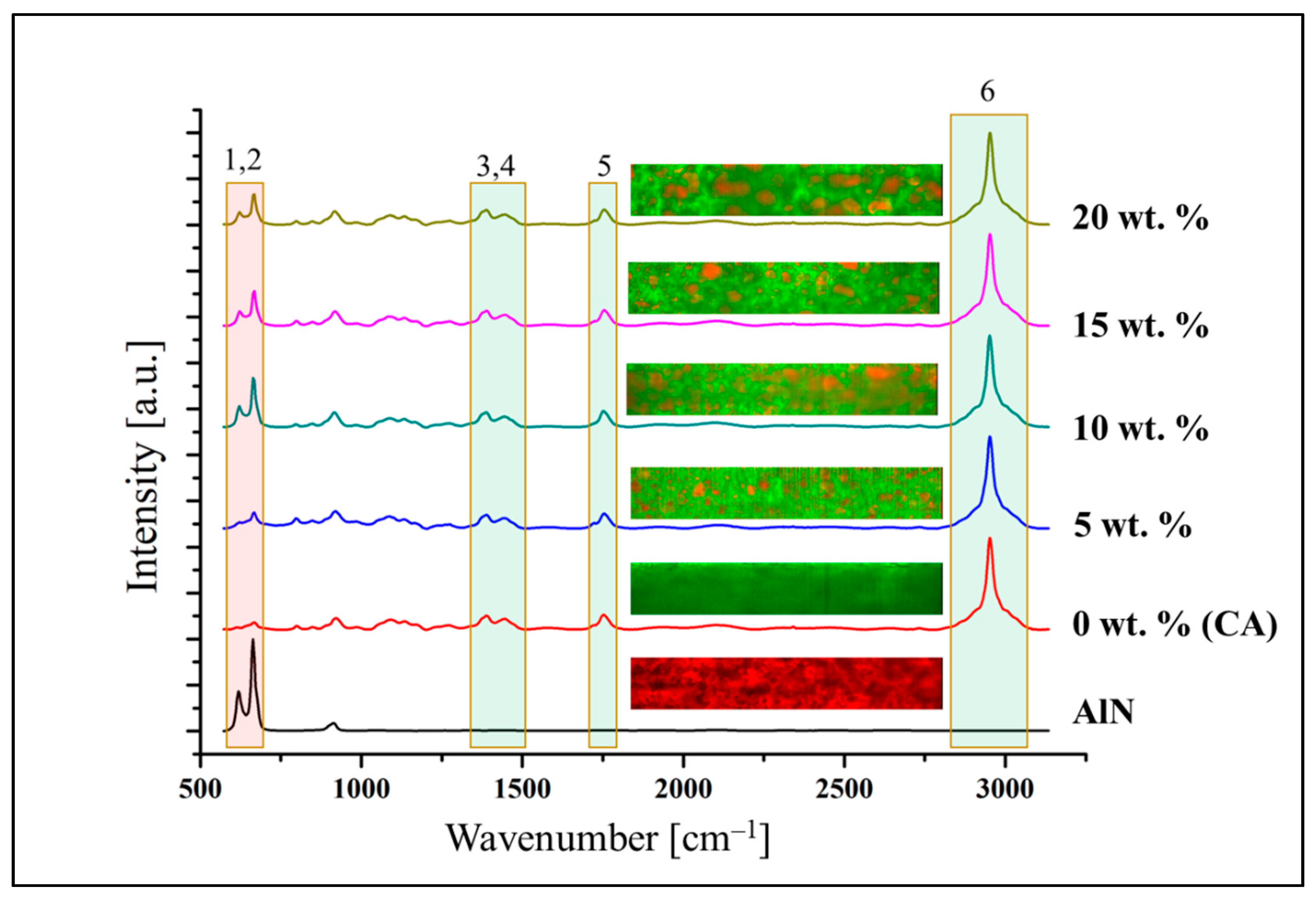
2.2.3. Raman Spectroscopy
2.3. Mechanical and Thermal Properties
2.3.1. Mechanical Properties
2.3.2. Thermal Properties
2.4. In Vitro Testing
2.4.1. Microbial Viability Assay (WST)
2.4.2. Crystal Violet Assay, Laser Microscopy Scanning, Viability Staining and ImageJ Analysis
2.5. Statistical Analysis
3. Results
3.1. Surface Characterization
3.1.1. Laser Microscopy
3.1.2. FTIR Spectroscopy
3.1.3. Raman Spectroscopy
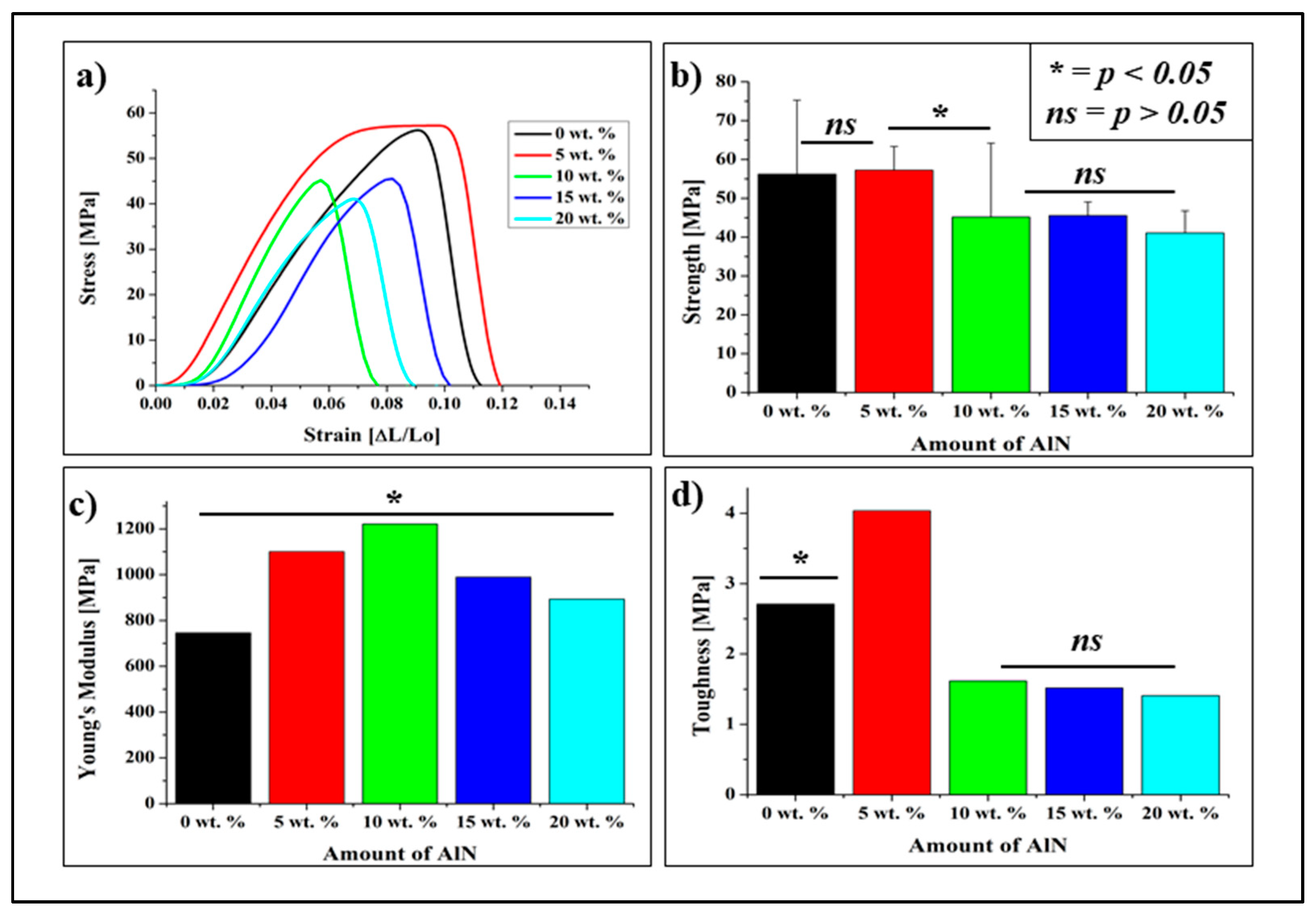
3.2. Mechanical Property
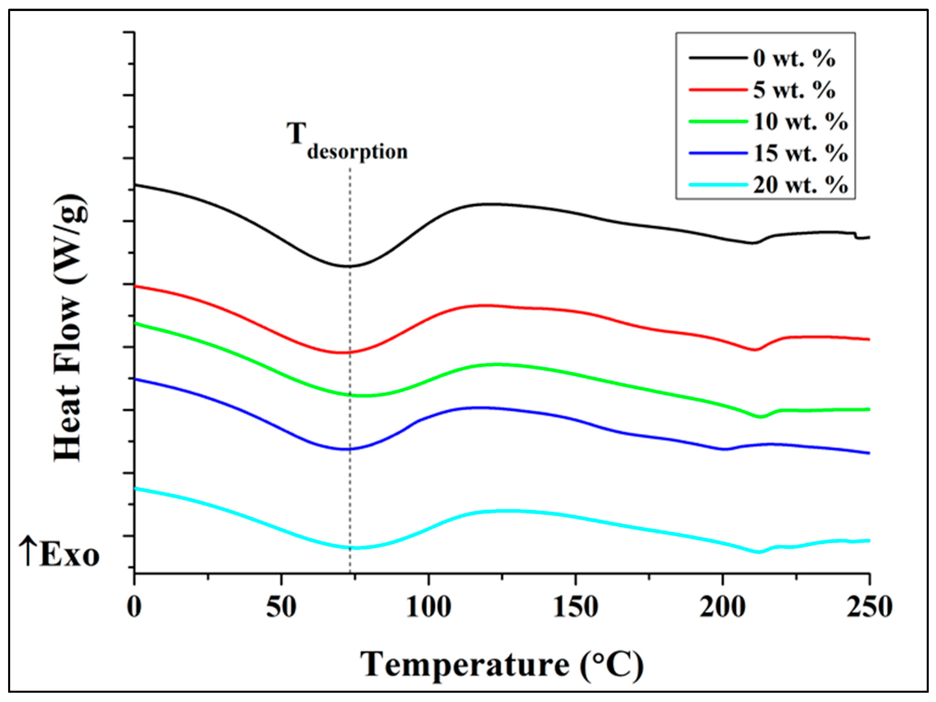
3.3. Thermal Properties
3.4. In Vitro Testing
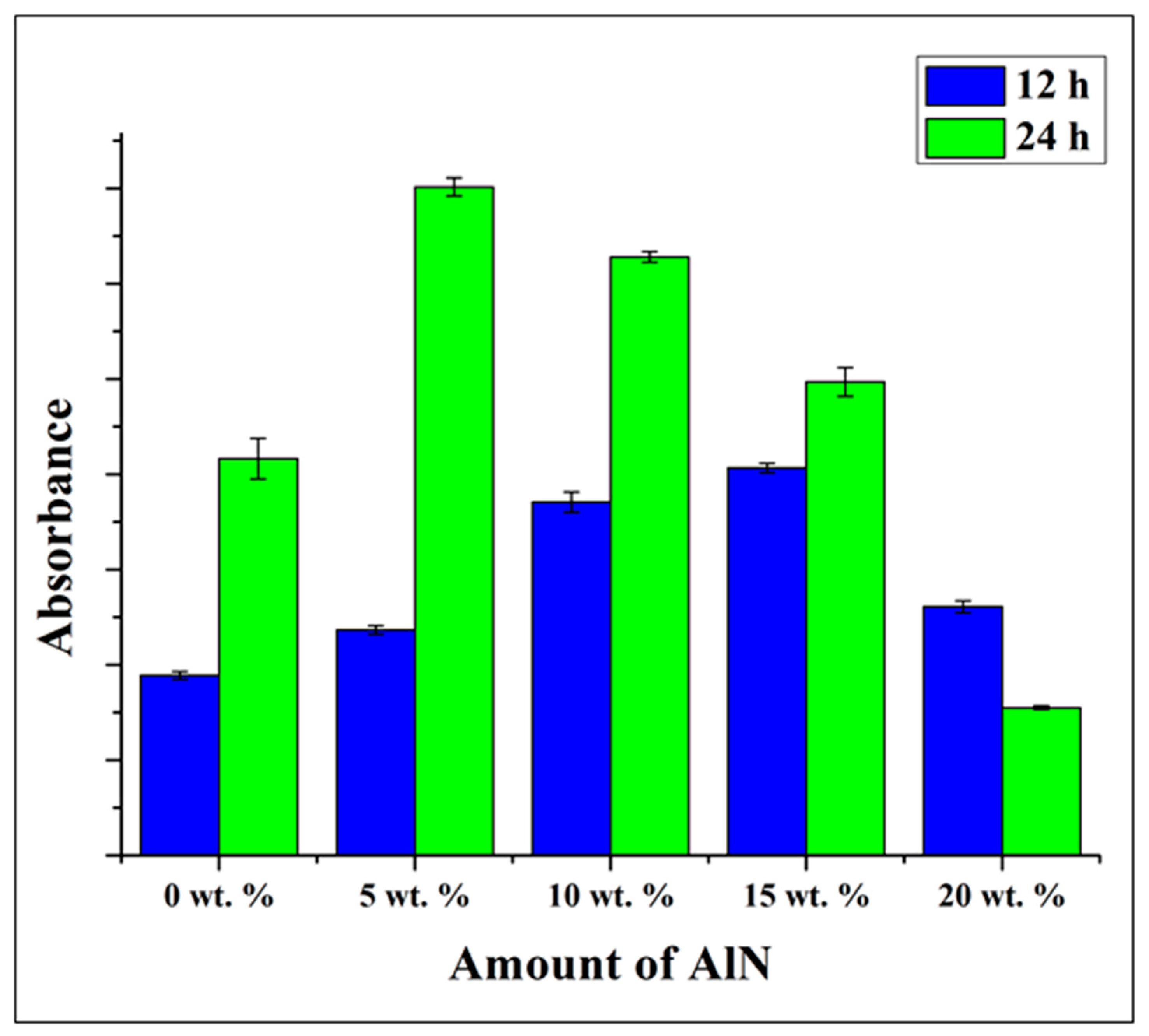
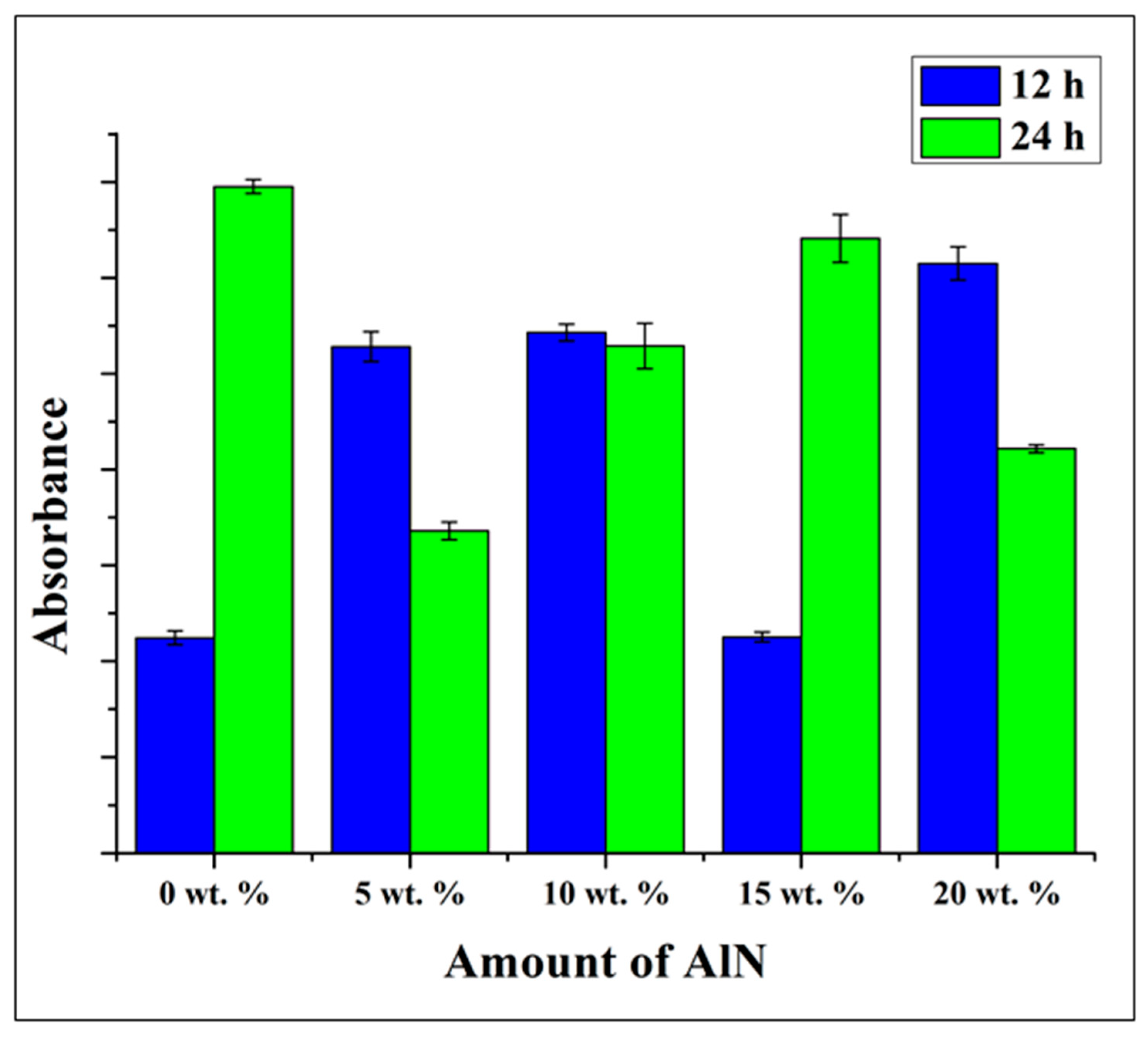
Microbial Viability Assay (WST)
4. Discussion
5. Conclusions
Supplementary Materials
Author Contributions
Funding
Data Availability Statement
Conflicts of Interest
References
- Sunthar, T.; Marin, E.; Boschetto, F.; Zanocco, M.; Sunahara, H.; Ramful, R.; Pezzotti, G. Antibacterial and ANTIFUNGAL properties of composite POLYETHYLENE Materials reinforced with Neem and turmeric. Antibiotics 2020, 9, 857. [Google Scholar] [CrossRef] [PubMed]
- Kamala, K.; Kumar, V.P. Food products and food contamination. Microb. Contam. Food Degrad. 2018, 9, 1–19. [Google Scholar] [CrossRef]
- Var, I. Active Antimicrobial Food Packaging; IntechOpen: London, UK, 2019; Available online: https://directory.doabooks.org/handle/20.500.12854/40079 (accessed on 19 October 2021).
- Han, J.H. Antimicrobial food packaging. Novel Food Packag. Tech. 2003, 8, 50–70. [Google Scholar] [CrossRef]
- Ahvenainen, R.; Ahvenainen, R. Novel Food Packaging Techniques; CRC Press: Boca Raton, FL, USA, 2003. [Google Scholar]
- Khaneghah, A.M.; Hashemi, S.M.B.; Limbo, S. Antimicrobial agents and packaging systems in antimicrobial active food packaging: An overview of approaches and interactions. Food Bioprod. Process. 2018, 111, 1–19. [Google Scholar] [CrossRef]
- Fischer, S.; Thümmler, K.; Volkert, B.; Hettrich, K.; Schmidt, I.; Fischer, K. Properties and applications of cellulose acetate. Macromol. Symp. 2008, 262, 89–96. [Google Scholar] [CrossRef]
- Law, R.C. 5. applications of cellulose acetate 5.1 cellulose acetate in textile application. Macromol. Symp. 2004, 208, 255–266. [Google Scholar] [CrossRef]
- Selvaduray, G.; Sheet, L. Aluminium nitride: Review of synthesis methods. Mater. Sci. Tech. 1993, 9, 463–473. [Google Scholar] [CrossRef]
- Jovanović, G.D.; Klaus, A.S.; Nikšić, M.P. Antimicrobial activity of chitosan coatings and films against Listeria monocytogenes on black radish. Rev. Argent. Microb. 2016, 48, 128–136. [Google Scholar] [CrossRef] [PubMed] [Green Version]
- Marin, E.; Boschetto, F.; Zanocco, M.; Honma, T.; Zhu, W.; Pezzotti, G. Explorative study on the antibacterial effects of 3D-printed PMMA/nitrides composites. Mater. Design 2021, 206, 109788. [Google Scholar] [CrossRef]
- Molleja, J.G.; Gómez, B.J.; Abdallah, B.; Djouadi, M.A.; Feugeas, J.; Jouan, P.-Y. Study of AlN thin films deposited by DC magnetron sputtering: Effect of pressure on texture. An. AFA 2016, 26, 190–194. [Google Scholar] [CrossRef]
- Ibáñez, J.; Hernández, S.; Alarcón-Lladó, E.; Cuscó, R.; Artús, L.; Novikov, S.V.; Foxon, C.T.; Calleja, E. Far-infrared transmission in gan, aln, AND algan thin films grown by molecular beam epitaxy. J. Appl. Phys. 2008, 104, 033544. [Google Scholar] [CrossRef] [Green Version]
- Murphy, D.; de Pinho, M.N. An atr-ftir study of water in cellulose acetate Membranes prepared by phase inversion. J. Membr. Sci. 1995, 106, 245–257. [Google Scholar] [CrossRef]
- Arkhangelsky, E.; Goren, U.; Gitis, V. Retention of organic matter by cellulose acetate membranes cleaned with hypochlorite. Desalination 2008, 223, 97–105. [Google Scholar] [CrossRef]
- Sudiarti, T.; Wahyuningrum, D.; Bundjali, B.; Made Arcana, I. Mechanical strength and ionic conductivity of polymer electrolyte membranes prepared from cellulose acetate-lithium perchlorate. IOP Conf. Ser. Mater. Sci. Eng. 2017, 223, 012052. [Google Scholar] [CrossRef] [Green Version]
- Dong, F.; Yan, M.; Jin, C.; Li, S. Characterization of Type-II Acetylated Cellulose Nanocrystals with Various Degree of Substitution and Its Compatibility in PLA Films. Polymers 2017, 9, 346. [Google Scholar] [CrossRef] [PubMed] [Green Version]
- Sánchez-Márquez, J.A.; Fuentes-Ramírez, R.; Cano-Rodríguez, I.; Gamiño-Arroyo, Z.; Rubio-Rosas, E.; Kenny, J.M.; Rescignano, N. Membrane Made of Cellulose Acetate with Polyacrylic Acid Reinforced with Carbon Nanotubes and Its Applicability for Chromium Removal. Int. J. Polym. Sci. 2015, 2015, 1–12. [Google Scholar] [CrossRef] [PubMed] [Green Version]
- Kubota, H.; Sakamoto, K.; Matsui, T. A confocal Raman Microscopic visualization of SMALL penetrants in cellulose acetate using A DEUTERIUM-LABELING TECHNIQUE. Sci. Rep. 2020, 10, 16426. [Google Scholar] [CrossRef] [PubMed]
- Musa, I.; Qamhieh, N.; Said, K.; Mahmoud, S.T.; Alawadhi, H. Fabrication and characterization of Aluminum Nitride NANOPARTICLES by Rf magnetron sputtering and inert Gas CONDENSATION TECHNIQUE. Coatings 2020, 10, 411. [Google Scholar] [CrossRef] [Green Version]
- Liu, L.; Liu, B.; Edgar, J.H.; Rajasingam, S.; Kuball, M. Raman characterization and stress analysis of AlN grown on SiC by sublimation. J. Appl. Phys. 2002, 92, 5183–5188. [Google Scholar] [CrossRef] [Green Version]
- Li, X.; Zhou, C.; Jiang, G.; You, J. Raman analysis of aluminum nitride at high temperature. Mater. Charact. 2006, 57, 105–110. [Google Scholar] [CrossRef]
- Yang, Z.-Y.; Wang, W.-J.; Shao, Z.-Q.; Zhu, H.-D.; Li, Y.-H.; Wang, F.-J. The transparency and mechanical properties of cellulose acetate nanocomposites using cellulose nanowhiskers as fillers. Cellulose 2013, 20, 159–168. [Google Scholar] [CrossRef]
- Fogagnolo, J.B.; Robert, M.H.; Velasco, F.; Torralba, J.M. Aluminium matrix COMPOSITES reinforced WITH Si3N4, AlN And ZrB2, produced by Conventional powder metallurgy and Mechanical Alloying. KONA Powder Part. J. 2004, 22, 143–150. [Google Scholar] [CrossRef] [Green Version]
- Vo, P.; Doan, H.; Kinashi, K.; Sakai, W.; Tsutsumi, N.; Huynh, D. Centrifugally Spun Recycled PET: Processing and Characterization. Polymers 2018, 10, 680. [Google Scholar] [CrossRef] [PubMed] [Green Version]
- de Freitas, R.R.M.; Senna, A.M.; Botaro, V.R. Influence of degree of substitution on thermal dynamic mechanical and physicochemical properties of cellulose acetate. Ind. Crops Prod. 2017, 109, 452–458. [Google Scholar] [CrossRef]
- Barud, H.S.; de Araújo Júnior, A.M.; Santos, D.B.; de Assunção, R.M.N.; Meireles, C.S.; Cerqueira, D.A.; Rodrigues Filho, G.; Ribeiro, C.A.; Messaddeq, Y.; Ribeiro, S.J.L. Thermal behavior of cellulose acetate produced from homogeneous acetylation of bacterial cellulose. Thermochim. Acta 2008, 471, 61–69. [Google Scholar] [CrossRef]
- Cerqueira, D.A.; Rodrigues Filho, G.; Assunção, R.M.N. A New Value for the Heat of Fusion of a Perfect Crystal of Cellulose Acetate. Polym. Bull. 2006, 56, 475–484. [Google Scholar] [CrossRef]
- Erdmann, R.; Kabasci, S.; Heim, H.-P. Thermal Properties of Plasticized Cellulose Acetate and Its β-Relaxation Phenomenon. Polymers 2021, 13, 1356. [Google Scholar] [CrossRef] [PubMed]
- Aoki, D.; Teramoto, Y.; Nishio, Y. Cellulose acetate/poly(methyl methacrylate) interpenetrating networks: Synthesis and estimation of thermal and mechanical properties. Cellulose 2011, 18, 1441–1454. [Google Scholar] [CrossRef] [Green Version]
- Gutierrez, J.; Carrasco-Hernandez, S.; Barud, H.S.; Oliveira, R.L.; Carvalho, R.A.; Amaral, A.C.; Tercjak, A. Transparent nanostructured cellulose acetate films based on the self assembly OF PEO-b-PPO-b-PEO block COPOLYMER. Carbohydr. Polym. 2017, 165, 437–443. [Google Scholar] [CrossRef] [PubMed] [Green Version]
- Iqhrammullah, M.; Marlina, M.; Khalil, H.P.; Kurniawan, K.H.; Suyanto, H.; Hedwig, R.; Karnadi, I.; Olaiya, N.G.; Abdullah, C.K.; Abdulmadjid, S.N. Characterization and performance evaluation of cellulose acetate–polyurethane film for lead ii ion removal. Polymers 2020, 12, 1317. [Google Scholar] [CrossRef] [PubMed]
- Yang, C.; Dong, J.; Fang, Y.; Ma, L.; Zhao, X.; Zhang, Q. Preparation of novel low-κ polyimide fibers with simultaneously excellent mechanical properties, UV-resistance and surface activity using chemically bonded hyperbranched polysiloxane. J. Mater. Chem. C 2018, 6, 1229–1238. [Google Scholar] [CrossRef]
- Liu, X.; Wang, T.; Chow, L.C.; Yang, M.; Mitchell, J.W. Effects of inorganic fillers on the thermal and mechanical properties of poly (lactic acid). Int. J. Polym. Sci. 2014, 2014, 827028. [Google Scholar] [CrossRef] [PubMed]
- Kleiner, D. The transport of NH3 and HN4+ across biological membranes. Biochim. Biophys. Acta (BBA)-Rev. Bioenerg. 1981, 639, 41–52. [Google Scholar] [CrossRef]
- Pezzotti, G.; Asai, T.; Adachi, T.; Ohgitani, E.; Yamamoto, T.; Kanamura, N.; Boschetto, F.; Zhu, W.; Zanocco, M.; Marin, E.; et al. Antifungal activity of polymethyl methacrylate/Si3N4 composites against Candida albicans. Acta Biomater. 2021, 126, 259–276. [Google Scholar] [CrossRef] [PubMed]
- Pezzotti, G.; Ohgitani, E.; Shin-Ya, M.; Adachi, T.; Marin, E.; Boschetto, F.; Zhu, W.; Mazda, O. Instantaneous “catch-and-kill” inactivation of SARS-CoV-2 by nitride ceramics. Clin. Transl. Med. 2020, 10, e212. [Google Scholar] [CrossRef] [PubMed]
- Li, J.; Nakamura, M.; Shirai, T.; Matsumaru, K.; Ishizaki, C.; Ishizaki, K. Mechanism and Kinetics of Aluminum Nitride Powder Degradation in Moist Air. J. Am. Ceram. Soc. 2006, 89, 937–943. [Google Scholar] [CrossRef]
- Bowen, P.; Highfield, J.G.; Mocellin, A.; Ring, T.A. ChemInform Abstract: Degradation of Aluminum Nitride Powder in an Aqueous Environment. ChemInform 1990, 21, 724–728. [Google Scholar] [CrossRef]
- Valko, E.I.; DuBois, A.S. Correlation between Antibacterial power and chemical structure of HIGHER ALKYL ammonium ions. J. Bacteriol. 1945, 50, 481–490. [Google Scholar] [CrossRef] [PubMed] [Green Version]
- Thilagavathi, G.; Viju, S. Antimicrobials for protective clothing. Antimicrob. Textiles 2016, 6, 305–317. [Google Scholar] [CrossRef]
- Xue, H.; Zhao, Z.; Chen, S.; Du, H.; Chen, R.; Brash, J.L.; Chen, H. Antibacterial coatings based on microgels containing quaternary ammonium ions: Modification with polymeric sugars for improved cytocompatibility. Colloid Interface Sci. Commun. 2020, 37, 100268. [Google Scholar] [CrossRef]
- Pandit, S.; Gaska, K.; Mokkapati, V.R.; Forsberg, S.; Svensson, M.; Kádár, R.; Mijakovic, I. Antibacterial effect of boron nitride flakes with controlled orientation in polymer composites. RSC Adv. 2019, 9, 33454–33459. [Google Scholar] [CrossRef] [Green Version]


Publisher’s Note: MDPI stays neutral with regard to jurisdictional claims in published maps and institutional affiliations. |
© 2021 by the authors. Licensee MDPI, Basel, Switzerland. This article is an open access article distributed under the terms and conditions of the Creative Commons Attribution (CC BY) license (https://creativecommons.org/licenses/by/4.0/).
Share and Cite
Sunthar, T.P.M.; Boschetto, F.; Doan, H.N.; Honma, T.; Kinashi, K.; Adachi, T.; Marin, E.; Zhu, W.; Pezzotti, G. Antibacterial Property of Cellulose Acetate Composite Materials Reinforced with Aluminum Nitride. Antibiotics 2021, 10, 1292. https://0-doi-org.brum.beds.ac.uk/10.3390/antibiotics10111292
Sunthar TPM, Boschetto F, Doan HN, Honma T, Kinashi K, Adachi T, Marin E, Zhu W, Pezzotti G. Antibacterial Property of Cellulose Acetate Composite Materials Reinforced with Aluminum Nitride. Antibiotics. 2021; 10(11):1292. https://0-doi-org.brum.beds.ac.uk/10.3390/antibiotics10111292
Chicago/Turabian StyleSunthar, Thefye P. M., Francesco Boschetto, Hoan Ngoc Doan, Taigi Honma, Kenji Kinashi, Tetsuya Adachi, Elia Marin, Wenliang Zhu, and Giuseppe Pezzotti. 2021. "Antibacterial Property of Cellulose Acetate Composite Materials Reinforced with Aluminum Nitride" Antibiotics 10, no. 11: 1292. https://0-doi-org.brum.beds.ac.uk/10.3390/antibiotics10111292