Article

Targeting Biofilm of MDR Providencia stuartii by Phages Using a Catheter Model

1
Institute of Dental Sciences, School of Dentistry, Hebrew University of Jerusalem, Jerusalem 91120, Israel
2
Hadassah-Hebrew University Medical Center, Department of Clinical Microbiology and Infectious Diseases, The Faculty of Medicine, Hebrew University of Jerusalem, Jerusalem 91120, Israel
3
Department of Pathology, University of California, San Diego, CA 92093, USA
*
Author to whom correspondence should be addressed.
Submission received: 8 March 2021 / Revised: 21 March 2021 / Accepted: 24 March 2021 / Published: 2 April 2021
(This article belongs to the Special Issue Benefits of Bacteriophages to Combat Antibiotic-Resistant Bacteria)

Abstract

:
Providencia spp. are emerging pathogens mainly in nosocomial infections. Providencia stuartii in particular is involved in urinary tract infections and contributes significantly to the high incidence of biofilm-formation in catheterized patients. Furthermore, recent reports suggested a role for multiple drug resistant (MDR) P. stuartii in hospital-associated outbreaks which leads to excessive complications resulting in challenging treatments. Phage therapy is currently one of the most promising solutions to combat antibiotic-resistant infections. However, the number of available phages targeting Providencia spp. is extremely limited, restricting the use of phage therapy in such cases. In the present study, we describe the isolation and characterization of 17 lytic and temperate bacteriophages targeting clinical isolates of Providencia spp. as part of the Israeli Phage Bank (IPB). These phages, isolated from sewage samples, were evaluated for host range activity and effectively eradicated 95% of the tested bacterial strains isolated from different geographic locations and displaying a wide range of antibiotic resistance. Their lytic activity is demonstrated on agar plates, planktonic cultures, and biofilm formed in a catheter model. The results suggest that these bacteriophages can potentially be used for treatment of antibiotic-resistant Providencia spp. infections in general and of urinary tract infections in particular.

1. Introduction

Providencia stuartii and Providencia rettgeri are members of the Enterobacteriaceae family [1]. The involvement of these opportunistic pathogens in human health-care-associated infections was recognized long ago, and indeed several health care outbreaks of Providencia spp. have been reported [2,3]. While it is considered uncommon, recent reports from various countries established the emergence of multiple drug resistant (MDR) isolates of Providencia species [4,5,6] and demonstrated their ability to disseminate amongst patients in hospital settings leading to nosocomial outbreaks with treatment challenges and complications [7,8,9].
Various studies showed that Providencia species that are involved in urinary tract infections are also biofilm-forming [10,11]. The urinary tract provides a unique niche for the colonization of this bacterium [12]; therefore, it is highly prevalent in bacteriuric long-term-catheterized patients [10,13]. Infections by P. stuartii may reach mortality rates of 30% in nosocomial outbreaks [4]. Recent outbreaks of Providencia rettgeri have also raised public health concerns [7].
Providencia spp. harbor several different virulence factors such as chromosomal ampC gene and plasmid-mediated resistance mechanisms such as Extended-spectrum beta-lactamases (ESBLs) and Metallo-beta-lactamases (MBLs) [14]. Additionally, intrinsic inherited resistance to aminopenicillins, colistin, beta-lactams such as early generation of cephalosporins, carbapenems, as well as aminoglycosides and many other antibiotic classes can also be found in this species [6,15,16,17]. Moreover, P. stuartii can create floating communities that facilitate adhesion and biofilm production leading to biofilm-associated infections [10]. These virulence factors make Providencia spp. difficult to treat [18].
Taking into account the increasing prevalence of Providencia spp. cases, their role in hospital-acquired infections, and the emergence of bacterial resistance, it is clear that additional or alternative tools for antibiotic treatments are necessary.
One such promising modality is phage therapy [19]. Indeed, the concept of harnessing bacteriophages to eradicate resistant infections has been re-discovered in Western medicine and is gathering significant attention worldwide as a new tool to combat infectious diseases [20]. In the last few years, reports of compassionate-use cases and early stage clinical trials are published, suggesting that bacteriophages against P. stuartii might also serve as a potential treatment. Nevertheless, to the best of our knowledge, the number of available Providencia spp. phages is rather limited [21] and their description and analysis is incomplete [22].
Here, we describe the isolation and characterization of 12 lytic and 5 temperate bacteriophages including full genome sequencing performed by the Israeli Phage Bank (IPB) [23]. The phages target multi-drug resistant clinical isolates of Providencia spp. across the world. We tested the treatment potential of these phages, by demonstrating their ability to eradicate biofilms built in a catheter model. As the phages present high efficiency in killing bacteria, we suggest that they might be useful as therapeutic agents.

2. Results

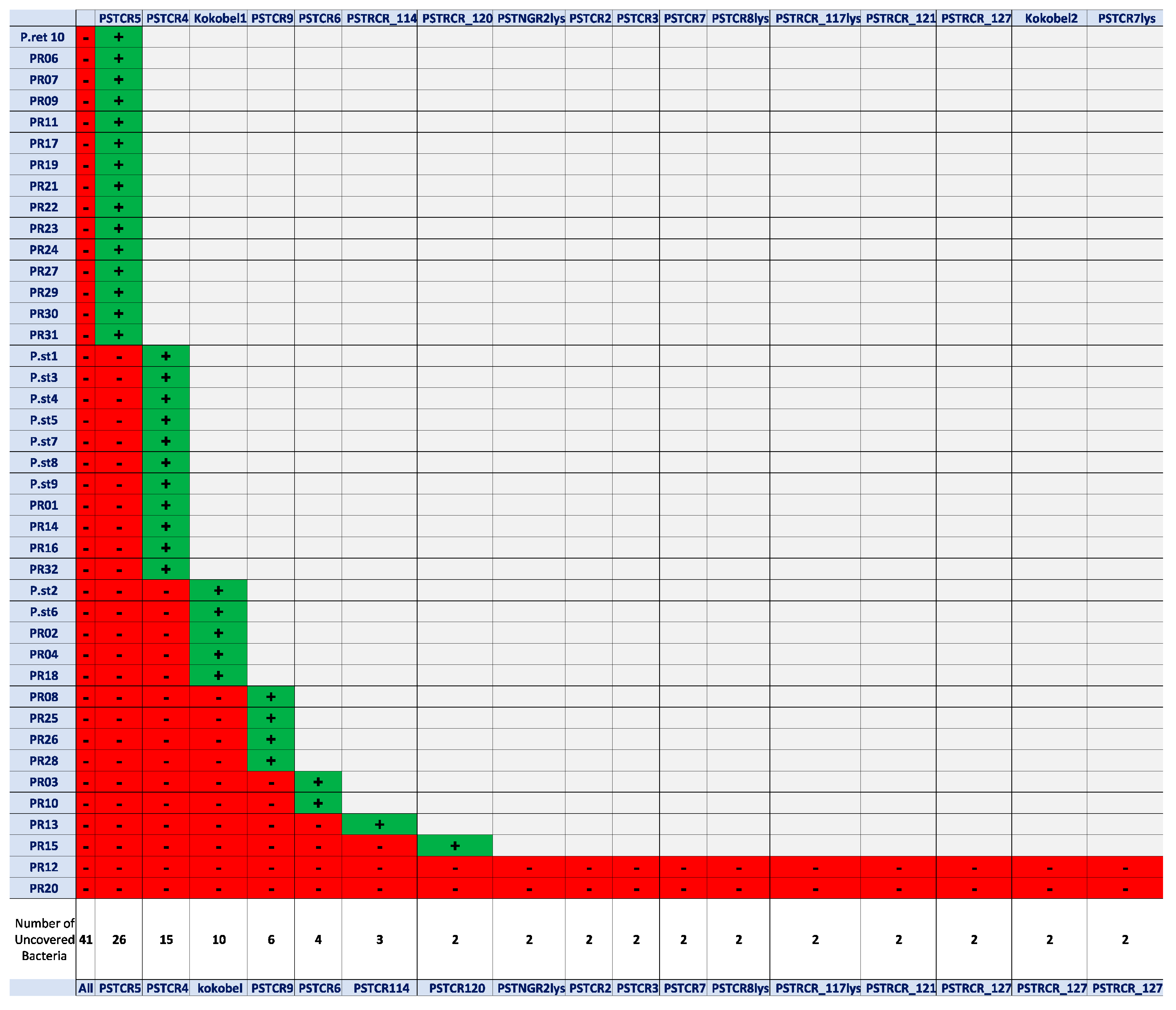
2.1. Phage Isolation and Assessment of Host-Range Coverage

A screening of sewage samples for the presence of potential anti-Providencia species bacteriophages was performed as described in “Materials and Methods” (phage isolation and propagation). After a preliminary enrichment step, and subsequent spotting of the lysate on bacterial lawns, numerous samples resulted in reproducible lysis of bacterial cells and exhibited clear plaques. A total of 17 phages were isolated from single plaques (Table 1) and further propagated in liquid culture as detailed in “Materials and Methods”, showing lysis after an overnight incubation in liquid culture. The phages were evaluated for host range activity by the drop test. As the goal here was to supply a satisfying coverage of Providencia spp., strains that showed sensitivity to one of the phages were excluded from the subsequent screening cycles. The results demonstrated that despite the bacteria’s wide-range antibiotic resistance, the phages targeted 39 out of 41 clinical isolates, providing a coverage of 95% of the isolates tested here (Figure 1).

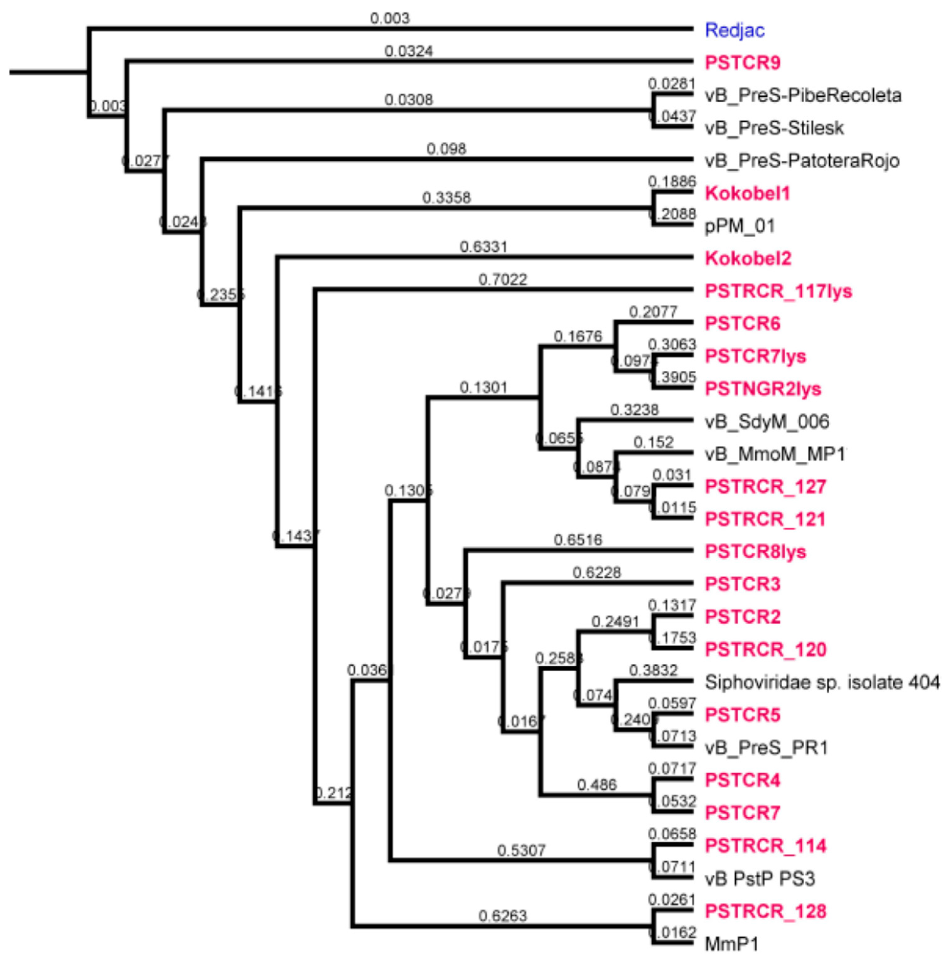
2.2. Genome Analysis

Bacteriophages’ genomes were subsequently sequenced and annotated. Phages’ names, their putative lifecycle type, topology, genome size, phylogeny, and GenBank accession number are denoted in Table 1.
Alignment of the genomes revealed a high diversity between the phages reported here. According to the NCBI database, the lytic phages PSTCR4, PSTCR6, PSTCR7 and Kokobel2 do not have high similarity to known phages. PSTCR4 and PSTCR7 do not overlap any other published genomes (0% query cover), those two phages have high similarity to each other with 93% coverage, 97% identity, and about 9% coverage with Kokobel2. PSTCR6 have only 20% coverage with the known Shigella phage vB_SdyM_006 (MK295204.1). The temperate phage PSTCR7lys which has some similarities with genomic regions of known Providencia strains was found to be unique and it seems that it belongs to a new phage subfamily. Some of the phages have genomic similarity to phages with different hosts; Kokobel1 genome displays similarity to Proteus phages and PSTRCR_121, PSTRCR_127 and PSRCR_128 display similarity to Morganella phages. The phylogenetic tree of our phages compared with the few published Providencia phages and other relevant phages (obtained from the NCBI database) is provided in Figure 2.
As potential candidates for phage therapy treatment, the phages were also scanned for virulence factors and antimicrobial resistance genes using Abricate (https://github.com/tseemann/abricate, release 24, accessed on 28 March 2020) with all its databases: NCBI, CARD, ARG-ANNOT, Resfinder, EcOH, MEGARES, PlasmidFinder, Ecoli_VF and VFDB. The phages were found to be free of virulence factors and antibiotic resistance genes (data not shown).

2.3. Characterization of Phage Lytic Activity

To further test the phages’ applicability against Providencia, lytic activity was assessed on agar plates, planktonic cultures, and biofilm formed on a catheter. The isolate P.st 8 (with its phages PSTCR4 and PSTCR6), was chosen to represent P. stuartii isolates for deeper investigation, since this bacterium was found to be one of the most resistant isolates (8 out of 10 antibiotics, see Table 2 in “Materials and Methods”). P.rett10 (with its phage PSTCR2), was selected arbitrarily, to represent P. rettgeri isolates.
Consistent with the fact that these phages were confirmed to be genotypically distinct from each other, the infectivity patterns of the different phages were found to be also unique phenotypically: bacteriophages (PSTCR4, PSTCR6, and PSTCR2) formed plaques of different sizes and transparencies levels, and some of them had a halo region surrounding the plaque (Figure 3a–c).

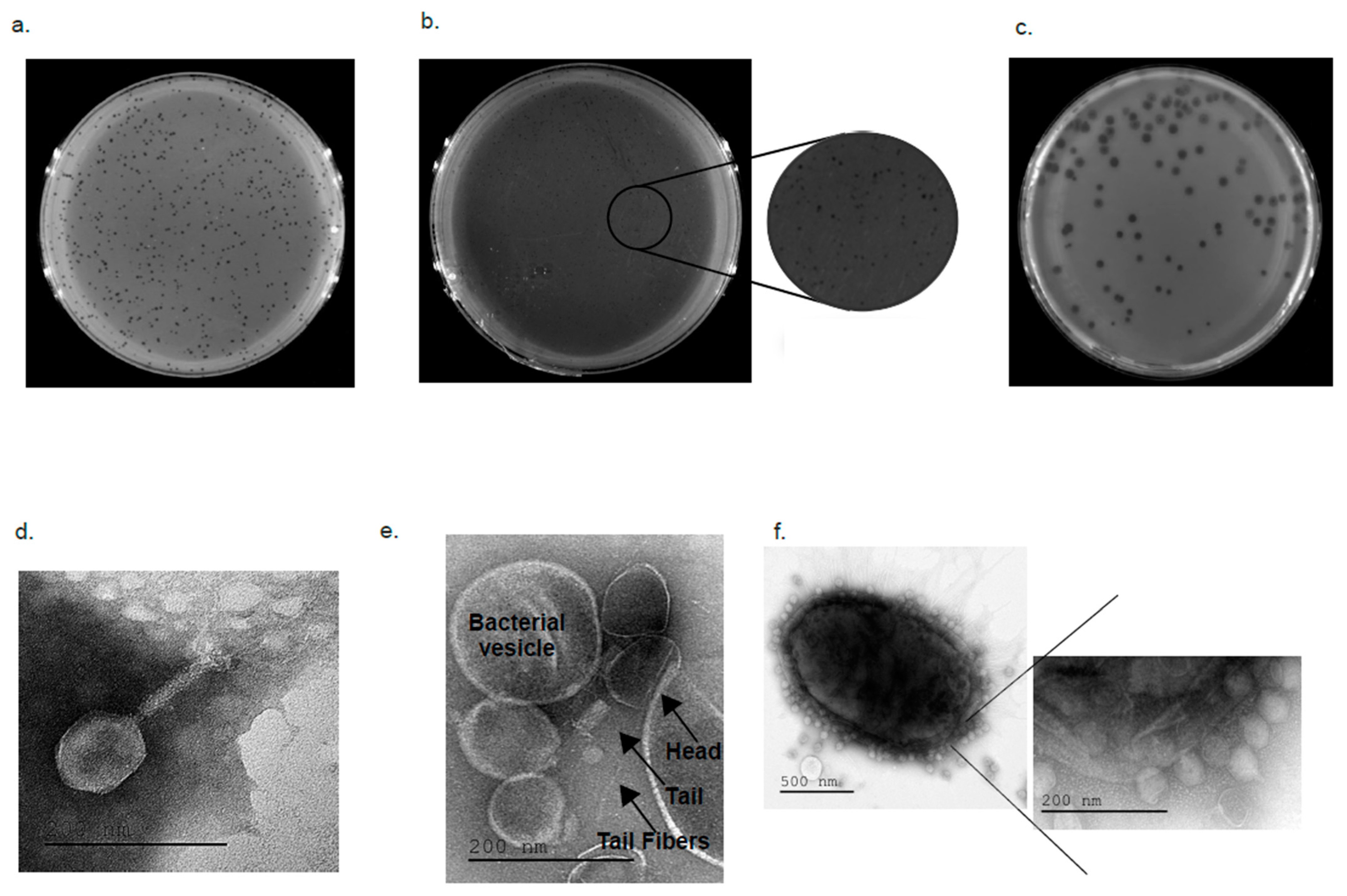
2.4. Electron Microscopy Visualizing

To observe and determine the geometric structure and morphological characteristics of PSTCR4, PSTCR6 and PSTCR2, these phages were visualized under a Transmission Electron Microscope (TEM). TEM microscopy showed that PSTCR4 has a hexagonal capsid with a measured diameter of 105/75 nm and a tail length of 61 nm with long fibers (Figure 3d). PSTCR6 has a hexagonal capsid with a measured diameter of 101/66 nm and a tail length of 60 nm with long-tailed fibers. In most images obtained, phages were attached or were hidden between vesicles secreted by the bacteria. (Figure 3e). PSTCR2 has a hexagonal capsid with a measured diameter of 55/50 nm and a very short tail length of 18 nm (Figure 3f).

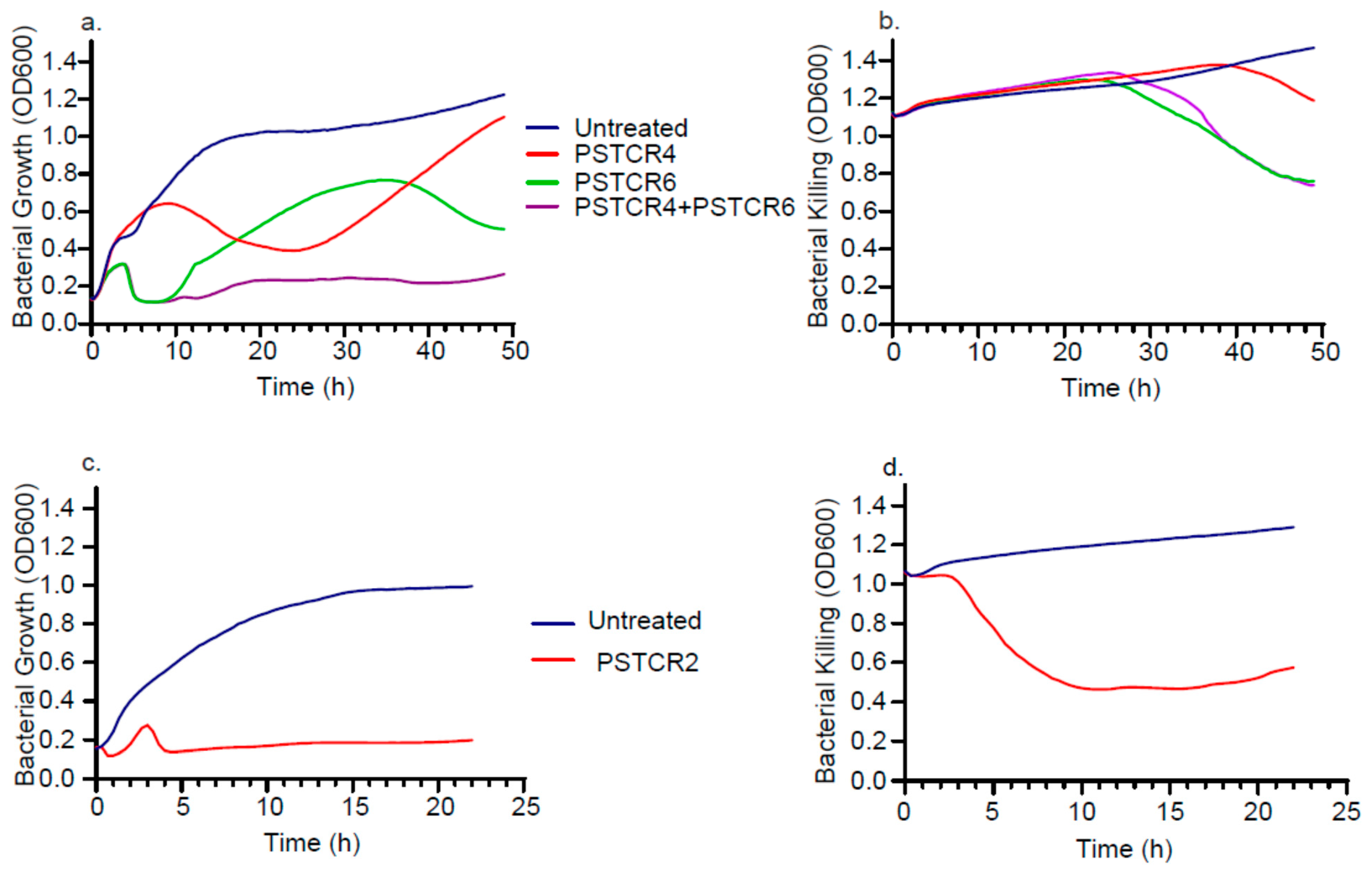
2.5. Kinetic of Lytic Activities in Liquid Culture

Phages also exhibited different inhibition patterns, as observed by the different kinetics of bacterial growth curves in the presence of the different phages: at the P. stuartii logarithmic stage, while treatment of PSCR4 showed an effect on bacterial growth only after 10 h with the best lysis after 23 h followed by a regrowth of the bacteria, PSTCR6 kept bacteria from growing for the first 10 h, followed by a regrowth that was disrupted by another lysis peak towards the stationary stage (Figure 4a). Cultures treated with both phages (PSTCR4 and PSTCR6) displayed a synergistic effect and kept bacteria from growing throughout 50 h of the experiment (Figure 4a). At the stationary phase of P.st 8, PSTCR4 exhibited a slight lytic effect after 40 h of growth, while PSTCR6 acted earlier, after 27 h, and no additive effect was obtained when both phages were added (Figure 4b). Impressive actual lysis of P. rettgeri cultures was observed by PSTCR2 totally inhibiting logarithmic bacterial growth throughout the time of the experiment (Figure 4c) and reducing stationary culture turbidity 2.4-fold (Figure 4d).

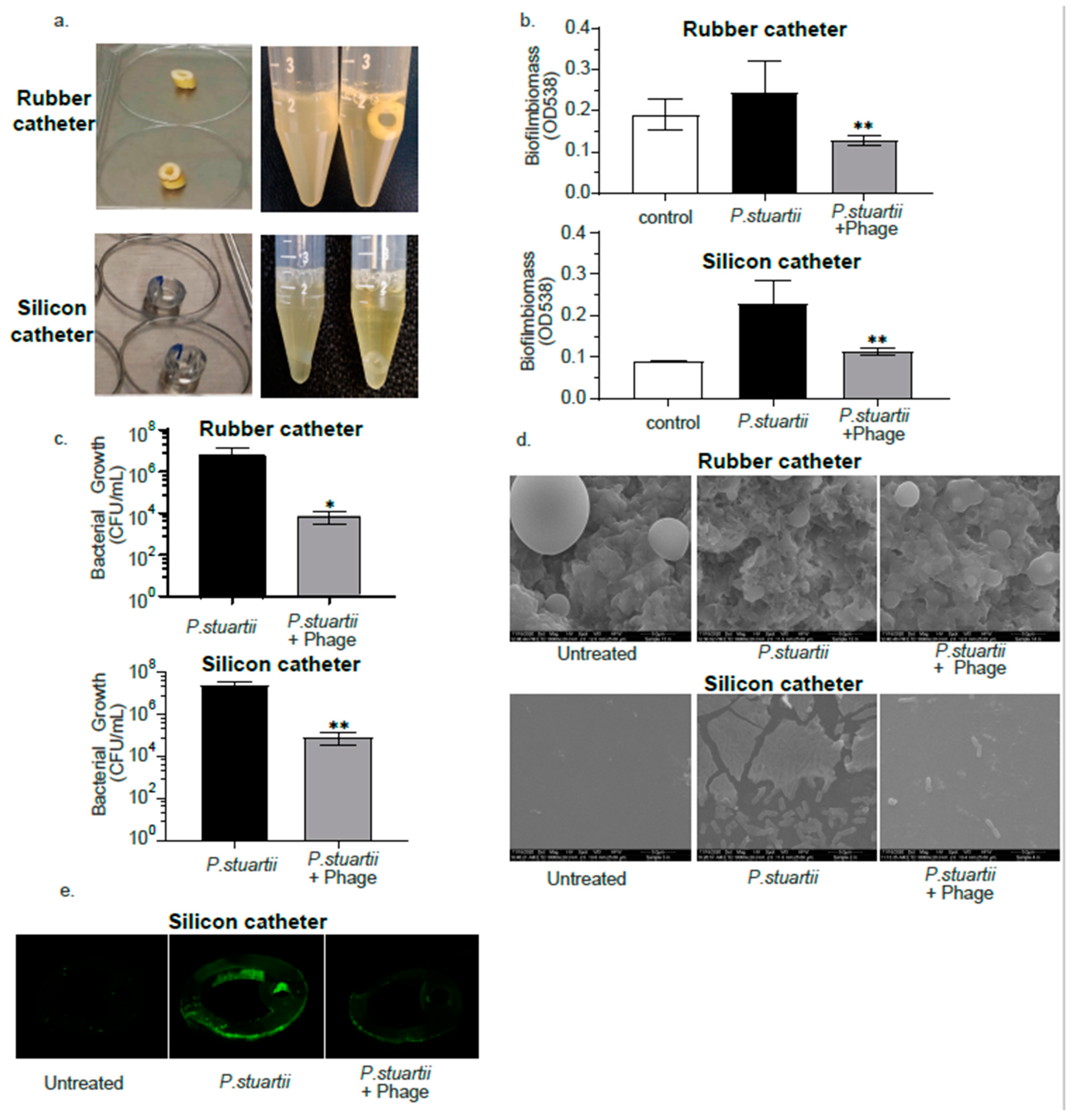
2.6. Biofilm Degradation Activity

Biofilm infections pose a huge challenge to human health as they are very difficult to treat by most antibacterial strategies, including conventional antibiotics [24]. Thus, an assessment of phages’ efficacy against P. stuartii biofilm formed on a catheter was performed using one of the most resistant isolates, P.st 8 and its two lytic phages (PSTCR4 and PSTCR6) as a representative model. Latex and silicone catheters were cut to ring-shaped pieces and infected with bacteria at the logarithmic stage, once the biofilm was established, phage was inoculated and incubated overnight. The cultures which contained phage were less turbid compared to the cultures without phage as can be seen in Figure 5a.
The biofilm degradation activity was assessed by biomass staining, viable counting of bacteria, SEM and confocal microscopy as described in detail in “Materials and Methods”. Crystal violet biofilm biomass evaluation showed a 1.9- and 2-fold reduction in 3-day-old P. stuartii biofilms built on latex or silicone catheters, respectively, following 24 h of treatment, in comparison with the untreated biofilm controls (Figure 5b). To exclude the interference of bacterial debris and to verify the eradication of live bacteria, we further tested the viable counts of the biofilm itself. Biofilms were disrupted by sonication and viable counts of live bacteria were performed. Viable counts showed a 2.86- and 2.46-log growth reduction by phage exposure in latex and silicone catheters, respectively, compared to untreated biofilms (Figure 5c).
To further assess phage efficacy against P. stuartii biofilm, SEM was used to visualize the effect of phage on a 3-day-old biofilm. Figure 5d shows the surface topography of naked latex and silicone catheters and biofilms formed on those catheters. The surface area of the latex catheter appeared to have protrusions and spherical structures. Exposure to phage revealed an extensive effect of bacterial lysis leaving cellular debris and almost no trace of the biofilm. (Figure 5d).
Finally, catheters were stained with the fluorescence stain syto and visualized by a confocal laser microscope. A significant difference was observed in biofilm formed on a silicone catheter treated with phage compared with the untreated controls (Figure 5e). This method was not appropriate for the latex catheters as for an unknown reason, the catheter itself absorbs the stain more efficiently than the biofilm treated with phage (data not shown).

3. Discussion

This article reports the isolation, characterization, and full genome sequencing of 17 novel phages targeting P. stuartii and P. rettgeri clinical isolates, providing a satisfying coverage of 95% of the host strains tested. The phages were active against Providencia isolates across the world, portending for their activity against yet unknown strains.
Some of the phages’ genomes were found to be unique exhibiting distinct differences compared to each other and compared to other known genomes in the NCBI databases. In our experience, this is rather unusual in comparison to other phage-analyses we previously conducted [23].
The extreme specificity of phages requires a personalized-medicine approach. Therefore, characterization of all 17 phages with all 41 bacterial hosts is tedious and irrelevant. When a candidate patient is ready for treatment, we screen all our phages to assess their killing abilities and characterize the most suitable one on the patient’s specific isolate. Therefore, here, characterization was carried out on representative phage and hosts. In vitro characterization of three of the lytic phages (PSTCR2, PSTCR4 and PSTCR6) showed killing capabilities in solid and liquid cultures in various patterns and levels of efficiencies.
Consistent with the results for the planktonic cultures, the phages PSTCR4 and PSTCR6 exhibited an efficient reduction of well-established P. stuartii biofilms formed in catheter models using various methods. These results, together with the finding that the phages are free of virulence factors and resistance genes, suggest the potential of those candidate phages as reliable therapeutic agents for phage therapy against P. stuartii biofilms in bacteriuric catheterized patients.
Nevertheless, as the ability to disrupt biofilms is not obvious for any phage, we postulate that the phages’ ability to remove biofilm should also be tested by adding small volumes of phage to the stationary culture in which the biofilm is formed, or alternatively to dialyze the phage suspension before inoculation. This is due to the possibility that additional nutrients that remain in the phage media might trigger bacteria in a biofilm state to switch to logarithmic growth. These circumstances might accentuate the killing effect of most phages.
Further steps should be taken to promote phage therapy against biofilms. These include: developing fast and accurate techniques for the prediction and matching of phages against biofilms, and establishing a large collection of anti-biofilm specialized phages by selective enrichment. Such improvement might have extensive clinical implications.
Though Providencia spp. are not defined yet as part of the ESCAPE group of challenging antibiotic resistant pathogens [25], the overuse of wide-spectrum antibiotics may result in the emergence of extreme resistance patterns and lead to more nosocomial outbreaks, which explains the growing concern among health professionals.
Currently, there are not enough available effective options to fight cases of Providencia biofilms infection: an extremely limited number of phages targeting P. stuartii and P. rettgeri were isolated, and no phage therapy was applied using Providencia phages. Thus, the results presented here open new avenues for bacteriuric long-term catheterized patients, and for the phage therapy field.
In conclusion, our results indicate that, though resistant to antibiotics, Providencia strains might be sensitive to phages, a promising direction to consider in the continuous struggle against the current emergence of multidrug resistant Providencia spp.

4. Materials and Methods

4.1. Bacterial Strains

In this work, 41 clinical isolates of Providencia stuartii and Providencia rettgeri were used (Table 2). A total of 10 of them were isolated locally, at the clinical microbiology lab, Hadassah-Hebrew University Medical Center, Jerusalem, Israel, and 31 strains were provided by the Center for Innovative Phage Applications and Therapeutics (IPATH) of the University of California, San Diego. Bacterial strains were grown in LB broth (Difco, Sparks, MD, USA) at 37 °C shaken on a rotary shaker at 200 rpm under aerobic conditions. Bacterial resistance or sensitivity to antibiotics was determined by the infectious disease unit of Hadassah Hospital, Jerusalem using VITEK (BioMérieux, Carponne, France).

4.2. Phage Isolation and Propagation

Isolation of phages was performed using the standard double-layer agar method previously described [26] with mild modifications. Preliminary enrichment of phages was carried out by overnight growth of the bacteria in an environmental sewage sample collected from various places in Jerusalem and the surrounding. Samples were centrifuged at 7800 rpm for 10 min (centrifuge 5430R, rotor FA-45-24-11HS; Eppendorf, Hamburg, Germany). The supernatant was filtered through 0.22-μm-pore-size filters (Merck-Millipore, Cork, Ireland) and LB 5X was then added to the filtered sewage. Grown bacterial cultures of Providencia stuartii and Providencia rettgeri were mixed and inoculated with filtered sewage and LB overnight at 37 °C shaken on a rotary shaker at 200 rpm under aerobic conditions. Following inoculation, the liquid was centrifuged and re-filtered through 0.22-μm-pore-size filters. The filtered lysate was then spotted on double-layered agarose plates which were incubated overnight at 37 °C. Double-layered agarose plates were prepared by covering LB plates with 3.5 mL of pre-warmed agarose (0.25%) containing 0.1 mL of an overnight culture of bacteria (Table 2). Clear plaques were observed and purified by a minimum of four passages (of repeatedly picking single plaques and spreading them on a new bacterial lawn). Finally, single plaques were collected and transferred into bacterial liquid cultures to collect high titer lysates, which were then filtered and stored at 4 °C. The concentration of the phages was determined by counting plaque-forming units (PFU) as described [26]. Briefly, the lysate was serially diluted 10-fold into LB broth, and drops of 5 µL were spotted on overlaid tested bacterial strain lawns of agarose (0.25%), and then incubated overnight at 37 °C. The plaques were counted, and the initial concentration of PFU/mL was calculated.

4.3. Phgae Genome Sequencing

The phages’ DNA was purified as described [27] using a phage DNA isolation kit (Norgen-Biotek, Thorold, Canada), and libraries were prepared with a Nextera XT DNA kit (catalog number FC-131-1096; Illumina, San Diego, CA, USA). Normalization, pooling, and tagging were performed in a common flow cell with 1 × 150 bp paired-end reads, which were used for sequencing with the Illumina NextSeq 500 platform. Sequencing was performed in the sequencing unit of The Hebrew University of Jerusalem at the Hadassah Campus. Trimming, quality control, assembly of reads, and analyses were performed using Geneious Prime and its plugins (https://www.geneious.com, accessed on 1 March 2021). Assembly was commonly performed using the SPAdes plugin of Geneious Prime. Annotation was performed using the RAST version 2 (https://rast.nmpdr.org/rast.cgi, accessed on 1 March 2021), PHAge Search Tool Enhanced Release (PHASTER) (https://phaster.ca, accessed on 1 March 2021), and BLAST servers.

4.4. Bacteriophages for Phylogenetic Tree

The genomes of published phages used for comparison are listed in Table 3.

4.5. TEM Visualization

To visualize the geometric structure of the phages, transmission electron microscopy (TEM) was used as described in OpenWetWare (http://openwetware.org/wiki/Gill:Preparingphage_specimens_for_TEM, accessed on 7 December 2017). Two ml of phage lysate with 109 PFU/mL were centrifuged at 14,000 rpm (centrifuge 5430R, rotor FA-45-24-11HS; Eppendorf) for 2 h at room temperature. The pellet was resuspended in 0.2 mL of 5 mM MgSO4 and incubated overnight at 4 °C. To visualize the phage–bacteria interactions, 1 mL of lysate with 108 PFU/mL was inoculated with 100 μL of overnight grown bacteria for 15 min. Then, 100 μL of the bacteria and phage mix were transferred into 100 μL of 5 mM MgSO4. On a strip of parafilm, 0.03 mL of 5 mM MgSO4 and 10 μL of the phage sample were mixed gently. Grids were prepared by placing on the drop of phage sample with the carbon side facing down for a minute, and then stain with a drop of uranyl acetate (2%) for another minute. After drying, a transmission electron microscope (Jeol, TEM 1400 plus, Peabody, MA, USA) with a charge-coupled device camera (Jeol, Gatan Orius 600) was used to capture images.

4.6. Assessment of Phage Lytic Activity in Planktonic Cultures

Lytic activity of the phage was assessed by inoculation of logarithmic or stationary Providencia cultures with phages in triplicates. The growth and lysis kinetics were recorded by measuring the optical density at a wavelength of 600 nm at 37 °C with 5 s shaking every 20 min using a 96-well plate reader (Synergy; BioTek, Winooski, VT, USA).

4.7. Assessment of Phage Lytic Activity in Biofilm

The 100% Latex (silicon coated) or silicone catheters (Foley Catheter) at a thickness of 16 Fr/Ch and volume of 5–15 mL were cut into ring-like pieces of ~2–3 mm width. The catheter pieces were placed in 15 mL tubes containing 3 mL LB broth with 0.1 mL of an overnight culture of bacteria. Biofilms were grown for 4 days at 37 °C shaken on a rotary shaker at 200 rpm under aerobic conditions. After transferring the catheter to a new tube and inoculation of the phage, incubation was continued overnight. Biofilms built on latex catheters were infected with PSTCR4 phage and biofilms built on silicone catheters were infected with a cocktail of PSTCR4 and PSTCR6 at the same concentration.
The catheter pieces were washed with phosphate-buffered saline (PBS), and the biomass was quantified using crystal violet staining as previously described [28]. Briefly, fixation was achieved by soaking the catheter in methanol, followed by incubation for 20 min, followed in turn by methanol aspiration and air drying. The biofilms were stained by crystal violet (1%) for 20 min at room temperature and then washed with water. The stain was extracted by adding ethanol (100%) and biomass was quantified by determining the optical density at 538 nm (OD538). In addition, the biofilm on the catheter was stained using the live stain (syto) of the Live/Dead cell viability kits (Life Technologies, Waltham, MA, USA) according to the manufacturer’s instructions. The fluorescence emission of the biofilms was detected using a confocal laser microscope (Nikon Yokogaha W1 spinning disk, Tokyo, Japan). The microscopy slices were combined to a 3D image using NIS program. For viable count, each catheter piece was transferred to 1 mL of water in 1.5 mL tubes and placed in a sonicating water bath (Bandelin Sonopuls HD 2200, Berlin, Germany) for 7 min to disrupt the biofilm, and 10 µL of 10-fold serial dilutions of each sample were plated on LB agar plates. Colonies were counted after 24 h at 37 °C and the number of colony-forming units (CFU)/mL was calculated.

4.8. Statistical Analysis

Prism (GraphPad, version 8.0.2 (263), San Diego, CA, USA) was used for the statistical analysis, graph formation, and design. The results were analyzed as the mean ± standard deviation in each experimental group. Statistical significance was calculated by a Student’s t-test two-tailed unpaired p values (significance level: p < 0.05).

Author Contributions

Conceptualization and experiment design: C.R., S.B.P., S.C.-G., R.N.-P. and R.H. Performing experiment: C.R., S.B.P., O.Y., M.A. and N.G. Bio-informatic analysis: S.A.-O. and R.H. Bacterial strains supply: L.H., D.P. and R.N.-P. Writing manuscript: C.R. and R.H. Editing: S.A.-O., S.C.-G., L.H., D.P. and R.N.-P. All authors have read and agreed to the published version of the manuscript.

Funding

This research was funded by: “United States–Israel Binational Science Foundation (BSF)” grant #2017123, The Israel Science Foundation (ISF) IPMP grant #ISF1349/20, The Rosetrees Trust grant A2232, The Milgrom Family Support Program.

Institutional Review Board Statement

Not applicable.

Informed Consent Statement

Not applicable.

Data Availability Statement

Sequences are available in NCBI GenBank (www.ncbi.nlm.nih.gov/genbank, accessed on 1 March 2021).

Acknowledgments

The authors would like to thank the “United States–Israel Binational Science Foundation (BSF)” grant #2017123, the Israel Science Foundation (ISF) IPMP grant #ISF1349/20, the Rosetrees Trust grant A2232, and the Milgrom Family Support Program. We would like also to thank the dedicated team of the Facility Core of Hadassah Campus of the Hebrew University for their help in sequencing and electron microscopy and Hanan Levi from the ‘Hagihon’ company, Jerusalem Region Water and Wastewater Utility, for supplying sewage samples.

Conflicts of Interest

The authors declare no conflict of interest

References

  1. O’Hara, C.M.; Brenner, F.W.; Miller, J.M. Classification, identification, and clinical significance of Proteus, Providencia, and Morganella. Clin. Microbiol. Rev. 2000, 13, 534–546. [Google Scholar] [CrossRef] [PubMed]
  2. Washington, J.A.; Senjem, D.H.; Haldorson, A.; Schutt, A.H.; Martin, W.J. Nosocomially Acquired Bacteriuria Due to Proteus Rettgeri and Providencia Stuartii. Am. J. Clin. Pathol. 1973, 60, 836–838. [Google Scholar] [CrossRef] [PubMed]
  3. Warren, J.W. Providencia Stuartii: A Common Cause of Antibiotic-Resistant Bacteriuria in Patients with Long-Term Indwelling Catheters. Rev. Infect. Dis. 1986, 8, 61–67. [Google Scholar] [CrossRef]
  4. Choi, H.K.; Kim, Y.K.; Kim, H.Y.; Park, J.E.; Uh, Y. Clinical and microbiological features of providencia bacteremia: Experience at a tertiary care hospital. Korean J. Intern. Med. 2015, 30, 219–225. [Google Scholar] [CrossRef] [PubMed]
  5. Molnár, S.; Flonta, M.M.M.; Almaş, A.; Buzea, M.; Licker, M.; Rus, M.; Földes, A.; Székely, E. Dissemination of NDM-1 carbapenemase-producer Providencia stuartii strains in Romanian hospitals: A multicentre study. J. Hosp. Infect. 2019, 103, 165–169. [Google Scholar] [CrossRef] [PubMed]
  6. Hayakawa, K.; Marchaim, D.; Divine, G.W.; Pogue, J.M.; Kumar, S.; Lephart, P.; Risko, K.; Sobel, J.D.; Kaye, K.S. Growing prevalence of Providencia stuartii associated with the increased usage of colistin at a tertiary health care center. Int. J. Infect. Dis. 2012, 16, e646–e648. [Google Scholar] [CrossRef] [PubMed] [Green Version]
  7. Sagar, S.; Narasimhaswamy, N.; d’Souza, J. Providencia Rettgeri: An Emerging Nosocomial Uropathogen in an Indwelling Urinary Catheterised Patient. J. Clin. Diagn. Res. 2017, 11, DD01. [Google Scholar] [CrossRef] [PubMed]
  8. Zavascki, A.P.; Carvalhaes, C.G.; da Silva, G.L.; Soares, S.P.T.; de Alcåntara, L.R.; Elias, L.S.; Sandri, A.M.; Gales, A.C. Outbreak of Carbapenem-Resistant Providencia stuartii in an Intensive Care Unit. Infect. Control Hosp. Epidemiol. 2012, 33, 627–630. [Google Scholar] [CrossRef] [PubMed] [Green Version]
  9. Rus, M.; Licker, M.; Musuroi, C.; Seclaman, E.; Muntean, D.; Cirlea, N.; Tamas, A.; Vulpie, S.; Horhat, F.G.; Baditoiu, L. Distribution of NDM1 carbapenemase-producing proteeae strains on high-risk hospital wards. Infect. Drug Resist. 2020, 13, 4751–4761. [Google Scholar] [CrossRef]
  10. El Khatib, M.; Tran, Q.T.; Nasrallah, C.; Lopes, J.; Bolla, J.M.; Vivaudou, M.; Pagès, J.M.; Colletier, J.P. Providencia stuartii form biofilms and floating communities of cells that display high resistance to environmental insults. PLoS ONE 2017, 12, e0174213. [Google Scholar] [CrossRef]
  11. Stickler, D.J. Bacterial biofilms in patients with indwelling urinary catheters. Nat. Clin. Pract. Urol. 2008, 5, 598–608. [Google Scholar] [CrossRef] [PubMed]
  12. Fletcher, M.; Oppenheimer, S.R.; Warren, J.W. Colonization of urinary catheters by Escherichia coli and providencia stuartii in a laboratory model system. J. Urol. 1994, 152, 232–236. [Google Scholar] [CrossRef]
  13. Fierer, J.; Ekstrom, M. An Outbreak of Providencia stuartii Urinary Tract Infections: Patients with Condom Catheters Are a Reservoir of the Bacteria. JAMA J. Am. Med. Assoc. 1981, 245, 1553–1555. [Google Scholar] [CrossRef]
  14. Tran, Q.T.; Mahendran, K.R.; Hajjar, E.; Ceccarelli, M.; Davin-Regli, A.; Winterhalter, M.; Weingart, H.; Pagès, J.M. Implication of porins in β-lactam resistance of Providencia stuartii. J. Biol. Chem. 2010, 285, 32273–32281. [Google Scholar] [CrossRef] [Green Version]
  15. Stock, I.; Wiedemann, B. Natural antibiotic susceptibility of providencia stuartii, P. rettgeri, P. alcalifaciens and P. rustigianii strains. J. Med. Microbiol. 1998, 47, 629–642. [Google Scholar] [CrossRef] [PubMed]
  16. McHale, P.J.; Keane, C.T.; Dougan, G. Antibiotic resistance in Providencia stuartii isolated in hospitals. J. Clin. Microbiol. 1981, 13, 1099–1104. [Google Scholar] [CrossRef] [PubMed] [Green Version]
  17. Tshisevhe, V.S.; Lekalakala, M.R.; Tshuma, N.; Janse van Rensburg, S.; Mbelle, N. Outbreak of carbapenem-resistant Providencia rettgeri in a tertiary hospital. S. Afr. Med. J. 2016, 107, 31–33. [Google Scholar] [CrossRef] [PubMed] [Green Version]
  18. Kurmasheva, N.; Vorobiev, V.; Sharipova, M.; Efremova, T.; Mardanova, A. The Potential Virulence Factors of Providencia stuartii: Motility, Adherence, and Invasion. Biomed Res. Int. 2018, 2018. [Google Scholar] [CrossRef] [Green Version]
  19. Lin, D.M.; Koskella, B.; Lin, H.C. Phage therapy: An alternative to antibiotics in the age of multi-drug resistance. World J. Gastrointest. Pharmacol. Ther. 2017, 8, 162. [Google Scholar] [CrossRef] [PubMed]
  20. Hanlon, G.W. Bacteriophages: An appraisal of their role in the treatment of bacterial infections. Int. J. Antimicrob. Agents 2007, 30, 118–128. [Google Scholar] [CrossRef]
  21. Oliveira, H.; Pinto, G.; Hendrix, H.; Noben, J.P.; Gawor, J.; Kropinski, A.M.; Łobocka, M.; Lavigne, R.; Azeredo, J. A lytic Providencia rettgeri virus of potential therapeutic value is a deepbranching member of the T5virus genus. Appl. Environ. Microbiol. 2017, 83. [Google Scholar] [CrossRef] [PubMed] [Green Version]
  22. Oliveira, H.; Pinto, G.; Mendes, B.; Dias, O.; Hendrix, H.; Akturk, E.; Noben, J.P.; Gawor, J.; Łobocka, M.; Lavigne, R.; et al. A tailspike with exopolysaccharide depolymerase activity from a new Providencia stuartii phage makes multidrug-resistant bacteria susceptible to serum-mediated killing. Appl. Environ. Microbiol. 2020, 86. [Google Scholar] [CrossRef] [PubMed]
  23. Yerushalmy, O.; Khalifa, L.; Gold, N.; Rakov, C.; Alkalay-Oren, S.; Adler, K.; Ben-Porat, S.; Kraitman, R.; Gronovich, N.; Ginat, K.S.; et al. The israeli phage bank (IPB). Antibiotics 2020, 9, 269. [Google Scholar] [CrossRef] [PubMed]
  24. Clokie, M.R.J.; Kropinski, A.M. Bacteriophage: Methods and Protocols Volume 1: Isolation, Characterization, and Interactions; Springer: New York, NY, USA, 2009; ISBN 9781588296825. [Google Scholar]
  25. Clokie, M.R.J.; Kropinski, A.M. Bacteriophages Methods and Protocols Volume 2: Molecular and Applied Aspects; Springer: New York, NY, USA, 2009; ISBN 978-1-60327-564-4. [Google Scholar]
  26. Stepanović, S.; Vuković, D.; Dakić, I.; Savić, B.; Švabić-Vlahović, M. A modified microtiter-plate test for quantification of staphylococcal biofilm formation. J. Microbiol. Methods 2000, 40, 175–179. [Google Scholar] [CrossRef]
  27. De la Fuente-Núñez, C.; Reffuveille, F.; Fernández, L.; Hancock, R.E.W. Bacterial biofilm development as a multicellular adaptation: Antibiotic resistance and new therapeutic strategies. Curr. Opin. Microbiol. 2013, 16, 580–589. [Google Scholar] [CrossRef]
  28. Rice, L.B. Federal funding for the study of antimicrobial resistance in nosocomial pathogens: No ESKAPE. J. Infect. Dis. 2008, 197, 1079–1081. [Google Scholar] [CrossRef]
Figure 1. Reduction screen. The bacterial isolates killed by each noted phage (green), and those that remained for the next screen (red) after phage exposure.
Antibiotics 10 00375 g001
Figure 2. Genome analysis of Providencia phages. Phylogenetic tree of Providencia phages. Phages of this work are shown in red. Close-similar genomes of fully sequenced phages from the NCBI database are given as reference points shown in black.
Antibiotics 10 00375 g002
Figure 3. Plaque morphologies and TEM microscopy of anti-Providencia phages. Single plaques of PSTCR4 (a), PSTCR6 (b) on P.st 8, and PSTCR2 (c) on P. rett10 on bacterial lawns. Geometric structure of PSTCR4 (d); PSTCR6 (e); and PSTCR2 attached to its host P. rett10 (f).
Antibiotics 10 00375 g003
Figure 4. Characterization of lytic activity in liquid cultures. Kinetic growth and killing curves of P.st 8 (blue) at the (a) logarithmic and (b) stationary stage in the presence of the phages PSTCR4 (red), PSTCR6 (green) and both of them (purple); growth curves of P.rett10 (blue) at the logarithmic (c) and killing curves of the stationary (d) stage in presence of the phage PSTCR2 (red).
Antibiotics 10 00375 g004
Figure 5. Biofilm degradation activity. (a) Model of P. stuartii biofilm formed on Latex and Silicone catheters in the absence (left tube) and presence (right tube) of phage. (b) Biofilm biomass quantification by crystal violet staining of P. stuartii formed on latex and silicone catheters; (c) viable colony-forming unit (CFU) counts of disrupted P. stuartii biofilms formed on latex and silicone catheters. (d) Scanning Electron Microscopy (SEM) of untreated, P.st 8 biofilm infected and phage-treated latex and silicone catheter surfaces; (e) confocal microscopy of untreated, P.st 8 biofilm infected and phage-treated silicone catheter biofilms. Asterisk (*, **) denotes p-value < 0.05 and <0.001 respectively.
Antibiotics 10 00375 g005
Table 1. Phage collection.
PhagePutative Life-Cycle Type 1Genome Organization 2Genome Size (bp)PhylogenyGenBank Accession
Kokobel1lyticcircular59,837SiphoridaeMW145139.1
Kokobel2lyticcircular45,880SiphoridaeMW145138.1
PSTNGR2lystemperatecircular50,958SiphoridaeMW145137.1
PSTCR2lyticcircular40,200AutographiviridaeMW057854.1
PSTCR3temperatecircular39,447MyoviridaeMW057855.1
PSTCR4lyticcircular57,214SiphoridaeMW057856.1
PSTCR5lyticcircular109,434SiphoridaeMW057857.1
PSTCR6lyticcircular155,737MyoviridaeMW057858.1
PSTCR7lyticcircular57,986SiphoviridaeMW057861.1
PSTCR7lystemperate—partial genomelinear22,938SiphoridaeMW057862.1
PSTCR8lystemperatelinear40,280SiphoridaeMW057859.1
PSTCR9lyticcircular58,873SiphoviridaeMW057860.1
PSTRCR_114lyticcircular42,750AutographiviridaeMW358930.1
PSTRCR_117lystemperatelinear41,059SiphoviridaeMW358929.1
PSTRCR_120lyticcircular39,396Autographiviridae;MW358928.1
PSTRCR_121lyticcircular165,776Myoviridae;MW385300.1
PSTRCR_127lyticcircular166,815Myoviridae;MW358927.1
1 The determination of lytic/temperate is based on the BLAST database. Cases where the full genome of the phage was found to be harbored by bacterial chromosome, served as an indication of lysogeny and were defined as temperate. Another indication for lysogeny is the similarities of the phage genes to elements involved in lysogenicity such as a lambda CI repressor, integrase, or transposon-like elements. 2 Linear organization refers to the constant end of the assembled genome and circular when terminal redundancy of reads was observed.
Table 2. Bacterial strains, their body site source, the provider of the isolates and antibiotic resistance.
Bacterial StrainsSourceProvided by 1Antibiotic Resistance 2
Providencia stuartii
P.st 1bloodHMCAMP, SAM, CRO, CXM, GEN, CFZ
P.st 2bloodHMCAMP, SAM, CIP, CRO, CXM, GEN, CFZ
P.st 3bloodHMCAMP, SAM, CIP, AMK, CRO, CXM, GEN, CFZ
P.st 4bloodHMCAMP, SAM, CRO, CXM, GEN, CFZ
P.st 5bloodHMCAMP, SAM, CIP, AMK, CRO, CXM, GEN, CFZ
P.st 6bloodHMCAMP, SAM, CIP, AMK, CRO, CXM, GEN, CFZ
P.st 7bloodHMCAMP, SAM, CIP, CXM, GEN, CFZ
P.st 8bloodHMCAMP, SAM, CIP, AMK, CXM, CRO, GEN, CFZ
P.st 9bloodHMCAMP, GEN
PR02urineUCSDAMP, SAM, CXM, GEN, CFZ
PR03urineUCSDAMP, SAM, CXM, GEN, CFZ
PR04urineUCSDAMP, SAM, AMK, CXM, GEN, CFZ
PR12leg lesionUCSDAMP, SAM, CIP, CFZ
PR13leg lesionUCSDAMP, SAM, CRO, CFZ
PR14urineUCSDAMP, SAM, CFZ
PR15bloodUCSDAMP, SAM, CIP
PR16urineUCSD
PR25bloodUCSDAMP, CXM, GEN
PR26bloodUCSDAMP, CXM, GEN
PR32urine catheterUCSDAMP, GEN, CFZ
Providencia rettgeri
P. rett10bloodHMCAMP, GEN
PR01breastUCSDAMP, SAM, GEN, CFZ
PR06bloodUCSDSAM, CFZ
PR07urineUCSDAMP, SAM, CFZ
PR08sputumUCSDAMP, SAM, CFZ
PR09urineUCSDAMP, SAM, CRO, CFZ
PR10heel lesionUCSDAMP, SAM, CIP, CFZ
PR11lesionUCSDAMP, SAM, CFZ
PR17foot cultureUCSDAMP, SAM
PR18urineUCSDAMP, SAM
PR19urineUCSDAMP, GEN
PR20body site, sacrumUCSD
PR21body tissue, footUCSDAMP, GEN
PR22urineUCSDAMP, SAM, GEN
PR23urineUCSDAMP, GEN
PR24urineUCSDAMP, GEN
PR27body site, bladder stoneUCSDAMP, CXM, GEN
PR28bloodUCSDAMP, CXM, GEN
PR29urineUCSDAMP, CXM, GEN
PR30sputumUCSDAMP, GEN
PR31nasopharyngealUCSDAMP, GEN
1 HMC stands for Hadassah Medical center, and UCSD stands for University of California San Diego. 2 The antibiotics names are given in the table as abbreviations: AMP stands for ampicillin, SAM for ampicillin/sulbactam, CIP for ciprofloxacin, MEM for meropenem, ETP for ertapenem, GEN for gentamicin, CXM for ceftriaxone, CRO for cefuroxime, AMK for amikacin, and CFZ for cefazolin.
Table 3. Published genomes.
TypePhage NameAccession Number
Providencia phageRedjacNC_018832.1
Providencia phage (P. stuartii)vB PstP PS3NC_048148.1
Providencia phage (P. stuartii)vB_PstP_StuartMK387869.1
Providencia phage (P. rettgeri)vB_PreS_PR1NC_041913.1
Providencia phage (P. rettgeri)vB_PreS-PatoteraRojoMT675126.1
Providencia phage (P. rettgeri)vB_PreS-StileskMT675125.1
Providencia phage (P. rettgeri)vB_PreS-PibeRecoletaMT675124.1
Morganella phageMmP1NC_011085.3
Morganella phagevB_MmoM_MP1NC_031020.1
Proteus phagepPM_01NC_028812.1
Shigella phagevB_SdyM_006MK295204.1
Siphoviridae sp. isolate 404MN855780.1
Publisher’s Note: MDPI stays neutral with regard to jurisdictional claims in published maps and institutional affiliations.

