Article

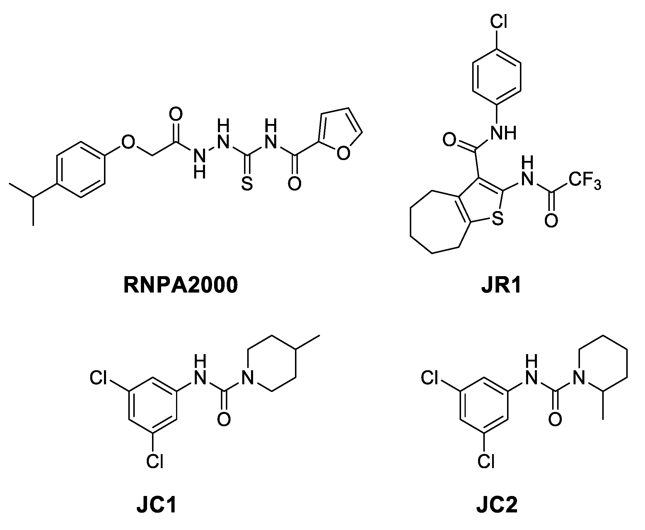
Staphylococcus aureus RnpA Inhibitors: Computational-Guided Design, Synthesis and Initial Biological Evaluation

by
Lorenzo Suigo
1,
Michaelle Chojnacki
2,
Carlo Zanotto
3,
Victor Sebastián-Pérez
4,
Carlo De Giuli Morghen
5,
Andrea Casiraghi
1,
Paul M. Dunman
2,
Ermanno Valoti
1 and
Valentina Straniero
1,*
1
Dipartimento di Scienze Farmaceutiche, Università degli Studi di Milano, Via Luigi Mangiagalli 25, 20133 Milano, Italy
2
Department of Microbiology and Immunology, University of Rochester Medical Center, 601 Elmwood Ave., Rochester, NY 14642, USA
3
Dipartimento di Biotecnologie Mediche e Medicina Traslazionale, Università degli Studi di Milano, Via Vanvitelli 32, 20129 Milano, Italy
4
Exscientia, The Schrödinger Building, Oxford Science Park, Oxford OX4 4GE, UK
5
Department of Chemical—Pharmaceutical and Biomolecular Technologies, Catholic University “Our Lady of Good Counsel”, Rr. Dritan Hoxha, 1025 Tirana, Albania
*
Author to whom correspondence should be addressed.
Submission received: 12 March 2021 / Revised: 12 April 2021 / Accepted: 12 April 2021 / Published: 14 April 2021
(This article belongs to the Special Issue FtsZ and RnpA as Valid Targets while Developing Novel Antimicrobials)

Abstract

:
Antibiotic resistance is spreading worldwide and it has become one of the most important issues in modern medicine. In this context, the bacterial RNA degradation and processing machinery are essential processes for bacterial viability that may be exploited for antimicrobial therapy. In Staphylococcus aureus, RnpA has been hypothesized to be one of the main players in these mechanisms. S. aureus RnpA is able to modulate mRNA degradation and complex with a ribozyme (rnpB), facilitating ptRNA maturation. Corresponding small molecule screening campaigns have recently identified a few classes of RnpA inhibitors, and their structure activity relationship (SAR) has only been partially explored. Accordingly, in the present work, using computational modeling of S. aureus RnpA we identified putative crucial interactions of known RnpA inhibitors, and we used this information to design, synthesize, and biologically assess new potential RnpA inhibitors. The present results may be beneficial for the overall knowledge about RnpA inhibitors belonging to both RNPA2000-like thiosemicarbazides and JC-like piperidine carboxamides molecular classes. We evaluated the importance of the different key moieties, such as the dichlorophenyl and the piperidine of JC2, and the semithiocarbazide, the furan, and the i-propylphenyl ring of RNPA2000. Our efforts could provide a foundation for further computational-guided investigations.

1. Introduction

The well-documented consequences of antimicrobial resistance have been exacerbated by the limited pharmaceutical investment in new molecules [1], together with antibiotic over-prescription and misuse [2]. Indeed, antimicrobial resistance is currently surging in every country of the world, as reported by the World Health Organization (WHO) [3]. In this context, the resistance of several pathogens, such as Klebsiella pneumoniae, Escherichia coli, Mycobacterium tuberculosis, and Plasmodium falciparum, to both front line and antibiotics of last resort, are progressively leading to higher incidences of morbidity and mortality, as well as increased healthcare costs that are related to prolonged stays in hospitals [4]. Furthermore, because most of the commercially available antibiotics belong to the same structural classes, the development of resistance to a single agent can rapidly broaden to the entire structural class [5].
Among the pathogenic and highly virulent bacteria comprised in the ESKAPE classification (Enterococcus faecium, Staphylococcus aureus, Klebsiella pneumoniae, Acinetobacter baumannii, Pseudomonas aeruginosa, and Enterobacter spp.), methicillin-resistant Staphylococcus aureus is responsible for the majority of nosocomial infections worldwide [6,7,8], and it is associated with a 64% increase of death risk in comparison to the drug-sensitive strains [3]. In addition to methicillin, S. aureus has developed resistance to virtually all other front line antibiotics, including vancomycin [9], ceftaroline, linezolid, and daptomycin [8,10,11,12,13,14]. These alarming data, together with a limited antibiotic development pipeline [15], necessitate screening chemical space for agents that inhibit novel antimicrobial targets as a means to develop new chemical classes of antimicrobials.
Post-transcriptional regulation has recently been recognized as a crucial process in both eukaryotic [16] and prokaryotic cells for the control of gene expression [17,18,19]. In S. aureus, mRNA turnover is able to finely regulate the transient expression of several virulence factors, allowing for the bacterium to colonize the host [20]. In both E. coli and S. aureus, the degradosome is also required for the maturation of several stable RNA species (rRNA, tRNA) [17,21]. For these reasons, very recently, the enzymes catalyzing bacterial RNA processing and degradation have emerged as potential targets for the development of novel antimicrobials [20].
While in E. coli, the main player of the degradosome is the essential endoribonuclease RNase E [17,20], S. aureus does not contain an RNase E ortholog, rather the 13 KDa protein RnpA seems to contribute to cellular mRNA degradation as well as a second RNA-metabolic process. Indeed, RnpA has been shown to catalyze the digestion of mRNA and it can interact with the ribozyme rnpB, thus forming the RNase P riboprotein complex, which is responsible for the removal of the 5′ leader sequences from precursor tRNA (ptRNA), thus promoting tRNA maturation [20]. As such, the protein may be a novel antimicrobial target that is required for two essential processes, mRNA degradation and ptRNA processing, and allows for the identification of agents that inhibit either one or both processes. Eidem and co-workers developed and fully studied one of the first and most promising RnpA inhibitors, RNPA2000 (Figure 1), which has been shown to inhibit RnpA mediated mRNA turnover and ptRNA maturation [22].
Starting from RNPA2000, Lounsbury and co-workers developed a series of analogues to overcome the structural weaknesses of this molecule, starting from the replacement of the 2-furanyl moiety [23]. In parallel, Colquhoun and colleagues discovered two additional structural classes of potential RnpA inhibitors, the piperidine carboxamides and phenylcarbamoyl cyclic thiophenes. Several compounds of these classes, such as JC2 and JR1 (Figure 1), demonstrated the ability to interfere with both RnpA-mediated mRNA degradation and ptRNA processing, with very low IC50 values and low mammalian cell cytotoxicity.
JC2 was also evaluated in vivo using a Galleria melonella model of S. aureus infection, and it proved to be able to reduce the mortality rate at 16 h to 30%, which was comparable to the antibiotic vancomycin [24]. Yet, our understanding of the structure activity relationship (SAR) from previously described RnpA inhibitors remains at its infancy.
Therefore, expanding the SAR of the RnpA inhibitors is the main aim of the present work. Guided by a novel computational approach to predict the protein/compound binding sites, we designed and synthesized 14 RNPA2000 or JC1/JC2 derivatives (Figure 2, 114) to probe key features of each chemical scaffold.
Compounds 15 aimed to assess the importance of each single moiety of JC1/JC2, where compounds 13 bear, in lieu of the 3,5-dichlorophenyl scaffold, a biphenylic one. As a result, we could evaluate the importance of the phenylic ring and, at the same time, we evaluated whether a bulkier, more lipophilic, and electron rich substituent could be well tolerated by the binding site. On the other hand, derivatives 4 and 5 were designed to investigate the opposite end of the molecule, by replacing the piperidine ring with a tetrahydroquinoline (4) or a tetrahydroisoquinoline (5).
Following the idea of Lounsbury and co-workers, to assess the relevance of the 2-furanyl moiety of RNPA2000 [23], we also decided to evaluate some alternatives to the furanyl ring. Compounds 69 present phenyl rings, which are substituted with halogens: 3,5-dichloro (6), 3-chloro (7), 3-bromo (8), and 3,5-dibromo (9). We decided to mimic the same phenylic moiety of JC1/JC2, to appraise whether its specific electronic and steric combinations are required for a strong inhibition of RnpA. Moreover, the productivity of the substitution of heterocyclic moieties with halogen-phenyl rings has been widely demonstrated [25]. The derivative 10 presents the 4-biphenyl-moiety in lieu of the 4-i-propylphenyl one, as in compounds 13. Finally, compounds 1114 are characterized by differences in the linker between the two aromatic moieties. They were designed to evaluate the importance of the semi thiocarbazide moiety of RNPA2000 in terms of electronic density, linearity, polarity, and conformational freedom.

2. Results and Discussion

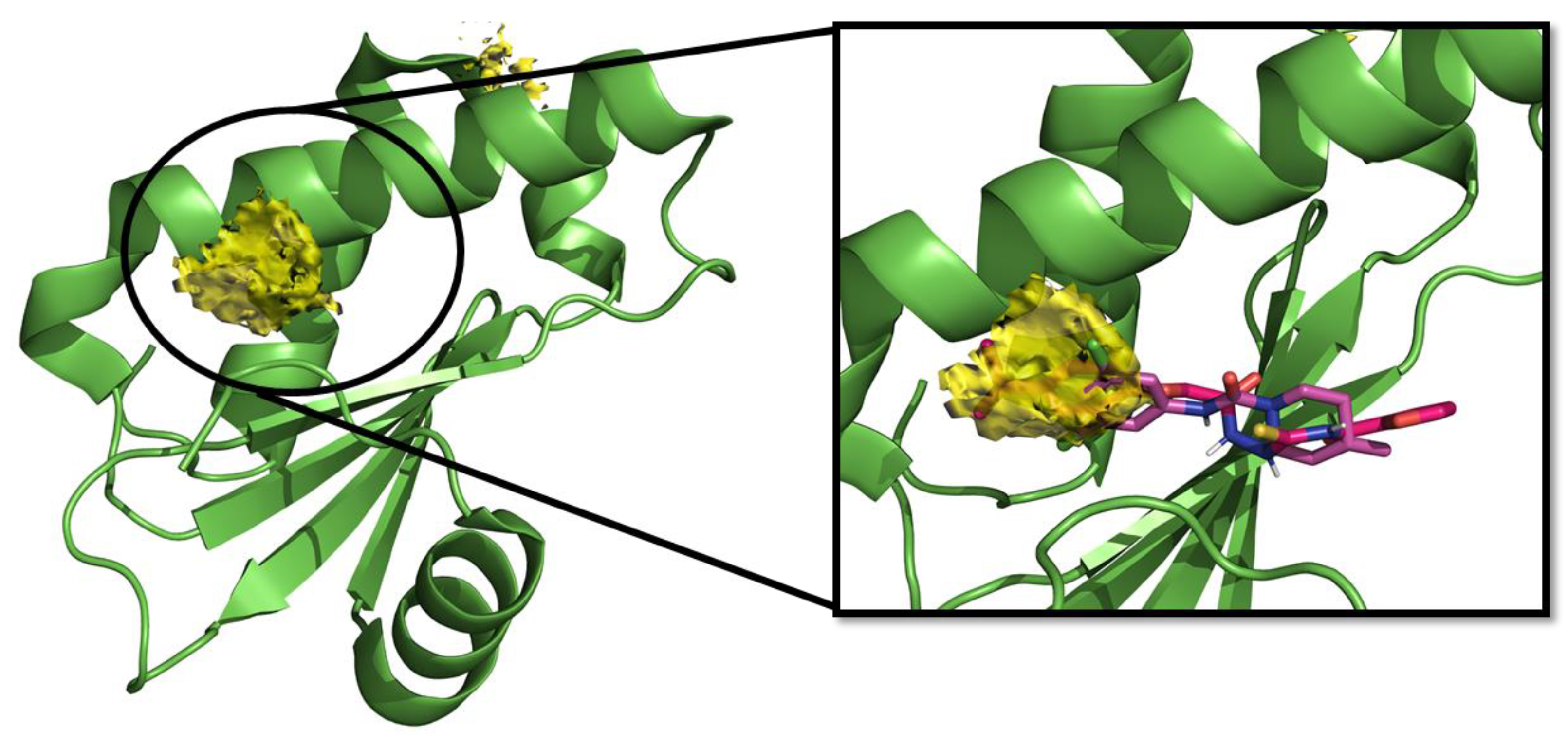
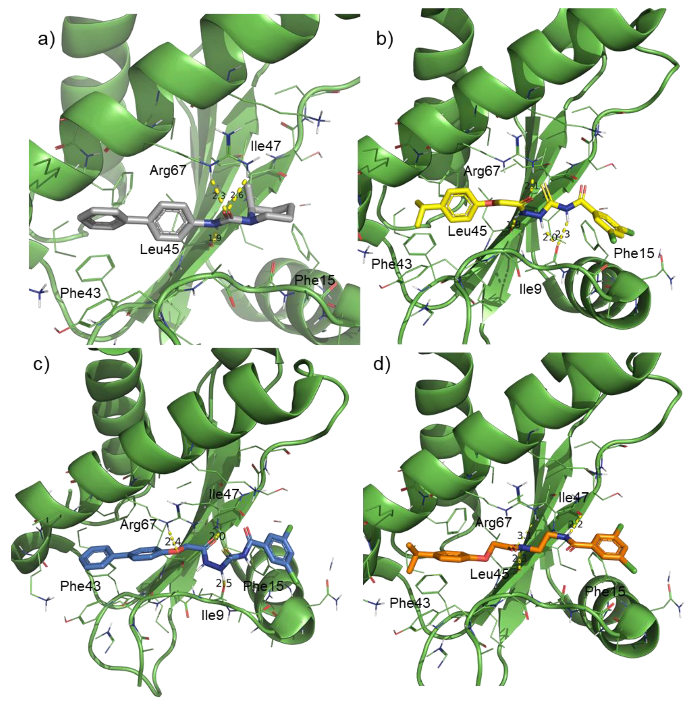
2.1. Computational Studies and Design

Using in-silico predictions, we aimed to estimate which cavity of the protein could be responsible for the binding of JC-like compounds, RNPA2000, the set of derivatives synthesized in this work and to decipher the main key interactions of these compounds in the binding site.
With this objective and using S. aureus RnpA protein, a detailed Hotspots Maps calculation was performed. These results revealed that RnpA displays low druggability when compared with other proteins, and only one low hotspot scoring area was identified as the main ligandable binding site. Both JC1 and RNPA2000 were docked in this hypothetical binding pocket in order to validate our hypothesis (Figure 3).
First, and given the unknown mechanism of action of RnpA inhibitors in RNA degradation and ptRNA maturation, a blind docking study was carried out. For this study, we considered the RnpA structure, and two reference compounds that were proposed in the literature as RnpA inhibitors (JC1 and RNPA2000) described above to assess their predicted binding cavity. The docking studies suggest that the reference molecules might be able to bind RnpA in the main area that was identified previously in the hotspots maps. The top part of this region involves the sequence of highest phylogenetic conservation in the RnpA proteins being named the RNR box motif and represented by residues 59–67. This positive-charged motif, containing three arginines and two lysines, resides at the top half of α2 being solvent exposed, being poised to form the required interactions with the catalytic RNA ribozyme (P-RNA) component of the complex [26].
Specifically, there are several key subareas in the solvent-exposed pocket that might drive the ligand-protein interaction:
  • in the subregion where the main hydrophobic hotspot was identified, there are several aromatic and hydrophobic residues, including Tyr7, Phe43, Leu45, and Phe70, where the first two are generating a cleft. This might help in anchoring aromatic systems, establishing π or CH–π interactions (such as the i-propylphenyl group that is present in RnpA2000);
  • the central part contains a solvent-exposed area, which might allow the binding of linear linkers by establishing hydrogen bonds interactions via the backbone of Ile9, Leu45, Ile47 or the guanidinic moiety of Arg67. H-bonds donor-acceptor that are predicted to interact with the ureidic portion of JC1 and with the semi-thiocarbazide moiety of RNPA2000; and,
  • finally, at the other end, Phe15 and Lys63 are suitable for π interaction or π-cation interactions with different aromatic moieties, such as the one present in RNA2000.
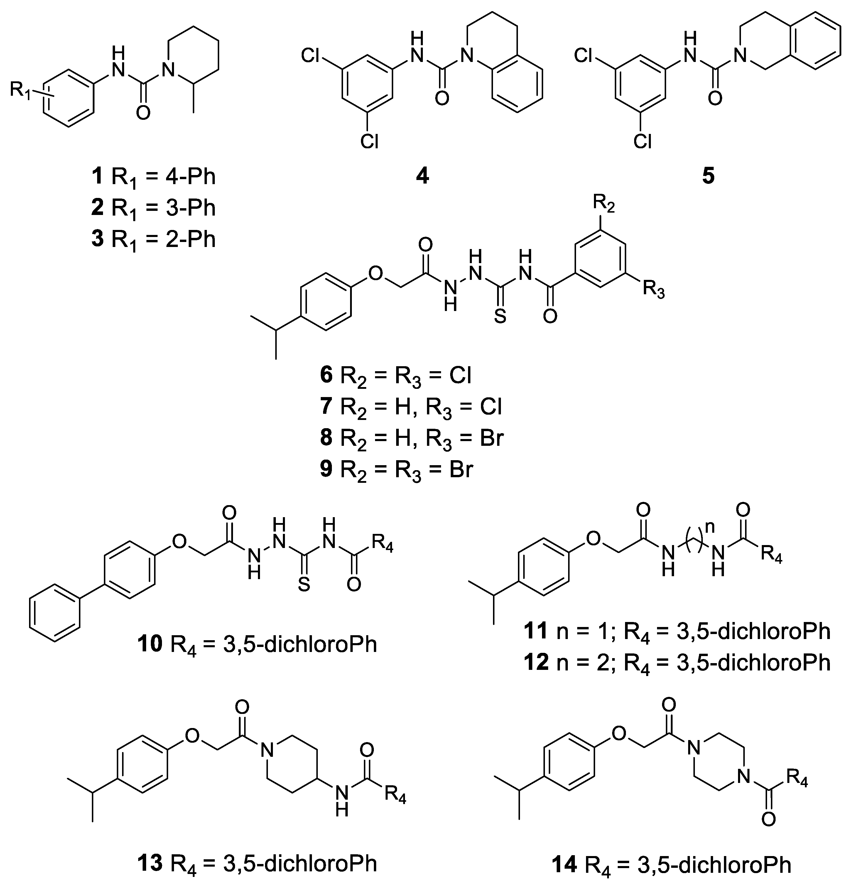
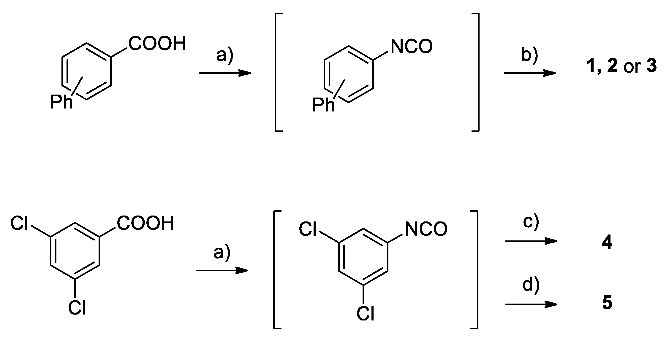
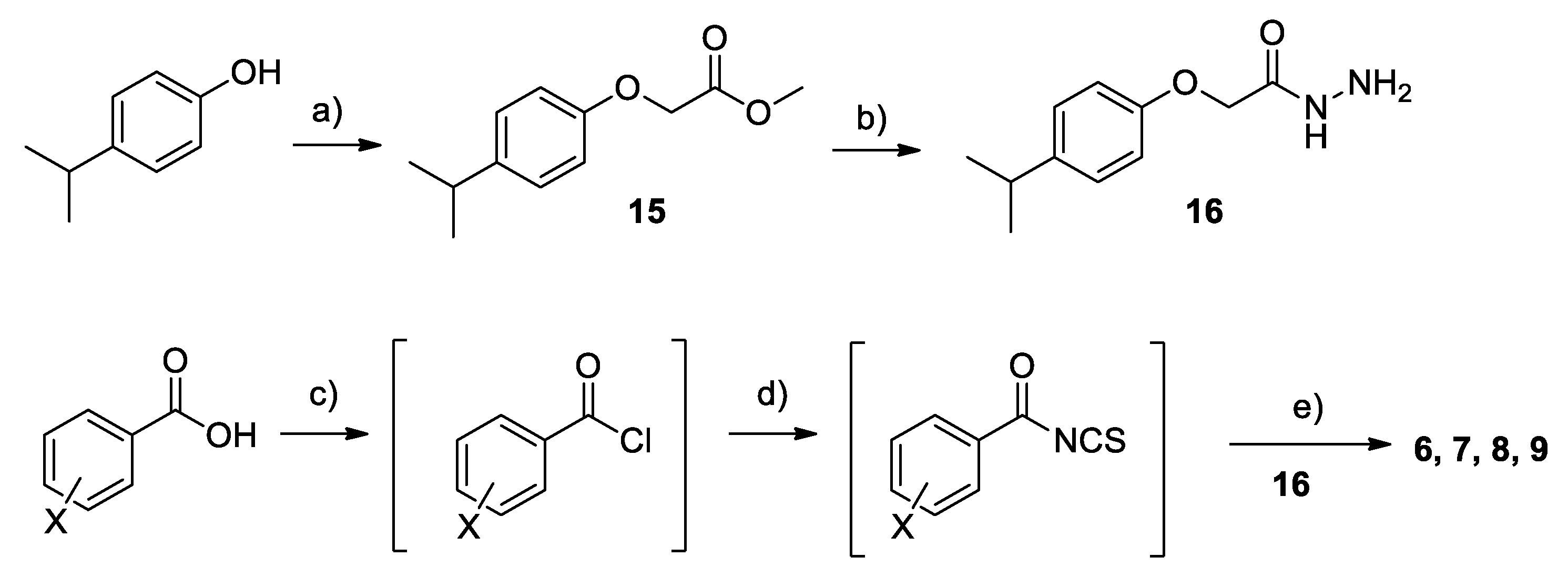
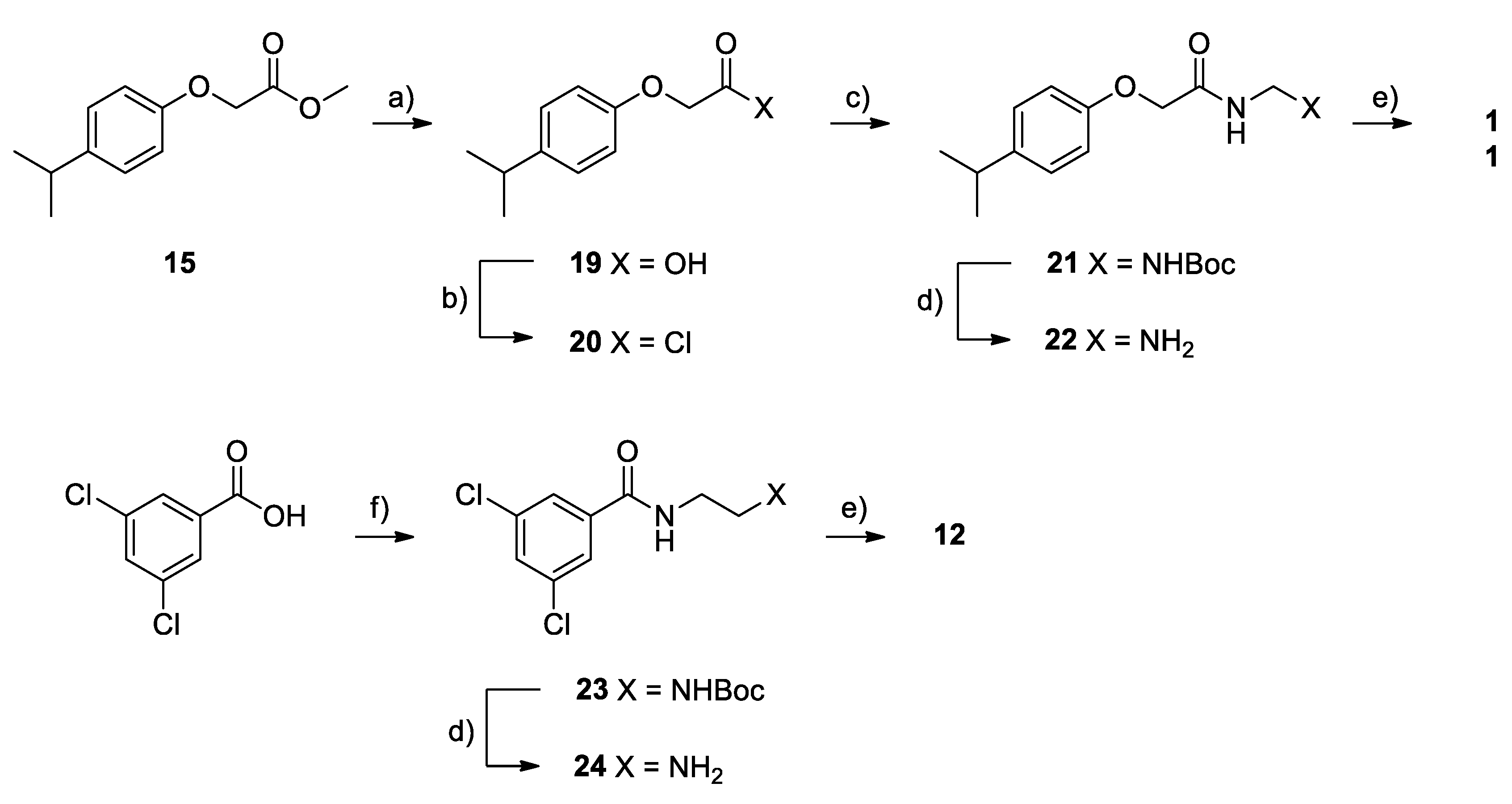
According to the docking results, compound 1 is predicted to bind the same cavity achieving interactions that are close to the reference compounds mentioned above (Figure 4a). The biphenyl motif engages with the aromatic residues on the cleft, while the amide makes H-bond with Leu45, and Arg67, as shown in Figure 4a. On the other hand, 2 and 3 with a different substitution pattern in the biphenyl system and with the vector growing at the 2 and 3 positions towards the solvent exposed area seem to be less attractive to fit in the cleft region as compared to 1.
Compounds 4 and 5 were designed to potentially achieve an extra-interaction with the Phe15 or Lys63 at the binding site, by the substitution of the piperidine ring of JC1 with a tetrahydroquinoline or a tetrahydroisoquinoline, respectively. Compounds 69 showed a very good interaction profile and fit in the pocket, with the i-propylphenyl and the semi-thiocarbazide showing a very similar shape to RNPA2000 and an optimal linearity given by the linker. At the other end, the replacement of the furanyl moiety by different halogen-phenyl systems are tolerated, as depicted in Figure 4b with compound 6. While considering derivative 10, the biphenylic portion was introduced to replace the characteristic i-propylic substituent present in other RNPA2000-like structures and evaluate its effects. Compound 10 keeps the H-bond interactions between the linker and Ile9, Ile47 and Arg67 and the biphenyl achieves a perfect fit in the cleft (Figure 4c).
Finally, compounds 1114 were designed to evaluate the importance of the semi-thiocarbazide moiety. Even removing the semi-thiocarbazide and replacing it with a di-amidic structure, these compounds maintain some of the key interactions with RnpA, as shown in Figure 4d. Moreover, this series of compounds should be more metabolically stable than RNPA2000-like derivatives, which might contain a liable nitrogen-nitrogen bond.
From a computational point of view, the hotspot maps analysis, together with the docking studies, suggested the binding cavity for this set of compounds proposing their putative binding mode and their main interactions with RnpA. In addition, several structural modifications were proposed to test different hypothesis and evaluate the effect of these modifications on both the phenotypic assay and the capability of these compounds to inhibit in-vitro RnpA-mediated ptRNA maturation. These results will need further structural confirmation in future studies, given the lack of structural information for inhibitors of this route.

2.2. Chemistry

Scheme 1 shows the synthesis of compounds 1 to 5. The synthetic route is the same for each compound. The proper carboxylic acid was converted into the corresponding isocyanate by treatment with diphenyl phosphoryl azide and subsequent Curtius rearrangement after heating, and soon reacted with the appropriate amine, yielding the desired ureidic derivative.
For the obtainment of the final compounds 6 to 9 (Scheme 2), we followed the synthetic route that was described by Lounsbury and colleagues [23], in which the key intermediate 2-(i-propylphenoxy)acetohydrazide is treated with the proper acyl isothiocyanate to obtain the thiosemicarbazide group, peculiar for this four compounds. To do so, we achieved the key intermediate 16 starting from the commercially available 4-i-propylphenol, which was alkylated by reaction with methyl 2-chloroacetate in the presence of K2CO3 as base and subsequently transformed into the corresponding hydrazide by treatment with hydrazine hydrate in methanol.
The synthetic scheme that was performed for the synthesis of compound 10 (Scheme 3) follows the one just described for compounds 6 to 9. In this case, the 4-phenylphenol was converted into the 2-(4-biphenyloxy)acetohydrazide, tracing the same strategy described before, and treated with the 3,5-dichlorobenzoyl isothiocyanate, yielding the desired compound 10.
The synthesis of compounds 1112 (Scheme 4), characterized by two amidic groups linked at the nitrogen atoms by methylenic or ethylenic linker (compounds 11 and 12), is based on the same strategy. Firstly, the two moieties were reacted with the appropriate monoprotected amine, generating the first amidic group. Thus, the intermediate was deprotected and linked to the second moiety to achieve the second amidic group. In particular, for compound 11, the process started from intermediate 15, which was hydrolyzed and treated with the N-Boc-methylendiamine, being adequately prepared, as described by DeBons and Loudon [27]. Therefore, this intermediate was deprotected and converted into the desired compound 11. For compound 12, conversely, the synthesis started from the 3,5-dichlorobenzoic acid, which undergoes the same reactions as before, using the appropriate amine.
Lastly, the obtainment of compounds 1314 (Scheme 5) was possible by following the same strategy previously described, since also these two derivatives are characterized by a diamidic structure. In this case, the intermediate 20 was treated with the corresponding amine, 4-N-Boc-aminopiperidine and N-Boc-piperazine, to achieve the first amidic group (2526). Therefore, these intermediates were deprotected (2728) and treated with 3,5-dichlorobenzoyl chloride yielding the final derivatives.

2.3. Biological Evaluation

2.3.1. Antimicrobial Activity

The antimicrobial activities of compounds 114 (Table 1) were evaluated on two S. aureus strains: a methicillin-sensitive S. aureus (MSSA, ATCC 29213) and a methicillin-resistant S. aureus (MRSA, ATCC 43300). The inhibitory ability of compounds 114 was evaluated by determining the minimum inhibitory concentration (MIC).
Some of the tested compounds displayed very promising antimicrobial activities, with reasonable MICs (6, 7, 8, 9, and 10), as shown in Table 1. Next, we assessed whether these antimicrobial activities correlated with the inhibition of RnpA activities.

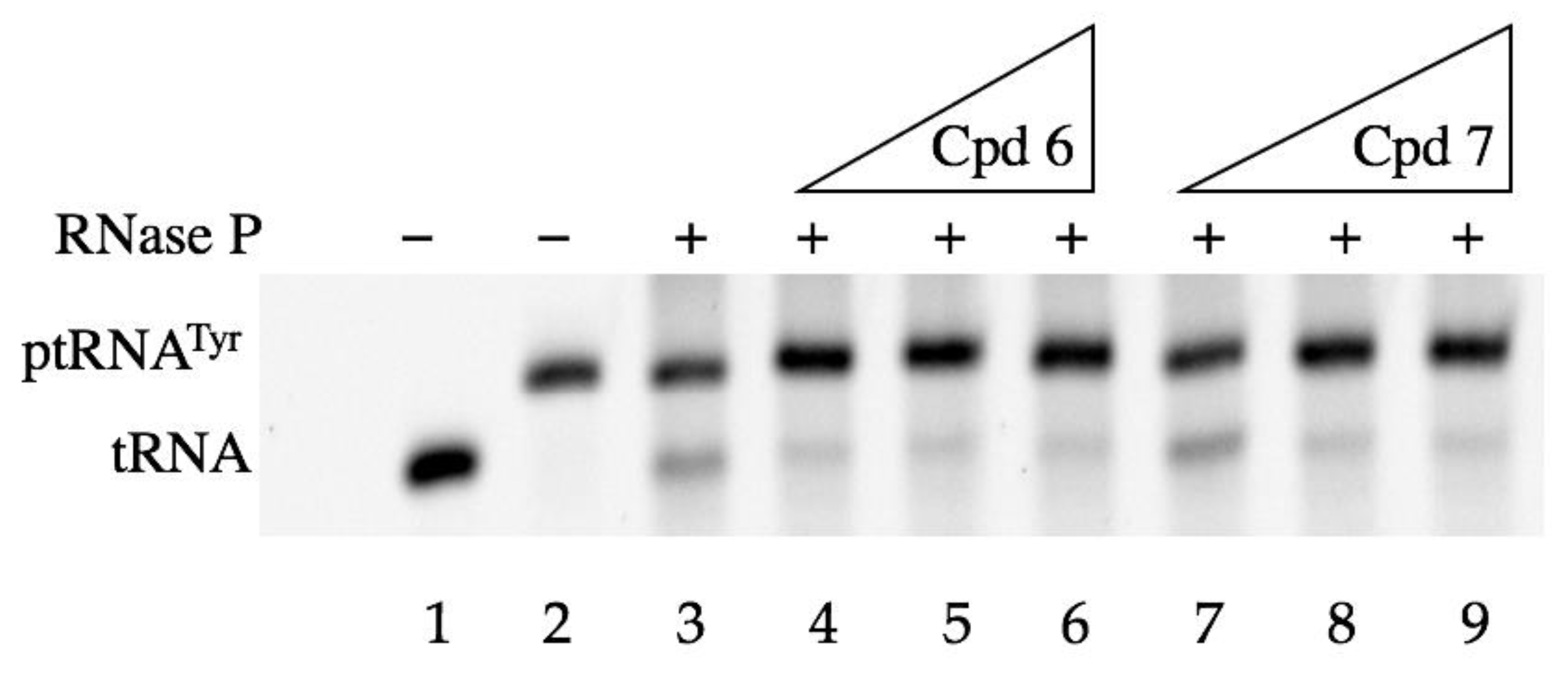
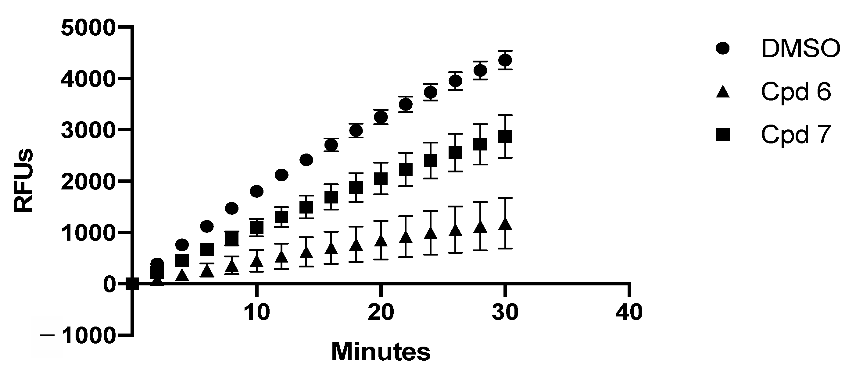
2.3.2. In-Vitro Assays

Firstly, we evaluated the capability of these compounds to inhibit in-vitro RnpA-mediated ptRNA maturation. To do so, Rnase P (RnpA + rnpB) was reconstituted and combined with the putative inhibitor or DMSO (as a negative control) and ptRNATyr for 30 min, and the relative quantities of ptRNATyr and mature tRNA were evaluated by Urea-PAGE. Figure 5 depicts representative data of the performance of compounds 6 and 7. The presence of RNase P alone (Figure 5, lane 3) catalyzes the conversion of ptRNA into mature tRNA. At the same time, increasing amounts of compounds 6 and 7 partially perturb the activity of RNase P, resulting in the accumulation of ptRNA (Figure 5, lanes 4 to 9). All of the compounds were tested at least twice and Table 2 records their IC50 values.
In parallel, the mRNA degradation inhibition profile of each compound was also evaluated. The time course experiments were performed by incubating 62.5 µM of the compound (or DMSO) with RnpA in the presence of a fluorescently labeled RNA substrate for 30 min. RnpA efficiently degraded RNA in the presence of DMSO (circles), but it was inhibited to 40–60% in the presence of the compounds 6 and 7, as shown in Figure 6 (triangles and squares, respectively). Subsequent dose response assays were carried out using increasing concentrations of each analogue to determine the IC50 value of each compound (Table 2).
Table 2 summarizes the IC50 values for each compound, which were calculated for both mRNA degradation inhibition and ptRNA maturation inhibition. The results revealed that the substitution of the 3,5-dichlorophenyl moiety of JC1/JC2 with a biphenylic one is always detrimental for the inhibition of the RnpA-mediated processes and, consequently, for the antimicrobial activity, although the 4-biphenilic derivative 1 presents a better overall profile when compared to the 3-biphenilic and the 2-biphenilic derivatives 2 and 3. These data match with the in-silico prediction. Conversely, the tetrahydroquinolinic compound 4 shows stronger RnpA inhibition properties as compared to the tetrahydroisoquilinic derivative 5. Despite that these results seem to be in contrast with the predicted docking data, the different activity might be due to other factors rather that the inability to correctly interact with RnpA. The IC50 values of compounds 6, 7, 8, and, partially, 9 confirm the productivity of the substitution of the 2-furanylmoiety with halogen-phenyl rings. In particular, the dichlorophenyl derivative 6 present comparative inhibition profiles for mRNA degradation and ptRNA processing, while derivatives 7 (m-chloro) and 8 (m-bromo) show, respectively, better ptRNA processing inhibition and mRNA degradation inhibition capability. Compounds 1114 present no antimicrobial activity, which suggests the importance of the semi-thiocarbazide moiety.

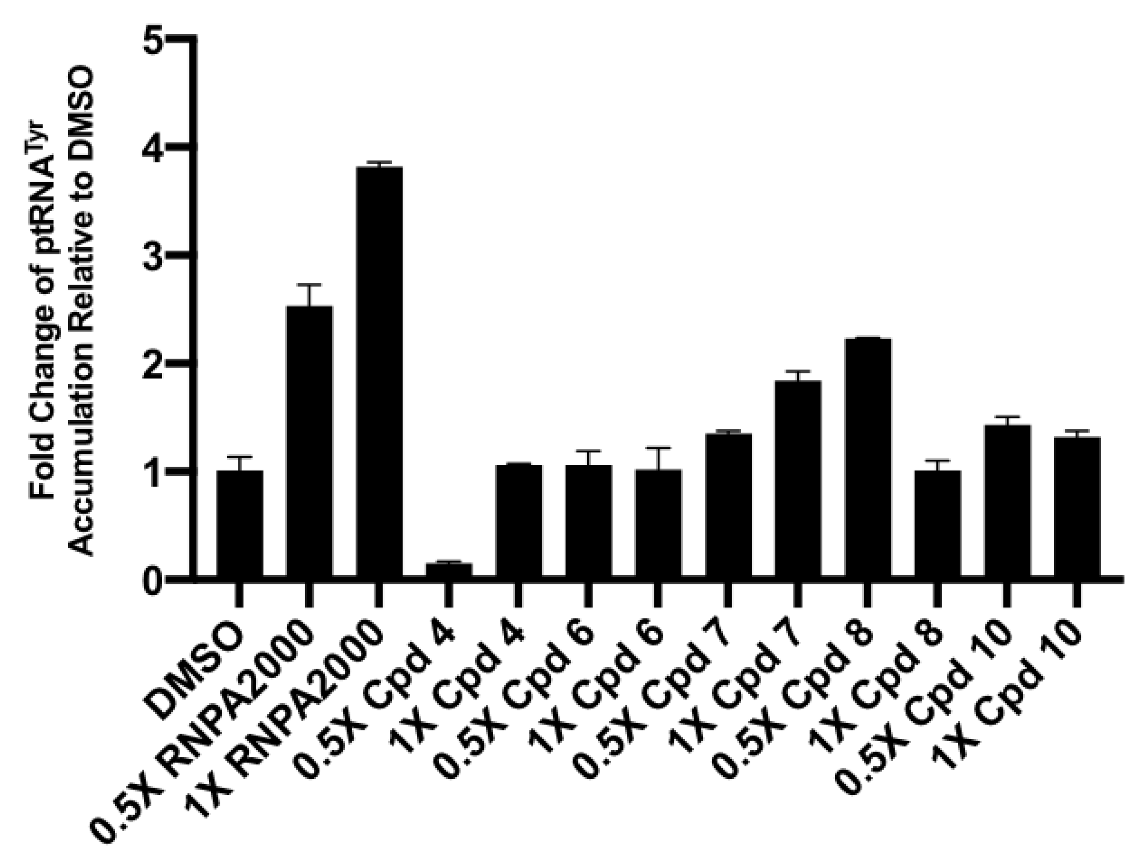
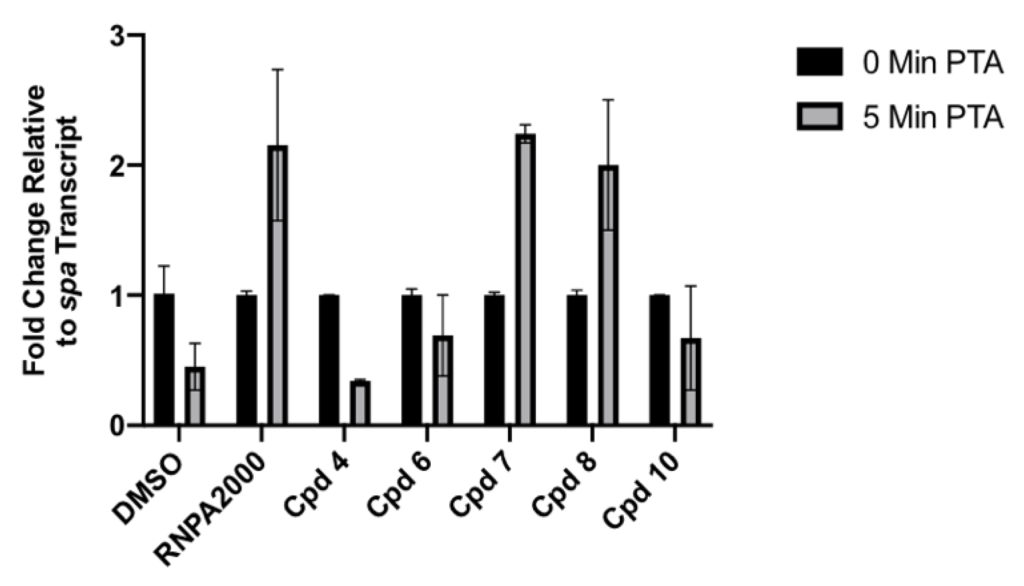
2.3.3. Cellular Assays

S. aureus were treated with 0.5× or 1× MIC of each compound for 1 h and qRT-PCR was used to quantify the accumulation of ptRNAtyr in order to evaluate the capability of each compound to affect RnpA-mediated ptRNA processing in the more complex cellular system, as previously described [22,24]. Cells that were treated with DMSO did not accumulate ptRNATyr, whereas RNPA2000 (positive control) resulted in a dose-dependent accumulation of 2.5-fold and 3.8-fold at 0.5× and 1× MIC, respectively, as shown in Figure 7. Compound 7 confirmed its ability to inhibit RnpA-mediated ptRNATyr processing and also demonstrated a dose-dependent effect on ptRNATyr accumulation. Treatment with compound 8, which had inhibitory activity in vitro, resulting in a 2.23-fold increase in ptRNATyr accumulation at 0.5× MIC, but did not inhibit ptRNATyr processing after treatment with 1× MIC, possibly due to the compound precipitation at this concentration. Compound 10, which had a good in-vitro inhibition of ptRNATyr processing, resulted in 1.4-fold and 1.3-fold increases in ptRNATyr accumulation at 0.5× and 1× of the MIC, respectively. Treatment with compounds 4 and 6 did not result in the accumulation of ptRNATyr, despite the antistaphylococcal and inhibitory activity in vitro.
As a more direct measure of the effect of each analogue, on cellular mRNA degradation, RnpA-mediated turnover of spa RNA, a natural substrate of RnpA, was evaluated. S. aureus UAMS-1 was treated with 0.5× the MIC for 30 min prior to post-transcriptional arrest (PTA) with rifampin and spa RNA was quantified via RT-PCR at 0- and 5-min PTA, as previously described [9]. In cells that were treated with DMSO, 55% of spa transcript was degraded after 5 min PTA, as shown in Figure 8. The RNPA2000 positive control, as well as compounds 7 and 8, showed a significantly reduced degradation of spa transcript. Compound 4 had no effect on cellular RNA turnover, with the treated cells degrading approximately 66% of spa after 5 min PTA, despite having in vitro activity. Compound 6, which also had in vitro activity, resulting in an average of 31% spa transcript degradation, an approximate 24% inhibition of degradation when compared to DMSO. Compound 10, which had weak in vitro degradation activity, had an average inhibition of 22% on cellular RNA turnover in comparison to DMSO.
Taken together, compounds 7 and 8, presenting a 3-halogenphenyl moiety in place of the 2-furanyl one, performed well when evaluated in in-vitro assays and they were the top performers in both cellular assays, significantly inhibiting both RnpA-mediated cellular RNA metabolism functions. Although compound 10, in which the i-propylphenyloxy portion was replaced by a more lypophilic one, had the most potent antibacterial activity, it had weak effects on RNA degradation (both in vitro and in vivo), but it was a good inhibitor of ptRNATyr processing in vitro and it had significantly increased ptRNATyr accumulation in cellular assays.

3. Conclusions

In the present work, we predicted key interactions that RNPA2000 and JC1/2 achieve with their molecular target RnpA. This information guided the design of new inhibitors, which increased the knowledge of the SAR of RNPA2000-like and JC1/2-like RnpA inhibitors. The lower activity of compounds 1 to 5 as compared to JC1/2 suggest that the combination of a small-aliphatic amine with a 3,5-dichlorophenyl moiety is required for RnpA inhibition. Nevertheless, further investigation regarding this class of compounds remains worthwhile. Compounds 7 and 8, characterized by a 3-halogenphenyl ring in place of the 2-furanyl moiety, demonstrated cellular activity that was comparable to RNPA2000, while avoiding the potentially metabotoxic furan. At the same time, the inactivity of compounds 11, 12, 13, and 14 underline the importance of the semi-thiocarbazide. This preliminary set of data, which are perfectly in line with the innovative computational model, trace the path for a deeper investigation of the SAR of these very promising classes of compounds. Moreover, an exciting aspect of the current work is the development of a putative computational-based path for RnpA inhibitor discovery and optimization. Building from our early modeling success is expected to allow for the rational design of more potent and presumably more selective molecules that, as they become available and evaluated for RnpA inhibitor properties, are expected to allow refining the computational modeling in iterative manner and yielding higher quality probes for mutagenesis based testing of the model.

4. Materials and Methods

4.1. Chemistry

All of the reagents were purchased from Sigma Aldrich (St. Louis, MO, USA) and they were used without further purification. The solvents, such as ACN, THF, DCM, DMF, methanol, and acetone, were purchased from Sigma Aldrich.
Silica gel matrix, with fluorescent indicator 254 nm, was used in analytical thin-layer chromatography (TLC on aluminum foils), and silica gel (particle size 40–63 µm, Merck) was used in flash chromatography on Sepachrom Puriflash XS 420. Visualizations were accomplished with UV light (λ 254 or 280 nm).
The 1H-NMR spectra were measured by Varian Mercury 300 NMR spectrometer/Oxford Narrow Bore superconducting magnet operating at 300 MHz. The 13C-NMR spectra were acquired operating at 75 MHz. Chemical shifts (δ) are reported in ppm relative to residual solvent as the internal standard. Signal multiplicity is used according to the following abbreviations: s = singlet, d = doublet, dd = doublet of doublets, t = triplet, q = quadruplet, dq = doublet of quadruplets, m = multiplet, bs = broad singlet, and set = septuplet. The final products, 114, were analyzed by reverse-phase HPLC using a Waters XBridge C-18 column (5 μm, 4.6 mm × 150 mm) on an Elite LaChrom HPLC system with a diode array detector, with different methods (Table 3). Their purity was quantified at peculiar λ max, depending on each sample, and it resulted to be >95%. The relative retention times are reported in each experimental section. The melting points were determined by DSC analysis over a TA Instruments DSC 1020 apparatus. The 1H- and 13C-NMR spectra of compounds 114, together with their HPLC profiles, are included in the supplementary material S1.

Synthesis

1-(4-Biphenyl)-3-(2-methylpiperdin-1-yl)urea (1): diphenyl phosphoryl azide (DPPA) (2.71 mL, 12.10 mmol) was added dropwise to a solution of biphenyl-4-carboxylic acid (2.00 g, 10.09 mmol) and triethylamine (TEA) (1.69 mL, 12.10 mmol) in toluene (20 mL). The reaction mixture was stirred at RT for 20 min, heated at 95 °C, and then stirred again for 20 min. The mixture was cooled at RT, added with 2-methylpiperidine (1.25 mL, 10.59 mmol), heated again at 95 °C and stirred overnight. Then, the mixture was diluted with ethyl acetate (30 mL), washed twice with 10% aqueous NaHCO3, dried over Na2SO4, filtered, and concentrated under vacuum to give a residue, which was purified by flash chromatography. Elution with cyclohexane/ethyl acetate 80/20 and subsequent crystallization from ethanol (15 vol) gave 0.85 g of 1 as a white solid. Yield: 29% M.p: 164.21 °C. Tr (HPLC): 12.65 min. (Method A), Purity = 99.7%. 1H NMR (300 MHz, CDCl3, δ) 7.55 (m, 4H), 7.46 (m, 4H), 7.29 (m, 1H), 6.49 (bs, 1H), 4.39 (m, 1H), 3.91 (d, J = 12.8 Hz, 1H), 3.02 (t, J = 12.8 Hz, 1H), 1.62 (m, 6H), 1.25 (d, J = 6.8 Hz, 3H). 13C NMR (75 MHz, CDCl3, δ) 154.94, 140.76, 138.79, 135.57, 128.68, 127.41, 126.77, 126.71, 120.13, 46.74, 39.13, 30.23, 25.63, 18.55, 15.76 ppm.
1-(3-Biphenyl)-3-(2-methylpiperdin-1-yl)urea (2): Prepared from biphenyl-3-carboxylic acid as described for 1 using DPPA (1.2 eq), TEA (1.2 eq), and 2-methylpiperdine (1.05 eq) in toluene (10 vol), and then purified by flash chromatography on silica gel. Elution with cyclohexane/ethyl acetate 80/20 and subsequent crystallization from EtOH (4 vol) and recrystallization from IPA (4 vol) gave 0.52 g of 2 as a white solid. Yield: 17% M.p: 126.53 °C. Tr (HPLC): 12.67 min. (Method A), Purity = 99.3%. 1H NMR (300 MHz, CDCl3, δ) 7.65 (m, 3H), 7.32 (m, 6H), 6.46 (bs, 1H), 4.41 (m, 1H), 3.91 (d, J = 13.0 Hz, 1H), 3.02 (t, J = 13.0 Hz, 1H), 1.64 (m, 6H), 1.25 (d, J = 6.9 Hz, 3H). 13C NMR (75 MHz, CDCl3, δ) 154.94, 141.91, 140.98, 139.75, 129.16, 128.61, 127.26, 127.18, 121.64, 118.72, 118.68, 46.76, 39.13, 30.23, 25.63, 18.55, 15.77 ppm.
1-(2-Biphenyl)-3-(2-methylpiperdin-1-yl)urea (3): Prepared from biphenyl-2-carboxylic acid as described for 1 using DPPA (1.2 eq), TEA (1.2 eq). and 2-methylpiperdine (1.05 eq) in toluene (10 vol), and the purified by flash chromatography on silica gel. Elution with cyclohexane/ethyl acetate 80/20 and subsequent crystallization from IPA (4 vol) gave 1.02 g of 3 as a white solid. Yield: 35% M.p: 104.05 °C. Tr (HPLC): 10.67 min. (Method A), Purity = 96.4%. 1H NMR (300 MHz, CDCl3, δ) δ 8.11 (d, J = 8.1 Hz, 1H), 7.41 (m, 6H), 7.20 (dd, J = 7.6, 1.6 Hz, 1H), 7.07 (td, J = 7.6, 0.9 Hz, 1H), 6.47 (bs, 1H), 3.98 (m, 1H), 3.70 (d, J = 12.6 Hz, 1H), 2.78 (t, J = 12.6 Hz, 1H), 1.44 (m, 6H), 1.01 (d, J = 6.9 Hz, 3H). 13C NMR (75 MHz, CDCl3, δ) 154.68, 138.72, 136.43, 131.52, 129.52, 129.32, 128.98, 128.38, 127.77, 122.49, 120.85, 46.62, 38.57, 30.19, 25.46, 18.52, 15.58 ppm.
1-(3,5-Dichlorophenyl)-3-(1,2,3,4-tetrahydroquinolin-1-yl)urea (4): Prepared from 3,5-dichlorobenzoic acid, as described for 1 using DPPA (1.2 eq), TEA (1.2 eq), and 1,2,3,4-tetrahydroquinoline (1.05 eq) in toluene (10 vol) and purified by flash chromatography on silica gel. Elution with cyclohexane/ethyl acetate 80/20 and subsequent crystallization from EtOH (6 vol) gave 0.80 g of 4 as a white solid. Yield: 32% M.p: 143.34 °C. Tr (HPLC): 12.46 min. (Method C), Purity = 99.8%. 1H NMR (300 MHz, CDCl3, δ) 7.34 (d, J = 1.9 Hz, 2H), 7.25 (m, 3H), 7.15 (m, 1H), 7.06 (bs, 1H), 7.00 (t, J = 1.8 Hz, 1H), 3.81 (t, J = 6.3 Hz, 2H), 2.79 (t, J = 6.7 Hz, 2H), 1.98 (m, 2H). 13C NMR (75 MHz, CDCl3, δ) 152.98, 140.59, 138.25, 135.11, 133.21, 129.89, 127.05, 125.32, 122.88, 122.82, 117.15, 43.43, 26.90, 23.94 ppm.
1-(3,5-Dichlorophenyl)-3-(1,2,3,4-tetrahydroisoquinolin-1-yl)urea (5): Prepared from 3,5-dichlorobenzoic acid as described for 1 using DPPA (1.2 eq), TEA (1.2 eq), and 1,2,3,4-tetrahydroisoquinoline (1.05 eq) in toluene (10 vol), and then purified by flash chromatography on silica gel. Elution with cyclohexane/ethyl acetate 85/15 and subsequent crystallization from IPE (20 vol) gave 0.60 g of 5 as a white solid. Yield: 24% M.p: 128.79 °C. Tr (HPLC): 9.00 min. (Method C). Purity = 99.9%. 1H NMR (300 MHz, CDCl3, δ) 7.36 (d, J = 1.5 Hz, 2H), 7.18 (m, 4H), 6.98 (s, 1H), 6.75 (bs, 1H), 4.64 (s, 2H), 3.71 (t, J = 5.8 Hz, 2H), 2.92 (t, J = 5.7 Hz, 2H). 13C NMR (75 MHz, CDCl3, δ) 154.29, 141.04, 134.92, 134.68, 132.73, 128.39, 126.99, 126.61, 126.29, 122.85, 118.15, 45.77, 41.73, 28.89 ppm.
Methyl 4-i-propylphenoxyacetate (15): methyl chloroacetate (0.71 mL, 8.01 mmol) was added to a solution of 4-isopropylphenol (1.00 g, 7.34 mmol) and potassium carbonate (1.12 g, 8.01 mmol) in DMF (10 mL). The reaction mixture was stirred at 50 °C for 1.5 h, concentrated under vacuum, diluted with ethyl acetate (30 mL), washed with brine (4 × 10 mL), dried over Na2SO4, filtered, and then concentrated under vacuum to give 1.4 g of 15 as a yellowish oil. Yield: 92%. 1H NMR (300 MHz, CDCl3, δ) 7.14 (d, J = 8.7 Hz, 2H), 6.84 (d, J = 8.7 Hz, 2H), 4.61 (s, 2H), 3.80 (s, 3H), 2.86 (set, J = 6.9 Hz, 1H), 1.22 (d, J = 6.9 Hz, 6H).
4-i-Propylphenoxyacetohydrazide (16): hydrazine hydrate (2.1 mL, 33.60 mmol) was added to a solution of 15 (1.40 g, 6.72 mmol) in methanol (15 mL). The reaction mixture was stirred at reflux for 16 h, concentrated under vacuum, diluted with ethyl acetate (30 mL), washed with brine (10 mL), dried over Na2SO4, filtered, and concentrated under vacuum to give 1,36 g of 16 as a white solid. Yield = 97%. M.p: 105.21 °C. 1H NMR (300 MHz, CDCl3, δ): 7.16 (d, J = 8.2 Hz, 2H), 6.83 (d, J = 8.2 Hz, 2H), 4.55 (s, 2H), 2.87 (set, J = 6.9 Hz, 1H), 1.22 (d, J = 6.9 Hz, 6H).
1-(4-i-Propylphenoxyacetyl)-4-(3,5-dichlorobenzoyl)thiosemicarbazide (6): a solution of 3,5-dichlorobenzoic acid (1.30 g, 6.8 mmol) in SOCl2 (6.5 mL) was stirred at reflux for 1 h. Thus, the solution was concentrated under vacuum, diluted with acetonitrile (15 mL), and then added with potassium thiocyanate (1.12 g, 11.56 mmol). The reaction mixture was stirred at RT for 1 h, added with a solution of 16 (1.40 g, 6.8 mmol) in DMF (5 mL), stirred at RT for 30 min, concentrated under vacuum, diluted with ethyl acetate (50 mL), washed with brine (4 × 10 mL), dried over Na2SO4, filtered, and then concentrated under vacuum to give a solid. Digestion firstly with MeOH (15 vol) at reflux and secondly with ACN (15 vol) at reflux gave 1.3 g of 6 as a yellowish solid. Yield: 43%. M.p: 195.69 °C. Tr (HPLC): 17.60 min. (Method C), Purity = 99.6%. 1H NMR (300 MHz, DMSO-d6, δ) 12.38 (bs, 1H), 11.95 (bs, 1H), 11.00 (bs, 1H), 7.96 (d, J = 1.8 Hz, 2H), 7.89 (t, J = 1.8 Hz, 1H), 7.15 (d, J = 8.6 Hz, 2H), 6.91 (d, J = 8.6 Hz, 2H), 4.68 (s, 2H), 2.82 (set, J = 6.9 Hz, 1H), 1.16 (d, J = 6.9 Hz, 6H). 13C NMR (75 MHz, DMSO-d6, δ) 177.66, 165.66, 165.61, 156.23, 141.70, 135.56, 134.63, 132.58, 127.88, 127.55, 115.02, 66.24, 33.06, 24.50 ppm.
1-(4-i-Propylphenoxyacetyl)-4-(3-chlorobenzoyl)thiosemicarbazide (7): Prepared from 3-chlorobenzoic acid, as described for 6 using SOCl2 (5 vol), potassium thiocyanate (1.6 eq) and 16 (1 eq) in acetonitrile (10 vol) at RT for 30 min. Digestion of the crude product with IPA (10 vol) at reflux gave 1.06 g of 7 as a pale yellow solid. Yield: 27%. M.p: 187.27 °C. Tr (HPLC): 9.38 min. (Method C), Purity = 98.5%. 1H NMR (300 MHz, DMSO-d6, δ) 12.47 (bs, 1H), 11.90 (bs, 1H), 11.01 (bs, 1H), 8.00 (t, J = 1.9 Hz, 1H), 7.86 (m, 1H), 7.70 (ddd, J = 8.0, 1.9, 1.0 Hz, 1H), 7.54 (t, J = 8.0 Hz, 1H), 7.15 (d, J = 8.7 Hz, 2H), 6.91 (d, J = 8.7 Hz, 2H), 4.68 (s, 2H), 2.82 (set, J = 6.9 Hz, 1H), 1.15 (d, J = 6.9 Hz, 6H). 13C NMR (75 MHz, DMSO-d6, δ) 178.09, 167.00, 156.66, 156.24, 141.69, 134.31, 133.56, 133.25, 130.81, 128.92, 127.91, 127.59, 115.00, 66.16, 33.05, 24.54 ppm.
1-(4-i-Propylphenoxyacetyl)-4-(3-bromobenzoyl)thiosemicarbazide (8): Prepared from 3-bromobenzoic acid as described for 6 using SOCl2 (5 vol), potassium thiocyanate (1.6 eq) and 16 (1 eq) in acetonitrile (10 vol) at RT for 30 min. Digestion of the crude product with IPA (5 vol) at reflux gave 1.96 g of 8 as a white solid. Yield: 62%. M.p: 184.01 °C. Tr (HPLC): 10.13 min. (Method C), Purity = 98.5%. 1H NMR (300 MHz, DMSO-d6, δ) 12.46 (bs, 1H), 11.90 (bs, 1H), 11.03 (bs, 1H), 8.14 (t, J = 1.7 Hz, 1H), 7.92 (d, J = 7.9 Hz, 1H), 7.83 (m, 1H), 7.47 (t, J = 7.9 Hz, 1H), 7.15 (d, J = 8.7 Hz, 2H), 6.91 (d, J = 8.7 Hz, 2H), 4.68 (s, 2H), 2.81 (set, J = 6.9 Hz, 1H), 1.15 (d, J = 6.9 Hz, 6H). 13C NMR (75 MHz, DMSO-d6, δ) 178.13, 166.89, 165.67, 156.24, 141.69, 136.13, 134.50, 131.73, 131.04, 128.28, 127.59, 121.92, 114.99, 66.15, 33.05, 24.54 ppm.
1-(4-i-Propylphenoxyacetyl)-4-(3,5-dibromobenzoyl)thiosemicarbazide (9): prepared from 3,5-dibromobenzoic acid, as described for 6 using SOCl2 (5 vol), potassium thiocyanate (1.6 eq), and 16 (1 eq) in acetonitrile (10 vol) at RT for 30 min. Digestion of the crude product firstly with MeOH (10 vol) and secondly with IPA (15 vol) at reflux gave 2.00 g of 9 as a white solid. Yield: 62%. M.p: 201.83 °C. Tr (HPLC): 6.19 min. (Method D), Purity = 99.0%. 1H NMR (300 MHz, DMSO-d6, δ) 12.36 (bs, 1H), 11.97 (bs, 1H), 11.03 (bs, 1H), 8.11 (s, 3H), 7.15 (d, J = 8.3 Hz, 2H), 6.87 (dd, J = 8.3 Hz, 2H), 4.67 (s, 2H), 2.82 (set, J = 6.9 Hz, 1H), 1.15 (d, J = 6.9 Hz, 7H). 13C NMR (75 MHz, DMSO-d6, δ) 177.89, 165.68, 165.48, 156.24, 141.68, 137.88, 136.01, 131.04, 131.04, 127.59, 122.81, 114.99, 66.14, 33.05, 24.54 ppm.
Methyl 4-biphenyloxyacetate (17): prepared from 4-hydoxybiphenyl as described for 15 using methyl 2-chloroacetate (1.25 eq) and potassium carbonate (1.2 eq) in DMF (10 vol) to give 2.85 g of 17 as a white solid. Yield: 93%. M.p: 100.98 °C. 1H NMR (300 MHz, CDCl3, δ) 7.53 (m, 4H), 7.41 (m, 2H), 7.31 (m, 1H), 6.99 (d, J = 8.8 Hz, 2H), 4.68 (s, 2H), 3.83 (s, 3H).
4-Biphenyloxyacetohydrazide (18): prepared from 17 as described for 16 using hydrazine hydrate (5 eq) in MeOH (30 mL) to give 1,85 g of 18 as a white solid. Yield: 64%. M.p: 112.86 °C. 1H NMR (300 MHz, DMSO-d6, δ): δ 9.43 (bs, 1H), 7.58 (m, 4H), 7.41 (t, J = 7.4 Hz, 2H), 7.29 (t, J = 7.4 Hz, 1H), 7.02 (dd, J = 9.3, 2.4 Hz, 2H), 4.53 (s, 2H), 4.33 (s, 2H).
1-(4-Biphenyloxyacetyl)-4-(3,5-dichlorobenzoyl)thiosemicarbazide (10): prepared from 3,5-diclorobenzoic acid as described for 6 using SOCl2 (5 vol), potassium thiocyanate (1.6 eq) and 18 (1 eq) in acetonitrile (10 vol) at RT for 1 h. Digestion of the crude product with MeOH (10 vol) at reflux gave 2.77 g of 10 as a white solid. Yield: 73%. M.p: 200.71 °C. Tr (HPLC): 13.81 min. (Method C), Purity = 95.2%. 1H NMR (300 MHz, DMSO-d6, δ) 12.37 (bs, 1H), 11.98 (bs, 1H), 11.12 (bs, 1H), 7.96 (d, J = 1.9 Hz, 2H), 7.89 (t, J = 1.9 Hz, 1H), 7.59 (m, 4H), 7.42 (t, J = 7.6 Hz, 2H), 7.33–7.25 (m, 1H), 7.08 (d, J = 8.8 Hz, 2H), 4.77 (s, 2H). 13C NMR (75 MHz, DMSO-d6, δ) 178.03, 165.63, 165.56, 157.78, 140.12, 135.70, 134.58, 133.79, 132.60, 129.31, 128.20, 127.92, 127.27, 126.69, 115.64, 66.11 ppm.
4-i-Propylphenoxyacetic acid (19): 2.5 N aqueous NaOH (6 mL, 15 mmol) was added to a solution of 15 (3.00 g, 14.40 mmol) in MeOH (30 mL). The reaction mixture was stirred at RT for 1.5 h, concentrated under vacuum, diluted with water, acidified to pH = 1 with conc. HCl, extracted twice with ethyl acetate (2 × 30 mL), dried over Na2SO4, filtered, and concentrated under vacuum to give 2.60 g of 19 as a white solid. Yield: 95%. M.p: 82.14 °C. 1H NMR (300 MHz, CDCl3, δ) 7.16 (d, J = 8.6 Hz, 2H), 6.86 (d, J = 8.6 Hz, 2H), 4.66 (s, 2H), 2.86 (set, J = 6.9 Hz, 1H), 1.22 (d, J = 6.9 Hz, 6H).
4-i-Propylphenoxyacetyl chloride (20): SOCl2 (10 mL, 5 vol) was added to 19 (2.00 g, 10.30 mmol). The reaction mixture was stirred at reflux for 1 h and concentrated under vacuum to give 2.15 g of 20 as a dark oil. Yield: 98%. 1H NMR (300 MHz, CDCl3, δ) 7.17 (d, J = 8.6 Hz, 2H), 6.83 (d, J = 8.6 Hz, 2H), 4.92 (s, 2H), 2.87 (set, J = 6.9 Hz, 1H), 1.22 (d, J = 6.9 Hz, 6H).
N-(t-Butoxycarbonylaminomethyl)-4-i-propylphenoxyacetamide (21): A solution of N-Boc methylendiamine (1.20 g, 8.21 mmol) in DCM (5 mL) was added to a solution of 20 (1.6 g, 8.21 mmol) and TEA (1.37 mL, 9.85 mmol) in DCM (5 mL) at 0 °C. The reaction mixture was stirred at RT for 3 h, diluted with DCM (20 mL), washed twice with 10% aqueous NaHCO3 (2 × 10 mL) and once with brine (10 mL), dried over Na2SO4, filtered and concentrated under vacuum to give 2.17 g of 21 as a yellowish oil. 1H NMR (300 MHz, CDCl3, δ) 7.50 (bs, 1H), 7.15 (d, J = 8.6 Hz, 2H), 6.83 (d, J = 8.6 Hz, 2H), 5.57 (bs, 1H), 4.62 (t, J = 6.4 Hz, 2H), 4.45 (s, 2H), 2.86 (set, J = 6.9 Hz, 1H), 1.43 (s, 9H), 1.22 (d, J = 6.9 Hz, 6H).
N-(Aminomethyl)-2-(4-i-propylphenoxy)acetamide (22): 10% aqueous HCl (2.5 mL, 7.5 mmol) was added to a solution of 21 (1.00 g, 3.10 mmol) in MeOH (10 mL) at RT. The reaction mixture was stirred at reflux for 0.5 h, concentrated under vacuum, diluted with Et2O (30 mL) and extracted twice with 10% aqueous HCl (2 × 15 mL). The aqueous phase was thus basified to pH = 12 and extracted three times with ethyl acetate (3 × 25 mL), dried over Na2SO4, filtered and concentrated under vacuum to give 0.40 g of 22 as a yellowish oil. Yield: 58%. 1H NMR (300 MHz, CDCl3, δ) 7.26 (bs, 1H), 7.16 (d, J = 8.4 Hz, 1H), 6.84 (d, J = 8.4 Hz, 1H), 4.46 (s, J = 20.8 Hz, 2H), 4.30 (s, 2H), 2.86 (set, J = 6.9 Hz, 1H), 2.36 (bs, 2H), 1.24 (dd, J = 14.8, 7.0 Hz, 6H).
3,5-Dichloro-N-((2-(4-i-propylphenoxy)acetamido)methyl)benzamide (11): prepared from 22 as described for 21 using 3,5-dichlorobenzoyl chloride (1 eq), TEA (1.2 eq) in DCM (20 mL) at RT for 1.5 h. Digestion of the crude product with IPA (10 vol) at reflux gave 0.85 g of 11 as a white solid. Yield: 70%. M.p: 189.88 °C. Tr (HPLC): 9.94 min. (Method B), Purity = 97.0%. 1H NMR (300 MHz, DMSO-d6, δ) 9.30 (t, J = 5.4 Hz, 1H), 8.63 (t, J = 5.4 Hz, 1H), 7.88 (d, J = 1.7 Hz, 2H), 7.80 (s, 1H), 7.10 (d, J = 8.6 Hz, 2H), 6.83 (d, J = 8.6 Hz, 2H), 4.68 (t, J = 5.4 Hz, 2H), 4.46 (s, 2H), 2.79 (set, J = 6.9 Hz, 1H), 1.12 (d, J = 6.9 Hz, 6H).13C NMR (75 MHz, DMSO-d6, δ) 168.74, 164.30, 156.20, 141.50, 137.40, 134.73, 131.32, 127.51, 126.69, 114.91, 67.21, 44.63, 33.01, 24.49 ppm.
N-(t-Butoxycarbonylaminoethyl)-3,5-dichlorobenzamide (23): prepared from 3,5-dichlorobenzoyl chloride as described for 21 using N-boc ethylendiamine (1.5 eq) and TEA (1.5 eq) in DCM (10 vol) to give 2.85 g of 23 as a pale yellow oil. Yield: 81%. 1H NMR (300 MHz, CDCl3, δ) 7.71 (d, J = 1.7 Hz, 2H), 7.57 (bs, 1H), 7.45 (t, J = 1.7 Hz, 1H), 5.07 (bs, 1H), 3.52 (m, 2H), 3.40 (m, 2H), 1.44 (s, 9H).
N-(2-Aminoethyl)-3,5-dichlorobenzamide (24): prepared from 23 as described for 22 using 10% aqueous HCl (2 eq) in MeOH (10 vol) to give 1.61 g of 24 as a brownish oil. Yield: 80%. 1H NMR (300 MHz, CDCl3, δ) 7.64 (d, J = 1.6 Hz, 2H), 7.43 (t, J = 1.6 Hz, 1H), 7.14 (bs), 3.46 (dd, J = 10.9, 5.4 Hz, 2H), 2.93 (t, J = 5.7 Hz, 2H), 1.67 (bs, 2H).
3,5-Dichloro-N-((2-(4-i-propylphenoxy)acetamido)ethyl)benzamide (12): prepared from 24 as described for 21 using 20 (1 eq), TEA (1.2 eq) in DCM (20 mL) at RT for 1 h. Digestion of the crude product with IPA (10 vol) at reflux gave 1.68 g of 12 as a white solid. Yield: 59%. M.p: 181.73 °C. Tr (HPLC): 7.21 min. (Method C), Purity = 97.1%. 1H NMR (300 MHz, DMSO-d6, δ) 8.70 (t, J = 4.6 Hz, 1H), 8.16 (t, J = 4.6 Hz, 1H), 7.82 (d, J = 1.7 Hz, 2H), 7.78 (t, J = 1.7 Hz, 1H), 7.09 (d, J = 8.6 Hz, 2H), 6.84 (d, J = 8.6 Hz, 2H), 4.40 (s, 2H), 3.33 (m, 4H), 2.77 (sec, J = 6.9 Hz, 1H), 1.12 (d, J = 6.9 Hz, 6H). 13C NMR (75 MHz, DMSO-d6, δ) 168.87, 164.17, 156.31, 141.52, 138.12, 134.65, 130.89, 127.41, 126.52, 115.01, 67.70, 38.51, 33.02, 24.44 ppm.
4-t-Butoxycarbonylamino-1-(4-i-propylphenoxyacetyl)piperidine (25): prepared from 20 as described for 21 using 4-N-Boc-aminopiperidine (1 eq), TEA (1.2 eq) in DCM (20 mL) at RT for 3 h. Crystallization of the crude product with MeOH (7.5 vol) gave 2.80 g of 25 as a white solid. Yield: 60%. M.p: 160,53 °C 1H NMR (300 MHz, CDCl3, δ) 7.13 (d, J = 8.5 Hz, 2H), 6.85 (d, J = 8.5 Hz, 2H), 4.63 (s, 1H), 4.44 (m, 2H), 3.95 (d, J = 13.1 Hz, 1H), 3.66 (bs, 1H), 3.13 (t, J = 12.2 Hz, 1H), 2.83 (m, 1H), 2.82 (set, J = 6.9 Hz, 1H) 1.97 (t, J = 12.5 Hz, 2H), 1.43 (s, 9H), 1.20 (d, J = 6.9 Hz, 6H).
4-Amino-1-(4-i-propylphenoxyacetyl)piperidine (27): TFA (5.66 mL, 73.80 mmol) was added to a solution of 25 (2.78 g, 7.38 mmol) in DCM (30 mL) at 0 °C. The reaction mixture was stirred at RT for 18 h, diluted with DCM, washed once with 10% aqueous NaOH (15 mL), once with brine (15 mL), dried over Na2SO4, filtered, and concentrated under vacuum to give 2.04 g of 27 as a yellowish oil. Yield: Quantitative. 1H NMR (300 MHz, CDCl3, δ) 7.13 (d, J = 8.6 Hz, 2H), 6.86 (d, J = 8.6 Hz, 2H), 4.64 (s, 2H), 4.44 (d, J = 13.6 Hz, 1H), 3.96 (d, J = 13.6 Hz, 1H), 3.10 (t, J = 12.4 Hz, 1H), 2.85 (m, 2H), 2.82 (set, J = 6.9 Hz, 1H), 1.84 (m, 2H), 1.30 (m, 2H), 1.21 (d, J = 6.9 Hz, 6H).
4-(3,5-Dichlorobenzoylamino)-1-(4-i-propylphenoxyacetyl)piperidine (13): prepared from 27 as described for 21 using 3,5-dichlorobenzoyl chloride (1 eq), TEA (1.2 eq) in DCM (20 mL) at RT for 3 h. Crystallization of the crude product with MeOH (5 vol) at reflux gave 0.81 g of 13 as a white solid. Yield: 43%. M.p: 135.90 °C. Tr (HPLC): 14.83 min. (Method B), Purity = 98.4%. 1H NMR (300 MHz, DMSO-d6, δ) 8.53 (d, J = 7.5 Hz, 1H), 7.86 (m, 2H), 7.78 (m, 1H), 7.12 (d, J = 8.2 Hz, 2H), 6.83 (d, J = 8.2 Hz, 2H), 4.79 (d, J = 14.0 Hz, 1H), 4.71 (d, J = 14.0 Hz, 1H), 4.28 (d, J = 13.1 Hz, 1H), 4.02 (m, 1H), 3.86 (d, J = 13.1 Hz, 1H), 3.15 (t, J = 12.1 Hz, 1H), 2.82 (set, J = 6,8 Hz, 1H), 2.80 (m, 1H), 1.85 (m, 2H), 1.53 (m, 1H), 1.37 (m, 1H), 1.15 (d, J = 6.8 Hz, 6H). 13C NMR (75 MHz, DMSO-d6, δ) 166.23, 163.25, 156.59, 141.21, 138.11, 134.66, 131.00, 127.47, 126.59, 114.79, 66.69, 47.27, 43.69, 33.03, 32.16, 31.40, 24.55 ppm.
4-t-Butoxycarbonyl-1-(4-i-propylphenoxyacetyl)piperazine (26): Prepared from 20 as described for 21 using N-Boc-piperazine (1 eq), TEA (1.2 eq) in DCM (20 mL) at RT for 3 h to give 1.86 g of 26 as a yellow oil. Yield: Quantitative. 1H NMR (300 MHz, CDCl3, δ) 7.14 (d, J = 7.1 Hz, 2H), 6.86 (d, J = 7.1 Hz, 2H), 4.66 (s, 2H), 3.57 (m, 4H), 3.43 (m, 4H), 2.85 (set, J = 6.8 Hz, 1H), 1.45 (s, 9H), 1.21 (d, J = 6.8 Hz, 6H).
1-(4-i-Propylphenoxyacetyl)piperazine (28): prepared from 26 as described for 27 using TFA (10 eq) in DCM (10 vol) at RT for 18 h to give 1.35 g of 28 as a yellowish oil. Yield: Quantitative. 1H NMR (300 MHz, CDCl3, δ) 7.14 (d, J = 8.7 Hz, 2H), 6.87 (d, J = 8.7 Hz, 2H), 4.65 (s, 2H), 3.59 (m, 4H), 2.85 (m, 4H), 2.82 (set, J = 7.0 Hz, 1H) 1.22 (d, J = 7.0 Hz, 6H).
4-(3,5-Dichlorobenzoyl)-1-(4-i-propylphenoxyacetyl)piperazine (14): prepared from 28 as described for 21 using 3,5-dichlorobenzoyl chloride (1 eq), TEA (1.2 eq) in DCM (20 mL) at RT for 3 h. Crystallization of the crude product with 7/3 cyclohexane/ethyl acetate (5 vol total) at reflux gave 0.95 g of 14 as a white solid. Yield: 42%. M.p: 104.08 °C. Tr (HPLC): 6.67 min. (Method C), Purity = 98.0%. 1H NMR (300 MHz, DMSO-d6, δ) 7.72 (m, J = 1.7 Hz, 1H), 7.50 (m, 2H), 7.12 (d, J = 8.1 Hz, 2H), 6.82 (d, J = 8.1 Hz, 2H), 4.78 (s, 2H), 3.44 (m, 8H), 2.80 (set, J = 6.9 Hz, 1H), 1.14 (d, J = 6.9 Hz, 6H). 13C NMR (75 MHz, DMSO-d6, δ) 166.70, 166.62, 156.48, 141.27, 139.65, 134.76, 129.58, 127.47, 126.09, 114.85, 66.49, 33.02, 24.55 ppm.

4.2. Biological Evaluation

4.2.1. Bacterial Growth Conditions

Staphylococcus aureus strains UAMS-1 is a well-characterized antibiotic-susceptible osteomyleitus clinical isolate (Blevins Infect. Immun 2003). It was cultured in Mueller Hinton (MH) broth and processed. as described below. Escherichia coli strain BL21 (DE3) was cultured in Luria–Bertani (LB) and supplemented with ampicillin (50 µg mL−1).

4.2.2. Antimicrobial Susceptibility Testing

ATCC 29213 and ATCC 43300

The antibacterial activity was tested on both a methicillin-sensitive S. aureus strain (MSSA, ATCC 29213) and a methicillin-resistant S. aureus strain (MRSA, ATCC 43300). All of the compounds were dissolved in dimethyl sulphoxide (DMSO). Fresh cell cultures were used at 105 cells/mL in a final volume of 2 mL. Each bacterial sample was grown with different compound concentrations at 37 °C for 16 h in aerobic culture tubes. The concentration of bacterial cells was then determined by optical density measurement, at 600 nm (OD600) in a SmartSpecTM 3000 spectrophotometer (Bio-Rad, Oceanside, CA, USA). The presence of antibacterial activity, at any concentration tested, was established for values of absorbance <0.1 OD600. The MIC was determined as the lowest compound concentration at which the bacterial cell growth was inhibited.
All of the tests were performed in quadruplicate and, for each series of experiments, both positive (bacterial cells cultures with no compound) and negative (fresh medium tubes with compound in the absence of bacteria) controls were included.

4.2.3. RnpA Protein Purification

His-tagged RnpA was purified, as previously described (Eidem AAC 2015). E. coli BL21 (DE3) cells harboring plasmid pEXP5-nt containing a hexahistidine tag fused to the N-terminus of S. aureus RnpA coding region were grown in LB supplemented with 50 µg mL−1 ampicillin to OD600 = 0.6 and then induced with 1 mM isopropyl β-d-1-Thiogalactopyranoside (IPTG) for 3 h to induce protein expression. E. coli cells were subjected to centrifugation at 3.488× g for 20 min at 4 °C and the cell pellet was resuspended in 20 mL buffer A (300 mM NaCl, 50 mM Na2HPO4, pH 7.4) containing a mini EDTA-free protease inhibitor tablet (Roche; Bradford, CT, USA) and 20 mM imidazole. Cells were mechanically lysed by three passes at 18,000 psi through a French Pressure Cell Press (SLM-Aminco; Pittsford, NY, USA) and cell debris was removed by centrifugation at 17,000× g for 10 min at 4 °C. Supernatants were collected, filtered through a 0.2 µM syringe filter, and then loaded onto a 5 mL HisPur cobalt column (ThermoFisher Scientific) using the BioRad Maximizer Duo-Flow Medium Pressure Chromatography System. The protein was eluted using an imidazole gradient (80 mM to 500 mM). Fractions were assessed for RnpA presence and purity via SDS-PAGE analysis, Coomassie staining, and Western blotting using an anti-His antibody (Invitrogen; Grand Island, NY, USA). His-RnpA was eluted off the column at imidazole concentrations ranging between 200–300 mM. Fractions that only contained bands corresponding to His-RnpA were pooled and stored at 4 °C. No additional dialysis or concentration protocols were implemented. ptRNA processing assays were performed with RnpA and ptRNA substrate in the absence of rnpB, as controls to eliminate the possibility that the protein preparation contained undetected contaminating RNase proteins. Because ptRNA processing requires the RNase P enzyme (RnpA+rnpB), no processing activity was detected with RnpA alone, which confirmed the absence of contaminating RNases (data not shown).

4.2.4. In-Vitro Transcription of RNA

RnpB and RNA substrates (spa, ptRNATyr) for RnpA functional assays, as well as mature tRNA, were synthesized in vitro, as previously described (Eidem AAC 2015). Each gene was PCR amplified using S. aureus UAMS-1 chromosomal DNA as a template and the corresponding oligonucleotide primer pairs, in which the forward primers contained a RNA polymerase T7 promoter sequence. The following primer pairs were used in this study (T7 promoter underlined): T7-rnpB forward—GAT TAC ATA ATA CGA CTC ACT ATA GGG TGA TAT TTC GGG TAA TCG CTA TA and rnpB reverse—ACT AGT AGT GAT ATT TCT ATA AGC CAT G; T7-ptRNATyr forward—GAT TAC ATA A TA CGA CTC ACT ATA GGG CAC CAT TTA TGG AGG GGT AGC G and ptRNATyr reverse—TGGTGGAG GGGGGCAGATTC; T7-spa forward—GAT TAC ATA ATA CGA CTC ACT ATA GGG TTA TAG TTC GCG ACG ACG TCC AG and spa reverse—TTG AAA AAG AAA AAC ATT TAT TCA ATT CGT AAA CTA GG. The resulting PCR products were analyzed on a 0.8% agarose gel and then purified using the Qiagen QIAquick Gel Extraction kit, according to the manufacturer’s instructions. In-vitro transcription was performed using the TranscriptAid T7 High Yield Transcription kit (Fermentas; Burlington, ON, Canada) following the manufacturer’s instructions. Following in-vitro transcription, the RNA was treated with DNaseI for 90 min and then re-purified using the Qiagen RNeasy Mini kit. RNA was quantified using a NanoDrop2000 spectrophotometer.

4.2.5. In-Vitro ptRNA Processing Assays

S. aureus RNase P activity assays were performed as previously described (Eidem AAC 2015). Briefly, ptRNATyr and rnpB RNA was denatured by heating at 95 °C for 5 min and then slowly cooled to RT. RNA species were then combined with 2 × low salt buffer (50 mM Tris-HCl pH 8.0, 5 mM MgCl2) and incubated for 5 min at 37 °C. RNase P was reconstituted by mixing an equal molar ratio of His-RnpA and rnpB for 15 min at 37 °C. Precursor tRNA processing inhibition reactions were performed by combining 5 pmol of reconstituted RNase P with DMSO (negative control) or compound and incubating for 5 min at 37 °C. 5 pmol ptRNATyr was added to each reaction and incubated for an additional 30 min at 37 °C. Reactions were stopped with 2 × RNA loading dye (Thermo Scientific) and heating at 65 °C for 10 min. The samples were electrophoresed on a 7 M urea/8% polyacrylamide gel and then stained with 0.5 µg mL−1 ethidium bromide. The BioRad EZ Gel Doc imaging system was used to visualize the RNA and the relative abundance of mature tRNATyr in the DMSO control or in the samples containing the compound. Samples were analyzed using the BioRad Image Lab densitometry software. The percent inhibitory activity of each compound was calculated using the following equation: (% experimental processing/% processing negative control) × 100.

4.2.6. In-Vitro mRNA Degradation Assays

RnpA-mediated degradation was assessed using the RNase Alert QC System (Thermofisher Scientific). His-RnpA (20 pmol of was combined with 0.4 μM (final concentration) FRET substrate and either DMSO (negative control) or compound (≤500 µM in 1 × RNase Alert Buffer). Reactions were assembled in 96-well PCR plates (BioRad) and incubated at 37 °C for 30 min in the BioRad CFX 96 Connect Real Time instrument, measuring fluorescence every 2 min at 490 nm excitation/520 nm emission. Percent inhibition of each compound was calculated at the 30 min time-point based on the following formula: (RFU of compound treated RnpA/RFU of DMSO-treated RnpA) × 100.

4.2.7. Cellular tRNATyr Population Measures

Cellular S. aureus tRNATyr pools were measured, as previously described (Colquhoun Antibiotics 2019). Briefly, S. aureus was grown to OD600 = 0.18 in MHB and then treated with DMSO (negative control), 0.5× or 1× MIC of the indicated compound for 1 h with shaking at 37 °C. The culture was removed, combined with an equal volume of acetone:ethanol (1:1 v/v), and stored at −80 °C.

4.2.8. Cellular mRNA Turnover Assays

S. aureus RNA half-life determinations were performed, as described (Eidem AAC 2015). Briefly, S. aureus UAMS-1 was grown to an optical density of 0.18 at 600nm in MHB and then treated with DMSO (negative control) or 0.5× MIC of the indicated compound for 30 min with shaking at 37 °C. To inhibit de novo RNA synthesis, rifampin (Alfa Aesar, Haverhill, MA, USA) was added to the culture at a final concentration of 200 µg mL−1. Half of the culture was immediately removed and combined with an equal volume of ice-cold acetone:ethanol (1:1 v/v). The remainder of the culture was returned to the 37 °C incubator for an additional 5 min and then combined with an equal volume of ice-cold acetone: ethanol (1:1 v/v) and stored at −80 °C.

4.2.9. Bacterial RNA Isolation and Quantitative Reverse Transcription Polymerase Chain Reaction (qRT-PCR)

Cell suspensions in acetone:ethanol were thawed on ice and centrifuged at 1560× g for 10 min at 4 °C. Acetone:ethanol was decanted and the pellets were air-dried for 5 min Cell pellets were resuspended in 500 µL ice-cold TE buffer (10 mM Tris-HCl, pH 8.0, 1 mM EDTA) and transferred to FastPrep Lysing Matrix B tubes (MP Biomedicals, Santa Ana, CA, USA). Mixtures were homogenized at 5 m∙s−1 for 20 s, rested on ice for 5 min, then homogenized again at 4.5 m∙s−1 for 20 s in a FastPrep-24 instrument. Cell lysates were centrifuged at 16,200× g for 15 min at 4 °C to remove cell debris. The total bacterial RNA and tRNATyr populations were isolated from the supernatant using Qiagen RNeasy Mini kits and miRNeasy kits, respectively, following the manufacturer’s recommendations (Germantown, MD, USA). For qRT-PCR, 2 µg of RNA substrate was treated with two units of DNaseI (New England Biolabs, Ipswich, MA, USA) for 1 h at 37 °C and then re-purified using Qiagen RNeasy Mini kits, following the manufacturer’s instructions. RNA was measured for concentration and quality using a NanoDrop spectrophotometer. Quantabio (Beverly, MA, USA) qScript cDNA Supermix was used to convert 200 ng of RNA into cDNA, following the manufacturer’s instructions, which was subsequently amplified using Quantabio SYBR Green Fast Mix following the manufacturer’s instructions. Fluorescence was read on the BioRad (Hercules, CA, USA) CFX 96 Connect Real Time Machine. The transcript levels were compared to the internal control 16S rRNA (ΔΔCt) and plotted as fold change compared to the control. The following primer pairs were used in this study for qRT-PCR: spa forward—GCA GAT AAC AAA TTA GCT GAT AAA AAC AT; spa reverse—CTA ACG CTA ATG ATA ATC CAC CAA ATA C; −15 ptRNATyr forward—TTA ACT GAA TAA GCT GGA GGG G; tRNATyr reverse—TGG TGG AGG GGG GCA GAT TC; 16S rRNA forward—TAA CCT ACC TAT AAG ACT GGG ATA A; 16S rRNA reverse—GCT TTC ACA TCA GAC TTA AAA A.

4.3. Hotspot Maps

The Fragment Hotspot maps software [28] identifies the location and environment of binding sites on the protein by first calculating atomic hotspots and then producing Fragment Hotspot maps while using simple molecular probes. These maps specifically highlight fragment-binding sites and their corresponding pharmacophores. For this reason and given the limited knowledge about ligand binding in this target, we applied hotspot maps to decipher the potential binding site for the set of molecules synthesized in this work. For this study, we used the crystal structure of S. aureus RNase P protein already available as 6D1R [26] in the Protein Data Bank.

4.4. Computational Studies

Automated docking was used to assess the potential binding pocket of the set of molecules together with their binding conformations. A Lamarckian genetic algorithm [29] method that was implemented in the program, AutoDock 4.2 [30], was employed. For docking calculations, Gasteiger charges were added, rotatable bonds were set using AutoDock tools (ADT), and all of the torsions were allowed to rotate for the ligand. As a first approach, blind docking was carried out with the reference compounds RNPA2000 and JC1, including all the receptor as part of the grid to assess the potential binding site of these compounds. For docking the compounds that were synthesized in this work, we used grid maps with a grid box size of 60-by-60-by-60 Å3 points and a grid-point spacing of 0.375 Å. The docking protocol consisted of 100 independent Genetic Algorithm (GA) runs, a population size of 200, and a maximum of 250,000 evaluations, while the other parameters were set as default. The final best-docked clusters, within the default 2.0-Å root mean square deviation (RMSD), according to the binding energies and relative population data provided by Autodock, were analyzed by visual inspection.

Supplementary Materials

The following are available online at https://0-www-mdpi-com.brum.beds.ac.uk/article/10.3390/antibiotics10040438/s1, File S1: Supporting Information.

Author Contributions

Conceptualization, L.S. and M.C.; methodology, M.C.; software, V.S.-P.; validation, L.S., M.C. and C.Z.; formal analysis, E.V.; investigation, L.S. and M.C.; data curation, V.S. and C.D.G.M.; writing—original draft preparation, L.S.; writing—review and editing, V.S., A.C. and P.M.D.; supervision, V.S.; project administration, E.V. and P.M.D. All authors have read and agreed to the published version of the manuscript.

Funding

M.C. is partially supported by the Training Program in Oral Sciences, National Institutes of Health training grant T90DE021985.

Institutional Review Board Statement

Not applicable.

Conflicts of Interest

The authors declare no conflict of interest.

References

  1. Dutescu, I.A.; Hillier, S.A. Encouraging the Development of New Antibiotics: Are Financial Incentives the Right Way Forward? A Systematic Review and Case Study. Infect. Drug Resist. 2021, 14, 415–434. [Google Scholar] [CrossRef] [PubMed]
  2. Global Action Plan. Available online: https://www.who.int/antimicrobial-resistance/publications/global-action-plan/en/ (accessed on 12 February 2021).
  3. World Health Organization. Available online: https://www.who.int/news-room/fact-sheets/detail/antimicrobial-resistance#:~:text=What%20is%20antimicrobial%20resistance%3F,spread%2C%20severe%20illness%20and%20death (accessed on 12 February 2021).
  4. CDC. Antibiotic Resistance Threats in the United States; Department of Health and Human Services, CDC: Atlanta, GA, USA, 2019.
  5. Aminov, R.I. A brief history of the antibiotic era: Lessons learned and challenges for the future. Front. Microbiol. 2010, 1, 134. [Google Scholar] [CrossRef] [PubMed] [Green Version]
  6. Blanchard, C.; Brooks, L.; Beckley, A.; Colquhoun, J.; Dewhurst, S.; Dunman, P.M. Neomycin Sulfate Improves the Antimicrobial Activity of Mupirocin-Based Antibacterial Ointments. Antimicrob. Agents Chemother. 2016, 60, 862–872. [Google Scholar] [CrossRef] [PubMed] [Green Version]
  7. Rungelrath, V.; DeLeo, F.R. Staphylococcus aureus, Antibiotic Resistance, and the Interaction with Human Neutrophils. Antioxid. Redox Signal. 2021, 34, 452–470. [Google Scholar] [CrossRef]
  8. Straniero, V.; Suigo, L.; Casiraghi, A.; Sebastián-Pérez, V.; Hrast, M.; Zanotto, C.; Zdovc, I.; De Giuli Morghen, C.; Radaelli, A.; Valoti, E. Benzamide Derivatives Targeting the Cell Division Protein FtsZ: Modifications of the Linker and the Benzodioxane Scaffold and Their Effects on Antimicrobial Activity. Antibiotics 2020, 9, 160. [Google Scholar] [CrossRef] [Green Version]
  9. Holmes, N.E.; Johnson, P.D.R.; Howden, B.P. Relationship between vancomycin-resistant Staphylococcus aureus, vancomycin-intermediate S. aureus, high vancomycin MIC, and outcome in serious S. aureus infections. J. Clin. Microbiol. 2012, 50, 2548–2552. [Google Scholar] [CrossRef] [PubMed] [Green Version]
  10. Foster, T.J. Antibiotic resistance in Staphylococcus aureus. Current status and future prospects. FEMS Microbiol. Rev. 2017, 41, 430–449. [Google Scholar] [CrossRef] [PubMed]
  11. Li, Y.; Yang, L.; Fu, J.; Yan, M.; Chen, D.; Zhang, L. Microbial pathogenicity and virulence mediated by integrons on Gram-positive microorganisms. Microb. Pathog. 2017, 111, 481–486. [Google Scholar] [CrossRef]
  12. Solomon, S.L.; Oliver, K.B. Antibiotic Resistance Threats in the United States: Stepping Back from the Brink. AFP 2014, 89, 938–941. [Google Scholar]
  13. McGuinness, W.A.; Malachowa, N.; DeLeo, F.R. Vancomycin Resistance in Staphylococcus aureus. Yale J. Biol. Med. 2017, 90, 269–281. [Google Scholar]
  14. Straniero, V.; Sebastián Pérez, V.; Hrast, M.; Zanotto, C.; Casiraghi, A.; Suigo, L.; Zdovc, I.; Radaelli, A.; De Giuli Morghen, C.; Valoti, E. Benzodioxane-benzamides as antibacterial agents: Computational and SAR studies to evaluate the influence of the 7-substitution in FtsZ interaction. ChemMedChem 2020, 2, 195–209. [Google Scholar] [CrossRef] [PubMed]
  15. Wenzel, R.P. The antibiotic pipeline—Challenges, costs, and values. N. Engl. J. Med. 2004, 351, 523–526. [Google Scholar] [CrossRef] [PubMed]
  16. Mitchell, P.; Petfalski, E.; Shevchenko, A.; Mann, M.; Tollervey, D. The Exosome: A Conserved Eukaryotic RNA Processing Complex Containing Multiple 3’→5’ Exoribonucleases. Cell 1997, 91, 457–466. [Google Scholar] [CrossRef] [Green Version]
  17. Tejada-Arranz, A.; de Crécy-Lagard, V.; de Reuse, H. Bacterial RNA Degradosomes: Molecular Machines under Tight Control. Trends Biochem. Sci. 2020, 45, 42–57. [Google Scholar] [CrossRef] [PubMed]
  18. Wang, X.; Wang, C.; Wu, M.; Tian, T.; Cheng, T.; Zhang, X.; Zang, J. Enolase binds to RnpA in competition with PNPase in Staphylococcus aureus. FEBS Lett. 2017, 591, 3523–3535. [Google Scholar] [CrossRef] [Green Version]
  19. Roux, C.M.; DeMuth, J.P.; Dunman, P.M. Characterization of components of the Staphylococcus aureus mRNA degradosome holoenzyme-like complex. J. Bacteriol. 2011, 193, 5520–5526. [Google Scholar] [CrossRef] [Green Version]
  20. Olson, P.D.; Kuechenmeister, L.J.; Anderson, K.L.; Daily, S.; Beenken, K.E.; Roux, C.M.; Reniere, M.L.; Lewis, T.L.; Weiss, W.J.; Pulse, M.; et al. Small Molecule Inhibitors of Staphylococcus aureus RnpA Alter Cellular mRNA Turnover, Exhibit Antimicrobial Activity, and Attenuate Pathogenesis. PLoS Pathog. 2011, 7, e1001287. [Google Scholar] [CrossRef] [Green Version]
  21. Carpousis, A.J. The RNA degradosome of Escherichia coli: An mRNA-degrading machine assembled on RNase E. Annu. Rev. Microbiol. 2007, 61, 71–87. [Google Scholar] [CrossRef] [Green Version]
  22. Eidem, T.M.; Lounsbury, N.; Emery, J.F.; Bulger, J.; Smith, A.; Abou-Gharbia, M.; Childers, W.; Dunman, P.M. Small-molecule inhibitors of Staphylococcus aureus RnpA-mediated RNA turnover and tRNA processing. Antimicrob. Agents Chemother. 2015, 59, 2016–2028. [Google Scholar] [CrossRef] [Green Version]
  23. Lounsbury, N.; Eidem, T.; Colquhoun, J.; Mateo, G.; Abou-Gharbia, M.; Dunman, P.M.; Childers, W.E. Novel inhibitors of Staphylococcus aureus RnpA that synergize with mupirocin. Bioorg. Med. Chem. Lett. 2018, 28, 1127–1131. [Google Scholar] [CrossRef]
  24. Colquhoun, J.M.; Ha, L.; Beckley, A.; Meyers, B.; Flaherty, D.P.; Dunman, P.M. Identification of Small Molecule Inhibitors of Staphylococcus aureus RnpA. Antibiotics 2019, 8, 48. [Google Scholar] [CrossRef] [PubMed] [Green Version]
  25. Naclerio, G.A.; Abutaleb, N.S.; Li, D.; Seleem, M.N.; Sintim, H.O. Ultrapotent Inhibitor of Clostridioides difficile Growth, Which Suppresses Recurrence In Vivo. J. Med. Chem. 2020, 63, 11934–11944. [Google Scholar] [CrossRef] [PubMed]
  26. Ha, L.; Colquhoun, J.; Noinaj, N.; Das, C.; Dunman, P.M.; Flaherty, D.P. Crystal structure of the ribonuclease-P-protein subunit from Staphylococcus aureus. Acta Crystallogr. F Struct. Biol. Commun. 2018, 74, 632–637. [Google Scholar] [CrossRef] [PubMed]
  27. DeBons, F.E.; Loudon, G.M. Protected diaminomethane. J. Org. Chem. 1980, 45, 1703–1704. [Google Scholar] [CrossRef]
  28. Radoux, C.J.; Olsson, T.S.G.; Pitt, W.R.; Groom, C.R.; Blundell, T.L. Identifying Interactions that Determine Fragment Binding at Protein Hotspots. J. Med. Chem. 2016, 59, 4314–4325. [Google Scholar] [CrossRef] [PubMed]
  29. Morris, G.M.; Goodsell, D.S.; Halliday, R.S.; Huey, R.; Hart, W.E.; Belew, R.K.; Olson, A.J. Automated docking using a Lamarckian genetic algorithm and an empirical binding free energy function. J. Comput. Chem. 1998, 19, 1639–1662. [Google Scholar] [CrossRef] [Green Version]
  30. Morris, G.M.; Huey, R.; Lindstrom, W.; Sanner, M.F.; Belew, R.K.; Goodsell, D.S.; Olson, A.J. AutoDock4 and AutoDockTools4: Automated docking with selective receptor flexibility. J. Comput. Chem. 2009, 30, 2785–2791. [Google Scholar] [CrossRef] [Green Version]
Figure 1. Structure of RNPA2000, JC1, JC2, and JR1, known RnpA inhibitors.
Antibiotics 10 00438 g001
Figure 2. Structures of compounds 114, object of the present work.
Antibiotics 10 00438 g002
Figure 3. Hotspot maps calculated for the protein. Overlay between the hotspot maps and the predicted binding mode for RNPA2000 and JC1 in the hypothesized binding site.
Antibiotics 10 00438 g003
Figure 4. Predicted binding poses, based on docking studies, for different compounds designed in this work: (a) Compound 1 (light grey) docked in the putative binding site containing a biphenyl scaffold being the amide able to interact via H-bond with Leu45 and Arg67. (b) Compound 6 (yellow) where the carbonyl interacts with Arg67 and thiosemicarbazide makes H-bond with Ile9 and Leu45. (c) Compound 10 (blue) making H-bond interaction with Ile9 and Ile47, and Arg67. (d) Compound 11 (orange) making H-bond interaction with Leu45, Ile47, and Arg67 with the double amide moiety.
Antibiotics 10 00438 g004
Scheme 1. Reagents and solvents: (a) Diphenyl phosphoryl azide (DPPA), Triethylamine (TEA), toluene, 95 °C, 40 min; (b) 2-methylpiperidine, toluene, 95 °C; (c) 1,2,3,4-tetrahydroquinoline, toluene, 95 °C; and, (d) 1,2,3,4-tetrahydroisoquinoline, toluene, 95 °C.
Antibiotics 10 00438 sch001
Scheme 2. Reagents and solvents: (a) Methyl 2-chloroacetate, K2CO3, DMF, 50 °C, 1.5 h; (b) Hydrazine hydrate, Methanol, Reflux, 16 h; (c) SOCl2, Reflux, 1 h; (d) Potassium thiocyanate, Acetonitrile, room temperature (RT), 1 h; and, (e) Acetonitrile, RT, 30 min.
Antibiotics 10 00438 sch002
Scheme 3. Reagents and solvents: (a) Methyl 2-chloroacetate, K2CO3, DMF, 50 °C, 1.5 h; (b) Hydrazine hydrate, methanol, reflux, 16 h; (c) SOCl2, reflux, 1 h; (d) Potassium thiocyanate, acetonitrile, RT, 1 h; and, (e) Acetonitrile, RT, 30 min.
Antibiotics 10 00438 sch003
Scheme 4. Reagents and solvents: (a) 2.5 N aqueous NaOH, MeOH, RT, 1.5 h; (b) SOCl2, reflux, 1 h; (c) N-Boc methylenediamine, TEA, DCM, RT, 3 h; (d) 10% aqueous HCl, MeOH, reflux, 0.5 h; (e) 3,5-dichlorobenzoyl chloride, TEA, DCM, RT, 1.5 h; (f) N-Boc ethylenediamine, TEA, DCM, RT 3 h, ad (e) Intermediate 20, TEA, DCM, RT, 1 h.
Antibiotics 10 00438 sch004
Scheme 5. Reagents and solvents: (a) 4-N-Boc-aminopiperidine or N-Boc-piperazine, TEA, DCM, RT, 3 h; (b) TFA, DCM, RT, 18 h; (c) 3,5-dichlorobenzoyl chloride, TEA, DCM, RT, 2 h.
Antibiotics 10 00438 sch005
Figure 5. In-vitro ptRNA maturation assay for Compounds 6 and 7. 0, 62.5, 250, or 500 µM compound was incubated with 10 pmol RNase P and ptRNATyr for 30 min at 37 °C and conversion of ptRNA to tRNA was evaluated by urea-PAGE; migration of tRNATyr and ptRNATyr are shown.
Antibiotics 10 00438 g005
Figure 6. In-vitro mRNA degradation assay—Compounds 6 and 7. Representative analogue inhibition of in-vitro RnpA-mediated RNA degradation. DMSO or 62.5 µM of the compound was incubated with RnpA in the presence of the FRET RNA substrate for 30 min at 37 °C. Fluorescence measurements (RFUs) were collected every 2 min at excitation 490 nm, emission 520 nm.
Antibiotics 10 00438 g006
Figure 7. ptRNA accumulation cellular assay. Quantitative RT-PCR measures of ptRNATyr accumulation in S. aureus UAMS-1 cells treated with DMSO, analogues or RNPA2000 (positive control). Cells were treated with 0.5× or 1× MIC for 1 h. Each compound was evaluated twice, and the values were averaged.
Antibiotics 10 00438 g007
Figure 8. RT-PCR of spa transcript turnover. S. aureus UAMS-1 cells were treated with 0.5× MIC of each compound for 30 min prior to transcriptional arrest (PTA). Each compound was evaluated twice, and values were averaged.
Antibiotics 10 00438 g008
Table 1. Antimicrobial activity of compounds 114.
Compound NameMSSA (ATCC 29213)MRSA (ATCC 43300)
MIC (μM)MIC (μM)
1>500>500
2>500>500
3>500>500
4311311
5>500>500
621.121.1
724.724.7
822.222.2
9>50018.9
1021.121.1
11>500>500
12>500>500
13>500>500
14>500>500
Table 2. IC50 values of compounds 114.
Compound NameDegradation IC50 *Processing IC50 **
RNPA2000275140
172.536
223337
3324>500
46650
5>50075
65359
77728
84976
9--
1018833
1131153
12165423
13198>500
1417425
* Degradation IC50 = in vitro mRNA degradation IC50; ** Processing IC50 = in vitro ptRNA processing IC50.
Table 3. HPLC Methods for the analysis of compounds 114.
Method NameMobile PhaseFlow Rate (mL/min)
AH2O (1‰ TFA)/ACN (1‰ TFA) 55/451
BH2O (1‰ TFA)/ACN (1‰ TFA) 55/451.5
CH2O (1‰ TFA)/ACN (1‰ TFA) 1/11.5
DH2O (1‰ TFA)/ACN (1‰ TFA) 40/601.5
Publisher’s Note: MDPI stays neutral with regard to jurisdictional claims in published maps and institutional affiliations.

