Article

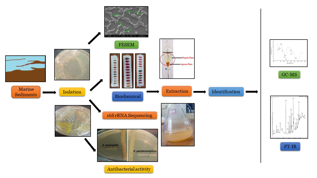
Isolation and Characterization of a Novel Actinomycete Isolated from Marine Sediments and Its Antibacterial Activity against Fish Pathogens

Centre for Nanobiotechnology, Vellore Institute of Technology (VIT), Vellore 632014, India
*
Author to whom correspondence should be addressed.
Submission received: 29 September 2022 / Revised: 21 October 2022 / Accepted: 1 November 2022 / Published: 3 November 2022
(This article belongs to the Special Issue Antimicrobial and Anti-infective Activity of Natural Products)

Abstract

:
Marine habitats are especially complex, with a varied diversity of living organisms. Marine organisms, while living in such intense conditions, have developed great physiological and metabolic potential to survive. This has led them to produce several potent metabolites, which their terrestrial counterparts are unable to produce. Over the past few years, marine Actinomycetes have been considered one of the most abundant sources of diverse and novel metabolites. In this work, an attempt was made to isolate Actinomycetes from marine sediments in terms of their ability to produce several novel bioactive compounds. A total of 16 different Actinomycete colonies were obtained from marine sediment samples. Among the 16 Actinomycete isolates, 2 isolates demonstrated in vitro antibacterial activity against Aeromonas hydrophila and Vibrio parahemolyticus. However, among them, only one isolate was found to have potent antibacterial activity, and hence, was taken for further analysis. This isolate was designated as Beijerinickia fluminensis VIT01. The bioactive components obtained were extracted and later subjected to Fourier transform infrared spectroscopy (FTIR) and gas chromatography–mass spectroscopy (GC-MS) analyses for identification. Several novel bioactive compounds were reported from the data obtained and were found to have potent antibacterial activity. Hence, they could be used as an alternative to antibiotics for treating several fish pathogens in the aquaculture industry.

Graphical Abstract

1. Introduction

Actinomycetes, belonging to the order Actinomycetales, are members of a heterogeneous group of Gram-positive bacteria, consisting of up to >55% GC content in their DNA [1]. They are anaerobic, with filamentous fungal morphology and branched growth patterns, resulting in either extensive mycelium or colony. Later, the mycelium may fall apart to form coccoid or rod-shaped forms. Most genera of Actinomycetes form a spore called sporangia or spore cases, found on the colony surface, aerial hyphae or maybe within the free environment. They exhibit diverse genetic, biological and functional activities and are a good source of many new secondary metabolites [2,3,4]. The sea has a vast treasure of resources. Due to this fact, marine Actinomycetes play a significant role by being a major part of it [5]. Apart from this reason, they differ from their terrestrial counterparts considerably because of distinct environmental changes prevailing in marine habitats [6,7].
The first marine Actinomycete was discovered in the year 1984 [4,8]. Since that period, many novel species of marine Actinomycetes have been found in aquatic environments worldwide [9,10,11,12,13]. In addition, a majority of the strains of Actinomycetes from the marine environment have been isolated from sediments [14]. The population size of Actinomycetes in the case of oceanic sediments has been reported to vary with various physicochemical parameters, such as pH, temperature, salinity, total organic carbon, pressure, etc. [4].
In general, Actinomycetes, and in particular, marine Actinomycetes have a vast range of applications in various fields. Be it in drug synthesis to cure certain diseases or biologically active products, synthesis of antibacterial, antiviral, antifungal as well as anticancer products, synthesis of enzymes, or their major role in biofouling, they have contributed a lot in various areas of the aquaculture industry [5]. Sharma et al. [15] reported the applications of Actinomycetes in bioremediation, cancer treatment and production of some valuable antibiotics, such as amphotericin, neomycin, vancomycin, chloramphenicol, novobiocin, gentamycin, nystatin, erythromycin, tetracycline, etc. They are also used as biocontrol tools, antifungal compounds, bio corrosion and as a source of agroactive compounds. The bioactive compounds extracted from these marine Actinomycetes possess different chemical structures and conformations, which hold the key to novel drug synthesis that, in future, may be potent enough to combat various resistant pathogens [16].
Actinomycetes are widely distributed in the marine environment, in marine species such as Streptomyces, Nocardia and Micromonospora. Their occurrence on the contact slides and dead marine algae suspended in the sea has been reported [17].
A study reported by Ref [18] revealed that some new marine Actinomycetes have produced a variety of bioactive metabolites. The antibacterial activity of fermentation products from Actinomycetes isolated from the marine environment also showed that several bioactive compounds had activities against vaccinia virus replication multidrug-resistant Gram-positive bacteria and cancer cells.
Actinomycetes contribute 70% of antibiotic sources and numerous non-antibiotic bioactive metabolites, including enzymes, enzyme inhibitors, anti-oxidation reagents, immunological regulators, etc. [19]. In other words, currently, almost two-thirds of the antibiotics developed are derived from Actinomycetes. Marine Actinomycetes have been reportedly studied for their efficacy as a potent novel antibiotic producer [20,21,22,23]. Although the use of several antibiotics has been banned in the aquaculture industry [24], they are still being used and are causing resistance. In this context, the aim of the current work was to isolate a novel bioactive compound from marine Actinomycetes, which is effective against pathogens in aquaculture.

2. Results

2.1. Isolation of Actinomycetes

A total of 16 different Actinomycete colonies were obtained from marine sediment samples. To isolate the Actinomycetes, specific agar medium Actinomycetes Isolation Agar (AIA) was used, since it was found to be good for the growth of actinomycetes. Some morphologically different strains of Actinomycetes were obtained from the sediment samples collected off the coast of Digha, West Bengal, India.

2.2. Characterization of Isolates

Among the 16 Actinomycete isolates, 2 isolates demonstrated in vitro antibacterial activity against Aeromonas hydrophila and Vibrio parahemolyticus. However, among them, only one isolate was found to have potent antibacterial activity, and hence, was taken for further characterization. This isolate was designated as Beijerinckia fluminensis VIT01.

2.2.1. Screening of Potential Actinomycete Isolates

The antibacterial activity of the isolate was confirmed by primary screening using the cross-streak method. This was followed by secondary screening using the agar well diffusion method. The crude extract of Actinomycete isolates showed a zone of inhibition against the two bacterial pathogens tested. Among the two pathogens, Vibrio parahemolyticus showed better results when compared to Aeromonas hydrophila. In the case of primary screening using the cross-streak method, the zones of inhibition observed in the isolate against pathogens A. hydrophila and V. parahemolyticus were 11 mm and 13 mm, respectively. In addition, the zone of inhibition observed in the secondary screening of the isolate was also found to be better in V. parahemolyticus compared to A. hydrophila.

2.2.2. Screening of the Potential Isolate in Comparison to the Antibiotic

Gentamicin was found to be sensitive to both pathogens A. hydrophila and V. parahemolyticus, showing a zone of inhibition of 11 mm and 12 mm in the case of both V. parahemolyticus and A. hydrophila, respectively, using the disc diffusion method. On the other hand, the Actinomycete isolate gave a better zone of inhibition compared to the antibiotic.
The zone of inhibition revealed in vitro antibacterial activity of Beijernickia fluminensis against both pathogens Aeromonas hydrophila and Vibrio parahemolyticus, as shown in Figure 1A. Figure 1B reveals the zone of inhibition of Gentamicin against A. hydrophila and V. parahemolyticus.
Table 1 summarizes the antibacterial activity of some Actinomycete isolates against Aeromonas hydrophila and Vibrio parahemolyticus compared to Gentamicin. The data table shows that compared to Gentamicin, Beijernickia fluminensis gave a better zone of inhibition against both pathogens A. hydrophila and V. parahemolyticus.

2.2.3. Morphological Characterization
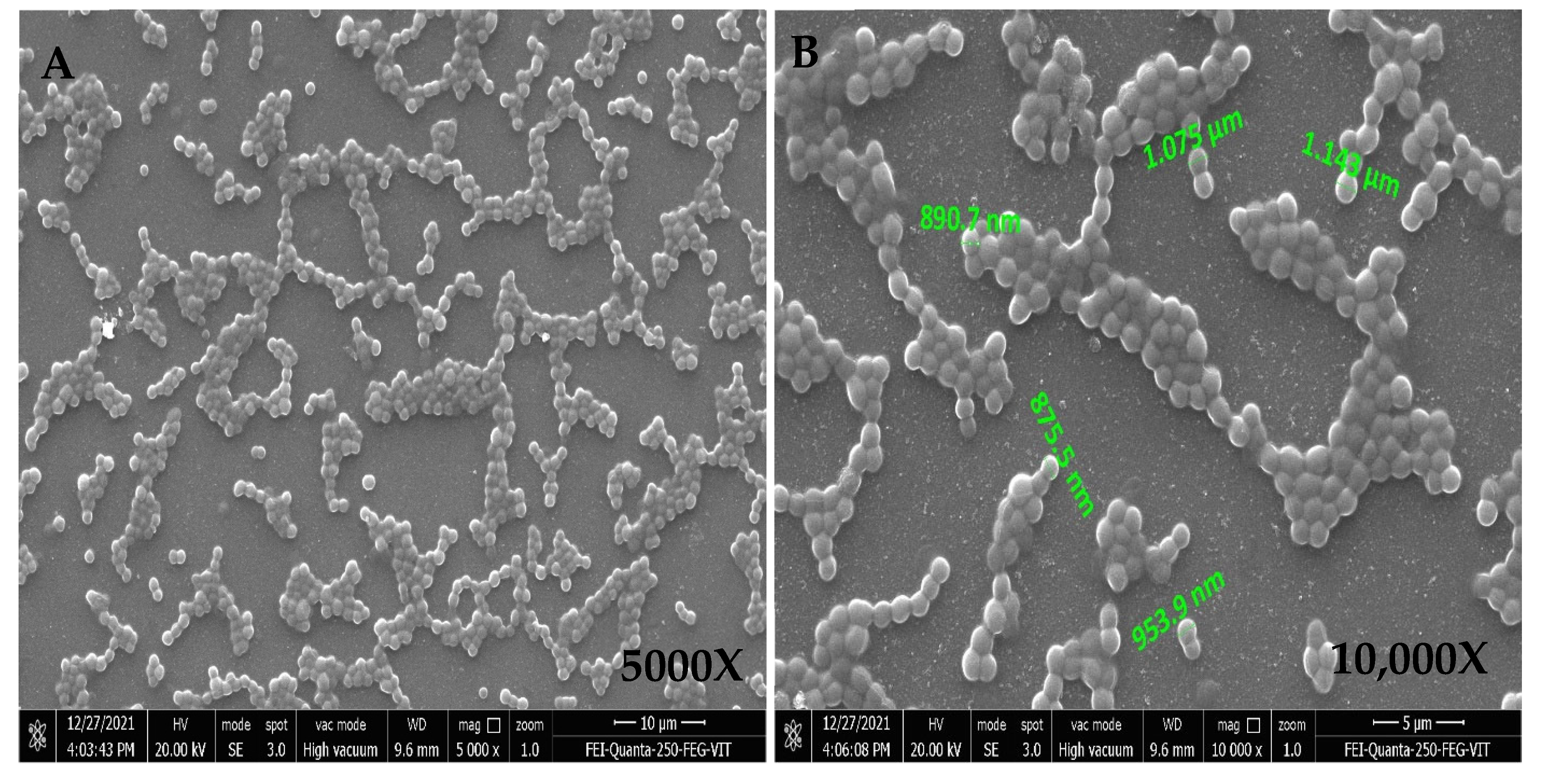
Actinomycetes were isolated, and their morphological appearance was observed. The staining results showed that the bacterium was Gram-positive, cocci-shaped, non-motile, capsulated and spore-forming.

FESEM Analysis

The FESEM micrograph revealed the shape and arrangement of the isolate under different magnifications, as shown in Figure 2.

Biochemical Characterization

The biochemical tests performed showed a positive result for Gram-positive bacteria. The results indicate that the isolate was capable of starch, gelatin and casein hydrolysis. The results of biochemical tests are presented in Table 2.

Physiological Characterization

The tested Actinomycete isolate was able to grow in 1%, 3.5%, 5% and 7% NaCl concentration but showed resistance at 10% and 20% NaCl. The ideal temperature for the growth of Actinomycetes was found to be between 28 and 30 °C, although they could even grow in temperatures up to 40 °C.

Molecular Characterization

The 16S rRNA sequencing revealed that the strain belongs to Beijerinckia fluminensis under Actinomycetes. This was designated as Beijerinckia fluminensis VIT01.

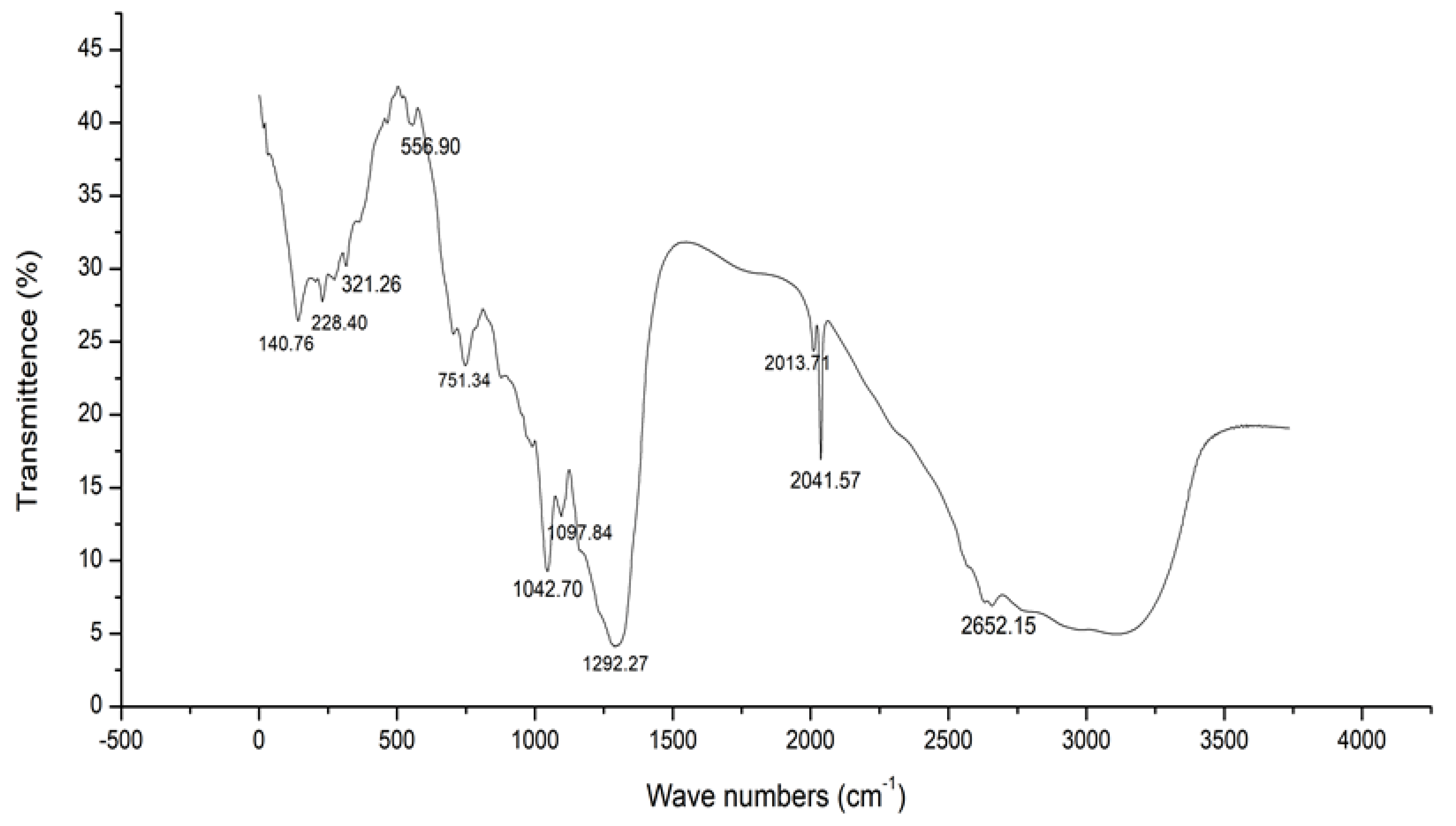
2.3. FTIR Analysis of Crude Extracts from Actinomycetes

Figure 3 summarizes the FTIR spectra of the sample ranging from 400 to 4000 cm−1. The characteristic bands observed from Sample 1 are 2652.15, 2041.57, 1292.27, 1042.70, 751.34 and 556.90. The data shown in Table 2 reveal the vibrational type of the functional groups, their band strength and the compounds present in the respective peaks of the ethyl acetate extract of B. fluminensis. The peak at 2652.15 corresponds to the stretching vibrational type of weak thiol groups. The peak at 2041.57 was assigned to the stretching vibration of strong isothiocyanate groups. Similarly, the peaks at 1292.27 and 1042.70 correspond to the stretching vibration type of strong fluoro compound and medium amine groups, respectively. In addition, the peak at 751.34 corresponds to the bending vibration type of strong 1,2-disubstituted groups, and the peak at 556.90 corresponds to the stretching type of vibration of strong halo compound groups.
The peak values corresponding to their respective groups in the sample are shown in Table 3.

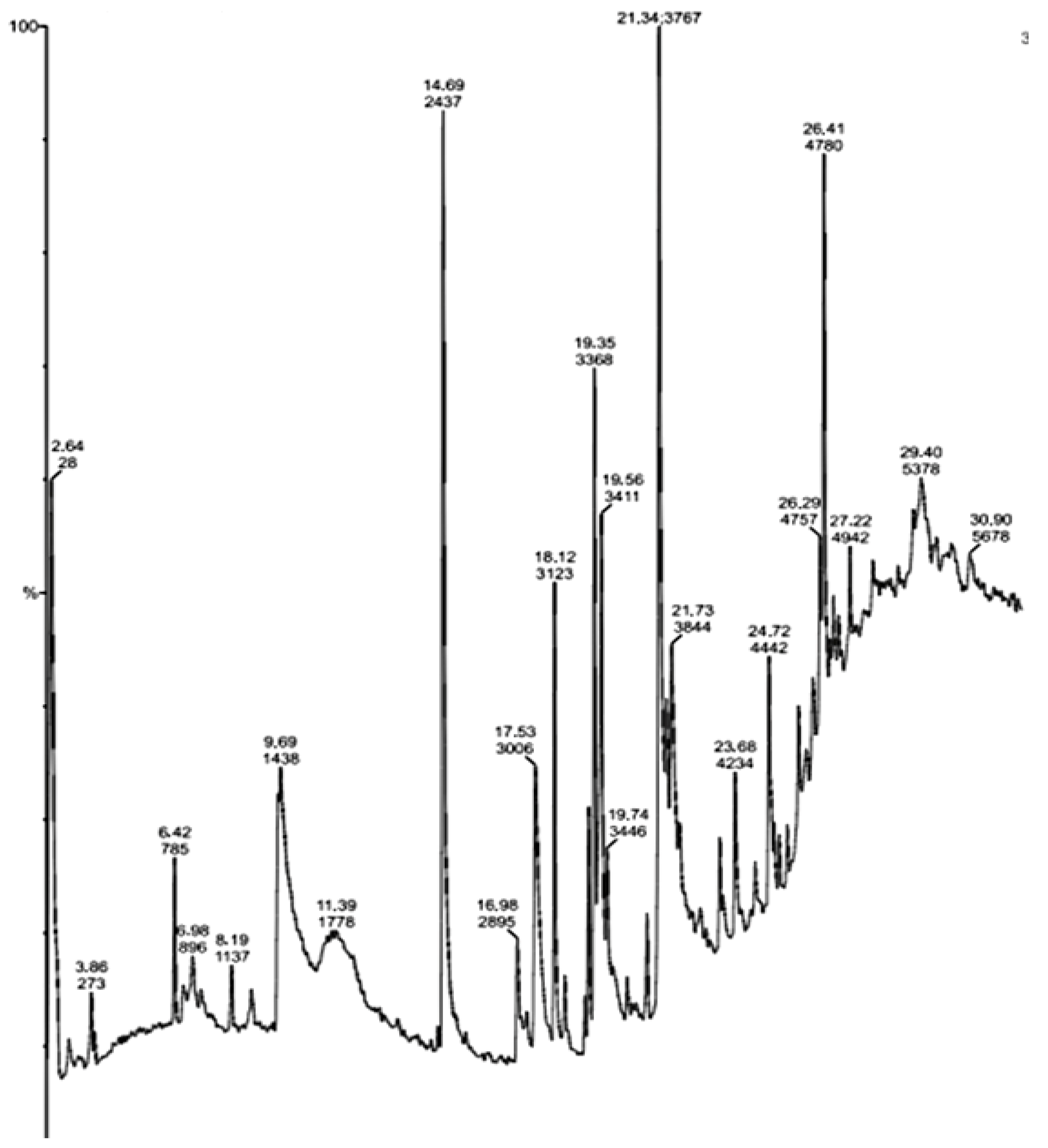
2.4. GC-MS Analysis of Crude Extracts from Actinomycetes

The GC-MS analysis of the identified active compounds isolated from the ethanolic extract of Actinomycetes displayed their peak percentage expressed with their retention indices in the chromatogram.
The bioactive compounds identified from the ethanolic extract of Beijerinckia fluminensis VIT01 showed that they contain many active components. They are responsible for several properties, including antimicrobial and antibacterial, as per Dr. Duke’s Phytochemical and Ethnobotanical Database. Figure 4 represents the chromatogram of the bioactive compounds extracted from the ethanolic extract of the Actinomycete strain sample. The compounds are 1,6;3,4-Dianhydro-2-Deoxy-Beta-D-Lyxo-Hexopyrano; N,N-Dimethylheptanamide, Butanamide,3,N-Dihydroxy; (S)-Isopropyl Lactate; 3-O-Acetyl-exo-1,2-O-Ethylidene-Alpha-D-Erythrof; Glycine,N-Octyl-,Ethyl Ester; D-Glucitol,1-Deoxy-1-(Octylamino); Glycyl-L-Proline; 7-Tetradecene, (E); 2,3-Anhydro-D-Galactosan, etc. The data from Table 4 show the major peak, the retention time of the compounds and the activities present in the ethyl acetate extract of B. fluminensis. The results revealed that compounds such as N, N-Dimethylheptanamide, Glycine,N-Octyl-,Ethyl Ester, Glycyl-L-Proline, Actinomycin C2, (S)-3,4-Dimethylpentanol and 7-Tetradecene, (E), have antibacterial properties. Therefore, these compounds can be used as potent antibacterial agents.
Table 4 summarizes the main compounds, retention time, major peak area and major activities present in the sample. These bioactive compounds were later used for in vivo and in vitro studies.

3. Discussion

Actinomycetes have proven to be one of the best sources of secondary metabolites, and they have potent bioactive compounds [26]. Currently, the incidence of infectious diseases in the aquaculture industry is increasing, leading to significant loss in the aquaculture industry. Hence, there is a dire need to develop novel bioactive compounds that can be effective against various pathogens. While screening for Actinomycetes, various fast growing microbial colonies, which have the ability to inhibit the colonization of Actinomycetes on their particular isolation medium, were detected. In order to isolate Actinomycetes and prevent the growth of unwanted as well as ubiquitous bacteria, pre-treatment of the sediment samples was preferred. Gebreyohannes et al. [27] reported that potent antibiotic-producing actinomycetes isolated from the marine sediment were subjected to physical pre-treatment methods to stimulate the growth of Actinomycetes. In the present study, similar aseptic methods were followed to prevent the growth of unwanted bacteria.
In recent times, there has been a growing interest in the isolation of various novel and potent bioactive Actinomycetes from the marine environment, since they contain bioactive compounds. Many culture-independent studies [28] have reported that the marine environment contains a vast and rich diversity of rare Actinomycetes. They have been useful in designing successful isolation schemes to isolate a wide variety of marine Actinomycetes. Oggerin et al. [29] isolated Beijerinickia fluminensis strains UQM 1685T and CIP 106281T from soil. Similarly, a recent study was conducted by Shwaaiman et al. [30] who isolated a novel bacterium Beijerinickia fluminensis BFC-33 from soil. In the present study, we isolated Beijerinickia fluminensis from marine sediments. Based on the literature survey, this might be the first report on the isolation of Beijerinickia fluminensis from marine sediments.
Trakunjae et al. [31] reported that they isolated a rare Actinomycete, Rhodococcus so. BSRT1-1, which was Gram-positive, non-motile, with short rods that later converted to cocci. Oggerin et al. [29] isolated Beijerinickia fluminensis from a soil sample. In the present work, the bacterium Beijerinickia fluminensis was isolated from marine sediments. The present work revealed that the isolated bacterium is light orange colored. It is Gram positive, with cocci in clusters, and non-motile. This corroborated the study of Oggerin et al. [29] who reported that a white and cream colored isolate was non-motile. Several studies reported on the biochemical and physiological characteristics of Actinomycetes. A study by Ref [32] reported the biochemical and physiological characteristics of Actinomycetes. They reported the presence of starch and gelatin. Casein hydrolysis was also reported. They also reported negative results for indole, Voges–Proskauer, urease and lipase, and positive results for methyl-red and citrate. In the present study, B. fluminensis gave positive results for starch, casein and gelatin hydrolysis, citrate and urease, while negative results were obtained for methyl-red, Voges–Proskauer and indole tests. Rajkumar et al. [33] isolated actinomycetes from different environments. They reported that the isolated Actinomycetes showed negative Indole, MR and VP. They also reported that the isolates from Actinomycetes CAHSH-2 and Actinomycetes CAHSH-3 were positive for citrate, urease, starch, casein and gelatin. This is in agreement with our report. In the case of physiological characterization, Sivanandhini et al. [32] revealed that the Actinomycetes were able to survive in high salt concentrations. Similarly, our present work showed that B. fluminensis was able to tolerate up to 7% salt concentration. Another study reported by Ref [27] on the Actinomycetes isolated from water and sediments showed starch and urea hydrolysis. It was also able to tolerate NaCl concentration up to 5%. Our present study showed that both starch and urea were able to hydrolyze. It was also able to survive in 7% salt concentration. Similarly, Undabarrena et al. [34] reported the physiological characteristics of several species of Actinobacteria. They reported that they were able to survive at 1%, 3.5%, 5%, 7% and 10% NaCl concentration. In the present work, we observed that they were able to tolerate up to 7% salt concentration. Rajkumar et al. [33] also reported that the Actinomycetes were able to survive in 9% NaCl. In the present study, equally similar biochemical and physiological characteristics, such as NaCl tolerance (salinity), temperature tolerance, as well as biochemical characteristics of Actinomycetes, were reported.
Primary and secondary screening was performed to evaluate the antimicrobial potential of the microorganism against the pathogens. Singh et al. [35] screened thirty-six Actinomycete isolates against both Gram-positive and Gram-negative bacterial pathogens, such as Staphylococcus aureus MTCC 96, Bacillus subtilis MTCC 441 and Escherichia coli MTCC 64, and a fungal pathogen Candida albicans MTCC 183. Among them, fifteen showed strong and moderate antimicrobial activity. Later, they were subjected to secondary screening, where out of fifteen active isolates, thirteen exhibited strong antimicrobial activity. Another study by Ref. [36] reported that different strains of Streptomyces sp. showed antibacterial activity against Vibrio spp. In the present study, good antibacterial activity was seen in the primary screening of the isolate, which exhibited a zone of inhibition of 11 mm and 13 against pathogens A. hydrophila and V. parahemolyticus, respectively, followed by secondary screening.
Norouzi et al., 2018 [37] reported the antibacterial activity potential of several Actinomycete isolates against many bacterial pathogens in comparison to antibiotics such as Gentamicin using the Kirby–Bauer disc diffusion method. Antibiotic sensitivity test and disc diffusion were carried out to compare the activity of the crude extract of the Actinomycete isolate with the antibiotic Gentamycin. The disc diffusion revealed that, among the three isolates, two potent marine Actinomycetes isolates MN2 and MN39 were able to produce biomolecules with antibacterial activity against multidrug-resistant (MDR) bacteria. In our present study, we observed that our isolate gave a better result compared to Gentamicin. Gentamicin exhibited zones of inhibition of 11 mm and 12 mm in the case of both pathogens, V. parahemolyticus and A. hydrophila. The zones of inhibition observed in the isolate against pathogens A. hydrophila and V. parahemolyticus were 11 mm and 13 mm, respectively.
Currently, the number of many novel secondary metabolites reported from marine Actinomycetes has surpassed the number of their terrestrial counterparts [38]. Dholakiya et al. [39] reported the results of a study where the bacteria Streptomyces variabilis exhibited a wide spectrum of antibacterial activities against several Gram-negative and Gram-positive bacteria. Merochlorin E and F isolated from a marine bacterium Streptomyces sp. displayed antibacterial activities against several bacteria, such as Staphylococcus. aureus, Bacillus. subtilis and Kocuria. rhizophila [40,41]. Voon et al. [42] reported the FTIR spectrum of biocellulose from Beijerinickia fluminensis WAUPM53 in different media. Another study by Ref [43] reported the FTIR spectra of melanin produced by Beijerinickia fluminensis. In the current study, several potent antibacterial compounds were found in the FTIR and GC-MS analyses of Actinomycetes.

4. Materials and Methods

4.1. Sample Collection

The samples for screening were collected in the month of May 2021, off the coast of Digha, West Bengal, India. The marine sediment samples were collected from Digha (21°37′35.82″ N latitude and 87°30′26.75″ E longitude) and transferred to autoclavable bags and then stored in cell frost at 4 °C [27,34].

4.2. Media Used

Media

The Actinomycetes Isolation Agar (AIA) medium (Hi Media) was used for the isolation of Actinomycetes. Nalidixic acid (50 µg/mL) and Mycostatin (100 µg/mL) were added to the medium. The International Streptomyces Project (ISP-4) medium (TM media, Delhi, India) was used for purification of the isolates [44]. The Mueller–Hinton Agar (MHA) medium (HiMedia, Mumbai, India) was used for the determination of antimicrobial activity of the Actinomycete isolates.

4.3. Pre-Treatment of Sediment Samples

To enable the isolation of Actinomycetes, samples were subjected to pre-treatment. Sediment samples were kept overnight at 70 °C in a hot air oven for drying [45]. They were crushed and ground aseptically using mortar and pestle under aseptic condition.

4.4. Isolation of Samples

From the pre-treated sediment sample, 1 g was weighed and dispersed into a 100 mL conical flask containing distilled water with 0.9% saline and shaken continuously at room temperature using an orbital shaker for 10 min. This was considered as the stock culture for the sediment samples. A volume of 1 mL was taken from the stock solution and transferred to 9 mL sterile test tube containing water and mixed vigorously to reach a dilution factor of 10−1. Afterward, serial dilutions from 10−2 to 10−6 were carried out.
After serial dilution, a 0.1 mL aliquot sample of each dilution from 10−3 to 10−6 was plated in the AIA medium (HiMedia) containing 0.9% NaCl using the spread plate technique. The medium was amended with 50 µg/mL of Nalidixic acid (HiMedia) and Mycostatin (100 µg/mL) (HiMedia) to prevent the growth of other microorganisms. The Petri plates were incubated at 30 °C for 7–14 days until visible colonies were observed [39,46].

4.5. Screening of Samples

The pure colonies were counted using a colony counter. Their morphological characteristics and pigmentation were recorded.
After isolation, the isolates were screened and sub-cultured by individual streaking into AIA medium prepared with 0.9% NaCl. They were then transferred to new Petri plates containing AIA medium to obtain pure colonies. In addition, they were also sub-cultured and maintained in AIA slants at 4 °C [47]. The isolated bacteria were later stored at −20 °C in 20% glycerol [34,48].

4.6. Characterization of Isolates

Strains were identified based on morphological, cultural, biochemical characteristics and molecular analysis using 16S rRNA sequencing.

4.6.1. Morphological Characterization

The selected isolates were streaked into an ISP4 medium to study their morphological characters by the macroscopic method. The colonies, morphology of the substrate and spores, aerial hyphae, branching and pigment production on the plates were observed in the preliminary stage. Gram staining, motility test and capsule staining were carried out to check the structure and motility of the isolated colonies.

Gram Staining

Gram staining was performed to identify the isolates, i.e., whether they are Gram positive or Gram negative. Later, the organism was first observed under 10× and 40× magnification and then under 100× with oil immersion [49].

Motility Test

A motility test was carried out by inoculating the isolate using the Sulphide Indole Motility (SIM) medium with an inoculation loop up to half or one-third inch above the bottom in a straight line. It was then incubated at 30 °C for 7 days. In addition, it was also observed under 10× and 40× magnification through the microscope to check its motility.

Capsule Staining

For capsule staining, Nigrosin was used as the primary stain and crystal violet as a counterstain. It was observed under 100× oil immersion microscope.

Field Emission Scanning Electron Microscope (FESEM) Analysis

The isolates selected were examined microscopically for spore chain morphology under 3000×, 5000×, 8000×, 10,000× and 13,000×. FESEM analysis was performed by two different methods to check the arrangement of the spores, the sporulating structures and the external surface morphology of the isolates. In the first method, the isolate was inoculated in a broth medium and incubated in a rotary shaker at 30 °C for 7 days until there was growth of bacteria. The broth culture was centrifuged at 7000 rpm for 10 min, and the supernatant was discarded. An amount of 1 mL autoclaved 1× Phosphate Buffer Saline (PBS) was added to the cell-free supernatant (CFS) and mixed well. Later, 10–20 µL of this culture was transferred to a clean grease-free slide. An amount of 20 µL of 0.25% Glutaraldehyde solution was dispensed into the slide containing the culture and oven dried overnight at 40 °C. In the second method, a clean, grease-free slide was taken, and a thin smear of the isolate was made in the center of it, air dried completely and kept overnight in a hot air oven at +40 °C.

4.6.2. Biochemical Characterization

Biochemical Tests

Biochemical tests, such as ONPG, Ornithine, Lysine, Phenylalanine, Urease, H2S Production, Nitrate Reduction, Methyl Red–Voges–Proskauer’s Tests (MR-VP), Indole Production, Esculin Hydrolysis, Malonate, Catalase, Oxidase, Citrate Utilization, Crystal violet, Casein Utilization and Growth in MacConkey Agar, were carried out using a readily prepared KB003 Hi 25 Kit (HiMedia). Both Strip-I and Strip-II reaction wells of the kit were inoculated with Actinomycete culture using an inoculation loop incubated at 30 °C for 7–14 days.
In addition to the above-conducted tests, readily prepared KB009 Hi CarboTM Kit B009C HiMedia kit was used to check the capability of the isolated Actinomycetes to use several carbon compounds as an energy source. The kit consisted of 11 sugars, including Melezitose, Xylitol, Cellobiose, D-Arabinose, Sorbose, Rhamnose, alpha-Methyl-D-Mannoside, Malonate, Esculin, Citrate, O-Nitrophenyl-β-D-galactopyranoside (ONPG) and one control. Parts C of the wells in the kit were inoculated similarly to the Actinomycetes and incubated at 30 °C for 7–14 days [33].

Starch Utilization Test

A starch hydrolysis test was performed in which the isolates were inoculated on sterile starch agar plates and incubated at 30 °C for seven days. Following the incubation period, iodine solution was flooded into the plates. Starch hydrolysis was confirmed by the presence of a clear zone of hydrolysis around the bacterial growth [27].

Gelatin Hydrolysis Test

In the gelatin hydrolysis test, the isolates were inoculated on gelatin agar plates and incubated at 30 °C for seven days to check the gelatin hydrolysis. At the end of the incubation period, 1 mL mercuric chloride solution was dispersed into the agar medium to observe the zone of hydrolysis [27].

Casein Hydrolysis Test

The isolates were similarly streaked on skimmed milk agar medium plates. They were then incubated at 30 °C for seven days to see whether there was a zone of hydrolysis [27].

Citrate Utilization Test

The isolates were also inoculated on Simon’s citrate slant agar test tubes incubated at 30 °C for seven days until a color change was found [27].

4.6.3. Physiological Characterization

To test the NaCl resistance, the AIA medium was prepared in seven batches and amended with 0%, 1%, 3.5%, 5%, 7%, 10% and 20% NaCl. The isolates were streaked on the media and incubated at 30 °C for seven days. The growth of the isolates at the highest salt concentration was observed and recorded.
The sterile AIA plates were inoculated with the isolates and incubated at 4 °C, 20 °C, 25 °C, 30 °C, 37 °C and 40 °C for seven days. The maximum growth at the optimal temperature was recorded by the visual observation of growth [50].

4.6.4. Molecular Characterization

Based on the biochemical characteristics and morphology of the isolate, the isolate that showed good antibacterial activity was chosen for further study. The 16S ribosomal RNA (rRNA) sequence analysis was carried out to confirm the isolated bacterium.

4.7. Bacterial Cultures Used

The two bacterial fish pathogens selected for this study were Vibrio parahemolyticus (MTCC 451) and Aeromonas hydrophila (MTCC 1739). The stock and slant cultures of these test organisms were obtained from the Microbial Type Culture Collection (MTCC). Both cultures were inoculated into the nutrient broth and sub-cultured on nutrient agar plates before performing the test.

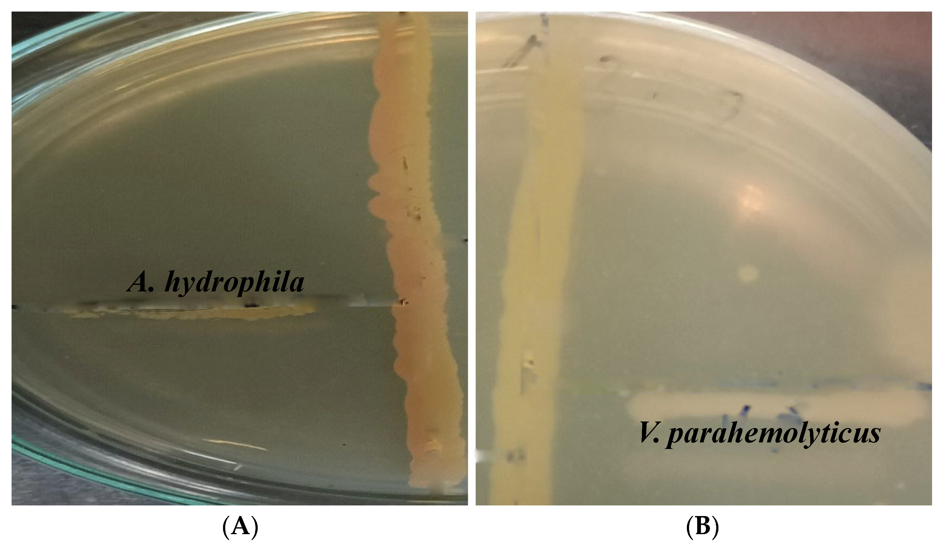
4.8. In Vitro Antibacterial Activity

4.8.1. Primary Screening

Preliminary screening was carried out by the cross-streak method in which the isolates were screened against the selected test pathogens. Mueller–Hinton Agar (MHA) plates were prepared and supplemented with 2% sodium chloride. The Actinomycete isolates were inoculated with a single streak in the center of MHA plates and incubated at 30 °C for 7 days. Later, the sub-cultured test cultures were streaked perpendicular to the Actinomycetes isolates at a 90° angle and incubated at 37 °C for 24 h. The zone of inhibition was recorded [35]. Based on the zone of inhibition observed against the test organism, the potential isolates of Actinomycetes were selected for secondary screening.

4.8.2. Secondary Screening

The Actinomycete isolates that showed prospective antibacterial activities in primary screening were subjected to solvent extraction to obtain the crude extracts. The isolates were inoculated into a Starch Casein broth/ISP2 broth and kept in a rotary shaker incubator for seven days at 30 °C and 120 rpm until growth was visible. Centrifugation was performed at 4 °C at 10,000 rpm for 20 min, and the filtrate was aseptically transferred into conical flasks. Equal volumes of organic solvents with ethanol and an aqueous extract with distilled water were added to the cell-free supernatant (CFS) and shaken well for two hours to extract the antibacterial compounds [51].

Agar Well Diffusion Method

The antibacterial activities of the crude extracts were tested at different concentrations, ranging from 50 µg/mL to 250 µg/mL, by the agar well diffusion method. Lawn cultures of the pathogenic organisms were spread on the solidified MHA agar plates using sterile cotton swabs. The wells were prepared on MHA plates using a sterile cork borer. The different concentrations of both ethanolic and aqueous extracts of Actinomycete isolates were poured into each well and allowed to diffuse completely. Culture plates were incubated at 37 °C for 24 h. Experiments were carried out three times, and the mean of the zone of inhibition (in mm) was recorded [52].

Disc Diffusion Method

Gentamycin (10 mcg) was taken for the antibiotic sensitivity test, which was performed for both pathogens. The Actinomycete isolate showing the maximum zone of inhibition was chosen for further study by the Kirby–Bauer agar disc diffusion method [36] to check the antibacterial activity against pathogens. In this method, the pathogens were first swabbed entirely from the surface of sterile MHA plates. Then, the concentration of the isolate showing positive results was added inside the disc and placed on the surface of the medium and incubated at 37 °C for 24 h. The zone of inhibition was recorded in triplicate to calculate the mean value of the inhibition zones.

4.9. Extraction of the Active Compound

The inoculum was prepared by inoculating the selected Actinomycete isolate into a Starch Casein Broth/ISP-2 Broth kept in a rotary shaker incubator at 30 °C and harvested after 7 days. The broth culture was then filtered through Whatman No. 1 filter paper. The filtrate obtained was centrifuged at 4 °C, 10,000 rpm, for 20 min. The CFS collected was further used for the extraction of metabolites. The CFS was extracted three times with an equal volume of ethyl acetate by shaking manually at a 1:1 ratio by using a separating funnel. Then, it was kept in a shaker for 48 h, after which it was removed and the solvent layer collected. The concentration of the solvent layer was carried out using a rotary evaporator to obtain the crude metabolites [33]. As per some earlier reports, studies were conducted using various solvents, which gave better results in the extraction of bioactive compounds from the CFS of Actinomycetes. Among them, ethyl acetate was found to be a good polar solvent [53,54].

4.10. Identification of the Crude Bioactive Compound

Compounds obtained from the Actinomycete isolate were dissolved in ethyl acetate and identified using gas chromatography–mass spectroscopy (GC-MS) and Fourier transform infrared spectroscopy (FTIR) analyses.
A 13 mm potassium bromide (KBr) powder was mixed and ground thoroughly using a mortar and pestle with the crude bioactive compounds isolated from potential Actinomycete isolates. The sample was placed inside a pellet dye in a quantity just sufficient to cover its bottom. It was pressed at 5000–10,000 psi using the pellet press to form a pellet disc and placed in the sample holder. It was then analyzed by FTIR to obtain the measurement in the spectrum wave number range between 4 and 4000/cm at a resolution of 4 cm−1 using a FTIR spectrometer (Jasco, Victoria, BC, Canada).
GC-MS was performed for the identification of the active components present in the ethanolic extract of Actinomycetes. The unknown components were identified using a GC-MS (Agilent 6890/Hewlett-Packard 5975 (Agilent, Palo Alto, CA, USA/Hewlett Packard Labs, Milpitas, CA, USA) under the electron impact (EI) mode. Several acquisition parameters were followed with respect to the analysis. The interpretation of the mass spectrum was carried out using the National Institute Standard and Technology (NIST) database. The NIST library was used for searching for the spectrum of unknown compounds from the Actinomycetes [55].

5. Conclusions

Sixteen Actinomycetes were successfully isolated from marine sediments. Among them, two strains displayed antibacterial activity, but one strain, Beijerinickia fluminensis, was reported to have potent antibacterial activity and was hence selected for further analysis. FTIR and GC-MS revealed several antibacterial compounds that can be used as an alternative to antibiotics for the treatment of several pathogens in aquaculture. Based on the literature survey, this might be the first report on the isolation of Beijerinickia fluminensis from marine sediments.

Author Contributions

All authors contributed to the study conceptualization and design. Formal analysis, methodology, validation and writing of the original draft were performed by H.M. Conceptualization, investigation, supervision, validation, review and editing of the manuscript were performed by J.T. All authors commented on previous versions of the manuscript. All authors have read and agreed to the published version of the manuscript.

Funding

This research received no external funding.

Institutional Review Board Statement

Not applicable.

Informed Consent Statement

Not applicable.

Data Availability Statement

Not applicable.

Acknowledgments

The authors are thankful to VIT Vellore for providing the required facilities to carry out this work.

Conflicts of Interest

The authors declare no conflict of interest.

References

  1. Mo, E.; Wa, E.; Gm, D. Marine Actinomycetes the Past, the Present and the Future. Pharm. Res. 2021, 5, 000241. [Google Scholar] [CrossRef]
  2. Arbat, A.B.; Zodpe, S.N. Biodiversity of Actinomycetes Species Isolated from Saline Belt of Akola District. Indian J. Appl. Res. 2011, 4, 450–452. [Google Scholar] [CrossRef]
  3. Rashad, F.M.; Fathy, H.M.; El-Zayat, A.S.; Elghonaimy, A.M. Isolation and Characterization of Multifunctional Streptomyces Species with Antimicrobial, Nematicidal and Phytohormone Activities from Marine Environments in Egypt. Microbiol. Res. 2015, 175, 34–47. [Google Scholar] [CrossRef]
  4. Jagannathan, S.V.; Manemann, E.M.; Rowe, S.E.; Callender, M.C.; Soto, W. Marine Actinomycetes, New Sources of Biotechnological Products. Mar. Drugs 2021, 19, 365. [Google Scholar] [CrossRef] [PubMed]
  5. Rasool, U.; Hemalatha, S. Diversified Role and Applications of Marine Actinomycetes in the Field of Biology. World Sci. News 2017, 74, 1–14. [Google Scholar]
  6. Imada, C.; Koseki, N.; Kamata, M.; Kobayashi, T.; Hamada-Sato, N. Isolation and Characterization of Antibacterial Substances Produced by Marine Actinomycetes in the Presence of Seawater. Actinomycetologica 2007, 21, 27–31. [Google Scholar] [CrossRef] [Green Version]
  7. Valli, S.; Sugasini, S.S.; Aysha, O.S.; Nirmala, P.; Vinoth Kumar, P.; Reena, A. Antimicrobial Potential of Actinomycetes Species Isolated from Marine Environment. Asian Pac. J. Trop. Biomed. 2012, 2, 469–473. [Google Scholar] [CrossRef] [Green Version]
  8. Helmke, E.; Weyland, H. Rhodococcus Marinonascens. Int. J. Syst. Bacteriol. 1984, 34, 127–138. [Google Scholar] [CrossRef] [Green Version]
  9. Colquhoun, J.A.; Mexson, J.; Goodfellow, M.; Ward, A.C.; Horikoshi, K.; Bull, A.T. Novel Rhodococci and Other Mycolate Actinomycetes from the Deep Sea. Antonie Van Leeuwenhoek Int. J. Gen. Mol. Microbiol. 1998, 74, 27–40. [Google Scholar] [CrossRef]
  10. Mincer, T.J.; Jensen, P.R.; Kauffman, C.A.; Fenical, W. Widespread and Persistent Populations of a Major New Marine Actinomycete Taxon in Ocean Sediments. Appl. Environ. Microbiol. 2002, 68, 5005–5011. [Google Scholar] [CrossRef] [Green Version]
  11. Yi, H.; Schumann, P.; Sohn, K.; Chun, J. Serinicoccus Marinus Gen. Nov., Sp. Nov., a Novel Actinomycete with L-Ornithine and L-Serine in the Peptidoglycan. Int. J. Syst. Evol. Microbiol. 2004, 54, 1585–1589. [Google Scholar] [CrossRef] [PubMed]
  12. Das, S.; Lyla, P.S.; Khan, S.A. Distribution and Generic Composition of Culturable Marine Actinomycetes from the Sediments of Indian Continental Slope of Bay of Bengal. Chin. J. Oceanol. Limnol. 2008, 26, 166–177. [Google Scholar] [CrossRef]
  13. Freel, K.C.; Edlund, A.; Jensen, P.R. Microdiversity and Evidence for High Dispersal Rates in the Marine Actinomycete “Salinispora Pacifica”. Environ. Microbiol. 2012, 14, 480–493. [Google Scholar] [CrossRef] [PubMed] [Green Version]
  14. Ghanem, N.B.; Sabry, S.A.; El-Sherif, Z.M.; Abu El-Ela, G.A. Isolation and Enumeration of Marine Actinomycetes from Seawater and Sediments in Alexandria. J. Gen. Appl. Microbiol. 2000, 46, 105–111. [Google Scholar] [CrossRef] [Green Version]
  15. Sharma, M.; Dangi, P.; Choudhary, M. Actinomycetes: Source, identification, and their applications. Int. J. Curr. Microbiol. Appl. Sci. 2014, 3, 801–832. [Google Scholar]
  16. Solanki, R.; Khanna, M.; Lal, R. Bioactive Compounds from Marine Actinomycetes. Indian J. Microbiol. 2008, 48, 410–431. [Google Scholar] [CrossRef] [Green Version]
  17. Okami, Y.; Okazaki, T. Nocardia and Streptomyces; Gaustav Fischer: Stuttgart, Germany, 1978; pp. 145–151. [Google Scholar]
  18. Magarvey, N.A.; Keller, J.M.; Bernan, V.; Dworkin, M.; Sherman, D.H. Isolation and Characterization of Novel Marine-Derived Actinomycete Taxa Rich in Bioactive Metabolites. Appl. Environ. Microbiol. 2004, 70, 7520–7529. [Google Scholar] [CrossRef] [Green Version]
  19. Dilip, C.; Mulaje, S.S.; Mohalkar, R.Y. A Review on Actinomycetes and Their Biotechnological Application. Int. J. Pharm. Sci. Res. 2013, 4, 1730–1742. [Google Scholar] [CrossRef]
  20. Riedlinger, J.; Reicke, A.; Zähner, H.; Krismer, B.; Bull, A.T.; Maldonado, L.A.; Ward, A.C.; Goodfellow, M.; Bister, B.; Bischoff, D.; et al. Abyssomicins, Inhibitors of the Para-Aminobenzoic Acid Pathway Produced by the Marine Verrucosispora Strain AB-18-032. J. Antibiot. 2004, 57, 271–279. [Google Scholar] [CrossRef] [Green Version]
  21. Soria-Mercado, I.E.; Prieto-Davo, A.; Jensen, P.R.; Fenical, W. Antibiotic Terpenoid Chloro-Dihydroquinones from a New Marine Actinomycete. J. Nat. Prod. 2005, 68, 904–910. [Google Scholar] [CrossRef]
  22. El-Gendy, M.M.A.; Shaaban, M.; Shaaban, K.A.; El-Bondkly, A.M.; Laatsch, H. Essramycin: A First Triazolopyrimidine Antibiotic Isolated from Nature. J. Antibiot. 2008, 61, 149–157. [Google Scholar] [CrossRef] [PubMed] [Green Version]
  23. Manivasagan, P.; Venkatesan, J.; Sivakumar, K.; Kim, S.K. Pharmaceutically Active Secondary Metabolites of Marine Actinobacteria. Microbiol. Res. 2014, 169, 262–278. [Google Scholar] [CrossRef] [PubMed]
  24. Newman, S.G. Antibiotics in Aquaculture. Is Responsible Use Possible? 2007. Available online: https://www.globalseafood.org/advocate/antibiotics-in-aquaculture-is-responsible-use-possible/ (accessed on 28 September 2022).
  25. Duke, J.U.S. Dr. Duke’s Phytochem. Ethanobotanical Databases; Department of Agriculture, Agricultural Research Service: Washington, DC, USA, 1992. [CrossRef]
  26. Suthindhiran, K.; Kannabiran, K. Cytotoxic and Antimicrobial Potential of Actinomycete Species Saccharopolyspora Salina VITSDK4 Isolated from the Bay of Bengal Coast of India. Am. J. Infect. Dis. 2009, 5, 90–98. [Google Scholar] [CrossRef]
  27. Gebreyohannes, G.; Moges, F.; Sahile, S.; Raja, N. Isolation and Characterization of Potential Antibiotic Producing Actinomycetes from Water and Sediments of Lake Tana, Ethiopia. Asian Pac. J. Trop. Biomed. 2013, 3, 426–435. [Google Scholar] [CrossRef] [Green Version]
  28. Chen, Y.; Zeng, D.; Ding, L.; Li, X.L.; Liu, X.T.; Li, W.J.; Wei, T.; Yan, S.; Xie, J.H.; Wei, L.; et al. Three-Dimensional Poly-(ε-Caprolactone) Nanofibrous Scaffolds Directly Promote the Cardiomyocyte Differentiation of Murine-Induced Pluripotent Stem Cells through Wnt/β-Catenin Signaling. BMC Cell Biol. 2015, 16, 1–13. [Google Scholar] [CrossRef] [Green Version]
  29. Oggerin, M.; Arahal, D.R.; Rubio, V.; Marín, I. Identification of Beijerinckia Fluminensis Strains CIP 106281T and UQM 1685T as Rhizobium Radiobacter Strains, and Proposal of Beijerinckia Doebereinerae Sp. Nov. to Accommodate Beijerinckia Fluminensis LMG 2819. Int. J. Syst. Evol. Microbiol. 2009, 59, 2323–2328. [Google Scholar] [CrossRef]
  30. Shwaaiman, H.A.; Shahid, M.; Elgorban, A.M.; Siddique, K.H.M.; Syed, A. Beijerinckia Fluminensis BFC-33, a Novel Multi-Stress-Tolerant Soil Bacterium: Deciphering the Stress Amelioration, Phytopathogenic Inhibition and Growth Promotion in Triticum Aestivum L. (Wheat). Chemosphere 2022, 295, 133843. [Google Scholar] [CrossRef]
  31. Trakunjae, C.; Boondaeng, A.; Apiwatanapiwat, W.; Kosugi, A.; Arai, T.; Sudesh, K.; Vaithanomsat, P. Enhanced Polyhydroxybutyrate (PHB) Production by Newly Isolated Rare Actinomycetes Rhodococcus Sp. Strain BSRT1-1 Using Response Surface Methodology. Sci. Rep. 2021, 11, 1896. [Google Scholar] [CrossRef]
  32. Sivanandhini, T.; Subbaiya, R.; Gopinath, M.; Mahavinod Angrasan, J.K.V.; Kabilan, T.; Masilamani Selvam, M. An Investigation on Morphological Characterization of Actinomycetes Isolated from Marine Sediments. Res. J. Pharm. Biol. Chem. Sci. 2015, 6, 1234–1243. [Google Scholar]
  33. Rajkumar, T.; Manimaran, M.; Taju, G.; Vimal, S.; Majeed, S.A.; Kannabiran, K.; Sivakumar, S.; Kumar, K.M.; Madhan, S.; Hameed, A.S. Antiviral Viral Compound from Streptomyces Ghanaensis like Strain against White Spot 2 Syndrome Virus (WSSV) of Shrimp. bioRxiv 2018. [Google Scholar] [CrossRef] [Green Version]
  34. Undabarrena, A.; Beltrametti, F.; Claverías, F.P.; González, M.; Moore, E.R.B.; Seeger, M.; Cámara, B. Exploring the Diversity and Antimicrobial Potential of Marine Actinobacteria from the Comau Fjord in Northern Patagonia, Chile. Front. Microbiol. 2016, 7, 1135. [Google Scholar] [CrossRef] [PubMed] [Green Version]
  35. Singh, V.; Haque, S.; Singh, H.; Verma, J.; Vibha, K.; Singh, R.; Jawed, A.; Tripathi, C.K.M. Isolation, Screening, and Identification of Novel Isolates of Actinomycetes from India for Antimicrobial Applications. Front. Microbiol. 2016, 7, 1921. [Google Scholar] [CrossRef] [PubMed]
  36. Azhar, N.; Yudiati, E.; Subagiyo; Alghazeer, R. Producing Active Secondary Metabolite Against Pathogenic Vibrio Spp. by Actinobacteria-Sodium Alginate Co-Culture. Ilmu Kelaut. Indones. J. Mar. Sci. 2021, 26, 254–264. [Google Scholar] [CrossRef]
  37. Norouzi, H.; Danesh, A.; Mohseni, M.; Khorasgani, M.R. Marine Actinomycetes with Probiotic Potential and Bioactivity against Multidrugdrug-Resistant Bacteria. Int. J. Mol. Cell. Med. 2018, 7, 44. [Google Scholar]
  38. Subramani, R.; Aalbersberg, W. Marine Actinomycetes: An Ongoing Source of Novel Bioactive Metabolites. Microbiol. Res. 2012, 167, 571–580. [Google Scholar] [CrossRef]
  39. Dholakiya, R.N.; Kumar, R.; Mishra, A.; Mody, K.H.; Jha, B. Antibacterial and Antioxidant Activities of Novel Actinobacteria Strain Isolated from Gulf of Khambhat, Gujarat. Front. Microbiol. 2017, 8, 2420. [Google Scholar] [CrossRef]
  40. Ryu, M.J.; Hwang, S.; Kim, S.; Yang, I.; Oh, D.C.; Nam, S.J.; Fenical, W. Meroindenon and Merochlorins e and F, Antibacterial Meroterpenoids from a Marine-Derived Sediment Bacterium of the Genus Streptomyces. Org. Lett. 2019, 21, 5779–5783. [Google Scholar] [CrossRef]
  41. Wang, C.; Lu, Y.; Cao, S. Antimicrobial Compounds from Marine Actinomycetes. Arch. Pharm. Res. 2020, 43, 677–704. [Google Scholar] [CrossRef]
  42. Voon, W.W.Y.; Muhialdin, B.J.; Yusof, N.L.; Rukayadi, Y.; Meor Hussin, A.S. Bio-Cellulose Production by Beijerinckia Fluminensis WAUPM53 and Gluconacetobacter Xylinus 0416 in Sago By-Product Medium. Appl. Biochem. Biotechnol. 2019, 187, 211–220. [Google Scholar] [CrossRef]
  43. Joshi, M.H.; Patil, A.A.; Adivarekar, R.V. Characterization of Brown-Black Pigment Isolated from Soil Bacteria, Beijerinckia Fluminensis. bioRxiv Microbiol. 2021. [Google Scholar] [CrossRef]
  44. Imada, C.; Masuda, S.; Kobayashi, T.; Hamada-Sato, N.; Nakashima, T. Isolation and Characterization of Marine and Terrestrial Actinomycetes Using a Medium Supplemented with NaCl. Actinomycetologica 2010, 24, 12–17. [Google Scholar] [CrossRef]
  45. Manimaran, M.; Rajkumar, T.; Vimal, S.; Taju, G.; Majeed, S.A.; Hameed, A.S.S.; Kannabiran, K. Antiviral Activity of 9(10H)-Acridanone Extracted from Marine Streptomyces Fradiae Strain VITMK2 in Litopenaeus Vannamei Infected with White Spot Syndrome Virus. Aquaculture 2018, 488, 66–73. [Google Scholar] [CrossRef]
  46. Selvan, G.P.; Ravikumar, S.; Ramu, A.; Neelakandan, P. Antagonistic Activity of Marine Sponge Associated Streptomyces Sp. Against Isolated Fish Pathogens. Asian Pac. J. Trop. Dis. 2012, 2, S724–S728. [Google Scholar] [CrossRef]
  47. Malviya, M.K.; Pandey, A.; Sharma, A.; Tiwari, S.C. Characterization and Identification of Actinomycetes Isolated from “fired Plots” under Shifting Cultivation in Northeast Himalaya, India. Ann. Microbiol. 2013, 63, 561–569. [Google Scholar] [CrossRef]
  48. El Karkouri, A.; Assou, S.A.; El Hassouni, M. Isolation and Screening of Actinomycetes Producing Antimicrobial Substances from an Extreme Moroccan Biotope. Pan Afr. Med. J. 2019, 33, 1–9. [Google Scholar] [CrossRef] [PubMed]
  49. Cheesbrough, M. Adaptive or Specific Immune Response, District Laboratory in Tropical Countries; Cambridge University Press: Cape Town, South Africa, 2010; p. 11. [Google Scholar]
  50. Muiru, W.M.; Mutitu, E.W.; Mukunya, D.M. Identification of Selected Actinomycetes Isolates and Characterization of Their Antibiotic Metabolites. J. Biol. Sci. 2008, 8, 1021–1026. [Google Scholar] [CrossRef] [Green Version]
  51. Thirumurugan, D.; Vijayakumar, R. A Potent Fish Pathogenic Bacterial Killer Streptomyces Sp. Isolated from the Soils of East Coast Region, South India. J. Coast. Life Med. 2013, 1, 175–180. [Google Scholar] [CrossRef]
  52. Sapkota, A.; Thapa, A.; Budhathoki, A.; Sainju, M.; Shrestha, P.; Aryal, S. Isolation, Characterization, and Screening of Antimicrobial-Producing Actinomycetes from Soil Samples. Int. J. Microbiol. 2020, 2020, 2716584. [Google Scholar] [CrossRef]
  53. Barakat, K.M.; Beltagy, E.A. Bioactive Phthalate from Marine Streptomyces Ruber EKH2 against Virulent Fish Pathogens. Egypt. J. Aquat. Res. 2015, 41, 49–56. [Google Scholar] [CrossRef] [Green Version]
  54. Ramalingam, V.; Rajaram, R. Antioxidant Activity of 1-Hydroxy-1-Norresistomycin Derived from Streptomyces Variabilis KP149559 and Evaluation of Its Toxicity against Zebra Fish Danio Rerio. RSC Adv. 2016, 6, 16615–16623. [Google Scholar] [CrossRef]
  55. Thanigaivel, S.; Vijayakumar, S.; Mukherjee, A.; Chandrasekaran, N.; Thomas, J. Antioxidant and Antibacterial Activity of Chaetomorpha Antennina against Shrimp Pathogen Vibrio Parahaemolyticus. Aquaculture 2014, 433, 467–475. [Google Scholar] [CrossRef]
Figure 1. Primary screening of the antimicrobial activity of isolate 1 (B. fluminensis VIT01) against selected pathogens (A) A. hydrophila (B) V. parahemolyticus.
Antibiotics 11 01546 g001
Figure 2. Scanning electron microscope images under 5000X (A) and 10,000X (B). Cocci in clusters.
Antibiotics 11 01546 g002
Figure 3. FTIR analysis of ethyl acetate extract of Beijerinickia fluminensis.
Antibiotics 11 01546 g003
Figure 4. GC-MS chromatogram of ethyl acetate extract of Beijerinickia fluminensis.
Antibiotics 11 01546 g004
Table 1. Antibacterial activity of Actinomycete isolates compared to Gentamicin.
S.No.IsolatesAntibacterial Activity/Zone of Inhibition (in mm)Zone of Inhibition in Gentamicin (in mm)
Aeromonas hydrophilaVibrio parahemolyticusAeromonas hydrophilaVibrio parahemolyticus
1Isolate 1 (Beijernickia fluminensis)11131211
2Isolate 29121211
Table 2. Biochemical tests of Beijerinickia fluminensis VIT01 strain.
Biochemical CharacteristicsBeijerinickia fluminensis
ONPGPositive
Lysine UtilizationPositive
Ornithine UtilizationPositive
UreasePositive
Phenylalanine DeaminationNegative
Nitrate ReductionPositive
H2S ProductionNegative
Citrate UtilizationPositive
Voges–ProskauerNegative
Methyl RedNegative
IndoleNegative
Malonate UtilizationNegative
Esculin HydrolysisPositive
L-ArabinoseNegative
XyloseNegative
AdonitolNegative
RhamnoseNegative
CellobioseNegative
MelibioseNegative
SaccharosePositive
RaffinoseNegative
TrehalosePositive
GlucosePositive
LactosePositive
OxidaseNegative
Casein UtilizationPositive
MelezitoseNegative
α–Methyl–D-MannosideNegative
XylitolPositive
D-ArabinoseNegative
SorbosePositive
Table 3. FTIR analysis of compounds exhibiting their functional groups.
WavelengthFunctional Group/BondsBand StrengthCompound
Ethyl Acetate Extract of Actinomycetes (Sample)
2652.15S-H stretchWeakThiol
2573.22O-H stretchStrong, BroadCarboxylic acid
2041.57N=C=S stretchStrongIsothiocyanate
2013.71N=C=S stretchStrongIsothiocyanate
1292.27C-F stretchStrongFluoro compound
1167.49C-O stretchStrongTertiary alcohol
1097.84C-N stretchMediumAmine
1042.705C-N stretchMediumAmine
991.63C=C bendingStrongAlkene
885.41C=C bendingStrongAlkene
751.34C-H bendingStrong1,2-disubstituted
709.55C=C bendingStrongAlkene
556.909C-Cl stretchingStrongHalo compound
464.62C-l stretchingStrongHalo compound
Table 4. Activity of bioactive compounds found in ethyl acetate extract of Beijerinickia fluminensis.
S.NoCompoundsMajor Peak Area (%)Retention Time (RT)Major Activities * Present in Sample
1N, N-Dimethylheptanamide282.64Antibacterial, Antitumor
2Butanamide,3,N-Dihydroxy2733.86Antitumor
33-O-Acetyl-Exo-1,2-O-Ethylidene- Alpha-D-Erythrof7856.42Anticancer
45-Aminovaleric acid14389.69Anticarcinogenic
5Glycine,N-Octyl-,Ethyl Ester243714.69Antibacterial, Antitumor
6As-Triazine-3,5(2H,4H)-Dione, 6-(Dimethylamino)289516.98
7Glycyl-L-Proline300617.53Antibacterial
8Propyl Aldoxime, 2-Methyl-, Syn312318.12DNA synthesis inhibitor
9Actinomycin C2336819.35Antibacterial, Anticancer
10(S)-3,4-Dimethylpentanol341119.56Antibacterial, Anticancer, Antidiabetic, DNA synthesis inhibitor
117-Tetradecene, (E)376721.34Antibacterial, Anticancer
122-Decenioc acid384421.73Anticarcinogenic
131-Decene, 8-Methyl423423.68Methyl-guanidine inhibitor
14Heneicosane, 11-Phenyl444224.72-
15Dodecane, 1-ChloroHeneicosane, 11-Phenyl475726.29-
16Dichloroacetic Acid, 2-Ethylhexyl Ester478026.41-
17Pterin-6-Carboxylic acid494227.22Anticarcinogenic
182,3-Anhydro-D-Galactosan537829.40Anticancer
* Dr. Duke’s Phytochemical and Ethnobotanical Database [25].
Publisher’s Note: MDPI stays neutral with regard to jurisdictional claims in published maps and institutional affiliations.

