Article

Should We Consider Them as a Threat? Antimicrobial Resistance, Virulence Potential and Genetic Diversity of Campylobacter spp. Isolated from Varsovian Dogs

by
Małgorzata Murawska
1,*,
Monika Sypecka
2,
Justyna Bartosik
3,
Ewelina Kwiecień
1,
Magdalena Rzewuska
1 and
Agnieszka Sałamaszyńska-Guz
1,*
1
Division of Microbiology, Department of Preclinical Sciences, Institute of Veterinary Medicine, Warsaw University of Life Sciences, Ciszewskiego 8, 02-786 Warsaw, Poland
2
Translational Platform for Regenerative Medicine, Mossakowski Medical Research Institute, Polish Academy of Sciences, Pawińskiego 5, 02-106 Warsaw, Poland
3
Division of Parasitology and Invasive Diseases, Department of Preclinical Sciences, Institute of Veterinary Medicine, Warsaw University of Life Sciences, Ciszewskiego 8, 02-786 Warsaw, Poland
*
Authors to whom correspondence should be addressed.
Submission received: 29 June 2022 / Revised: 14 July 2022 / Accepted: 15 July 2022 / Published: 18 July 2022
(This article belongs to the Special Issue Antimicrobial Resistance and Zoonoses)

Abstract

:
Campylobacteriosis seems to be a growing problem worldwide. Apart from the most common sources of numerous Campylobacter species, such as poultry and other farm animals, dogs may be an underrated reservoir of this pathogen. Our goal was to establish the frequency of occurrence, antimicrobial resistance, and detection of chosen virulence factor genes in genomes of canine Campylobacter isolates. Campylobacter isolates frequency in dogs from shelters, and private origin was 13%. All of the tested virulence factor genes were found in 28 of 31 isolates. We determined high resistance levels to the ciprofloxacin and ampicillin and moderate tetracycline resistance. For C. jejuni shelter isolates, genetic diversity was also determined using PFGE. Our results indicate that dogs may be the reservoir of potentially diverse, potentially virulent, and antimicrobial-resistant Campylobacter strains.

1. Introduction

Campylobacter spp. is the most common etiological agent of human gastroenteritis in Europe, with a reported number of cases of 220,682 in 2019 and a notification rate of 59.7 cases per 100,000 population [1]. Poland has one of the lowest notification rates in the EU (less than 5.8 cases/100,000 population). However, the number of cases of campylobacteriosis in Poland has been growing slowly in recent years, from 552 cases and a 1.4% notification rate in 2013 up to 874 cases and a notification rate of 2.3% in 2017 [1]. Clinical manifestation of campylobacteriosis is non-specific and includes diarrhoea (bloody or watery), abdominal pain and fever. Campylobacteriosis is most frequently a self-limiting disease; however, sometimes antimicrobial treatment is required, especially in immunodeficient patients or when the disease seems to be severe or persistent. Campylobacteriosis may also be followed by complications: gastrointestinal such as Inflammatory Bowel Disease, Barret’s Oesophagus, and extra-gastrointestinal—such as Guillain–Barré Syndrome and Miller–Fisher Syndrome [2].
The main sources of numerous Campylobacter species are poultry and other farm animals and their products (e.g., meat and milk), contaminated soil and water, as well as wild birds (e.g., gulls) [1,3]. Many studies indicate companion animals (dogs and cats) as a huge, underestimated reservoir of genetically diverse, potentially virulent and antimicrobial-resistant Campylobacter species [4,5,6,7]. It has been shown that owning more than one dog or cat, including at least one puppy or kitten, significantly increases the risk of Campylobacter infection occurrence in humans [8]. Dogs and cats are mainly asymptomatic carriers of Campylobacter spp. Some studies reported that animals with symptoms of gastrointestinal disease (e.g., diarrhoea) as well as animals invaded with intestinal parasites are more likely to be Campylobacter-positive. Moreover, increased species richness in diarrheic dogs was highlighted [5,9]. Some factors such as age, stress, living conditions, season, geographic area, model of feeding and breed are indicated as risk factors for Campylobacter shedding. Few studies showed that young and senior animals, as well as kennelled or shelter animals are more likely to be Campylobacter-positive as well as pure-breed pets in comparison with cross-breed animals [4,5,7,10,11,12,13]. The prevalence of Campylobacter spp. in companion animals varies widely from 5.9% to 97% and depends on many factors: animal host species (dog or cat), season of sampling, geographic area, as well as pathogen detection and identification method (culture-based or molecular), study design and the number of samples analysed [4,6,7,8,9,10,11,12,13,14,15,16,17,18,19,20,21,22].
The study goals were to establish the frequency of occurrence, virulence potential and antimicrobial resistance of Campylobacter strains isolated from dogs of private and shelter origin. Additionally, the genetic diversity among Campylobacter jejuni isolates from shelter dogs was determined.

2. Results

2.1. Occurrence of Campylobacter spp.

A total number of 196 faecal samples were examined by direct inoculation on mCCD agar, including 124 samples from dogs living in the animal shelter and 72 samples taken from private dogs.
In total, faecal samples from 26 dogs were Campylobacter—positive (13.3%, CI 95%: 8.85–18.83%). The frequency of Campylobacter occurrence in shelters and in private dogs was 13.7% (CI 95%: 8.19–21.04%) and 12.5% (CI 95%: 5.88–22.41%), respectively, and did not vary significantly (p = 0.81). Eleven of the Campylobacter-positive animals (42%) were puppies (≤1 y/o), whereas fifteen (58%) were adults (>1 y/o). Additionally, 16 of the Campylobacter-positive animals were concurrently invaded with intestinal parasites: Giardia intestinalis, Toxocara canis, Isospora spp. and non-specified coccidia and nematodes. Exactly 75% of the invaded animals (12 of 16) were from the animal shelter.
Based on the various colony morphology, two different Campylobacter strains were found in the samples from five dogs, including three dogs from a shelter (C. jejuni, C. lari) and two private dogs (C. jejuni); thus, finally, 31 Campylobacter spp. strains were tested. PCR and biochemical identification allowed us to identify 19 strains of Campylobacter jejuni (9/19 of a shelter origin), nine strains of Campylobacter lari (8/9 of a shelter origin) and three strains of Campylobacter upsaliensis (3/3 of a shelter origin). A species profile noted for shelter and private dogs varied significantly (p = 0.017). Almost 65% of Campylobacter spp. strains (n = 20) were isolated from shelter dogs, whereas the remaining strains (n = 11) were derived from private animals. Neither Campylobacter helveticus nor Campylobacter coli were isolated. Further information on Campylobacter-positive dogs is available in the supplementary material (Table S1).

2.2. Detection of Virulence Factor Genes

PCR screening for selected virulence factor genes confirmed that all tested genes responsible for the cytolethal distending toxin production (cdtA, cdtB and cdtC) and the motility (flaA, flaB) were present in all studied Campylobacter spp. strains. The presence of four selected genes responsible for adhesion and internalisation to the host’s cells (ciaB, cadF, pldA and flpA) was confirmed in 28 strains (90%); however, the cadF gene was not found in three C. upsaliensis strains. The summary of the information on the presence of the virulence factor genes is available in the supplementary material (Table S2).

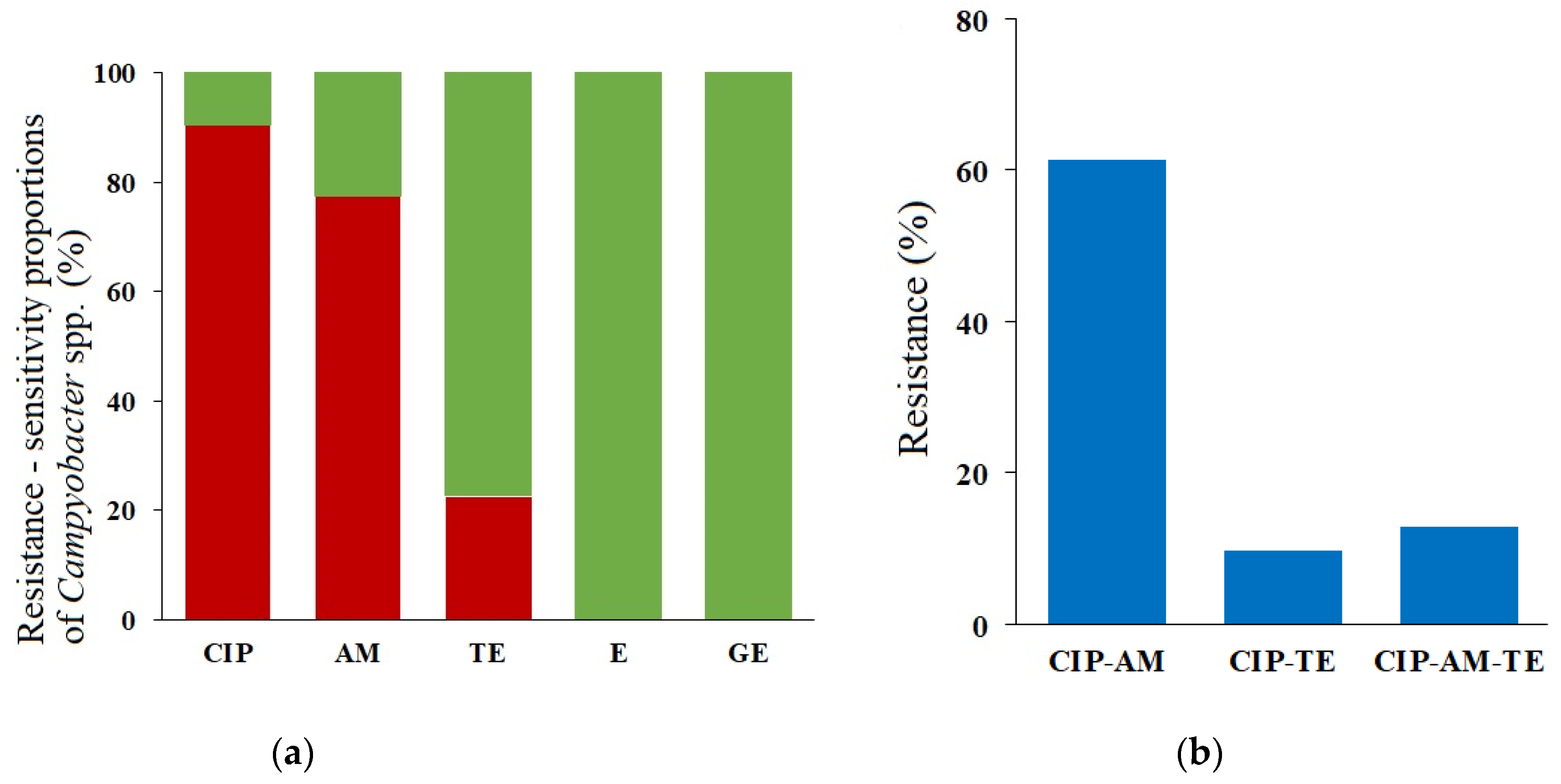
2.3. Phenotypes and Genotypes of the Antimicrobial Resistance

The Minimum Inhibitory Concentration (MIC) values of the tested antimicrobials were determined using a gradient strip method to establish an antimicrobial resistance phenotype. For selected antimicrobial agents, resistance mechanisms were also studied by PCR detection of appropriate resistance genes.
The significantly high level of the ciprofloxacin resistance (90.3%; in 28 out of 31 isolates) was determined (Figure 1a). A point mutation C257T (codon 86) in the gyrA gene, resulting in amino acid substitution (Thr-86-Ile), was found in the 16 ciprofloxacin-resistant C. jejuni strains and two C. upsaliensis strains. However, in all resistant C. lari and one C. upsaliensis isolate, no amino acid substitution was found.
Seven C. jejuni strains (22.6%) were resistant to tetracycline (Figure 1a), and all of them harboured the tetO gene in plasmid DNA.
The resistance to ampicillin was at a high level (77.4%), and it was detected in all Campylobacter species, but its determinants in the studied strains were not determined.
No resistance to gentamicin and erythromycin was noted (Figure 1a).
Twenty-six of thirty-one Campylobacter spp. strains (83.9%) were resistant to two or more tested antimicrobial agents, including 4/31 strains (12.9%) classified as multidrug-resistant (resistant to three or more of the antimicrobial classes; MDR) (Figure 1b). Two MDR strains were isolated from dogs of shelter origin and the other two from dogs of private owners. Six resistance patterns could be distinguished, the predominant was CIPR AMR TES, and the less frequent pattern was CIPS AMR TES, found only in one strain (Table 1).

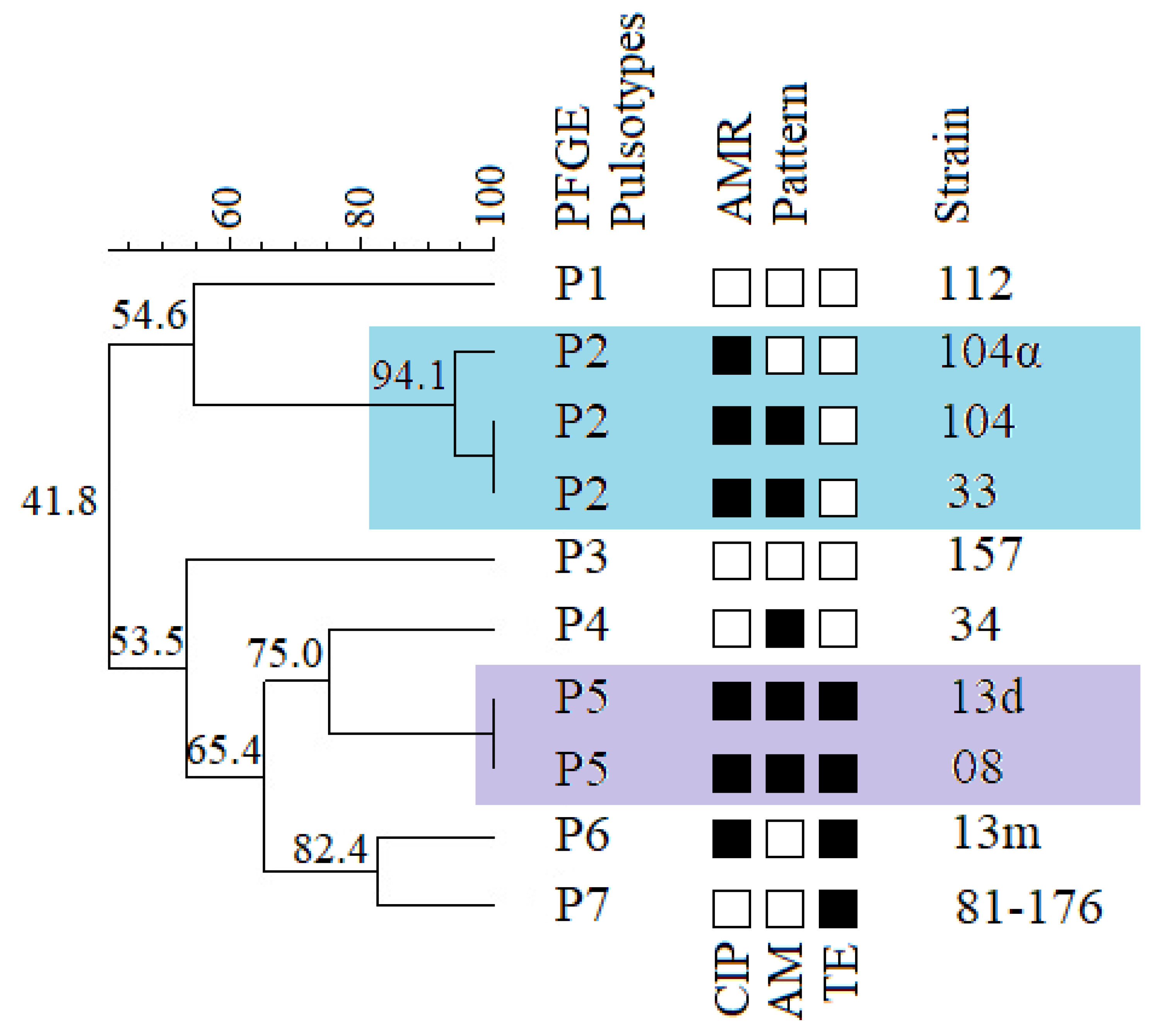
2.4. Genetic Diversity of Campylobacter jejuni

Pulsed-field gel electrophoresis (PFGE) was performed to establish the genetic diversity of C. jejuni isolates obtained from the shelter dogs.
Nine C. jejuni strains isolated from faeces of the shelter dogs were classified into six pulsotypes (P1–P6). The similarity between those pulsotypes varied from 53.5% to 75% (Figure 2). The Simpson’s index of discrimination of the studied molecular typing method was 0.944 (95% CI: 0.870–1.018).
Interestingly, two C. jejuni strains (13d and 13m), isolated from the same dog of shelter origin, have been classified into separate pulsotypes (Figure 2). However, strain 13d clustered together with strain 08 into the pulsotype P5. The pulsotype P2 consisted of three strains; two of them, 104 and 104α, isolated from the same dog were similar at 94.1%, and curiously enough, the third strain isolated from another dog was genetically identical to the strain 104α (Figure 2).
The results of PFGE typing were compared with the antimicrobial resistance patterns. The strains 13d and 13m obtained from the same dog had different resistance patterns. However, strain 104 had a resistance pattern similar to strain 33 and different from the 104α strain, which was isolated from the same dog (Figure 2).

3. Discussion

Our study showed that dogs might be an important reservoir of Campylobacter species potentially dangerous for human health. The previous studies reported the prevalence of these bacteria in pets’ faeces strongly varied, as well as various virulence potential, antimicrobial resistance, and genetic diversity of Campylobacter isolates. It seems that the obtained results may depend on some factors, such as the number of samples, the age and origin of tested animals or the detection and identification methods used.
In this research, we obtained the frequency of the Campylobacter spp. occurrence in dog faeces much lower (13.3%) than in most literature reports. In many studies, the prevalence of these bacteria above 20% in dogs was noted [4,7,10,11,12,15,17,18,19]. In a few reports, the prevalence of Campylobacter spp. in animals living in shelters varied from 45.4% to 87% [10,11,15]. However, there are also few reports showing similar or lower Campylobacter prevalence than in our study, and it varied from 5.9% to 18.3% [6,13,16,21,22,23]. It should be highlighted that in this study, the frequency of Campylobacter spp. occurrence did not vary significantly between shelter animals and animals of private owners (13.7% vs. 12.5%).
Most of the previous studies were focused on two species, C. jejuni and C. coli, which are the most common causes of human gastroenteritis, but C. coli was rarely isolated from dogs [7,8,17,22]. However, Chaban et al. [9] showed, using a quantitive PCR, that dogs may be a reservoir of numerous Campylobacter species other than C. jejuni and C. coli, such as C. upsaliensis, C. hyointestinalis, C. showae, C. sputorum, C. fetus and C. lari.
In our study, three species of Campylobacter: C. jejuni, C. upsaliensis and C. lari were found. C. jejuni was the predominant species, which is in accordance with the literature data [6,7,12,16,17,19,20,22]. Curiously, in this study, the C. upsaliensis strains represented only 9.7% of the isolates, even though this species is reported as commonly occurring in Campylobacter spp. in dogs, similarly to C. jejuni [4,8,12,18,19,20,24]. Interestingly, we identified nine strains of C. lari, which was 29% of all isolates. C. lari is rarely reported in dogs and cats [7,8,19,20] but is mostly associated with animals living in coastal regions and the marine environment (gulls, molluscs, aquatic mammals, and birds) [3,25]. Thus, such a high frequency of this species in dogs in the central region of Poland (Warsaw and the surrounding area) is difficult to explain. However, we lack information on the history of the origin of those dogs. Eight of nine C. lari strains were isolated from shelter dogs, as well as all C. upsaliensis strains. It suggests that animals kept in shelters potentially may be a reservoir of more diverse Campylobacter species than dogs or cats of private owners. We did not isolate any C. coli and C. helveticus strains, which is consistent with some previous reports [4,11,13,16,19].
The motility, cytolethal distending toxin production and ability to adhere and invade the host’s cells are pointed to as the main virulence factors among Campylobacter species. Most research focused on detection of genes encoding flagellar proteins (FlaA, FlaB, FlaC), subunits of cytolethal distending toxin (CdtA, CdtB, CdtC), adhesins (CadF, FlpA) and invasion factors (CiaB, IamA). Some of them also indicated a product of the pldA gene encoding outer membrane phospholipase, which is involved in the invasion and colonisation process [24,26,27,28,29,30,31].
In all examined strains in this study, both the presence of the flaA and flaB genes was detected. They are encoding two proteins, FlaA and FlaB, responsible for the flagellum’s filament formation and thus are responsible for the Campylobacter motility. These results are consistent with the available literature data, where a high prevalence of the flaA gene, between 82–100% in human, canine and feline isolates, was detected [6,24,26,28].
The other important virulence factor of Campylobacter spp. is the cytolethal distending toxin (CDT) with DNase activity, which is composed of three subunits encoded by the cdtA, cdtB and cdtC genes. While the CdtB subunit is an active compound (DNase I), CdtA and CdtC mediate in binding and internalisation to the host’s cells [31]. Therefore, most of the studies focused on the presence of the cdtB gene [24,26,29,32], but some of them investigated all of the three genes or the cdtABC cluster [28,30]. Unlike the flaA gene, the prevalence of CDT encoding genes varied from 29.5–100% [6,24,28]. In our study, cdtA, cdtB and cdtC were found in all of the examined strains belonging to C. jejuni, C. lari and C. upsaliensis species; thus, a fully functional toxin may be produced by those isolates.
The ability to adhere to and invade of host’s enterocytes is also crucial for the pathogenesis of campylobacteriosis. The Campylobacter adhesion proteins, CadF and FlpA (fibrin-like peptide A), are outer membrane proteins responsible for adhesion initiation by binding to fibronectin of epithelium [27,31]. Products of the pldA gene, phospholipase A, and CiaB protein are involved in the internalisation of Campylobacter into the host’s cells [27,31]. The frequency of flpA, ciaB and pldA genes obtained in our study was 100%, while the cadF gene occurred in 90.3% of the isolates overall. We did not find cadF in C. upsaliensis strains, but 100% of the C. jejuni and C. lari strains were positive for this gene. The gene cadF was usually detected in 100% of isolates from different sources: dogs, cats, broilers, pigs, cattle, and humans [6,24,26,30,32]. Only in research by Selwet cadF was found in 83.6% of isolates from dogs [28].
Almost the 100% frequency of selected genes occurrence, responsible for adhesion and invasion process makes obtained isolates potentially able to colonise and damage intestinal epithelium, which may result in symptomatic infection development.
As for all emerging pathogens, the antimicrobial resistance (AMR) in Campylobacter spp. has been a growing problem over the years. For the Campylobacter genus, increasing resistance to fluoroquinolones, quinolones, tetracyclines and penicillins is constantly observed. Preventive administration of those antimicrobials in animal husbandries (chicken and turkey broilers, pigs and cattle) and pet stores (puppies and kittens) may be indicated as one of the main reasons for increasing AMR and more frequent occurrence of multidrug-resistant strains [33,34]. Thus, it seems reasonable to monitor AMR and the occurrence of multidrug resistance (MDR) in Campylobacter strains of pet origin.
In this study, antimicrobial susceptibility testing was performed for all isolated Campylobacter spp. strains. It is worth noting that in the case of Campylobacter antimicrobial susceptibility testing, the interpretation of the results may be problematic due to the fact that breakpoints are available for few antimicrobials and are only established for C. jejuni and C. coli, as these species are the main etiological factors of human campylobacteriosis.
A very high level of ciprofloxacin resistance (90.3%) was noted among the studied Campylobacter spp. strains (Figure 1). Our results are similar to those performed on canine and feline isolates in Iran, where the ciprofloxacin resistance was determined at the level of 75% [13]. The main mechanism of resistance to fluoroquinolones is the weakening of the antimicrobial binding to the so-called quinolone-binding pocket at the GyrA subunit of DNA gyrase, caused by amino acid substitution [35,36,37]. We found the most common point mutation, C257T, in codon 86 of the gyrA gene, resulting in amino acid substitution (Thr-86-Ile) in 19 (67.9%) of the ciprofloxacin-resistant strains, 17 C. jejuni and two C. upsaliensis strains. No other mutations in the gyrA gene have been found in the studied ciprofloxacin-resistant strains.
In this study, the level of tetracycline resistance was at 22.6%, which may be considered moderate in comparison with varied data from a few reports on tetracycline resistance in canine and feline Campylobacter strains. In some studies, a low tetracycline resistance level (0% to 8.6%) was noted, however other studies revealed high levels of tetracycline resistance, up to 87.5% [13,14,22,28].
Tetracycline activity is based on binding to the ribosome and inhibiting the protein synthesis via blocking the accommodation of aminoacyl-tRNA into the A site of the ribosome. One of the main mechanisms of tetracycline resistance is associated with the presence of ribosomal protection proteins (RPP’s) encoded by tet genes [38]. Among the Campylobacter genus, the presence of the tetO gene on a plasmid or chromosomal DNA, encoding Tet(O) RPP, is the most widespread mechanism of tetracycline resistance [39]. In this study, in all seven tetracycline-resistant Campylobacter strains, the tetO gene was found in plasmid DNA. This result is in accordance with the literature data [30,40,41,42], where Tet(O) was found as a main tetracycline resistance determinant.
β-lactam antibiotics are not commonly used for campylobacteriosis treatment, whereas macrolides (azithromycin in particular) are now applied as a gold standard [30]. However, few studies included the determination of the β-lactam resistance; thus, we decided to perform susceptibility testing for ampicillin. A total of 77.4% of the studied strains were ampicillin-resistant, which is much more than found in literature data, where the prevalence of aminopenicillin resistance ranged from 25% to 58.1% [13,22,30,43]. However, the study of Rozynek et al. showed that the level of ampicillin resistance increased over the years from 8% to 35.5% and from 5.8% to 30.4% in human and broiler isolates, respectively [40].
We did not obtain any macrolide and aminoglycoside resistant strains of Campylobacter spp. Most of the studies reported low levels of resistance to those two classes of antimicrobials, and, in most cases, only single isolates were resistant [23,28,29,30,40,41,43].
Despite the fact that we did not determine any resistance to macrolides or aminoglycosides, four of the isolated strains were classified as multidrug-resistant because they revealed the resistance to three classes of antimicrobial agents: fluoroquinolones, tetracyclines and β-lactams. The report of Montgomery et al. on the outbreak of MDR C. jejuni in the United States after puppy exposure indicated that preventive usage of antimicrobials in animals in pet stores could contribute to developing multidrug resistance and may lead to more outbreaks of potentially untreatable infections in humans [33].
As the animal shelter is a closed environment in which the strains of bacteria can circulate within the hosts, we decided to perform PFGE typing of nine strains of C. jejuni isolated from shelter dogs and additionally the reference strain C. jejuni 81176 of human origin, to determine genetical diversity among those strains. We showed that one dog might be a source of different C. jejuni strains, e.g., genetically diverse strains 13d and 13m from one animal, belonging to the two different pulsotypes. Our results also indicate that strains clustering into one pulsotype share identical or very similar antimicrobial resistance patterns (Figure 2) which comes along with the research of Du et al. [44] on human and poultry isolates but stays in contrast with the results of Bakhshi et al. [45] where the genetically similar isolates of poultry meat origin showed different AMR patterns. It has been confirmed that Campylobacter spp. strains could spread between animals in a shelter.
Most of the studies on the genetic diversity of animal Campylobacter spp. strains were focused on the investigation of isolates from slaughter animals (chicken broilers, bovine meat, pig fatteners) due to the fact that meat contamination is indicated as the most common source of pathogenic Campylobacter for humans. Those studies showed that mentioned animal-origin isolates are genetically highly diverse [46,47,48,49,50]. However, little is still known about the genetic diversity of canine Campylobacter strains. Our data and previously published data clearly indicate that dogs and cats may be a reservoir of diverse Campylobacter species and an important source of infections caused by these pathogens in humans [6,7].

4. Materials and Methods

4.1. Sampling and Shipment

Samples of faeces were taken from 196 dogs, including 124, mostly healthy, dogs from one of the Varsovian animal shelters and 72 dogs of private owners, subjected to veterinary cabinets in the Warsaw agglomeration (Poland) due to needing to perform faeces examination. The study included animals, both males and females, aged from one month to 16 years old. The samples were collected between February 2019 and June 2020.
Faecal samples in sterile containers were stored at a temperature of 2–8 °C until shipping and were delivered to the microbiological laboratory within 24–48 h from sampling. Apart from a bacteriological study, additionally, a parasitology examination using the routine methodology was performed for all samples.

4.2. Isolation and Identification of Campylobacter spp.

Specimens were streaked on selective mCCD agar plates (GRASO Biotech, Starogard Gdański, Poland) using sterile cotton swabs, followed by two reducing streaks with a laboratory wire loop. Plates were incubated at 42 °C for 48 h under microaerophilic conditions using the GasPak Campy Container System (BD Biosciences, USA). After incubation, one or two colonies (two colonies were taken if they differed) from each selective plate were cultured on Columbia Blood Agar (GRASO Biotech, Starogard Gdański, Poland) at 42 °C for 48 h under microaerophilic conditions. When pure cultures were obtained, preliminary identification to the genus level was performed based on specific colony morphology, cell morphology, and the motility observed by phase-contrast microscopy, as well as catalase and oxidase test results. Preliminary identification was followed by DNA isolation using the Genomic Mini isolation kit (A&A Biotechnology, Gdańsk, Poland), performed according to the manufacturer’s instructions. Further recognition of isolates to the species level was performed using PCR with species-specific primers (Table 2). Reaction conditions were as follows: pre-denaturation at 95 °C for two minutes; 25 cycles of denaturation at 95 °C for 30 s, primer annealing for 30 s, elongation at 72 °C for 30 s or 60 s (for C. upsaliensis and C. helveticus identification); and an additional elongation step at 72 °C for five minutes; annealing temperatures for all used primers are presented in Table 2. For positive control, Campylobacter jejuni 81-176 ATCC® BAA2151 and Campylobacter coli 605 strains were used. Identification of C. lari and C. upsaliensis strains was also confirmed with the API Campy test (bioMérieux, Marcy-l’Étoile, France), following the manufacturer’s protocol.

4.3. Virulence Factor Genes Occurrence

For identified strains of Campylobacter spp., PCR assays were performed to detect selected virulence factor genes: flaA, flaB, cdtA, cdtB, cdtC, ciaB, pldA, cadF and flpA. The used primers are listed in Table 3. A template chromosomal DNA was isolated using the Genomic Mini isolation kit (A&A Biotechnology, Gdańsk, Poland). For positive control, Campylobacter jejuni 81-176 ATCC® BAA2151 was used. All reactions were performed under the following conditions: pre-denaturation at 95 °C for two minutes; 30 cycles of denaturation at 95 °C for 30 s, primer annealing for 30 s, elongation at 72 °C for 30 s (90 s for the flaA); an additional elongation step at 72 °C for five minutes; annealing temperatures for the used primers are presented in Table 3.

4.4. Antimicrobial Susceptibility Testing

Antimicrobial susceptibility to ciprofloxacin (CIP), tetracycline (TE), gentamicin (GE), erythromycin (E) and ampicillin (AM) was tested by MIC determination using the Etest gradient strips (bioMérieux, Marcy-l’Étoile, France), according to the CLSI or EUCAST guidelines [59,60]. Briefly, bacterial suspensions were prepared in 1 mL of sterile buffered saline to obtain 0.5 McFarland density and streaked on Columbia Blood Agar plates with a sterile cotton swab. Plates were dried for a few minutes, then gradient strips were placed, and cultures were incubated at 42 °C for 48 h under microaerophilic conditions. After incubation, the MIC values were read. Results of antimicrobial susceptibility testing were interpreted according to the CLSI and EUCAST guidelines [59,60]. Campylobacter jejuni 81-176 ATCC® BAA2151 strain was used as the quality control. All tested antimicrobials and interpretation criteria used are listed in Table 4.

4.5. Investigation of Antimicrobial Resistance Mechanisms

For tetracycline-resistant strains, PCR assays using both chromosomal and plasmid DNA were performed to detect the tetO gene. A plasmid DNA was isolated using a Plasmid Mini isolation kit (A&A Biotechnology, Gdańsk, Poland) and a chromosomal DNA using the Genomic Mini isolation kit (A&A Biotechnology, Gdańsk, Poland), according to manufacturer protocols.
For ciprofloxacin-resistant strains, a 290-bp fragment of the gyrA gene (codons 32-127) was amplified by PCR using genomic DNA as a template. The following conditions for both reactions were applied: pre-denaturation at 95 °C for two minutes; 25 cycles of denaturation at 95 °C for 30 s, primer annealing for 30 s, elongation at 72 °C for 30 s (for tetO and gyrA amplification); an additional elongation step at 72 °C for three minutes; primer sequences and annealing temperatures are listed in Table 3. PCR products of gyrA were sequenced (Genomed, Poland) and then analysed for point mutations using a DNA Baser Assembler software v. 5.11.3 (Heracle Biosoft SRL, Mioveni, Romania).

4.6. Pulsed-Field Gel Electrophoresis (PFGE)

Pulse-field gel electrophoresis (PFGE) was performed only for C. jejuni strains isolated from the shelter dogs, according to Ribot et al. rapid PFGE protocol for subtyping of Campylobacter jejuni [61]. Briefly, pure cultures of the strains were streaked on the Columbia Agar plates (GRASO, Starogard Gdański, Poland) and cultured at 42 °C for 24 h in microaerophilic conditions. After incubation, bacterial suspensions were prepared in sterile buffered saline (OD600 = 0.6–0.8) with the addition of a proteinase K (A&A Biotechnology, Gdańsk, Poland) and then were mixed with 2% low melting point agarose (Bio-rad, Hercules, CA, USA) in a ratio of 1:1, and agarose plugs were prepared on glass slides. Plugs were incubated in the cell lysis buffer with proteinase K and then washed once with sterile deionised water and three times with Tris-EDTA (TE) buffer. Plugs were stored in TE buffer at 4 °C until digestion. Digestion was preceded by equilibration of plugs in a solution containing restriction enzyme buffer and sterile deionised water. Plugs were then digested with 40 U of SmaI enzyme (Thermo Fisher Scientific, Waltham, MA, USA) at 25 °C for two hours, then washed in 0.5× TRIS-borate-EDTA (TBE) buffer once and equilibrated in 200 µL of fresh 0.5× TBE buffer. Electrophoresis was performed in 1.5% agarose in 0.5× TBE buffer on the CHEF-DR II apparatus (Bio-Rad, Hercules, CA, USA) under the following conditions: an initial pulse time of 6.8 s, a final pulse time of 38.4 s, gradient 6 V/cm, 19 h, angle 120°. The gel was stained with ethidium bromide (0.5 µg/mL) and washed in distilled water, and then visualised on a Gel Doc™ EZ Imaging System with Image Lab software v. 5.2.1 (Bio-Rad, Hercules, CA, USA). The images were analysed using BioNumerics v. 7.6 software (Applied Maths, Sint-Martens-Latem, Belgium). Obtained patterns were compared, and a UPGMA dendrogram was generated, using a Dice coefficient with a 1.5% tolerance window and 90% cut-off value. Simpson’s index of discrimination was also calculated. The analysed strains were compared to the reference Campylobacter jejuni 81-176 (ATCC® BAA2151) strain.

4.7. Statistical Analysis

Statistical analysis was performed using a IBM SPSS software Statistics for Windows, version 28 (IBM Corp., Armonk, N.Y., USA) software [62]. Confidence intervals were calculated using a Sample Size Calculators online tool [63].

5. Conclusions

Companion animals may be a potential source of Campylobacter strains causing zoonotic infections in humans; therefore, it seems reasonable to monitor the occurrence of these bacteria in pets. In this study, we obtained the relatively low frequency of the Campylobacter spp. occurrence in dogs; however, the isolated strains seem to be potentially virulent and antimicrobial-resistant.
Since, from the epidemiological point of view, C. jejuni and C. coli are a significant threat to human health, most studies are focused only on those species and are based on phenotypic bacteriological methods, while it was shown that molecular detection and identification methods are more effective and may allow detection of a higher prevalence and a species richness. Results of this research showed that dogs might play a role as reservoirs of different, potentially virulent Campylobacter species, which can be antimicrobial-resistant and even multidrug-resistant. Therefore, it seems reasonable to monitor the dissemination of various Campylobacter species in different populations of dogs, including shelters and veterinary cabinets from a large area, to obtain more meaningful results, and even though C. lari has been connected with single cases of human campylobacteriosis, it seems important to control its occurrence, as many virulence factors and antimicrobial resistance mechanisms are conserved and similar to those present in C. jejuni.

Supplementary Materials

The following supporting information can be downloaded at: https://0-www-mdpi-com.brum.beds.ac.uk/article/10.3390/antibiotics11070964/s1, Table S1: The summary of information on Campylobacter-positive dogs; Table S2: The summary of information on the presence of the selected virulence factor genes among Campylobacter isolates.

Author Contributions

Conceptualization, M.M., M.R and A.S-G.; methodology, M.M.; formal analysis, M.M., E.K.; investigation, M.M., M.S., J.B.; writing—original draft preparation, M.M.; writing—review and editing, A.S-G., M.R. All authors have read and agreed to the published version of the manuscript.

Funding

This research received no external funding.

Institutional Review Board Statement

Not applicable.

Informed Consent Statement

Not applicable.

Data Availability Statement

The data presented in this study are available on request from the corresponding authors.

Conflicts of Interest

The authors declare no conflict of interest.

References

  1. EFSA and ECDC (European Food Safety Authority and European Centre for Disease Prevention and Control). The European Union One Health 2019 Zoonoses Report. EFSA J. 2021, 19, 6406. [Google Scholar] [CrossRef]
  2. Kaakoush, N.O.; Castaño-Rodríguez, N.; Mitchell, H.M.; Man, S.M. Global Epidemiology of Campylobacter Infection. Clin. Microbiol. Rev. 2015, 28, 687–720. [Google Scholar] [CrossRef] [Green Version]
  3. Costa, D.; Iraola, G. Pathogenomics of emerging Campylobacter species. Clin. Microbiol. Rev. 2019, 32, e00072-18. [Google Scholar] [CrossRef] [PubMed]
  4. Holmberg, M.; Rosendal, T.; Engvall, E.O.; Ohlson, A.; Lindberg, A. Prevalence of thermophilic Campylobacter species in Swedish dogs and characterization of C. jejuni isolates. Acta Vet. Scand 2015, 57, 19. [Google Scholar] [CrossRef] [PubMed] [Green Version]
  5. Acke, E. Campylobacteriosis in dogs and cats: A review. New Zeal Vet. J. 2018, 66, 221–228. [Google Scholar] [CrossRef]
  6. Andrzejewska, M.; Szczepańska, B.; Klawe, J.J.; Spica, D.; Chudzińska, M. Prevalence of Campylobacter jejuni and Campylobacter coli species in cats and dogs from Bydgoszcz (Poland) region. Pol. J. Vet. Sci. 2013, 16, 115–120. [Google Scholar] [CrossRef] [Green Version]
  7. Thépault, A.; Rose, V.; Queguiner, M.; Chemaly, M.; Rivoal, K. Dogs and Cats: Reservoirs for Highly Diverse Campylobacter jejuni and a Potential Source of Human Exposure. Animals 2020, 10, 838. [Google Scholar] [CrossRef]
  8. Mughini Gras, L.; Smid, J.H.; Wagenaar, J.A.; Koene, M.G.; Havelaar, A.H.; Friesema, I.H.; French, N.P.; Flemming, C.; Galson, J.D.; Graziani, C.; et al. Increased risk for Campylobacter jejuni and C. coli infection of pet origin in dog owners and evidence for genetic association between strains causing infection in humans and their pets. Epidemiology Infect. 2013, 141, 2526–2535. [Google Scholar] [CrossRef] [Green Version]
  9. Chaban, B.; Ngeleka, M.; Hill, J.E. Detection and quantification of 14 Campylobacter species in pet dogs reveals an increase in species richness in feces of diarrheic animals. BMC Microbiol. 2010, 10, 73. [Google Scholar] [CrossRef] [Green Version]
  10. Acke, E.; Whyte, P.; Jones, B.R.; McGill, K.; Collins, J.D.; Fanning, S. Prevalence of termophilic Campylobacter species in cats and dogs in two animal shelters in Ireland. Vet. Rec. 2006, 158, 51–54. [Google Scholar] [CrossRef]
  11. Leahy, A.M.; Cummings, K.J.; Rodriguez-Rivera, L.D.; Hamer, S.A.; Lawhon, S.D. Faecal Campylobacter shedding among dogs in animal shelters across Texas. Zoonoses Public Hlth. 2017, 64, 623–627. [Google Scholar] [CrossRef] [PubMed]
  12. Karama, M.; Cenci-Goga, B.T.; Prosperi, A.; Etter, E.; El-Ashram, S.; McCrindle, C.; Ombui, J.N.; Kalake, A. Prevalence and risk factors associated with Campylobacter spp. occurrence in healthy dogs visiting four rural community veterinary clinics in South Africa. Onderstepoort J. Vet. Res. 2019, 86, e1–e6. [Google Scholar] [CrossRef] [PubMed]
  13. Torkan, S.; Vazirian, B.; Khamesipour, F.; Dida, G.O. Prevalence of thermotolerant Campylobacter species in dogs and cats in Iran. Vet. Med. Sci. 2018, 4, 296–303. [Google Scholar] [CrossRef] [PubMed]
  14. Olkkola, S.; Kovanen, S.; Roine, J.; Hänninen, M.L.; Hielm-Björkman, A.; Kivistö, R. Population Genetics and Antimicrobial Susceptibility of Canine Campylobacter Isolates Collected before and after a Raw Feeding Experiment. PLoS ONE 2015, 10, e0132660. [Google Scholar] [CrossRef] [Green Version]
  15. LaLonde-Paul, D.; Cummings, K.J.; Rodriguez-Rivera, L.D.; Wu, J.; Lawhon, S.D. Ciprofloxacin resistance among Campylobacter jejuni isolates obtained from shelter dogs in Texas. Zoonoses Public Hlth. 2019, 66, 337–342. [Google Scholar] [CrossRef]
  16. Pölzler, T.; Stüger, H.P.; Lassnig, H. Prevalence of most common human pathogenic Campylobacter spp. in dogs and cats in Styria, Austria. Vet. Med. Sci. 2018, 4, 115–125. [Google Scholar] [CrossRef] [Green Version]
  17. Santaniello, A.; Varriale, L.; Dipineto, L.; Borrelli, L.; Pace, A.; Fioretti, A.; Menna, L.F. Presence of Campylobacter jejuni and C. coli in Dogs under Training for Animal-Assisted Therapies. Int. J. Environ. Res. Public Health 2021, 18, 3717. [Google Scholar] [CrossRef]
  18. Runesvärd, E.; Wikström, C.; Fernström, L.L.; Hansson, I. Presence of pathogenic bacteria in faeces from dogs fed raw meat-based diets or dry kibble. Vet. Rec. 2020, 5, e002231. [Google Scholar] [CrossRef] [Green Version]
  19. Bojanić, K.; Midwinter, A.C.; Marshall, J.C.; Rogers, L.E.; Biggs, P.J.; Acke, E. Isolation of Campylobacter spp. from Client-Owned Dogs and Cats, and Retail Raw Meat Pet Food in the Manawatu, New Zealand. Zoonoses Public Hlth. 2017, 64, 438–449. [Google Scholar] [CrossRef]
  20. Marks, S.L.; Rankin, S.C.; Byrne, B.A.; Weese, J.S. Enteropathogenic bacteria in dogs and cats: Diagnosis, epidemiology, treatment, and control. J. Vet. Intern. Med. 2011, 25, 1195–1208. [Google Scholar] [CrossRef]
  21. Queen, E.V.; Marks, S.L.; Farver, T.B. Prevalence of selected bacterial and parasitic agents in feces from diarrheic and healthy control cats from northern California. J. Vet. Intern. Med. 2012, 26, 54–60. [Google Scholar] [CrossRef] [PubMed]
  22. Rodrigues, C.G.; Melo, R.T.; Fonseca, B.B.; Martins, P.A.; Ferreira, F.A.; Araujo, M.B.J.; Rossi, D.A. Occurrence and characterization of Campylobacter spp. isolates in dogs, cats and children. Pesquisa Vet. Brasil 2015, 35, 365–370. [Google Scholar] [CrossRef] [Green Version]
  23. Szczepanska, B.; Andrzejewska, M.; Spica, D.; Klawe, J.J. Prevalence and antimicrobial resistance of Campylobacter jejuni and Campylobacter coli isolated from children and environmental sources in urban and suburban areas. BMC Microbiol. 2017, 17, 80. [Google Scholar] [CrossRef] [PubMed] [Green Version]
  24. Selwet, M.; Cłapa, T.; Galbas, M.; Słomski, R.; Porzucek, F. The prevalence of Campylobacter spp. and occurrence of virulence genes isolated from dogs. Pol. J. Microbiol. 2015, 64, 73–76. [Google Scholar] [CrossRef] [Green Version]
  25. Igwaran, A.; Okoh, A.I. Human campylobacteriosis: A public health concern of global importance. Heliyon 2019, 5, e02814. [Google Scholar] [CrossRef]
  26. Andrzejewska, M.; Klawe, J.J.; Szczepańska, B.; Śpica, D. Occurence of virulence genes among Campylobacter jejuni and Campylobacter coli isolates from domestic animals and children. Pol. J. Vet. Sci. 2011, 14, 207–211. [Google Scholar] [CrossRef] [Green Version]
  27. Bang, D.D.; Borck, B.; Nielsen, E.M.; Scheutz, F.; Pedersen, K.; Madsen, M. Detection of seven virulence and toxin genes of Campylobacter jejuni isolates from Danish turkeys by PCR and cytolethal distending toxin production of the isolates. J. Food Protect. 2004, 67, 2171–2177. [Google Scholar] [CrossRef]
  28. Selwet, M. The Prevalence of Virulence Genes and Multidrug Resistance in Thermophilic Campylobacter Spp. Isolated from Dogs. Open Life Sci. 2019, 14, 681–687. [Google Scholar] [CrossRef]
  29. Andrzejewska, M.; Szczepańska, B.; Śpica, D.; Klawe, J.J. Prevalence, Virulence, and Antimicrobial Resistance of Campylobacter spp. in Raw Milk, Beef, and Pork Meat in Northern Poland. Foods 2019, 8, 420. [Google Scholar] [CrossRef] [Green Version]
  30. Wysok, B.; Wojtacka, J.; Hänninen, M.L.; Kivistö, R. Antimicrobial Resistance and Virulence-Associated Markers in Campylobacter Strains from Diarrheic and Non-diarrheic Humans in Poland. Front. Microbiol. 2020, 11, 1799. [Google Scholar] [CrossRef]
  31. Kreling, V.; Falcone, F.H.; Kehrenberg, C.; Hensel, A. Campylobacter sp.: Pathogenicity factors and prevention methods-new molecular targets for innovative antivirulence drugs? Appl. Microbiol. Biotechnol. 2020, 104, 10409–10436. [Google Scholar] [CrossRef] [PubMed]
  32. Selwet, M.; Galbas, M. Monitoring of selected genes in Campylobacter jejuni and Campylobacter coli isolates from domestic animals. Bull. Veter-Inst. Pulawy 2012, 56, 507–511. [Google Scholar] [CrossRef] [Green Version]
  33. Montgomery, M.P.; Robertson, S.; Koski, L.; Salehi, E.; Stevenson, L.M.; Silver, R.; Sundararaman, P.; Singh, A.; Joseph, L.A.; Weisner, M.B.; et al. Multidrug-Resistant Campylobacter jejuni Outbreak Linked to Puppy Exposure—United States, 2016–2018. MMWR. Morb. Mortal. Wkly. Rep. 2012, 67, 1032–1035. [Google Scholar] [CrossRef] [PubMed]
  34. Landers, T.F.; Cohen, B.; Wittum, T.E.; Larson, E.L. A review of antibiotic use in food animals: Perspective, policy, and potential. Public Health Rep. 2012, 127, 4–22. [Google Scholar] [CrossRef] [PubMed] [Green Version]
  35. Piddock, L.J.; Ricci, V.; Pumbwe, L.; Everett, M.J.; Griggs, D.J. Fluoroquinolone resistance in Campylobacter species from man and animals: Detection of mutations in topoisomerase genes. J. Antimicrob. Chemoth. 2003, 51, 19–26. [Google Scholar] [CrossRef] [Green Version]
  36. Beckmann, L.; Müller, M.; Luber, P.; Schrader, C.; Bartelt, E.; Klein, G. Analysis of gyrA mutations in quinolone-resistant and -susceptible Campylobacter jejuni isolates from retail poultry and human clinical isolates by non-radioactive single-strand conformation polymorphism analysis and DNA sequencing. J. Appl. Microbiol. 2004, 96, 1040–1047. [Google Scholar] [CrossRef] [PubMed]
  37. Chatur, Y.A.; Brahmbhatt, M.N.; Modi, S.; Nayak, J.B. Fluoroquinolone resistance and detection of topoisomerase gene mutation in Campylobacter jejuni isolated from animal and human sources. Int. J. Curr. Microbiol. 2014, 3, 773–783. [Google Scholar]
  38. Connell, S.R.; Tracz, D.M.; Nierhaus, K.H.; Taylor, D.E. Ribosomal protection proteins and their mechanism of tetracycline resistance. Antimicrob. Agents Chemother. 2003, 47, 3675–3681. [Google Scholar] [CrossRef] [Green Version]
  39. Gibreel, A.; Tracz, D.M.; Nonaka, L.; Ngo, T.M.; Connell, S.R.; Taylor, D.E. Incidence of antibiotic resistance in Campylobacter jejuni isolated in Alberta, Canada, from 1999 to 2002, with special reference to tet(O)-mediated tetracycline resistance. Antimicrob. Agents Chemother. 2004, 48, 3442–3450. [Google Scholar] [CrossRef] [Green Version]
  40. Rozynek, E.; Dzierzanowska-Fangrat, K.; Korsak, D.; Konieczny, P.; Wardak, S.; Szych, J.; Jarosz, M.; Dzierzanowska, D. Comparison of antimicrobial resistance of Campylobacter jejuni and Campylobacter coli isolated from humans and chicken carcasses in Poland. J. Food Protect. 2008, 71, 602–607. [Google Scholar] [CrossRef] [Green Version]
  41. Wardak, S.; Szych, J.; Zasada, A.A.; Gierczynski, R. Antibiotic resistance of Campylobacter jejuni and Campylobacter coli clinical isolates from Poland. Antimicrob. Agents Chemother. 2007, 51, 1123–1125. [Google Scholar] [CrossRef] [PubMed] [Green Version]
  42. Woźniak-Biel, A.; Bugla-Płoskońska, G.; Kielsznia, A.; Korzekwa, K.; Tobiasz, A.; Korzeniowska-Kowal, A.; Wieliczko, A. High Prevalence of Resistance to Fluoroquinolones and Tetracycline Campylobacter Spp. Isolated from Poultry in Poland. Microb. Drug Resist. 2018, 24, 314–322. [Google Scholar] [CrossRef] [PubMed]
  43. Wysok, B.; Wojtacka, J.; Wiszniewska-Łaszczych, A.; Szteyn, J. Antimicrobial Resistance and Virulence Properties of Campylobacter Spp. Originating from Domestic Geese in Poland. Animals 2020, 10, 742. [Google Scholar] [CrossRef] [PubMed]
  44. Du, Y.; Wang, C.; Ye, Y.; Liu, Y.; Wang, A.; Li, Y.; Zhou, X.; Pan, H.; Zhang, J.; Xu, X. Molecular Identification of Multidrug-Resistant Campylobacter Species from Diarrheal Patients and Poultry Meat in Shanghai, China. Front. Microbiol. 2018, 9, 1642. [Google Scholar] [CrossRef] [PubMed]
  45. Bakhshi, B.; Kalantar, M.; Rastegar-Lari, A.; Fallah, F. PFGE genotyping and molecular characterization of Campylobacter spp. isolated from chicken meat. Iran. J. Vet. Res. 2016, 17, 177–183. [Google Scholar] [PubMed]
  46. Di Giannatale, E.; Calistri, P.; Di Donato, G.; Decastelli, L.; Goffredo, E.; Adriano, D.; Mancini, M.E.; Galleggiante, A.; Neri, D.; Antoci, S.; et al. Thermotolerant Campylobacter spp. in chicken and bovine meat in Italy: Prevalence, level of contamination and molecular characterization of isolates. PLoS ONE 2019, 14, e0225957. [Google Scholar] [CrossRef] [Green Version]
  47. Di Donato, G.; Marotta, F.; Nuvoloni, R.; Zilli, K.; Neri, D.; Di Sabatino, D.; Calistri, P.; Di Giannatale, E. Prevalence, Population Diversity and Antimicrobial Resistance of Campylobacter coli Isolated in Italian Swine at Slaughterhouse. Microorganisms 2020, 8, 222. [Google Scholar] [CrossRef] [Green Version]
  48. Zhang, L.; Li, Y.; Shao, Y.; Hu, Y.; Lou, H.; Chen, X.; Wu, Y.; Mei, L.; Zhou, B.; Zhang, X.; et al. Molecular Characterization and Antibiotic Resistant Profiles of Campylobacter Species Isolated from Poultry and Diarrheal Patients in Southeastern China 2017–2019. Front. Microbiol. 2020, 11, 1244. [Google Scholar] [CrossRef]
  49. Marotta, F.; Garofolo, G.; Di Donato, G.; Aprea, G.; Platone, I.; Cianciavicchia, S.; Alessiani, A.; Di Giannatale, E. Population Diversity of Campylobacter jejuni in Poultry and Its Dynamic of Contamination in Chicken Meat. BioMed Res. Int. 2015, 2015, 1–10. [Google Scholar] [CrossRef] [Green Version]
  50. Di Giannatale, E.; Di Serafino, G.; Zilli, K.; Alessiani, A.; Sacchini, L.; Garofolo, G.; Aprea, G.; Marotta, F. Characterization of antimicrobial resistance patterns and detection of virulence genes in Campylobacter isolates in Italy. Sensors 2014, 14, 3308–3322. [Google Scholar] [CrossRef] [Green Version]
  51. On, S.L.W.; Jordan, P.J. Evaluation of 11 PCR Assays for Species-Level Identification of Campylobacter jejuni and Campylobacter coli. J. Clin. Microbiol. 2003, 41, 330–336. [Google Scholar] [CrossRef] [PubMed] [Green Version]
  52. Linton, D.; Owen, R.J.; Stanley, J. Rapid identification by PCR of the genus Campylobacter and of five Campylobacter species enteropathogenic for man and animals. Res. Microbiol. 1996, 147, 707–718. [Google Scholar] [CrossRef]
  53. Klena, J.D.; Parker, C.T.; Knibb, K.; Ibbitt, J.C.; Devane, P.M.; Horn, S.T.; Miller, W.G.; Konkel, M.E. Differentiation of Campylobacter coli, Campylobacter jejuni, Campylobacter lari, and Campylobacter upsaliensis by a multiplex PCR developed from the nucleotide sequence of the lipid A gene lpxA. J. Clin. Microbiol. 2004, 42, 5549–5557. [Google Scholar] [CrossRef] [Green Version]
  54. Nachamkin, I.; Bohachick, K.; Patton, C.M. Flagellin gene typing of Campylobacter jejuni by restriction fragment length polymorphism analysis. J. Clin. Microbiol. 1993, 31, 1531–1536. [Google Scholar] [CrossRef] [Green Version]
  55. Goon, S.; Kelly, J.F.; Logan, S.M.; Ewing, C.P.; Guerry, P. Pseudaminic acid, the major modification on Campylobacter flagellin, is synthesized via the Cj1293 gene. Mol. Microbiol. 2003, 50, 659–671. [Google Scholar] [CrossRef]
  56. Hickey, T.E.; McVeigh, A.L.; Scott, D.A.; Michielutti, R.A.; Bixby, A.; Carroll, S.A.; Bourgeois, A.L.; Guerry, P. Campylobacter jejuni cytolethal distending toxin mediates release of interleukin-8 from intestinal epithelial cells. Infect. Immun. 2000, 68, 6535–6541. [Google Scholar] [CrossRef] [Green Version]
  57. Datta, S.; Niwa, H.; Itoh, K. Prevalence of 11 pathogenic genes of Campylobacter jejuni by PCR in strains isolated from humans, poultry meat and broiler and bovine faeces. J. Med. Microbiol. 2003, 52, 345–348. [Google Scholar] [CrossRef] [PubMed]
  58. Zheng, J.; Meng, J.; Zhao, S.; Singh, R.; Song, W. Adherence to and invasion of human intestinal epithelial cells by Campylobacter jejuni and Campylobacter coli isolates from retail meat products. J. Food Prot. 2006, 69, 768–774. [Google Scholar] [CrossRef]
  59. Breakpoints tables for interpretation of MICs and zone diameters, Version 12.0; The European Committee on Antimicrobial Susceptibility Testing: 2022. Available online: http://eucast.org (accessed on 1 January 2022).
  60. CLSI. Methods for Antimicrobial Susceptibility Testing of Infrequently Isolated or Fastidious Bacteria Isolated From Animals, 1st ed.; CLSI supplement VET06; Clinical and Laboratory Standards Institute: Wayne, PA, USA, 2017. [Google Scholar]
  61. Ribot, E.M.; Fitzgerald, C.; Kubota, K.; Swaminathan, B.; Barrett, T.J. Rapid Pulsed-Field Gel Electrophoresis Protocol for Subtyping of Campylobacter jejuni. J. Clin. Microbiol. 2001, 39, 1889–1894. [Google Scholar] [CrossRef] [Green Version]
  62. IBM SPSS Statistics for Windows, version 28; IBM Corp: Armonk, NY, USA, 2022.
  63. Kohn, M.A.; Senyak, J. Sample Size Calculators. UCSF CTSI. 20 December 2021. Available online: https://www.sample-size.net/ (accessed on 26 May 2022).
Figure 1. Summary of antimicrobial resistance testing: (a) Diagram showing level of susceptibility and resistance to antimicrobial agents, green—susceptible; red—resistant; CIP—ciprofloxacin; TE—tetracycline; AM—ampicillin; E—erythromycin; GE—gentamicin; (b) Percentage of resistance to two or more classes of antimicrobials.
Antibiotics 11 00964 g001
Figure 2. PFGE/SmaI typing of C. jejuni strains of shelter origin in compilation with antimicrobial resistance patterns (black square—R; white square—S). A dendrogram was generated using the UPGMA method, a Dice coefficient with a 1.5% tolerance window and a cut-off value of 90%. Bootstrap values are given as a percentage.
Antibiotics 11 00964 g002
Table 1. Frequency of occurrence of the AMR patterns obtained in this study.
Resistance ProfileNumber of IsolatesFrequency [%]Species Included [%]
CIPR AMR TES1961.3C. jejuni (47.4), C. lari (15.8), C. upsaliensis (36.8)
CIPR AMR TER412.9C. jejuni (100)
CIPR AMS TER39.6C. jejuni (100)
CIPR AMS TES26.5C. jejuni (100)
CIPS AMS TES26.5C. jejuni (100)
CIPS AMR TES13.2C. jejuni (100)
Total31
Table 2. Primers used in this study for Campylobacter species identification.
Target SpeciesPrimerAmplicon Size [bp]Primer Sequence 5′-3′Tm * [°C]Ref.
C. jejunimapAF604ATGTTTAAAAAATTTTTG55[51]
mapARAAGTTCAGAGATTAAACTAG
C. upsaliensisCHCU146F879GGGACAACACTTAGAAATGAG55[52]
CU1024RCACTTCCGTATCTCTACAGA
C. helveticusCHCU146F1226GGGACAACACTTAGAAATGAG52[52]
CH1371RCCGTGACATGGCTGATTCAC
C. larilpxAF233TRCCAAATGTTAAAATAGGCGA50[53]
lpxARCAATCATGDGCDATATGASAATAHGCCAT
C. coliMu3364AGGCAAGGGAGCCTTTAATC61[51]
Mu4TATCCCTATCTACAATTCGC
* Tm—temperature of primer annealing.
Table 3. Primers used in this study for the detection of virulence factor genes and determination of the molecular mechanisms of antimicrobial resistance.
Target GenePrimerAmplicon
Size [bp]
Primer Sequence 5′- 3′Tm * [°C]Ref.
flaAflaA-F1728GGATTTCGTATTAACACAAATGGTGC45[54]
flaA-RCTGTAGTAATCTTAAAACATTTTG
flaBfB1260AAGGATTTAAAATGGGTTTTAGAATAAACACC54[55]
fA2GCTCATCCATAGCTTTATCTGC
cdtAcdtA-F370CCTTGTGATGCAAGCAATC46[56]
cdtA-RACACTCCATTTGCTTTCTG
cdtBcdtB-F620CAGAAAGCAAATGGAGTGTT47[57]
cdtB-RAGCTAAAAGCGGTGGAGTAT
cdtCcdtC-F182TTGGCATTATAGAAAATACAGTT46[57]
cdtC-RCGATGAGTTAAAACAAAAAGATA
ciaBciaB-F527TGCGAGATTTTTCGAGAATG47[58]
ciaB-RTGCCCGCCTTAGAACTTACA
pldApldA-F385AAGAGTGAGGCGAAATTCCA49[58]
pldA-RGCAAGATGGCAGGATTATCA
flpAflpAF1017GCTTTTGAATGGGAGTCTTTATAT49This study
flpARATCAATAGCAATAACTTCATAACTATA
cadFcadF_F580TTTGAGTGCTATTAAAGGTATTG47This study
cadF_RTCTTTCTGAAAGCTTTTGATTATA
cadF
(C. lari)
cadF_LF589GCGCACGACCTTCTTTAGT50This study
cadF_LRGCTTATGAAAATAAAAGCGGTATG
cadF
(C. upsaliensis)
cadF_UF510CTCTCTTGGTTCTTCAGGACA52This study
cadF_URGATAATCGCTATGCACCAGGGA
tetOtetO_F559GGCGTTTTGTTTA49[37]
tetO_RATGGACAACCCGACAGAAGC
gyrAgyrA_F290ATTATAGGTCGTGCTTTGCCT50This study
gyrA_RGCTTCAGTATAACGCATCGCA
* Tm—temperature of primer annealing
Table 4. Interpretation criteria (breakpoints) for antimicrobial susceptibility determination.
Antimicrobial AgentAntimicrobial ClassConcentration Range Tested [mg/L]Breakpoints for MIC Testing
S ≤IR ≥
CIP b *Fluoroquinolones0.002–320.001-0.5
TE aTetracyclines0.016–2564816
AM bβ-lactams0.016–2562-8
E aMacrolides0.016–25681632
GE bAminoglycosides0.016–2560.5-0.5
* Interpretation according to: a CLSI or b EUCAST
Publisher’s Note: MDPI stays neutral with regard to jurisdictional claims in published maps and institutional affiliations.

