Article

Sub-MIC Antibiotics Modulate Productions of Outer Membrane Vesicles in Tigecycline-Resistant Escherichia coli

1
School of Animal Science and Technology, Guangxi University, Nanning 530004, China
2
Key Laboratory of Food Quality and Safety of Jiangsu Province-State Key Laboratory Breeding Base, Key Laboratory of Agro-Product Safety Risk Evaluation (Nanjing) of Ministry of Agriculture and Rural Affairs, Institute of Food Safety and Nutrition, Jiangsu Academy of Agricultural Sciences, Nanjing 210014, China
*
Authors to whom correspondence should be addressed.
These authors contributed equally to this work.
Submission received: 21 February 2024 / Revised: 13 March 2024 / Accepted: 16 March 2024 / Published: 19 March 2024

Abstract

:
Antimicrobial resistance (AMR) has been recognized as one of the most important crises affecting global human health in the 21st century. Tigecycline is one of the last resort antibiotics for treating severe infections caused by multi-drug resistant Enterobacteriaceae. However, the mobile resistance gene tet(X4), which could mediate high-level tigecycline resistance, was discovered in 2019. The outer membrane vesicle (OMV) has been recognized as a new route for horizontal gene transfer; antimicrobial resistant bacteria also have the ability to secret OMVs, while little is known about the impact of antibiotics on the secretion and characteristics of OMVs from tigecycline resistant bacteria till now. This study aimed to investigate the effects of antibiotics on the production and traits of a tigecycline resistant Escherichia coli strain of 47EC. The results showed that sub-inhibitory (1/2 MIC or 1/4 MIC) concentrations of gentamicin, meropenem, ceftazidime, chloramphenicol, tigecycline, ciprofloxacin, polymycin, rifaximin and mitomycin C could significantly increase the secretion of OMVs (0.713 ± 0.05~6.333 ± 0.15 mg/mL) from E. coli 47EC compared to the respective untreated control (0.709 ± 0.03 mg/mL). In addition, the particle sizes of OMVs were generally larger, and the zeta potential were lower in the antibiotics-treated groups than those of the antibiotic-free group. The copy numbers of the tigecycline resistance gene of tet(X4) in the OMVs of most antimicrobial-treated groups were higher than that of the control group. Moreover, transcriptome analysis on ciprofloxacin-treated E. coli 47EC indicated that the SOS response and prophage activation might participate in the ciprofloxacin-induced OMV formation. In conclusion, the clinical application of antibiotics in treating bacterial infections, especially multi-drug resistant bacteria, might lead to the increased secretion of bacterial OMVs and the enrichment of antimicrobial-resistant genes in the OMVs.

1. Introduction

Antimicrobial resistance is an increasingly severe threat to global public health [1]. If no action is taken, the human deaths caused by antibiotic-resistant bacteria are estimated to be 10 million per year by 2050, which is 1.8 million more than cancer death [2]. The multi-drug-resistant bacteria, particularly those resistant to last-resort drugs (carbapenems, colistin and tigecycline) cause the most serious concern [3].
Tigecycline, a third-generation semi-synthetic tetracycline, has been developed for the treatment of polymicrobial infections caused by Gram-positive and Gram-negative pathogens and is classified as one of the critically important antibacterial (CIA) drugs by the WHO (World Health Organization) [4]. Resistance to tigecycline is mediated by different mechanisms including ribosomal protein binding site mutation, cell membrane variation, efflux pump transport system activation and modification enzyme degradation [5]. In 2019, a novel plasmid-encoded transmissible tet(X4) gene, which could confer high-level tigecycline resistance, was discovered in an Enterobacteriaceae isolate from animals in China [6,7]. The tet(X4) gene encodes a FAD-development monooxygenase that could regioselectively hydroxylate tetracycline substrates. Most of the tet(X4) genes are located on different types of plasmids such as IncQ1, IncX1, IncFIB, IncHI1 and hybrid plasmids [5].
The outer membrane vesicle is a newly found mechanism of horizontal gene transfer apart from transformation, conjugation and transduction [8,9]. OMVs are 20- to 400-nm spherical bilayer liposomes that are naturally secreted by bacteria during growth [10,11]. OMVs could enclose the cargos of lipopolysaccharide (LPS), phospholipids, genetic elements (DNA, RNA, and plasmids), and periplasmic as well as cytoplasmic protein components [12]. The outer membrane vesicles have diverse functions, such as protein transport, horizontal gene transfer, nutrient acquisition, cell-intercellular communication, antibacterial activity, toxin delivery and host-immune response modulation [13,14]. The biogenesis of OMVs was influenced by many factors, such as pH, temprature, hypoxia, nutrition-deficient, etc., especially antibiotic stress. For instance, Mi Hyun Kim et al. found that Enterococcus faecium ATCC700221 cultured with 1/2 MIC of vancomycin or linezolid produced 3.0 and 1.5 times more membrane vesicles than bacteria cultured without antibiotics, respectively [15]. Simon Devos et al. reported that treatment with the β-lactam antibiotic imipenem led to a dramatic increase in the secretion of OMVs in the nosocomial pathogen Stenotrophomonas maltophilia [16].
Antimicrobial resistant bacteria have been reported to have the ability to secret OMVs [8], but the production under antibiotic stress conditions and the characteristics thereof have yet to be determined. The aim of this study was to investigate the production and traits of OMVs in a tigecycline resistant E. coli strain of 47EC under exposure of sub-inhibitory concentrations of different antibiotics.

2. Results

2.1. AMR Phenotypes of E. coli 47EC

The AMR phenotype of E. coli 47EC was determined using the broth microdilution method according to the CLSI M100 standard, 28th Edition [17]. The antimicrobial susceptibility testing results showed that the MICs of the control strain ATCC25922 were all within the quality control ranges (Table 1), indicating the validity of the MIC tests. E. coli 47EC was resistant to tigecycline, chloramphenicol and rifaximin, mediate resistant to gentamicin, and sensitive to ceftazidime, ciprofloxacin, polymyxin B and meropenem.

2.2. Effects of Antibiotics on the Production of OMVs from E. coli 47EC

To investigate the effects of different antibiotics on the production and characteristics of OMVs, the bacterial cultures of E. coli 47EC were treated with gentamicin, meropenem, ceftazidime, chloramphenicol, tigecycline, ciprofloxacin, polymycin, rifaximin and mitomycin C at sub-inhibitory concentrations (1/2 MIC or 1/4 MIC). The OMVs were extracted from E. coli 47EC treated with or without antibiotics by using the ultracentrifugation method. The BCA results showed that the protein concentration of OMVs without antibiotic treatment was 0.709 ± 0.03 mg/mL (Figure 1). After being treated with different antibiotics, the productions of OMVs from E. coli 47EC were significantly increased (0.713 ± 0.05~6.333 ± 0.15 mg/mL). The positive control mitomycin C exhibited the strongest stimulation effect on the production of OMVs. Among the different antibiotics, the concentrations of OMVs in the ciprofloxacin group (1/2 MIC: 4.938 ± 1.19 mg/mL and 1/4 MIC: 6.03 ± 0.90 mg/mL) were relatively higher than in other antibiotic groups.

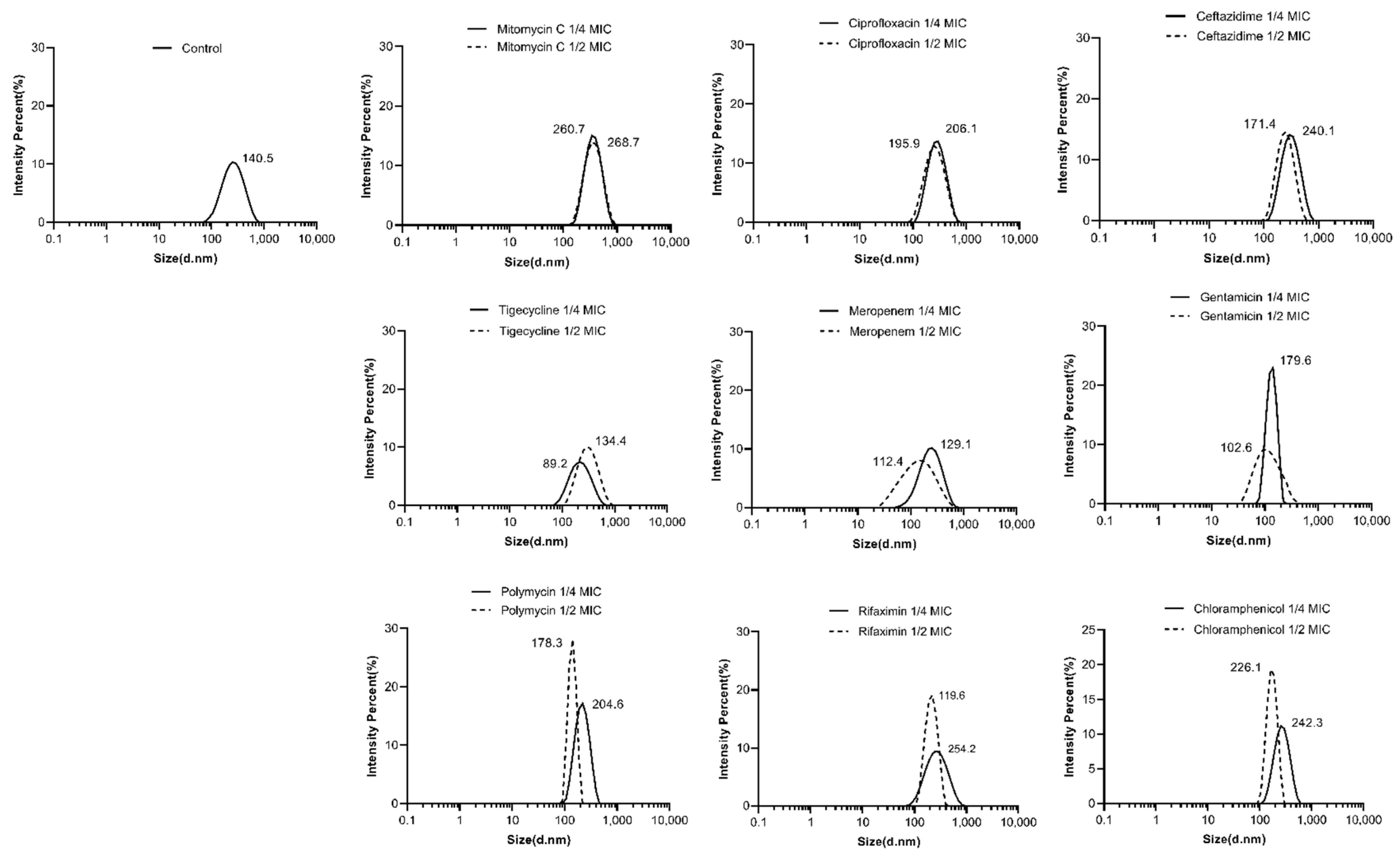
2.3. Effects of Antibiotics on the Characterizations of OMVs from E. coli 47EC

The DLS-intensity weighted particle size distribution of the OMV was determined using a Zeta-sizer nano-ZS (Malvern Instruments, Worcestershire, UK). The particle size of the OMV without antibiotic treatment was 140.5 nm (Figure 2). After being treated with antibiotics such as mitomycin C, ciprolfoxacin, ceftazidime, polymycin, chloramphenicol, gentamicin (at 1/4 MIC) and rifaximin (at 1/4 MIC), the particle sizes of the OMVs were increased (178.3~268.7 nm), whereas other antimicrobialssuch as tigecycline, meropenem, gentamicin (at 1/2 MIC) and rifaximin (at 1/2 MIC) induced smaller OMVs. Most of the antimicrobials induced larger-size OMVs at concentrations of 1/4 MICs than at 1/2 MICs except tigecycline, whose OMVs sizes were 137.4 nm at 1/2 MIC and 89.2 nm at 1/4 MIC.
The differences in the size of the OMVs under antibiotics treatment were also demonstrated by the OMV morphology observed by TEM. TEM showed that the OMVs were spherical (Figure 3). The size of the OMV from the ciprofloxacin-treated group was slightly larger than that of the control group.
The zeta potential of the OMV in the control group was −5.057 ± 0.956 (Figure 4). When treated with rifaximin, tigecycline, chloramphenicol, meropenem, gentamicin, ciprofloxacin, mitomycin C, ceftazidime (1/4 MIC) and polymycin (1/4 MIC), the zeta potentials of the OMVs were decreased to −5.6 ± 2.229~−14.725 ± 1.366. Only in the groups of ceftazidime (1/2 MIC, −3.167 ± 1.456) and polymycin (1/2 MIC, −3.126 ± 1.243) were the zeta potentials of the OMVs higher than that of the antibiotic-free group.

2.4. Abundance of tet(X4) in OMVs

The existence of the tigecycline-resistant gene tet(X4) in the OMVs was examined by PCR, and the copy numbers of tet(X4) were quantified by qPCR. The PCR result confirmed that the OMVs originating from either the control group or the antibiotics-treated groups all contained the resistant gene of tet(X4) (Figure 5). The standard curve for tet(X4) was linear (R2 > 0.99) at the tested range of 1 × 10² to 1 × 10¹⁰ copies/µL (Figure 6). The absolute qPCR reuslts showed that the copies of tet(X4) in the OMV of the control group was 2.937 × 10⁴ copies/µL, and the copy numbers in the OMVs from different antimicrobial-treated groups varied from 1.755 × 10³ to 6.875 × 10⁵ copies/µL (Table 2). For most of the antimicrobials, the copy numbers of tet(X4) in the OMVs were generally higher than those of the control group, expect for rifaximin and tigecycline (1/2 MIC). Also, it seemed that the copy numbers of tet(X4) in the OMVs were not positively correlated with the production of the OMVs induced by different antibiotics, which might be due to the different action mechanism of the antimicrobials, thereby resulting in the variation of cargos in the OMVs.

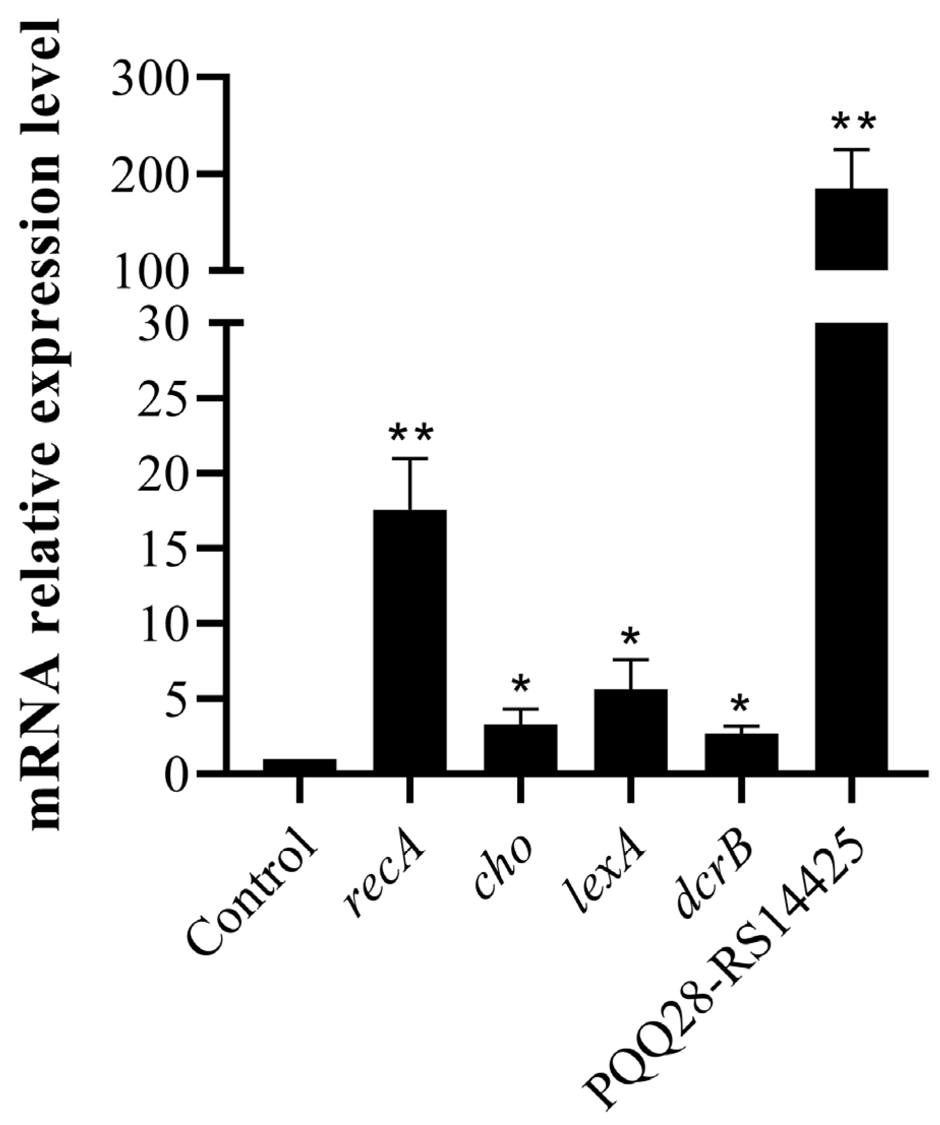
2.5. Transcriptome Changes in E. coli 47EC Induced by Ciprofloxacin

Because ciprofloxacin showed the strongest stimulation effects on the production of OMVs among the tested antimicrobials, we applied a transcriptome analysis to investigate the underlying mechanism. E. coli 47EC was treated with ciprofloxacin at a concentration of 1/2 MIC for 6 h; several changes were revealed in the transcriptomes of E. coli 47EC. Analysis of the RNA-seq data identified 386 genes with statistically significantly (i.e., adjusted p value, <0.05) altered transcription levels, with 195 genes and 191 genes showing increases and decreases of at least twofold (i.e., log2 > 1) in the mRNA amounts in cells growth in the presence of ciprofloxacin as compared to those grown in the absence of antibiotic, respectively. The 386 differentially expressed genes were classified into different functional categories according to the GO and the KEGG database. In the presence of ciprofloxacin, the genes coding for the proteins involved in SOS response (GO: 0009432), the cellular response to extracellular stimulus (GO: 0031668) and the DNA repair (GO: 0006281) were significantly induced, whereas the genes coding for proteins involved in the regulation of cell division (GO: 0051302), the regulation of gene expression (GO: 0010468), the regulation of cellular macromolecule biosynthetic process (GO: 2000112) and the regulation of DNA-templated transcription (GO: 0006355) were significantly repressed. At the transcription level, the SOS response, the DNA-repair-related genes (rtcB, dinD, recA, recX, recN, ruvA, yebG, cho, ydjM, umuC, umuD, dinI, sulA, dinG, uvrB, yafP, dinB, polB, ssb1, uvrA, lexA and uvrD) and the prophage-related genes (RS01710, RS14425 and RS14905) were significantly up-regulated in the ciprofloxacin-treated E. coli 47EC than in the untreated bacteria at different levels (Table 3). qRT-PCR was performed on five SOS response and prophage-related genes to validate the transcriptome data. The qRT-PCR results showed that the expressions of recA, cho, lexA, dcrB and PQQ28-RS14425 were increased by 17.64, 3.31, 5.68, 2.72 and 185.29-fold, respectively (Figure 7). The expression patterns of the five genes determined by qRT-PCR were found to be consistent with the transcriptome data.
It is known that quinolones, such as ciprofloxacin, kill sensitive bacteria by binding to its DNA gyrase and topoisomerase IV, leading to the formation of quinolone-gyrase-DNA complexes and resulting in bacterial DNA double-strand breaks. Several studies reported that to repair the DNA damage, bacteria would trigger the SOS response and DNA repair pathways, and this bacterial reaction would further activate prophage and promote the production of OMVs. Considering the elevated transcription of the SOS response and prophage-related genes, these pathways might participate in the formation of OMVs in the E. coli 47EC induced by ciprolfoxacin.

3. Discussion

OMVs have been recognized as a fourth pathway for horizontal gene transfer and play a key role in the dissemination of antimicrobial resistance genes [8]. However, the impact of antibiotics on the OMVs derived from antimicrobial-resistant bacteria has not been fully illustrated. To our knowledge, this is the first study investigating the modulations of different classes of antibiotics on the production and characteristics of OMVs from a tigecycline-resistant E. coli strain of 47EC.
This study found that treatment with different antibiotics stimulate the production of OMVs, as indicated by the protein concentration of OMVs determined by BCA. Moreover, ciprofloxacin exhibited a stronger stimulation effect than other antibiotics. The results were consistent with previous publications, showing that sub-MIC ampicillin or ciprofloxacin increased OMV secretion at 1.4–5.6-folds in Geobacter sulfurreducens [18]; ampicillin induction increased OMV production at 22.4-fold in a methicillin-resistant S. aureus [19]; imipenem and ciprofloxacin lead to dramatic increases in the OMVs secretion in Stenotrophomonas maltophilia [16,20]; polymyxin B, meropenem and gentamicin increased OMV production in Pseudomonas aeruginosa [21,22,23]; ciprofloxacin, flucloxacillin, and ceftaroline increased the OMV production in Staphylococcus aureus through different routes [24]. Andreas Bauwens et al. [25] reported that several antibiotics could promote OMV formation in E. coli strains of LB226692 (O104:H4) and 5791/99 (O157:H7) at different levels, including ciprofloxacin (250-fold and 183-fold), mitomycin C (568-fold and 470-fold), fosfomycin (77-fold and 24-fold), meropenem (27-fold and 14-fold) and polymyxin B (ninefold and sevenfold). However, gentamicin, rifaximin, tigecycline, chloramphenicol and azithromycin did not influence the OMV production [25]. Antibiotics are thought to be prominent stressors and can be considered as the most potent inducers of defense mechanisms in pathogens. The overproduction of bacterial membrane vesicles can be taken as a form of bacterial defense against stress.
In this study, the antibiotic-treated OMVs generally exhibited larger particle sizes and more negative zeta potentials as compared to the antibiotic-free group, as determined by DLS. The differences in the particle sizes of the ciprofloxacin-treated group and antibiotic-free group were also demonstrated by the OMV morphology observed by TEM. The results were consistent with other reports. Fang et al. [18] detected that the mean size of the ciprolfoxacin-treated OMVs (63.69 ± 0.2 nm) and the ampicillin-treated OMVs (62.37 ± 0.1 nm) were larger than the antibiotic-free group (60.28 ± 0.2 nm) in Geobacter sulfurreducens. By using TEM, they also observed some double-layer OMVs with larger sizes in addition to the classic OMVs in the ciprofloxacin-induced group. Shweta Fulsundar et al. [26] also found that gentamicin (0.1 and 0.3 μg/mL) and chloramphenicol (1 μg/mL) led to a significant increase in the diameters of the OMVs from Acinetobacter baylyi. In addition, OMVs from gentamicin (0.1 μg/mL)-treated cells had significantly more negative zeta potentials (−27.6 ± 0.7 mV, p < 0.001) than the respective control (−19.4 ± 0.4 mV). However, chloramphenicol treatment resulted in OMVs with significantly less negative surface charges (−11.2 ± 1.1 mV, p < 0.001) than those of the OMVs from untreated cells. Another study performed on carbapenem-resistant hypervirulent Klebsiella pneumoniae showed that meropenem-induced OMVs were larger (78.8 to 396 nm, median size of 161.77 nm) than the OMV without antibiotic treatment (68.1 to 396 nm, median size 147.30 nm) [27]. The differences in the production and the characteristics (size distribution and zeta potential) of the OMVs between our study and previous publications might be due to the distinctions in the bacterial species and strains, the antibiotic treatment concentration and duration, the OMV isolation and experimental methods, etc.
Reports showed that antibiotic treatment might influence the abundance of antimicrobial-resistant gens carried by the OMVs. By performing PCR, we detected the tigecycline resistant gene tet(X4) in all OMV groups, including the antibiotic-free group. Furthermore, qPCR results showed that most antibiotics could increase the abundance (copy numbers) of tet(X4) within the OMVs of E. coli 47EC. Similarly, Li et al. reported that the copy numbers of extended-spectrum β-lactamase (ESBL) gene blaCTX-M-55 in the OMVs from an avian pathogenic Escherichia coli strain of SCAO22 was 30,615 copies/µL in the enrofloxacin treatment group and 1196 copies/µL in the amoxicillin group, which were higher than the 208 copies/µL in the control group [28]. In addition to the antimicrobial-resistant genes, bacterial virulence factors such as Shiga toxin (Stx) 2a could also be enriched in the ciprolfoxacin-induced OMVs from Enterohemorrhagic Escherichia coli (EHEC) strains [25]. These facts indicate that the clinical usage of antibiotics for treating bacterial infections might stimulate the production of OMVs and increase the abundance of antimicrobial-resistant genes or virulence factors within the OMVs, thereby increasing the risk of them being vectors for disseminating AMR and virulence. More studies should be conducted in the future to evaluate the frequency of antibiotic-induced OMVs’ mediated horizontal gene transfer and the mechanisms of the cargo selection and packaging in the OMVs.
It has been reported that antibiotics could stimulate the production of OMVs through three pathways: envelope stress response, SOS response and inhibition of cell wall biosynthesis [29,30]. The SOS response appears to be the main mechanism for the biogenesis of OMVs [20]. Previous studies showed that ciprofloxacin could trigger bacterial SOS response to breaking the DNA double-strand, activating the prophage to encode endolysin and thus causing bacterial explosive cell lysis to produce OMVs [20,31]. Indeed, by applying a transcriptome analysis, we verified that the SOS response, DNA repair and several prophage-related genes were significantly up-regulated in the ciprofloxacin-treated E. coli 47EC, indicating that these pathways might participate in the ciprolfoxacin-induced OMV formation. Similarly, earlier studies reported that quinolones provoke the bacterial SOS response to DNA damage and/or to the inhibition of DNA replication and induce the stx-harbouring prophage activation in E. coli O157:H7 [32] and an enterohemorrhagic E. coli O104:H4 strain [33]. Recently, Fang et al., reported that ciprolfoxacin-induced OMVs contain more redox active cytochromes, facilitating iron oxides reduction [18]. Moreover, by applying electron microscopy and proteomic analysis, they also found that the SOS response triggered by ciprofloxacin induced prophage and led to the formation of outer-inner membrane vesicles (OIMVs) in the Geobacter sulfurreducens strain PCA. Similarly, Federica Andreoni et al. showed that ciprofloxacin induces the SOS response, triggering vesicle formation in the lysogenic strains of Staphylococcus aureus but not in their phage-devoid counterparts [24]. Also, the DNA amount associated with the ciprofloxacin-induced OMVs was significantly greater than that of the β-lactam antibiotic-induced OMVs.

4. Conclusions

For the first time, the effects of sub-MIC concentrations of different antibiotics on the production and characteristics of OMVs were investigated in a tigecycline-resistant and tet(X4)-positive strain of E. coli 47EC. Generally, treatment with antibiotics could induce E. coli 47EC to secrete more OMVs with larger particle sizes and lower zeta potentials and carry more resistant genes of tet(X4), with the exception of a few antibiotics. The underlying mechanism of ciprofloxacin’s strong induction on the production of OMV was examined by transcriptome analysis, showing that the SOS response and prophage activation pathway might be contributing factors. Considering that the OMV plays an important role in the horizontal gene transfer, further studies should be conducted to determine whether the clinical usage of antibiotics for treating bacterial infections, especially against multi-drug resistant bacteria, would increase the risk of OMVs as vectors for disseminating AMR genes. Inhibitors for OMV production should also be developed in the future to inhibit the transmission of antimicrobial resistance.

5. Materials and Methods

5.1. Bacterial Strain

A tigecycline-resistant clinical strain of E. coli 47EC was isolated from a pig farm in Shandong, China in 2018. E. coli 47EC was used to isolate the OMVs in this study. This strain carries a plasmid of p47EC (with a size of 170,312 bp), which belongs to IncFIB(K) and harbors the tigecycline resistance gene tet(X4). The genome sequence of plasmid p47EC was deposited in GenBank (accession number: MK134376).

5.2. Antimicrobial Susceptibility Testing

The antimicrobial susceptibility of E. coli 47EC was evaluated by determining the MIC values of different kinds of antibiotics, following the Clinical & Laboratory Standards Institute (CLSI, United States) M100 protocol [17]. A total of nine drugs (all purchased from Solabio), including aminoglycosides (gentamicin), carbapenems (meropenem), cephalosporins (ceftazidime), phenicols (chloramphenicol), tetracyclines (tigecycline), fluoroquinolones (ciprofloxacin), polymyxins (polymycin), rifaximin and mitomycin C, were included in the tests. The results were interpreted using the CLSI breakpoint (CLSI M100, 28th Edition). If the CLSI breakpoint was not available, the European Committee on Antimicrobial Susceptibility Testing (EUCAST) breakpoint (v8.1) was used for interpretation. Each antibiotic was tested with three duplicates. E. coli ATCC25922 was used for quality control.

5.3. Isolation of OMV

OMVs derived from E. coli 47EC were isolated and purified according to the previously described method [25]. Mitomycin C has been proven to confer a strong stimulation effect on OMV production, so it was used as the positive control in this study [34,35]. Briefly, E. coli 47EC were cultured in 250 mL Luria-Bertani (LB) broth to the middle exponential phase (optical density of 0.5 at 600 nm) at 37 °C and at a speed of 180 rpm. Then, antibiotics were added to the culture at final concentrations of 1/4 MICs and 1/2 MICs. Both the E. coli 47EC with and without antibiotics treatment were incubated for 12 h and then used for the isolation of the OMVs. The bacterial cells in the cultures were removed by centrifugation at 4 °C and 10,000 rpm (12,000× g) for 10 min, and the supernatants were filtered through a 0.2 µm vacuum filter to remove residual cells and cellular debris. The filtrates were precipitated by ultracentrifugation at 4 °C and 35,000 rpm (100,000× g) for 2 h using a 45 Ti rotor (Beckman, Brea, CA, USA). The pellets were washed with phosphate-buffered saline (PBS) (pH 7.4) and ultracentrifugated again to collect the OMVs. The pellet containing the OMVs was resuspended in 1 mL of PBS and sterilized through 0.22 µm filters. The OMVs were verified by culturing aliquots on blood agar to ensure no contamination of bacteria and stored at −80 °C until use.

5.4. Quantification of OMVs

The OMV samples (10 µg) were treated with 1 U DNase (Beyotime Biotechnology, Shanghai, China) to remove extravesicular DNA and 100 µg/mL proteinase K (Takara, Shiga, Japan) to digest the phage coats. DNase- and proteinase K-treated OMVs were subsequently quantified using a bicinchoninic acid (BCA) protein assay kit (Beyotime Biotechnology, Shanghai, China), according to the manufacturer’s protocol.

5.5. Size Distribution and Zeta Potential of OMVs

The OMV samples were diluted to a concentration of 20 µg/mL with sterile PBS, and 1 mL of the OMV aliquots was pipetted into a sterile cuvette. The particle size distributions and zeta potentials of the OMVs were detected using Dynamic Light Scattering (DLS) on a Zetasizer Nano ZSE (Malvern Instruments, Worcestershire, UK). All measurements were performed in triplicate at room tempreture.

5.6. Detection of tet(X4) in OMVs

The detection of the tigecycline resistant gene of tet(X4) in the OMVs was conducted using PCR according to the our previous publication [6]. Real-time quantitative PCR (qPCR) was used to quantify the copy numbers of the tet(X4) gene within the OMVs from the antimicrobial-treated groups and the control group. qPCR was conducted using Taq Pro Universal SYBR qPCR Master Mix (Vazyme, Nanjing, China) according to the manufacturer’s instructions. The OMVs were treated with DNase I- and proteinase K first, and then the OMV-DNA was extracted using a Bacterial DNA Extraction Kit (Tiangen Biotech, Beijing, China). The qPCR reaction system includes components of OMVs-DNA (1 µL), 2 × Taq Pro Universal SYBR qPCR Master Mix (10 µL), forward primer (10 µM, 0.4 µL), reverse primer (10 µM, 0.4 µL) and ddH2O (8.2 µL). The primers used for qPCR were tet(X4)-qPCR-F: (5′-TTCAATGCTTGCCCACAAGG-3′) and tet(X4)-qPCR-R: (5′-ATGAGCAGCATCGCCAATC-3′). The amplification was initially activated at 95 °C for 30 s and then subjected to 40 PCR cycles of denaturation at 95 °C for 15 s and extension and annealing at 60 °C for 1 min, then was heated to 95 °C for 15 s. The standard curves were constructed using constructed recombinant plasmid pUC18-tet(X4). The plasmid copy number (tet(X4) concentration) within the OMVs was converted in plasmid copy number, using the following equation: DNA (copy) = [6.02 × 10²³(copy/mol) × DNA amount(g)]/[DNA length(dp) × 660 (g/mol/dp)] [36].

5.7. Morphology of OMVs

Among all the tested antibiotics, ciprofloxacin exhibited the strongest stimulation effect on the production of OMVs. Thus, the OMVs from the ciprofloxacin-treated group and the antibiotic-free group were compared for their morphology by using a transmission electron microscope (TEM). The OMV sample was dropped on a copper grid, stained with 1% phosphotungstic acid and then observed using a Tecnai high-field transmission electron microscope (Phillips company, Amsterdam, Holland).

5.8. Transcriptome Analysis of E. coli 47EC under Ciprofloxacin Treatment

The ciprofloxacin-treated E. coli 47EC were subjected to transcriptome sequencing to analyze the underlying mechanism of the antibiotic-induced formation of the OMVs. The 47EC cultures at OD600 0.5 were incubated with a 1/2 MIC concentration of ciprofloxacin for 6 h. Then, the total RNA was isolated using the Trizol Reagent (Invitrogen Life Technologies, Carlsbad, CA, USA), and the Zymo-Seq RiboFree Total RNA Library Kit was used to remove the rRNA from the total RNA. Sequencing libraries were generated using the AMPure XP system (Beckman Coulter, Beverly, CA, USA) and sequenced on a NovaSeq 6000 platform (Illumina, San Diego, CA, USA) by the Shanghai Personal Biotechnology Cp. Ltd. The raw data were filtered using fastp (0.22.0) software and the clean data were mapped to the reference genome using Bowtie2. v2.5.1. HTSeq (v0.9.1) was used to quantify the original expression level of the gene. FPKM (fragments per kilobaseof exon per million fragments mapped) was used to normalize the expression. DEGSeq (v1.38.3) was conducted to analyze the differential expressed mRNA; transcripts with |log2FoldChang| > 1 and p-value < 0.05 were considered as differentially expressed mRNA. Bidirectional cluster analysis on all mRNA and samples was performed using the R language Pheatmap (v1.0.12) software package. The Euclidean method was used to calculate the distance, and the hierarchical clustering longest-distance method was used to accomplish the clustering. GO enrichment analysis on differential genes was performed using topGO; p-value was calculated by the hypergeometric distribution method (the standard of significant enrichment is p-value < 0.05). ClusterProfiler (v4.6.0) software was used to carry out the enrichment analysis of the KEGG pathway of the differential genes, focusing on the significant enrichment pathway with p-value < 0.05. The ggplot2 package was used for figure drawing. Raw and processed data of the RNA-seq have been submitted to the Sequence Read Archive (SRA) at the National Center for Biotechnology Information (NCBI) under accession no. PRJNA1049989.
qRT-PCR was performed on five genes to validate the transcriptome data. The qRT-PCR was performed using the RNA extracted under similar conditions to those of theRNA-seq analysis. cDNA was synthesized using the HiScript II 1st Strand cDNA Synthesis Kit (+gDNA wiper) (Vazyme, Nanjing, China) according to the manufacture’s instructions. Primers were designed using Primer3Plus (https://www.primer3plus.com/index.html, accessed on 12 December 2023) (Table 4) and the 16S rRNA gene was used as the housekeeping control. All qRT-PCR reactions were run on the LightCycler 480 real-time PCR system (Roche, Indianapolis, IN, USA) using the Taq Pro Universal SYBR qPCR Master Mix (Vazyme, Nanjing, China) according to the manufacture’s protocol. qRT-PCR was performed with three biological replicates for each sample. The relative expression of each gene was determined by the 2−ΔΔct method.

Author Contributions

Conceptualization, J.L. and T.H.; data curation, X.J.; formal analysis, M.Y.; funding acquisition, J.L.; methodology, Q.L.; supervision, R.W. (Ruicheng Wei) and R.W. (Ran Wang); validation, Q.L.; writing—original draft, Q.L.; writing—review and editing, J.L., R.W. (Ruicheng Wei) and R.W. (Ran Wang). All authors have read and agreed to the published version of the manuscript.

Funding

This work was supported by the National Key Research and Development Program of China (2022YFD1800400), the National Natural Science Foundation of China (32102728), the Exploration and Disruptive Innovation Projects of the Jiangsu Academy of Agricultural Sciences [ZX(21)1224], the Natural Science Foundation of Jiangsu province (BK20220746) and the National Agricultural Product Quality and Safety Risk Assessment (GJFP20230305).

Institutional Review Board Statement

Not applicable.

Informed Consent Statement

Not applicable.

Data Availability Statement

The original contributions presented in the study are included in the article, further inquiries can be directed to the corresponding author/s.

Conflicts of Interest

The authors declare no conflicts of interest.

References

  1. Djordjevic, S.P.; Jarocki, V.M.; Seemann, T.; Cummins, M.L.; Watt, A.E.; Drigo, B.; Wyrsch, E.R.; Reid, C.J.; Donner, E.; Howden, B.P. Genomic surveillance for antimicrobial resistance—A One Health perspective. Nat. Rev. Genet. 2023, 25, 142–157. [Google Scholar] [CrossRef] [PubMed]
  2. McAllister, T.A.; Wang, Y.X.; Diarra, M.S.; Alexander, T.; Stanford, K. Challenges of a one-health approach to the development of alternatives to antibiotics. Anim. Front. 2018, 8, 10–20. [Google Scholar] [CrossRef] [PubMed]
  3. Zhu, L.; Shuai, X.Y.; Lin, Z.J.; Sun, Y.J.; Zhou, Z.C.; Meng, L.X.; Zhu, Y.G.; Chen, H. Landscape of genes in hospital wastewater breaking through the defense line of last-resort antibiotics. Water Res. 2022, 209, 117907. [Google Scholar] [CrossRef] [PubMed]
  4. Yaghoubi, S.; Zekiy, A.O.; Krutova, M.; Gholami, M.; Kouhsari, E.; Sholeh, M.; Ghafouri, Z.; Maleki, F. Tigecycline antibacterial activity, clinical effectiveness, and mechanisms and epidemiology of resistance: Narrative review. Eur. J. Clin. Microbiol. Infect. Dis. 2022, 41, 1003–1022. [Google Scholar] [CrossRef] [PubMed]
  5. Zhang, S.; Wen, J.; Wang, Y.; Wang, M.; Jia, R.; Chen, S.; Liu, M.; Zhu, D.; Zhao, X.; Wu, Y.; et al. Dissemination and prevalence of plasmid-mediated high-level tigecycline resistance gene tet(X4). Front. Microbiol. 2022, 13, 969769. [Google Scholar] [CrossRef]
  6. He, T.; Wang, R.; Liu, D.; Walsh, T.R.; Zhang, R.; Lv, Y.; Ke, Y.; Ji, Q.; Wei, R.; Liu, Z.; et al. Emergence of plasmid-mediated high-level tigecycline resistance genes in animals and humans. Nat. Microbiol. 2019, 4, 1450–1456. [Google Scholar] [CrossRef]
  7. Sun, J.; Chen, C.; Cui, C.; Zhang, Y.; Liu, X.; Cui, Z.; Ma, X.; Feng, Y.; Fang, L.; Lian, X.; et al. Plasmid-encoded tet(X) genes that confer high-level tigecycline resistance in Escherichia coli. Nat. Microbiol. 2019, 4, 1457–1464. [Google Scholar] [CrossRef]
  8. Dell’Annunziata, F.; Folliero, V.; Giugliano, R.; De Filippis, A.; Santarcangelo, C.; Izzo, V.; Daglia, M.; Galdiero, M.; Arciola, C.R.; Franci, G. Gene transfer potential of outer membrane vesicles of Gram-Negative bacteria. Int. J. Mol. Sci. 2021, 22, 5985. [Google Scholar] [CrossRef]
  9. Schwechheimer, C.; Kuehn, M.J. Outer-membrane vesicles from gram-negative bacteria: Biogenesis and functions. Nat. Rev. Microbiol. 2015, 13, 605–619. [Google Scholar] [CrossRef] [PubMed]
  10. Toyofuku, M.; Nomura, N.; Eberl, L. Types and origins of bacterial membrane vesicles. Nat. Rev. Microbiol. 2019, 17, 13–24. [Google Scholar] [CrossRef] [PubMed]
  11. Sartorio, M.G.; Pardue, E.J.; Feldman, M.F.; Haurat, M.F. Bacterial outer membrane vesicles: From discovery to applications. Annu. Rev. Microbiol. 2021, 75, 609–630. [Google Scholar] [CrossRef] [PubMed]
  12. Hendrix, A.; De Wever, O. Systemically circulating bacterial extracellular vesicles: Origin, fate, and function. Trends Microbiol. 2022, 30, 213–216. [Google Scholar] [CrossRef]
  13. Avila-Calderón, E.D.; Ruiz-Palma, M.D.; Aguilera-Arreola, M.G.; Velázquez-Guadarrama, N.; Ruiz, E.A.; Gomez-Lunar, Z.; Witonsky, S.; Contreras-Rodríguez, A. Outer membrane vesicles of gram-negative bacteria: An outlook on biogenesis. Front. Microbiol. 2021, 12, 557902. [Google Scholar] [CrossRef]
  14. Juodeikis, R.; Carding, S.R. Outer membrane vesicles: Biogenesis, functions, and issues. Microbiol. Mol. Biol. Rev. 2022, 86, e0003222. [Google Scholar] [CrossRef]
  15. Kim, M.H.; Kim, S.Y.; Son, J.H.; Kim, S.I.; Lee, H.; Kim, S.; Lee, H.; Kim, S.; Shin, M.; Lee, J.C. Production of membrane vesicles by cultured with or without subinhibitory concentrations of antibiotics and their pathological effects on epithelial cells. Front. Cell Infect. Microbiol. 2019, 9, 295. [Google Scholar] [CrossRef] [PubMed]
  16. Devos, S.; Van Oudenhove, L.; Stremersch, S.; Van Putte, W.; De Rycke, R.; Van Driessche, G.; Vitse, J.; Raemdonck, K.; Derveese, B. The effect of imipenem and diffusible signaling factors on the secretion of outer membrane vesicles and associated Ax21 proteins in Stenotrophomonas maltophilia. Front. Microbiol. 2015, 6, 298. [Google Scholar] [CrossRef]
  17. Clinical and Laboratory Standards Institute. Performance Standards for Antimicrobial Susceptibility Testing. M100, 28th ed.; CLSI: Wayne, PA, USA, 2018. [Google Scholar]
  18. Fang, Y.; Yang, G.; Wu, X.; Qin, B.; Xie, Y.; Zhuang, L. Sub-MIC antibiotics affect microbial ferrihydrite reduction by extracellular membrane vesicles. J. Hazard. Mater. 2023, 458, 131876. [Google Scholar] [CrossRef]
  19. Cho, H.; Sondak, T.; Kim, K.S. Characterization of increased extracellular vesicle-mediated tigecycline resistance in Acinetobacter baumannii. Pharmaceutics 2023, 15, 1251. [Google Scholar] [CrossRef] [PubMed]
  20. Devos, S.; Van Putte, W.; Vitse, J.; Van Driessche, G.; Stremersch, S.; Van Den Broek, W.; Raemdonck, K.; Braeckmans, K.; Stahlberg, H.; Kudryashev, M.; et al. Membrane vesicle secretion and prophage induction in multidrug-resistant Stenotrophomonas maltophilia in response to ciprofloxacin stress. Environ. Microbiol. 2017, 19, 3930–3937. [Google Scholar] [CrossRef]
  21. Macdonald, I.A.; Kuehn, M.J. Stress-induced outer membrane vesicle production by Pseudomonas aeruginosa. J. Bacteriol. 2013, 195, 2971–2981. [Google Scholar] [CrossRef]
  22. Siqueira, V.L.; Cardoso, R.F.; Caleffi-Ferracioli, K.R.; Scodro, R.B.; Fernandez, M.A.; Fiorini, A.; Ueda-Nakamura, T.; Dias-Filho, B.P.; Nakamura, C.V. Structural changes and differentially expressed genes in Pseudomonas aeruginosa exposed to meropenem-ciprofloxacin combination. Antimicrob. Agents Chemother. 2014, 58, 3957–3967. [Google Scholar] [CrossRef]
  23. Kadurugamuwa, J.L.; Clarke, A.J.; Beveridge, T.J. Surface action of gentamicin on Pseudomonas aeruginosa. J. Bacteriol. 1993, 175, 5798–5805. [Google Scholar] [CrossRef]
  24. Andreoni, F.; Toyofuku, M.; Menzi, C.; Kalawong, R.; Shambat, S.M.; Francois, P.; Zinkernagel, A.; Eberl, L. Antibiotics stimulate formation of vesicles in Staphylococcus aureus in both phage-dependent and -independent fashions and via different routes. Antimicrob. Agents Chemother. 2019, 63, e01439-18. [Google Scholar] [CrossRef]
  25. Bauwens, A.; Kunsmann, L.; Karch, H.; Mellmann, A.; Bielaszewska, M. Antibiotic-mediated modulations of outer membrane vesicles in Enterohemorrhagic Escherichia coli O104:H4 and O157:H7. Antimicrob. Agents Chemother. 2017, 61, e00937-17. [Google Scholar] [CrossRef]
  26. Fulsundar, S.; Harms, K.; Flaten, G.E.; Johnsen, P.J.; Chopade, B.A.; Nielsen, K.M. Gene transfer potential of outer membrane vesicles of Acinetobacter baylyi and effects of stress on vesiculation. Appl. Environ. Microbiol. 2014, 80, 3469–3483. [Google Scholar] [CrossRef] [PubMed]
  27. Tang, B.; Yang, A.; Liu, P.; Wang, Z.; Jian, Z.; Chen, X.; Yan, Q.; Liang, X.; Liu, W. Outer membrane vesicles transmitting mediate the emergence of carbapenem-resistant hypervirulent Klebsiella pneumoniae. Antimicrob. Agents Chemother. 2023, 67, e0144422. [Google Scholar] [CrossRef] [PubMed]
  28. Li, C.; Wen, R.; Mu, R.; Chen, X.; Ma, P.; Gu, K.; Huang, Z.; Ju, Z.; Lei, C.; Tang, Y.; et al. Outer membrane vesicles of Avian Pathogenic Escherichia coli mediate the horizontal transmission of blaCTX-M-55. Pathogens 2022, 11, 481. [Google Scholar] [CrossRef]
  29. Yang, J.; Jia, F.; Qiao, Y.; Hai, Z.; Zhou, X. Correlation between bacterial extracellular vesicles and antibiotics: A potentially antibacterial strategy. Microb. Pathog. 2023, 181, 106167. [Google Scholar] [CrossRef] [PubMed]
  30. Turnbull, L.; Toyofuku, M.; Hynen, A.L.; Kurosawa, M.; Pessi, G.; Petty, N.K.; Osvath, S.R.; Cárcamo-Oyarce, G.; Gloag, E.S.; Shimoni, R.; et al. Explosive cell lysis as a mechanism for the biogenesis of bacterial membrane vesicles and biofilms. Nat. Commun. 2016, 7, 11220. [Google Scholar] [CrossRef] [PubMed]
  31. Dutta, S.; Iida, K.I.; Takade, A.; Meno, Y.; Nair, G.B.; Yoshida, S. Release of Shiga toxin by membrane vesicles in Shigella dysenteriae serotype 1 strains and in vitro effects of antimicrobials on toxin production and release. Microbiol. Immunol. 2004, 48, 965–969. [Google Scholar] [CrossRef] [PubMed]
  32. Zhang, X.; McDaniel, A.D.; Wolf, L.E.; Keusch, G.T.; Waldor, M.K.; Acheson, D.W.K. Quinolone antibiotics induce Shiga toxin-encoding bacteriophages, toxin production, and death in mice. J. Infect. Dis. 2000, 181, 664–670. [Google Scholar] [CrossRef] [PubMed]
  33. Bielaszewska, M.; Idelevich, E.A.; Zhang, W.; Bauwens, A.; Schaumburg, F.; Mellmann, A.; Peters, G.; Karch, H. Effects of antibiotics on Shiga toxin 2 production and bacteriophage induction by epidemic Escherichia coli O104:H4 strain. Antimicrob. Agents Chemother. 2012, 56, 3277–3282. [Google Scholar] [CrossRef] [PubMed]
  34. Crispim, J.S.; Dias, R.S.; Laguardia, C.N.; Araújo, L.C.; da Silva, J.D.; Vidigal, P.M.P.; Sousa, M.P.; da Silva, C.C.; Santana, M.F.; de Paula, S.O. Prophages and their possible involvement in the horizontal transfer of genes by outer membrane vesicles. Gene 2019, 703, 50–57. [Google Scholar] [CrossRef]
  35. Kharina, A.; Podolich, O.; Faidiuk, I.; Zaika, S.; Haidak, A.; Kukharenko, O.; Zaets, I.; Tovkach, F.; Reva, O.; Kremenskoy, M.; et al. Temperate bacteriophages collected by outer membrane vesicles in Komagataeibacter intermedius. J. Basic Microbiol. 2015, 55, 509–513. [Google Scholar] [CrossRef] [PubMed]
  36. Lee, C.; Kim, J.; Shin, S.G.; Hwang, S. Absolute and relative QPCR quantification of plasmid copy number in Escherichia coli. J. Biotechnol. 2006, 123, 273–280. [Google Scholar] [CrossRef]
Figure 1. The protein concentrations of OMVs isolated from E. coli 47EC treated with different antibiotics. Each test was performed with three independent repeats, and the error bars show the standard deviations (SDs). The significant difference of the protein concentrations between the antibiotic-treated groups and the antibiotic-free group was tested using the unparied t test; * p < 0.05; ** p < 0.01; *** p < 0.001.
Antibiotics 13 00276 g001
Figure 2. The particle size distribution of the OMVs isolated from E. coli 47EC and treated with different antimicrobials.
Antibiotics 13 00276 g002
Figure 3. The morphology of the OMVs observed by TEM. (A) Without antibiotic treatment group and (B) Ciprofloxacin treatment group. Scale bars: 100 nm.
Antibiotics 13 00276 g003
Figure 4. The zeta potential of the OMVs isolated from E. coli 47EC and treated with different antibiotics. Each test was performed with three independent repeats, and the error bars show the standard deviations (SDs). The significant difference of the zeta potentials between the antibiotic-treated groups and the antibiotic-free group was tested using the unparied t test; * p < 0.05; ** p < 0.01.
Antibiotics 13 00276 g004
Figure 5. PCR detection of the tigecycline-resistant gene tet(X4) in the OMVs from E. coli 47EC in the different antibiotic treatment groups and the control group. Plasmid 47EC and the genome DNA extracted from E. coli 47EC bacteria were used as positive controls. Sterile water was used as the negative control.
Antibiotics 13 00276 g005
Figure 6. Construction of the standard curve of the quantitative PCR to determine the copy numbers of tet(X4) in the OMVs.
Antibiotics 13 00276 g006
Figure 7. Relative expressions of recA, cho, lexA, dcrB and PQQ28-RS14425 genes in E. coli 47EC under treatment with ciprofloxacin. Bacteria without antibiotic treatment were taken as the control with the basal level expression indicated as 1. Data are representative of three independent experiments. Bars represent the mean ± SD. * p < 0.05; ** p < 0.01.
Antibiotics 13 00276 g007
Table 1. MICs of the drugs against E. coli 47EC and ATCC25922.
AntimicrobialMICs (μg/mL) for
E. coli 47ECATCC25922
Rifaximin256.00016.000
Tigecycline16.0000.125
Ceftazidime0.2500.250
Ciprofloxacin1.0000.008
Meropenem0.0300.031
Chloramphenicol256.0004.000
Polymyxin B0.1250.500
Gentamicin8.0001.000
Mitomycin C16.0001.000
Table 2. The copy numbers of tet(X4) in the OMVs from E. coli 47EC under exposure to antimicrobials at different concentrations.
AntimicrobialCopies of tet(X4) Gene in OMVs from E. coli 47EC at Different Conditions (Copies/µL)
Without Antimicrobial1/4 MIC1/2 MIC
Control2.937 × 104
Mitomycin C 1.757 × 1056.875 × 105
Ciprofloxacin 1.657 × 1055.769 × 105
Meropenem 2.488 × 1057.783 × 104
Tigecycline 4.125 × 1041.500 × 104
Ceftazidime 3.226 × 1055.890 × 105
Gentamicin 5.471 × 1052.478 × 105
Rifaximin 1.755 × 1032.867 × 104
Chloramphenicol 3.067 × 1051.384 × 105
Polymycin 3.954 × 1058.425 × 104
Table 3. Differentially regulated genes in ciprofloxacin-treated E. coli 47EC associated with SOS response and prophage.
Gene No.Gene NameProduct NameGene StartGene EndRNA-Seq Fold ChangeAdjusted p Value
gene-PQQ28_RS02015rtcBRNA-splicing ligase RtcB 351,645352,8716.6161.43 × 10−3
gene-PQQ28_RS00805dinDDNA damage-inducible protein D86,76387,58715.8682.47 × 10−13
gene-PQQ28_RS06340recADNA recombination and repair protein RecA1,229,1871,230,24828.0955.40 × 10−14
gene-PQQ28_RS06345recXRecombination regulator RecX1,230,3171,230,81727.8853.46 × 10−14
gene-PQQ28_RS06590recNDNA repair protein RecN1,270,9491,272,61051.5913.16 × 10−5
gene-PQQ28_RS10705ruvAHolliday junction ATP-dependent DNA helicase RuvA 2,197,6262,198,2372.28301.90 × 10−2
gene-PQQ28_RS10770yebGDNA damage-inducible protein YebG2,212,8552,213,14510.5055.99 × 10−5
gene-PQQ28_RS11320choExcinuclease Cho2,317,0272,317,9144.0244.23 × 10−2
gene-PQQ28_RS11380ydjMInner membrane protein YdjM2,329,3762,329,9664.4934.49 × 10−2
gene-PQQ28_RS14070umuCDNA polymerase V catalytic protein2,872,7222,873,9906.8387.53 × 10−7
gene-PQQ28_RS14075umuDTranslesion error-prone DNA polymerase V autoproteolytic subunit 2,873,9902,874,40912.5898.69 × 10−11
gene-PQQ28_RS15315dinIDNA damage-inducible protein I3,067,1493,067,3948.9744.42 × 10−5
gene-PQQ28_RS16320sulACell division inhibitor SulA3,263,0183,263,52732.8728.06 × 10−7
gene-PQQ28_RS17075dinGATP-dependent DNA helicase DinG3,436,6993,438,8493.1121.16 × 10−3
gene-PQQ28_RS17175uvrBExcinuclease ABC subunit B 3,455,9193,457,9405.1364.98 × 10−6
gene-PQQ28_RS19460yafPGNAT family N-acetyltransferase3,933,6383,934,0908.4695.61 × 10−3
gene-PQQ28_RS19465dinBDNA polymerase IV3,934,0873,935,1425.4584.21 × 10−6
gene-PQQ28_RS20795polBDNA polymerase II 4,218,2004,220,5517.9831.56 × 10−8
gene-PQQ28_RS23100ssb1Single-stranded DNA-binding protein4,690,5734,691,1093.3671.96 × 10−2
gene-PQQ28_RS23105uvrAExcinuclease ABC subunit UvrA4,691,3644,694,1865.9206.92 × 10−7
gene-PQQ28_RS23255lexATranscriptional repressor LexA 4,728,0414,728,6498.8162.20 × 10−9
gene-PQQ28_RS24565uvrDDNA helicase II5,014,7955,016,9572.9636.24 × 10−3
gene-PQQ28_RS01710dcrBPhage sensitivity protein DcrB287,181287,7382.6434.40 × 10−3
gene-PQQ28_RS14425PQQ28_RS14425Phage NinB-Orf DNA recombination2,924,8942,925,334586.6604.67 × 10−2
gene-PQQ28_RS14905PQQ28_RS14905Phage protein2,993,4782,993,741352.6514.72 × 10−2
Table 4. RT-qPCR primers used in this study.
Target GenesPrimersPrimer Sequences (5′-3′)Length (bp)
recArecA-FGGCGTCGATATCGACAACCTG138
recA-RCGCTTTCGGCGTCAGTGC
chocho-FGGATGAAGCCGCCATGCTAC94
cho-RGATTAATCGCGCTTCAAGG
lexAlexA-FTCATCCGTGATCACATCAGC113
lexA-RTGCCAGCGCCTTCAGATG
dcrBdcrB-FCATGCATGTCTGGTCCGACG133
dcrB-RTTGCAGCTGCGGATCGCG
PQQ28-RS14425PQQ28-RS14425-FCACTACCCATCGACGACAAG98
PQQ28-RS14425-RATCGTTCAGCATCGGCCAC
Disclaimer/Publisher’s Note: The statements, opinions and data contained in all publications are solely those of the individual author(s) and contributor(s) and not of MDPI and/or the editor(s). MDPI and/or the editor(s) disclaim responsibility for any injury to people or property resulting from any ideas, methods, instructions or products referred to in the content.

