Article

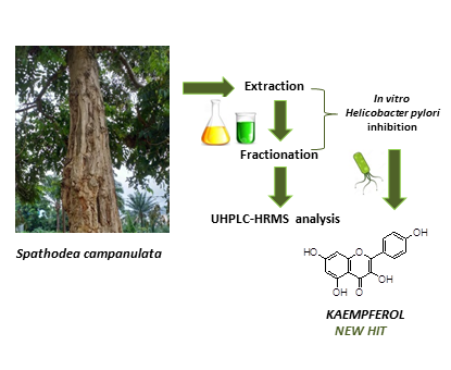
New Insights into Bioactive Compounds from the Medicinal Plant Spathodea campanulata P. Beauv. and Their Activity against Helicobacter pylori

by
Corinne Raïssa Ngnameko
1,2,
Lucia Marchetti
1,3,
Barbara Zambelli
4,
Antonio Quotadamo
1,3,
Davide Roncarati
4,
Davide Bertelli
1,
Frederic Nico Njayou
2,
Stella I. Smith
5,
Paul F. Moundipa
2,*,
Maria Paola Costi
1 and
Federica Pellati
1,*
1
Department of Life Sciences, University of Modena and Reggio Emilia, Via G. Campi 103, 41125 Modena, Italy
2
Department of Biochemistry, Faculty of Science, The University of Yaounde I, P. Box 812 Yaounde, Cameroon
3
Doctorate School in Clinical and Experimental Medicine (CEM), University of Modena and Reggio Emilia, 41125 Modena, Italy
4
Department of Pharmacy and Biotechnology, Alma Mater Studiorum University of Bologna, Viale Fanin 44, 40127 Bologna, Italy
5
Nigerian Institute of Medical Research, PMB 2013, Yaba, Lagos 100001, Nigeria
*
Authors to whom correspondence should be addressed.
Submission received: 21 March 2020 / Revised: 12 May 2020 / Accepted: 13 May 2020 / Published: 15 May 2020
(This article belongs to the Special Issue Novel Targets and Mechanisms in Antimicrobial Drug Discovery)

Abstract

:
The medicinal plant Spathodea campanulata P. Beauv. (Bignoniaceae) has been traditionally applied for the prevention and treatment of diseases of the kidney and urinary system, the skin, the gastrointestinal tract, and inflammation in general. The present work shows for the first time how chemical components from this plant inhibit Helicobacter pylori growth by urease inhibition and modulation of virulence factors. The crude extract and the main fractions of S. campanulata bark were tested on H. pylori isolated strains and the active ones were further fractionated. Fractions and sub-fractions of the plant crude extract were characterized by ultra-high-performance liquid chromatographic tandem high resolution-mass spectrometry detection (UHPLC-HRMS). Several phenolics and triterpenoids were identified. Among the sub-fractions obtained, SB2 showed the capacity to inhibit H. pylori urease in a heterologous bacterial model. One additional sub-fraction (SE3) was able to simultaneously modulate the expression of two adhesins (HopZ and BabA) and one cytotoxin (CagA). The flavonol kaempferol was identified as the most interesting compound that deserves further investigation as a new hit for its capacity to modulate H. pylori virulence factors.

Graphical Abstract

1. Introduction

Helicobacter pylori (H. pylori) is a Gram-negative bacterium that persistently colonizes the stomach of more than half the human population and its prevalence is significantly higher in the developing countries [1,2]. It causes gastric inflammation, gastric mucosa-associated lymphoid tissue lymphoma, and cancer. Due to its role in cancer, it was listed as a class I carcinogen by the World Health Organization (WHO) in 1994 [3].
Several meta-analyses have pointed out that the mass eradication of H. pylori would have beneficial effects, such as reduction in gastric cancer incidence, peptic ulcer development, dyspepsia symptoms, and anemia occurrence. Nonetheless, the efficacy of current treatments remains a major concern. The medical therapy for H. pylori still relies on a combination of antibiotics and anti-secretory agents, e.g., proton pump inhibitors (PPIs) [4]. However, several studies have described high resistance to antibiotic treatment [5,6,7]. Indeed, in 2017 WHO included H. pylori in the list of antibiotic resistant bacterium for which the identification and development of new antimicrobial drugs represent a global priority [8].
To grow in the gastric acid medium H. pylori takes advantage of the Ni(II)-dependent urease enzyme, which catalyzes the hydrolysis of urea to produce ammonia and carbamate, the latter subsequently decomposes to ammonia and bicarbonate. The effect of this process is the increase of the medium pH, hence making the environment comfortable for H. pylori colonization, despite the harsh acidic conditions of the stomach [9,10]. Urease is therefore a target for the development of alternative and specific antibacterial strategies to overcome H. pylori gastric infection.
H. pylori uses adhesins to bind and enter to the gastric mucosa. Adhesins are cell-surface proteins that enable bacterial adherence to cells. H. pylori major adhesive factors, which belong to the largest outer membrane protein (OMP) family, include blood group antigen-binding adhesion (BabA), sialic acid-binding adhesion (SabA), H. pylori outer membrane protein (HopZ), adherence-associated lipoprotein A and B (AlpA-B), H. pylori adhesin A (HpaA) and Lewisx–LPS. H. pylori adhesins are considered bacterial virulence factors and they are involved in several processes during the early and chronic phases of the infection. The most studied virulence factors of H. pylori are cytotoxin-associated protein A (CagA) and vacuolating cytotoxin A (VacA) [11,12]. CagA is able to initiate in host cells NF-κB, MAPK, and SHP-2/ERK pathways, producing inflammatory factors and pro-inflammatory cytokines (IL-6, IL-8, INF-γ, TNF-α). These substances may cause extensive infection sites and inflammation, leading to gastritis or gastric cancer [11,12].
The capacity to inhibit the growth of this bacterium has been ascribed to a variety of medicinal plants and natural compounds [13,14]. However, only a few papers have described the mechanisms of action of natural products against H. pylori [13,14]. In general, these mechanisms include urease activity inhibition, anti-adhesion activity, DNA damage, protein synthesis inhibition, and oxidative stress [15,16,17].
Spathodea campanulata P. Beauv. (Bignoniaceae) is a medicinal plant traditionally used in Africa for the prevention and treatment of diseases of the kidney and urinary systems, the skin, the gastrointestinal tract, and inflammation in general [18,19]. Extracts of this plant have been found to be active against proliferative diseases, involving cancer cells and bacteria [20]. More recently, anti-H. pylori, anti-adhesive, and growth inhibitory activities of a methylene chloride (DCM)/methanol (MeOH) extract from the bark of the plant have been described [21]. Data about the chemical characterization of S. campanulata are limited. Nevertheless, previous studies undertaken on the stem bark and leaves have shown the presence of phenolic acids, flavonoids, triterpenoids, iridoids, and sterols [22,23,24,25,26,27].
Consequently, this is the first chemical characterization study of the main compounds present in the extracts, fractions and sub-fractions of S. campanulata bark using UHPLC-HRMS, which were also assessed for their anti-H. pylori, anti-urease, and anti-adhesins effects.

2. Materials and Methods

2.1. Chemicals and Solvents

Analytical grade solvents, HPLC grade solvents and ammonium formate were from Sigma–Aldrich (Milan, Italy). Ultrapure water (H2O) was obtained from a Millipore water purification system (Merck Life Science, Milan, Italy). Pre-coated aluminum Kieselgel 60 F254 plates (layer thickness 0.2 mm, Merck, Darmstadt, Germany) were used for analytical thin-layer chromatography (TLC).

2.2. Plant Material and Preparation of the Crude Extract

The bark from S. campanulata was collected in July 2018 in Foumbot (West Region, Cameroon). A sample of the bark was deposited at the HNC-Cameroon National Herbarium, with the voucher number 50085/HNC. The bark used in this study was harvested from at least three different trees, in order to have a representative sample. The plant material was washed with H2O and dried at room temperature for several weeks. The dried plant material was then powdered using a grinder. The obtained powder was kept at 4 °C until the preparation of the extracts. A portion of 500 g of powdered plant material was soaked in 2 L of solvent solution composed by DCM/MeOH (1:1, v/v) for 48 h and paper filtered. The filtrate was dried using a rotary evaporator to obtain the crude extract.

2.3. Fractionation Procedure

The crude extract was purified on silica gel by preparative flash column chromatography using in sequence n-hexane (Hex), ethyl acetate (EtOAc), and MeOH. Eight stages of polarity were used: Hex (fraction A), Hex/EtOAc 25% (fraction B), Hex/EtOAc 50% (fraction C), Hex/EtOAc 75% (fraction D), EtOAc (fraction E), and MeOH (fraction F). The fractions were further partitioned using a silica gel column chromatography with an Isolera apparatus (Biotage, Charlotte, NC, USA) with different solvents, according to the result of TLC. In this way, different sub-fractions were collected: SA1 (90% Cyhex/EtOAc) from fraction A, SB1 and SB2 (70% Cyhex/EtOAc and 90% Cyhex/EtOAc, respectively) from fraction B, SC1 and SC2 (95% DCM/MeOH) from fraction C, SD1 and SD2 (20% Cyhex/EtOAc) from fraction D, SE1 and SE3 (50% MeOH/H2O and 70% MeOH/H2O, respectively) from fraction E, and SF (80% Cyhex/EtOAc) from fraction F. The solvent was removed from the collected fractions and sub-fractions by using a rotary evaporator.

2.4. Determination of Total Polyphenols and Total Flavonoids

For the determination of total polyphenols and total flavonoids, the crude extract and its fractions were dissolved in MeOH at a concentration of 100 µg/mL. The content of total polyphenols was analyzed according to the Folin–Ciocalteu’s method, as previously described [28]. Briefly, 50 µL of sample was added to a solution composed of 2.4 mL of distilled water, 200 µL of Folin–Ciocalteu’s reagent (2 N), and 500 µL of Na2CO3 20%. The reaction mixture was incubated at 25 °C in dark for 1 h, and the absorbance was read at 765 nm. The results were compared to a gallic acid calibration curve, and the total phenolic content was expressed as milligrams of gallic acid equivalents (GAE) per gram of extract.
The total flavonoid content was analyzed according to the aluminum chloride method [28]. Briefly, 50 µL of sample was mixed with 0.95 mL of MeOH and 1.0 mL of AlCl3 (2%). The reaction mixture was incubated at 25 °C for 30 min, and the absorbance was read at 420 nm. The results were compared to quercetin calibration curve and the total flavonoids content was expressed as milligrams of quercetin equivalents (QE) per gram of extract.

2.5. UHPLC-HRMS Analysis

For UHPLC-HRMS analysis, 2 mg of each fraction and sub-fraction obtained from the crude plant extract were dissolved in 1.0 mL of acetonitrile (ACN), filtered using a PTFE filter, and vortex mixed for 20 s, spun at 5000 g for 5 min. Then, 5 µL of this solution were injected in duplicate, for the analysis. A Thermo Scientific Dionex Ultimate 3000 UHPLC system was used, equipped with an autosampler, a quaternary pump and a thermostated column compartment controlled by the Chromeleon 7.2 Software (Thermo Scientific, Waltham, MA, USA). The UHPLC system was coupled to a high-resolution Q Exactive mass spectrometer (Thermo Scientific, Bremen, Germany). The mass spectrometer was calibrated before the analyses. Nitrogen (N2) (purity > 99.999%), obtained from a Zefiro zero 60 LC-MS nitrogen generator (CINEL, Vigonza, Italy), was employed both as the source gas and the collision gas. The chromatographic system was hyphenated to the MS with an electrospray ionization (ESI) source, which was operated both in the positive and in the negative ion modes. The capillary temperature was set at 270 °C and the following N2 flows (arbitrary units) were used: sheat gas 40, auxiliary gas 30, and sweep gas 3. The capillary voltage was set at −3.5 and +3 kV. The chromatographic separation was performed on an Ascentis Express C18 (10 cm × 2.1 mm i.d., 2.7 µm) (Supelco, Bellefonte, PA, USA). The column was kept at 25 °C. The mobile phase was composed of 80% H2O + 0.1% formic acid (FA) (A) and 20% ACN (B) at 0.3 mL/min. The following gradient was used: from 0.1 to 10.1 min from 20% to 98% B, which was kept constant for 8.8 min, before the reconditioning step, for a total analysis time of 25 min.
MS and MS2 spectra were recorded from 100 to 1000 m/z, at a resolution of 70,000 and 17,500, respectively. The three most intense ions were selected for MS2 N2-promoted collision-induced dissociation (CID) (stepped CE = 20, 28 eV). A precursor active exclusion of 6 s was set. In parallel, an all ion fragmentation experiment (CE = 28 eV) was scheduled for post-hoc analysis. The mass calibration was performed weekly, both in the negative and in the positive ion modes, to ensure a mass accuracy ≤ 2 ppm. XCalibur 2.9 (Thermo Scientific, Bremen, Germany) and FreeStyle 1.3 software (Thermo Scientific, San José, CA, USA) were used for UHPLC control and MS data processing, respectively.

2.6. Antimicrobial Disk Diffusion Test

H. pylori strain G27 was obtained from the University of Bologna, Italy. H. pylori cells were recovered from glycerol stocks on Brucella broth agar plates, containing 5% fetal calf serum (FCS), added with Dent’s antibiotic supplement in an atmosphere of 9% CO2/91% air, maintained at 37 °C, and 95% humidity in water-jacketed incubator (Thermo Fisher Scientific, Waltham, MA, USA). All reagents were purchased from Oxoid, United Kingdom. The assay was carried out following the method described by Balouiri et al. [29]. H. pylori cells were collected from Brucella broth agar plates, suspended in 500 µL of liquid Brucella broth with 5% Dent’s antibiotic supplement and subsequently the cell density (OD600) of bacterial suspension was determined. Then, H. pylori cells were diluted in melted Brucella broth Soft Agar medium (Brucella broth agar plates containing 0.5% agar) to obtain a final OD600 of 0.07 and 6.5 mL of this suspension was poured into a sterile standard Brucella broth agar plate. After that, 5 mm sterile paper disks were deposited into the agar. A total of 4 µL of the crude extract, fractions, and sub-fractions (10 mg/mL), prepared using DMSO/H2O (25% v/v), were dropped into the disk. The DMSO/H2O solution was used as the negative control. Kanamycin (10 µg) was used as the reference antibiotic. The plates were incubated under microaerophilic conditions at 37 °C for 72 h. The products that showed a diameter of inhibition ≥ 8 mm were considered to be active [29]. The active products were serially diluted using sterile DMSO/H2O (25% v/v) to obtain three decreasing concentrations, which were re-tested in triplicate and the results expressed as a mean diameter of inhibition zone. The minimum active quantity (MAQ) for each extract was determined as the minimum quantity that produced an inhibition zone ≤ 8 mm.

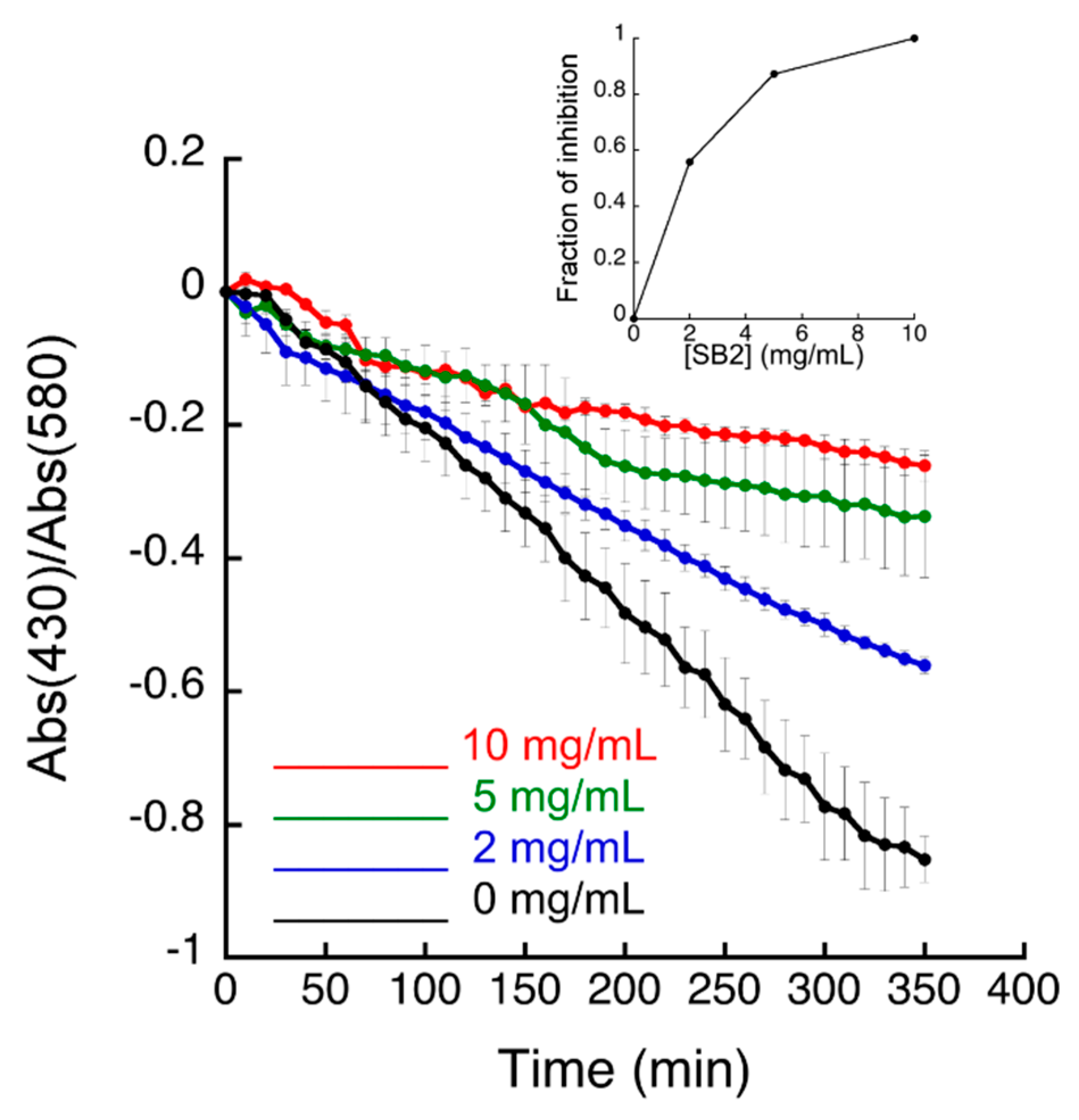
2.7. In-Cell Urease Activity Test

Escherichia coli TOP10 cells harboring pGEM-ureOP plasmid were pre-cultured at 37 °C in 1 mL of lysogeny broth (LB), containing 100 μg/mL of ampicillin (Ap). After 16 h, 40 μL of pre-culture were used to inoculate 2 mL of LB medium containing 100 μg/mL of Ap and 10 μg/L of cresol red pH indicator. When OD600 reached 0.7–0.9, 90 μL of each culture was inoculated in 96-well plates with 80 mM urea, with and without 10 µL of different concentrations of either the fractions or the sub-fractions of the plant extract or DMSO. Immediately before running the experiment, 2.5 µL of 10 mM NiSO4 was added to each culture to a final concentration of 250 µM. In each well, 30 μL of mineral oil was added to prevent the medium evaporation during the culture. The color change of cresol red indicator was monitored over time spectrophotometrically in a multi-plate reader, measuring the absorption at 430 and 580 nm.

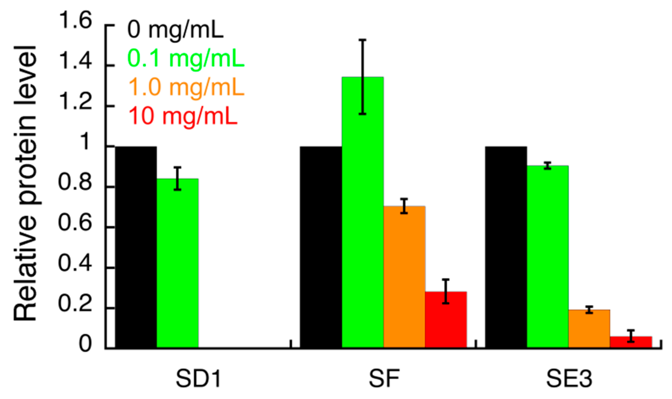
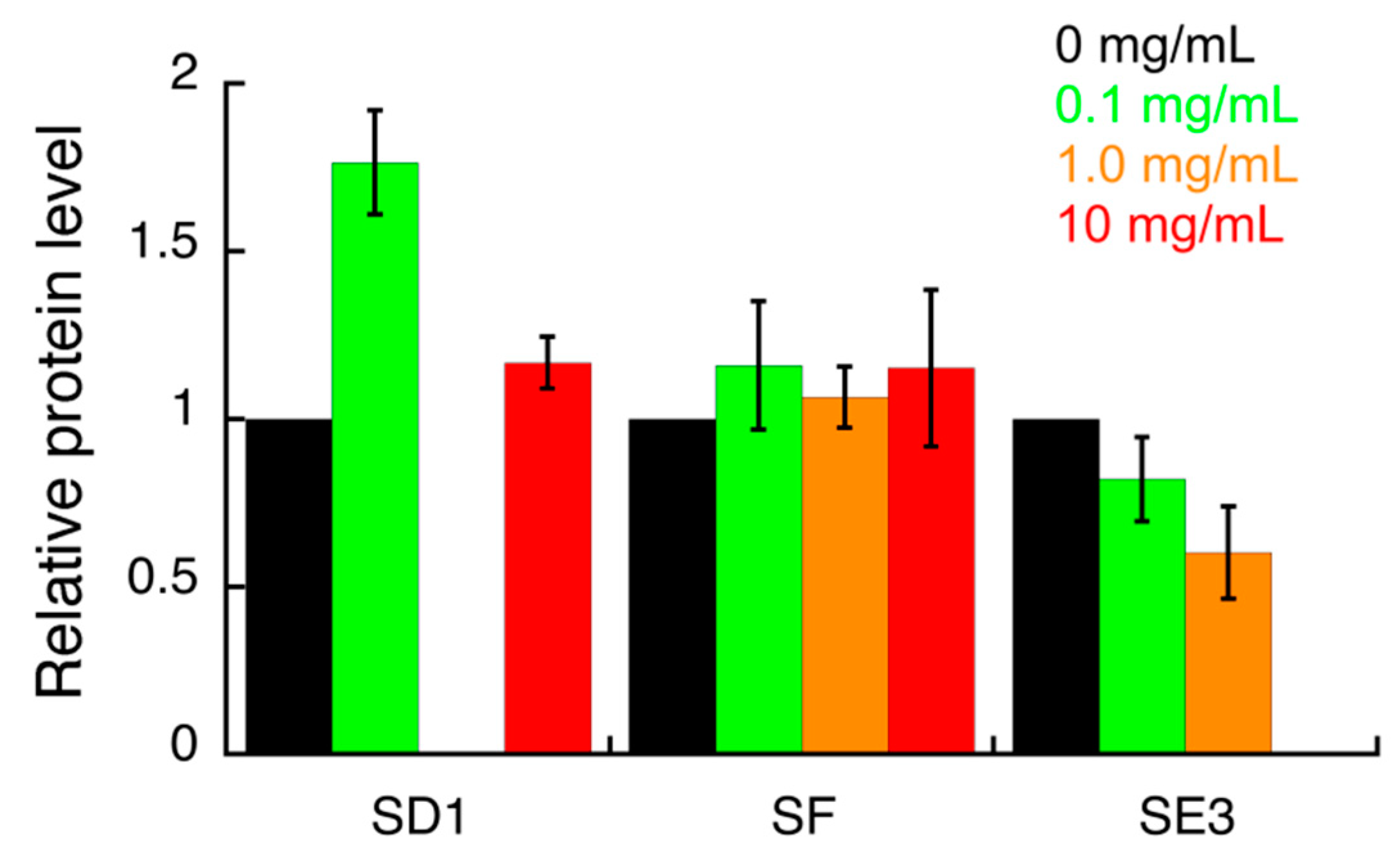
2.8. Western Blot Analysis

Three days old cultures of the H. pylori G27 strain were harvested into Brucella broth medium supplemented with 10% FBS to 0.8 OD600. Then, 100 µL of the mixture was added to each well in a 96-well plate. A total of 100 µL of the plant extract at the three concentrations was used. The plates were incubated at 37 °C under microaerophilic conditions for 4 h. After incubation, 200 µL of the mixture was transferred into a microcentrifuge tube and centrifuged at 14,000 rpm for 5 min at 4 °C. The supernatant was discarded, and the pellet was washed with phosphate buffer saline (PBS). Protein in Laemmli loading buffer was separated by 12% sodium dodecyl sulfate poly-acrylamide gel electrophoresis (SDS-PAGE) and electro-transferred into a polyvinylidene difluoride paper (PVDF) blotting membrane (GE Healthcare, Germany). The membranes were blocked with 5% w/v non-fat milk in phosphate buffered saline Tween-20, incubated overnight at 4 °C with primary antibodies (BabA, CagA, HopZ, and HP1043 as the loading control), rinsed, and then incubated for 1 h at 25 °C with horseradish peroxidase-conjugated secondary antibodies. The blots were then developed by using a chemiluminescent method (Sigma, Germany). The densitometric analysis of the protein bands was performed using the ImageJ Software (NIH, USA).

2.9. Statistical Analysis

Data analysis was carried out using the GraphPad Prism 5.0 software. For the diameter of inhibition, the results were reported as mean ± standard deviation (SD). For the IC50 of urease and adhesion, the results were expressed as mean ± SD of three independent experiments. Regarding protein expression, data were presented as mean ± SD. Comparisons were made between untreated group and DMSO control groups.

3. Results and Discussion

3.1. Chemical Composition of S. campanulata Fractions and Sub-Fractions

To identify new anti-H. pylori growth inhibitors, we first tested fractions and sub-fractions of bark of S. campanulata against the bacterium and then we focused on the urease enzyme and adhesin proteins involved in adhesion to gastric mucosa.
After the extraction of the plant material, its fractionation and further sub-fractionation were performed by means of preparative flash column chromatography. Six fractions were obtained, which were further sub-fractioned using silica gel column chromatography to provide 10 sub-fractions. The content of total polyphenols and total flavonoids in the crude plant extract and in the six fractions is shown in Table 1.
MS and MS/MS data of all the identified compounds are shown in Table S1. The chemical structure of the 11 characterized compounds, which was confirmed by comparison with existing structures [22,23,24,25,26,27,30], is shown in Table 2. Compound 5,7-dihydroxy-4-metilcoumarin was detected in this plant for the first time, through the mass spectral database mzCloud.

3.2. Inhibitory Activity of S. campanulata Fractions and Sub-Fractions against H. pylori

The inhibitory activity on H. pylori of all the fractions and sub-fractions is shown in Table 3. The minimum active quantity (MAQ) values ranged from 0.1 mg to 10 mg/mL. The lowest MAQ value of 0.1 mg/mL was obtained with fractions B, C, and E and sub-fractions SA1 and SB1 on H. pylori.
The measurement of the cellular urease activity was performed in a heterologous E. coli bacterial model overexpressing the enzyme (Figure 1, Figures S1–S3), as previously described [31].
The results indicated that sub-fractions SD2, SE1 and SE3 produced minor urease inhibition (Figures S1–S3), while the sub-fraction SB2 inhibited urease in living bacteria with an IC50 value of 3.3 mg/mL (Figure 1). All the other fractions showed either low solubility or limited activity.

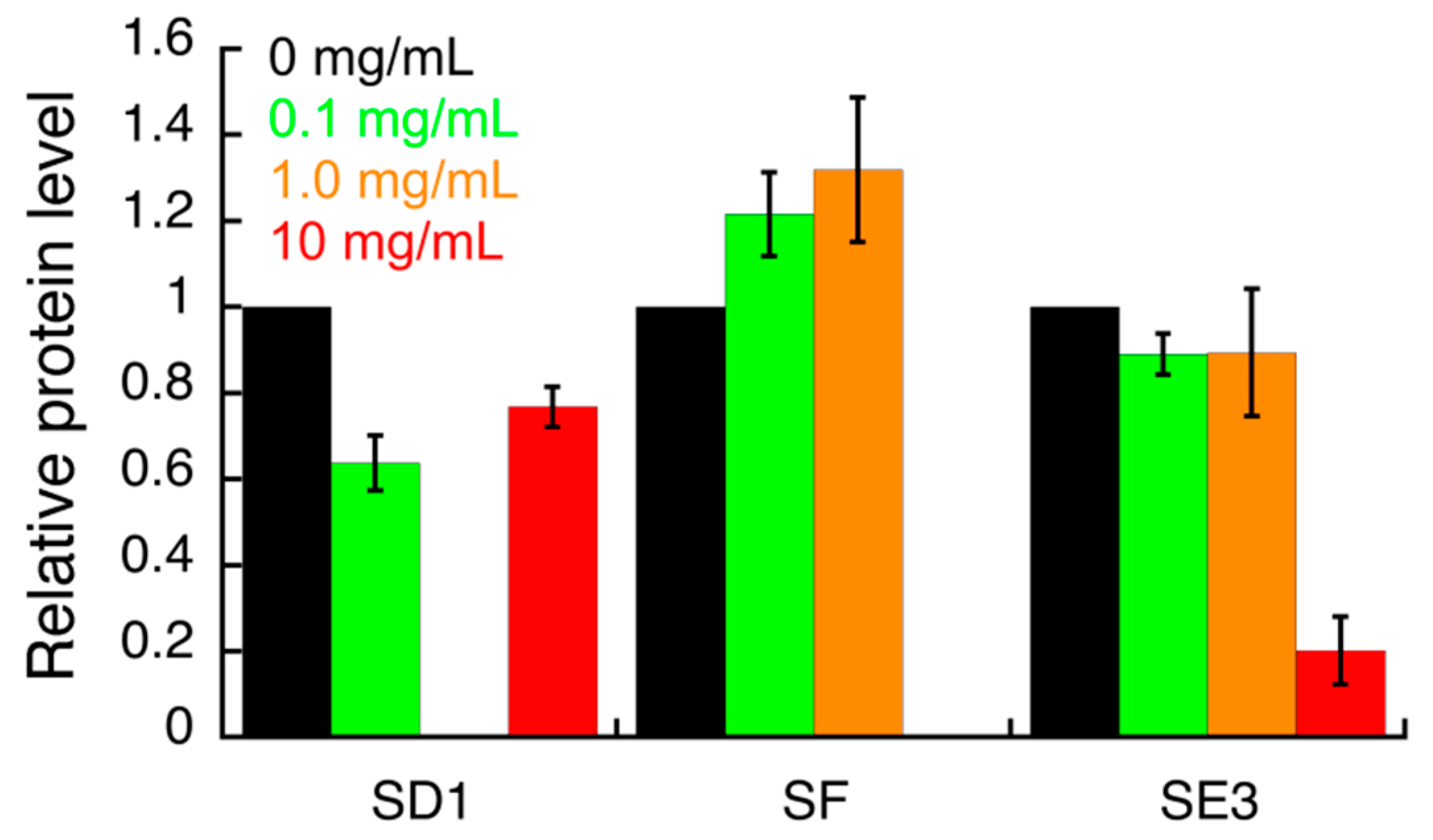
3.3. Effect of S. campanulata Fractions and Sub-Factions on H. pylori Virulence Factors

The effect of all S. campanulata fractions and sub-fractions on the expression of specific H. pylori virulence factors, including the two adhesins BabA and HopZ, and the cytotoxin-associated gene A (CagA), was investigated by Western blot. The samples that showed a significant effect in this preliminary assay were further investigated (Figure S4; Figure 2, Figure 3 and Figure 4). HP1043 staining was used as the loading control. Densitometric analysis of blot was performed and quantification data are reported in the bar chart. Values are expressed as means ± SD of two independent experiments. Specifically, sub-fractions SD1, SE3, and SF were able to inhibit HopZ expression (Figure 2). Sub-fractions SD1 and SE3 were found to slightly inhibit the expression of BabA (Figure 3). As for CagA expression, only sub-fraction SE3 inhibited the expression of this cytotoxin (Figure 4).
The compounds identified in this work by UHPLC-HRMS may be involved in the observed biological activity either by inhibiting urease activity or by modulating the expression of the virulence factors mentioned above. The plant sub-fraction SE3, able to simultaneously modulate the expression of two adhesins (HopZ and BabA) and one cytotoxin (CagA), was obtained from fraction E, which was the richest one in total polyphenols and total flavonoids (Table 1); the flavonol kaempferol was identified in sub-fraction SE3 by UHPLC-HRMS analysis. As for the other two sub-fractions SD1 and SF, their chemical composition was not disclosed by UHPLC-HRMS analysis.
Phenolics have general antimicrobial activity and some of them are also active against H. pylori [13,32]. Several phytochemicals may act by inhibiting enteric microbial growth, inducing cellular membrane perturbations, interfering with certain microbial metabolic processes and modulating signal transduction or gene expression pathways [32]. Notably, it has been recently observed that the flavonoid kaempferol, a widely diffused natural flavonol, has an anti-inflammatory effect on H. pylori-induced inflammation by suppressing the translocation of CagA and VacA proteins and leading to the downregulation of pro-inflammatory cytokines [33]. Recently, kaempferol has been found to inhibit the in vitro DNA binding activity of the homeostatic stress regulator (HsrA), a protein which plays as a global homeostatic regulator, synchronizing metabolic functions and virulence with availability of nutrients and cell division, mediating also the response to oxidative stress. HsrA represents a novel and effective therapeutic target in H. pylori infection [34]. In the present work, the sub-fraction SE3 from S. campanulata, which contains kaempferol, was found to simultaneously modulate the expression of the two H. pylori adhesins HopZ and BabA and the cytotoxin CagA; in addition, this sub-fraction showed a mild urease inhibition. Regarding the effect of natural compounds on the expression of HopZ, to the best of our knowledge no information is available in the literature. N-phenylpropenoyl-l-amino acids (NPAs) has been identified as polyphenol/amino acid conjugates in the seeds of theobroma cacao as well as in a variety of herbal drugs; they have been described for their inhibitors of H. pylori BabA outer membrane protein [35]. As for CagA, in addition to kaempferol, binding interactions between this virulence factor and natural compounds have been investigated by molecular docking and four hit compounds have been identified, including one phytosterol, one chalcone, and two carotenoids [36]. Due to the reduced number of natural compounds able either to inhibit urease enzymatic activity or to modulate the expression of H. pylori virulence factors, the research in this field deserves a great attention in the medicinal chemistry field in order to develop the identified hit kaempferol, alone or in combination with known drugs. This outcome is in agreement with a recent study [34], where kaempferol has exhibited a potent bactericidal activity against H. pylori, with an MBC value from 28 to 56 µM, the latter in resistant strains. However, the wide group of intracellular targets and the high number of natural compounds potentially effective as therapeutic agents against H. pylori present in this plant extract require further evidence to better understand the role of these substances using in vivo models, as previously described in the literature for kampferol isolated from Polygonum tinctorium Lour. [37].

4. Conclusions

It can be assumed from this study that natural compounds present in S. campanulata bark extract could be effective as potential therapeutic agents against H. pylori, by inhibiting urease activity and some virulence factors and by modulating the expression of the adhesins BabA and HopZ and the cytotoxin CagA. These results suggest that enriched extracts from this plant could be useful for the treatment of H. pylori mediated gastric diseases and they could be used synergistically with antibiotics in conventional therapy. The flavonol kaempferol, identified by UHPLC-HRMS, has to be further assessed as a new hit for its potential therapeutic activity against H. pylori infection.

Supplementary Materials

The following files are available online at https://0-www-mdpi-com.brum.beds.ac.uk/2079-6382/9/5/258/s1, Figure S1: Urease activity of recombinant E. coli cells in the presence of 250 μM Ni(II) and of 80 mM urea, measured as a change of pH detected by the cresol red indicator. Increasing concentrations of the sub-fraction SD2 were added to the E. coli culture before performing the colorimetric assay. Data are shown as mean ± SD of the triplicates. Sub-fraction SD2 (20% Cyhex/EtOAc) was obtained from fraction D. Figure S2: Urease activity of recombinant E. coli cells in the presence of 250 μM Ni(II) and of 80 mM urea, measured as a change of pH detected by the cresol red indicator. Increasing concentrations of the sub-fraction SE1 were added to the E. coli culture before performing the colorimetric assay. Data are shown as mean ± SD of the triplicates. Sub-fraction SE1 (50% MeOH/H2O) was obtained from fraction E. Figure S3: Urease activity of recombinant E. coli cells in the presence of 250 μM Ni(II) and of 80 mM urea, measured as a change of pH detected by the cresol red indicator. Increasing concentrations of the sub-fraction SE3 were added to the E. coli culture before performing the colorimetric assay. Data are shown as mean ± SD of the triplicates. Sub-fraction SE3 (70% MeOH/H2O) was obtained from fraction E. Figure S4: Western blot analysis of H. pylori cells, treated with various concentrations of sub-fractions, to quantify the expression levels of HopZ, BabA and CagA proteins. DMSO was used as the control treatment (C), while HP1043 staining was used as the loading control. Sub-fraction SD1 (20% Cyhex/EtOAc), sub-fraction SE3 (70% MeOH/H2O) and sub-fraction SF (80% Cyhex/EtOAc) were from fractions D, E and F, respectively. Table S1: Compounds identified in the active fractions and sub-fractions from Spathodea campanulata by UHPLC-HRMS.

Author Contributions

Conceptualization, P.F.M., M.P.C. and F.P.; methodology, C.R.N., L.M., B.Z., A.Q., D.R. and D.B.; software, D.B.; validation, M.P.C. and F.P.; formal analysis, B.Z., D.R., M.P.C. and F.P.; investigation, C.R.N., L.M., B.Z., A.Q. and D.R.; resources, M.P.C., D.B. and F.P.; data curation, B.Z., D.R., M.P.C. and F.P.; writing—original draft preparation, C.R.N., L.M., B.Z., A.Q. and D.R.; writing—review and editing, F.N.N., S.I.S., P.F.M., M.P.C. and F.P.; visualization, C.R.N., L.M., B.Z. and D.R.; supervision, P.F.M., M.P.C. and F.P.; project administration, P.F.M., M.P.C. and F.P.; funding acquisition, M.P.C., D.B. and F.P. All authors have read and agreed to the published version of the manuscript.

Funding

This research received no external funding.

Acknowledgments

Rainer Haas from Max von Pettenkofer (Institute Pettenkoferstr, Munich, Germany) is kindly acknowledged for providing the antibodies. The authors also acknowledge the Fondazione Cassa di Risparmio di Modena for funding the UHPLC-ESI-Q Exactive system at the Centro Interdipartimentale Grandi Strumenti (CIGS) of the University of Modena and Reggio Emilia, Italy. The authors are grateful to the Erasmus+ program (action KA 107) for providing Corinne Raïssa Ngnameko a grant for her research work at the University of Modena and Reggio Emilia and at the University of Bologna in Italy. Part of the work was funded by Research Modernization Fund of Ministry of Higher Education (Cameroon) and by the FAR2018 Fund of the University of Modena and Reggio Emilia (Italy).

Conflicts of Interest

The authors declare no conflict of interest.

References

  1. Cover, T.L.; Blaser, M.J. Helicobacter pylori in Health and Disease. Gastroenterology 2009, 136, 1863–1873. [Google Scholar] [CrossRef] [PubMed] [Green Version]
  2. Testerman, T.L.; Morris, J. Beyond the stomach: An updated view of Helicobacter pylori pathogenesis, diagnosis, and treatment. World J. Gastroenterol. 2014, 20, 12781–12808. [Google Scholar] [CrossRef] [PubMed]
  3. Ferlay, J.; Soerjomataram, I.; Dikshit, R.; Eser, S.; Mathers, C.; Rebelo, M.; Parkin, D.M.; Forman, D.; Bray, F. Cancer incidence and mortality worldwide: Sources, methods and major patterns in Globocan 2012: Globocan 2012. Int. J. Cancer 2015, 136, E359–E386. [Google Scholar] [CrossRef] [PubMed]
  4. Safavi, M.; Shams-Ardakani, M.; Foroumadi, A. Medicinal plants in the treatment of Helicobacter pylori infections. Pharm. Biol. 2015, 53, 939–960. [Google Scholar] [CrossRef] [PubMed]
  5. Al-Eraky, D.M.; Helmy, O.M.; Ragab, Y.M.; Abdul-Khalek, Z.; El-Seidi, E.A.; Ramadan, M.A. Prevalence of CagA and antimicrobial sensitivity of H. pylori isolates of patients with gastric cancer in Egypt. Inf. Agents Cancer 2018, 13, 24. [Google Scholar] [CrossRef] [PubMed] [Green Version]
  6. Harrison, U.; Fowora, M.A.; Seriki, A.T.; Loell, E.; Mueller, S.; Ugo-Ijeh, M.; Onyekwere, C.A.; Lesi, O.A.; Otegbayo, J.A.; Akere, A.; et al. Helicobacter pylori strains from a Nigerian cohort show divergent antibiotic resistance rates and a uniform pathogenicity profile. PLoS ONE 2017, 12, e0176454. [Google Scholar] [CrossRef] [PubMed] [Green Version]
  7. Ndip, R.N.; Takang, A.E.M.; Ojongokpoko, J.E.A.; Luma, H.N.; Malongue, A.; Akoachere, J.-F.T.K.; Ndip, L.M.; MacMillan, M.; Weaver, L.T. Helicobacter pylori isolates recovered from gastric biopsies of patients with gastro-duodenal pathologies in Cameroon: Current status of antibiogram. Trop. Med. Int. Health 2008, 13, 848–854. [Google Scholar] [CrossRef]
  8. WHO Global Priority List of Antibiotic-Resistant Bacteria to Guide Research, Discovery, and Development of New Antibiotics. Available online: http://www.who.int/medicines/publications/global-priority-list-antibiotic-resistant-bacteria/en/ (accessed on 6 November 2019).
  9. Konieczna, I.; Żarnowiec, P.; Kwinkowski, M.; Kolesińska, B.; Frączyk, J.; Kamiński, Z.; Kaca, W. Bacterial Urease and its Role in Long-Lasting Human Diseases. Curr. Protein Pept. Sci. 2012, 13, 789–806. [Google Scholar] [CrossRef]
  10. Rutherford, J.C. The Emerging Role of Urease as a General Microbial Virulence Factor. PLoS Pathog. 2014, 10, e1004062. [Google Scholar] [CrossRef] [Green Version]
  11. Hage, N.; Howard, T.; Phillips, C.; Brassington, C.; Overman, R.; Debreczeni, J.; Gellert, P.; Stolnik, S.; Winkler, G.S.; Falcone, F.H. Structural basis of Lewisb antigen binding by the Helicobacter pylori adhesin BabA. Sci. Adv. 2015, 1, 1–9. [Google Scholar] [CrossRef] [Green Version]
  12. Kalali, B.; Mejías-Luque, R.; Javaheri, A.; Gerhard, M. H. pylori Virulence Factors: Influence on Immune System and Pathology. Med. Inflamm 2014, 2014, 426309. [Google Scholar] [CrossRef] [PubMed] [Green Version]
  13. Escandón, R.A.; del Campo, M.; López-Solis, R.; Obreque-Slier, E.; Toledo, H. Antibacterial effect of kaempferol and (−)-epicatechin on Helicobacter pylori. Eur. Food Res. Technol. 2016, 242, 1495–1502. [Google Scholar] [CrossRef]
  14. Hassan, S.T.S.; Žemlička, M. Plant-derived urease inhibitors as alternative chemotherapeutic agents. Archiv. Pharmazie 2016, 349, 507–522. [Google Scholar] [CrossRef] [PubMed]
  15. Flavonoid glycosides of Polygonum capitatum protect against inflammation associated with Helicobacter pylori infection. PLoS ONE 2015, 10, e0126584.
  16. Ghasemian, A.; Fattahi, A.; Mostafavi, S.K.S.; Almarzoqi, A.H.; Memariani, M.; Braiek, O.B.; Yassine, H.M.; Mostafavi, N.S.S.; Ahmed, M.M.; Mirforughi, S.A. Herbal medicine as an auspicious therapeutic approach for the eradication of Helicobacter pylori infection: A concise review. J. Cell. Phys. 2019, 234, 16847–16860. [Google Scholar] [CrossRef] [PubMed]
  17. Wang, Y.-C. Medicinal plant activity on Helicobacter pylori related diseases. World J. Gastroenterol. 2014, 20, 10368–10382. [Google Scholar] [CrossRef]
  18. Ilodigwe, E.E.; Akah, P.A.; Nworu, C.S. Anticonvulsant activity of ethanol leaf extract of Spathodea campanulata P. Beauv (Bignoniaceae). J. Med. Food 2010, 13, 827–833. [Google Scholar] [CrossRef] [PubMed]
  19. Ofori-Kwakye, K.; Kwapong, A.A.; Adu, F. Antimicrobial activity of extracts and topical products of the stem bark of Spathodea campanulata for wound healing. Afr. J. Tradit. Complement. Altern. Med. 2009, 6, 168–174. [Google Scholar] [CrossRef] [Green Version]
  20. Teinkela, J.E.M.; Nguedia, J.C.A.; Meyer, F.; Donfack, E.V.; Ndjakou, B.L.; Ngouela, S.; Tsamo, E.; Adiogo, D.; Azebaze, A.G.B.; Wintjens, R. In vitro antimicrobial and anti-proliferative activities of plant extracts from Spathodea campanulata, Ficus bubu, and Carica papaya. Pharm. Biol. 2016, 54, 1086–1095. [Google Scholar] [CrossRef] [PubMed]
  21. Ngnameko, C.R.; Njayou, F.N.; Fowora, M.; Nemg, F.B.S.; Moundipa Fewou, P.; Smith, S.I. Inhibitory effect of medicinal plants from Cameroon on the growth and adhesion of Helicobacter pylori. Eur. J. Int. Med. 2019, 30, 100957. [Google Scholar] [CrossRef]
  22. Amusan, O.O.G.; Adesogan, E.K.; Makinde, J.M. Antimalarial Active Principles of Spathodea campanulata Stem Bark. Phytother. Res. 1996, 10, 692–693. [Google Scholar] [CrossRef]
  23. Boniface, P.K.; Singh, M.; Verma, S.; Shukla, A.; Khan, F.; Srivastava, S.K.; Pal, A. RP-HPLC-DAD method for the identification of two potential antioxidant agents namely verminoside and 1-O-(E)-caffeoyl-β-gentiobiose from Spathodea campanulata leaves. Nat. Prod. Res. 2015, 29, 676–680. [Google Scholar] [CrossRef] [PubMed]
  24. Heim, S.C.; Guarnier, F.A.; Ferreira, D.T.; Braz-Filho, R.; Cecchini, R.; Cecchini, A.L. Antioxidant activity of Spathodea campanulata (Bignoneaceae) extracts. Rev. Bras. Plantas Med. 2012, 14, 287–292. [Google Scholar] [CrossRef] [Green Version]
  25. Lifongo, L.L.; Simoben, C.V.; Ntie-Kang, F.; Babiaka, S.B.; Judson, P.N. A bioactivity versus ethnobotanical survey of medicinal plants from Nigeria, west Africa. Nat. Prod. Bioprospect. 2014, 4, 1–19. [Google Scholar] [CrossRef] [PubMed] [Green Version]
  26. Nazif, N.M. Phytochemical and antioxidant activity of Spathodea campanulata P. beauvois. Growing in Egypt. Nat. Prod. Sci. 2007, 13, 11–16. [Google Scholar]
  27. Ngouela, S.; Tsamo, E.; Sondengam, B.L.; Connolly, J.D. Spathodol, a New Polyhydroxysterol from the Leaves of Spathodea campanulata. J. Nat. Prod. 1991, 54, 873–876. [Google Scholar] [CrossRef]
  28. Ngoungoure, V.L.N.; Mfotie Njoya, E.; Ngamli Fewou, S.; Ella, A.F.; McGaw, L.J.; Moundipa, P.F. Acetylcholinesterase inhibitory, anti-inflammatory and antioxidant properties of some Cameroonian medicinal plants used to treat some neurological disorders. Investig. Med. Chem. Pharmacol. 2019, 2, 33–46. [Google Scholar] [CrossRef]
  29. Balouiri, M.; Sadiki, M.; Ibnsouda, S.K. Methods for in vitro evaluating antimicrobial activity: A review. J. Pharm. Anal. 2016, 6, 71–79. [Google Scholar] [CrossRef] [Green Version]
  30. Pianaro, A.; Pinto, J.; Trevisan Ferreira, D.; Ishikawa, N.; Braz-Filho, R. Iridoid glucoside and antifungal phenolic compounds from Spathodea campanulata roots. Semin. Cienc. Agrar. 2007, 28, 251–255. [Google Scholar] [CrossRef] [Green Version]
  31. Tarsia, C.; Danielli, A.; Florini, F.; Cinelli, P.; Ciurli, S.; Zambelli, B. Targeting Helicobacter pylori urease activity and maturation: In-cell high-throughput approach for drug discovery. Biochim. Biophsica Acta (BBA) Gen. Subj. 2018, 1862, 2245–2253. [Google Scholar] [CrossRef]
  32. Godstime, O.C.; García, A.; Augustina, J.O.; Christopher, E. Mechanisms of Antimicrobial Actions of Phytochemicals against Enteric Pathogens—A Review. J. Pharm. Chem. Biol. Sci. 2014, 2, 77–85. [Google Scholar]
  33. Yeon, M.J.; Lee, M.H.; Kim, D.H.; Yang, J.Y.; Woo, H.J.; Kwon, H.J.; Moon, C.; Kim, S.-H.; Kim, J.-B. Anti-inflammatory effects of Kaempferol on Helicobacter pylori-induced inflammation. Biosci. Biotechnol. Biochem. 2019, 83, 166–173. [Google Scholar] [CrossRef] [PubMed]
  34. González, A.; Salillas, S.; Velázquez-Campoy, A.; Espinosa Angarica, V.; Fillat, M.F.; Sancho, J.; Lanas, Á. Identifying potential novel drugs against Helicobacter pylori by targeting the essential response regulator HsrA. Sci. Rep. 2019, 9, 11294. [Google Scholar] [CrossRef] [PubMed] [Green Version]
  35. Niehues, M.; Stark, T.; Keller, D.; Hofmann, T.; Hensel, A. Antiadhesion as a functional concept for prevention of pathogens: N-Phenylpropenoyl-L-amino acid amides as inhibitors of the Helicobacter pylori BabA outer membrane protein. Mol. Nutr. Food Res. 2011, 55, 1104–1117. [Google Scholar] [CrossRef] [PubMed]
  36. Racha, S.; Wongrattanakamon, P.; Raiwa, A.; Jiranusornkul, S. Discovery of novel potent small natural molecules able to enhance attenuation of the pathobiology of gastric cancer-associated Helicobacter pylori by molecular modeling. Int. J. Pept. Res. Ther. 2019, 25, 881–896. [Google Scholar] [CrossRef]
  37. Kataoka, M.; Hirata, K.; Kunikata, T.; Ushio, S.; Iwaki, K.; Ohashi, K.; Ikeda, M.; Kurimoto, M. Antibacterial action of tryptanthrin and kaempferol, isolated from the indigo plant (Polygonum tinctorium Lour.), against Helicobacter pylori-infected Mongolian gerbils. J. Gastroenterol. 2001, 36, 5–9. [Google Scholar] [CrossRef]
Figure 1. Urease activity of recombinant Escherichia coli cells in the presence of 250 μM Ni(II) and of 80 mM urea, measured as a change of pH detected by the cresol red indicator. Increasing concentrations of the sub-fraction SB2 were added to the E. coli culture before performing the colorimetric assay. Data are shown as mean ± SD of the triplicates. The insert represents the dose-response plot for the SB2 sub-fraction. Sub-fraction SB2 (90% Cyhex/EtOAc) was obtained from fraction B.
Antibiotics 09 00258 g001
Figure 2. Densitometric analysis of the Western blot images (shown in the Supplementary Materials) indicating the effects of S. campanulata on H. pylori G27 HopZ adhesin expression. H. pylori cells were treated with different concentrations of S. campanulata sub-fractions for 4 h and protein level of HopZ adhesin was determined by Western blot, using a specific anti-HopZ antibody. Values are expressed as means ± SD of two independent experiments Sub-fraction SD1 (20% Cyhex/EtOAc), sub-fraction SE3 (70% MeOH/H2O), and sub-fraction SF (80% Cyhex/EtOAc) were from fractions D, E, and F, respectively.
Antibiotics 09 00258 g002
Figure 3. Densitometric analysis of the Western blot images (shown in the Supplementary Materials) indicating the effects of S. campanulata on H. pylori G27 BabA adhesin expression. H. pylori cells were treated with different concentrations of S. campanulata sub-fractions for 4 h and protein level of BabA adhesin was determined by Western blot, using a specific anti-BabA antibody. Values are expressed as means ± SD of two independent experiments. Sub-fraction SD1 (20% Cyhex/EtOAc), sub-fraction SE3 (70% MeOH/H2O), and sub-fraction SF (80% Cyhex/EtOAc) were from fractions D, E, and F, respectively.
Antibiotics 09 00258 g003
Figure 4. Densitometric analysis of the Western blot images (shown in the Supplementary Materials) indicating the effects of S. campanulata on H. pylori G27 CagA expression. H. pylori cells were treated with different concentrations of S. campanulata sub-fractions for 4 h and protein level of the virulence factor CagA was determined by Western blot, using a specific anti-CagA antibody. Values are expressed as means ± SD of two independent experiments. Sub-fraction SD1 (20% Cyhex/EtOAc), sub-fraction SE3 (70% MeOH/H2O), and sub-fraction SF (80% Cyhex/EtOAc) were from fractions D, E, and F, respectively.
Antibiotics 09 00258 g004
Table 1. Total polyphenols and total flavonoids content in Spathodea campanulata crude extract and fractions (A–F).
Total Polyphenols (mg GAE/g) a
Crude extractABCDEF
51.4 b46.4 ± 0.543.0 ± 0.153.0 b48.3 b68.8 ± 0.356.3 ± 1.1
Total Flavonoids (mg QE/g) a
Crude extractABCDEF
5.2 ± 0.22.8 ± 0.11.8 b1.9 b2.0 b7.1 b1.9 ± 0.1
a Data of total polyphenols are expressed as mg of gallic acid equivalents (GAE)/g; data of total flavonoids as mg of quercetin equivalents (QE)/g (± SD). b SD < 0.05.
Table 2. Chemical structures of the identified compounds and their distribution in the active fractions and sub-fractions of Spathodea campanulata.
CompoundStructureFraction/Sub-Fraction
CESA1SB1SC2SD2SE1SE3
4-Hydroxy Benzoic Acid Antibiotics 09 00258 i001
Methyl-4-Hydroxybenzoate Antibiotics 09 00258 i002
5,7-Dihydroxy-4-Metilcoumarin Antibiotics 09 00258 i003
Quercetin Antibiotics 09 00258 i004
Kaempferol Antibiotics 09 00258 i005
Kaempferol 3-O-Glucoside Antibiotics 09 00258 i006
Spathodol Antibiotics 09 00258 i007
Spathodic Acid Antibiotics 09 00258 i008
Tomentosolic Acid Antibiotics 09 00258 i009
Ursolic Acid Antibiotics 09 00258 i010
Corosolic Acid Antibiotics 09 00258 i011
Table 3. Mean diameter of inhibition (mm ± SD) and minimum active quantity (MAQ) values (mg/mL) of Spathodea campanulata crude plant extract, active fractions and sub-fractions against Helicobacter pylori.
Sample.Concentration
0.1 mg/mL1.0 mg/mL10.0 mg/mLMAQ (mg/mL)
Crude extractND8.6 ± 0.811.6 ± 0.71.0
B11.3 ± 1.211.7 ± 2.112.1 ± 1.00.1
C10.012.0 16.3 ± 2.50.1
E10.011.7 ± 0.615.8 ± 0.30.1
SA19.8 ± 0.410.0 ± 1.010.7 ± 1.20.1
SB19.3 ± 0.69.7 ± 0.610.7 ± 0.60.1
SB2ND8.7 ± 2.311.3 ± 2.51.0
SC2ND8.010.0 1.0
SD1NDND10.3 ± 0.610.0
SE3NDND13.0 ± 1.010.0
SFND9.3 ± 1.210.7 ± 0.61.0
Kanamycin (2.5 mg/mL)35.7 ± 0.6---

