Article

Occurrence, Virulence and Antimicrobial Resistance-Associated Markers in Campylobacter Species Isolated from Retail Fresh Milk and Water Samples in Two District Municipalities in the Eastern Cape Province, South Africa

by
Aboi Igwaran
1,2,* and
Anthony Ifeanyi Okoh
1,2
1
SAMRC Microbial Water Quality Monitoring Centre, University of Fort Hare, Alice 5700, South Africa
2
Applied and Environmental Microbiology Research Group (AEMREG), Department of Biochemistry and Microbiology, University of Fort Hare, Private Bag X1314, Alice 5700, South Africa
*
Author to whom correspondence should be addressed.
Submission received: 21 December 2019 / Revised: 11 February 2020 / Accepted: 14 February 2020 / Published: 21 July 2020

Abstract

:
Campylobacter species are among the major bacteria implicated in human gastrointestinal infections and are majorly found in faeces of domestic animals, sewage discharges and agricultural runoff. These pathogens have been implicated in diseases outbreaks through consumption of contaminated milk and water in some parts of the globe and reports on this is very scanty in the Eastern Cape Province. Hence, this study evaluated the occurrence as well as virulence and antimicrobial-associated makers of Campylobacter species recovered from milk and water samples. A total of 56 water samples and 72 raw milk samples were collected and the samples were processed for enrichment in Bolton broth and incubated for 48 h in 10% CO2 at 42 °C under microaerobic condition. Thereafter, the enriched cultures were further processed and purified. After which, presumptive Campylobacter colonies were isolated and later confirmed by PCR using specific primers for the detection of the genus Campylobacter, target species and virulence associated genes. Antimicrobial resistance profiles of the isolates were determined by disk diffusion method against a panel of 12 antibiotics and relevant genotypic resistance genes were assessed by PCR assay. A total of 438 presumptive Campylobacter isolates were obtained; from which, 162 were identified as belonging to the genus Campylobacter of which 36.92% were obtained from water samples and 37.11% from milk samples. The 162 confirmed isolates were further delineated into four species, of which, 7.41%, 27.16% and 8.64% were identified as C. fetus, C. jejuni and C. coli respectively. Among the virulence genes screened for, the iam (32.88%) was most prevalent, followed by flgR (26.87%) gene and cdtB and cadF (5.71% each) genes. Of the 12 antibiotics tested, the highest phenotypic resistance displayed by Campylobacter isolates was against clindamycin (95.68%), while the lowest was observed against imipenem (21.47%). Other high phenotypic resistance displayed by the isolates were against erythromycin (95.06%), followed by ceftriaxone (93.21%), doxycycline (87.65%), azithromycin and ampicillin (87.04% each), tetracycline (83.33%), chloramphenicol (78.27%), ciprofloxacin (77.78%), levofloxacin (59.88%) and gentamicin (56.17%). Relevant resistance genes were assessed in the isolates that showed high phenotypic resistance, and the highest resistance gene harbored by the isolates was catII (95%) gene while VIM, KPC, Ges, bla-OXA-48-like, tetC, tetD, tetK, IMI and catI genes were not detected. The occurrence of this pathogen and the detection of virulence and antimicrobial resistance-associated genes in Campylobacter isolates recovered from milk/water samples position them a risk to human health.

1. Introduction

Campylobacter species are frequent enteric pathogens that cause diarrhea [1,2], and these pathogens are of great significant to public health due to the increasing number of species implicated in human infections [3]. Most campylobacteriosis cases are through consumption of contaminated food [4], unpasteurized milk [5] and contaminated water [6]. Water is important to life, but a lot of persons lack access to safe and clean water. As a result of this problem, many persons die of waterborne bacterial infections [7]. Waterborne infection is a worldwide burden that is approximated to cause millions of deaths annually and daily cases of illness including systematic illnesses, diarrhea and gastroenteritis [8,9]. Water sources, including rivers, lakes, streams and ponds, have numerous potential contamination sources such as faecal droppings of animals on pasture, direct faecal contamination by wild birds within the watersheds [10] and discharge of poorly treated wastewater effluents or non-disinfected sewage [11]. In South Africa, gastroenteritis, viral hepatitis, cholera, typhoid fever and dysentery are among waterborne infections that pose a high risk to the citizens [12]. Gastroenteritis is one of the major symptoms of campylobacteriosis. Consumption of unpasteurized milk from cows have been reported to be implicated in human campylobacteriosis [13,14]. Globally, milk consumption is predicted to be in billions of liters and most of which are consumed as pasteurized. Though, in recent years, there has been a rise in the rate of consumption of unpasteurized milk compared to pasteurized milk [15]. Raw milk consumption is highly unsafe for infants, pregnant women, the aged and immunocompromised persons [16]; and the propensity towards consumption of unpasteurized raw milk is due to its health benefits, taste and higher nutritional qualities.
Thus, consumption of unpasteurized raw milk positions the consumers at high risk of ill-health which could leads to diseases outbreaks. Raw milk is sometimes contaminated with pathogenic microbes and this usually occurs from sick animals or from environmental sources [14]. Several reports, including the studies of Artursson et al. [17] and Del Collo et al. [18], have also detected Campylobacter species in raw milk samples. Campylobacter species are among the several pathogens that sometimes contaminate raw milk [19,20]. In several parts of the world, Campylobacter species have been reported to be implicated in disease outbreaks and Campylobacter infections are of varying severity ranges from abdominal pains, vomiting, nausea, fever and diarrhea [21,22,23]. In extreme cases, acute phase of Campylobacter infection is followed by sequelae: Guillain-Barré syndrome or even death [24]. Some Campylobacter species reported to be implicated in human infections includes C. fetus, C. jejuni, C. coli and C. lari [25]. In addition to the burden of infections caused by these bacteria pathogens, the spread of antibiotic resistant-Campylobacter strains is another burden of public health plight which might be more severe in developing countries where there is largely uncontrolled use of antimicrobials [26,27]. Antibiotic resistance is known as a One Health concern due to the rapid emergence and spreading of resistant bacteria and resistant genes on a global scale [28]. Antibiotic resistant bacteria (ARB) are majorly disseminated through discharge of animal manure, human waste and wastewater effluents into the environment which can lead to the development of antibiotic-resistant genes (ARGs) in the exposed bacteria [29]. Antibiotic resistance can be mediated through vertical gene transfer or through genetic exchanges between and within bacteria species [30]. ARGs are emerging environmental pollutants and aquatic environments are known as one of the major reservoirs of ARB and ARGs [31]. Hence, this study evaluated the occurrence as well as virulence and antimicrobial resistance-associated markers of Campylobacter species recovered from milk and water samples in the Chris Hani and Amathole District Municipalities in the Eastern Cape Province, South Africa.

2. Material and Methods

2.1. Ethical Clearance

Ethical clearance was applied for the study and granted by the University of Fort Hare research ethics committee with certificate reference number: OKO021IGW01.

2.2. Description of Study Area

The study was carried out in Chris Hani and Amathole District Municipalities, in the Eastern Cape Province, South Africa with geographical co-ordinates “31.8743° S, 26.7968° E′′ and “32.5842° S, 27.3616° E′′ respectively.

2.3. Collection of Samples

A total of 128 samples were collected, comprising of 40 water samples from rivers and 16 water samples from pond/dams (used for irrigation), 40 milk samples from cow/bulk milk tanks from farms, 15 milk samples from cars/roadside, 9 milk samples from retail markets and 8 milk samples from butcheries. The water samples were collected in sterile 1L polypropylene bottles while the milk samples were collected in sterile 250 mL polypropylene bottles. All the samples were collected in Amathole and Chris Hani District Municipalities in the Eastern Cape Province, South Africa, transported in cooler box with ice and were analysed within 6 h of collection.

2.4. Isolation of Campylobacter Species from Water Samples

The method described by Van Dyke et al. [32] was adopted for Campylobacter isolation. Briefly, 1000 mL of water samples were filtered through nitrocellulose membrane filters (0.45-μm pore size). The filter papers were picked with sterilized forceps, added into 20 mL of Bolton selective enrichment broth supplemented with Bolton broth selective supplement with 5% (v/v) defibrinated horse blood and incubated in 10% CO2 at 42 °C for 48 h under microaerophilic condition in HF151UV CO2 incubator. Thereafter, a loopful from the enriched cultures were streaked onto modified cefoperazone deoxycholate agar (mCCDA) plates supplemented with antibiotic selective supplement (CCDA selective supplement (cefoperazone and amphotericin)), incubated as before. Presumptive Campylobacter colonies were picked and re-streaked onto blood agar plates supplemented with 7% (v/v) defibrinated horse blood and incubated as before.

2.5. Isolation of Campylobacter Species from Milk Samples

The milk samples were processed following the method previously described by Bianchini et al. [33]. Briefly, 20 mL of the milk samples was introduced into 200 mL of Bolton selective enrichment broths (1:10 ratio) to which Bolton antibiotic supplement with 5% (v/v) defibrinated horse blood were added and incubated at 42 °C for 48 h under microaerophilic atmosphere in 10%CO2 in HF151UV CO2 incubator. Thereafter, isolation and purification process described in Section 2.4 were followed.

2.6. DNA Extraction

Bacteria DNA was extracted by boiling method following the method of Sierra-Arguello et al. [34] with slight modification. Briefly, single Campylobacter colonies from the blood agar plates were isolated and grown in 5 mL of Tryptone Soya Broth (TSB) incubated for 48 h at 42 °C in 10%CO2 in a HF151UV CO2 incubator. From which, 1 mL of the broth was centrifuged for 5 min at 12,800 rpm and the supernatants were decanted and the cells were suspended in 400 µL of sterile distilled water in 1.5 mL Eppendorf tubes. The suspensions were boiled at 100 °C for 10 min in a heating block and the cell debris were removed by centrifugation for 5 min at 12,800 rpm and the supernatants were collected and stored at −20 °C until ready for use.

2.7. Molecular Confirmation Characterization and Amplification of Virulence Genes

Presumptive Campylobacter isolates were confirmed by PCR for identification of the genus Campylobacter targeting a 439 base pairs of part of 16S rRNA gene as reported by Moreno et al. [35]. The confirmed Campylobacter isolates were further delineation into C. jejuni, C. lari, C. fetus and C. coli using the primer sets as listed in Table S1 targeting cj0414, glyA, cstA and asK genes respectively [36] and primers specific for virulence markers responsible for invasion (iam) gene [37], invasion protein gene (ciaB) [38] colonization gene (flaA), adherence (cadF) gene and toxin production (cdtB) gene [3] and flagella synthesis and modification (flgR) gene [39] by PCR. Both multiplex and singleplex PCR were carried out in a 25 μL reaction volume (1.0 μL of each PCR primer, 12.5 µL master mix (Inqaba Biotec, South Africa), 5.0 μL of extracted DNA and 5.50 μL of nuclease free water). The amplified PCR products were visualized by gel electrophoresis in a 1.5% (w/v) agarose stained with ethidium bromide in 5xTAE buffer.

2.8. Antibiotic Resistance of Campylobacter Isolates

The disc diffusion technique on Mueller Hinton agar plates supplemented with 5% defibrinated horse blood was used to characterized the sensitivity of Campylobacter isolates against antimicrobial agents [40]. In summary, bacterial growth in TSB incubated at 42 °C for 48 h in 10% CO2 were adjusted to 0.5 McFarland turbidity standard in sterile normal saline followed by a gentle spread of the solution with a cotton swab on the entire surface of Mueller Hinton agar plates. Afterward, antibiotic discs were impregnated on the plates and incubated for 24 h at 42 °C in CO2 incubator under microaerobic conditions. The selected antimicrobials used were doxycycline (30 μg), tetracycline (30 μg), ampicillin (10 μg), azithromycin (15 µg), erythromycin (15 μg), gentamicin (10 μg), clindamycin (2 μg), chloramphenicol (30 μg), ciprofloxacin (5 μg), levofloxacin (5 µg), ceftriaxone (30 μg) and imipenem (10 µg). The inhibition zones for tetracycline, doxycycline, ciprofloxacin and erythromycin were interpreted according to CLSI [40] guidelines for Campylobacter. As there are no guidelines available for Campylobacter against ampicillin, azithromycin, gentamicin, clindamycin, chloramphenicol, levofloxacin, ceftriaxone and imipenem; CLSI, [40] guideline for Enterobacteriaceae were used for the interpretation of results.

2.9. Multiple Antibiotic Resistance MAR Index

The MAR index of each of the Campylobacter isolates were calculated using the formula MAR = a/b as reported by Krumperman, [41]. Where a= is the number of antibiotics to which the test isolate showed resistance to and b= is the total number of antibiotics to which the test isolate has been evaluated for susceptibility.

2.10. Molecular Screening Of Antimicrobial Resistance Genes

The isolates that showed phenotypic resistance to the test antibiotics were subjected to molecular screening for the detection of genotypic resistance genes employing PCR method. The primer sets reported by Ng et al. [42] was used for the detection of tetA, tetB, tetC, and tetD genes, for tetK and tetM genes [43], gyrA gene [44], ermB gene [45], catI and catII genes [46] and the aac(3)-IIa-(aacC2) [47] and VIM, KPC, Ges, bla-OXA-48-like and IMI genes [48] and the primer sets are shown in Table S2.

2.11. Statistical Analysis

Statistical analysis was carried out by Microsoft office tools.

3. Results

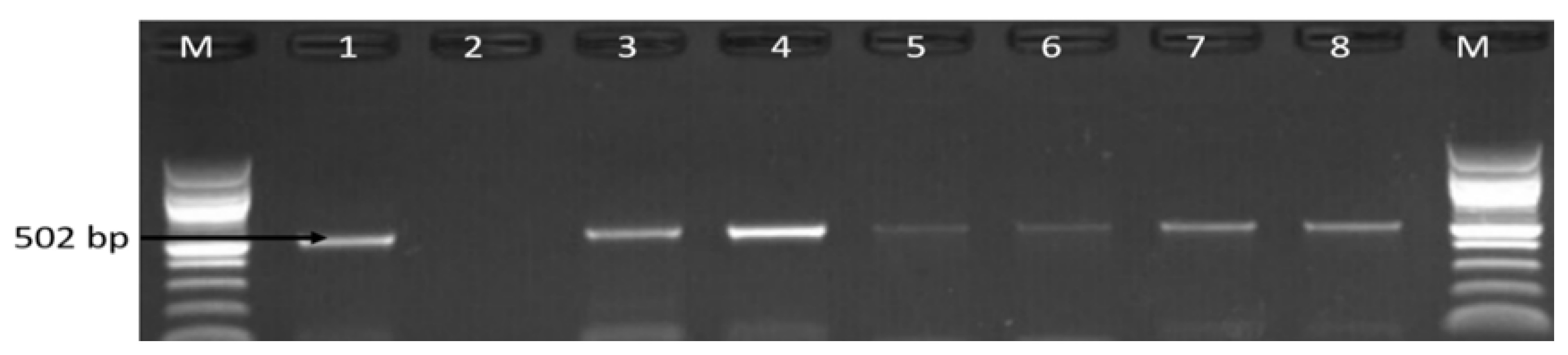
3.1. Molecular Identification of the Genus Campylobacter

A total of 438 presumptive Campylobacter isolates were obtained, from which 162 (36.99%) were identified as belonging to the genus Campylobacter of which 103 (36.92%) isolates out of the 279 presumptive isolates were detected in water samples from rivers/dams, and 33 (58.93%) water samples out of 56 water samples were positive for Campylobacter. In the milk samples, 59 (37.11%) isolates out of 159 presumptive isolates were detected to be Campylobacter and 19 (26.38%) milk samples out of 72 milk samples obtained from butcheries, farms, retail markets and car/roads were positive for Campylobacter (Figure 1). However, not all milk and water samples were positive for Campylobacter. Figure 1 is a pictorial representation of presumptive/confirmed Campylobacter isolates recovered from milk samples from different sources while Figure 2 is a representative gel picture of some PCR confirmed genus Campylobacter.

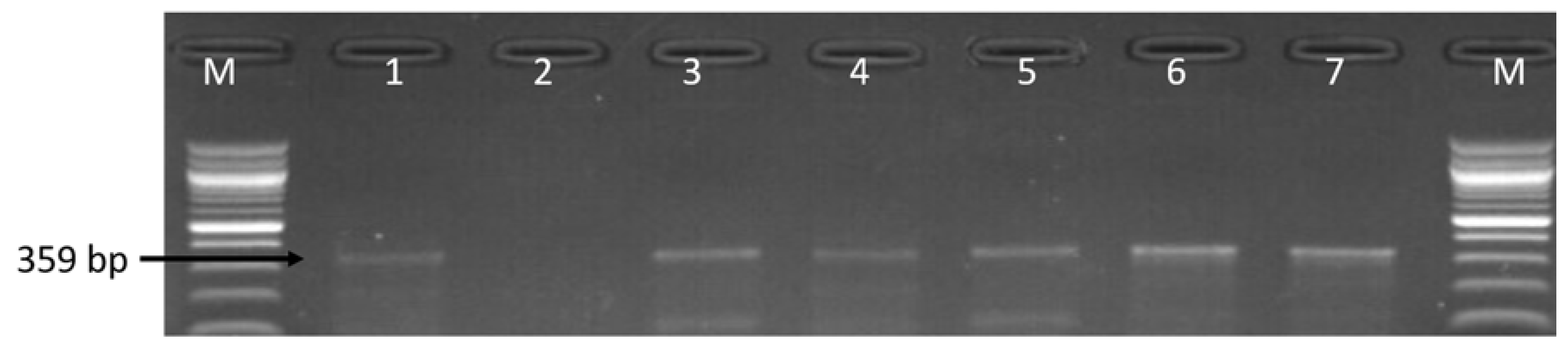
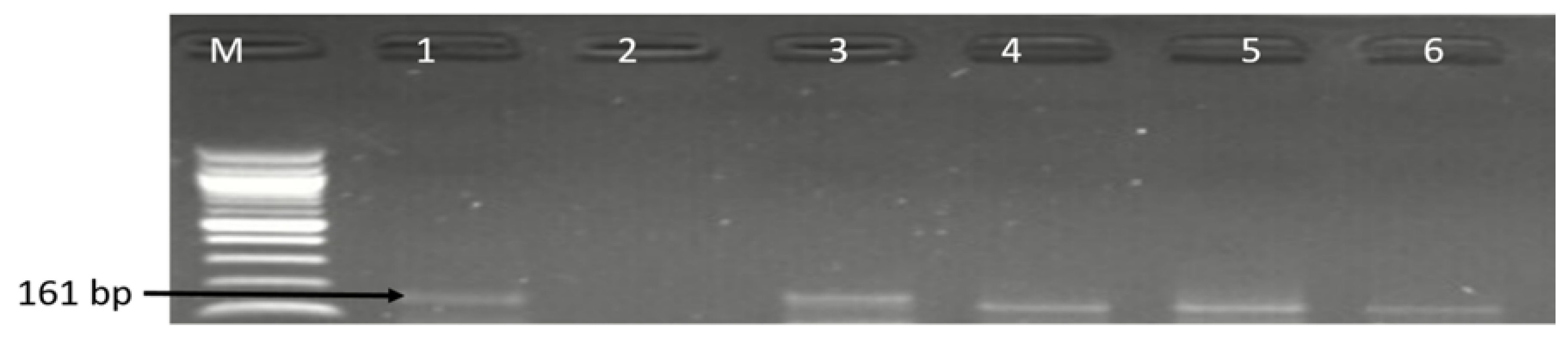
3.2. Molecular Detection of C. coli C. jejuni and C. fetus

The 162 confirmed isolates identified as belonging to the genus Campylobacter were further delineated into C. coli, C. fetus and C. jejuni while C. lari was not detected. The detailed distribution patterns of occurrence of the identified species is as shown in Table 1 while Figure 3, Figure 4 and Figure 5 are representative gel images of some identified C. coli, C. fetus, and C. jejuni isolates.

3.3. Molecular Detection of Virulence Genes in the Identified Campylobacter Species

Assessment of virulence genes were determined by PCR techniques and the virulence genes associated with toxin production (cdtB), invasion (iam and ciaB), adherence (cadF and flaA), and flagellia synthesis and regulator (flgR) genes were detected. From the six virulence genes screened for among the 70 isolates identified as C. coli, C. jejuni and C. fetus (Table 1), the iam (32.86%) gene was most prevalent in all the Campylobacter species, followed by flgR (20%) gene, and cdtB and cadF (5.71%) genes. The observed percentage occurrence of virulence-associated genes detected among C. fetus, C. jejuni and C. coli were different, except for the ciaB gene that was not detected in all the isolates. From the PCR results obtained, high occurrence of iam (35%) gene was detected in C. jejuni isolates while low incidence of flgR (4.55%) gene was detected in C. jejuni isolates. It was also observed that virulence-associated genes were more often detected in C. coli than in C. jejuni and C. fetus. In terms of classes of virulence genes co-harbored in the identified species, 4 (5.71%) C. coli isolates co-harbored the iam and flaR genes, 1 (1.43%) C. jejuni isolate co-harbored iam and flaR genes, 1(1.43%) C. coli isolates co-harbored iam and cadF genes, 1 (1.43%) C. coli strain co-harbored iam, cadF and cdtB genes and 2 (2.88%) isolates identified as C. jejuni and C. coli co-harbored iam and cdtB genes. The detailed distribution pattern of the virulence genes detected in Campylobacter species recovered from both water and milk samples are as shown in Table 2 while Figure 6 and Figure 7 are representative gel images of the detected virulence-associated genes.

3.4. Antibiotic Phenotypic Resistance Profiles of Campylobacter Isolates

The 162 Campylobacter isolates obtained from water and milk samples were tested against 12 antimicrobials agents. Of the 12 antibiotics tested, the highest phenotypic resistance displayed by Campylobacter isolates recovered from milk and water samples was against clindamycin (95.68%), while the lowest was observed against imipenem (21.47%). Other high phenotypic resistance displayed by tThe e isolates were against erythromycin (95.06%), followed by ceftriaxone (93.21%), doxycycline (87.65%), azithromycin and ampicillin (87.04% each), tetracycline (83.33%), chloramphenicol (78.27%), ciprofloxacin (77.78%), levofloxacin (59.88%) and gentamicin (56.17%) (Figure 8). Most of the isolates were resistance to more than three classes of antimicrobial agents and were classified as multi-drug resistance (MDR). The lowest phenotypic MDR rate observed in C. coli isolate was to CRO-E-CD-AP, in C. fetus was to CRO-E-CD-T-DXT-AP and in C. jejuni was to E-ATH-mCD-T-DXT-AP (Table 3). Mand majority of the isolates showed resistance to more than 2 to 9 classes of antimicrobial agents, and the highest resistance profiles observed in C. jejuni, C. coli and C. fetus isolates were to LEV-CRO-C-CIP-E-ATH-CD-T-GM-DXT-AP (22.86%) and LEV-CRO-C-CIP-E-ATH-IMI-CD-T-GM-DXT-AP (10%). The detailed multiple resistance patterns exhibited by C. coli, C. jejuni and C. fetus are showed in Table 3.

3.5. Molecular Detection of Genotypic Resistance Genes in Campylobacter Isolates

Genotypic resistance genes in Campylobacter isolates were detected by PCR and the prevalence of catII gene in chloramphenicol resistance Campylobacter isolates was the highest resistance gene detected, where 38 (95%) isolates identified as C. coli, C. jejuni and C. fetus harbored the catII gene. Tetracycline resistance genes were widespread in C. coli, C. jejuni and C. fetus where tetA, tetB and tetM were detected in 88.71%, 27.42% and 32.26% respectively. Other ARGs detected in C. coli, C. jejuni and C. fetus, including ermB (erythromycin resistance gene), gyrA (gentamycin resistance gene), ampC (ampicillin resistance gene) and aac(3)-IIa-(aacC2)a (gentamycin resistance gene), were 15.38%, 39.13%, 81.54% and 84.85% respectively. All C. coli, C. jejuni and C. fetus recovered from both milk and water samples were negative for VIM, KPC, Ges, bla-OXA-48-like, tetC, tetD, tetK, IMI, and catI genes. From the PCR results obtained, most of the isolates were observed to harbor multiple resistance genes and the highest number of resistance genes detected in C. jejuni isolates were tetA, tetM, ampC, catII, gyrA, aac(3)-IIa-(aacC2)a genes, in C. coli isolate were tetA, tetM, ampC, catII, ermB, aac(3)-IIa-(aacC2)a genes while in C. fetus isolates were tetA, tetM, ampC, catII, ermB, aaac(3)-IIa-(aacC2)a genes (Table 4). Detection of multiple resistance genes in the isolates indicates that the isolates simultaneously carry two or more classes of antimicrobial resistance genes. Table 4 showed the detailed pattern of multiple antibiotic resistance genes detected in C. fetus, C. jejuni and C. coli recovered from water and milk samples while Figure 9 and Figure 10 are representative gel electrophoreses images of the amplified PCR products.

4. Discussion

Globally, there is an increasing rate in the detection of Campylobacter species including reports from Africa, America, Asia, and Europe [49,50,51] and this is of great concern to public health [52]. Campylobacter species are implicated in both waterborne/milkborne infections, and it is vital to provide more information to existing reports on the risk of consumption of unchlorinated water and unpasteurized milk. Hence, this study evaluated the occurrence as well as virulence and antimicrobial resistance-associated makers of Campylobacter species isolated from retailed milk and water samples. Occurrence of Campylobacter species in water/milk samples was determined by culture-based and PCR techniques and reports on this is very scanty in the Eastern Cape Province which has the largest livestock in South Africa. In this study, Campylobacter was detected in 103 (36.92%) isolates recovered from water samples and 33 (58.93%) water samples out of 56 water samples were positive for Campylobacter. In the milk samples, 59 (37.11%) isolates were detected to be Campylobacter and 19 (26.38%) milk samples out of 72 milk samples were positive for Campylobacter. Results from this study showed that water samples were more contaminated with Campylobacter than the milk samples and our finding is in agreement with the report of Elmal and Can, [53]. Other studies carried out by Khan et al. [54], Szczepanska, et al. [55] and Van Dyke et al. [32] reported high detection rates of Campylobacter species in river water samples and our finding correspond with their reports.
Other studies conducted by Artursson et al. [17], Bianchini et al. [33] and Wysok et al. [56] have also detected Campylobacter species in raw milk samples and this current finding is also in line with their reports. Consumption of raw milk has been implicated in campylobacteriosis cases; a behaviour that has attracted attention lately. Worldwide, campylobacteriosis add ominously to the burden of human enteric illness [57]. In the United States and Europe, consumption of raw cow’s milk has been reported to be implicated in campylobacteriosis outbreaks [58,59]. In the Limpopo Province, South Africa, Campylobacter species were reported to be common causes of gastroenteritis in children [60,61] and the detection of these pathogens in retail raw milk/water samples in this study area, position them as a public health concern of provincial interest. In the water samples, occurrence of C. jejuni was most prevalent with percentage detection rate of 38.83%, followed by 7.77% for C. coli and 5.83% for C. fetus and a similar result was reported in the studies of Denis et al. [62], Pérez-Boto et al. [63] and Szczepanska et al. [55]. In the milk samples, C. coli and C. fetus were detected to be most prevalent and our finding is similar with the report of Mabote et al. [64] but contrary to the reports of Andrzejewska et al. [65], Kabir et al. [66] and Rahimi et al. [67].
The detection of pathogenic Campylobacter species in river water samples and retailed milk samples highlights the significance of river and raw milk as a potential reservoir of Campylobacter species. Of the 70 isolates identified as C. coli, C. fetus and C. lari, the major virulence genes detected were the iam, flgR, cdtB and cadF genes (Table 2). Our study showed the distribution patterns of the iam gene among the Campylobacter species and the iam gene is a virulence marker responsible for invasion of host cell. The iam gene was detected in both C. jejuni and C. coli isolates recovered from water and milk samples and this result is akin with the reports of Ghorbanalizadgan et al. [68], Pandey et al. [69] and Wysok et al. [70]. In another study of Bardoň et al. [71], the iam gene was majorly detected in C. jejuni than in C. coli and our result is contrary to this report. The cdtB gene is another virulence gene assessed responsible for toxin production, and studies have detected the cdtB gene in C. jejuni and C. coli strain recovered from beef, raw milk and pork [65], from chicken [60], from humans [72] and from cows’ cervical mucus [73] and our finding is also in line with these reports. Another virulence gene assessed was the flgR gene, the flgR gene was found in 50% of C. coli, 41.67% of C. fetus and 5% of C. jejuni and the flgR gene were detected in 5.56% water isolates and 62.5% in milk isolates (Table 2) and there is large variability of detection of flgR gene between water and milk samples. The study of Modi et al. [3] has also detected the flgR gene in C. coli and our finding correspond with this report. In this study area, no study has reported the detection of flgR gene in Campylobacter species. Furthermore, there are few reports on the detection of flgR gene in Campylobacter isolates recovered from milk and water. The flgR gene is liable for phase variation—a mechanism that help the bacteria to modify the antigenic make-up of its surface to adapt to new hosts [74]. Another gene detected was the cadF gene, and the cadF gene is a virulence gene that helps in binding to the intestinal epithelial cells [75,76]. The cadF gene (5.71%) was detected in C. coli and C. fetus recovered from water and milk samples. In the studies of Lluque et al. [77], Wieczorek et al. [78] and Selwet et al. [79], the cadF gene was detected in Campylobacter species from a Peruvian pediatric cohort, from meat samples and from Campylobacter isolates isolated from dogs and these reports corroborate our finding. The presence of one or more virulence genes in the Campylobacter genome give rise to the incidence of human infection (Abu-Madi et al. [80]). In our study, some Campylobacter species were observed to harbor multiple virulence genes and several studies including the studies of Aslantaş [81], Redondo et al. [82], Samad et al. [83] and Wei et al. [84] have also detected multiple virulence genes in Campylobacter species and our finding also corroborate with their reports. Detection of these virulence genes in Campylobacter isolates recovered from retail milk and water samples position them a risk to human health and continuous consumption of raw milk in the study area may put people at high risk of ill-health. The confirmed isolates were tested against a panel of 12 antibiotics, and the highest phenotypic resistant displayed by the Campylobacter species was to clindamycin (95.68%) and the lowest was observed against imipenem (21.47%) (Figure 8). Multidrug resistance to azithromycin, ampicillin and ciprofloxacin were observed in the study of Martín-Maldonado et al. [85] and in this study we also observed similar multiple resistance pattern (Table 3). In this study, high phenotypic Campylobacter resistance to ciprofloxacin (77.78%) was observed and this finding corroborate with the report of Meistere et al. [86], who also reported high phenotypic resistance to ciprofloxacin (93.6%) in Campylobacter isolates.
High resistance rate to tetracycline (83.33%) were also observed in the isolates and this result is similar with the report of Elhadidy et al. [87] who also reported high phenotypic Campylobacter resistance rate of 81.4% to tetracycline. Furthermore, high susceptibility level was observed against imipenem and our result also corroborate with this report of Noreen et al. [88]. Analysis of the MAR indices of the Campylobacter isolates showed that MAR indices values were all greater than 0.2 (Table 3). A MAR index value greater than 0.2 is said to have originated from commercial swine, poultry farms, dairy cattle and humans where antibiotics are often used and are at high-risk sources of antibiotic contamination [89,90]. The highest MAR value indices values were to twelve of the antimicrobials tested (LEV-CRO-C-CIP-E-ATH-IMI-CD-T-GM-DXT-AP). In this study, high resistance rates were observed against erythromycin (95.06%), ampicillin (87.04% each), tetracycline (83.33%), chloramphenicol (78.27%), ciprofloxacin (77.78%) and gentamicin (56.17%). Our result corresponds with the report of Abbasi et al. [91] who also observed high phenotypic Campylobacter isolates resistant to tetracycline, ciprofloxacin and erythromycin. The report of Nizar et al. [92] also showed high Campylobacter resistant to gentamycin (25.6%) and our finding is also in line with their report. Another study of Premarathne et al. [93] also observed high Campylobacter resistant to ampicillin and this report correspond with our result. Genotypic antimicrobial resistance genes were also determined by PCR and high gyrA gene (39.13%) was detected in Campylobacter isolates and our finding corroborate with the report of Meistere et al. [86], in which high gyrA gene was detected in their study. Primarily, macrolides remain the frontline antibiotic use for the treating of campylobacteriosis. However, in many countries, there have been reports on progressive increase in Campylobacter resistance to macrolide and this is a growing health threat concern of global concern [94]. In our study, erythromycin resistance gene erm (B) was detected in C. coli and C. jejuni isolates and our finding corroborate with the report of Liu et al. [95]. The tet-genes are other genes assessed and tetA gene is among the tet-genes responsible for tetracycline resistance and in our study, high rate of tetA (88.71%) gene was detected in tetracycline resistant-Campylobacter isolates and a similar result was also reported in the study of Divsalar et al. [96]. Furthermore, the high rate of ampC gene was also detected in ampicillin resistant-Campylobacter isolates and the detection of multiple resistance genes in Campylobacter isolates might limit the treatment option for campylobacteriosis cases.

5. Conclusions

The key step in the prevention of Campylobacter infection is monitoring of this pathogen that pose a great menace to human health. Our finding reveals that Campylobacter strains with important pathogenic factors responsible for toxin production (cdtB), invasiveness (iam, ciaB), motility (flaA, flgR) and adherence (cadF) were detected in the Campylobacter isolates recovered from river and milk samples. This study also highlights the importance of monitory of the spread of antibiotic resistant-Campylobacter isolates recovered from water and retail milk samples which will help determined the risk poses to human if appropriate measure is not put to hurt the distribution patterns. Furthermore, high rates of multiple phenotypic and associated genotypic antibiotic resistance genes were detected and this might further limit treatment options for Campylobacter infections.

Supplementary Materials

The following are available online at https://0-www-mdpi-com.brum.beds.ac.uk/2079-6382/9/7/426/s1, Table S1: Primers used for confirmation, characterization and amplification of virulence genes, Table S2: Primers sequences used for screening for antimicrobial resistance genes.

Author Contributions

A.I. carried out the experiment, analyse the result and wrote the paper. A.I.O. design the project, supervise the work, corrected and proof read the paper. All authors have read and agreed to the published version of the manuscript.

Funding

The research was supported by South Africa Medical Research Council.

Acknowledgments

The authors are grateful to South Africa Medical Research Council (SAMRC) for financial support.

Conflicts of Interest

The authors declare no competing interests.

References

  1. Hu, L.; Dennis, K.D. Campylobacter Species. Food Safety; Apple Academic Press Inc.: Waretown, NJ, USA, 2017; pp. 3–110. [Google Scholar] [CrossRef]
  2. Osbjer, K.; Tano, E.; Chhayheng, L.; Mac-Kwashie, A.O.; Fernström, L.L.; Ellström, P.; Sokerya, S.; Sokheng, C.; Mom, V.; Chheng, K.; et al. Detection of Campylobacter in human and animal field samples in Cambodia. APMIS 2016, 124, 508–515. [Google Scholar] [CrossRef] [PubMed]
  3. Modi, S.; Brahmbhatt, M.N.; Chatur, Y.A.; Nayak, J.B. Prevalence of Campylobacter species in milk and milk products, their virulence gene profile and antibiogram. Vet. World 2015, 8, 1–8. [Google Scholar] [CrossRef] [PubMed] [Green Version]
  4. Taylor, E.V.; Herman, K.M.; Ailes, E.C.; Fitzgerald, C.; Yoder, J.S.; Mahon, B.E.; Tauxe, R.V. Common source outbreaks of Campylobacter infection in the USA, 1997–2008. Epidemiol. Infect. 2013, 141, 987–996. [Google Scholar] [CrossRef] [PubMed] [Green Version]
  5. Davis, K.R. Campylobacter jejuni infections associated with raw milk consumption-Utah, 2014. MMWR 2016, 65, 301–305. [Google Scholar]
  6. Kuhn, K.G.; Falkenhorst, G.; Emborg, H.D.; Ceper, T.; Torpdahl, M.; Krogfelt, K.A.; Ethelberg, S.; Mølbak, K. Epidemiological and serological investigation of a waterborne Campylobacter jejuni outbreak in a Danish town. Epidemiol. Infect. 2017, 145, 701–709. [Google Scholar]
  7. Cabral, J.P. Water microbiology. Bacterial pathogens and water. Int. J. Environ. Res. Public Health 2010, 7, 3657–3703. [Google Scholar]
  8. Bitton, G. Microbiology of Drinking Water Production and Distribution, 1st ed.; John Wiley & Sons, Inc.: Hoboken, NJ, USA, 2014; Volume 312, pp. 1–298. [Google Scholar]
  9. Ramírez-Castillo, F.; Loera-Muro, A.; Jacques, M.; Garneau, P.; Avelar-González, F.; Harel, J.; Guerrero-Barrera, A. Waterborne pathogens: Detection methods and challenges. Pathogens 2015, 4, 307–334. [Google Scholar] [CrossRef]
  10. Muirhead, R.W.; Collins, R.P.; Bremer, P.J. Numbers and transported state of Escherichia coli in runoff direct from fresh cowpats under simulated rainfall. Lett. Appl. Microb. 2006, 42, 83–87. [Google Scholar]
  11. Hellein, K.N.; Battie, C.; Tauchman, E.; Lund, D.; Oyarzabal, O.A.; Lepo, J.E. Culture-based indicators of faecal contamination and molecular microbial indicators rarely correlate with Campylobacter spp. in recreational waters. J. Water Health 2011, 9, 695–707. [Google Scholar] [CrossRef] [Green Version]
  12. WHO. Country Cooperation Strategic at a Glance, South Africa, WHO/CCU/18.02/South Africa. 2018. Available online: https://apps.who.int/iris/handle/10665/136874 (accessed on 1 May 2018).
  13. Di Giannatale, E.; Garofolo, G.; Alessiani, A.; Di Donato, G.; Candeloro, L.; Vencia, W.; Decastelli, L.; Marotta, F. Tracing back clinical Campylobacter jejuni in the Northwest of Italy and assessing their potential source. Front. Microbiol. 2016, 7, 1–9. [Google Scholar]
  14. Sarkar, S. Microbiological Safety Concerns of Raw Milk. Safety 2016, 24, 1–7. [Google Scholar]
  15. Sugrue, I.; Tobin, C.; Ross, R.P.; Stanton, C.; Hill, C. Foodborne pathogens and zoonotic diseases. Raw Milk 2019, 12, 259–272. [Google Scholar]
  16. Maldonado, Y.A.; Glode, M.P.; Bhatia, J.; Brady, M.T.; Byington, C.L.; Davies, H.D.; Edwards, K.M.; Jackson, M.A.; Keyserling, H.L.; Murray, D.L.; et al. Consumption of raw or unpasteurized milk and milk products by pregnant women and children. Pediatrics 2014, 133, 175–179. [Google Scholar]
  17. Artursson, K.; Schelin, J.; Lambertz, S.T.; Hansson, I.; Engvall, E.O. Foodborne pathogens in unpasteurized milk in Sweden. Int. J. Food Microbiol. 2018, 284, 120–127. [Google Scholar] [CrossRef] [PubMed]
  18. Del Collo, L.P.; Karns, J.S.; Biswas, D.; Lombard, J.E.; Haley, B.J.; Kristensen, R.C.; Kopral, C.A.; Fossler, C.P.; Van Kessel, J.A.S. Prevalence, antimicrobial resistance, and molecular characterization of Campylobacter spp. in bulk tank milk and milk filters from US dairies. J. Dairy Sci. 2017, 100, 3470–3479. [Google Scholar] [CrossRef] [Green Version]
  19. El-Zamkan, M.A.; Hameed, K.G.A. Prevalence of Campylobacter jejuni and Campylobacter coli in raw milk and some dairy products. Vet. World 2016, 9, 1147–1151. [Google Scholar] [CrossRef] [Green Version]
  20. Rasolofo, E.A.; St-Gelais, D.; LaPointe, G.; Roy, D. Molecular analysis of bacterial population structure and dynamics during cold storage of untreated and treated milk. Int. J. Food Microbiol. 2010, 138, 108–118. [Google Scholar] [CrossRef]
  21. Langer, A.J.; Ayers, T.; Grass, J.; Lynch, M.; Angulo, F.J.; Mahon, B.E. Nonpasteurized dairy products, disease outbreaks, and state laws-United States, 1993–2006. Emerg. Infect. Dis. 2012, 18, 385–391. [Google Scholar] [CrossRef]
  22. Pölzler, T.; Stüger, H.P.; Lassnig, H. Prevalence of most common human pathogenic Campylobacter spp. in dogs and cats in Styria, Austria. Vet. Med. Sci. 2018, 4, 115–125. [Google Scholar] [CrossRef] [Green Version]
  23. Quigley, L.; O’sullivan, O.; Stanton, C.; Beresford, T.P.; Ross, R.P.; Fitzgerald, G.F.; Cotter, P.D. The complex microbiota of raw milk. FEMS Microbiol. Rev. 2013, 37, 664–698. [Google Scholar] [CrossRef] [Green Version]
  24. Janssen, R.; Krogfelt, K.A.; Cawthraw, S.A.; van Pelt, W.; Wagenaar, J.A.; Owen, R.J. Host-pathogen interactions in Campylobacter infections: The host perspective. Clin. Microbiol. Rev. 2008, 21, 505–518. [Google Scholar] [CrossRef] [Green Version]
  25. Heredia, N.; García, S. Animals as sources of food-borne pathogens: A review. Anim. Nutr. 2018, 4, 250–255. [Google Scholar] [CrossRef] [PubMed]
  26. Kariuki, S. Chapter 11: Antimicrobial resistance in enteric pathogens in developing countries. In Antimicrobial Resistance in Developing Countries; Sosa, A.J., Byarugaba, D.K., Amábile-Cuevas, C.F., Hsueh, P., Kariuki, S., Okeke, I.N., Eds.; Springer Publishing Co. Press: New York, NY, USA, 2010; pp. 177–197. [Google Scholar]
  27. Omulo, S.; Thumbi, S.M.; Lockwood, S.; Verani, J.R.; Bigogo, G.; Masyongo, G.; Call, D.R. Evidence of superficial knowledge regarding antibiotics and their use: Results of two cross-sectional surveys in an urban informal settlement in Kenya. PLoS ONE 2017, 12, e0185827. [Google Scholar]
  28. Rousham, E.K.; Unicomb, L.; Islam, M.A. Human, animal and environmental contributors to antibiotic resistance in low-resource settings: Integrating behavioural, epidemiological and One Health approaches. Proc. R. Soc. B Biol. Sci. 2018, 285, 1–9. [Google Scholar]
  29. Sivagami, K.; Vignesh, V.J.; Srinivasan, R.; Divyapriya, G.; Nambi, I.M. Antibiotic usage, residues and resistance genes from food animals to human and environment: An Indian scenario. J. Environ. Chem. Eng. 2018, 8, 1–8. [Google Scholar] [CrossRef]
  30. Holmes, A.H.; Moore, L.S.; Sundsfjord, A.; Steinbakk, M.; Regmi, S.; Karkey, A.; Guerin, P.J.; Piddock, L.J. Understanding the mechanisms and drivers of antimicrobial resistance. Lancet 2016, 387, 176–187. [Google Scholar]
  31. Lu, J.; Tian, Z.; Yu, J.; Yang, M.; Zhang, Y. Distribution and abundance of antibiotic resistance genes in sand settling reservoirs and drinking water treatment plants across the Yellow River, China. Water 2018, 10, 246. [Google Scholar] [CrossRef] [Green Version]
  32. Van Dyke, M.I.; Morton, V.K.; McLellan, N.L.; Huck, P.M. The occurrence of Campylobacter in river water and waterfowl within a watershed in southern Ontario, Canada. J. Appl. Microbiol. 2010, 109, 1053–1066. [Google Scholar]
  33. Bianchini, V.; Borella, L.; Benedetti, V.; Parisi, A.; Miccolupo, A.; Santoro, E.; Recordati, C.; Luini, M. Prevalence in bulk tank milk and epidemiology of Campylobacter jejuni in dairy herds in Northern Italy. Appl. Environ. Microbiol. 2014, 80, 1832–1837. [Google Scholar] [CrossRef] [Green Version]
  34. Sierra-Arguello, Y.M.; Furian, T.Q.; Perdoncini, G.; Moraes, H.L.; Salle, C.T.; Rodrigues, L.B.; dos Santos, L.R.; Gomes, M.J.P.; do Nascimento, V.P. Fluoroquinolone resistance in Campylobacter jejuni and Campylobacter coli from poultry and human samples assessed by PCR-restriction fragment length polymorphism assay. PLoS ONE 2018, 13, e0199974. [Google Scholar] [CrossRef] [Green Version]
  35. Moreno, Y.; Botella, S.; Luis Alonso, J.; Ferru’s, M.A.; Herna’ndez, M.; Herna’ndez, J. Specific detection of Arcobacter and Campylobacter strains in Water and Sewage by PCR and Fluorescent in Situ Hybridization. Appl. Environ. Microbiol. 2003, 69, 1181–1186. [Google Scholar] [CrossRef] [Green Version]
  36. Yamazaki-Matsune, W.; Taguchi, M.; Seto, K.; Kawahara, R.; Kawatsu, K.; Kumeda, Y.; Kitazato, M.; Nukina, M.; Misawa, N.; Tsukamoto, T. Development of a multiplex PCR assay for identification of Campylobacter coli, Campylobacter fetus, Campylobacter hyointestinalis subsp. hyointestinalis, Campylobacter jejuni, Campylobacter lari and Campylobacter upsaliensis. J. Med. Microbiol. 2007, 56, 1467–1473. [Google Scholar] [PubMed] [Green Version]
  37. Carvalho, A.C.; Ruiz-Palacios, G.M.; Ramos-Cervantes, P.; Cervantes, L.E.; Jiang, X.; Pickering, L.K. Molecular characterization of invasive and noninvasive Campylobacter jejuni and Campylobacter coli isolates. J. Clin. Microbiol. 2001, 39, 1353–1359. [Google Scholar] [PubMed] [Green Version]
  38. Casabonne, C.; Gonzalez, A.; Aquili, V.; Subils, T.; Balague, C. Prevalence of seven virulence genes of Campylobacter jejuni isolated from patients with diarrhea in Rosario, Argentina. Int. J. Infect. 2016, 3, 1–6. [Google Scholar] [CrossRef]
  39. Wilson, D.L.; Rathinam, V.A.; Qi, W.; Wick, L.M.; Landgraf, J.; Bell, J.A.; Plovanich-Jones, A.; Parrish, J.; Finley, R.L.; Mansfield, L.S.; et al. Genetic diversity in Campylobacter jejuni is associated with differential colonization of broiler chickens and C57BL/6J IL10-deficient mice. Microbiology 2010, 156, 2046–2057. [Google Scholar] [PubMed] [Green Version]
  40. Clinical and Laboratory Standards Institute (CLSI). Method for Antimicrobial Dilution and Disk Susceptibility Testing of Infrequently Isolated or Fastidious Bacteria, 3rd ed.; CLSI: Wayne, NJ, USA, 2015. [Google Scholar]
  41. Krumperman, P.H. Multiple antibiotic resistance indexing of Escherichia coli to identify high-risk sources of fecal contamination of foods. Appl. Environ. Microbiol. 1983, 46, 165–170. [Google Scholar] [CrossRef] [PubMed] [Green Version]
  42. Ng, L.K.; Martin, I.; Alfa, M.; Mulvey, M. Multiplex PCR for the detection of tetracycline resistant genes. Mol. Cell. Probes 2001, 15, 209–215. [Google Scholar] [CrossRef]
  43. Strommenger, B.; Kettlitz, C.; Werner, G.; Witte, W. Multiplex PCR assay for simultaneous detection of nine clinically relevant antibiotic resistance genes in Staphylococcus aureus. J. Clin. Microbiol. 2003, 41, 4089–4094. [Google Scholar] [CrossRef] [Green Version]
  44. Yan, S.S.; Fox, M.L.; Holland, S.M.; Stock, F.; Gill, V.J.; Fedorko, D.P. Resistance to multiple fluoroquinolones in a clinical isolate of Streptococcus pyogenes: Identification of gyrA and parC and specification of point mutations associated with resistance. Antimicrob. Agents Chemother. 2000, 44, 196–3198. [Google Scholar]
  45. Osode, A.N.; Okoh, A.I. Impact of discharged wastewater final effluent on the physicochemical qualities of a receiving watershed in a suburban community of the Eastern Cape Province. Clean-Soil Air Water 2009, 37, 938–944. [Google Scholar]
  46. Maynard, C.; Bekal, S.; Sanschagrin, F.; Levesque, R.C.; Brousseau, R.; Masson, L.; Lariviere, S.; Harel, J. Heterogeneity among virulence and antimicrobial resistance gene profiles of extraintestinal Escherichia coli isolates of animal and human origin. J. Clin. Microbiol. 2004, 42, 5444–5452. [Google Scholar] [PubMed] [Green Version]
  47. Velusamy, S.; Barbara, E.G.; Mark, J.L.; Lien, T.N.; Susan, I.H.; Ynte, H.S.; Stephen, P.O. Phenotypic and genotypic antimicrobial resistance patterns of Escherichia coli isolated from dairy cows with mastitis. Vet. Microbiol. 2007, 124, 319–328. [Google Scholar]
  48. Dallenne, C.; Da Costa, A.; Decré, D.; Favier, C.; Arlet, G. Development of a set of multiplex PCR assays for the detection of genes encoding important β-lactamases in Enterobacteriaceae. J. Antimicrob. Chemother. 2010, 65, 490–495. [Google Scholar] [PubMed] [Green Version]
  49. Reich, F.; Valero, A.; Schill, F.; Bungenstock, L.; Klein, G. Characterisation of Campylobacter contamination in broilers and assessment of microbiological criteria for the pathogen in broiler slaughterhouses. Food Control 2018, 87, 60–69. [Google Scholar] [CrossRef]
  50. Stella, S.; Soncini, G.; Ziino, G.; Panebianco, A.; Pedonese, F.; Nuvoloni, R.; Di Giannatale, E.; Colavita, G.; Alberghini, L.; Giaccone, V. Prevalence and quantification of thermophilic Campylobacter spp. in Italian retail poultry meat: Analysis of influencing factors. Food Microbiol. 2017, 62, 232–238. [Google Scholar]
  51. Wei, B.; Kang, M.; Jang, H.K. Genetic characterization and epidemiological implications of Campylobacter isolates from wild birds in South Korea. Transbound. Emerg. Dis. 2019, 66, 56–65. [Google Scholar]
  52. Han, X.; Guan, X.; Zeng, H.; Li, J.; Huang, X.; Wen, Y.; Zhao, Q.; Huang, X.; Yan, Q.; Huang, Y.; et al. Prevalence, antimicrobial resistance profiles and virulence-associated genes of thermophilic Campylobacter spp. isolated from ducks in a Chinese slaughterhouse. Food Control 2019, 104, 157–166. [Google Scholar]
  53. Elmal, M.; Can, H.Y. Antimicrobial susceptibility and virulence-associated genes in Campylobacter isolates from milk and wastewater in Hatay, Turkey. Cienc. Rural 2019, 49, 1–8. [Google Scholar] [CrossRef]
  54. Khan, I.U.; Gannon, V.; Jokinen, C.C.; Kent, R.; Koning, W.; Lapen, D.R.; Medeiros, D.; Miller, J.; Neumann, N.F.; Phillips, R.; et al. A national investigation of the prevalence and diversity of thermophilic Campylobacter species in agricultural watersheds in Canada. Water Res. 2014, 61, 243–252. [Google Scholar] [CrossRef]
  55. Szczepanska, B.; Andrzejewska, M.; Spica, D.; Klawe, J.J. Prevalence and antimicrobial resistance of Campylobacter jejuni and Campylobacter coli isolated from children and environmental sources in urban and suburban areas. BMC Microbiol. 2017, 17, 80. [Google Scholar] [CrossRef] [Green Version]
  56. Wysok, B.; Wiszniewska-Łaszczych, A.; Uradziński, J.; Szteyn, J. Prevalence and antimicrobial resistance of Campylobacter in raw milk in the selected areas of Poland. Polish J. Vet. Sci. 2011, 14, 473–477. [Google Scholar] [CrossRef] [Green Version]
  57. Christidis, T.; Pintar, K.D.M.; Butler, A.J.; Nesbitt, A.; Thomas, M.K.; Marshall, B.; Pollari, F. Campylobacter spp. prevalence and levels in raw milk: A systematic review and meta-analysis. J. Food Prot. 2016, 79, 1775–1783. [Google Scholar] [CrossRef]
  58. Claeys, W.L.; Cardoen, S.; Daube, G.; Block, J.D.; Dewettinck, K.; Dierick, K.; De Zutter, L.; Huyghebaert, A.; Imberechts, H.; Thiange, P.; et al. Raw or heated cow milk consumption: Review of risks and benefits. Food Control 2013, 31, 251–262. [Google Scholar] [CrossRef]
  59. EFSA. The European Union summary report on trends and sources of zoonoses, zoonotic agents and food-borne outbreaks in 2014. EFSA J. 2015, 13, 1–190. [Google Scholar]
  60. Bissong, M.E.; Ateba, C.N. Detection of virulent thermophilic Campylobacter species in communal chickens. S. Afr. J. Sci. 2019, 115, 1–5. [Google Scholar] [CrossRef]
  61. Samie, A.; Obi, C.L.; Barrett, L.J.; Powell, S.M.; Guerrant, R.L. Prevalence of Campylobacter species, Helicobacter pylori and Arcobacter species in stool samples from the Venda region, Limpopo, South Africa: Studies using molecular diagnostic methods. J. Infect. 2007, 54, 558–566. [Google Scholar] [CrossRef] [PubMed]
  62. Denis, M.; Tanguy, M.; Chidaine, B.; Laisney, M.J.; Mégraud, F.; Fravalo, P. Description and sources of contamination by Campylobacter spp. of river water destined for human consumption in Brittany, France. Pathol. Biol. 2011, 59, 256–263. [Google Scholar] [CrossRef]
  63. Pérez-Boto, D.; Garcia-Pena, F.J.; Abad-Moreno, J.C.; Hurtado-Pizarro, M.D.; Pérez-Cobo, I.; Aurora Echeita, M. Drinking water as the source of Campylobacter coli infection in grandparent heavy breeders. Avian Pathol. 2010, 39, 483–487. [Google Scholar] [CrossRef]
  64. Mabote, K.I.; Mbewe, M.; Ateba, C.N. Prevalence of Campylobacter contamination in fresh chicken meat and milk obtained from markets in the North-West Province, South Africa. J. Human Ecol. 2011, 36, 23–28. [Google Scholar] [CrossRef]
  65. Andrzejewska, M.; Szczepańska, B.; Śpica, D.; Klawe, J.J. Prevalence, virulence, and antimicrobial resistance of Campylobacter spp. in raw milk, beef, and pork meat in Northern Poland. Foods 2019, 8, 420. [Google Scholar] [CrossRef] [Green Version]
  66. Kabir, S.L.; Lubna, M.M.; Islam, M.; Haque, A.Z.; Neogi, S.B.; Yamasaki, S. Isolation, molecular identification and antimicrobial resistance patterns of Campylobacter species of dairy origin: First report from Bangladesh. Vet. Sci. Dev. 2018, 8, 16–20. [Google Scholar] [CrossRef]
  67. Rahimi, E.; Sepehri, S.; Momtaz, H. Prevalence of Campylobacter species in milk and dairy products in Iran. Rev. Med. Vet. 2013, 164, 283–288. [Google Scholar]
  68. Ghorbanalizadgan, M.; Bakhshi, B.; Najar-Peerayeh, S. Heterogeneity of cytolethal distending toxin sequence types of Campylobacter jejuni and correlation to invasion/cytotoxicity potential: The first molecular survey from Iran. Microb. Pathog. 2018, 114, 213–218. [Google Scholar] [CrossRef] [PubMed]
  69. Pandey, R. Virulence genes detection and antibiotic resistance study on the Campylobacter isolates obtained from poultry, domestic animals and humans. Int. J. Basic Appl. Agric. Res. 2018, 16, 157–164. [Google Scholar]
  70. Wysok, B.; Wojtacka, J. Detection of virulence genes determining the ability to adhere and invade in Campylobacter spp. from cattle and swine in Poland. Microb. Pathog. 2018, 115, 257–263. [Google Scholar] [CrossRef]
  71. Bardoň, J.; Pudova, V.; Koláčková, I.; Karpíšková, R.; Röderová, M.; Kolář, M. Virulence and antibiotic resistance genes in Campylobacter spp. in the Czech Republic. Epidemiol. Mikrobiol. Imunol. Cas. Spol. Epidemiol. Mikrobiol. Ceske Lek. Spol. JE Purkyne 2017, 66, 59–66. [Google Scholar]
  72. Do Nascimento, V.H.; Medeiros, P.H.; Ribeiro, S.A.; Freitas, T.M.; Santos, A.K.; Amaral, M.S.; Bona, M.D.; Havt, A.; Lima, I.F.; Lima, N.L.; et al. Campylobacter jejuni virulence genes and immune-inflammatory biomarkers association with growth impairment in children from Northeastern Brazil. Eur. J. Clin. Microbiol. Infect. Dis. 2018, 37, 2011–2020. [Google Scholar]
  73. Lúcio, É.C.; Barros, M.R.; Mota, R.A.; Maia, R.D.C.C.; Pinheiro, J.W. Identification of Campylobacter fetus subsp. venerealis virulence genes in cervical mucus from cows. Braz. J. Microb. 2019, 50, 1133–1137. [Google Scholar]
  74. Kärenlampi, R.; Rautelin, H.; Hänninen, M.L. Evaluation of genetic markers and molecular typing methods for prediction of sources of Campylobacter jejuni and C. coli infections. Appl. Environ. Microbiol. 2007, 73, 1683–1685. [Google Scholar]
  75. Konkel, M.E.; Garvis, S.G.; Tipton, S.L.; Anderson, D.E., Jr.; Cieplak, W., Jr. Identification and molecular cloning of a gene encoding a fibronectin-binding protein (CadF) from Campylobacter jejuni. Mol. Microbiol. 1997, 24, 953–963. [Google Scholar] [CrossRef]
  76. Konkel, M.E.; Gray, S.A.; Kim, B.J.; Garvis, S.G.; Yoon, J. Identification of the Enteropathogens Campylobacter jejuni and Campylobacter coli based on the cadF virulence gene and its product. J. Clin. Microbiol. 1999, 37, 510–517. [Google Scholar] [CrossRef] [PubMed] [Green Version]
  77. Lluque, A.; Riveros, M.; Prada, A.; Ochoa, T.J.; Ruiz, J. Virulence and antimicrobial resistance in Campylobacter spp. from a Peruvian pediatric cohort. Scientifica 2017, 2017, 7848926. [Google Scholar] [CrossRef] [PubMed] [Green Version]
  78. Wieczorek, K.; Szewczyk, R.; Osek, J. Prevalence, antimicrobial resistance, and molecular characterization of Campylobacter jejuni and C. coli isolated from retail raw meat in Poland. Vet. Med. 2012, 57, 293–299. [Google Scholar] [CrossRef] [Green Version]
  79. Selwet, M.; Clapa, T.; Galbas, M.; Slomski, R.; Porzucek, F. The prevalence of Campylobacter spp. and occurrence of virulence genes isolated from dogs. Polish J. Microbiol. 2015, 64, 73–76. [Google Scholar]
  80. Abu-Madi, M.; Behnke, J.M.; Sharma, A.; Bearden, R.; Al-Banna, N. Prevalence of virulence/stress genes in Campylobacter jejuni from chicken meat sold in Qatari retail outlets. PLoS ONE 2016, 11, e0156938. [Google Scholar]
  81. Aslantaş, Ö. Isolation and molecular characterization of thermophilic Campylobacter spp. in dogs and cats. Kafkas Univ. Vet. Fak. Derg. 2019, 25, 341–348. [Google Scholar]
  82. Redondo, N.; Carroll, A.; McNamara, E. Molecular characterization of Campylobacter causing human clinical infection using whole-genome sequencing: Virulence, antimicrobial resistance and phylogeny in Ireland. PLoS ONE 2019, 14, e0219088. [Google Scholar] [CrossRef] [Green Version]
  83. Samad, A.; Abbas, F.; Ahmed, Z.; Akbar, A.; Naeem, M.; Sadiq, M.B.; Ali, I.; Bugti, F.S.; Achakzai, S.K. Prevalence, antimicrobial susceptibility, and virulence of Campylobacter jejuni isolated from chicken meat. J. Food Saf. 2019, 39, 12600. [Google Scholar]
  84. Wei, B.; Cha, S.Y.; Yoon, R.H.; Kang, M.; Roh, J.H.; Seo, H.S.; Lee, J.A.; Jang, H.K. Prevalence and antimicrobial resistance of Campylobacter spp. isolated from retail chicken and duck meat in South Korea. Food Control 2016, 62, 63–66. [Google Scholar] [CrossRef]
  85. Martín-Maldonado, B.; Montoro-Dasi, L.; Pérez-Gracia, M.T.; Jordá, J.; Vega, S.; Marco-Jiménez, F.; Marin, C. Wild Bonelli’s eagles (Aquila fasciata) as carrier of antimicrobial resistant Salmonella and Campylobacter in Eastern Spain. Comp. Immunol. Microbiol. Infect. Dis. 2019, 67, 1–6. [Google Scholar] [CrossRef]
  86. Meistere, I.; Ķibilds, J.; Eglīte, L.; Alksne, L.; Avsejenko, J.; Cibrovska, A.; Makarova, S.; Streikiša, M.; Grantiņa-Ieviņa, L.; Bērziņš, A. Campylobacter species prevalence, characterisation of antimicrobial resistance and analysis of whole-genome sequence of isolates from livestock and humans, Latvia, 2008 to 2016. Eurosurveillance 2019, 24, 1–9. [Google Scholar] [CrossRef] [PubMed]
  87. Elhadidy, M.; Miller, W.G.; Arguello, H.; Álvarez-Ordóñez, A.; Dierick, K.; Botteldoorn, N. Molecular epidemiology and antimicrobial resistance mechanisms of Campylobacter coli from diarrhoeal patients and broiler carcasses in Belgium. Transbound. Emerg. Dis. 2019, 66, 463–475. [Google Scholar] [CrossRef] [Green Version]
  88. Noreen, Z.; Siddiqui, F.; Javed, S.; Wren, B.W.; Bokhari, H. Transmission of Multidrug Resistant Campylobacter jejuni to Children from Different Sources in Pakistan. J. Glob. Antimicrob. Resist. 2020, 20, 219–224. [Google Scholar] [PubMed]
  89. Al-Dulaimi, M.M.K.; Mutalib, S.A.; Ghani, M.A.; Zaini, N.A.M.; Ariffin, A.A. Multiple antibiotic resistance (MAR), plasmid profiles, and DNA polymorphisms among Vibrio vulnificus isolates. Antibiotics 2019, 8, 2–13. [Google Scholar]
  90. Dale, J.W.; Park, S. Molecular Genetics of Bacteria, 1st ed.; John Wiley & Sons Inc.: Chichester, UK, 2010; pp. 147–148. [Google Scholar]
  91. Abbasi, E.; van Belkum, A.; Ghaznavi-Rad, E. Quinolone and Macrolide-Resistant Campylobacter jejuni in Pediatric Gastroenteritis Patients from Central Iran. Microb. Drug Resist. 2019, 25, 1080–1086. [Google Scholar] [CrossRef]
  92. Nisar, M.; Mushtaq, M.H.; Shehzad, W.; Hussain, A.; Muhammad, J.; Nagaraja, K.V.; Goyal, S.M. Prevalence and antimicrobial resistance patterns of Campylobacter spp. isolated from retail meat in Lahore, Pakistan. Food Control 2017, 80, 327–332. [Google Scholar] [CrossRef]
  93. Premarathne, J.M.; Anuar, A.S.; Thung, T.Y.; Satharasinghe, D.A.; Jambari, N.N.; Abdul-Mutalib, N.A.; Huat, J.T.Y.; Basri, D.F.; Rukayadi, Y.; Nakaguchi, Y.; et al. Prevalence and antibiotic resistance against tetracycline in Campylobacter jejuni and C. coli in cattle and beef meat from Selangor, Malaysia. Front. Microbiol. 2017, 8, 1–9. [Google Scholar]
  94. Lurchachaiwong, W.; Ruksasiri, S.; Wassanarungroj, P.; Serichantalergs, O.; Bodhidatta, L.; Crawford, J.; Shrestha, S.K.; Pandey, P. Determination of azithromycin heteroresistant Campylobacter jejuni in traveler’s diarrhea. Gut Pathog. 2019, 11, 3–5. [Google Scholar] [CrossRef]
  95. Liu, D.; Liu, W.; Lv, Z.; Xia, J.; Li, X.; Hao, Y.; Zhou, Y.; Yao, H.; Liu, Z.; Wang, Y.; et al. Emerging erm (B)-mediated macrolide resistance associated with novel multidrug resistance genomic islands in Campylobacter. Antimicrob. Agents Chem. 2019, 63, 1–9. [Google Scholar] [CrossRef] [Green Version]
  96. Divsalar, G.; Kaboosi, H.; Khoshbakht, R.; Shirzad-Aski, H.; Ghadikolaii, F.P. Antimicrobial resistances, and molecular typing of Campylobacter jejuni isolates, separated from food-producing animals and diarrhea patients in Iran. Comp. Immunol. Microbiol. Infect. Dis. 2019, 65, 194–200. [Google Scholar]
Figure 1. A pictorial representation of presumptive/confirmed Campylobacter isolates recovered from milk samples from different sources.
Antibiotics 09 00426 g001
Figure 2. A representative gel image of PCR confirmed genus Campylobacter. Lane M: (100 bp DNA ladder), lane 1: positive control (C. jejuni ATCC 3356), lane 2: negative control, lane 3–10: some positive Campylobacter isolates.
Antibiotics 09 00426 g002
Figure 3. Gel image of PCR detected aspK (502 bp) gene of C. coli. Lane M: DNA ladder (100 bp), lane 1: positive control (C. coli ATCC 33559), lane 2: negative control, lane 3–8: some positive C. coli isolates (502 bp).
Antibiotics 09 00426 g003
Figure 4. Gel electrophoresis image of PCR detected cstA (359 bp) gene of C. fetus. Lane M: molecular marker (100 bp), lane 1: positive control (C. fetus ATCC 27374), lane 2: negative control, lane 3–7: some positive C. fetus isolates (359 bp).
Antibiotics 09 00426 g004
Figure 5. Gel electrophoresis image of identified C. jejuni cj0414 gene at 161 bp. Lane M: DNA ladder (100 bp), lane 1: positive control (C. jejuni ATCC 33560), lane 3–6: some positive C. jejuni isolates.
Antibiotics 09 00426 g005
Figure 6. A representative gel image of some of the PCR detected iam, cadF and fldR genes. Lane 1–4: positive Campylobacter isolates that harbor cadF gene (400 bp), lane 5–8: positive Campylobacter isolates that harbor iam gene (519 bp), lane 9–12: positive Campylobacter isolates that harbored flgR gene (390 bp).
Antibiotics 09 00426 g006
Figure 7. Gel image of some of the PCR detected cdtB gene. Lane M: DNA ladder, lane: 1–10: some positive Campylobacter isolates that harbor cdtB gene (495 bp).
Antibiotics 09 00426 g007
Figure 8. Resistance proportions of C. jejuni, C. coli, C. fetus and other Campylobacter species isolated from milk and water samples to 12 antimicrobial agents. Levofloxacin (LEV), ciprofloxacin (CIP), azithromycin (ATH), imipenem (IMI), ampicillin (AP), clindamycin (CD), tetracycline (TET), ceftriaxone (CRO), chloramphenicol (C), erythromycin (E), gentamicin (GM) and doxycycline (DXT).
Antibiotics 09 00426 g008
Figure 9. A representative electrophoresis picture of various amplified antibiotics resistance genes of Campylobacter isolates. Lanes M: DNA ladder (100 bp), lane 1: negative control, lane 2: ampC gene (530 bp), lane 3: tetA (201 bp) and tetB gene (359 bp), lane 4: ermB gene (320 bp), lane 5: gyrA gene (441 bp), lane 6: aac(3)-IIa (aacC2)a gene (740 bp) and lane 7: tetM gene (159 bp).
Antibiotics 09 00426 g009
Figure 10. Electrophoresis gel image of PCR confirmed catII gene. Lane M: DNA ladder (100 bp), lane 1: negative control, lane 2–7, Campylobacter isolates that harbored the catII gene (543 bp).
Antibiotics 09 00426 g010
Table 1. Distribution patterns of Campylobacter species identified in the sample sources.
Sample SourcesC. fetus (%)C. jejuni (%)C. coli (%)C. lari (%)No of Isolates That Belong to Other Campylobacter Species (%)
Milk6 (10.17)4 (6.78)6 (10.17)043 (72.88)
Water6 (5.83)40 (38.83)8 (7.77)049 (47.57)
Table 2. Prevalence of virulence genes detected in Campylobacter species.
Water SamplesMilk Samples
Campylobacter spp.No of IsolateVirulence Genes Screened (%) No of IsolatesVirulence Genes Screened (%)
iamflaAcadFflgRcdtBciaB iamflaAcadFflgRcdtBciaB
C. coli84 (50)-3 (37.)1 (12.5)1 (12.5)-64 (66.7)--6 (100)--
C. jejuni4014 (35)--2 (5)3 (7.5)-4------
C. fetus61 (16.7)--1 (16.7)--6--1 (16.7)4 (66.7)--
Table 3. Antibiotics phenotypic resistance patterns of Campylobacter isolates.
NoAntimicrobial Resistance PatternsSample SourceNo of Isolates TotalMAR Index
WaterMilkC. coliC. jejuniC. fetus
1CRO-E-CD-AP 11--10.33
2CRO-E-CD-T-DXT-AP 1--110.5
3E-ATH-CD-T-DXT-AP2 -1-20.5
4CRO-C-E-ATH-CD-AP1 -1-10.5
5LEV-C-CIP-E-ATH-CD1 --110.5
6LEV-CRO-CIP-E-ATH-CD-AP1 -1-10.58
7CRO-E-ATH-CD-T-DXT-AP3 -3-30.58
8E-ATH-CD-T-GM-DXT-AP1 -1-10.58
9CRO-E-ATH-CD-T-GM-AP1 -1-10.58
10CRO-E-ATH-CD-T-DXT-AP1 -1-10.58
11CRO-E-ATH-CD-T-GM-DXT-AP3213150.67
12CRO-C-E-ATH-CD-T-GM-AP 1--110.67
13CRO-C-CIP-E-CD-T-DXT-AP 3-1-10.67
14CRO-C-E-ATH-CD-T-DXT-AP3 -3-30.67
15CRO-E-ATH-IMI-CD-T-DXT-AP2 -2-20.67
16CRO-CIP-E-ATH-CD-T-DXT-AP1 -1-10.67
17LEV-CRO-C-CIP-E-ATH-CD-DXT 11--10.67
18CRO-E-ATH-IMI-CD-T-GM-AP2 -2-20.67
19CRO-CIP-E-ATH-CD-T-DXT-AP1 -1-10.67
20CRO-C-E-ATH-IMI-CD-T-DXT-AP 1--110.75
21CRO-C-E-ATH-CD-T-GM-DXT-AP -2-20.75
22LEV-C-CIP-E-ATH-CD-T-GM-AP 1--110.75
23CRO-CIP-E-ATH-CD-T-GM-DXT-AP2 11-20.75
24C-CIP-E-ATH-IMI-CD-T-DXT-AP 1-1-10.75
25LEV-CRO-CIP-E-ATH-CD-T-DXT-AP1 -1-10.75
26LEV-CRO-C-CIP-E-ATH-CD-T-DXT-AP1 -1230.83
27LEV-CRO-CIP-E-ATH-CD-T-GM-DXT-AP4 11240.83
28CRO-C-CIP-E-ATH-CD-T-GM-DXT-AP1 1--10.83
29CRO-CIP-E-ATH-IMI-CD-T-GM-DXT-AP1 -1-10.83
30LEV-CRO-C-CIP-E-ATH-CD-T-GM-DXT-AP1155101160.92
31CRO-C-CIP-E-ATH-IMI-CD-T-GM-DXT-AP1 -1-10.92
32LEV-CRO-C-CIP-E-ATH-IMI-CD-T-GM-DXT-AP7 33171
Table 4. Multiple antibiotic resistance genes in C. fetus, C. jejuni and C. coli isolates.
NoSample SourceCampylobacter SpeciesMultiple Resistance Genes Harbored
Water SampleMilk SampleC. jejuniC. coliC. fetus
1+-2--tetA, catII
2-+1--catII, ermB
3--1-1tetA, ampC
4-+--1tetA, tetM, ampC
5+---1tetA, ampC, gyrA
6+-1--tetK, ampC, catII
7+-1--tetA, catII, gyrA
8+-6--tetA, tetB, ampC
9++4--tetA, ampC, catII
10+--1-tetM, ampC, gyrA
11-+-2-tetA, ampC, aac(3)-IIa-(aacC2)a
12-+--1tetA, catII, aac(3)-IIa-(aacC2)a
13++--2tetA, tetM, ampC, catII
14+-2--tetA, tetB, ampC, aac(3)-IIa-(aacC2)a
15+-1--tetA, tetB, ampC, ermB
16+-1--tetA, tetB, ampC, catII
17+--1-tetM, ampC, catII, aac(3)-IIa-(aacC2)a
18+-1--tetA, catII, ermB, aac(3)-IIa-(aacC2)a
19+--11tetA, ampC, gyrA, aac(3)-IIa-(aacC2)a
20+-2--tetA, tetB, ampC, catII, gyrA
21+ 43-tetA, ampC, catII, gyrA, aac(3)-IIa-(aacC2)a
22+-1--tetA, tetM, catII, ermB, gyrA
23+-1--tetA, tetB, ampC, catII, gyrA
24-+-1-tetM, ampC, catII, gyrA, aac(3)-IIa-(aacC2)a
25++2 2tetA, tetM, ampC, catII, aac(3)-IIa-(aacC2)a
26+-111tetA, tetM, ampC, catII, gyrA
27+-3--tetA, tetB, ampC, ermB, aac(3)-IIa-(aacC2)a
28+-2--tetA, tetB, ampC, ermB, gyrA
29+--1-tetA, tetM, ampC, catII, ermB, aac(3)-IIa-(aacC2)a
30+-2--tetA, tetM, ampC, catII, gyrA, aac(3)-IIa-(aacC2)a
Note: + = presence, − = absence.

