A Significant Question in Cancer Risk and Therapy: Are Antibiotics Positive or Negative Effectors? Current Answers and Possible Alternatives


Abstract
:1. Bacterial Contributions to Eukaryotic Origins and Human Biology
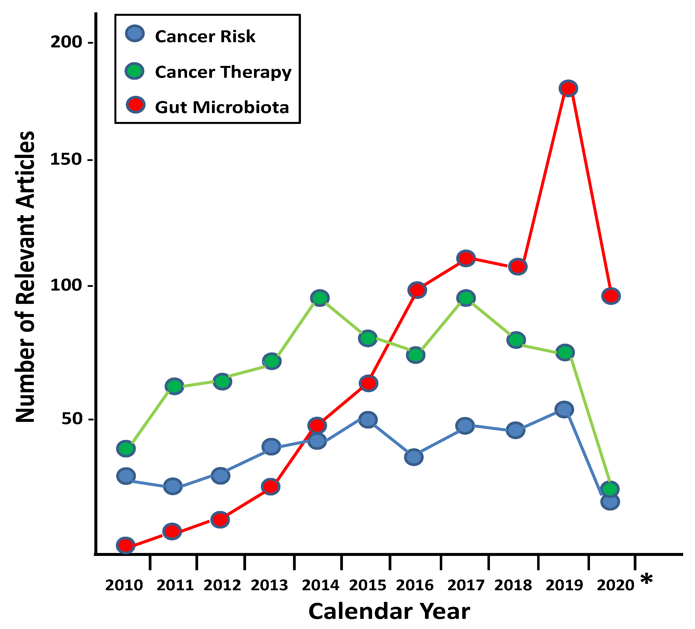
2. Bacteria and Cancer
3. Antibiotics and Cancer Risk
4. Antibiotics and Cancer Therapy Outcomes
5. Central Regulatory Role of the Microbiota
6. Alternative Approaches
7. Conclusions and New Perspectives
Author Contributions
Funding
Conflicts of Interest
References
- Sagan, L. On the origin of mitosing cells. J. Theor. Biol. 1967, 14, 255–274. [Google Scholar] [CrossRef]
- Margulis, L. Symbiotic theory of the origin of eukaryotic organelles; criteria for proof. Symp. Soc. Exp. Biol. 1975, 29, 21–38. [Google Scholar] [PubMed]
- Booth, A.; Doolittle, W.F. Eukaryogenesis, how special really? Proc. Natl. Acad. Sci. USA 2015, 112, 10278–10285. [Google Scholar] [CrossRef] [PubMed] [Green Version]
- Sato, N. Revisiting the theoretical basis of the endosymbiotic origin of plastids in the original context of Lynn Margulis on the origin of mitosing, eukaryotic cells. J. Theor. Biol. 2017, 434, 104–113. [Google Scholar] [CrossRef] [PubMed]
- Martin, W.F.; Garg, S.; Zimorski, V. Endosymbiotic theories for eukaryote origin. Philos. Trans. R. Soc. B Biol. Sci. 2015, 370, 20140330. [Google Scholar] [CrossRef] [Green Version]
- Martin, W.F. Physiology, anaerobes, and the origin of mitosing cells 50 years on. J. Theor. Biol. 2017, 434, 2–10. [Google Scholar] [CrossRef] [PubMed]
- Lane, N. Serial endosymbiosis or singular event at the origin of eukaryotes? J. Theor. Biol. 2017, 434, 58–67. [Google Scholar] [CrossRef] [Green Version]
- Gould, S.B.; Garg, S.G.; Martin, W.F. Bacterial Vesicle Secretion and the Evolutionary Origin of the Eukaryotic Endomembrane System. Trends Microbiol. 2016, 24, 525–534. [Google Scholar] [CrossRef]
- Zimorski, V.; Ku, C.; Martin, W.F.; Gould, S.B. Endosymbiotic theory for organelle origins. Curr. Opin. Microbiol. 2014, 22, 38–48. [Google Scholar] [CrossRef]
- Aanen, D.K.; Eggleton, P. Symbiogenesis: Beyond the endosymbiosis theory? J. Theor. Biol. 2017, 434, 99–103. [Google Scholar] [CrossRef]
- López-García, P.; Eme, L.; Moreira, D. Symbiosis in eukaryotic evolution. J. Theor. Biol. 2017, 434, 20–33. [Google Scholar] [CrossRef] [PubMed]
- Lazcano, A.; Peretó, J. On the origin of mitosing cells: A historical appraisal of Lynn Margulis endosymbiotic theory. J. Theor. Biol. 2017, 434, 80–87. [Google Scholar] [CrossRef]
- Marchesi, J.R.; Ravel, J. The vocabulary of microbiome research: A proposal. Microbiome 2015, 3, 31. [Google Scholar] [CrossRef] [Green Version]
- Prescott, S.L. History of medicine: Origin of the term microbiome and why it matters. Hum. Microbiome J. 2017, 4, 24–25. [Google Scholar] [CrossRef]
- Hugon, P.; Lagier, J.-C.; Colson, P.; Bittar, F.; Raoult, D. Repertoire of human gut microbes. Microb. Pathog. 2017, 106, 103–112. [Google Scholar] [CrossRef] [PubMed]
- Lederberg, J.; McCray, A.T. ‘Ome Sweet’ Omics—A genealogical treasury of words. Scientist 2001, 15, 8. [Google Scholar]
- Dworkin, M. Sergei Winogradsky: A founder of modern microbiology and the first microbial ecologist. FEMS Microbiol. Rev. 2012, 36, 364–379. [Google Scholar] [CrossRef] [PubMed]
- Farré-Maduell, E.; Casals-Pascual, C. The origins of gut microbiome research in Europe: From Escherich to Nissle. Hum. Microbiome J. 2019, 14, 100065. [Google Scholar] [CrossRef]
- Clavel, T.; Lagkouvardos, I.; Hiergeist, A. Microbiome sequencing: Challenges and opportunities for molecular medicine. Expert Rev. Mol. Diagn. 2016, 16, 795–805. [Google Scholar] [CrossRef]
- Schlaberg, R. Microbiome Diagnostics. Clin. Chem. 2020, 66, 68–76. [Google Scholar] [CrossRef]
- Gotschlich, E.C.; Colbert, R.A.; Gill, T. Methods in microbiome research: Past, present, and future. Best Pract. Res. Clin. Rheumatol. 2019, 33, 101498. [Google Scholar] [CrossRef] [PubMed]
- Fujimura, K.E.; Slusher, N.A.; Cabana, M.D.; Lynch, S.V. Role of the gut microbiota in defining human health. Expert Rev. Anti Infect. Ther. 2010, 8, 435–454. [Google Scholar] [CrossRef] [PubMed] [Green Version]
- Quigley, E.M.M. Gut bacteria in health and disease. Gastroenterol. Hepatol. 2013, 9, 560–569. [Google Scholar]
- Ruan, W.; Engevik, M.A.; Spinler, J.K.; Versalovic, J. Healthy Human Gastrointestinal Microbiome: Composition and Function after a Decade of Exploration. Dig. Dis. Sci. 2020, 65, 695–705. [Google Scholar] [CrossRef] [PubMed] [Green Version]
- Ogunrinola, G.A.; Oyewale, J.O.; Oshamika, O.O.; Olasehinde, G.I. The Human Microbiome and Its Impacts on Health. Int. J. Microbiol. 2020, 2020, 8045646. [Google Scholar] [CrossRef]
- Ahlawat, S.; Sharma, K.K. Gut-organ axis: A microbial outreach and networking. Lett. Appl. Microbiol. 2020. [Google Scholar] [CrossRef]
- Lawson, R.D.; Coyle, W.J. The noncolonic microbiome: Does it really matter? Curr. Gastroenterol. Rep. 2010, 12, 259–262. [Google Scholar] [CrossRef]
- Eberl, G. The microbiota, a necessary element of immunity. Comptes Rendus Biol. 2018, 341, 281–283. [Google Scholar] [CrossRef]
- Fitzgibbon, G.; Mills, K.H.G. The microbiota and immune-mediated diseases: Opportunities for therapeutic intervention. Eur. J. Immunol. 2020, 50, 326–337. [Google Scholar] [CrossRef] [Green Version]
- Ruff, W.E.; Greiling, T.M.; Kriegel, M.A. Host-microbiota interactions in immune-mediated diseases. Nat. Rev. Microbiol. 2020. [Google Scholar] [CrossRef]
- Fenneman, A.C.; Rampanelli, E.; Yin, Y.S.; Ames, J.; Blaser, M.J.; Fliers, E.; Nieuwdorp, M. Gut microbiota and metabolites in the pathogenesis of endocrine disease. Biochem. Soc. Trans. 2020, 48, 915–931. [Google Scholar] [CrossRef] [PubMed]
- Muscogiuri, G.; Cantone, E.; Cassarano, S.; Tuccinardi, D.; Barrea, L.; Savastano, S.; Colao, A.; on behalf of the Obesity Programs of nutrition, Education, Research and Assessment (OPERA) group. Gut microbiota: A new path to treat obesity. Int. J. Obes. Suppl. 2019, 9, 10–19. [Google Scholar] [CrossRef] [PubMed]
- Knezevic, J.; Starchl, C.; Berisha, A.T.; Amrein, K. Thyroid-Gut-Axis: How Does the Microbiota Influence Thyroid Function? Nutrients 2020, 12, 1769. [Google Scholar] [CrossRef] [PubMed]
- Schmalle, V.; Lorentz, A. Role of the microbiota in circadian rhythms of the host. Chronobiol. Int. 2020, 37, 301–310. [Google Scholar] [CrossRef] [PubMed]
- Gao, W.; Baumgartel, K.L.; Alexander, S.A. The Gut Microbiome as a Component of the Gut-Brain Axis in Cognitive Health. Biol. Res. Nurs. 2020, 1099800420941923. [Google Scholar] [CrossRef] [PubMed]
- Novakovic, M.; Rout, A.; Kingsley, T.; Kirchoff, R.; Singh, A.; Verma, V.; Kant, R.; Chaudhary, R. Role of gut microbiota in cardiovascular diseases. World J. Cardiol. 2020, 12, 110–122. [Google Scholar] [CrossRef] [PubMed]
- Anselmi, G.; Gagliardi, L.; Egidi, G.; Leone, S.; Gasbarrini, A.; Miggiano, G.A.D.; Galiuto, L. Gut microbiota and cardiovascular diseases: A critical review. Cardiol. Rev. 2020. [Google Scholar] [CrossRef] [PubMed]
- Hartmann, P.; Chu, H.; Duan, Y.; Schnabl, B. Gut microbiota in liver disease: Too much is harmful, nothing at all is not helpful either. Am. J. Physiol. Gastrointest. Liver Physiol. 2019, 316, G563–G573. [Google Scholar] [CrossRef]
- Nogueira, A.R.; Shoenfeld, Y. Microbiome and autoimmune diseases: Cause and effect relationship. Curr. Opin. Rheumatol. 2019, 31, 471–474. [Google Scholar] [CrossRef]
- Libertucci, J.; Young, V.B. The role of the microbiota in infectious diseases. Nat. Microbiol. 2019, 4, 35–45. [Google Scholar] [CrossRef]
- Baffy, G. Gut Microbiota and Cancer of the Host: Colliding Interests. Adv. Exp. Med. Biol. 2020, 1219, 93–107. [Google Scholar] [CrossRef]
- Hooks, K.B.; O’Malley, M.A. Dysbiosis and Its Discontents. mBio 2017, 8, e01492-17. [Google Scholar] [CrossRef] [Green Version]
- Petersen, C.; Round, J.L. Defining dysbiosis and its influence on host immunity and disease. Cell. Microbiol. 2014, 16, 1024–1033. [Google Scholar] [CrossRef] [PubMed]
- Markova, N.D. Eubiotic vs. dysbiotic human blood microbiota: The phenomenon of cell wall deficiency and disease-trigger potential of bacterial and fungal L-forms. Discov. Med. 2020, 29, 31–40. [Google Scholar] [PubMed]
- Nicolas, S.; Blasco-Baque, V.; Fournel, A.; Gilleron, J.; Klopp, P.; Waget, A.; Ceppo, F.; Marlin, A.; Padmanabhan, R.; Iacovoni, J.S.; et al. Transfer of dysbiotic gut microbiota has beneficial effects on host liver metabolism. Mol. Syst. Biol. 2017, 13, 921. [Google Scholar] [CrossRef] [PubMed]
- Precup, G.; Vodnar, D.-C. Gut Prevotella as a possible biomarker of diet and its eubiotic versus dysbiotic roles: A comprehensive literature review. Br. J. Nutr. 2019, 122, 131–140. [Google Scholar] [CrossRef]
- McBurney, M.I.; Davis, C.; Fraser, C.M.; Schneeman, B.O.; Huttenhower, C.; Verbeke, K.; Walter, J.; Latulippe, M.E. Establishing What Constitutes a Healthy Human Gut Microbiome: State of the Science, Regulatory Considerations, and Future Directions. J. Nutr. 2019, 149, 1882–1895. [Google Scholar] [CrossRef] [PubMed]
- Bresalier, R.S.; Chapkin, R.S. Human Microbiome in Health and Disease: The Good, the Bad, and the Bugly. Dig. Dis. Sci. 2020, 65, 671–673. [Google Scholar] [CrossRef] [Green Version]
- Rook, G.; Bäckhed, F.; Levin, B.R.; McFall-Ngai, M.J.; McLean, A.R. Evolution, human-microbe interactions, and life history plasticity. Lancet 2017, 390, 521–530. [Google Scholar] [CrossRef]
- Achtman, M. How old are bacterial pathogens? Proc. Biol. Sci. 2016, 283, 20160990. [Google Scholar] [CrossRef] [Green Version]
- Bos, K.I.; Kühnert, D.; Herbig, A.; Esquivel-Gomez, L.R.; Andrades Valtueña, A.; Barquera, R.; Giffin, K.; Lankapalli, A.K.; Nelson, E.A.; Sabin, S.; et al. Paleomicrobiology: Diagnosis and Evolution of Ancient Pathogens. Annu. Rev. Microbiol. 2019, 73, 639–666. [Google Scholar] [CrossRef] [PubMed]
- Spyrou, M.A.; Bos, K.I.; Herbig, A.; Krause, J. Ancient pathogen genomics as an emerging tool for infectious disease research. Nat. Rev. Genet. 2019, 20, 323–340. [Google Scholar] [CrossRef] [PubMed]
- Andam, C.P.; Worby, C.J.; Chang, Q.; Campana, M.G. Microbial Genomics of Ancient Plagues and Outbreaks. Trends Microbiol. 2016, 24, 978–990. [Google Scholar] [CrossRef]
- Bos, K.I.; Harkins, K.M.; Herbig, A.; Coscolla, M.; Weber, N.; Comas, I.; Forrest, S.A.; Bryant, J.M.; Harris, S.R.; Schuenemann, V.J.; et al. Pre-Columbian mycobacterial genomes reveal seals as a source of New World human tuberculosis. Nature 2014, 514, 494–497. [Google Scholar] [CrossRef] [PubMed]
- Rasmussen, S.; Allentoft, M.E.; Nielsen, K.; Orlando, L.; Sikora, M.; Sjögren, K.-G.; Pedersen, A.G.; Schubert, M.; Van Dam, A.; Kapel, C.M.O.; et al. Early divergent strains of Yersinia pestis in Eurasia 5000 years ago. Cell 2015, 163, 571–582. [Google Scholar] [CrossRef]
- Dobson, A.P.; Varper, E.R. Infectious Diseases and Human Population History. Bioscience 1996, 46, 115–126. [Google Scholar] [CrossRef] [Green Version]
- Key, F.M.; Posth, C.; Esquivel-Gomez, L.R.; Hübler, R.; Spyrou, M.A.; Neumann, G.U.; Furtwängler, A.; Sabin, S.; Burri, M.; Wissgott, A.; et al. Emergence of human-adapted Salmonella enterica is linked to the Neolithization process. Nat. Ecol. Evol. 2020, 4, 324–333. [Google Scholar] [CrossRef]
- Vouga, M.; Greub, G. Emerging bacterial pathogens: The past and beyond. Clin. Microbiol. Infect. 2016, 22, 12–21. [Google Scholar] [CrossRef] [Green Version]
- Laroche, M.; Raoult, D.; Parola, P. Insects and the Transmission of Bacterial Agents. Microbiol. Spectr. 2018, 6. [Google Scholar] [CrossRef]
- Abebe, E.; Gugsa, G.; Ahmed, M. Review on Major Food-Borne Zoonotic Bacterial Pathogens. J. Trop. Med. 2020, 2020, 4674235. [Google Scholar] [CrossRef]
- Bujalkova, M.; Straka, S.; Jureckova, A. Hippocrates’ humoral pathology in nowaday’s reflections. Bratisl. Lek. Listy 2001, 102, 489–492. [Google Scholar] [PubMed]
- Yapijakis, C. Hippocrates of Kos, the father of clinical medicine, and Asclepiades of Bithynia, the father of molecular medicine. In Vivo 2009, 23, 507–514. [Google Scholar] [PubMed]
- Nam, J.K. Medieval European Medicine and Asian Spices. Korean J. Med. Hist. 2014, 23, 319–342. [Google Scholar] [CrossRef] [PubMed]
- Aminov, R.I. A brief history of the antibiotic era: Lessons learned and challenges for the future. Front. Microbiol. 2010, 1, 134. [Google Scholar] [CrossRef] [Green Version]
- Durand, G.A.; Raoult, F.D.; Dubourg, G. Antibiotic discovery: History, methods and perspectives. Int. J. Antimicrob. Agents 2019, 53, 371–382. [Google Scholar] [CrossRef]
- MacLean, R.C.; San Millan, A. The evolution of antibiotic resistance. Science 2019, 365, 1082–1083. [Google Scholar] [CrossRef] [PubMed]
- Culp, E.J.; Waglechner, N.; Wang, W.; Fiebig-Comyn, A.A.; Hsu, Y.-P.; Koteva, K.; Sychantha, D.; Coombes, B.K.; Van Nieuwenhze, M.S.; Brun, Y.V.; et al. Evolution-guided discovery of antibiotics that inhibit peptidoglycan remodeling. Nature 2020, 578, 582–587. [Google Scholar] [CrossRef]
- Stokes, J.M.; Yang, K.; Swanson, K.; Jin, W.; Cubillos-Ruiz, A.; Donghia, N.M.; MacNair, C.R.; French, S.; Carfrae, L.A.; Bloom-Ackermann, Z.; et al. A Deep Learning Approach to Antibiotic Discovery. Cell 2020, 181, 475–483. [Google Scholar] [CrossRef]
- Thales, A.F. Albuquerque1, Luisa Drummond do Val2, Aoife Doherty2 and João Pedro de Magalhães. From humans to hydra: Patterns of cancer across the tree of life. Biol. Rev. 2018, 93, 1715–1734. [Google Scholar] [CrossRef]
- Boddy, A.M.; Harrison, T.M.; Abegglen, L.M. Comparative Oncology: New insights into an ancient disease. iSience 2020. [Google Scholar] [CrossRef]
- Capasso, L.L. Antiquity of cancer. Int. J. Cancer 2005, 113, 2–13. [Google Scholar] [CrossRef] [PubMed]
- David, A.R.; Zimmerman, M.R. Cancer: An old disease, a new disease or something in between? Nat. Rev. Cancer 2010, 10, 728–733. [Google Scholar] [CrossRef] [PubMed]
- Wang, Y.; Zhang, T.; Wang, W. An old disease, a new disease or something in between: Evidence from China. Nat. Rev. Cancer 2011, 11, 76. [Google Scholar] [CrossRef] [PubMed]
- Merczi, M.; Marcsik, A.; Bernert, Z.; Józsa, L.; Buczkó, K.; Lassányi, G.; Kelemen, M.H.; Zádori, P.; Vandulek, C.; Biró, G.; et al. Skeletal metastatic carcinomas from the Roman period (1st to 5th Century AD) in Hungary. Pathobiology 2014, 81, 100–111. [Google Scholar] [CrossRef]
- Halperin, E.C. Paleo-oncology: The role of ancient remains in the study of cancer. Perspect. Biol. Med. 2004, 47, 1–14. [Google Scholar] [CrossRef]
- Faltas, B. Cancer is an ancient disease: The case for better palaeoepidemiological and molecular studies. Nat. Rev. Cancer 2011, 11, 76. [Google Scholar] [CrossRef] [Green Version]
- Fornaciari, G. Histology of ancient soft tissue tumors: A review. Int. J. Paleopathol. 2018, 21, 64–76. [Google Scholar] [CrossRef]
- Hunt, K.J.; Roberts, C.; Kirkpatrick, C. Taking stock: A systematic review of archaeological evidence of cancers in human and early hominin remains. Int. J. Paleopathol. 2018, 21, 12–26. [Google Scholar] [CrossRef] [Green Version]
- Chene, G.; Lamblin, G.; Le Bail-Carval, K.; Beaufils, E.; Chabert, P.; Gaucherand, P.; Mellier, G.; Coppens, Y. Lucy’s cancer(s): A prehistorical origin? Gynecol. Obstet. Fertil. 2016, 44, 690–700. [Google Scholar] [CrossRef]
- Zaid, H.; Saad, B. Cancer treatment in the Arab-Ialamic medicine; Integration of tradition with modern experimental trails. Jamia 2010, 14, 13–40. [Google Scholar]
- Karpozilos, A.; Pavlidis, N. The treatment of cancer in Greek antiquity. Eur. J. Cancer 2004, 40, 2033–2040. [Google Scholar] [CrossRef] [PubMed]
- Reed, A.B. The history of radiation use in medicine. J. Vasc. Surg. 2011, 53, 3S–5S. [Google Scholar] [CrossRef] [PubMed] [Green Version]
- DeVita, V.T., Jr.; Chu, E. A History of Cancer Chemotherapy. Cancer Res. 2008, 68, 8643–8653. [Google Scholar] [CrossRef] [PubMed] [Green Version]
- Dobosz, P.; Dzieciątkowski, T. The Intriguing History of Cancer Immunotherapy. Front. Immunol. 2019, 10, 2965. [Google Scholar] [CrossRef] [PubMed] [Green Version]
- Abdou, Y.; Pandey, M.; Sarma, M.; Shah, S.; Baron, J.; Ernstoff, M.S. Mechanism-based treatment of cancer with immune checkpoint inhibitor therapies. Br. J. Clin. Pharmacol. 2020. [Google Scholar] [CrossRef] [PubMed]
- Joyce, K.; Saxena, S.; Williams, A.; Damurjian, C.; Auricchio, N.; Aluotto, S.; Tynan, H.; Demain, A.L. Antimicrobial spectrum of the antitumor agent, cisplatin. J. Antibiot. 2010, 63, 530–532. [Google Scholar] [CrossRef]
- Parsonnet, J. Bacterial infection as a cause of cancer. Environ. Health Perspect. 1995, 103 (Suppl. 8), 263–268. [Google Scholar] [CrossRef] [Green Version]
- Nguewa, P.A.; Villa, T.G.; Notario, V. Microbiome control in the prevention and early management of cancer. In New Weapons to Control Bacterial Growth; Villa, T.G., Viñas, M., Eds.; Springer International Publishing: Cham, Switzerland, 2016; pp. 219–237. [Google Scholar]
- Limburg, P.J.; Stolzenberg-Solomon, R.Z.; Colbert, L.H.; Perez-Perez, G.I.; Blaser, M.J.; Taylor, P.R.; Virtamo, J.; Albanes, D. Helicobacter pylori seropositivity and colorectal cancer risk: A prospective study of male smokers. Cancer Epidemiol. Biomark. Prev. 2002, 11, 1095–1099. [Google Scholar] [PubMed]
- de Martel, C.; Llosa, A.E.; Friedman, G.D.; Vogelman, J.H.; Orentreich, N.; Stolzenberg-Solomon, R.Z.; Parsonnet, J. Helicobacter pylori infection and development of pancreatic cancer. Cancer Epidemiol. Biomark. Prev. 2008, 17, 1188–1194. [Google Scholar] [CrossRef] [Green Version]
- Koshiol, J.; Flores, R.; Lam, T.K.; Taylor, P.R.; Weinstein, S.J.; Virtamo, J.; Albanes, D.; Perez-Perez, G.; Caporaso, N.E.; Blaser, M.J. Helicobacter pylori seropositivity and risk of lung cancer. PLoS ONE 2012, 7, e32106. [Google Scholar] [CrossRef] [Green Version]
- Stolzenberg-Solomon, R.Z.; Blaser, M.J.; Limburg, P.J.; Perez-Perez, G.; Taylor, P.R.; Virtamo, J.; Albanes, D. ATBC Study. Helicobacter pylori seropositivity as a risk factor for pancreatic cancer. J. Natl. Cancer Inst. 2001, 93, 937–941. [Google Scholar] [CrossRef]
- Shiotani, A.; Cen, P.; Graham, D.Y. Eradication of gastric cancer is now both possible and practical. Semin. Cancer Biol. 2013, 23, 492–501. [Google Scholar] [CrossRef] [PubMed]
- Kim, S.Y.; Choi, D.J.; Chung, J.-W. Antibiotic treatment for Helicobacter pylori: Is the end coming? World J. Gastrointest. Pharmacol. Ther. 2015, 6, 183–198. [Google Scholar] [CrossRef] [PubMed]
- Matsumoto, H.; Shiotani, A.; Graham, D.Y. Current and Future Treatment of Helicobacter pylori Infections. Adv. Exp. Med. Biol. 2019, 1149, 211–225. [Google Scholar] [CrossRef] [PubMed]
- Hodgson, S. Mechanisms of inherited cancer susceptibility. J. Zhejiang Univ. Sci. B 2008, 9, 1–4. [Google Scholar] [CrossRef] [PubMed] [Green Version]
- Wu, S.; Powers, S.; Zhu, W.; Hannun, Y.A. Substantial contribution of extrinsic risk factors to cancer development. Nature 2016, 529, 43–47. [Google Scholar] [CrossRef]
- Wu, S.; Zhu, W.; Thompson, P.; Hannun, Y.A. Evaluating intrinsic and non-intrinsic cancer risk factors. Nat. Commun. 2018, 9, 3490. [Google Scholar] [CrossRef]
- Gulland, A. Quarter of patients think it’s fine to use friend or family member’s antibiotics, survey finds. BMJ 2015, 351, h6196. [Google Scholar] [CrossRef]
- Lima, S.I.V.C.; Diniz, R.S.; Egito, E.S.T.; Azevedo, P.R.M.; Oliveira, A.G.; Araujo, I.B. Rationality of Antimicrobial Prescriptions in Community Pharmacy Users. PLoS ONE 2015, 10, e0141615. [Google Scholar] [CrossRef]
- Ashraf, M.S.; Cook, P.P. Antibiotic Misuse in Hospital, Outpatient, and Long-Term Care Settings. N. C. Med. J. 2016, 77, 346–349. [Google Scholar] [CrossRef] [Green Version]
- López Romo, A.; Quirós, R. Appropriate use of antibiotics: An unmet need. Ther. Adv. Urol. 2019, 11, 1756287219832174. [Google Scholar] [CrossRef] [PubMed] [Green Version]
- Anonymous. Antibiotics in the 21st century: Are we really safe? EBioMedicine 2018, 38, 1–2. [Google Scholar] [CrossRef] [PubMed]
- Leeds, I.L.; Fabrizio, A.; Cosgrove, S.E.; Wick, E.C. Treating Wisely: The Surgeon’s Role in Antibiotic Stewardship. Ann. Surg. 2017, 265, 871–873. [Google Scholar] [CrossRef] [PubMed] [Green Version]
- Edwards, B.L.; Stukenborg, G.J.; Brenin, D.R.; Schroen, A.T. Use of prophylactic postoperative antibiotics during surgical drain presence following mastectomy. Ann. Surg. Oncol. 2014, 21, 3249–3255. [Google Scholar] [CrossRef]
- Rolston, K.V.I. Infections in Cancer Patients with Solid Tumors: A Review. Infect. Dis. Ther. 2017, 6, 69–83. [Google Scholar] [CrossRef] [Green Version]
- Gallagher, M.; Jones, D.J.; Bell-Syer, S.V. Prophylactic antibiotics to prevent surgical site infection after breast cancer surgery. Cochrane Database Syst. Rev. 2019, 9, CD005360. [Google Scholar] [CrossRef]
- Liss, B.; Cornely, O.A. Burden and benefit of antibiotic prophylaxis in cancer chemotherapy. Lancet Infect. Dis. 2016, 16, 640. [Google Scholar] [CrossRef]
- Taplitz, R.A.; Kennedy, E.B.; Bow, E.J.; Crews, J.; Gleason, C.; Hawley, D.K.; Langston, A.A.; Nastoupil, L.J.; Rajotte, M.; Rolston, K.V.; et al. Antimicrobial Prophylaxis for Adult Patients with Cancer-Related Immunosuppression: ASCO and IDSA Clinical Practice Guideline Update. J. Clin. Oncol. 2018, 36, 3043–3054. [Google Scholar] [CrossRef]
- McCormack, V.A.; Boffetta, P. Today’s lifestyles, tomorrow’s cancers: Trends in lifestyle risk factors for cancer in low- and middle-income countries. Ann. Oncol. 2011, 22, 2349–2357. [Google Scholar] [CrossRef]
- Siegel, R.L.; Miller, K.D.; Jemal, A. Cancer statistics, 2020. CA Cancer J. Clin. 2020, 70, 7–30. [Google Scholar] [CrossRef]
- Friedman, G.D.; Oestreicher, N.; Chan, J.; Quesenberry, C.P., Jr.; Udaltsova, N.; Habel, L.A. Antibiotics and risk of breast cancer: Up to 9 years of follow-up of 2.1 million women. Cancer Epidemiol. Biomark. Prev. 2006, 15, 2102–2106. [Google Scholar] [CrossRef] [PubMed] [Green Version]
- Velicer, C.M.; Heckbert, S.R.; Rutter, C.; Lampe, J.W.; Malone, K. Association between antibiotic use prior to breast cancer diagnosis and breast tumour characteristics (United States). Cancer Causes Control 2006, 17, 307–313. [Google Scholar] [CrossRef] [PubMed]
- Velicer, C.M.; Heckbert, S.R.; Lampe, J.W.; Potter, J.D.; Robertson, C.A.; Taplin, S.H. Antibiotic use in relation to the risk of breast cancer. JAMA 2004, 291, 827–835. [Google Scholar] [CrossRef] [PubMed] [Green Version]
- Tamim, H.M.; Hanley, J.A.; Hajeer, A.H.; Boivin, J.-F.; Collet, J.-P. Risk of breast cancer in relation to antibiotic use. Pharmacoepidemiol. Drug Saf. 2008, 17, 144–150. [Google Scholar] [CrossRef]
- Didham, R.C.; Reith, D.M.; McConnell, D.W.; Harrison, K.S. Antibiotic exposure and breast cancer in New Zealand. Breast Cancer Res. Treat. 2005, 92, 163–167. [Google Scholar] [CrossRef]
- Zhang, H.; García Rodríguez, L.A.; Hernández-Díaz, S. Antibiotic use and the risk of lung cancer. Cancer Epidemiol. Biomark. Prev. 2008, 17, 1308–1315. [Google Scholar] [CrossRef] [Green Version]
- Cao, Y.; Wu, K.; Mehta, R.; Drew, D.A.; Song, M.; Lochhead, P.; Nguyen, L.H.; Izard, J.; Fuchs, C.S.; Garrett, W.S.; et al. Long-term use of antibiotics and risk of colorectal adenoma. Gut 2018, 67, 672–678. [Google Scholar] [CrossRef]
- Sanyaolu, L.N.; Oakley, N.J.; Nurmatov, U.; Dolwani, S.; Ahmed, H. Antibiotic exposure and the risk of colorectal adenoma and carcinoma: A systematic review and meta-analysis of observational studies. Colorectal Dis. 2019. [Google Scholar] [CrossRef]
- Armstrong, D.; Dregan, A.; Ashworth, M.; White, P.; McGee, C.; de Lusignan, S. The association between colorectal cancer and prior antibiotic prescriptions: Case control study the association between colorectal cancer and prior antibiotic prescriptions: Case control study. Br. J. Cancer 2020, 122, 912–917. [Google Scholar] [CrossRef]
- Zhang, J.; Haines, C.; Watson, A.J.M.; Hart, A.R.; Platt, M.J.; Pardoll, D.M.; Cosgrove, S.E.; Gebo, K.A.; Sears, V.L. Oral antibiotic use and risk of colorectal cancer in the United Kingdom, 1989–2012: A matched case-control study. Gut 2019, 68, 1971–1978. [Google Scholar] [CrossRef]
- Wan, Q.-Y.; Zhao, R.; Wang, Y.; Wu, Y.; Wu, X.-T. Antibiotic use and risk of colorectal cancer: A meta-analysis of 412 450 participants. Gut 2020. [Google Scholar] [CrossRef] [PubMed]
- Qu, G.; Sun, C.; Sharma, M.; Uy, J.P.; Song, E.J.; Bhan, C.; Shu, L. Is antibiotics use really associated with increased risk of colorectal cancer? An updated systematic review and meta-analysis of observational studies. Int. J. Colorectal Dis. 2020, 35, 1397–1412. [Google Scholar] [CrossRef]
- Boursi, B.; Mamtani, R.; Haynes, K.; Yang, Y.-X. Recurrent antibiotic exposure may promote cancer formation--Another step in understanding the role of the human microbiota? Eur. J. Cancer 2015, 51, 2655–2664. [Google Scholar] [CrossRef] [PubMed] [Green Version]
- Bao, C.; Wang, K.; Ding, Y.; Kong, J. Association between Anti-bacterial Drug Use and Digestive System Neoplasms: A Systematic Review and Meta-analysis. Front. Oncol. 2019, 9, 1298. [Google Scholar] [CrossRef] [PubMed]
- Kaae, J.; Boyd, H.A.; Hansen, A.V.; Wulf, H.C.; Wohlfahrt, J.; Melbey, M. Photosensitizing medication use and risk of skin cancer. Cancer Epidemiol. Biomark. Prev. 2010, 19, 2942–2949. [Google Scholar] [CrossRef] [Green Version]
- Robinson, S.N.; Zens, M.S.; Perry, A.E.; Spencer, S.K.; Duell, E.J.; Karagas, M.R. Photosensitizing agents and the risk of non-melanoma skin cancer: A population-based case-control study. J. Investig. Dermatol. 2013, 133, 1950–1955. [Google Scholar] [CrossRef] [Green Version]
- Gerber, S.R.; Seifert, B.; Inci, I.; Serra, A.L.; Kohler, M.; Benden, C.; Hofbauer, G.F.L.; Schuurmans, M.M. Exposure to moxifloxacin and cytomegalovirus replication is associated with skin squamous cell carcinoma development in lung transplant recipients. J. Eur. Acad. Dermatol. Venereol. 2015, 29, 2451–2457. [Google Scholar] [CrossRef]
- Li, W.-Q.; Drucker, A.M.; Cho, E.; Laden, F.; VoPham, T.; Li, S.; Qureshi, A.A. Tetracycline use and risk of incident skin cancer: A prospective study. Br. J. Cancer 2017, 118, 294–298. [Google Scholar] [CrossRef] [Green Version]
- Oshyvalova, O.O.; Ziukov, L.O.; Gurianov, G.V. Prognostic Model of Skin Cancer Risk Assessment. Wiad. Lek. 2019, 72, 817–822. [Google Scholar] [PubMed]
- Kilkkinen, A.; Rissanen, H.; Klaukka, T.; Pukkala, E.; Heliövaara, M.; Huovinen, P.; Männistö, S.; Aromaa, A.; Knekt, P. Antibiotic use predicts an increased risk of cancer. Int. J. Cancer 2008, 123, 2152–2155. [Google Scholar] [CrossRef]
- Petrelli, F.; Ghidini, M.; Ghidini, A.; Perego, G.; Cabiddu, M.; Khakoo, S.; Oggionni, E.; Abeni, C.; Hahne, J.C.; Tomasello, G.; et al. Use of Antibiotics and Risk of Cancer: A Systematic Review and Meta-Analysis of Observational Studies. Cancers 2019, 11, 1174. [Google Scholar] [CrossRef] [PubMed] [Green Version]
- Zhang, Z.; Yu, X.; Wang, Z.; Wu, P.; Huang, J. Anthracyclines potentiate anti-tumor immunity: A new opportunity for chemoimmunotherapy. Cancer Lett. 2015, 369, 331–335. [Google Scholar] [CrossRef] [PubMed]
- Kaderbhai, C.; Richard, C.; Fumet, J.D.; Aarnink, A.; Foucher, P.; Coudert, B.; Favier, L.; Lagrange, A.; Limagne, E.; Boidot, R.; et al. Antibiotic Use Does Not Appear to Influence Response to Nivolumab. Anticancer Res. 2017, 37, 3195–3200. [Google Scholar] [CrossRef] [PubMed]
- Hakozaki, T.; Okuma, Y.; Omori, M.; Hosomi, Y. Impact of prior antibiotic use on the efficacy of nivolumab for non-small cell lung cancer. Oncol. Lett. 2019, 17, 2946–2952. [Google Scholar] [CrossRef] [PubMed] [Green Version]
- Kuczma, M.P.; Ding, Z.-C.; Li, T.; Habtetsion, T.; Chen, T.; Hao, Z.; Bryan, L.; Singh, N.; Kochenderfer, J.N.; Zhou, G. The impact of antibiotic usage on the efficacy of chemoimmunotherapy is contingent on the source of tumor-reactive T cells. Oncotarget 2017, 8, 111931–111942. [Google Scholar] [CrossRef] [PubMed] [Green Version]
- Derosa, L.; Hellmann, M.D.; Spaziano, M.; Halpenny, D.; Fidelle, M.; Rizvi, H.; Long, N.; Plodkowski, A.J.; Arbour, K.C.; Chaft, J.E.; et al. Negative association of antibiotics on clinical activity of immune checkpoint inhibitors in patients with advanced renal cell and non-small-cell lung cancer. Ann. Oncol. 2018, 29, 1437–1444. [Google Scholar] [CrossRef]
- Huang, X.-Z.; Gao, P.; Song, Y.-X.; Xu, Y.; Sun, J.-X.; Chen, X.-W.; Zhao, J.-H.; Wang, Z.-N. Antibiotic use and the efficacy of immune checkpoint inhibitors in cancer patients: A pooled analysis of 2740 cancer patients. Oncoimmunology 2019, 8, e1665973. [Google Scholar] [CrossRef] [Green Version]
- Galli, G.; Triulzi, T.; Proto, C.; Signorelli, D.; Imbimbo, M.; Poggi, M.; Fucà, G.; Ganzinelli, M.; Vitali, M.; Palmieri, D.; et al. Association between antibiotic-immunotherapy exposure ratio and outcome in metastatic non-small cell lung cancer. Lung Cancer 2019, 132, 72–78. [Google Scholar] [CrossRef] [PubMed]
- Zhao, S.; Gao, G.; Li, W.; Li, X.; Zhao, C.; Jiang, T.; Jia, Y.; He, Y.; Li, A.; Su, C.; et al. Antibiotics are associated with attenuated efficacy of anti-PD-1/PD-L1 therapies in Chinese patients with advanced non-small cell lung cancer. Lung Cancer 2019, 130, 10–17. [Google Scholar] [CrossRef]
- Wilson, B.E.; Routy, B.; Nagrial, A.; Chin, V.T. The effect of antibiotics on clinical outcomes in immune-checkpoint blockade: A systematic review and meta-analysis of observational studies. Cancer Immunol. Immunother. 2020, 69, 343–354. [Google Scholar] [CrossRef]
- Lalani, A.-K.A.; Xie, W.; Braun, D.A.; Kaymakcalan, M.; Bossé, D.; Steinharter, J.A.; Martini, D.J.; Simantov, R.; Lin, X.; Wei, X.X.; et al. Effect of Antibiotic Use on Outcomes with Systemic Therapies in Metastatic Renal Cell Carcinoma. Eur. Urol. Oncol. 2020, 3, 372–381. [Google Scholar] [CrossRef] [PubMed] [Green Version]
- Pérez-Ruiz, E.; Jiménez-Castro, J.; Berciano-Guerrero, M.-A.; Valdivia, J.; Estalella-Mendoza, S.; Toscano, F.; de la Rodriguez Borbolla Artacho, M.; Garrido-Siles, M.; Martínez-Bautista, M.J.; Villatoro Roldan, R.; et al. Impact of intestinal dysbiosis-related drugs on the efficacy of immune checkpoint inhibitors in clinical practice. Clin. Transl. Oncol. 2020. [Google Scholar] [CrossRef] [PubMed]
- Tinsley, N.; Zhou, C.; Tan, G.; Rack, S.; Lorigan, P.; Blackhall, F.; Krebs, M.; Carter, L.; Thistlethwaite, F.; Graham, D.; et al. Cumulative Antibiotic Use Significantly Decreases Efficacy of Checkpoint Inhibitors in Patients with Advanced Cancer. Oncologist 2020, 25, 55–63. [Google Scholar] [CrossRef] [PubMed] [Green Version]
- Nenclares, P.; Bhide, S.A.; Sandoval-Insausti, H.; Pialat, P.; Gunn, L.; Melcher, A.; Newbold, K.; Nutting, C.M.; Harrington, K.J. Impact of antibiotic use during curative treatment of locally advanced head and neck cancers with chemotherapy and radiotherapy. Eur. J. Cancer 2020, 131, 9–15. [Google Scholar] [CrossRef]
- Rea, D.; Coppola, G.; Palma, G.; Barbieri, A.; Luciano, A.; Del Prete, P.; Rossetti, S.; Berretta, M.; Facchini, G.; Perdonà, S.; et al. Microbiota effects on cancer: From risks to therapies. Oncotarget 2018, 9, 17915–17927. [Google Scholar] [CrossRef] [Green Version]
- McKee, A.M.; Hall, L.J.; Robinson, S.D. The microbiota, antibiotics and breast cancer. Breast Cancer Manag. 2019, 8, BMT29. [Google Scholar] [CrossRef] [Green Version]
- Rosean, C.B.; Bostic, R.R.; Ferey, J.C.M.; Feng, T.-Y.; Azar, F.N.; Tung, K.S.; Dozmorov, M.G.; Smirnova, E.; Bos, P.D.; Rutkowski, M.R. Preexisting commensal dysbiosis is a host-intrinsic regulator of tissue inflammation and tumor cell dissemination in hormone receptor-positive breast cancer. Cancer Res. 2019, 79, 3662–3675. [Google Scholar] [CrossRef] [Green Version]
- Pinato, D.J.; Gramenitskaya, D.; Altmann, D.M.; Boyton, R.J.; Mullish, B.H.; Marchesi, J.R.; Bower, M. Antibiotic therapy and outcome from immune-checkpoint inhibitors. J. Immunother. Cancer 2019, 7, 287. [Google Scholar] [CrossRef]
- Abid, M.B.; Shah, N.N.; Maatman, T.C.; Hari, P.N. Gut microbiome and CAR-T therapy. Exp. Hematol. Oncol. 2019, 8, 31. [Google Scholar] [CrossRef] [Green Version]
- Anfossi, S.; Calin, G.A. Gut microbiota: A new player in regulating immune- and chemo-therapy efficacy. Cancer Drug Resist. 2020, 3. [Google Scholar] [CrossRef] [Green Version]
- Abril, A.G.; Lanzi, P.G.; Notario, V. Implications of Lateral or Horizontal Gene Transfer from Bacteria to the Human Gastrointestinal System for Cancer Development and Treatment. In Horizontal Gene Transfer—Breaking Borders between Living Kingdoms; Villa, T.G., Viñas, M., Eds.; Springer: Cham, Switzerland, 2019; pp. 377–397. [Google Scholar]
- Becattini, S.; Taur, Y.; Pamer, E.G. Antibiotic-Induced Changes in the Intestinal Microbiota and Disease. Trends Mol. Med. 2016, 22, 458–478. [Google Scholar] [CrossRef] [Green Version]
- Fong, W.; Li, Q.; Yu, J. Gut microbiota modulation: A novel strategy for prevention and treatment of colorectal cancer. Oncogene 2020, 39, 4925–4943. [Google Scholar] [CrossRef] [PubMed]
- Choi, I.J.; Kim, C.G.; Lee, J.Y.; Kim, Y.-I.; Kook, M.-C.; Park, B.; Joo, J. Family History of Gastric Cancer and Helicobacter pylori Treatment. N. Engl. J. Med. 2020, 382, 427–436. [Google Scholar] [CrossRef] [PubMed]
- Galloway-Peña, J.R.; Jenq, R.R.; Shelburne, S.A. Can Consideration of the Microbiome Improve Antimicrobial Utilization and Treatment Outcomes in the Oncology Patient? Clin. Cancer Res. 2017, 23, 3263–3268. [Google Scholar] [CrossRef] [PubMed] [Green Version]
- Vivarelli, S.; Salemi, R.; Candido, S.; Falzone, L.; Santagati, M.; Stefani, S.; Torino, F.; Banna, G.L.; Tonini, G.; Libra, M. Gut Microbiota and Cancer: From Pathogenesis to Therapy. Cancers 2019, 11, 38. [Google Scholar] [CrossRef] [Green Version]
- Villéger, R.; Lopès, A.; Carrier, G.; Veziant, J.; Billard, E.; Barnich, N.; Gagnière, J.; Vazeille, E.; Bonnet, M. Intestinal Microbiota: A Novel Target to Improve Anti-Tumor Treatment? Int. J. Mol. Sci. 2019, 20, 4584. [Google Scholar] [CrossRef] [Green Version]
- Heshiki, Y.; Vazquez-Uribe, R.; Li, J.; Ni, Y.; Quainoo, S.; Imamovic, L.; Li, J.; Sørensen, M.; Chow, B.K.C.; Weiss, G.J.; et al. Predictable modulation of cancer treatment outcomes by the gut microbiota. Microbiome 2020, 8, 28. [Google Scholar] [CrossRef] [Green Version]
- Luke, J.J.; Pal, S.K. Further evidence to support judicious use of antibiotics in patients with cancer. Ann. Oncol. 2018, 29, 1349–1351. [Google Scholar] [CrossRef]
- Heianza, Y.; Ma, W.; Li, X.; Cao, Y.; Chan, A.T.; Rimm, E.B.; Hu, F.B.; Rexrode, K.M.; Manson, J.E.; Qi, L. Duration and Life-Stage of Antibiotic Use and Risks of All-Cause and Cause-Specific Mortality: Prospective Cohort Study. Circ. Res. 2020, 126, 364–373. [Google Scholar] [CrossRef]
- Ye, X.; Monchka, B.A.; Righolt, C.H.; Mahmud, S.M. Maternal use of antibiotics and cancer incidence risk in offspring: A population-based cohort study in Manitoba, Canada. Cancer Med. 2019, 8, 5367–5372. [Google Scholar] [CrossRef] [Green Version]
- Zhao, H.; Donnelly, A.C.; Kusuma, B.R.; Brandt, G.E.L.; Brown, D.; Rajewski, R.A.; Vielhauer, G.; Holzbeierlein, J.; Cohen, M.S.; Blagg, B.S.J. Engineering an antibiotic to fight cancer: Optimization of the novobiocin scaffold to produce anti-proliferative agents. J. Med. Chem. 2011, 54, 3839–3853. [Google Scholar] [CrossRef] [PubMed] [Green Version]
- Ude, Z.; Kavanagh, K.; Twamley, B.; Pour, M.; Gathergood, N.; Kellett, A.; Marmion, C.J. A new class of prophylactic metallo-antibiotic possessing potent anti-cancer and anti-microbial properties. Dalton Trans. 2019, 48, 8578–8593. [Google Scholar] [CrossRef]
- Viswesh, V.; Gates, K.; Sun, D. Characterization of DNA damage induced by a natural product antitumor antibiotic leinamycin in human cancer cells. Chem. Res. Toxicol. 2010, 23, 99–107. [Google Scholar] [CrossRef] [PubMed] [Green Version]
- Matic, I. The major contribution of the DNA damage-triggered reactive oxygen species production to cell death: Implications for antimicrobial and cancer therapy. Curr. Genet. 2018, 64, 567–569. [Google Scholar] [CrossRef] [PubMed]
- Wakao, K.; Watanabe, T.; Takadama, T.; Ui, S.; Shigemi, Z.; Kagawa, H.; Higashi, C.; Ohga, R.; Taira, T.; Fujimuro, M. Sangivamycin induces apoptosis by suppressing Erk signaling in primary effusion lymphoma cells. Biochem. Biophys. Res. Commun. 2014, 444, 135–140. [Google Scholar] [CrossRef] [PubMed]
- Sengupta, A.; Rahman, M.; Mateo-Lozano, S.; Tirado, O.M.; Notario, V. The dual inhibitory effect of thiostrepton on FoxM1 and EWS/FLI1 provides a novel therapeutic option for Ewing’s sarcoma. Int. J Oncol. 2013, 43, 803–812. [Google Scholar] [CrossRef]
- Seto, B. Rapamycin and mTOR: A serendipitous discovery and implications for breast cancer. Clin. Transl. Med. 2012, 1, 29. [Google Scholar] [CrossRef] [Green Version]
- Yedery, R.D.; Jerse, A.E. Augmentation of Cationic Antimicrobial Peptide Production with Histone Deacetylase Inhibitors as a Novel Epigenetic Therapy for Bacterial Infections. Antibiotics 2015, 4, 44–61. [Google Scholar] [CrossRef]
- Grabiec, A.M.; Potempa, J. Epigenetic regulation in bacterial infections: Targeting histone deacetylases. Crit. Rev. Microbiol. 2018, 44, 336–350. [Google Scholar] [CrossRef] [Green Version]
- Quarni, Q.; Dutta, R.; Green, R.; Katiri, S.; Patel, B.; Mohapatra, S.S.; Mohapatra, S. Mithramycin A Inhibits Colorectal Cancer Growth by Targeting Cancer Stem Cells. Sci. Rep. 2019, 9, 15202. [Google Scholar] [CrossRef] [Green Version]
- Ōmura, S.; Crump, A. Lactacystin: First-in-class proteasome inhibitor still excelling and an exemplar for future antibiotic research. J. Antibiot. 2019, 72, 189–201. [Google Scholar] [CrossRef] [PubMed] [Green Version]
- DuPont, H.L. Review article: The antimicrobial effects of rifaximin on the gut microbiota. Aliment. Pharmacol. Ther. 2016, 43 (Suppl. 1), 3–10. [Google Scholar] [CrossRef] [PubMed]
- Harris, L.A.; Baffy, N. Modulation of the gut microbiota: A focus on treatments for irritable bowel syndrome. Postgrad. Med. 2017, 129, 872–888. [Google Scholar] [CrossRef] [PubMed]
- Bruzzese, E.; Pesce, M.; Sarnelli, G.; Guarino, A. Pharmacokinetic drug evaluation of rifaximin for treatment of diarrhea-predominant irritable bowel syndrome. Expert Opin. Drug Metab. Toxicol. 2018, 14, 753–760. [Google Scholar] [CrossRef] [PubMed]
- Krumbeck, J.A.; Maldonado-Gomez, M.X.; Ramer-Tait, A.E.; Hutkins, R.W. Prebiotics and synbiotics: Dietary strategies for improving gut health. Curr. Opin. Gastroenterol. 2016, 32, 110–119. [Google Scholar] [CrossRef]
- Markowiak, P.; Śliżewska, K. Effects of Probiotics, Prebiotics, and Synbiotics on Human Health. Nutrients 2017, 9, 1021. [Google Scholar] [CrossRef]
- Taur, Y.; Coyte, K.; Schluter, J.; Robilotti, E.; Figueroa, C.; Gjonbalaj, M.; Littmann, E.R.; Ling, L.; Miller, L.; Gyaltshen, Y.; et al. Reconstitution of the gut microbiota of antibiotic-treated patients by autologous fecal microbiota transplant. Sci. Transl. Med. 2018, 10, eaap9489. [Google Scholar] [CrossRef] [Green Version]
- Kaźmierczak-Siedlecka, K.; Daca, A.; Fic, M.; van de Wetering, T.; Folwarski, M.; Makarewicz, W. Therapeutic methods of gut microbiota modification in colorectal cancer management—Fecal microbiota transplantation, prebiotics, probiotics, and synbiotics. Gut Microbes 2020, 11, 1518–1530. [Google Scholar] [CrossRef]
- Jiménez-Avalos, J.A.; Arrevillaga-Boni, G.; González-López, L.; García-Carvajal, Z.Y.; González-Avila, M. Classical methods and perspectives for manipulating the human gut microbial ecosystem. Crit. Rev. Food Sci. Nutr. 2020, 2, 1–25. [Google Scholar] [CrossRef]
- DuPont, H.L.; Jiang, Z.D.; DuPont, A.W.; Utay, N.S. Abnormal Intestinal Microbiome in Medical Disorders and Potential Reversibility by Fecal Microbiota Transplantation. Dig. Dis. Sci. 2020, 65, 741–756. [Google Scholar] [CrossRef] [Green Version]
- Chen, D.; Wu, J.; Jin, D.; Wang, B.; Cao, H. Fecal microbiota transplantation in cancer management: Current status and perspectives. Int. J. Cancer 2019, 145, 2021–2031. [Google Scholar] [CrossRef] [PubMed] [Green Version]
- Adhya, S.; Merril, C.R.; Biswas, B. Therapeutic and prophylactic applications of bacteriophage components in modern medicine. Cold Spring Harb. Perspect. Med. 2014, 4, a012518. [Google Scholar] [CrossRef] [PubMed] [Green Version]
- Dąbrowska, K.; Kaźmierczak, Z.; Majewska, J.; Miernikiewicz, P.; Piotrowicz, A.; Wietrzyk, J.; Lecion, D.; Hodyra, K.; Nasulewicz-Goldeman, A.; Owczarek, B.; et al. Bacteriophages displaying anticancer peptides in combined antibacterial and anticancer treatment. Future Microbiol. 2014, 9, 861–869. [Google Scholar] [CrossRef] [PubMed]
- Harada, L.K.; Silva, E.C.; Campos, W.F.; Del Fiol, F.S.; Vila, M.; Dąbrowska, K.; Krylov, V.N.; Balcão, V.M. Biotechnological applications of bacteriophages: State of the art. Microbiol. Res. 2018, 212–213, 38–58. [Google Scholar] [CrossRef] [PubMed]
- Paule, A.; Frezza, D.; Edeas, M. Microbiota and Phage Therapy: Future Challenges in Medicine. Med. Sci. 2018, 6, 86. [Google Scholar] [CrossRef] [Green Version]
- Sudhakar, G.K.; Kamath, B.; Pai, A. Enzybiotics—A Review. Int. J. Pharmacol. Res. 2013, 3, 60–71. [Google Scholar] [CrossRef]
- Datta, S.; Rajnish, K.N.; Doss, C.G.P.; Samuel, S.M.; Selvarajan, E.; Zayed, H. Enzyme therapy: A forerunner in catalyzing a healthy society? Expert Opin. Biol. Ther. 2020, 1–23. [Google Scholar] [CrossRef]
- Kashani, H.H.; Schmelcher, M.; Sabzalipoor, H.; Hosseini, E.S.; Moniri, R. Recombinant Endolysins as Potential Therapeutics against Antibiotic-Resistant Staphylococcus aureus: Current Status of Research and Novel Delivery Strategies. Clin. Microbiol. Rev. 2017, 31, e00071-17. [Google Scholar] [CrossRef] [Green Version]
- Röhrig, C.; Huemer, M.; Lorgé, D.; Luterbacher, S.; Phothaworn, P.; Schefer, C.; Sobieraj, A.M.; Zinsli, L.V.; Shambat, S.M.; Leimer, N.; et al. Targeting Hidden Pathogens: Cell-Penetrating Enzybiotics Eradicate Intracellular Drug-Resistant Staphylococcus aureus. mBio 2020, 11, e00209-20. [Google Scholar] [CrossRef] [Green Version]
- Hodyra, K.; Dąbrowska, K. Molecular and chemical engineering of bacteriophages for potential medical applications. Arch. Immunol. Ther. Exp. 2015, 63, 117–127. [Google Scholar] [CrossRef] [Green Version]
- Nejman, D.; Livyatan, I.; Fuks, G.; Gavert, N.; Zwang, Y.; Geller, L.T.; Rotter-Maskowitz, A.; Weiser, R.; Mallel, G.; Gigi, E.; et al. The human tumor microbiome is composed of tumor type-specific intracellular bacteria. Science 2020, 368, 973–980. [Google Scholar] [CrossRef]
- Xavier, J.B.; Young, V.B.; Skufca, J.; Ginty, F.; Testerman, T.; Pearson, A.T.; Macklin, P.; Mitchell, A.; Shmulevich, I.; Xie, L.; et al. The Cancer Microbiome: Distinguishing Direct and Indirect Effects Requires a Systemic View. Trends Cancer 2020, 6, 192–204. [Google Scholar] [CrossRef] [PubMed] [Green Version]
- Poljsak, B.; Kovac, V.; Dahmane, R.; Levec, T.; Starc, A. Cancer Etiology: A Metabolic Disease Originating from Life’s Major Evolutionary Transition? Oxid. Med. Cell. Longev. 2019, 2019, 7831952. [Google Scholar] [CrossRef] [PubMed] [Green Version]
- Mazzocca, A. The Systemic-Evolutionary Theory of the Origin of Cancer (SETOC): A New Interpretative Model of Cancer as a Complex Biological System. Int. J. Mol. Sci. 2019, 20, 4885. [Google Scholar] [CrossRef] [PubMed] [Green Version]


| Time Period | Main Bacterial Agents(s) | Geographic Distribution |
|---|---|---|
| 5000–1500 BC | Yersinia pestis Helicobacter pylori | Paleomicrobiology records suggest frequent infectious diseases |
| XIV Century BC | Francisella tularensis | The Hittite Plague was most likely a case of tularemia, a zoonotic, possibly fatal disease in humans, that spread through the Middle East |
| 430–426 BC | Salmonella enterica, serovar Typhi identified as a possible cause | Plague of Athens, ancient Greece, later spread through war with infected animals to a wider geographical region |
| 541–544 AD | Yersinia pestis | Justinian Plague, spread through Asia, North Africa, Europe and the Arabian Peninsula |
| 1347–1351 Later outbreaks: 1616–1619 1629–1631 1656–1658 1665–1666 1720–1722 | Yersinia pestis | Black Death—Bubonic plague, widely spread through Europe and Asia Massachusetts Plague, North America Italian Plague—Milan —Naples Great Plague of London (England) Great Plague of Marseille (France) |
| 1817–1824 | Vibrio cholerae | Cholera epidemic—India, China and Southeastern Asia |
| 1894 | Yersinia pestis | Bubonic plague—India and China |
| 1899–1923 | Vibrio cholerae | Cholera pandemic—Started in India and spread over the years to the Middle East, North Africa, Eastern Europe and Russia |
| 1994 | Yersinia pestis | Indian Bubonic Plague |
| Time Period | Civilization(s) | People/Events |
|---|---|---|
| 2500–1500 BC ca. 1825 BC ca. 1538 BC | Ancient Egypt | Earliest descriptions Medical Papyri Papyrus Cahun Papyrus Ebers |
| 1400–1100 | Chinese | Oracles written to provide earliest documentation on cancer cases |
| 475–221 BC | Chinese | Writings “Inner Cannon of Yellow Emperor” on etiological factors (e.g., diet, depression, body deficiencies), symptoms and pathology “The Classic Mountains and Seas” on treatments with different types of seaweeds. |
| 460–310 BC | Greek | Hippocrates Described several cancer types, with drawings Coined the term “karkinos” (for “crab”) based on the appearance of tumors Treatments based on “Humor Theory” (diet, bloodletting, laxatives) |
| 25 BC–50 AC | Roman | Celsus Coined the term “cancer” (Latin for “crab”) Cancer was appreciated as being common enough to be widely studied and recorded |
| 130 AD–210 AC | Greece | Galen Coined the term “oncos” to refer to the swelling associated with all tumors Recognized the differences between malignant (“karkinos”) and non-malignant tumors Use the suffix “-oma” (still used for tumor types) Established modern concept of Oncology |
| III–VII Century AC | Western Europe | Medical Handbooks (Orebasius, Aetios of Amida, Paul of Aegina) compiled with more detailed descriptions and drawings of various tumor types |
| 648 AC | Chinese | Surgery used for the first time to remove tumors |
| VII–XIV Century AC | ARAB and MuslimCultures | Scholars (Avicenna, Rhazes, Al Zahrawi, Ibn al Nafis), mainly in the Caliphate of Cordoba (what is now Spain) made important advances: Invention of surgical tools First removal of early-stage breast cancer Realization that successful treatment was possible if detected early |
| XV–XVIII Century AC | European | Avicena’s “The Cannon of Medicine” remained the Standard in cancer management |
| XVIII Century AC to Present ca. 1940 ca. 1970 recently | Worldwide | Advances in surgical techniques Discovery of Radiation and therapeutic use of X-rays First use of Chemotherapeutic protocols First use of Immunotherapy approach Introduction of Immune Checkpoint Inhibitors |
© 2020 by the authors. Licensee MDPI, Basel, Switzerland. This article is an open access article distributed under the terms and conditions of the Creative Commons Attribution (CC BY) license (http://creativecommons.org/licenses/by/4.0/).
Share and Cite
Amadei, S.S.; Notario, V. A Significant Question in Cancer Risk and Therapy: Are Antibiotics Positive or Negative Effectors? Current Answers and Possible Alternatives. Antibiotics 2020, 9, 580. https://0-doi-org.brum.beds.ac.uk/10.3390/antibiotics9090580
Amadei SS, Notario V. A Significant Question in Cancer Risk and Therapy: Are Antibiotics Positive or Negative Effectors? Current Answers and Possible Alternatives. Antibiotics. 2020; 9(9):580. https://0-doi-org.brum.beds.ac.uk/10.3390/antibiotics9090580
Chicago/Turabian StyleAmadei, Steffanie S., and Vicente Notario. 2020. "A Significant Question in Cancer Risk and Therapy: Are Antibiotics Positive or Negative Effectors? Current Answers and Possible Alternatives" Antibiotics 9, no. 9: 580. https://0-doi-org.brum.beds.ac.uk/10.3390/antibiotics9090580