Pectin-Based Films Loaded with Hydroponic Nopal Mucilages: Development and Physicochemical Characterization


 , ,
, ,  and
and
Abstract
:1. Introduction
2. Materials and Methods
2.1. Materials
2.2. Film Preparation
2.3. Films’ Characterization
2.3.1. Optical Properties
2.3.2. Scanning Electron Microscopy (SEM)
2.3.3. Thickness
2.3.4. Moisture Content (MC) and Water Solubility (WS)
2.3.5. Water Contact Angle (WCA)
2.3.6. Water Vapour Permeability (WVP)
2.3.7. Mechanical Properties
2.3.8. X-ray Diffraction (XRD)
2.3.9. Fourier Transform Infrared (FTIR) Spectroscopy
2.3.10. Thermal Analysis
2.4. Statistical Analysis
3. Results and Discussion
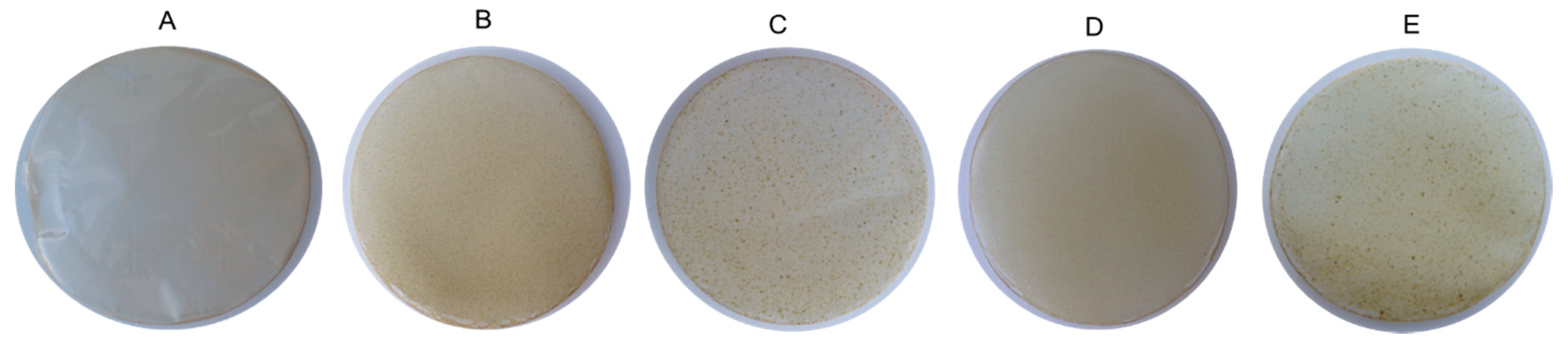
3.1. Visual Appearance and Optical Properties
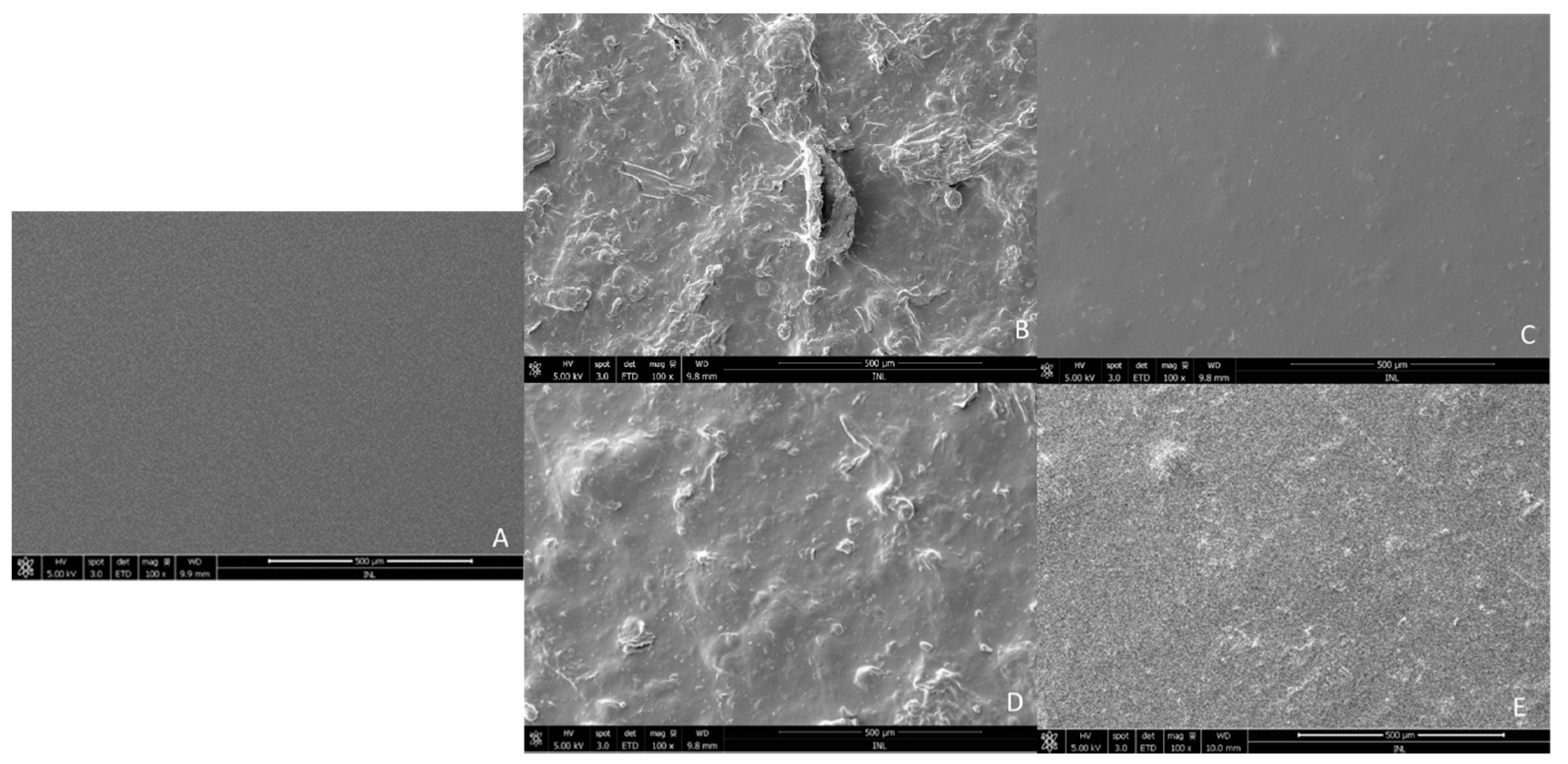
3.2. Scanning Electron Microscopy (SEM)
3.3. Thickness, Moisture Content (MC), Water Contact Angle (WCA) and Water Solubility (WS)
3.4. Water Vapour Permeability (WVP)
3.5. Mechanical Properties
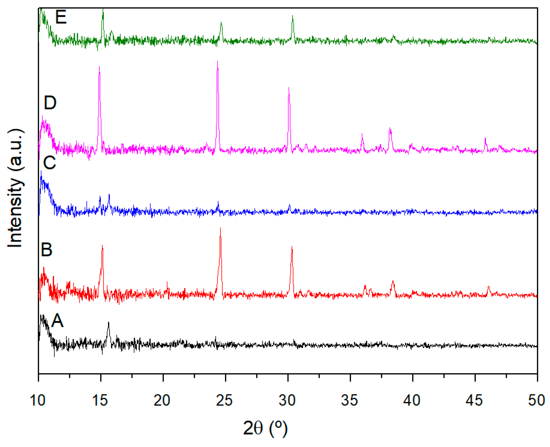
3.6. X-ray Diffraction (XRD)
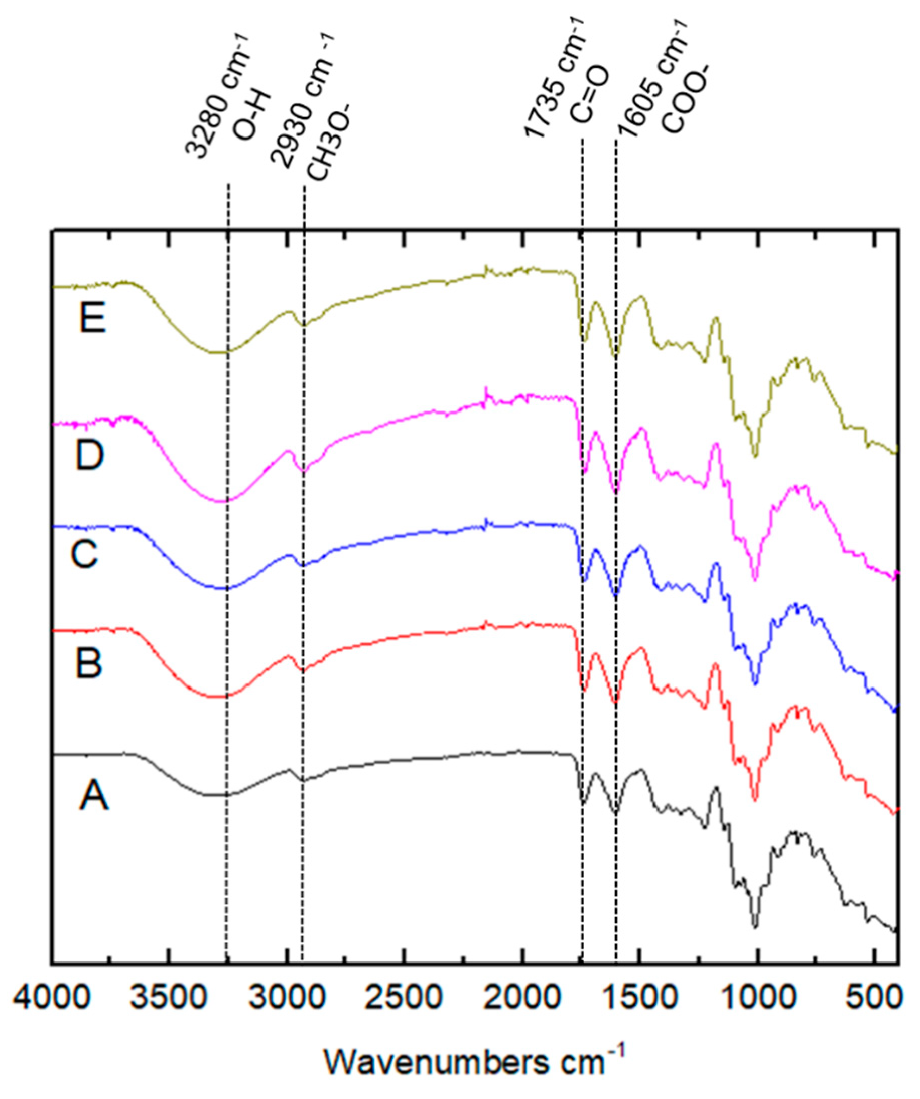
3.7. Fourier-Transform Infrared Spectroscopy
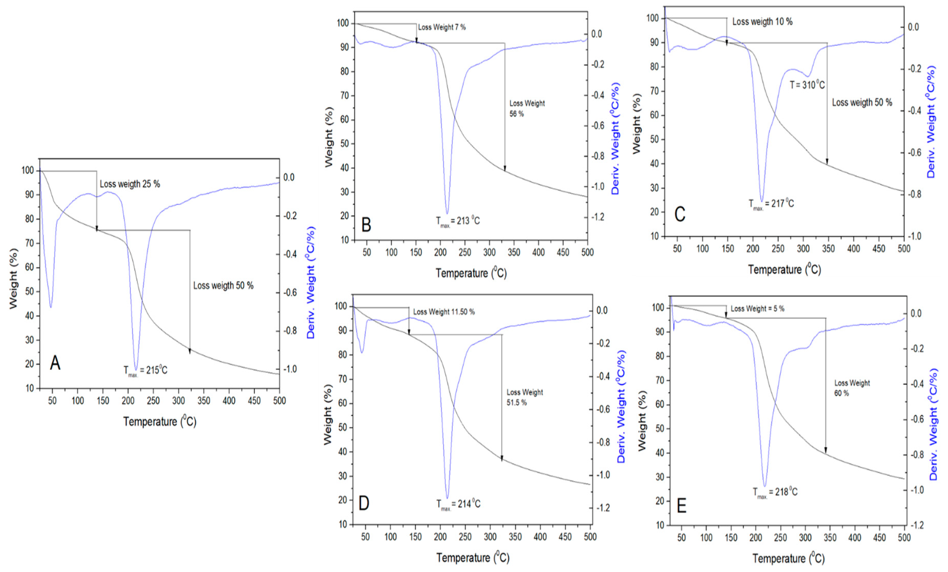
3.8. Thermogravimetric Analysis (TGA)
4. Conclusions
Author Contributions
Funding
Acknowledgments
Conflicts of Interest
References
- Costa, M.J.; Marques, A.M.; Pastrana, L.M.; Teixeira, J.A.; Sillankorva, S.M.; Cerqueira, M.A. Physicochemical properties of alginate-based films: Effect of ionic crosslinking and mannuronic and guluronic acid ratio. Food Hydrocoll. 2018, 81, 442–448. [Google Scholar] [CrossRef] [Green Version]
- Kraśniewska, K.; Pobiega, K.; Gniewosz, M. Pullulan-biopolymer with potential for use as food packaging. Int. J. Food Eng. 2019, 15. [Google Scholar] [CrossRef]
- Cerqueira, M.A.; Bourbon, A.I.; Pinheiro, A.C.; Martins, J.T.; Souza, B.W.S.; Teixeira, J.A.; Vicente, A.A. Galactomannans use in the development of edible films/coatings for food applications. Trends Food Sci. Technol. 2011, 22, 662–671. [Google Scholar] [CrossRef] [Green Version]
- Mellinas, C.; Ramos, M.; Jim, A. Recent trends in the use of pectin from agro-waste residues as a natural-based. biopolymer for food packaging applications. Materials 2020, 13, 673. [Google Scholar] [CrossRef] [Green Version]
- Mohamed, S.A.A.; El-Sakhawy, M.; El-Sakhawy, M.A.-M. Polysaccharides, protein and lipid-based natural edible films in food packaging: A review. Carbohydr. Polym. 2020, 238, 116178. [Google Scholar] [CrossRef]
- Dhanapal, A.; Rajamani, L.; Banu, M. Edible films from polysaccharides. Food Sci. Qual. Manag. 2012, 3, 9–18. [Google Scholar]
- FAO Versión resumida. Futuro Agric. Aliment. 2017, 44. [CrossRef]
- Behbahani, B.A.; Imani Fooladi, A.A. Shirazi balangu (Lallemantia royleana) seed mucilage: Chemical composition, molecular weight, biological activity and its evaluation as edible coating on beefs. Int. J. Biol. Macromol. 2018, 114, 882–889. [Google Scholar] [CrossRef]
- Contreras-Padilla, M.; Pérez-Torrero, E.; Hernández-Urbiola, M.I.; Hernández-Quevedo, G.; del Real, A.; Rivera-Muñoz, E.M.; Rodríguez-García, M.E. Evaluation of oxalates and calcium in nopal pads (Opuntia ficus-indica var. redonda) at different maturity stages. J. Food Compos. Anal. 2011, 24, 38–43. [Google Scholar] [CrossRef]
- Oliveira, N.L.; Rodrigues, A.A.; Oliveira Neves, I.C.; Teixeira Lago, A.M.; Borges, S.V.; de Resende, J.V. Development and characterization of biodegradable films based on Pereskia aculeata Miller mucilage. Ind. Crops Prod. 2019, 130, 499–510. [Google Scholar] [CrossRef]
- Dick, M.; Dal Magro, L.; Rodrigues, R.C.; de Rios, A.O.; Flôres, S.H. Valorization of Opuntia monacantha (Willd.) Haw. cladodes to obtain a mucilage with hydrocolloid features: Physicochemical and functional performance. Int. J. Biol. Macromol. 2019, 123, 900–909. [Google Scholar] [CrossRef] [PubMed]
- Mondragón-Jacobo, C.; González, S.P. El Nopal (Opuntia spp.) Como Forraje; Food and Agriculture Organization: Rome, Italy, 2003. [Google Scholar]
- Sandoval, D.C.G.; Sosa, B.L.; Martínez-Ávila, G.C.G.; Fuentes, H.R.; Abarca, V.H.A.; Rojas, R. Formulation and characterization of edible films based on organic mucilage from Mexican Opuntia ficus-indica. Coatings 2019, 9. [Google Scholar] [CrossRef] [Green Version]
- Willats, W.G.; Knox, J.P.; Mikkelsen, J.D. Pectin: New insights into an old polymer are starting to gel. Trends Food Sci. Technol. 2006, 17, 97–104. [Google Scholar] [CrossRef]
- Gheribi, R.; Puchot, L.; Verge, P.; Jaoued-Grayaa, N.; Mezni, M.; Habibi, Y.; Khwaldia, K. Development of plasticized edible films from Opuntia ficus-indica mucilage: A comparative study of various polyol plasticizers. Carbohydr. Polym. 2018, 190, 204–211. [Google Scholar] [CrossRef]
- Torres, L.; Gumecindo, C.; Bautista, E.; Gómez, Y. Opuntia ficus-indica mucilage and edible chitosan biofilms including brassica olearacea extract for extending the shell-life of capsicum annuum var serrano. Asian Res. J. Agric. 2018, 8, 1–9. [Google Scholar] [CrossRef]
- Del-Valle, V.; Hernández-Muñoz, P.; Guarda, A.; Galotto, M.J. Development of a cactus-mucilage edible coating (Opuntia ficus indica) and its application to extend strawberry (Fragaria ananassa) shelf-life. Food Chem. 2005, 91, 751–756. [Google Scholar] [CrossRef]
- Treviño-Garza, M.Z.; García, S.; Heredia, N.; Alanís-Guzmán, M.G.; Arévalo-Niño, K. Layer-by-layer edible coatings based on mucilages, pullulan and chitosan and its effect on quality and preservation of fresh-cut pineapple (Ananas comosus). Postharvest Biol. Technol. 2017, 128, 63–75. [Google Scholar] [CrossRef]
- Ganesan, A.R.; Shanmugam, M.; Palaniappan, S.; Rajauria, G. Development of edible film from Acanthophora spicifera: Structural, rheological and functional properties. Food Biosci. 2018, 23, 121–128. [Google Scholar] [CrossRef]
- Allegra, A.; Inglese, P.; Sortino, G.; Settanni, L.; Todaro, A.; Liguori, G. The influence of Opuntia ficus-indica mucilage edible coating on the quality of “Hayward” kiwifruit slices. Postharvest Biol. Technol. 2016, 120, 45–51. [Google Scholar] [CrossRef]
- Cortés-Camargo, S.; Acuña-Avila, P.E.; Rodríguez-Huezo, M.E.; Román-Guerrero, A.; Varela-Guerrero, V.; Pérez-Alonso, C. Effect of chia mucilage addition on oxidation and release kinetics of lemon essential oil microencapsulated using mesquite gum—Chia mucilage mixtures. Food Res. Int. 2019, 116, 1010–1019. [Google Scholar] [CrossRef]
- Jouki, M.; Mortazavi, S.A.; Yazdi, F.T.; Koocheki, A.; Khazaei, N. Use of quince seed mucilage edible films containing natural preservatives to enhance physico-chemical quality of rainbow trout fillets during cold storage. Food Sci. Hum. Wellness 2014, 3, 65–72. [Google Scholar] [CrossRef] [Green Version]
- Allegra, A.; Sortino, G.; Inglese, P.; Settanni, L.; Todaro, A.; Gallotta, A. The effectiveness of Opuntia ficus-indica mucilage edible coating on post-harvest maintenance of ‘Dottato’ fig (Ficus carica L.) fruit. Food Packag. Shelf Life 2017, 12, 135–141. [Google Scholar] [CrossRef]
- Soukoulis, C.; Gaiani, C.; Hoffmann, L. Plant seed mucilage as emerging biopolymer in food industry applications. Curr. Opin. Food Sci. 2018, 22, 28–42. [Google Scholar] [CrossRef]
- Beigomi, M.; Mohsenzadeh, M.; Salari, A. Characterization of a novel biodegradable edible film obtained from Dracocephalum moldavica seed mucilage. Int. J. Biol. Macromol. 2018, 108, 874–883. [Google Scholar] [CrossRef]
- Sadeghi-Varkani, A.; Emam-Djomeh, Z.; Askari, G. Physicochemical and microstructural properties of a novel edible film synthesized from Balangu seed mucilage. Int. J. Biol. Macromol. 2018, 108, 1110–1119. [Google Scholar] [CrossRef]
- Luo, M.; Cao, Y.; Wang, W.; Chen, X.; Cai, J.; Wang, L.; Xiao, J. Sustained-release antimicrobial gelatin film: Effect of chia mucilage on physicochemical and antimicrobial properties. Food Hydrocoll. 2019, 87, 783–791. [Google Scholar] [CrossRef]
- Rodríguez-González, S.; Martínez-Flores, H.E.; Chávez-Moreno, C.K.; Macías-Rodríguez, L.I.; Zavala-Mendoza, E.; Garnica-Romo, M.G.; Chacõn-García, L. Extraction and characterization of mucilage from wild species of opuntia. J. Food Process Eng. 2014, 37, 285–292. [Google Scholar] [CrossRef]
- Espino-Díaz, M.; De Jesús Ornelas-Paz, J.; Martínez-Téllez, M.A.; Santillán, C.; Barbosa-Cánovas, G.V.; Zamudio-Flores, P.B.; Olivas, G.I. Development and characterization of edible films based on mucilage of Opuntia ficus-indica (L.). J. Food Sci. 2010, 75, 347–352. [Google Scholar] [CrossRef]
- Martins, J.T.; Cerqueira, M.A.; Vicente, A.A. Influence of α-tocopherol on physicochemical properties of chitosan-based films. Food Hydrocoll. 2012, 27, 220–227. [Google Scholar] [CrossRef] [Green Version]
- Casariego, A.; Souza, B.W.S.; Cerqueira, M.A.; Teixeira, J.A.; Cruz, L.; Díaz, R.; Vicente, A.A. Chitosan/clay films’ properties as affected by biopolymer and clay micro/nanoparticles’ concentrations. Food Hydrocoll. 2009, 23, 1895–1902. [Google Scholar] [CrossRef] [Green Version]
- ASTM E96: Standard Test Methods for Water Vapor Transmission of Materials; ASTM: West Conshohocken, PA, USA, 1995.
- ASTM D 882-91: Standard Test Method for Tensile Properties of Thin Plastic Sheeting; ASTM: West Conshohocken, PA, USA, 1991.
- Pereira, R.N.; Souza, B.W.S.; Cerqueira, M.A.; Teixeira, J.A.; Vicente, A.A. Effects of electric fields on protein unfolding and aggregation: Influence on edible films formation. Biomacromolecules 2010, 11, 2912–2918. [Google Scholar] [CrossRef] [Green Version]
- Da Silva Oliveira, C.I.; Martinez-Martinez, D.; Al-Rjoub, A.; Rebouta, L.; Menezes, R.; Cunha, L. Development of a statistical method to help evaluating the transparency/opacity of decorative thin films. Appl. Surf. Sci. 2018, 438, 51–58. [Google Scholar] [CrossRef]
- Lira-Vargas, A.A.; Joel, J.; Corrales-Garcia, E.; Valle-Guadarrama, S.; Beatriz Peña-Valdivia, C.; Trejo-Marquez, M.A.; Cantu, J.; Izcalli, C.; Mexico, M. Biopolymeric films based on cactus (Opuntia ficus-indica) mucilage incorporated with gelatin and beeswax. J. Prof. Assoc. Cactus 2014, 16, 51–70. [Google Scholar]
- Lutz, R.; Aserin, A.; Wicker, L.; Garti, N. Structure and physical properties of pectins with block-wise distribution of carboxylic acid groups. Food Hydrocoll. 2009, 23, 786–794. [Google Scholar] [CrossRef]
- Contreras-Padilla, M.; Rivera-Muñoz, E.M.; Gutiérrez-Cortez, E.; del López, A.R.; Rodríguez-García, M.E. Characterization of crystalline structures in Opuntia ficus-indica. J. Biol. Phys. 2015, 41, 99–112. [Google Scholar] [CrossRef] [Green Version]
- Contreras-Padilla, M.; Rodríguez-García, M.E.; Gutiérrez-Cortez, E.; del Valderrama-Bravo, M.C.; Rojas-Molina, J.I.; Rivera-Muñoz, E.M. Physicochemical and rheological characterization of Opuntia ficus mucilage at three different maturity stages of cladode. Eur. Polym. J. 2016, 78, 226–234. [Google Scholar] [CrossRef]
- Vanitha Kumari, G.; Ananth, N.; Mathavan, T.A.; Jothi Rajan, M.A. Synthesis and characterization of folic acid conjugated silver/gold nanoparticles for biomedical applications. Mater. Today Proc. 2016, 3, 4215–4219. [Google Scholar] [CrossRef]
- Guadarrama-Lezama, A.Y.; Castaño, J.; Velázquez, G.; Carrillo-Navas, H.; Alvarez-Ramírez, J. Effect of nopal mucilage addition on physical, barrier and mechanical properties of citric pectin-based films. J. Food Sci. Technol. 2018, 55, 3739–3748. [Google Scholar] [CrossRef]
- Cai, W.; Gu, X.; Tang, J. Extraction, purification, and characterization of the polysaccharides from Opuntia milpa alta. Carbohydr. Polym. 2008, 71, 403–410. [Google Scholar] [CrossRef]
- El Achaby, M.; El Miri, N.; Hannache, H.; Gmouh, S.; Ben youcef, H.; Aboulkas, A. Production of cellulose nanocrystals from vine shoots and their use for the development of nanocomposite materials. Int. J. Biol. Macromol. 2018, 117, 592–600. [Google Scholar] [CrossRef]
- Mujtaba, M.; Akyuz, L.; Koc, B.; Kaya, M.; Ilk, S.; Cansaran-Duman, D.; Martinez, A.S.; Cakmak, Y.S.; Labidi, J.; Boufi, S. Novel, multifunctional mucilage composite films incorporated with cellulose nanofibers. Food Hydrocoll. 2019, 89, 20–28. [Google Scholar] [CrossRef]
- Chaturvedi, K.; Sharma, N.; Yadav, S.K. Composite edible coatings from commercial pectin, corn flour and beetroot powder minimize post-harvest decay, reduces ripening and improves sensory liking of tomatoes. Int. J. Biol. Macromol. 2019, 133, 284–293. [Google Scholar] [CrossRef]


| Film Samples | L* | a* | b* | Opacity (%) |
|---|---|---|---|---|
| Control | 99.19 ± 0.22a | 15.50 ± 0.60a | 13.86 ± 1.36a | 4.38 ± 0.32a |
| Copwtf | 82.90 ± 0.14d | 0.96 ± 0.12d | 18.09 ± 1.13b | 6.21 ± 0.24b |
| Copwf | 88.43 ± 1.94e | 0.05 ± 0.03c | 13.11 ± 1.57a | 3.92 ± 0.09d |
| Viwtf | 69.22 ± 0.65b | 3.29 ± 0.43b | 28.43 ± 1.07c | 6.39 ± 0.05b |
| Viwf | 92.60 ±1.22c | 0.07 ± 0.02c | 15.91 ± 2.83ab | 3.23 ± 0.02c |
| Film Samples | Thickness (mm) | MC (%) | WCA (°) | WVP (×10−11 g (m·s·Pa)−1) |
|---|---|---|---|---|
| Control | 0.08 ± 0.01a | 8.85 ± 0.06a | 87.22 ± 6.02a | 10.05 ± 1.16a |
| Copwtf | 0.17 ± 0.03c | 12.41 ± 0.08d | 37.80 ± 7.57b | 44.28 ± 4.68c |
| Copwf | 0.22 ± 0.03d | 12.44 ± 0.01d | 39.32 ± 8.75b | 20.95 ± 2.60d |
| Viwtf | 0.11 ± 0.05ab | 13.18 ± 0.02b | 37.12 ± 9.92b | 33.83 ± 5.65b |
| Viwf | 0.15 ± 0.06bc | 9.29 ± 0.01c | 39.89 ± 5.74b | 26.96 ± 0.47bd |
| Film Samples | TS (MPa) | EB (%) | YM (MPa) |
|---|---|---|---|
| Control | 21.11 ± 3.95a | 9.57 ± 2.51a | 6.78 ± 1.56ab |
| Copwtf | 11.07 ± 2.92b | 2.91 ± 0.64b | 5.13 ± 0.90a |
| Copwf | 3.34 ± 1.71c | 0.89 ± 0.20c | 4.83 ± 1.44a |
| Viwtf | 16.11 ± 2.76ab | 2.33 ± 0.32b | 8.41 ± 0.68b |
| Viwf | 14.75 ± 8.66a | 2.60 ± 0.88b | 6.56 ± 2.65ab |
© 2020 by the authors. Licensee MDPI, Basel, Switzerland. This article is an open access article distributed under the terms and conditions of the Creative Commons Attribution (CC BY) license (http://creativecommons.org/licenses/by/4.0/).
Share and Cite
Luna-Sosa, B.; Martínez-Ávila, G.C.G.; Rodríguez-Fuentes, H.; Azevedo, A.G.; Pastrana, L.M.; Rojas, R.; Cerqueira, M.A. Pectin-Based Films Loaded with Hydroponic Nopal Mucilages: Development and Physicochemical Characterization. Coatings 2020, 10, 467. https://0-doi-org.brum.beds.ac.uk/10.3390/coatings10050467
Luna-Sosa B, Martínez-Ávila GCG, Rodríguez-Fuentes H, Azevedo AG, Pastrana LM, Rojas R, Cerqueira MA. Pectin-Based Films Loaded with Hydroponic Nopal Mucilages: Development and Physicochemical Characterization. Coatings. 2020; 10(5):467. https://0-doi-org.brum.beds.ac.uk/10.3390/coatings10050467
Chicago/Turabian StyleLuna-Sosa, Brenda, Guillermo C.G. Martínez-Ávila, Humberto Rodríguez-Fuentes, Ana G. Azevedo, Lorenzo M. Pastrana, Romeo Rojas, and Miguel A. Cerqueira. 2020. "Pectin-Based Films Loaded with Hydroponic Nopal Mucilages: Development and Physicochemical Characterization" Coatings 10, no. 5: 467. https://0-doi-org.brum.beds.ac.uk/10.3390/coatings10050467