
Investigation on the Painting Materials and Profile Structures Used in Ancient Chinese Folk Architectural Paintings by Multiple Analytical Methods


Abstract
:1. Introduction
2. Methods and Experimental Design
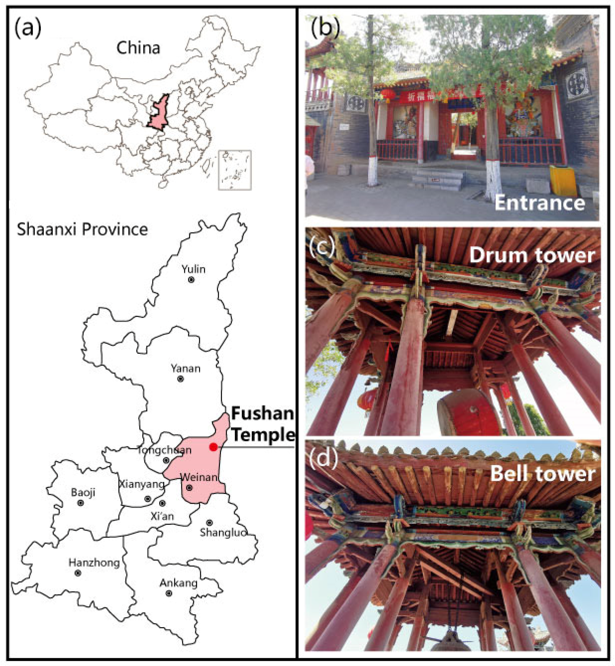
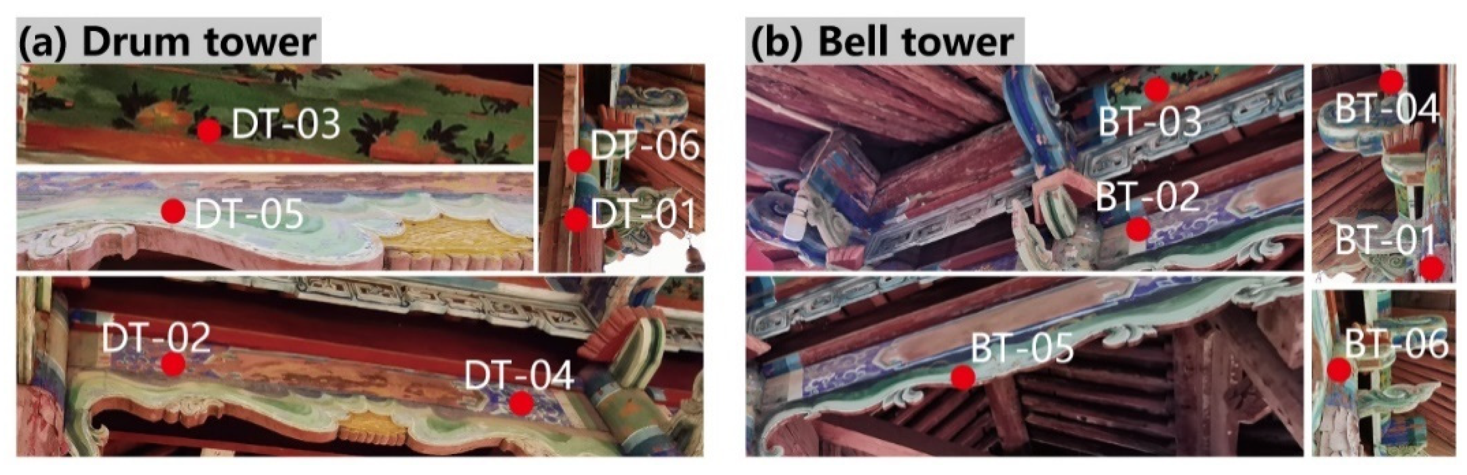
2.1. Samples and Dyeing Experiments
2.2. Experimental Equipment
2.2.1. Optical Microscope and SEM-EDS
2.2.2. Micro-Raman Microscopy and FTIR Spectroscopy
3. Results and Discussion
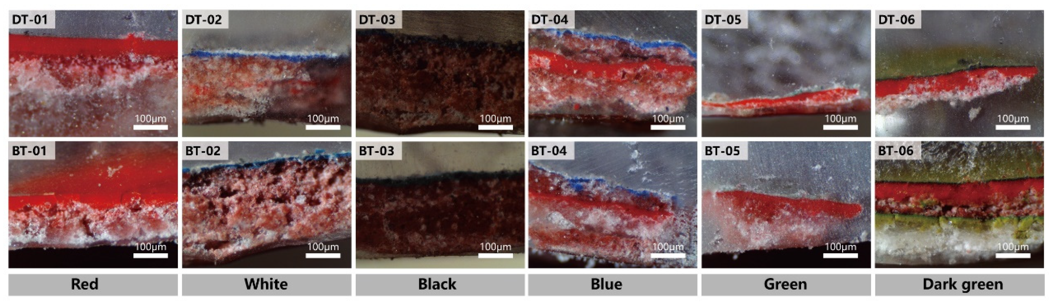
3.1. Painting Profile Structures Analysis
3.2. Materials Used in Architectural Paintings
3.2.1. Pigments
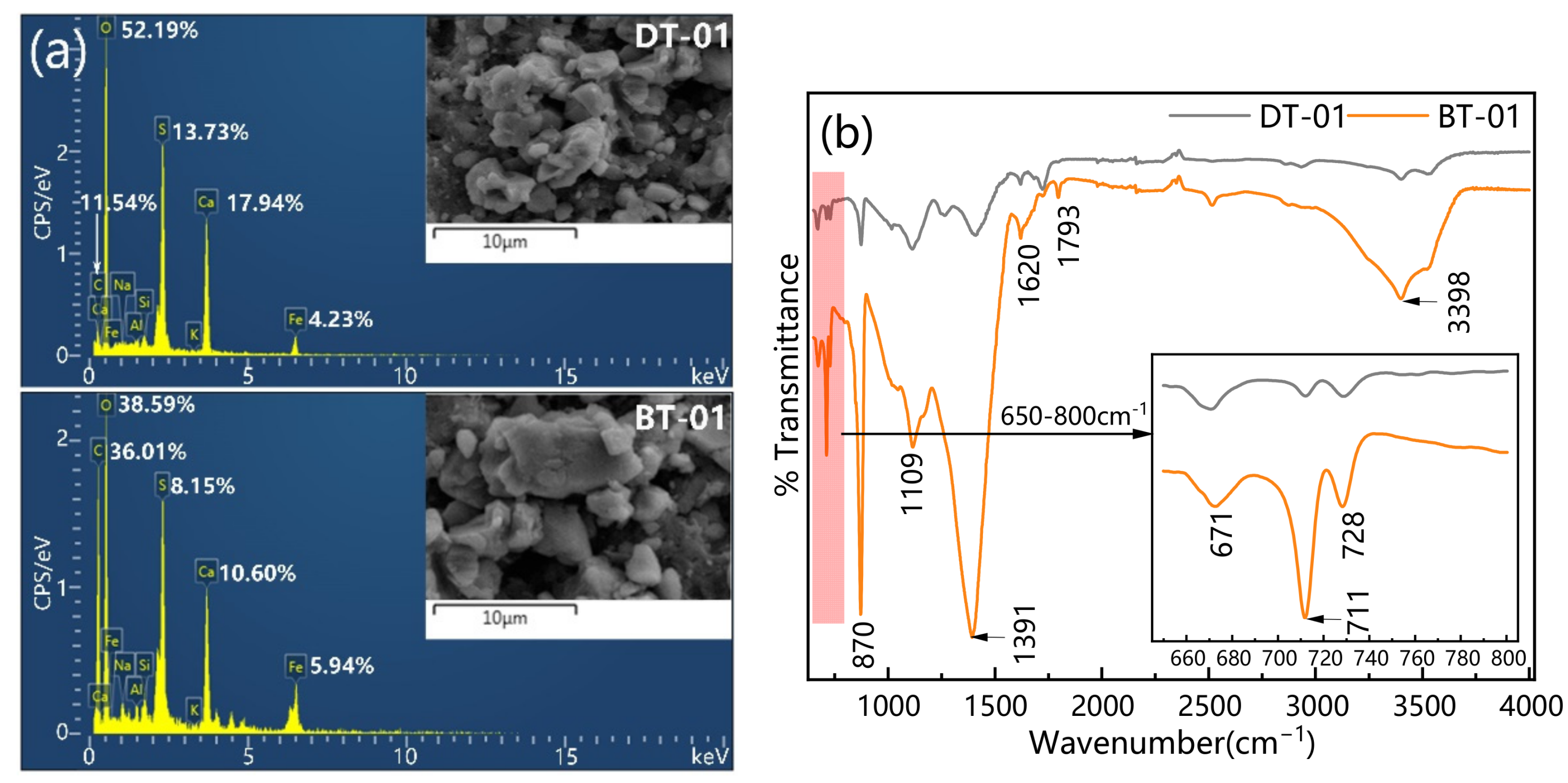
Red Pigment
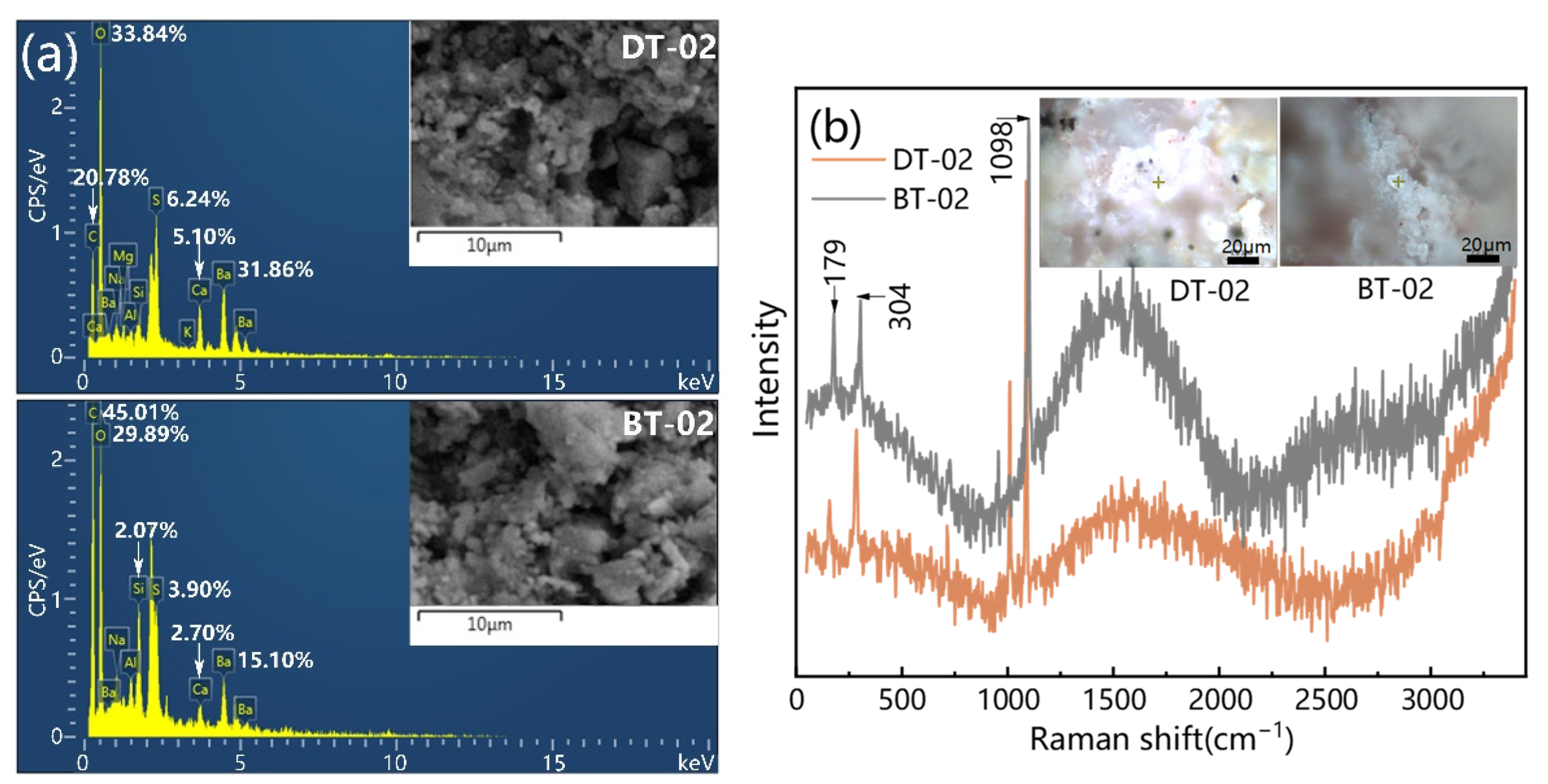
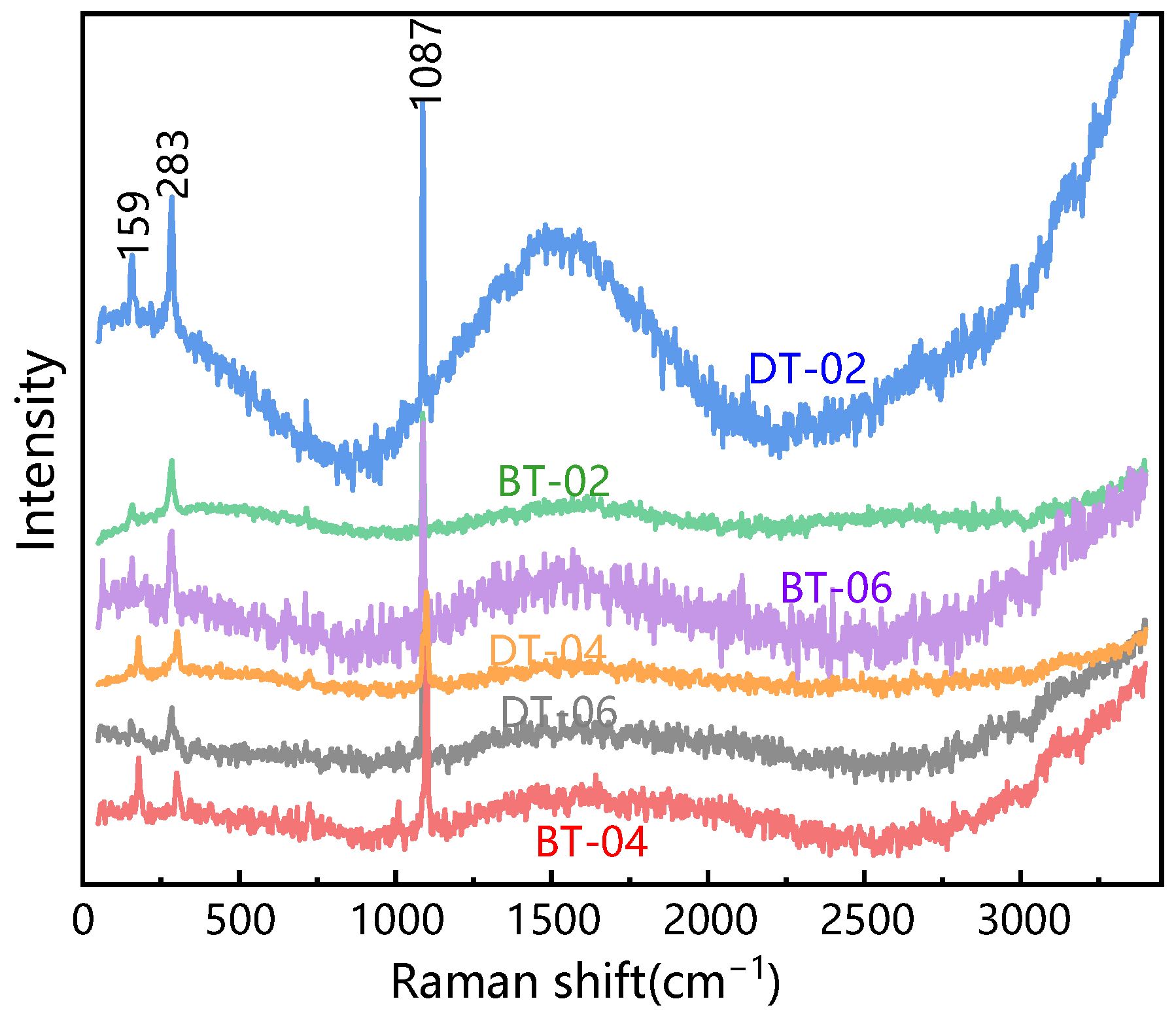
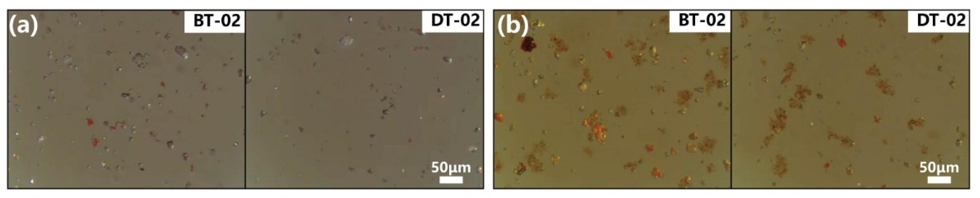
White Pigment
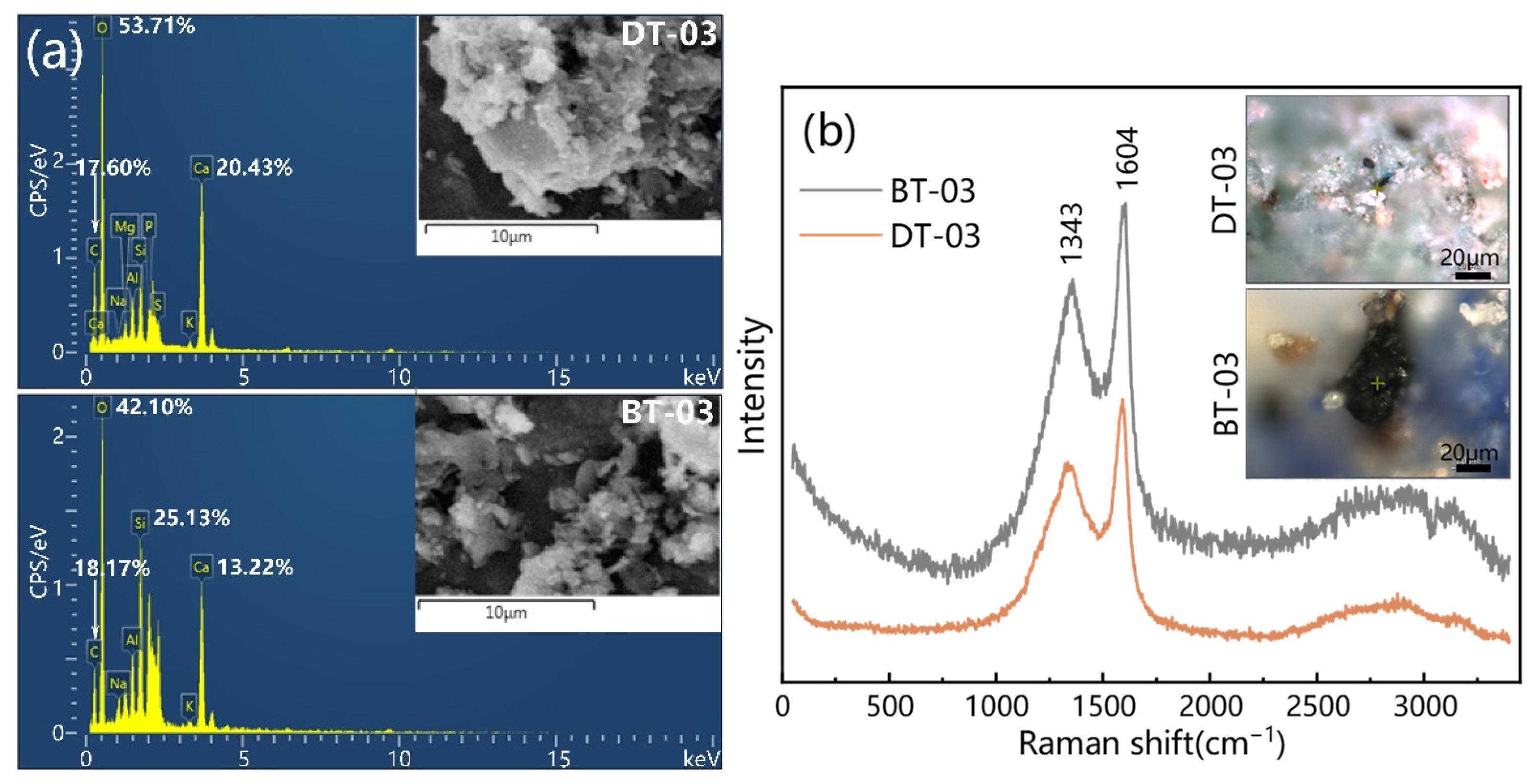
Black Pigment
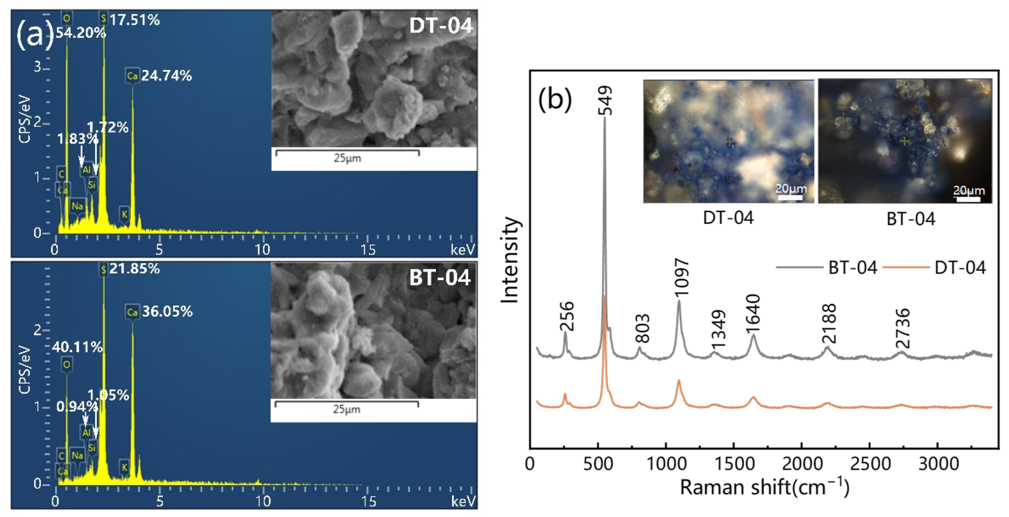
Blue Pigment
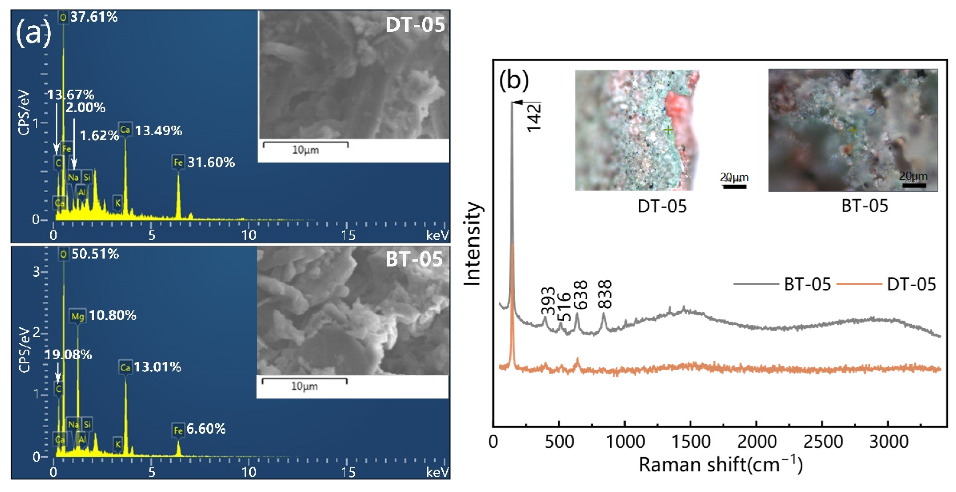
Green Pigment
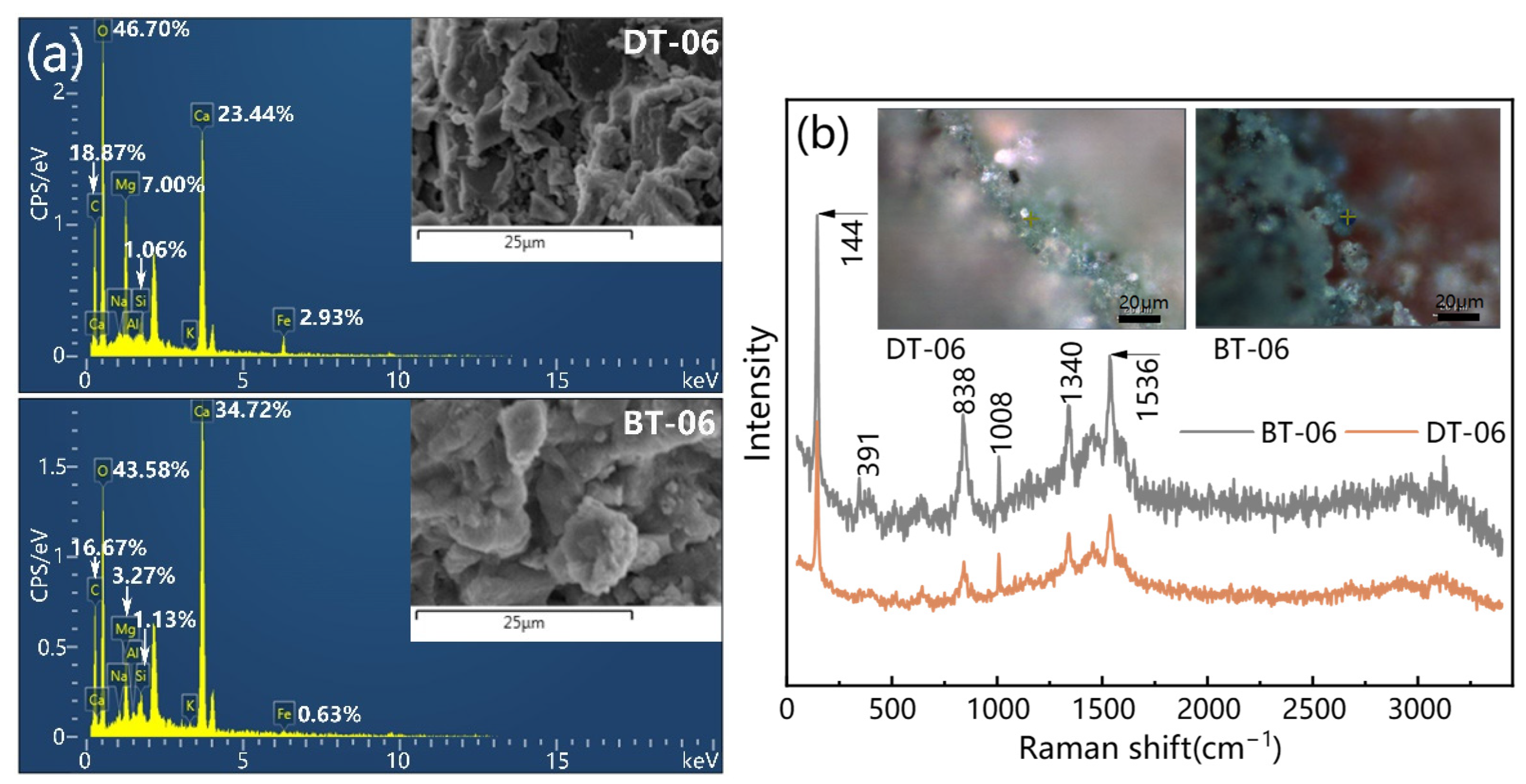
Dark Green Pigment
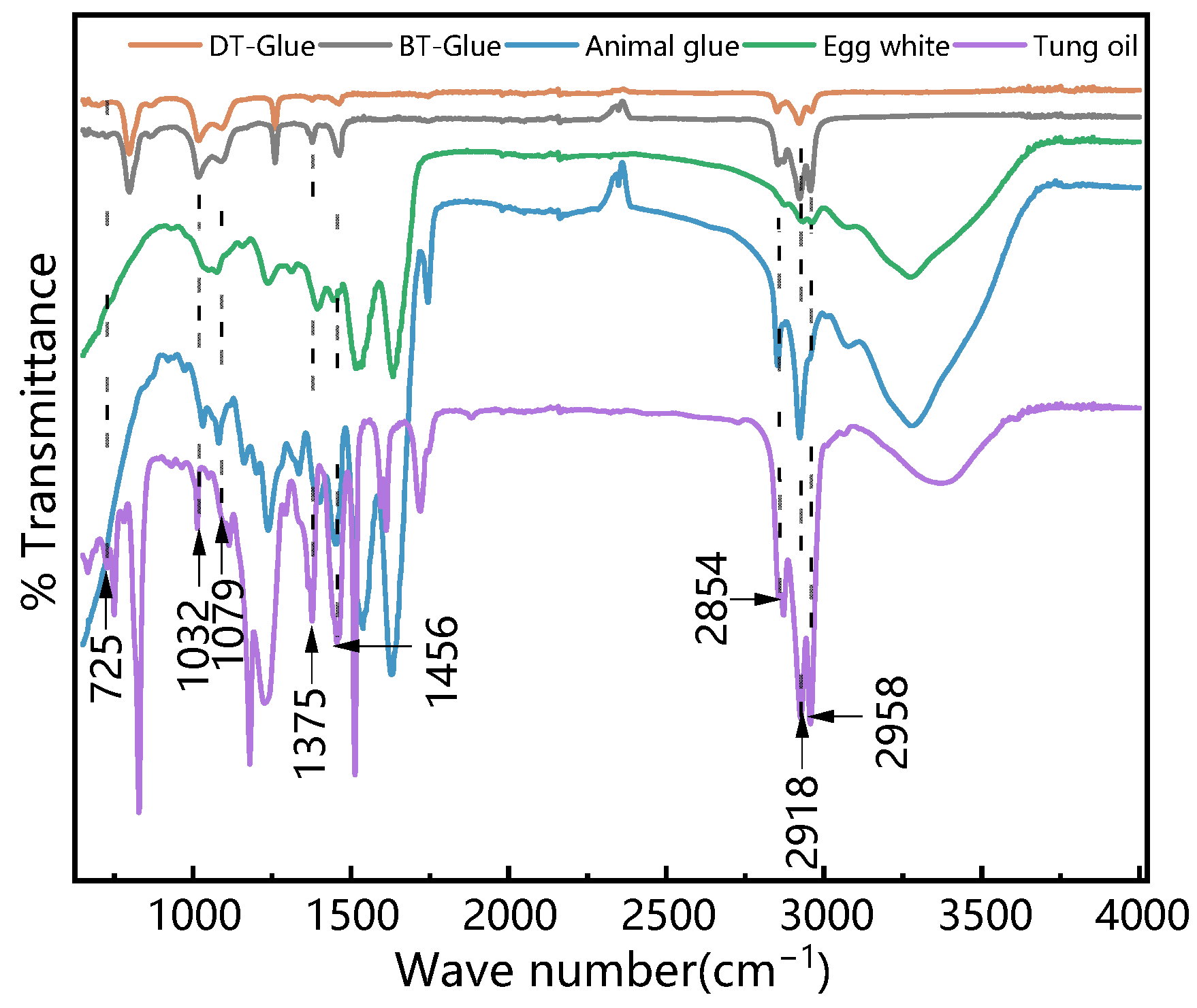
3.2.2. Binding Media
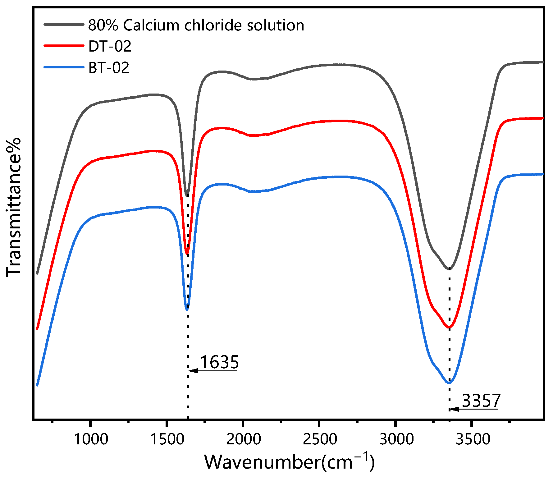
3.2.3. Ground Layer
4. Conclusions
Author Contributions
Funding
Institutional Review Board Statement
Informed Consent Statement
Data Availability Statement
Acknowledgments
Conflicts of Interest
References
- Li, J. The composition of pigments of decorative paintings on ancient buildings of Qufu’s temple Confucius. China Cult. Herit. Sci. Res. 2014, 4, 86–89. [Google Scholar] [CrossRef]
- Mazzeo, R.; Cam, D.; Chiavari, G.; Fabbri, D.; He, L. Analytical study of traditional decorative materials and techniques used in Ming dynasty wooden architecture. the case of the drum tower in Xi’an, P.R. of China. J. Cult. Herit. 2004, 5, 273–283. [Google Scholar] [CrossRef]
- Li, Y.; Liu, M.Y. A Study of The Materials and Techniques Used in The Polychrome Ceiling Decoration of The Linxi Pavilion in The Garden of The Cining Palace. Palace Mus. J. 2018, 6, 45–63+159. [Google Scholar]
- Zhang, Y.; Zhang, Z.J.; Rong, B.; Dang, H.Y. Structural survey and pigment analysis of the paint layer on the cultural relics from the Han Yangling. Sci. Conserv. Archaeol. 2013, 25, 88–92. [Google Scholar] [CrossRef]
- Li, Y.; Wang, F.P.; Fu, X.Y.; Sun, Z.J.; Xu, Y.Q. Analysis of the pigments for smoked mural by confocal micro-Raman spectroscopy. J. Raman Spectrosc. 2017, 48, 1479–1486. [Google Scholar] [CrossRef]
- Zhang, Y.; Wang, J.; Zhang, T. Analysis on mural structures and components of the tombs in Liao dynasty (A.D. 907-A.D. 1125). Spectrosc. Lett. 2015, 48, 732–740. [Google Scholar] [CrossRef]
- Cheilakou, E.; Troullinos, M.; Koui, M. Identification of pigments on byzantine wall paintings from Crete (14th century AD) using non-invasive fiber optics diffuse reflectance spectroscopy. J. Archaeol. Sci. 2014, 41, 541–555. [Google Scholar] [CrossRef]
- Iordanidis, A.; Garcia-Guinea, J.; Strati, A.; Gkimourtzina, A.; Papoulidou, A. Byzantine wall paintings from Kastoria, northern Greece: Spectroscopic study of pigments and efflorescing salts. Spectrochim. Acta Part A Mol. Biomol. Spectrosc. 2011, 78, 874–887. [Google Scholar] [CrossRef]
- Souto, J.; Prieto, A.C.; Gutiérrez-Vicente, V. Raman analysis of gothic wall paintings in the apse of the Santiago Apóstol church in Alcazarén. J. Cult. Herit. 2016, 22, 1061–1065. [Google Scholar] [CrossRef]
- Vandenabeele, P.; Bodé, S.; Alonso, A.; Moens, L. Raman spectroscopic analysis of the Maya wall paintings in Ek’Balam, Mexico. Spectrochim. Acta Part A Mol. Biomol. Spectrosc. 2005, 61, 2349–2356. [Google Scholar] [CrossRef]
- Proietti, N.; Tullio, V.D.; Presciutti, F.; Gentile, G.; Brunetti, B.G.; Capitani, D. A multi-analytical study of ancient Nubian detached mural paintings. Microchem. J. 2016, 124, 719–725. [Google Scholar] [CrossRef]
- Moretti, P.; Gallegos, D.; Marte, F.; Brunetti, B.; Sgamellotti, A.; Miliani, C. Materials and techniques of twentieth century Argentinean murals. Procedia Chem. 2013, 8, 221–230. [Google Scholar] [CrossRef] [Green Version]
- Dallongeville, S.; Garnier, N.; Rolando, C.; Tokarski, C. Proteins in Art, Archaeology, and Paleontology: From Detection to Identification. Chem. Rev. 2015, 116, 2–79. [Google Scholar] [CrossRef] [PubMed]
- Demir, S.; Şerifaki, K.; BöKe, H. Execution technique and pigment characteristics of byzantine wall paintings of Anaia church in western Anatolia. J. Archaeol. Sci. Rep. 2018, 17, 39–46. [Google Scholar] [CrossRef]
- Želinská, J.; Kopecká, I.; Svobodová, E.; Milovská, S.; Vratislav, H. Stratigraphic EM-EDS, XRF, Raman and FT-IR analysis of multilayer paintings from the Main Altar of the St. James Church in Levoča (Slovakia). J. Cult. Herit. 2018, 33, 90–99. [Google Scholar] [CrossRef]
- Tomasini, E.; Rodríguez, D.C.; Gómez, B.A.; Faria, D.L.A.; Landa, C.R.; Siracusano, G.; Maier, M.S. A multi-analytical investigation of the materials and painting technique of a wall painting from the church of Copacabana de Andamarca (Bolivia). Microchem. J. 2016, 128, 172–180. [Google Scholar] [CrossRef]
- Li, J. Ying Zao Fa Shi; Commercial Press: Shanghai, China, 1954; Volumes 1–4. (In Chinese) [Google Scholar]
- Liang, S.C. Qing-Dynasty Municipal Engineering; Tsinghua University Press: Beijing, China, 2006. (In Chinese) [Google Scholar]
- ICOMOS China. Principles for the Conservation of Heritage Sites in China (Billingual); ICOMOS China: Beijing, China, 2015; Available online: http://openarchive.icomos.org/id/eprint/1650/ (accessed on 29 January 2022).
- Yang, L.; Huang, J.H.; Chen, X.N.; Wang, L.Q.; Wei, Y.M. PCA-LDA analysis of binders used in the theater colour painting of Jiayuguan pass based on FTIR spectra. Spectrosc. Spectr. Anal. 2021, 41, 796–800. [Google Scholar] [CrossRef]
- Ji, L.; Shao, Z.Y. Analytical study on the painting of ancient buildings in Xi’an-Determination of starch particles in the ground layer. Identif. Apprec. Cult. Relics 2010, 1, 30–35. [Google Scholar] [CrossRef]
- Nurdini, N.; Ilmi, M.M.; Maryanti, E.; Kadja, G. Investigation on the crystal structures of hematite pigments at different sintering temperatures. Key Eng. Mater. 2021, 6139, 20–27. [Google Scholar] [CrossRef]
- Akyuz, A.S.; Akyuz, T.A.; Basaran, S.B.; Kocabas, I.B.; Gulec, A.B.; Cesmeli, H.B.; Uca, B.B. FT-IR and EDXRF analysis of wall paintings of ancient Ainos Hagia Sophia Church. J. Mol. Struct. 2009, 924, 400–403. [Google Scholar] [CrossRef]
- Jin, D.L.; Yue, L.H.; Xu, Z.D. Infrared and Raman analysis of spherical CaCO3 composite. Chin. J. Inorg. Chem. 2004, 20, 715–720. [Google Scholar] [CrossRef]
- Bell, I.M.; Clark, R.; Gibbs, P.J. Raman spectroscopic library of natural and synthetic pigments (pre-1850 AD). Spectrochim. Acta Part A Mol. Biomol. Spectrosc. 1997, 53, 2159–2179. [Google Scholar] [CrossRef]
- Goler, S.; Yardley, J.T.; Cacciola, A.; Hagadorn, A.; Ratzan, D.; Bagnall, R. Characterizing the age of ancient Egyptian manuscripts through micro-Raman spectroscopy. J. Raman Spectrosc. 2016, 47, 1185–1193. [Google Scholar] [CrossRef]
- Choi, S.; Lee, S.K.; Kim, N.H.; Kim, S.; Lee, Y.N. Raman spectroscopy detects amorphous carbon in an enigmatic egg from the upper cretaceous Wido Volcanics of south Korea. Front. Earth Sci. 2020, 7, 349. [Google Scholar] [CrossRef]
- Rosina, P.; Gomes, H.; Collado, H.; Nicoli, M.; Volpe, L.; Vaccaro, C. Μicro-Raman spectroscopy for the characterization of rock-art pigments from Abrigo del Guila (Badajoz-Spain). Opt. Laser Technol. 2018, 102, 274–281. [Google Scholar] [CrossRef]
- Barone, G.; Mazzoleni, P.; Cecchini, A.; Russo, A. In situ Raman and PXRF spectroscopic study on the wall paintings of Etruscan Tarquinia tombs. Dye. Pigment. 2018, 150, 390–403. [Google Scholar] [CrossRef]
- Wang, Y. The chromophore fading and spectroscopy analysis of lazurite in annealing treatment. Spectrochim. Acta Part A Mol. Biomol. Spectrosc. 2020, 247, 119117. [Google Scholar] [CrossRef]
- Li, Q.Q.; Zhou, L.L.; Wei, S.Y.; Ma, Q.L. Study on the Materials of the Paintings on the Head of Buddha Statue Excavated from Longxing Temple in Qingzhou, Shandong, China. Meseum 2017, 2, 28–37. [Google Scholar]
- Fanost, A.; Gimat, A.; Viguerie, L.; Martinetto, P.; Giot, A.C.; Clémancey, M.; Jaber, M. Revisiting the identification of commercial and historical green earth pigments. Colloids Surf. A Physicochem. Eng. Asp. 2020, 584, 124035. [Google Scholar] [CrossRef]
- Baraldi, P.; Bracci, S.; Cristoferi, E.; Fiorentino, S.; Venturi, E. Pigment characterization of drawings and painted layers under 5th–7th centuries wall mosaics from Ravenna (Italy). J. Cult. Herit. 2016, 21, 802–808. [Google Scholar] [CrossRef]
- Fu, Q.; Xia, Y.; Wang, W.; Yang, J.; Lv, Z.; Xi, N. Study of green ground layer in an eastern Han dynasty tomb mural painting at Haotan, Dingbian county, Shaanxi province. Sci. Conserv. Archaeol. 2012, 24, 38–43. [Google Scholar] [CrossRef]


| Sampling Location | Drum Tower (DT) | |||||
| Sampling number | DT-01 | DT-02 | DT-03 | DT-04 | DT-05 | DT-06 |
| Sampling colour | Red | White | Black | Blue | Green | Dark green |
| Sampling position | Beam (West) | Beam (West) | Beam (North) | Beam (West) | Queti (South) | Wooden column (West) |
| Sampling location | Bell Tower (BT) | |||||
| Sampling number | BT-01 | BT-02 | BT-03 | BT-04 | BT-05 | BT-06 |
| Sampling colour | Red | White | Black | Blue | Green | Dark green |
| Sampling position | Beam (West) | Beam (North) | Beam (North) | Beam (West) | Queti (East) | Wooden column (East) |
| Element | C | O | Ca | Si | Mg | Na | Al | K | |
|---|---|---|---|---|---|---|---|---|---|
| (wt.%) Sample | |||||||||
| DT-02 | 14.04 | 47.96 | 38.01 | 0.00 | 0.00 | 0.00 | 0.00 | 0.00 | |
| DT-04 | 20.53 | 42.74 | 34.51 | 0.00 | 0.97 | 1.24 | 0.00 | 0.00 | |
| DT-06 | 14.43 | 47.42 | 25.10 | 0.00 | 13.06 | 0.00 | 0.00 | 0.00 | |
| BT-02 | 16.76 | 47.47 | 35.04 | 0.00 | 0.74 | 0.00 | 0.00 | 0.00 | |
| BT-04 | 12.83 | 44.72 | 42.46 | 0.00 | 0.00 | 0.00 | 0.00 | 0.00 | |
| BT-06 | 9.94 | 31.50 | 46.42 | 0.00 | 10.32 | 1.82 | 0.00 | 0.00 | |
Publisher’s Note: MDPI stays neutral with regard to jurisdictional claims in published maps and institutional affiliations. |
© 2022 by the authors. Licensee MDPI, Basel, Switzerland. This article is an open access article distributed under the terms and conditions of the Creative Commons Attribution (CC BY) license (https://creativecommons.org/licenses/by/4.0/).
Share and Cite
Zou, W.; Yeo, S.-Y. Investigation on the Painting Materials and Profile Structures Used in Ancient Chinese Folk Architectural Paintings by Multiple Analytical Methods. Coatings 2022, 12, 320. https://0-doi-org.brum.beds.ac.uk/10.3390/coatings12030320
Zou W, Yeo S-Y. Investigation on the Painting Materials and Profile Structures Used in Ancient Chinese Folk Architectural Paintings by Multiple Analytical Methods. Coatings. 2022; 12(3):320. https://0-doi-org.brum.beds.ac.uk/10.3390/coatings12030320
Chicago/Turabian StyleZou, Weihan, and Sok-Yee Yeo. 2022. "Investigation on the Painting Materials and Profile Structures Used in Ancient Chinese Folk Architectural Paintings by Multiple Analytical Methods" Coatings 12, no. 3: 320. https://0-doi-org.brum.beds.ac.uk/10.3390/coatings12030320