
Electrochemical Detection of Hormones Using Nanostructured Electrodes

Department of Chemistry and Biochemistry, University of Missouri-Saint Louis, St. Louis, MO 63121, USA
*
Author to whom correspondence should be addressed.
Coatings 2023, 13(12), 2040; https://0-doi-org.brum.beds.ac.uk/10.3390/coatings13122040
Submission received: 31 October 2023
/
Revised: 29 November 2023
/
Accepted: 30 November 2023
/
Published: 4 December 2023
(This article belongs to the Special Issue Advances in the Preparation and Characterization Techniques for Developing Coating Materials and Applications)
Abstract
:Hormones regulate several physiological processes in living organisms, and their detection requires accuracy and sensitivity. Recent advances in nanostructured electrodes for the electrochemical detection of hormones are described. Nanostructured electrodes’ high surface area, electrocatalytic activity, and sensitivity make them a strong hormone detection platform. This paper covers nanostructured electrode design and production using MOFs, zeolites, carbon nanotubes, metal nanoparticles, and 2D materials such as TMDs, Mxenes, graphene, and conducting polymers onto electrodes surfaces that have been used to confer distinct characteristics for the purpose of electrochemical hormone detection. The use of aptamers for hormone recognition is producing especially promising results, as is the use of carbon-based nanomaterials in composite electrodes. These materials are optimized for hormone detection, allowing trace-level quantification. Various electrochemical techniques such as SWV, CV, DPV, EIS, and amperometry are reviewed in depth for hormone detection, showing the ability for quick, selective, and quantitative evaluation. We also discuss hormone immobilization on nanostructured electrodes to improve detection stability and specificity. We focus on real-time monitoring and tailored healthcare with nanostructured electrode-based hormone detection in clinical diagnostics, wearable devices, and point-of-care testing. These nanostructured electrode-based assays are useful for endocrinology research and hormone-related disease diagnostics due to their sensitivity, selectivity, and repeatability. We conclude with nanotechnology–microfluidics integration and tiny portable hormone-detection devices. Nanostructured electrodes can improve hormone regulation and healthcare by facilitating early disease diagnosis and customized therapy.
1. Introduction
The term “hormone” is used in a comprehensive manner, encompassing molecules that are not conventionally classified as hormones, like eicosanoids, neurogenic amines, interleukins, and cytokines, along with the secretions of the principal endocrine glands, namely, the pancreas, adrenal glands, gonads, and thyroid [1]. Hormones are chemicals that are made and secreted by cells, and they can influence the behavior of neighboring cells. Both the actions that hormones perform and the molecular structures that they possess allow for their classification [2]. Hormones can be classified or categorized according to many criteria. The topics of interest include (a) region, (b) mechanism, (c) composition, (d) endocrine stimulation, and (e) effect. The initial classification of hormones consisted of three distinct categories, namely, steroid hormones, amino acid-related hormones, and protein hormones. Nevertheless, the classification of hormones into solely three categories grew increasingly insufficient and imprecise with the discovery of additional hormones. There are four distinct hormonal transmission systems, including neurotransmitters, systemic, paracrine, and autocrine [3]. Hormones can be broadly classified into two categories: endogenously synthesized hormones, which are naturally created by the human body, and exogenously synthesized hormones, which are artificially produced with industrial means [4]. The probability of developing breast cancer is related to increased exposure to elevated amounts of both exogenous and endogenous hormones [5].
Several glands in the body release hormones, which make it easier to control important biochemical processes. Hormone imbalances (either too much or not enough) can be caused by several conditions, many of which lead to serious illness and a wide range of systemic effects. So, measuring and monitoring hormone levels can help diagnose a person’s condition. Also, hormones from outside the body can be used to treat many diseases, like diabetes, so being able to measure hormone concentrations is important for personalized and effective treatment. Since diseases that affect the endocrine system are becoming more common, the global endocrine testing market is growing quickly [6]. Hormones serve as signaling molecules within the human body and are synthesized and secreted by many endocrine glands, including the adrenal, thymus, pituitary, thyroid, pancreatic, and pineal glands. Male and female biological hormones in males and females are created by their respective reproductive organs, namely, the testicles in males and the ovaries in females. They are transported throughout the body via the circulatory system and ultimately reach their intended destinations within diverse tissues and organs. Hormones are recognized for their multifaceted functions within the human body, encompassing several aspects such as metabolism, sexual function, development, reproduction, and growth. Moreover, they play a crucial role in regulating circadian rhythms and the innate sleep–wake patterns of individuals. Furthermore, there have been documented instances of their interaction affecting the immune response. They perform a fundamental function in the regulation of metabolism within the human body. In addition, hormones are known to fulfill crucial functions in maintaining homeostasis, such as regulating blood pressure and glucose levels. The functionality of these hormones is typically facilitated with the utilization of positive and negative feedback mechanisms.
Maintaining hormonal equilibrium is essential for promoting overall well-being and reducing the risk of cancer, irrespective of an individual’s religious convictions. The natural occurrence of hormone fluctuations is observed in various conditions, including premenopause, puberty, menopause, and detrimental lifestyle choices. These fluctuations can disrupt hormone levels, resulting in a range of symptoms such as hair damage, hair loss, infertility, irregular periods, unwanted weight gain or loss (not attributable to intentional dietary changes), insomnia, anxiety, depression, fatigue, reduced libido, digestive problems, changes in appetite, and numerous other manifestations [7]. Hormonal disorders, diseases of the cardiovascular system, eating disorders, and osteoporosis are just some of the serious conditions that can develop as a result of hormonal imbalances or an inability to produce normal amounts of hormones [8]. The ability to detect and analyze hormones in both invasive and non-invasive ways using bodily fluids is a powerful diagnostic tool that has been used for years to identify or confirm diseases and anomalies and to distinguish between distinct physiological states or changes. Doping in sports can also be checked using hormone detection methods. Over the years, other approaches have emerged, each one more refined and precise than the last. Bioassays, immunoassays, and receptor assays are the big three conventional methods for detecting and analyzing hormones [3]. Certain cells, such as Leydig cells, mostly found in glands, are responsible for the production of testosterone hormones, which can subsequently reach their desired target cells either via the process of simple diffusion or by the circulation of blood. The identification of hormones is essential for the disease diagnosis process since hormones often circulate at low concentrations (less than or equal to 1 nM) [9]. Hormones, being complex molecules, have a crucial function in animal growth and several biochemical processes. Both synthetic and natural hormones are extensively used in the agricultural and dairy industries, despite their significant health risks. They are responsible for overseeing substantial modifications in organisms that impact several biological processes, including glucose metabolism, stress response, pigmentation, and reproductive well-being. Given the limited presence of hormones in organisms, it is imperative to use efficient techniques for hormone detection. Nevertheless, traditional analytical techniques used for the recognition of animal hormones exhibit many restrictions. These downsides encompass prolonged analysis duration, high costs, frequent occurrence of inaccurate outcomes, and inherent complexity in their application [10].
The ability to frequently monitor hormones is constrained by the limitations of existing conventional centralized analytical instruments, which lack the capability to provide a rapid response. The utilization of electrochemical sensing has been widely regarded as an optimal method for hormone detection due to its several advantages, including a rapid response time, ease of use, affordability, and potential applicability in point-of-care environments [11]. Scientists have placed a premium on plant hormone detection for several reasons, including the identification of novel hormones and metabolites and tracking the presence of individual hormones within a cell. Science has progressed to the point where we can categorize the analytical tools used to assess plant hormones and other elements as either accurate, fast, or sensitive. Existing analytical methods are tested using traditional detection procedures. Extensive processes are required for sample preparation prior to running a bioassay, immunoassay, or chromatographic method. In addition, current methods are not advanced enough to isolate and quantify compounds deep within plant tissues. Wearable devices, electrochemical sensors, biosensors, and spectroscopic methods have replaced traditional laboratory approaches because of their improved quality and sensitivity. These have allowed for sensitive and selective recognitions at the specific tissue level, greatly enhancing the detection limits and sample recoveries [12].
Biomolecules play a crucial role in the human body since they participate in numerous metabolic pathways [13]. Endocrine cells are responsible for the secretion of hormones, which are subsequently transported to target tissues by the circulatory system. Due to their minimal presence, a significant level of sensitivity is required for their detection. Endocrine cells, located within endocrine glands, are responsible for the synthesis and release of hormones [4]. For hormones to reach their targets, a specific cell type—typically located in a gland—must first secrete them. Without entering the target cell, certain hormones trigger reactions on the inside by binding to certain receptors on the plasma membrane. Some hormones, however, penetrate the cell membrane and bind to specific receptors inside the target cell. Hormones are often difficult to detect because they are secreted at such low amounts (1 nM or less) [9]. Hormones possess significant physiological roles encompassing metabolic regulation, developmental processes, somatic expansion, and reproductive functions. They also facilitate a range of environmental signals, injuries, and stress factors. The agricultural industry extensively utilizes both natural and synthetic hormones despite the potential for serious health risks associated with their use. Therefore, the expeditious and precise identification of hormones holds significant significance. Nevertheless, conventional chromatographic methods are characterized by their extended duration, high cost, frequent lack of precision, and challenging implementation. Biosensors are devices that have three essential constituents: a transducer, a biorecognition element, and an output mechanism. The utilization of biosensors for hormone detection is becoming increasingly significant due to their rapidity, convenience, and affordability, as well as their notable selectivity and sensitivity. Therefore, the selection of biosensors as a means for hormone detection offers numerous advantages and has numerous applications when compared with conventional chromatographic approaches. Figure 1 shows the broad application of hormone biosensors in various fields covering medicine, healthcare, and agriculture [14].
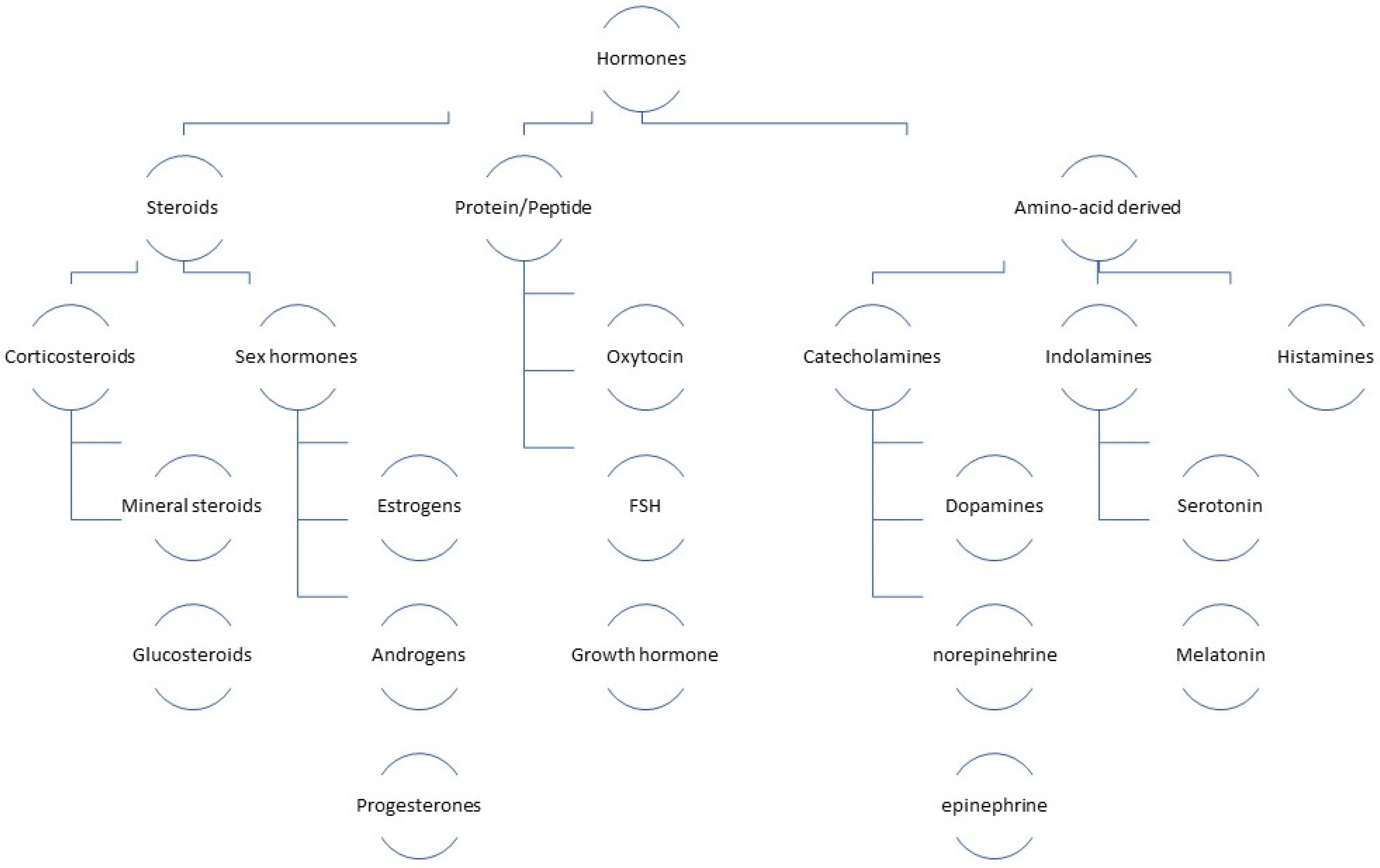
1.1. Classification of Hormones
Hormones can be categorized based on their chemical composition, mode of operation, functional characteristics, physiological consequences, and the stimulus they provide to endocrine glands as shown in Figure 2 [7]. Hormones can originate from lipid or peptide sources [14]. The structure of hormones discussed in this review are shown in Table 1, along with an example and reference of how their electrochemical detection is related to the hormone structure undergoing a redox change. Not all electrochemical hormone detection relies on a hormone undergoing oxidation or reduction but may be associated with a change in the amount or activity of a redox-active marker.
1.1.1. Steroid Hormones
Steroid hormones are a vital group of chemical compounds that play a crucial role in regulating numerous physiological activities. Thorough detection of steroid hormones can provide valuable insights into the physiopathologic mechanisms behind illnesses associated with these hormones. Identifying steroid hormones in biological samples poses significant challenges because of their limited endogenous levels and inadequate ionization efficiency. Steroid hormones play a crucial role as signaling molecules in various physiological activities, including transcriptional regulation, maintenance of secondary sexual characteristics, and coordination of the immunological and endocrine systems. Consequently, their significance to the human body is exceptional. Recent studies have demonstrated a direct correlation between irregularities in steroid hormone metabolism in humans and several ailments, such as endocrine disorders, breast cancer, prostate cancer, endometrial cancer, and cardiovascular disease. To comprehend the pathophysiology of different illnesses, it is imperative to use an analytical approach that is highly sensitive, stable, and trustworthy, enabling quantitative profiling of steroid hormones. The utilization of a research methodology that specifically targets the identification of a limited quantity of steroid hormones has the potential to introduce inaccuracies in the process of pathological characterization and the diagnosis of illnesses. Quantitative metabolic profiling of a broader range of steroid hormones not only enhances the comprehensive characterization of the steroid hormone metabolism network but also facilitates the provision of more precise and reliable diagnostic outcomes, hence reducing the occurrence of false positives or false negatives. Researchers encounter significant challenges and obstacles while investigating the metabolic network of steroid hormones due to the predominantly low concentrations of these hormones within the human body [15]. The amount of steroid hormones is an important part of investigating endocrinological disorders that affect how the adrenal glands or gonads work [16].
1.1.2. Peptide Hormones
Peptide hormones are a class of hormones composed of amino acid chains, which exert their primary physiological effects on the endocrine system. Hormones can be categorized into two systems, namely, amino acid-based or steroid-based, depending on their building units. Peptide hormones can exert their effects on target cells with the utilization of secondary messengers, owing to the inclusion of amino acids within their composition. There is a distinction between steroid hormones, which possess lipid solubility, enabling them to traverse the plasma membranes of target cells and exert their effects within the nuclei [17]. Peptide hormones play a crucial role as messengers within the signaling network that connects neurological coordination, endocrine glands, the gastrointestinal tract, and energy storage, hence governing the regulation of metabolism and feeding behavior [18]. They are known to play a crucial role in mediating endocrine transmission between the gonads and brain in vertebrates, hence regulating reproductive development. However, the impact of these molecules on reproductive growth in invertebrates remains relatively understudied [19].
1.1.3. Amino Acid-Derived Hormones
Regular hormone release is crucial for sustaining optimal health. The hypothalamus’s intrinsic pacemaker functions as the principal regulator of circadian periodicity, whereas several hormones exhibit oscillatory patterns with varying frequencies and amplitudes. The aforementioned rhythms are required to exhibit responsiveness to external stimuli, retain their resilience when confronted with significant disruptions, and convey pertinent data regarding the state of sound physiological operation [20].

Table 1.
Summary of hormone structures and their electrochemically detectable groups.
| Hormones | Structures of Hormones | Purpose/Function |
|---|---|---|
| Cortisol |  | The single-bonded keto group becomes oxidized during electrochemical detection [21]. |
| 17β-Estradiol |  | The irreversible oxidation of the hydroxyl group in the aromatic ring of the 17β-estradiol molecule is ascribed to a singular oxidation mechanism, resulting in the formation of its associated ketone derivative. The cause of this irreversible behavior is thought to be an electron transfer control mechanism used by CV and DPV to regulate the 17β-estradiol reaction on the electrode surface [22,23]. |
| Estriol |  | During the oxidation of estriol, a highly reactive phenoxy radical and a C=O were formed, corresponding to anodic peaks seen at LSV [24]. |
| Testosterone |  | The C-3 keto group of testosterone is first subjected to one-electron reduction, forming an unprotonated radical; then, those above radicals undergo protonation and then participate in a reaction with another radical, ultimately leading to the creation of a dimeric configuration [25]. |
| Progesterone |  | The P4 molecule undergoes a single electron reaction that reduces the C-3 keto group and has a larger positive electron density than the C-20 ketone group, making it simpler to acquire electrons for reduction [26]. |
| Melatonin |  | The process of electrooxidation of MT involves the loss of two electrons and a proton, resulting in the formation of an intermediate compound. This intermediate compound is susceptible to nucleophilic attack, leading to the formation of a derivative known as 4,7-dihydroxy indole. This derivative exhibits a pair of quasi-reversible redox peaks when compared with its quinone counterpart [27]. |
| Thyroxine |  | The first anodic peak attributed to the oxidation of hydroxyl (OH) groups present on the phenol of T4 and the first cathodic peak attributed to the reduction of iodine atoms, since the iodine atoms present on the phenol group of T4 exhibited significant reactivity. Additionally, the subsequent peaks observed to the oxidation and reduction products resulting from the first step [28]. |
| Oxytocin |  | The redox reaction attributed to the reaction involving a quinoic structure, which is one of the products resulting from the oxidation of phenolic OH of oxytocin [29]. |
| Insulin |  | Oxidation of amino acids in insulin [30]. |
Several methods have been used to determine the identity of hormones. These include high-performance liquid chromatography (HPLC), gas chromatography/mass spectrometry (GC-MS), and high-performance liquid chromatography/mass spectrometry (HPLC-MS) [9]. Tests for estrogens are often conducted using tried-and-true analytical methods such as gas chromatography, HPLC, MS, and immunoassays. HPLC and GC methods are typically highly automated, exceedingly sensitive, and extremely specific, making them ideal for the quantification and identification of a wide range of species at trace levels. These methods offer sensitive and accurate detection, but are impractical due to their high cost, requirement of specially trained operators, extensive time required between sample collection and analysis, and inability to be used in the field [31]. Electrochemical sensing techniques present a comparatively more streamlined and expeditious approach for hormone analysis in comparison with methodologies such as high-performance liquid chromatography/tandem mass spectrometry (LC-MS/MS) [4] because of their low cost, rapid response, high sensitivity, and user-friendliness. These techniques also do not require specialized tools or expert technicians. Electrochemical techniques possess notable benefits over alternative approaches in hormone detection, including cost-effectiveness, heightened sensitivity, rapid response, and operational simplicity. Moreover, these methodologies do not necessitate the use of costly apparatuses or highly skilled personnel [9]. Enzymes or antibodies are often used as biorecognition elements in electrochemical biosensors to detect hormones. Electrochemical biosensors work on the basic idea that specific molecules undergo reactions with target substances and produce a product that can be oxidized or reduced on an electrode surface. There are a few problems that biosensing systems have to deal with: enzymes have a limited lifespan, biosensors sometimes need extra treatments before each use, and some of the sensors we have now do not give a steady response over time [4]. Electroanalytical techniques offer several advantages in the field of analytical chemistry. These techniques demonstrate high selectivity and sensitivity toward electroactive species, allowing for the precise detection of trace amounts of analytes. Additionally, they possess a wide linear range, enabling the quantification of analytes across a broad concentration range. Furthermore, electroanalytical techniques exhibit very low detection limits, ensuring the detection of even minute quantities of analytes. Moreover, the portability and affordability of instruments used in these techniques make them a practical and cost-effective option for routine analysis of various analytes in diverse sample media [30].
2. Electrochemical Methods for the Detection of Hormones
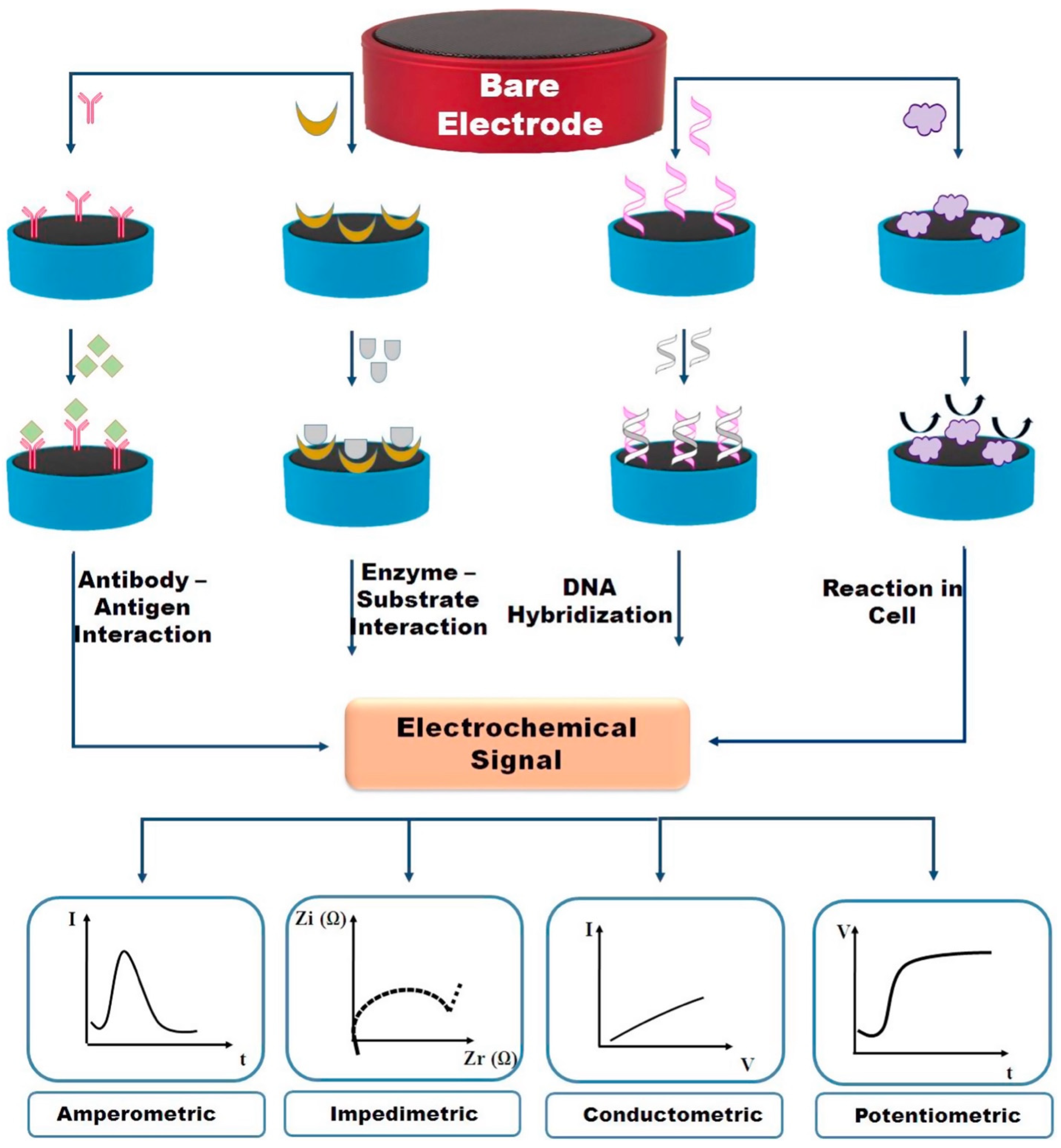
High-performance liquid chromatography, fluorescence and calorimetric tests, spectrometry, enzyme-linked immunosorbent assay, surface plasmon resonance (SPR), and electrochemical methods are among the analytical techniques commonly used for the assessment of purity and efficacy. Except for electrochemical methods, all treatments require demanding and time-consuming steps, specialized equipment, and skilled operators. Many pharmaceutical compounds have electrochemical activity, rendering electrochemical methods capable of detecting and differentiating them, even within biological fluids. Electrochemical technologies possess several notable advantages, including their compact dimensions, rapid response time, heightened sensitivity, cost-effectiveness, extended linear detection range, and exceptional selectivity; these options are favored over other choices, as deemed suitable [32]. The main advantages of electrochemical detection methods are their low detection threshold, high accuracy, fast reaction times, and easy system incorporation [33]. Due to their sensitivity, affordability, and intrinsic compactness, electrochemical detection technologies have drawn a lot of interest. Simple devices and affordable electrodes can be combined to make quick measurements in portable devices that are easy to use and small. Electrochemical biosensors’ relatively straightforward nature is one of their significant benefits. One of the best things about electrochemical biosensors is how easy they are to use. Simple electronics can be easily put together with cheap electrodes to make portable systems that are small and easy to use [34]. Figure 3 illustrates the structure and components of an electrochemical biosensor and different electrochemical methods [9].
Potentiometry is a method used to determine the electrical potential of a two-electrode system when there is no current flowing between the electrodes. The solution composition remains unaltered. Ion-selective electrodes (ISEs) are highly favored potentiometric sensors that have found extensive application in the assessment of ion activity in several types of samples, for example, those of biological, chemical, and environmental nature. These electrodes are recognized for their simplicity and effectiveness in this regard. Ion-selective electrodes (ISEs) provide the capability to selectively identify ions in the presence of other chemicals, making them a cost-effective platform. Potentiometry is widely recognized for its utility in the determination of pH levels in various liquids. Moreover, the utilization of potentiometric methods for trace analysis of metals has been widely used in environmental studies [35]. Amperometry is a technique used to quantify the current variations versus time due to oxidation or reduction of an indicator while maintaining a continuous potential relative to a reference electrode. Chemical compounds may undergo either oxidation or reduction processes when exposed to inert electrodes under conditions of constant potential in a suitable range. Amperometry typically offers enhanced sensitivity in terms of detection limits, albeit with the limitation of being applicable just to electroactive species, and systems are commonly used in enzyme-based sensors, wherein the enzyme facilitates a redox process [35].
Electrodeposition for forming nanomaterials on the surface of a working electrode can be provided using chronoamperometry (CA) [36]. The electrochemical technique of chronoamperometry also finds widespread application in enzymatic biosensors. Electrochemical analyses are simplified by the constant potential configuration because enzymes catalyze only certain redox reactions. The sensor activity can be measured using CA [37]. The effective explanation of a chronoamperogram requires a comprehensive depiction of the concentration profiles of all species present in the solution relative to the working electrode surface. The Cottrell equation predicts the occurrence of a diffusion-only line, which eventually reaches a steady-state current as time progresses. The two sets of graphs exhibit significant differences, particularly in terms of the involvement of both Faradic and non-Faradic processes. It is evident that the bulk of the current may be attributed to non-Faradaic effects. The Faradaic current’s behavior exhibits a notable level of detail, which may be ascribed to the small delay in the electric field’s reaction near the electrode when there is a modification in the applied potential. The observed substantial rise in Faradaic current may be attributed to the enhanced favorability of the potential driving force for electrolysis, which occurs when a more negatively charged double layer is formed. At significantly extended durations, the rate of electron transfer becomes rapid enough to the extent that the current is only constrained by the transport of the electroactive species. In the context of this limit, the current exhibits a diminishing trend over time due to the expansion of the depletion layer away from the electrode. In the scenario when the electroactive species is in an uncharged state, the observed behavior of current over time aligns precisely with the predictions made using a diffusion-only model. Nevertheless, in the case where the species carries a negative charge, there is an observed increase in current when compared with the diffusion-only model over extended periods of time. This may be attributed to the flow of an anion down the positive potential gradient toward the electrode, which provides an extra contribution to the overall mass transport [38].
Voltammetry is a technique used to determine the current generated by an electrochemical cell, which is then measured in relation to a potential that varies with time. In the field of voltammetry, it is possible to manipulate the applied potential and afterward measure the resulting current over a specified duration. In the context of voltammetric measurements, it is common practice to use a three-electrode system. Voltammetric methods can be categorized into several subtypes, including cyclic voltammetry (CV), alternating current voltammetry (ACV), differential pulse voltammetry (DPV), linear sweep voltammetry (LSV), square wave voltammetry (SWV), anodic stripping voltammetry (ASW), and cathodic stripping voltammetry (CSW). Voltammetric measures primarily yield qualitative and quantitative information regarding both direct and indirect redox reactions [35]. Voltammetry is widely recognized as a prominent technology used in the field of electrochemical immunosensors, and such approaches are used to manipulate the decay rates of both the charging and Faradaic currents using various waveforms to scan the potential. The enhanced proportion of Faradaic current relative to non-Faradaic current facilitates a reduction in the limit of detection and an increase in sensitivity for both reversible and irreversible events. In comparison with the impedance method, voltammetric methods require less expensive equipment [39].
The potentiostat, which is the most widely used instrument in electrochemical analysis, is frequently utilized for the acquisition of CV data. In this method, a composite working electrode, integrating both a reference electrode and a counter electrode, is used. The application of the source of voltage for the potential scan occurs in the region between the counter electrode and the working electrode. To maintain the appropriate potential at the working electrode relative to the reference electrode, the overall voltage is adjusted after the measurement of the potential between the reference electrode and the working electrode utilizing a voltmeter. The term “scan rate” refers to the linear rate at which the potential is scanned in this particular scenario [40]. For example, 3-thiophene acetic acid and palladium nanoparticles (Pd NPs) formed the basis of a new type of electrochemical sensor for progesterone detection, which was then characterized with CV. Palladium nanoparticles (Pd NPs) were used as a cross-linking agent to fabricate a three-dimensional network film exhibiting remarkable conductivity. This enhanced conductivity was due to improved interparticle charge transfer [41].
Differential pulse voltammetry (DPV) is another electroanalytical technique used in biosensor development. DPV is the utilization of linear sweep voltammetry, wherein a linear potential sweep is accompanied by a sequence of regular voltage step pulses. The measurement of the current is conducted over a short time window immediately prior to the upward (i1) and downward (i2) steps of a pulse and then the difference between these two currents (i2–i1) is recorded. Therefore, by minimizing the contribution of the charging current, it is possible to attain a heightened level of sensitivity [42]. DPV responses are assessed using varying doses of hormone according to the ideal experimental circumstances. It is evident that the peak currents of DPV displayed an upward trend in conjunction with the rise in thyroid-stimulating hormone concentration. A strong linear correlation may exist between the peak currents observed in DPV and the logarithmic values of the concentrations of the hormone under investigation, as observed for a sensor detecting thyroid stimulating hormone [43]. The findings of one study show that DPV has greater sensitivity than CV in determining the concentration of parathyroid hormone with an LOD of 0.17 pM and 0.33 pM for DPV and CV, respectively [35]. A progressive decline in the peak current was seen as the amount of hormone increased in both the DPV and CV methods. The observed phenomenon involves a reduction in the number of binding sites present on the molecular layer that recognizes the PTH molecule. This drop may be attributed to an increase in steric hindrance on the surface, generated by the binding of parathyroid hormone (PTH). Consequently, electron transport is suppressed because of these combined effects. A correlation was established between the peak current and the concentration for PTH, indicating a dynamic linear relationship [34,44].
Electrochemical impedance spectroscopy (EIS) is a technique used to examine the response of an electrochemical system to an alternate current (AC) as it varies with frequency. The analysis of EIS data involves examining the variations in current response to an oscillating potential at a specific series of frequencies over a wide range, enabling the determination of the electrochemical characteristics of the system. EIS, a technique known for its responsiveness and non-destructive nature, has found extensive application in several domains including corrosion, protective coatings, conductors, fuel cells, batteries, electrocatalytic reactions, and the study of interfacial parameters of biosensors. The elimination of the requirement for labeled electroactive groups or indicators is the primary distinguishing characteristic of the EIS approach. However, it is important to note that EIS should be used in conjunction with other analytical approaches to fully comprehend interfacial properties [35]. The utilization of surface-modified electrodes presents several advantages when using the EIS approach. The impedance spectra represented as a Nyquist plot usually exhibit two distinct regions, namely, a semicircular segment and a linear segment. The linear portion of the graph represents the diffusion-limited phase of the electrochemical process, while the semicircular region corresponds to the electron transfer resistance (Rct). The rate at which electron transfer occurs for an added redox probe is governed by the charge transfer resistance (Rct) at the electrode surface [45]. Impedance measurements are often conducted under open circuit potential conditions. The acquisition of each impedance spectrum typically requires a duration of around 3 to 4 min [46]. The utilization of EIS proves to be quite advantageous in the evaluation and analysis of interfacial properties pertaining to surface-modified electrodes. Impedance data are usually treated using two distinct sorts of graphs, which are the Bode and Nyquist plots [32]. Nyquist plots were obtained with EIS to investigate the interfacial characteristics of an electrode, including electrocatalytic and conductivity capabilities, both before and after changes. The [Fe(CN)6]3−/4− redox probe was used for this purpose [34]. There is a correlation between the diffusion-limited process and the semicircular region of the EIS curve [47].
Square-wave voltammetry is a pulse-based electrochemical technique that possesses the notable advantages of rapid analysis and high sensitivity for both reversible and irreversible electrochemical reactions [48]. The electrode potential is modified by SWV with the utilization of a distinct waveform composed of a square wave combined with staircase potential, serving to regulate the decay rates of both the charging and Faradaic currents. The enhanced proportion of Faradaic to non-Faradaic current results in a reduced limit of detection and heightened sensitivity in both reversible and irreversible events. The primary variables associated with SWV are prepotential, start potential, final potential, time at pre-potential, pulse period, pulse amplitude, pulse frequency, and potential step. SWV is considered to possess a superior sensitivity [49]. The utilization of SWV is deemed highly suitable for the electroanalysis of species that have been adsorbed onto the electrode surface [50].
3. Electrodes for Hormone Detection
The selection of an appropriate electrode is of paramount importance in analyte detection, notwithstanding the numerous advantages offered by electrochemical methods. For example, the utilization of bare electrodes is commonly linked to electrode fouling, low selectivity, inadequate repeatability, elevated overpotential, and slow electrode kinetics. To mitigate these limitations, it is common practice to improve the catalytic activity of electrodes by including noble metals such as palladium (Pd), platinum (Pt), gold (Au), and ruthenium (Ru), which not only improve conductivity but also augment catalytic performance. The implementation of modifications serves to mitigate the development of fouling layers [32]. Microelectrodes are also useful for electrochemical sensors because of their fast mass transfer, low electroactive area, low interfacial resistance, and low ohmic drop at the surface of the electrode due to radial diffusion. The total enhanced surface area is useful to obtain improved ranges of detection, a better signal-to-noise ratio, and a wider dynamic range. However, due to a weak response from the analyte, these microelectrodes can only be used to test for a certain range of hormones. Strategies for changing electrode platforms are very important for improving our ability to analyze and identify hormones. Nanostructured electrodes with a high surface area have often been shown to be amongst the best platforms for electrochemical biosensors [34].
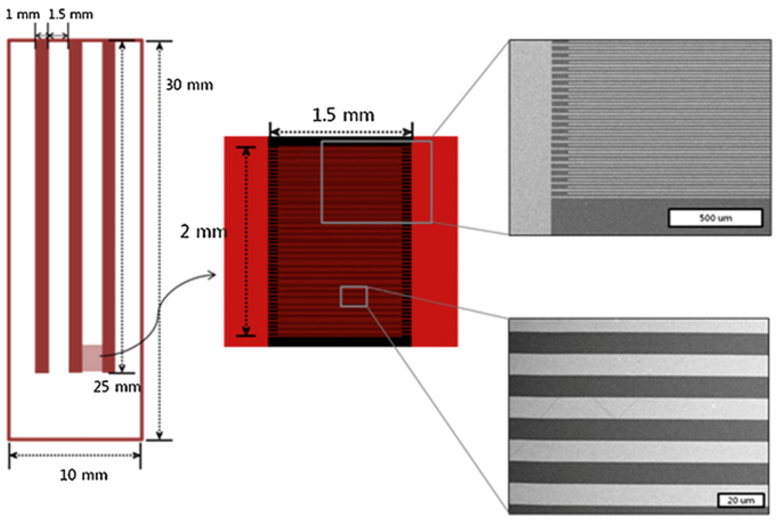
3.1. Interdigitated Array Electrodes
The interdigitated electrode array (IDA) as shown in Figure 4 was made by using a standard photolithographic method to make gold designs on a silicon (Si) chip that was treated with boron to give it a resistance of about 1 to 30 Ω cm. Before depositing the gold, an oxide layer 100 nm thick was formed on the Si base to stop any leaky current from going through the Si chip. The oxidized Si wafer was then treated with LORTM (lift-off resist) and positive photoresist that was a few hundred nanometers thick. It was then soft baked, exposed to UV light via a chrome mask with negative IDA electrode patterns that were lined up, and developed in MIF solution. The area of photoresist that was exposed to UV light became easy to dissolve during development, making it possible for the negative pattern in the IDA electrode structure to be seen on the Si chip base after the lithographic process. A binding layer of 5 nm thick Cr on top of the printed Si base and a layer of 50 nm thick platinum or gold was then placed on top of these. The IDA electrode on the Si substrate was completed using the standard lift-off process to get rid of the last few resists [51]. Since electrochemical assay measurements are homogeneous, they are not impacted by dispersion in bulk solution, and the basic equipment can be downsized to make detection in tiny volumes more accessible. Ino et al. used two-band microelectrodes for the detection of an enzyme produced by cells; however, IDA electrodes and a reversible redox couple can provide additional amplification. The metal fingers of an IDA electrode are flat, parallel, and arranged in two interdigitated comb arrays. Molecules oxidized at an electrode finger of one array can be reduced at the nearby fingers of the other array, making the molecule available for a subsequent reoxidation, thanks to the independent regulation of the potential of each array. The electrochemical responses are improved by the redox cycling across the arrays. To maximize the signal strength, the electrode finger width and the spacing between them are critical. Electrochemistry coupled with spectroscopy, dielectrophoretic cell separation, and the use of IDA electrodes with biological substances are all examples of applications. Electrochemical monitoring has been used to track the activities of enzymes such as glucose oxidase and glutamate oxidase immobilized in hydrogels, urease immobilized in a sol-gel, and alkaline phosphatase. A homogeneous competitive test for hormones was connected with an IDA electrode to dual-electrode voltammetry [52].
3.2. Screen-Printed Electrodes
The usage of screen-printed electrodes (SPEs) has allowed for the mass manufacture of disposable sensors that are both cheap, remarkably repeatable, and dependable. Different inks can be printed on various substrates including circuit boards, plastic, or ceramic to create SPEs. The analytical procedure’s needed selectivity and sensitivity are based on the chemical makeup of the various printing inks. Carbonaceous materials (carbon black (CB), graphene (GR), carbon nanotubes (CNTs), etc., are just a few examples of the types of nanomaterials that have been successfully used in SPE surface modifications to enhance analytical features and performance. Graphite working/counter/reference electrodes and an Ag/AgCl reference electrode make up the SPEs in use. Graphite is a working electrode surface with an external counter and reference electrodes used for uniformity [53]. The utilization of screen-printed carbon electrodes (SPCEs) is crucial in the progress of recyclable biosensors and electrochemical sensors because of their advantageous surface features, straightforward manufacturing procedure, cost-effectiveness, environmentally friendly nature, and production suitability for large-scale manufacturing [26].
3.3. Gold Electrodes
Gold’s versatility as a substrate for thiol conjugation chemistry has led to its extensive adoption in sensor development, where it is used to tailor surface functionality for a wide range of chemical and biosensing applications. Fabricating and characterizing gold or gold nanostructure-modified electrodes is a crucial first step in creating reproducible and resilient thiol conjugated surfaces, which are necessary for accurate sensor performance. The most common metals for solid electrodes in the fields of electrochemistry and electroanalysis are gold and platinum. This is due to the metals’ high quality, ease of use in manufacturing, and inertness in the presence of almost any chemical. Two major applications are driving the rise in demand for gold electrodes: stripping analysis and research into surface changes via self-assembly. A gold working electrode is used for numerous voltammetric procedures, while a platinum counter electrode and silver/silver chloride (Ag/AgCl) reference electrode complete the standard three-electrode electrochemical setup [54].
3.4. Glassy Carbon Electrode
Glassy carbon electrodes (GCEs) have been extensively used in numerous electrochemical investigations due to their affordability, wide potential range, and ease of surface modification. There exist many techniques for preparing and enhancing a GCE’s surface prior to utilization, including mechanical polishing, ultrasonication, and electrochemical treatments. The electrochemical treatment approach is widely utilized due to its straightforward operation, resulting in the formation of a nanoporous coating on the GCE with a significantly increased effective surface area. The proposed technique exhibits potential for utilization as a surface modification method for GCE due to its remarkable reproducibility and efficiency [45]. Glassy carbon is extensively utilized in the field of electrochemistry owing to its exceptional characteristics, including excellent tolerance to elevated temperatures, low electrical resistance, low density, and hardness. Glassy carbon (GC) is a variety of non-graphitic carbon that is produced with the process of pyrolysis of specific polymeric precursors at a temperature exceeding 2000 °C. The microstructures of graphene composite consist of separate pieces characterized by curved carbon planes, resembling nanoparticles with imperfections like those found in fullerene structures. Under these conditions, the resulting graphene structure manifests as a network comprising stacked ribbon-like molecules that resemble graphite. The utilization of GC as an electrode material for electroanalysis is prevalent due to its characteristics of high hardness, low reactivity, impermeability, and superior electrical conductivity and impermeability [55].
3.5. BDD Electrodes
Boron-doped diamond, a novel and highly promising solid material has many applications in the field of electrochemistry. The application of this material is prevalent in the examination of pharmaceuticals and their metabolites, bioactive compounds, metallic ions, and organic contaminants [56]. Researchers can produce BDD electrodes using either home-built systems or commercial growth systems such as hot filament (HF), plasma chemical vapor deposition (CVD) reactors, and microwave (MW) reactors. Key factors that are important when evaluating the electrochemical behavior include the boron content, surface morphology, surface termination, surface polish, and non-diamond-carbon (NDC) presence despite the source [57]. It has been used as a viable substitute for high-performance electrodes in electrochemical investigations due to its inert nature, hardness, elevated thermal conductivity, and electrical conductivity. Furthermore, the BDD electrode is often favored in this domain because of its notable characteristics, including a decreased background current, a wide electrochemical potential range, and exceptional resilience and longevity in both alkaline and acidic environments. Additionally, the BDD electrode has several advantages in comparison with traditional solid electrodes, including enhanced hardness, durability, and optical characteristics, and improved response parameters, such as time, stability, and precision. In the realm of electrochemical investigations, it is imperative to ensure the cleanliness or activation of the exterior of the BDD electrode. This is necessary to achieve consistent and highly responsive outcomes on the electrode’s outermost layer, a requirement shared by other solid electrodes. The presence of impurities on the electrode surface might result in either a reduction or enhancement in the strength of the signal used for analysis. Consequently, this leads to inaccuracies in the outcomes of the analysis. Because of the high sensitivity of the BDD electrode surface, it is imperative to exercise caution and use a gentle mechanical cleaning procedure. The cleaning procedure described in this study involves the activation of an electrode that has been rendered free from surface contamination. This activation process occurs in various solutions, with the specific potential applied to the electrode being dependent on the prevailing working conditions. In the existing body of literature, various studies have been conducted to investigate the cleaning and activation of the BDD electrode. These studies used the application of potentials in both the anodic and cathodic directions, albeit in different solutions [58]. Boron-doped diamond (BDD) electrodes, when subjected to varying levels of boron doping, exhibit distinct variations in binding energies and carrier densities, which have significant implications for their utilization in semiconductor applications. The aforementioned features have a notable impact on the present state and oxidation peak potential of the substance, hence influencing the overall effectiveness of analyte detection [59].
3.6. Fused Deposition Modeling
To fabricate electrodes using fused deposition modeling (FDM), a combination of carbon-based conductive particles and thermoplastic materials is used to create conductive filaments. In the recent literature, there have been reports on various conductive polymeric filaments, including polylactic acid (PLA)–graphene filament, carbon nanofiber–graphite–polystyrene, PLA-carbon black (PLA-CB), polybutylene terephthalate–carbon nanotube–graphene, polypropylene-CB, and acrylonitrile butadiene styrene (ABS)-CB. The PLA-CB filament has received significant attention in recent research endeavors due to its use in the practical fabrication of printable electrodes that can be utilized in electroanalytical applications. The utilization of printed electrodes has demonstrated encouraging outcomes in the identification and quantification of biomolecular species. Up to the present time, there has been an absence of exploration of the utilization of 3D-printed electrodes for the purpose of hormone analysis [13]. For electrochemical hormone determination, a wide variety of carbon electrodes, including glassy carbon (GCE), pencil graphite (PGE), edge plane pyrolytic graphite (EPPGE), carbon paste (CPE), and screen-printed carbon (SPCE), have been utilized widely [4].
4. Nanostructures for the Detection of Hormones
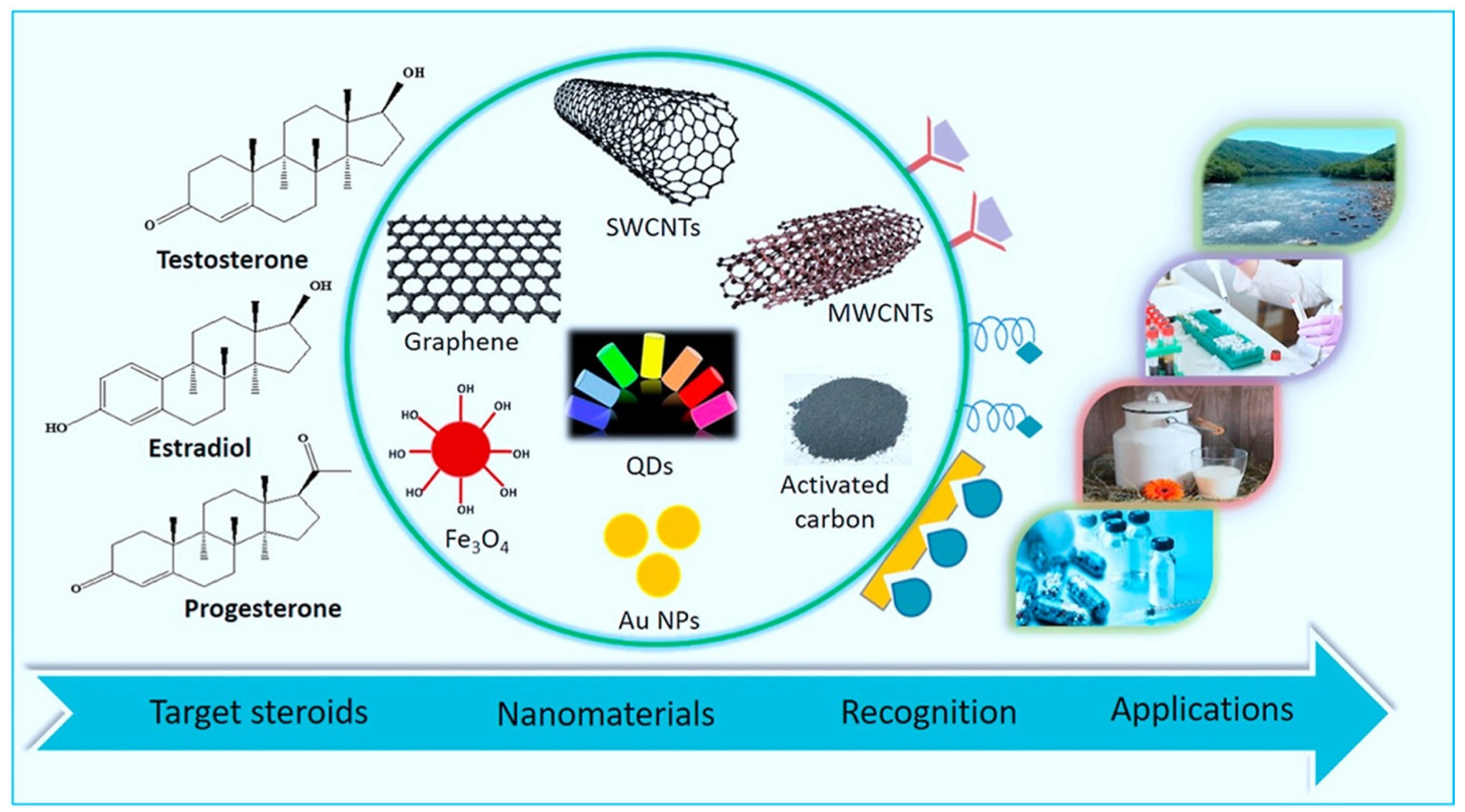
The scientific community has increasingly shown interest in the advantages of various nanomaterials for a broad range of biosensor functions [60]. Nanomaterials have garnered considerable attention for their capacity to augment catalytic processes and their substantial surface area, rendering them promising candidates for deployment in sensors and several other technological devices [24]. Numerous nanomaterials have been produced, modified, created, and investigated within the realm of electrochemical biosensors in order to leverage their exceptional intrinsic benefits and achieve heightened sensitivity and selectivity [61]. Figure 5 shows the usage of various nanomaterials along with biorecognition elements for detecting steroidal hormones [62].
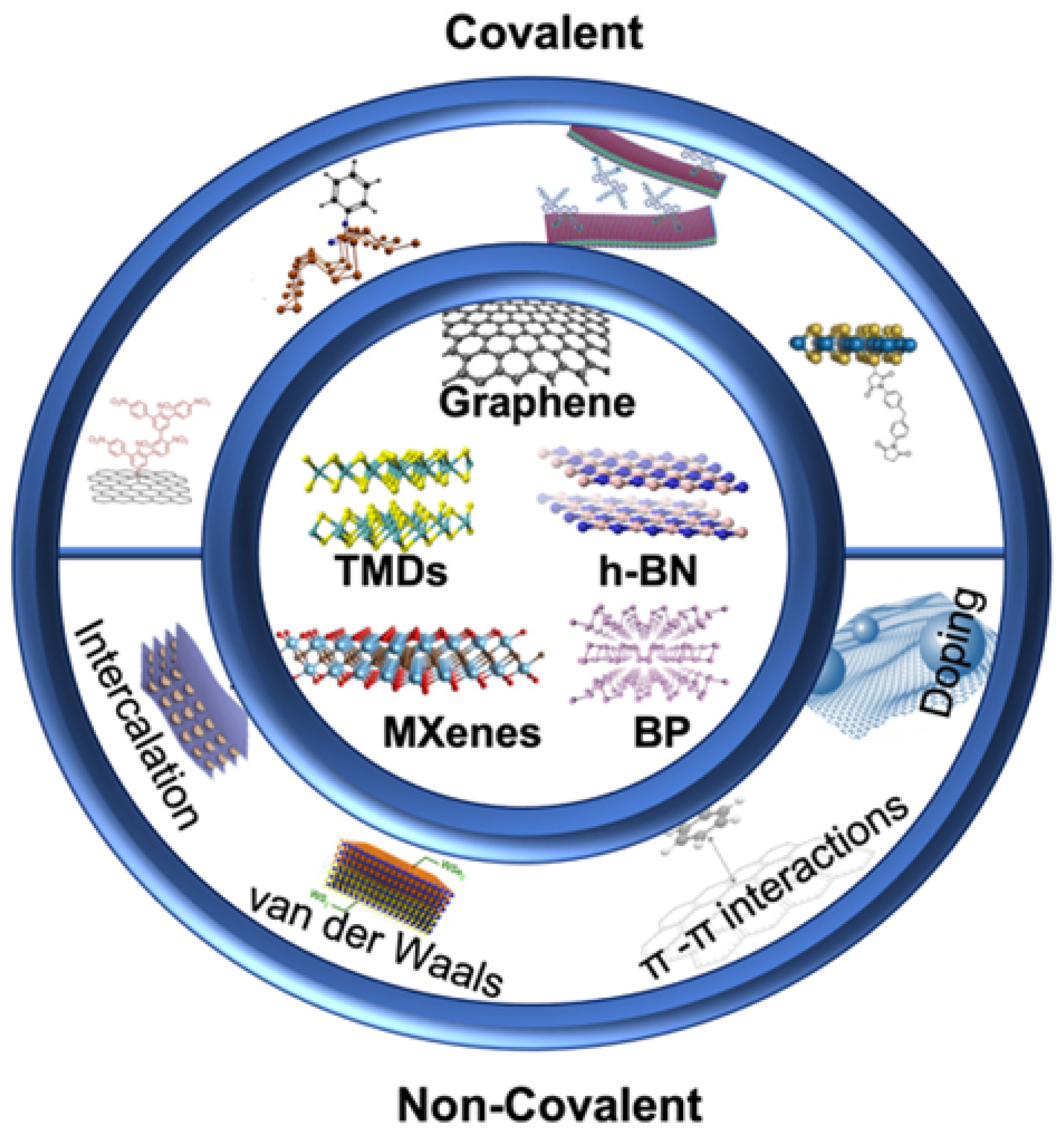
4.1. Two-Dimensional (2D) Nanomaterials
Of all the nanomaterials, two-dimensional (2D) nanomaterials are among the newest and most interesting. Since Andre Geim and Konstantin Novoselov found graphene in 2004, other materials like boron nitride (BN), graphite carbon nitride (g-C3N4), transition metal dichalcogenides (TMDs, like MoS2 and WS2), transition metal oxides (like MoO3, WO3, and MnO2), MXenes, silicene, germanene (2D germanium), hexagonal boron nitride, borophene, and black phosphorus have been found as depicted in Figure 6. So far, 2D nanoparticles and their nanocomposites have shown great chemical, physical, electronic, and optical characteristics. These qualities make them useful for many applications, like catalysis, drug delivery, antibacterial, therapy, and bioimaging [63,64,65].
4.1.1. Graphene
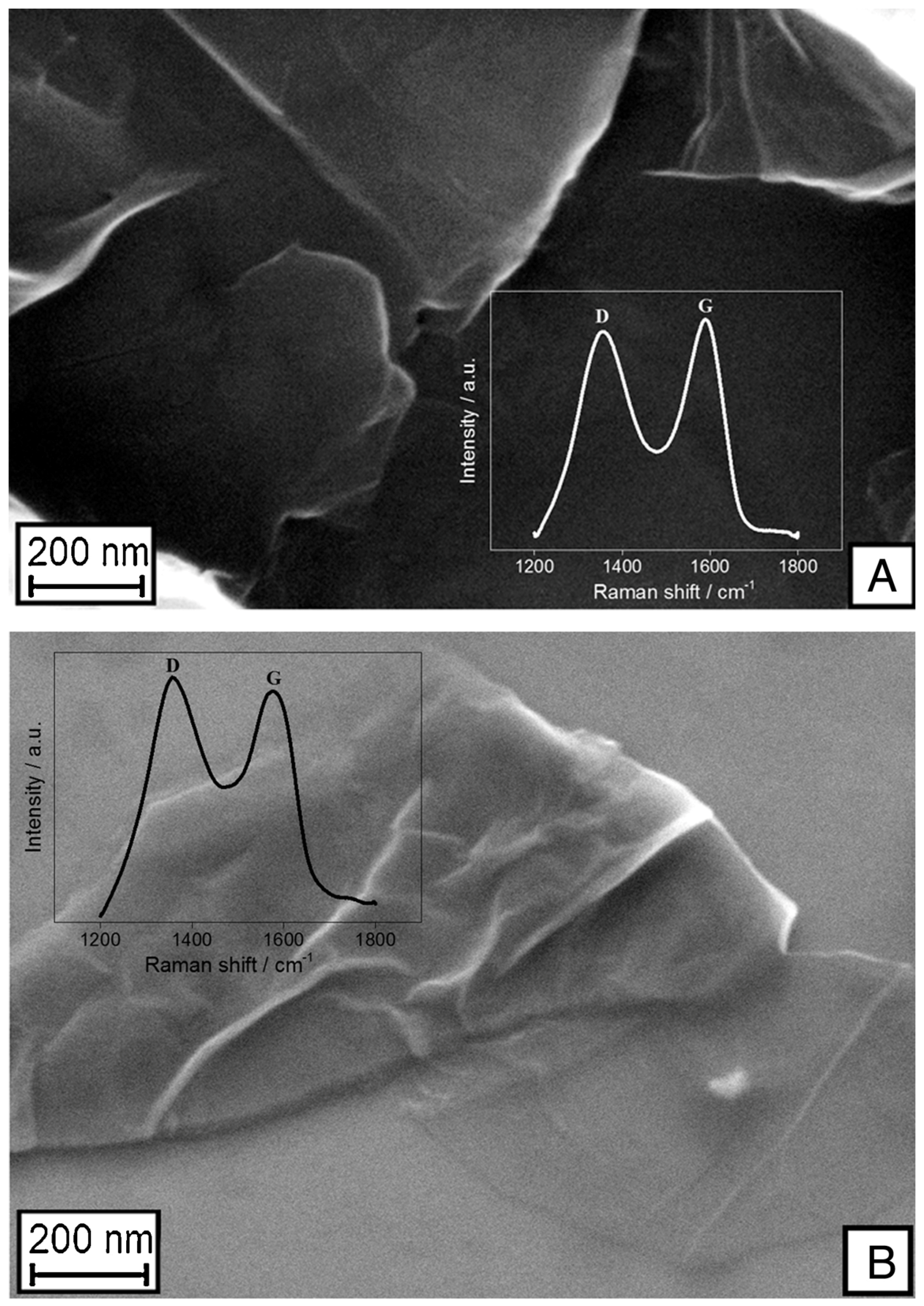
Since its initial identification in 2004, graphene has sparked a significant surge in attention within the realm of electrochemistry owing to its remarkable electronic transport characteristics, electrocatalytic capabilities, and expansive surface area. Graphene, akin to carbon nanotubes (CNTs), can also serve as a substrate that can be subjected to various modifications with distinct species [66]. It is a special two-dimensional (2D) material made of carbon atom monolayers and has a honeycomb-like structure. Due to its novel physical and chemical properties, it has attracted attention since its introduction in 2004. The oxidized form graphene oxide (GO) has a useful surface chemistry because of the existence of oxygen groups that permit functionalization [24]. SEM images of graphene oxide and the reduced form of graphene oxide are should in Figure 7. Research has been conducted on the utilization of graphene as an electrode material in the realm of high-sensitivity detection of biological molecules. This interest stems from graphene’s remarkable catalytic activity, electrical conductivity, and intrinsic physicochemical features. Graphene nano-sheets experience restacking due to robust π–π interactions and van der Waals attractive forces among them. Consequently, the layered structure of graphite is partially restored, resulting in a reduced specific surface area of graphene when compared with its theoretical value [67]. Graphene oxide magnetic nanocomposites were used as electrodes for the loading of bioreceptors. The sensitivity of these sensors was improved with the large increase in electrically conductive surface area [68].
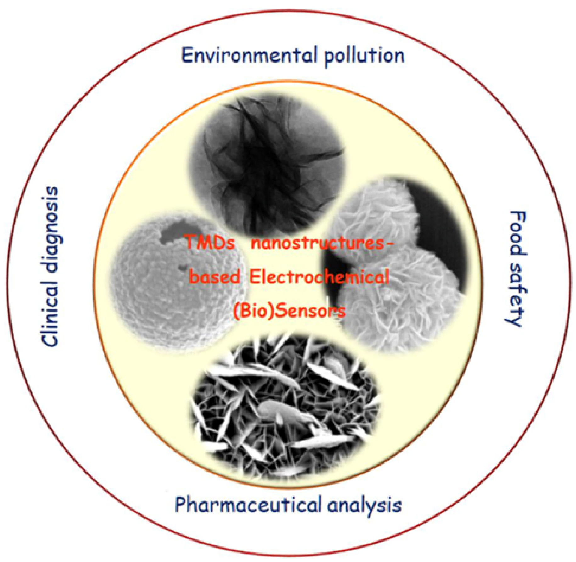
4.1.2. Transition Metal Dichalcogenides (TMDs)
Since the discovery of graphene in 2004, research on two-dimensional (2D) materials has increased exponentially. Transition metal dichalcogenides (TMDs) as shown in Figure 8 belong to a category of 2D materials in which the bonding between individual covalently bound X–M–X layers (where M represents the transition metal and X represents the chalcogen) is facilitated by weak van der Waals forces. This characteristic enables the synthesis of these materials with precise control over the number of layers. Single-layer TMDs exhibit a noteworthy shift from indirect to direct band gaps, accompanied by intriguing optical and electrical characteristics. The presence of limited layer-dependent opto-electronic characteristics and band gaps render single-layer TMDs more advantageous compared with graphene, hence facilitating its use in many domains [69]. The commendable characteristics of TMDs include their remarkable capacity for charge transfer, their substantial surface-to-volume ratio (S/V), the tunability of their energy band gap based on the number of layers, their significant interaction with light, and their mechanical resilience. Additionally, these resources are both economically efficient and readily available. The distinctive characteristics, structural properties, and current use of nanostructured TMDs render them very suitable for producing electrochemical biosensors. The commonly recognized 2D TMDs include tungsten diselenide (WSe2), molybdenum disulfide (MoS2), tungsten disulfide (WS2), and molybdenum diselenide (MoSe2) [64,70]. For example, molybdenum diselenide (MoSe2) was used to make a sensor for 17β-estradiol. MoSe2 nanosheets mixed with carbon aerogel spheres (MoSe2-CA sensor) using a chemical method worked very well with 17β-estradiol, detecting as little as 0.2 pM in a concentration range of 5 pM to 5 nM. The RSDs of 1.7% and 3.1% found in two sets of six detection tests showed that the MoSe2-CA sensor was very reliable. Its stability was also shown by the fact that it retained 94.8% of its initial current response after being stored at a low temperature. It was found that carbon aerogel not only kept MoSe2 from sticking together and improved electron transfer, but it also made the efficiency of composites more stable, all of which were important for the 17β-estradiol detection application [71].
4.1.3. Mxenes
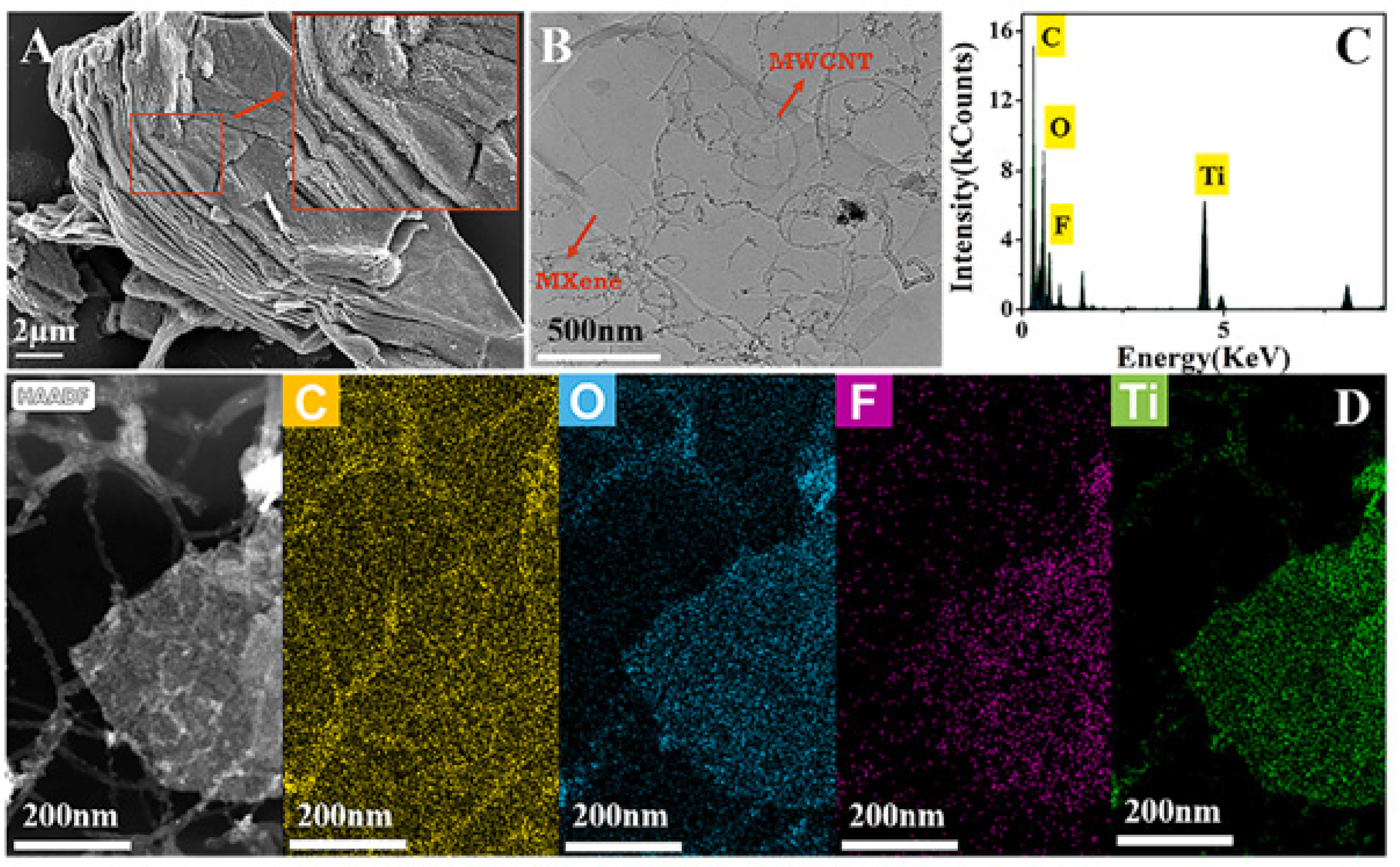
MXene (transition metal carbides and nitrides) have been the subject of intensive study because they are 2D materials. Mn+1XnTx is the chemical formula used for MXene. M can be any early transition metal, like Mo, V, Nb, or Ti. X can be N or C, and n can be any value from 1 to 3. Tx stands for different surface endings, which can be F, O, or OH groups. MXene has special physical and chemical features, including the ability to carry large amounts of charge and conduct electricity, and have good mechanical qualities. In addition to these traits, one of the attractive features of MXene is that it is biocompatible. This makes it a promising material for designing advanced biosensor systems [72]. SEM, TEM and EDS data for a MXene-MWCNT composite are shown in Figure 9. Due to its unique layered structure, high conductivity, high surface area, high thermal stability, and low environmental impact, MXenes have recently attracted a lot of scientific interest for use in sensing, catalysis, electronics, and energy storage. It is well-suited for the development of rapid-performance electrochemical biosensors due to its great hydrophilicity from surface functional groups, high electrical conductivity, and exceptional ion intercalation behavior. Recent years have seen the development of high-performance materials in the form of ultrathin MXenes for stretchy and flexible conductive coverings [73,74]. A new reusable electrochemical impedimetric immunosensor using a Ti3C2Tx MXene-loaded laser-burned graphene (LBG) flakes 3D electrode network and PDMS was developed to detect cortisol in human sweat without touching the person. The sensor has a microfluidic path and chamber. The sensor was put on the skin to collect sweat and sweat moved through the tube to the detection chamber with its own weight. The Ti3C2Tx MXene/LBG/PDMS-based patch cortisol immunosensor worked well with linearity and a LOD of 0.01–100 nM and 88 pM, respectively. It also worked well for the detection of cortisol at the point of care [75].
4.1.4. Black Phosphorous
BP is a semiconductor material characterized by a layered structure; in which discrete atomic layers are assembled using van der Waals forces. The formation of a puckered honeycomb structure occurs because each phosphorus atom is covalently bonded to three neighboring phosphorus atoms. BP has a direct bandgap, notable wrinkled structure, elevated hole mobility, and good mechanical, electrical, and optical characteristics, making it a material with considerable potential for several applications. However, the limited use of this substance in many industries is mostly attributed to its inherent instability and rapid chemical deterioration when exposed to typical environmental conditions. In recent times, BP has been used in several domains including sensing, energy storage devices, photocatalysis, and medicinal applications. The commonly used method for synthesizing few-layer BP involves liquid-phase exfoliation followed by chemical vapor deposition (CVD) [65]. BP has garnered significant attention as a novel nanomaterial that presents the establishment of a unique immunofiltration strip approach that utilizes temperature as the readout signal, leveraging the photothermal impact of BP nanosheets. Using an indirect competitive approach, this method offered a straightforward, expeditious, highly responsive, and cost-effective framework for the identification of 17β-estradiol, an endocrine-disrupting chemical often found in ambient water or food samples. A negative correlation was shown between the content of 17β-estradiol in the sample and the binding of BP nanosheets to the strip surface. Additionally, a decrease in temperature variation was observed when the sample was subjected to intense laser irradiation. A detection limit of 0.104 ng mL−1 was attained under optimal circumstances. The test’s practicality was evaluated using a standard addition procedure in samples of water and milk. The results demonstrated satisfactory performance and suggested that the assay has potential usefulness for convenient, cost-effective, and direct monitoring of 17β-estradiol [76].
4.1.5. Two-Dimensional Metal–Organic Frameworks
Most of the current research in the field of synthesizing metal–organic frameworks (MOFs) in two-dimensional (2D) structures has been primarily concerned with augmenting the advantages of porous materials with the amplification of the exposed surface area. There have been reports on the synthesis procedures and applications of 2D MOFs in energy-related research, which has generated significant interest. The secondary building units (SBUs) consist of metal-containing nodes and organic linkers that serve as bridges. These components combine to produce ordered network structures with large pore volumes and surface areas in crystal formations, which may be one, two, or three-dimensional in nature. There are several techniques available for the integration of 2D conductive MOFs into devices. One such approach involves the use of drop casting, where an MOF is suspended in a solvent and then placed as a coating onto the desired substrate. The framework architectures, pore layouts, and sizes of MOFs may be modified using various metal centers and organic linkers. With the chemical modification of linkers and other alterations, it is possible to modify their chemical properties. The dimensionality of the emerging framework structure is influenced significantly by the coordination geometry of the structural components. Metal–ligand coordination bonds have been widely used for the purpose of arranging molecular building blocks into diverse supramolecular structures, which give rise to one-dimensional (1D), two-dimensional (2D), and three-dimensional (3D) networks, usually known as coordination polymers (CPs) or metal–organic frameworks (MOFs) [77]. In the process of immune recognition, a Cd2+/Au/polydopamine/Ti3C2 (Cd2+/Au/pDA/Ti3C2) composite-modified electrode was utilized for immobilizing the E2 antibody (E2-Ab). Subsequently, the E2-conjugated bovine serum albumin (E2-BSA) was labeled with a Cu-MOF and competed with E2 for binding to the E2-Ab. The Cu-MOF exhibited both electroactivity and effective electrocatalytic performance toward H2O2. Therefore, the quantification of E2 was determined by analyzing the peak current change in the Cu-MOF on the differential pulse voltammetry (DPV) curve or by measuring the variation in the current of H2O2 reduction.
4.1.6. Two-Dimensional-Doped Materials
The incorporation of metal into 2D materials or the addition of metal decorations to 2D materials has shown to be a useful approach for modifying the electrical and chemical characteristics of layered materials. This modification often leads to improved applications, particularly in the field of sensors. The addition of metal atoms or the incorporation of metal decorations has been shown to be beneficial in several cases for enhancing catalytic activity, improving the optical characteristics of layered materials, and enhancing the sensitivity and selectivity of sensors [78].
The electronic properties of TMDs can be modified by introducing metal dopants or functionalizing them with metal nanostructures. This process involves the transfer of electrons or holes, which reduces the activation energy required for reactions with gaseous molecules. Consequently, the sensitivity of TMDs toward nonpolar gas molecules is enhanced, leading to improved selectivity [78]. N-doped MoS2 nanoflowers were better at photocatalysis because they had a larger surface area, which meant more active sites for surface adsorption. This was because of their shape, the narrow band gap that came from N doping, which made the material respond better to visible light, and the absorption edge moving farther out in the visible light region [64].
A novel approach for the synthesis of polydopamine-functionalized black phosphorus (PDA/BP) with enhanced biocompatibility and chemical stability indicated that the application of polydopamine as a coating on black phosphorus surfaces has the dual effect of preventing oxidation and creating a biocompatible matrix. This matrix offers a favorable microenvironment for protein immobilization, allowing proteins to maintain their biological activity. Consequently, polydopamine-coated black phosphorus shows great potential as a versatile component in the development of innovative biological probes and biosensing platforms. Subsequently, PDA/BP (polydopamine-coated black phosphorus) facilitated the activation of an ultrasensitive fluorometric immunoassay, where the PDA/BP complex, which was coupled with antibodies, served as a quencher. The outcome yielded a detection limit of 83 (pg mL−1) for diethylstilbestrol, which showed that the immunosensor had exceptional sensitivity, specificity, and stability, making it suitable for a wide range of applications. Its versatility allowed for the quantitative detection of many targets in both food and medical specimens [79].
The stable configurations, electrical characteristics, and interactions among pristine and Al- and Si-doped boron nitride nanosheets (BNNSs) with methimazole medication (MM) were studied using density functional theory (DFT) computations. The results suggested that MM exhibited physical interactions with pristine BNNSs, whereas it demonstrated chemical interactions with Al- and Si-doped BNNSs. Due to the poor association and small change in the energy gap (Eg) seen between BNNSs and the sensing material, it seemed that this system was not well-suited for possible sensing applications. However, the introduction of silicon-doped BNNS demonstrated a more favorable interaction and a significant shift in the energy gap upon the absorption of the sensing material. This suggests that silicon-doped BNNSs might serve as a promising option for the development of a sensing device. Aluminum-doped BNNSs exhibited a pronounced affinity toward MM, resulting in a rapid recovery period. This observation suggests that this particular system has favorable characteristics for the degradation of MM [80].
In this situation, the measurement of DPV was conducted by including Cd2+ as an internal reference, resulting in the acquisition of a ratio readout that exhibited a high level of reproducibility. The dual-mode E2 electrochemical immunosensor, as produced, exhibited a satisfactory linear correlation throughout the concentration ranges of 1 pg mL−1–10 ng mL−1 (DPV) and 10 pg mL−1–10 ng mL−1 (chronoamperometry technique). The respective detection limits were determined to be 0.47 and 5.4 pg mL−1. Moreover, the dual-mode electrochemical immunosensor showed favorable practicality in the examination of actual samples [81].
4.2. Carbon-Based Nanostructures
Various carbonaceous materials, such as fullerenes, graphene, graphene oxide, reduced graphene oxide, carbon dots, carbon black, carbon nanofibers, and carbon nanotubes have garnered significant interest and have been extensively studied in the field of electrochemical sensing for a wide range of metabolites. The rapid flow of electrons between electroactive metabolites and the electrode surface is accelerated by the large surface area and the high degree of electrical conductivity of these materials and their utilization around sensors and biosensors. Furthermore, carbon-based materials have the potential to improve the functioning of biosensors, owing to their broad potential window, exceptional electrochemical stability, significant mechanical resilience, and compatibility with biological systems. Nevertheless, the high cost of their manufacture and the considerable challenges associated with immobilizing them on electrode surfaces present an inherent drawback. These limitations may account for the current lack of large-scale production of carbon nanostructures intended for utilization in sensing applications [82,83]. Carbon-based materials, particularly the ones at the nanoscale, have exhibited numerous benefits compared with traditional electrode materials. This is primarily attributed to their distinctive properties and functionalities, including but not limited to a functionalized structure, elevated electrical conductivity, and large surface area. These characteristics can be used efficiently in chemically modified electrochemical devices [84].
4.2.1. Carbon Nanotubes (CNTs)
Carbon nanotubes (CNTs) possess exceptional strength and rigidity as an allotropic form of carbon, which are characterized by their cylindrical nanostructure at the nanoscale. Carbon nanotubes (CNTs) possess unique qualities that make them appropriate for various applications, specifically in the field of electrochemical determination of electrically active moieties. These properties include great mechanical strength, improved electrical conductivity, chemical stability, increased surface area, and distinctive electronic properties. In addition to carbon nanotubes (CNTs), graphite paste-based electrodes have also been found to exhibit similar capabilities. Consequently, both of these materials are used as crucial sensing materials were used in a previous study, albeit in composite form [85]. CNTs make biosensors more stable, biocompatible, and electrocatalytic and provide improved electron transport. Enhanced electron transfer capacities for biomolecule electrochemical activity can be provided by CNT-based modified electrodes, which make biosensors more stable, biocompatible, electrocatalytic, and better at moving electrons [86]. Multi-walled carbon nanotubes (MWCNTs) are interesting one-dimensional nanomaterials that are used to make biosensors because they have special properties like the ability to transfer electrons easily, electrocatalytic effects, a large specific surface area, and the ability to adsorb molecular species strongly. When MWCNTs are added to surfaces, they not only change how electrochemically reactive analytes are, but they also lower electrolyte resistance. Because of these features, MWCNTs are very appealing as surface enhancers in numerous electrochemical devices [87].
4.2.2. Carbon-Based Quantum Dots
Since their discovery in the 1980s, quantum dots (QDs) have emerged as versatile tools in various fields including the development of optical biosensors, bio-sensing, gene delivery, glucose monitoring, drug delivery, pharmaceutical purposes, and cellular imaging. These applications encompass a broad range of areas, such as the examination of HeLa cells, human lung cancer cells, and bio-imaging, among others. The diverse range of applications of QDs renders them very versatile materials with several uses [88]. Many types of biomedical applications have been established using quantum dots (QDs), including in vivo bioimaging, cellular labeling, targeted medication administration, and disease detection. The primary reason behind this is the high quality of their optics. In addition to their usage in medicine, QDs are also poised for rapid expansion in the fields of solar cells, sensors, and LEDs (light-emitting diodes) [89]. Carbon-based quantum dots (CQDs) have the potential to serve as a novel platform for the electrochemical detection of insulin at physiological pH due to their appropriate antifouling features, stability, and enhanced insulin oxidation current. Furthermore, the straightforward and cost-effective technique for synthesizing colloidal quantum dots (CQDs) on graphene-based substrates, such as reduced graphene oxide, and graphene oxide presents a promising avenue for using CQDs as an attractive substitute substance for numerous electrochemical applications [90]. Graphene quantum dots (GQDs) have been extensively used in the development of electrochemical bio-sensing techniques within the realm of nanomaterials. This is mostly attributed to their intrinsic properties, including biological compatibility, stability, excellent conductivity, quick electron transfer, and a significant surface area [91].
4.2.3. Laser-Induced Graphitized Surfaces
Recently, laser-induced graphitized (LIG) surfaces have received consideration as a favorable novel material for electrochemical sensing applications due to their unique features. For instance, graphene generated with laser ablation has been formed on the surface of polyimide (PI) films using a thermal procedure. This technology is characterized by its high speed and cost-effectiveness, making it particularly suitable for applications in versatile electronics and energy storage devices, such as supercapacitors. In addition to their fascinating features, porous carbonaceous materials are inexpensive and readily available, making them ideal for a broad range of ablation applications. Electronics, motion sensors, electrochemical sensors, ultraviolet (UV) photodetectors, and supercapacitors are just some of the areas where LIG structures have been put to good use. Heat transmission from a laser to the PI or another target substrate is reported to be the mechanism underlying LIG porosity creation. Other methods, such as controlling the laser head using a 3D printer, have been reported more recently using diode lasers operating at distinctive wavelengths. Three-dimensional printing is a cutting-edge technology that offers exciting prospects for improving the production of sensors and analytical instruments. Research and industrial institutions are increasingly using 3D printing setups due to the adaptability of the manufacturing approach; this can be taken even further by integrating low-cost diode lasers into the production process. A 3D printer/laser hybrid system was used to make electrochemical sensor platforms. The effect of varying processing factors was investigated on the final product in terms of electrical conductivity and morphology. These parameters included substrate distance, scan speed, and laser power to focus. The flexible 3D printer/laser system was also used to demonstrate the primary use of LIG for the electrochemical detection of estradiol at a low cost [92,93]. A multimodal analysis device that incorporates machine learning (ML) and utilizes LIG electrodeposited with polysulfide molybdenum (eMoSx) as the sensing material facilitated the concurrent identification of uric acid and tyrosine in sweat and oral fluid. The sensor’s analytical capabilities make it suitable for many applications in medicinal products, environmental toxin detection, and research in the life sciences [94]. Yoon et al. effectively developed an acetic acid-treated LIG electrochemical glucose sensor. An electrochemical and physical investigation revealed that the use of the acetic acid treatment improved electrical properties and allowed for stable, efficient electrodeposition of PtNPs onto LIG without accumulation, resulting in a homogeneous distribution. The reduced electron-transfer rate and hydrophobicity of the acetic acid-treated LIG facilitated a minimal background current in PBS, owing to fewer interactions with species like sweat and PBS. The newly developed glucose sensor using acetic acid-treated LIG showed an excellent sensitivity of 4.622 μA/mM, an extremely low LOD of 300 nM, a linear dynamic range of up to 2.1 mM, and an R-square of above 0.99 after a linear regression [95].
4.3. Zeolites
In the present day, there is much emphasis placed on the application of zeolite-modified electrodes (referred to as ZMEs) in the domain of electroanalysis. The interest garnered by zeolites can be mostly attributed to their unique capabilities in the areas of cation exchange and electrocatalysis. Noteworthy energies have been dedicated to the advancement of ZMEs to facilitate the creation of biosensors utilizing voltammetric techniques. Notably, these techniques have been useful in the detection and analysis of several analytes, including NADH, cytochrome c, and glucose. In this context, a notable use of ZMEs pertains to the integration of metal ions and metal nanoparticles. This prospect arises from the complicated three-dimensional architectures of ZMEs, which provide diverse sites for accommodating these metallic species. Significant improvements in detection sensitivity have been observed in several instances, such as the analysis of ascorbic acid (AA) and dopamine (DA). Notably, the utilization of zeolite-modified electrodes included with Fe3+ and Cu2+ ions has resulted in heightened levels of detection sensitivity [96]. Zeolitic imidazolate frameworks (ZIFs) are made up of transition metal particles (like Zn, Co, Fe, and Cu) that are connected by imidazolate or its derivatives. ZIFs are different from other MOFs in that they have great properties like high temperature and hydrothermal stability, resistance to chemical changes, hydrophobic properties, and low cytotoxicity. ZIFs have a lot of promise for use in many different fields, such as storing energy, separating gases, sensing chemicals, delivering drugs, and catalyzing reactions. In the field of zeolitic imidazolate frameworks (ZIFs), zeolitic imidazolate framework-8 (ZIF-8) has received a lot of attention because of its well-defined pores and high temperature and chemical stability. Even though ZIF-8 is being used more and more to make sensors, its low conductivity makes it difficult for electrons to move between the sensor surface and the analyte contact. But this problem with conduction can be solved by adding nanomaterials like metal nanoparticles and carbon-based nanomaterials, which make ZIF-8 much better at conducting electricity [97].
4.4. Metal–Organic Frameworks (MOFs)
Metal–organic frameworks possessing adjustable active functional sites, porosity, and fluorescence properties have acquired substantial interest as capable materials for the advancement of opto-electrochemical sensors. MOFs possess a surface that is readily amenable to functionalization, allowing for facile modification. Additionally, their pore size may be adjusted to meet specific requirements. MOFs also exhibit inherent luminescence and possess a favorable adsorption capacity. These characteristics collectively contribute to an enhancement in MOF-target analyte interactions and enable the conversion of these interactions into quantifiable optical responses [98]. MOFs have garnered significant interest because of their potential utilization across diverse domains, including but not limited to adsorption, environmental applications, storage, separation processes, and sensing technologies. The field of biomedical applications experienced increased attention in the previous decade, primarily due to the remarkable structural characteristics exhibited by these materials, such as enduring porosity, exceptional specific surface areas, customizable pore size and structure, adaptable modifications, and compatibility with biological systems. These characteristics exemplify a broad range of MOF applications as biosensing platforms. These platforms are designed to facilitate the rapid discovery of various diseases such as cancer or diabetes, the identification of pathogens, the quantification of drugs and their metabolites, and the detection of analytes in biological samples. Consequently, these MOFs enable the expedited diagnosis of diseases with the use of rapid testing methods [99].
4.5. Nanoparticles
In the field of electrochemical sensing, different metal nanoparticles have been used to find chemical and biochemical species with a very high level of sensitivity [100]. Electroanalyses use a lot of different metallic nanoparticles (MNPs), like palladium, antimony, silver, and gold, because they are good conductors, have a large surface area, are chemically stable, can have their surface chemistry changed, and are cheap.
4.5.1. Metallic Nanoparticles
Silver nanoparticles (AgNPs) are one of these MNPs, and they have antibacterial, catalytic, optical, electrical, and surface-enhanced Raman activity. AgNPs are used in spectroscopy, antibacterial materials, and sensors and are used to enhance the functioning of solar materials because of these qualities [101]. The utilization of gold nanoparticles (AuNPs) in the development of drug delivery systems and biosensors has garnered significant and ongoing interest in the last decade. The use of AuNPs is motivated by their distinctive physicochemical characteristics, which facilitate advancements in biomarker identification and the development of drug delivery systems [102]. Electrochemical biosensors, immunoassays, vaccine development, biosensorics, drug transport, and detection are only some of the more standard uses of gold nanoparticles in the biomedical industry. The distinctive chemical and physical features of these particles form the basis for their extensive range of applications. However, the detection of biological and chemical materials in environmental and biomedical settings is also critically important, making features like cost-effectiveness, high specificity and sensitivity, convenience, and low detection of limits (LOD) crucial. Nanoparticles, especially gold nanoparticles, can be used as biological and chemical sensors in various material and analyte detection methods because of their unique chemical and physical properties [103]. Nanomaterials composed of palladium (Pd) have notable electro-catalytic efficacy toward specific objectives, such as hydrogen evolution, fuel cells, and electrochemical sensors [67].
4.5.2. Metal Oxide Nanoparticles (MO-NPs)
Metal oxide nanoparticles possess several advantageous characteristics, including a significant surface area-to-volume ratio, exceptional endurance, precise control over morphology, and remarkable chemical stability [104]. Cobalt-based nanoparticles have garnered significant interest within the realm of nanomaterials. Due to their notable reactivity, CoO nanoparticles (NPs) have been widely used in diverse fields including energy storage devices, sensing, biosensing, field-effect transistors, heterogeneous catalysis, Li-ion battery anode materials, photocatalysis, solar energy absorption, pigments, and various biomedical applications. Cobalt oxide (CoO) nanoparticles (NPs) have been used in a diverse range of electrochemical sensors. An experimental setup involving the utilization of a glassy carbon electrode (GCE) that was coated with cobalt oxide nanoparticles (CoO NPs) was used for the purpose of detecting arsenic (III) ions. Cobalt oxide nanoparticles (CoO NPs) were manufactured using a surfactant-assisted hydrothermal process and were utilized for the detection of glucose [104].
4.6. Plasmonic Nanostructures
Plasmonic nanostructures possessing specific textures and topologies have been found to boost the sensitivity of surface-enhanced Raman spectroscopy (SERS)-based molecular analysis. Furthermore, these nanostructures have been observed to improve the intensity of Raman scattering. The distinctive optical characteristics exhibited by gold (Au) and silver (Ag) have resulted in their extensive utilization as nanoparticles (NPs) in plasmonic resonance methodologies. The intense scattering of light by plasmon nanoparticles (NPs) results in the localization, measurement, and molecular imaging of analytes. Various types of nanostructures, such as nanospheres, nanowires, hollow nanocages, and top-down nanochips, with large aspect ratio nanorods (NR) are subjected to modifications in plasmonic NP structures based on specific requirements. Examples of methods that enhance the chemical catalytic performances of modified plasmonic nanoparticles (NPs) while also possessing notable biochemical sensing capabilities include nanohybrids based on molybdenum disulfide (MoS2) and a revolutionary green technique involving photochemical reduction using femtosecond laser pulses [105].
4.7. PEDOT Nanostructures
PEDOT NPs, or poly(3,4-ethylenedioxythiophene) nanoparticles, have been utilized frequently because they have a low bandgap and are conductive, stable, and doped transparent [88]. Poly(3,4-ethylenedioxythiophene), or PEDOT, is a PTh family derivative that is one of the most stable and hopeful CPs on the market today. It has been found to be interesting because it has a high and stable conductivity, relatively few structural defects caused by wrong linking, and good qualities for making films. Because of these favorable qualities, there has been a recent focus on PEDOT, and it can be used in a very broad range of fields. Specifically, PEDOT has been used to make chemo/bio-sensors, nerve probes (prostheses), and drug-releasing vehicles because it is biocompatible and stable in water and electrolytes [106]. The structure of imprinted PEDOT films showed more compactness than that of non-imprinted PEDOT fields [107].
4.8. Co3O4/g-C3N4 Heterojunctions
The utilization of heterojunctions comprising g-C3N4 and Co3O4 nanoparticles exhibits potential as an electrocatalyst for the detection of phenolic hormones in the environment. The fabrication of Co3O4/g-C3N4 heterojunctions was achieved using a straightforward one-pot thermal decomposition process. The Co3O4/g-C3N4 heterojunctions were thoroughly examined using a range of advanced analytical techniques. When compared with g-C3N4, the Co3O4/g-C3N4 heterojunctions exhibited superior characteristics such as a higher donor density, a narrower band gap, and a greater surface area, as determined with Brunauer–Emmett–Teller (BET) isotherm analysis, which provides information about the porosity of a material. These attributes are beneficial for enhancing the electrocatalytic activity of the heterojunctions in the degradation of environmental phenolic hormones. Furthermore, the Co3O4/g-C3N4 heterojunctions were effectively used for the electrochemical detection of phenolic hormones in the environment [108].
5. Applications of Nanostructures for Hormone Detection
Materials with nanoscale characteristics have garnered significant attention due to their unique surface qualities, which make them extremely desirable for the development of sensors that exhibit rapid response times, as well as providing excellent sensitivity and selectivity in detecting chemical groups and biological molecules [109].
5.1. Applications in Cortisol Detection
Cortisol is a steroid hormone that has been used as a biomarker for assessing stress exposure due to its status as a physiological consequence of the hypothalamic–pituitary–adrenal (HPA) axis [110,111]. The impact of stress extends to both the physical and emotional well-being of individuals. The aforementioned issue is widely acknowledged as a significant risk element and an inconspicuous contributor to a range of health-related ailments [112]. Cortisol, the primary human glucocorticoid, helps keep blood pressure stable, but too much of it, either systemically or locally, causes hypertension [113]. In contemporary society, stress has emerged as a significant contributing factor to a range of pathological conditions in the human population. The implementation of real-time monitoring of stress-related biomarker levels in bodily fluids can considerably improve the efficacy of stress management strategies [114].
Cortisol is an adrenal hormone crucial for the preservation of homeostasis that plays a vital role in supporting life. It is secreted by the hypothalamic–pituitary–adrenal system and is synthesized as a component of the body’s physiological reaction to stress. The cortisol hormone, commonly known as the “stress hormone”, plays a significant role in influencing and regulating a range of physiological processes, including but not limited to blood pressure, sugar levels, immunological responses, cardiac contractions, field-effect transistors, and central nervous system activation. The diurnal variation in cortisol levels has been well-documented, exhibiting a circadian rhythm characterized by peak levels during the early morning hours and a gradual decline throughout the day, ultimately reaching its nadir during the nighttime. Therefore, it is crucial to comprehend the implications of deviant cortisol levels. Aberrations in cortisol levels serve as diagnostic markers for chronic ailments such as Cushing’s disease, which is characterized by excessive cortisol production, and Addison’s disease, which is characterized by inadequate cortisol production and adrenal insufficiencies. The measurement of cortisol at point-of-care (POC) has become crucial in comprehending human behavioral patterns due to the influence of changes in behavioral and environmental factors on cortisol secretion. Recently, electrochemical immunosensing has been recognized as a potentially useful method for point-of-care (POC) detection of cortisol in bio-fluids because of its simplicity, inexpensive price, and lack of a need for a tag [115]. Human cortisol is widely assessed using urine, saliva, interstitial fluid (ISF), sweat, and blood samples (including feces and milk from animals). Despite their reliability, these methods can only detect cortisol levels that have been rapidly circulating (saliva or plasma) or absorbed for the course of the examined sampling period (urine), which is usually a little more than 24 h [116].
The precise determination of cortisol in saliva was proved with the development of an electrochemical-based aptasensor. Because of the direct covalent connection that can be achieved with thiol bonding, gold nanoparticles have received significant attention as a potential immobilization substrate for thiol-terminated aptamers. In a previous study, a truncated 14-mer aptamer with a thiol on the 5′ end that is specific to cortisol was immobilized onto gold nanoparticles that were electrodeposited onto screen-printed gold electrodes. The unmodified sites on the Au nanoparticle surfaces were blocked with 6-mercapto-1-hexanol. The binding of cortisol resulted in a change in the aptamer conformation that resulted in a decrease in the current seen for the oxidation of the [Fe (CN)6]3−/4− redox probe as measured with DPV. With a detection limit of 0.25 pg/mL in buffer and 0.28 pg/mL in artificial saliva matrix, the aptasensor had a large dynamic range from 0.1 pg/mL to 100 ng/mL. In PBS buffer, the sensor had a sensitivity of 1.53 µA (pg/mL)−1, while the sensitivity of the sensor in the artificial saliva matrix was 1.33 µA (pg/mL)−1. The suggested aptasensor was shown to have a high selectivity for cortisol and did not show any change in the signal when exposed to structural analogs of cortisol [117].
Continuous stress monitoring using biological indicators like cortisol helps spot diseases earlier. Continuous cortisol measurement was made possible using an electrochemical aptamer sensor with a conjugated redox probe that changed conformation after the binding of cortisol. In the study, a 40-mer cortisol-specific aptamer with a thiol on the 3′-end was used to modify gold disc electrodes. Methylene blue was used as a redox probe and was covalently conjugated to the 5′-NH2 ends of the aptamer with an amide linkage. The binding of cortisol caused a conformational change in the aptamer that resulted in a decrease in log(current) from methylene blue oxidation, as measured with SWV, which was linearly related to log([cortisol]). The sensor could detect clinically significant cortisol concentrations from 0.05 to 100 ng/mL, and a detection limit of 0.25 ng/mL was found for a 100-fold diluted serum sample. The measurements without reagent were also shown using unadulterated human blood serum. The novel sensor can measure cortisol to help diagnose and treat cortisol-related diseases [118].
The efficient detection of the steroid hormone cortisol holds potential advantages for the diagnosis of medical conditions associated with adrenal gland problems and chronic stress. Yeasmin et al. presented a new electrochemical sensor that uses a poly o-phenylenediamine (poly-o-PD) molecularly imprinted polymer (MIP) doped with Au nanoparticles formed with electropolymerization in the presence of simultaneous reduction of HAuCl4. This sensor selectively detected low cortisol concentrations with improved sensitivity, and the measurement of cortisol levels was accomplished using the sensor by determining the decrease in the current from the oxidation of the redox-active probe [Fe(CN)6]3−/4−. This decrease in current was a result of the binding of imprinted sites existing in the polymer with the target cortisol. The Au@MIP sensor was fabricated with a simple one-step process including in situ electropolymerization and gold reduction. This method effectively dispersed gold nanoparticles with a high density near the binding sites. The process of the polymerization reaction was facilitated with in situ gold, hence increasing the sensor’s effective surface area. The incorporation of doped nano gold additionally promoted charge transfer upon exposure to redox reagents. The effective blockage of charge transfer was made possible with the occupation of the cavities by cortisol, leading to an increased sensitivity and an improved detection response. The Au@MIP sensor had a strong binding affinity for cortisol, as indicated by a dissociation constant (Kd) of around 0.47 nM. It also displayed a linear detection range spanning from 1 pM to 500 nM, with a detection limit of approximately 200 fM. Furthermore, the sensor exhibited specificity toward cortisol against other steroid hormones that possess structurally comparable characteristics. The effective demonstration of the sensor involved the determination of cortisol concentrations spiked into saliva within both normal and high limits. The efficacy of this cortisol-detection approach, which does not require the use of antibodies, was demonstrated to possess a high level of sensitivity and selectivity. Consequently, it was deemed appropriate for utilization in point-of-care testing scenarios. Figure 10 depicts a simplified version of this strategy and the corresponding CV response [119].
A silver nanoparticle-doped, molecularly imprinted polymer electrochemical sensor showed sensitive cortisol detection. Shama et al. developed molecularly imprinted poly (hydroxyethyl methacrylate-N-methacryloyl-(l)-histidine methyl ester)-coated pencil graphite electrodes doped with silver nanoparticles (AgNPs). DPV was used to detect cortisol in aqueous solution and biological samples. The cortisol-imprinted pencil graphite electrode (PGE) had an enhanced surface area due to doped AgNPs that also provided improved electroactivity. Cortisol-imprinted polymer-coated PGEs (MIP), MIP@AgNPs, and NIP@AgNPs were tested for selective and sensitive detection of cortisol in aqueous solution. The MIP@AgNPs were exposed to five cortisol doses (0.395, 0.791, 1.32, 2.64, and 3.96 nM), and a DPV response due to cortisol oxidation was detected. The modified electrode had a low limit of detection of 0.214 nM and better cortisol electrocatalytic activity from 0.395 to 3.96 nM. The MIP@AgNPs sensor showed promise for selective and sensitive cortisol measurement in biological trials [120].
Biosensing techniques based on textiles and clothing make it possible to put diagnostic devices in a worn format to create non-invasive ways to measure biomarkers in bodily fluids. The modification of textiles with nanostructured materials, onto which bioreceptors can be immobilized, is a new and attractive strategy. In one study, conductive carbon fibers (CCY) were modified with the deposition of SnO2 nanoflakes (SnO2/CCY) and used to sense cortisol. Anti-cortisol antibodies were noncovalently assembled onto SnO2/CCY, likely with hydrogen bonding. In the presence of cortisol, the current observed with DPV for the oxidation of the redox probe [Fe(CN)63/4−] decreased linearly with log([cortisol]). The electrochemical immunosensor reacted to a wide range of cortisol concentrations, from 10 fg/mL to 1 g/mL, with a detection limit of 1.6 fg/mL for the detection of cortisol. Under optimum circumstances, the immunosensor was tested for measuring the amount of cortisol spiked into human sweat, and an excellent recovery of 99.02 to 102.93% was achieved [121].
A thread-based electrochemical immunosensor was developed for non-invasive cortisol detection in human sweat, which was achieved by immobilizing anti-cortisol antibodies on a conductive thread electrode that was modified with L-cys/AuNPs/MXene. The utilization of AuNPs and MXene in the context of the conductive thread electrode served to augment the surface area, hence facilitating the immobilization of anti-cortisol antibodies and consequently allowing for an improved sensitivity of the sensor. The immobilization of anti-cortisol antibodies onto the electrode was achieved with the utilization of N-hydroxysulfosuccinimide and 1-ethyl-3-(3-dimethylaminopropyl) carbodiimide coupling agents, in conjunction with L-cys/AuNPs/MXene modification. The electrochemical detection method for cortisol relied on the reduction of oxidation current for the redox probe [Fe(CN)63/4−] due to the hindrance of electron transport caused by the binding contact between the antigen and antibody. This immunosensor had excellent sensitivity, a broad linearity range of 5–180 ng/mL, and a low detection limit of 0.54 ng mL−1 while being less affected by interferences. In addition, this immunosensor exhibited a notable degree of reproducibility and demonstrated a remarkable capacity for long-term storage stability, with a minimum duration of six weeks. In conclusion, this approach was effectively utilized for the identification of cortisol in synthetic perspiration, with good outcomes. Figure 11 shows that this particular technology exhibits potential suitability for utilization as a wearable electrochemical sensor for the detection of cortisol in human sweat when integrated into a watchstrap [122].
Zubarev et al. investigated the sensing characteristics of multi-layer graphene in conjunction with pyrrole to develop an affordable and highly sensitive material for the detection of cortisol [123]. Pyrrole and graphene nanoplatelets were dispersed in 1 M HNO3 with the utilization of an intense ultrasound probe for a duration of 10 min. Subsequently, centrifugation was carried out for a period of 30 min at a rotational speed of 4000 revolutions per minute. Polymerization was subsequently conducted using cyclic voltammetry. The pyrrole–graphene composite was subjected to testing for ultra-low concentrations of cortisol in synthetic saliva, which closely matched the quantities found in human oral samples. Raman spectroscopy was used to conduct a more in-depth analysis of the structure of the composite. Additionally, the interactions between cortisol and the layer were simulated using Marvin Beans® software (https://chemaxon.com/marvin, accessed on 30 October 2023). The results demonstrated a notable degree of sensitivity in the measurement of salivary cortisol levels using cyclic voltammetry, with the capability to detect cortisol concentrations as low as 0.5 ng/mL [123].
The synthesis of nitrogen-doped carbon nanotubes with a bamboo-like structure, incorporating nickel nanoclusters, was achieved using a single-pot pyrolysis technique. The study focused on the investigation of a molecularly imprinted electrochemical sensor (MIECS) that possessed recognition sites for cortisol. Molecularly imprinted polymer films were fabricated with a straightforward and manageable electropolymerization technique, using o-phenylenediamine as the functional monomer and cortisone as the template. Following a sequence of optimized experimental settings, the MIECS (microfluidic immunoassay electrochemical sensor) demonstrated a linear range spanning from 10−14 M to 10−9 M for the detection of cortisol. Moreover, this sensor exhibited a low detection limit of 2.37 × 10⁻¹⁵ M. Furthermore, this sensing system was successfully utilized for identifying the presence of cortisol in saliva samples, yielding a good recovery [124]. Table 2 summarizes studies reported using nanostructure-modified electrodes to detect cortisol.
5.2. Applications in Melatonin Detection
Melatonin (MLT) is a hormone with electroactive properties that plays a crucial role in regulating and controlling the circadian rhythms of human beings. The indoleamine hormone is situated within the epithalamus of the vertebrate pineal gland and synthesized during periods of darkness. However, it is also produced by certain meninges, glial cells, and neurons. Melatonin (MT), often known as the hormone responsible for regulating sleep, plays a crucial role in governing a range of physiological processes within the brain. Additionally, it exhibits properties such as neuroprotective, anti-apoptotic, antioxidant, and anti-inflammatory actions. Moreover, music therapy has been found to be advantageous in the treatment of brain damage due to its potential to efficiently permeate the blood–brain barrier. Over the course of the previous decade, a substantial number of studies demonstrated the restoration of tissue after MT treatment in numerous conditions associated with oxidative stress. Additionally, these studies indicated encouraging therapeutic outcomes of MT in diverse sleep and neurological disorders, encompassing depression, Parkinson’s disease, epilepsy, Alzheimer’s disease, and alcoholism [125,126]. Because of its great sensitivity, potential for miniaturization, and extreme compatibility with micro- and nanotechnologies, electrochemical detection of melatonin has been demonstrated to be an appropriate and effective analytical approach [127].
Melatonin is therapeutically significant for immune system, circadian cycle, blood pressure, and cortisol maintenance. Thus, real-time detection is essential for body function monitoring. Electrochemical sensors can detect melatonin with high sensitivity in point-of-care analyses with a rapid reaction time, ease of use, and low cost. For biosensing applications, natural polymer-based biocomposites such chitosan, gum acacia, xanthan gum, and chitin are commonly used due to their availability, low cost, biocompatibility, and high surface area. In one study, tungsten oxide (WO3) nanospheres coated with functionalized chitosan (FCH) were used for melatonin detection. Chitosan functionalization created abundant amine groups and the inter-hydrogen bonding needed to generate a WO3/FCH biocomposite. The nanocomposite’s conductivity and melatonin detection were enhanced by the high density of amine groups, which bound WO3 efficiently. An electroanalysis showed the biocomposite’s good electrocatalytic efficacy against melatonin with a 4.9 nM detection limit. The proposed nanocomposite was selective, reproducible, and stable. Its actual applicability was tested using real samples, which proved its effectiveness in detecting therapeutically significant biomolecules [128].
An electrochemical sensor for melatonin was developed using sonogel–carbon electrode material (SNGCE) and gold nanoparticles. The ultrasound-assisted sonogel technology produced an inexpensive and eco-friendly SNGCE material. Chemically produced AuNPs were 12.3 ± 3.9 nm in size. The SNGCE/AuNP sensor was characterized using electrochemical impedance spectroscopy, and it was found that the Au nanoparticles reduced the charge transfer resistance. The SNGCE and SNGCE/AuNP sensor’s linear response range, accuracies, selectivities, and detection limits were examined using chronoamperometry. The SNGCE sensor displayed a linear current response to melatonin oxidation over the range of 0.5–20 μM with a detection limit of 100.2 nM. The optimized SNGCE/AuNP sensor displayed a second linear current response over the range of 0.02–0.3 μM and a detection limit of 8.4 nM. The recovery of melatonin spiked into human peripheral blood serum showed good accuracy for the suggested sensor’s real sample analysis and anti-matrix performance [129].
An electrochemical impedance monitor was developed for melatonin. Polydopamine formed on glassy carbon electrodes was coated with Au nanoparticles (AuNPs/PDA/GCE). Melatonin was detected using a method that combined the signal-off strategy and electrochemical impedance spectroscopy (EIS). When the AuNPs/PDA/GCE electrode was put into a buffered solution with melatonin and the oxidation current of AuNPs was measured, it was seen that melatonin molecules made the oxidation current of AuNPs decrease. When −0.2 V was applied for 150 s in an acetate buffer solution (ABS) with a pH of 5, melatonin molecules adsorbed to the electrode surface and slowed the electron transfer process, which is important for oxidizing AuNPs. This resulted in a decrease in the electrical current. EIS was used to detect melatonin over the range of 1 to 18 pM. The sensor was able to reach an outstanding detection limit of 0.32 pM. Importantly, these new sensors showed that they had very good sensitivity, specificity, stability, and repeatability. The use of the sensors was proposed for a wide range of fields, such as medical diagnosis, drug evaluation, environmental surveillance, and production control [130].
The voltammetric signals for melatonin and tryptophan overlap, making the detection of melatonin in the presence of tryptophan difficult. A new method to detect melatonin was developed using SWV measurement of an electroactive product of melatonin. This product was generated by applying a positive prepotential of +0.8 V (vs. Ag/AgCl) for 10 s to a carbon paste. Using this approach, the electrochemical signal of melatonin was successfully separated from that of tryptophan by generating an electroactive product that can be oxidized at +0.4 V. The optimal response was found at pH 2 with a prepotential of +0.8 V applied for 10 s. Over the range of 5.00 × 10⁻⁷–8.00 × 10⁻⁵ mol L−1, a linear relation was found between the peak current and melatonin concentration. This method gave a detection limit of 8.30 × 10⁻⁸ mol L−1 (S/N = 3). The method was very selective because the signal from melatonin did not change when there were more substances that could interfere, such as ascorbic acid, tryptophan, glucose, etc. The sensor was used to determine melatonin concentration in samples of drugs and food [131].
The precise identification of melatonin, a potent antioxidant with broad-ranging effects and a crucial hormone produced by the pineal gland, holds considerable importance. A new ternary nanocomposite α-Fe2O3/CeFe-gC3N4 with exceptional electrocatalytic activity for the nanomolar-level electrochemical detection of melatonin was synthesized by embedding porous flower-like hematite (α-Fe2O3) nanoparticles into carbon and iron-codoped graphitic carbon nitride (C-Fe-gC3N4) using a sonochemical method. The manufacture of CeFe-gC3N4 involved the use of a thermal polymerization strategy, whereas the synthesis of α-Fe2O3 nanoparticles was achieved with a hydrothermal process. A comprehensive analysis of the synthesized materials was conducted with a thorough materials characterization. Additionally, electrochemical investigations were carried out using CV and DPV techniques to assess the electrochemical performance of the prepared nanocomposite in quantifying melatonin. The melatonin sensor exhibited a remarkably low detection limit of 4 nM, along with exceptional selectivity, stability, reproducibility, and repeatability. The study presented an investigation into the development of a screen-printed carbon electrode incorporating a composite of α-Fe2O3/CeFe-gC3N4 for the purpose of detecting melatonin, as shown in Figure 12 [132].
The efficient fabrication of an electrochemical sensing platform was developed using a probe sonication approach to self-assemble gold nanoparticles (Au NPs) on MoS2 nanoflakes (NFs). The investigation focused on an initial electrochemical sensor (Au-MoS2) utilized for the simultaneous detection of uric acid and melatonin using DPV. The Au-MoS2 sensing platform was applied onto a glassy carbon electrode (GCE) using a drop-casting method. The utilization of Au-MoS2 resulted in an increased electrochemical active surface area (EASA) of the GCE, reaching a value of 0.980 cm2 due to its high surface-to-volume ratio. The electron transport at the interface of the gold–molybdenum disulfide (Au-MoS2)-modified glassy carbon electrode (GCE) is accelerated due to the low charge transfer resistance (197.7 Ω), the high conductivity of the Au-MoS2 nanocomposite, and the synergistic effect between the gold nanoparticles and molybdenum disulfide nanofibers (MoS2 NFs). The simultaneous electrochemical detection of uric acid and melatonin was facilitated with the significant electrocatalytic activity and extensive surface area present at the Au-MoS2 interface. The limits of detection for uric acid and melatonin were determined to be 18.2 and 15.7 nM, respectively, within a linear range of 0.033–10.0 μM. The sensor that was created had a high degree of selectivity, exact reproducibility, and outstanding repeatability, and it demonstrated good stability in both operational and storage conditions. The experiment involved conducting the spiking of uric acid and melatonin into a urine sample under optimal conditions. The calculated recoveries obtained from the experiment fell within the range of 92.0% to 105.6%. The potential for parallel electrochemical detection of several electrochemical active species may be achievable by adjusting the sensing platform to accommodate varied conditions, thanks to the advantageous features of the simple Au-MoS2 sensing platform [133].
Zhou et al. presented the concurrent quantification of serotonin and melatonin with a composite substrate consisting of highly ordered and vertically oriented mesoporous silica nanochannel films (VMSF) deposited on a highly electrochemically reduced graphene oxide–carbon nanotube (HErGO-CNT) platform [134]. A compact 2D-2D layered structure consisting of VMSF/HErGO-CNT was produced on indium tin oxide (ITO) electrodes using a two-step electrochemical process. The first step involved the electrodeposition of an ErGO-CNT film onto the ITO surface with CV. In the second step, VMSF is grown on the ErGO-CNT/ITO surface using an electrochemically assisted self-assembly method. This process involved subjecting graphene oxide (GO) to two rounds of electrochemical reduction to form HErGO. The carbon nanotubes (CNT) enclosed within the graphene oxide (GO) sheets functioned as conductive pathways for electron transfer. This not only enhanced the electrochemical reduction of GO but also imparted a certain level of water repellency, thereby facilitating the controlled electrodeposition of composite materials consisting of reduced graphene oxide (ErGO) and CNT onto indium tin oxide (ITO) substrates within a safe electrochemical potential range. Furthermore, the introduction of doping carbon nanotubes (CNTs) had the capability to increase the interlayer spacing between graphene sheets, thus enhancing the electroactive surface area and mass transfer properties of the nanocarbon composite substrate. In addition, the HErGO-CNT layer served as an effective electroactive component, while the outer VMSF layer exhibited electrostatic preconcentration and anti-fouling properties. The separation of the peaks observed in DPV was about 0.3 V, and for serotonin, the peak appeared near 0.45 V, and that for melatonin appeared near 0.76 V (vs. Ag/AgCl), on the ErGO-CNT/ITO, which were negatively shifted relative to the peaks on the bare ITO electrode. For serotonin, two linear ranges in the peak current response of 0.1–10 μM and 10–30 μM were observed with a detection limit of 5.4 nM, and for melatonin, two linear ranges of 1–20 μM and 20–100 μM were observed with a detection limit of 14.3 nM. This composite electrode enabled the direct electrochemical analysis of serotonin and melatonin spiked into two challenging biological fluids, namely, human whole blood and artificial cerebrospinal fluid [134].
A new melatonin sensor that utilized unfunctionalized macroporous graphene networks adorned with gold nanoparticles was designed for the purpose of detecting melatonin in pharmaceutical products using DPV. Metallic template-assisted chemical vapor deposition was used to create graphene structures with a high degree of porosity. Subsequently, they enhanced the active surface area and electrocatalytic activity of these structures by electrochemically depositing gold nanoparticles (ranging from 50 to 250 nm) onto their struts. The electrodes composed of graphene and gold demonstrated a remarkable level of sensitivity and performance for the electro-oxidation of melatonin. This sensitivity was characterized by a linear range spanning from 0.05 to 50 μM, a detection limit of 0.0082 μM (calculated as 3 times the standard deviation divided by the slope of the calibration curve), and a notable sensitivity value of 16.219 μA μM−1 cm−2. Hence, the sensor’s performance in terms of the achieved figures of merit surpassed that of numerous other electrodes used for melatonin detection. The electrochemical active surface area of the glassy carbon electrode was increased by a factor of 18. Additionally, the gold–graphene composites exhibited good conductivity and a synergistic catalytic effect, resulting in a reduction in electron transport resistance by 87%. Furthermore, the study demonstrated the long-term stability of the signal for a duration of around 14 days. The reproducibility of the results was deemed satisfactory. The electrodes used in the study exhibited selectivity against various interfering substances. The study demonstrated the possible application of the sensors for the accurate measurement of melatonin in medicinal samples of dissolved tablets [135].
Another study presented a melatonin sensor platform that exhibits good performance, utilizing carbon nanofiber arrays that are adorned with FeCo alloy nanoparticles. A carbon nanofiber variant, derived from the utilization of FeCo bimetallic alloy, was synthesized with the electrospinning technique. The electrocatalytic capability and electron transfer potential of the materials were increased with the synergistic effect of FeCo. Furthermore, the authors investigated the electrooxidation activity of melatonin and the corresponding reaction mechanism based on the composite material. Subsequently, they developed a highly efficient electrochemical sensor for the quantitative detection of melatonin. The sensor exhibited a wide detection range of 0.08–400 μM and a detection limit of 2.7 nM. In contrast to earlier studies, the melatonin sensor under consideration exhibited exceptional characteristics and a broad range of detectability [27]. Table 3 summarizes studies reported using nanostructure-modified electrodes to detect melatonin.
5.3. Applications in Androgens and Testosterone Detection
Androgens play a crucial role in the development and maintenance of sexually mature hair and sebaceous glands. Research findings have provided evidence that androgens have the capacity to augment the diameter of hair fibers, enlarge hair follicles, and extend the duration of the anagen phase for terminal hairs. Acne vulgaris, a dermatological condition affecting the sebaceous gland, has been shown to be influenced by the increase in androgen levels throughout puberty. Consequently, androgens play a crucial role in the growth and differentiation of sebaceous glands [136]. Testosterone is an androgen that functions as the principal sex hormone for men. Testosterone is a type of anabolic steroid. Both free testosterone (testosterone that is not bound) and bound testosterone (testosterone that is attached to albumin or sex hormone-bound globulin) are possible forms of testosterone. Testosterone levels, including total testosterone and free testosterone, are intimately connected to a large variety of physiological events that occur within the body, and having low testosterone levels can cause a wide variety of serious problems [8].
A novel aptasensor utilizing graphene oxide (GO) adorned with gold nanoparticles (AuNPs) was developed to detect the androgen receptor (AR) secreted by prostate cells. This innovative approach aimed to enhance our understanding of the correlation between AR and prostate cancer, ultimately leading to improved diagnostic accuracy for cancer detection. The characterization of the surface-modified graphene electrode (GE) was conducted with the utilization of cyclic voltammetry and electrochemical impedance spectroscopy techniques. Ferrocene was used as a redox probe in these analyses. The electrochemical biosensor used in the study demonstrated a detection limit of 0.5 ng/mL (3σ/slope) and a linear range spanning from 0 to 110 ng/mL, under the optimum experimental conditions. Due to its exceptional sensitivity, specificity, and reliability, the current electrochemical biosensor exhibited promising potential for utilization in clinical diagnostics [137].
For medical diagnostics, therapy, pharmaceutical quality control, and doping detection, sensitive, easy, and rapid testosterone (TST) blood monitoring is essential. Human athletic abilities, protein synthesis, and muscular growth depend on TST, the principal male sex hormone. Thus, sportsmen abuse TST and its equivalents to increase muscle growth and performance. Steroid use is prohibited for fair play. One study measured TST using a pencil graphite electrode electrochemically modified with CuO nanoparticles (CuONPs). Electrodeposition of CuONPs onto the PGE surface at −0.6 V for 200 s resulted in a four-fold increase in electrode responsiveness relative to the original pencil graphite electrode (PGE). A morphological investigation with scanning electron microscopy and energy-dispersive X-ray spectroscopy verified the success of the modification. A square wave adsorptive stripping voltammetry analysis in Britton–Robinson buffer at pH 6.0 showed that the proposed sensor could detect TST within a linear range of 5–200 nM. The sensor has a detection limit of 4.6 nM (1.32 ng/mL). The sensor platform for precise, sensitive, and specific TST determination has great potential for point-of-care devices and lab-on-a-chip research [25].
Moura et al. investigated the voltammetric oxidation of testosterone (4-androsten-17-ol-3-one) using a cobalt oxide-modified edge plane glassy carbon electrode [138]. A distinct and precise peak was observed at a peak potential of 0.50 V vs. SCE, which was related to the reduction reaction of CoIV/CoIII. This reduction process is responsible for the oxidation of the steroid. A calibration curve was generated for testosterone by conducting measurements in triplicate within the concentration range of 0.33 to 2.00 M. The resulting correlation coefficient exceeded 0.99, indicating a strong relationship between the measured values. The detection limit for testosterone was determined to be 0.16 M. The relative standard deviations (R.S.Ds) ranged from 1.33 × 10⁻⁵ to 4.00 × 10⁻⁴% with a 95% significance level (p < 0.05). The electrode that underwent modifications exhibited a consistent and repeatable reaction when used to detect this steroid. Consequently, the findings indicated that the electrode that underwent modification with cobalt oxide exhibited potential as a viable substitute for conducting straightforward, cost-effective, and expeditious analysis of testosterone [138].
A biosensor for the determination of androsterone was developed by immobilizing the enzyme 3a-hydroxysteroid dehydrogenase (3a-HSD) on a composite electrode platform. The platform consisted of a mixture of multi-walled carbon nanotubes (MWCNTs), octylpyridinium hexafluorophosphate (OPPF6) ionic liquid, and NAD+ cofactor. The arrangement facilitated the rapid, highly responsive, and consistent electrochemical identification of NADH produced during enzymatic activity. The optimization of all experimental factors pertaining to the making and functioning of the enzyme biosensor was conducted. Amperometry was conducted in agitated solutions at a potential of +400 mV, resulting in a linear calibration graph for androsterone within the concentration range of 0.5–10 mM. The slope value obtained from the study was found to be more than 200 times greater than the value reported in prior research. The achieved detection limit was 0.15 mM, and a low value of the apparent Michaelis–Menten constant (KMapp) of 36.0 mM, which was comparable to the value recorded for the enzyme in solution, was computed. The biosensor consisting of 3a-HSD/MWCNTs/OPPF6/NAD+ showed favorable outcomes when utilized for the quantification of androsterone in human serum samples that were artificially enriched with the compound [139].
A very sensitive electrochemical method for detecting the androgen receptor (AR) was devised, utilizing the safeguarding of DNA duplex with AR against restriction endonuclease-mediated digestion, followed by a hybridization chain reaction (HCR). Two DNA probes, namely, P1 and P2, were strategically chosen to create a hybridized probe known as the androgen receptor binding probe (ARBP). The ARBP molecule is characterized by the presence of a duplex structure at one terminus and two single-stranded tails at the opposite terminus. The section of the duplex that includes the recognition sites for the AR and NspI restriction endonucleases was affixed to the gold electrode. In contrast, the single-stranded portions were utilized as the capture probes to initiate a hybridization chain reaction (HCR). If AR is not present, the enzyme NspI can cleave the double-stranded DNA and subsequently release the capture probes. Consequently, the hybridization chain reaction (HCR) does not take place. Nevertheless, it was observed that AR can interact with ARBP, safeguarding the duplex from undergoing cleavage. Consequently, the capture probes can initiate the hybridization chain reaction (HCR) involving four meticulously engineered G-quadruplex generating hairpin probes and the capture probes. This process leads to the generation of a substantial number of G-quadruplex structures. The quantification of AR was performed using differential pulse voltammetry (DPV). The experimental analysis demonstrated a detection limit of 7.64 femtomolar (fM). The excellent specificity and practicality of this method in analyzing serum samples suggest its potential utility in clinical evaluation and illness diagnosis [140]. Table 4 summarizes studies reported using nanostructure-modified electrodes to detect androgens and testosterone.
5.4. Applications in Estrogens and the Detection of Its Related Categories
Estrogen is a powerful steroid hormone that is found in elevated concentrations in females over the period from adolescence to menopause, while males typically have lower amounts of this hormone. The notion that estrogen supplementation after menopause has a beneficial effect on reducing cardiovascular disease has been substantiated with a substantial body of anecdotal evidence gathered over an extended period. There exist two well-established estrogen receptors, namely, α and β. The modulation of the tissue response to estrogen is believed to be influenced by variations in the distribution of these receptors across different tissues. However, the confirmation of this hypothesis is still pending [141]. Estrogen, a type of sex steroid hormone, demonstrates a wide range of physiological actions that encompass the management of the menstrual cycle and reproductive processes, as well as the modulation of bone density, brain function, and cholesterol mobilization. Although endogenous estrogen has normal and advantageous physiological effects in women, unusually elevated levels of estrogen are linked to a higher occurrence of specific types of cancer, particularly breast and endometrial cancer [142]. Estrogens belong to a category of steroid hormones encompassing estrone (E1), estradiol (E2), and estriol (E3) [143].
For the examination of contraceptive pills and products used in hormone replacement therapy, a simple electroanalytical approach for simultaneous detection of estrogens, namely, synthetic 17-ethinylestradiol (EE2) or 17-estradiol (E2), was devised. The methodology used in the study utilized SWV to investigate the anodic oxidation on carbonaceous electrode materials in a pure acetonitrile solvent. The investigation focused on examining the electrochemical properties of these hormones when subjected to a glassy carbon electrode (GCE) in a nonaqueous solvent. CV was used as a technique to validate and understand the underlying reaction mechanisms involved. During the optimization process, it was observed that commercially available glassy carbon electrode (GCE) and boron-doped diamond electrodes had comparable analytical performance. This performance was characterized by a large linear range exceeding 100 μmol/L, with limits of quantification of around 1.6 μmol L−1 for each hormone. Mixtures of E2 and progestin hormone dienogest (DGN) were found to have clearly separated current peaks with SWV. The electroanalytical method that was developed was subsequently utilized for the quantification of female hormones found in pharmaceutical preparations. The obtained results were found to be similar to those obtained using a reference method that involved reversed-phase high-performance liquid chromatography coupled with a diode array detector. This correspondence between the two methods validates the practical applicability of the developed electroanalytical method for routine pharmaceutical analysis [144].
In another study, a magnetic molecularly imprinted sensing film (MMISF) was developed to detect estradiol (E2). The fabrication process involved the use of a magnetic glassy carbon electrode (MGCE) and magnetic molecularly imprinted polymers (MMIPs). The synthesis of the molecularly imprinted polymers (MMIPs) involved the in situ polymerization of glutathione (GSH)-functionalized gold (Au)-coated Fe3O4 (Fe3O4@Au-GSH) nanocomposites and aniline. The construction of the MMISF involved the utilization of MMIPs with a process known as “soft modification”. This process entailed the assembly and immobilization of MMIPs onto the surface of MGCE, which could be achieved by either inserting a magnet into MGCE or leaving it magnet-free, hence allowing for the removal of MMIPs from the surface. The E2-MMIPs were synthesized using molecularly imprinted polymers (MMIPs) to selectively recognize E2 in the sample. Subsequently, an electrochemical detection method was used by including a modified glassy carbon electrode (MGCE) into the suspension liquid of E2-MMIPs, generating a “soft modification” sensing film. Subsequently, the “soft modification” magnetic microparticle immobilization system (MMISF) was detached from the electrode with the removal of the magnet from the magnetically guided carbon electrode (MGCE). The electrode contact had the potential to be rapidly rejuvenated with a straightforward treatment for subsequent detection. The investigation of the structures and morphologies of Fe3O4@Au-GSH, MMIPs, and MMISF was conducted using FTIR, UV-VIS spectroscopy, scanning electron microscopy, and atomic force microscopy. Furthermore, the MMISF (magnetic molecularly imprinted solid-phase extraction) technique demonstrated effective detection of E2 (estradiol) in milk powder, exhibiting favorable attributes such as high sensitivity, selectivity, repeatability, and efficiency. The linear range of the MMISF (microfluidic molecularly imprinted solid-phase extraction) method for detecting E2 (estradiol) was found to be 0.025–10.0 μmol/L. The limit of detection for E2 using this method was shown to be 2.76 nM, using a signal-to-noise ratio of 3 [145].
Arvand et al. established an electroanalytical technique for the concurrent quantification of steroid hormones, marking the first instance of such an approach. The selection of appropriate electrode materials is a crucial determinant in electrochemical methodologies. To achieve the desired objective, the immobilization of graphene quantum dot (GQD)-doped poly (sulfosalicylic acid) (PSSA) was carried out on a glassy carbon electrode (GCE). In addition to exhibiting a robust and consistent electrocatalytic response toward estradiol (E2) and progesterone (P4), the sensor under consideration demonstrated the capability to effectively differentiate between the two hormones’ oxidation peaks. Under ideal circumstances, a study was conducted to selectively determine the concentration of E2 and P4. The results showed that there were strong linear associations between the peak current in linear sweep voltammetry and the concentrations of E2 and P4 in the range of 0.001–6.0 μmol L−1. The detection limit for E2 was found to be 0.23 nmol L−1, while the detection limit for P4 was 0.31 nmol L−1. The sensor that was constructed demonstrated precise and prompt detection capabilities toward E2 and P4, exhibiting enhanced stability, selectivity, and repeatability. Furthermore, the electrochemical construction strategy presented in the study holds significant importance due to its simplicity and environmentally friendly nature. This approach has the potential to offer a cost-effective means of establishing nanocomposites or nanohybrid-based sensing platforms. Consequently, it expands the potential applications of electrochemical sensors for the efficient and sensitive analysis of electroactive compounds in biological systems and drugs while also promoting sustainability and ease of use [146].
Li et al. effectively synthesized Pd-decorated N-doped reduced graphene oxide (Pd/N-RGO) utilizing a straightforward wet-chemical approach, devoid of capping agents or stabilizers. The Pd/N-RGO composite was thoroughly characterized using a variety of techniques including XRD, XPS, and TEM. The efficient integration of nitrogen (N) into nitrogen-reduced graphene oxide (N-RGO) sheets was supported by the XPS spectra. Additionally, the presence of palladium (Pd) nanoparticles, ranging in size from 5 to 20 nm, on the N-RGO sheets was confirmed using TEM. The electrocatalytic activity of the Pd/N-RGO-based sensor was enhanced due to the combination of the wide surface area of N-RGO sheets and the efficient electron transfer capability of Pd nanoparticles. This integration resulted in the improved oxidation of estradiol. Hence, the Pd/N-RGO composite exhibits potential as a proficient sensing platform for the non-enzymatic identification of estradiol. Additionally, it was evident that the artificially created sensor exhibited two distinct linear concentration ranges, specifically 0.1–2 μM and 2–400 μM, while also possessing a notably low detection limit of 1.8 nM for the detection of estradiol. Ultimately, the sensor that was developed proved to be effective in detecting estradiol in actual samples, yielding good recoveries ranging from 96.0% to 103.9%. The Pd/N-RGO composite has several notable advantages, such as its straightforward fabrication process, favorable repeatability, and long-term stability. Consequently, it holds great potential as a viable option for implementing improved electrode materials in sensing applications [147].
The levels of estradiol in women have been found to be correlated with the occurrence of lung, uterine (endometrial), ovarian, and breast cancers. However, the precise mechanism by which these malignancies form is not yet comprehensively known. A novel, economically efficient sensor was developed by integrating conductive NiFe2O4 metal oxide into the electrochemical sensor platform of mesoporous carbon (MC) generated from cotton. This sensor was designed for the electrochemical detection of estradiol. A novel electrode, denoted as GCE/NiFe2O4-MC, was developed with the application of the drop casting technique. This modified electrode demonstrated remarkable electrocatalytic properties and was specifically designed for the detection of the electroactive estradiol molecule. The electrochemical determination of estradiol was conducted using SWV. The detection limit achieved was 6.88 nM, within a linear range of 20.0–566 nM in a phosphate buffer solution at pH 7.4. The study utilized a unique modified glassy carbon electrode (GCE) with a NiFe2O4-MC modification, which demonstrated the highest electron transfer rate among the tested electrodes. The study successfully conducted the simultaneous detection of estradiol in the presence of testosterone using a 0.1 M tetrabutylammonium tetrafluoroborate supporting electrolyte solution produced in acetonitrile, an anhydrous medium. Empirical experiments were conducted to demonstrate the practicality and accuracy of the newly developed electrode. The quantification of estradiol from pills was successfully achieved using the standard addition method, resulting in good recovery values [148].
A unique microfluidic electrochemical array device (μFED) that is entirely disposable was used for the detection of the biomarker estrogen receptor alpha (ERα). The microfluidic electrophoresis device (μFED) was fabricated utilizing affordable materials and a cost-effective home cutter printer, allowing for the production of several μFEDs within a short span of 2 h. The manufacturing cost per device was less than USD 0.20 in terms of material expenses. The microfluidic electrochemical device (μFED) includes counter and reference electrodes, as well as eight working electrodes made of carbon. These working electrodes were modified with DNA sequences called estrogen response elements (DNA-ERE), which have a specific binding affinity for ERα. In the study, paramagnetic particles that were extensively coated with anti-ERα antibody and horseradish peroxidase (MP-Ab-HRP) were used for the effective capture of ERα from the solution containing the sample. The bioconjugate ERα-MP-Ab-HRP was introduced into the microfluidic electrochemical detector (μFED) and allowed to interact with the DNA-ERE-modified electrodes. Subsequently, amperometric detection was performed by applying a potential of −0.2 V vs. Ag|AgCl. Simultaneously, a combination of H2O2 and hydroquinone was introduced into the microfluidic device. The proposed approach achieved an ultralow limit of detection of 10.0 femtograms per milliliter (fg mL−1). The assay’s performance, specifically its sensitivity and repeatability, was evaluated using undiluted calf serum. Notably, the detection of ERα in MCF-7 cell lysate yielded exceptional recoveries ranging from 94.7% to 108%. The μFED system possesses the capability to be readily assembled and utilized for the purpose of multiplex biomarker detection. This characteristic renders the device a highly commendable and economically viable substitute for cancer diagnosis, particularly in regions with limited resources and infrastructure [149].
A new electrochemical biosensor was developed for the quantitative analysis of natural estrogens. The biosensor was constructed using a carbon paste electrode modified with a composite of electrodeposited graphene and ordered mesoporous carbon (GR/OMC/CPE). The measurement of natural estrogens was carried out using SWV as the analytical technique. A modified electrode consisting of a composite of graphene oxide (GO) and ordered mesoporous carbon (OMC) was fabricated using electrodeposition. The fabrication process involved cyclically sweeping the electrode potential in the range of −0.8 to +2.4 V at a scan rate of 100 mV s−1 in solutions containing 0.1 mg/mL of OMC and GO, respectively. SEM was used to characterize the morphology of the GR/OMC composite. The study aimed to explore the electrochemical behaviors of estrogens at the graphene oxide-modified carbon paste electrode (GR/OMC/CPE) using CV. The obtained results demonstrated that the GR/OMC/CPE exhibited remarkable electrocatalytic activity for the oxidation of natural estrogens, specifically estrone, estradiol, and estriol. The solid-phase extraction (SPE) with vacuum technique was used for the quantitative analysis of estrogens. Under ideal circumstances, it was observed that the current response exhibited a linear relationship with the concentration of estrogens throughout the range of 5.0 × 10⁻⁹ to 2.0 × 10⁻⁶ mol/L. Furthermore, the detection limit was determined to be 2.0 × 10⁻⁹ mol/L using a signal-to-noise ratio of 3. The sensor that was suggested was effectively utilized to assess the concentration of estrogens in serum samples obtained from females [150]. Table 5 summarizes studies reported using nanostructure-modified electrodes to detect estrogens.
5.5. Applications in Progesterone Detection
Progesterone (a 12-carbon steroid hormone) is secreted mainly by the corpus luteum [152] and is synthesized from cholesterol. It has a crucial function in ensuring the stability and upkeep of pregnancy in mammalian species [153]; has a significant role in the female reproductive system, specifically in regulating the menstrual cycle, facilitating pregnancy, and supporting embryogenesis in both humans and animals [154]; and also has a significant function in other non-reproductive tissues, including the mammary gland in the anticipation of lactation, the circulatory system, the central nervous system, and skeletal structure. Progesterone (P4), synthesized by the gonads, is primarily transported through the bloodstream to exercise its physiological effects. It originates from the adrenal glands and undergoes significant conversion into glucocorticoids and androgens [155]. It is present in the bloodstream, where it is bound to cortisol-binding globulin (about 10%) and serum albumin [155,156], and exhibits a comparatively brief half-life of about five minutes within the human body. The liver primarily synthesizes sulfates and glucuronides of reduced derivatives as metabolites, which are then eliminated through urinary excretion [155].
A hydrogel conjugated aptamer-based electrochemical detector for detecting progesterone (P4) was synthesized with thiol-modified aptamers specific for P4 that were assembled onto gold nanocubes (AuNCs). Chitosan (CS) and hydroxyethyl cellulose (HEC) were mixed to make CS-gHEC hybrid hydrogels that were then mixed with the aptamer-modified AuNCs, as shown in Figure 13. SWV was used measure P4 aptamer binding of P4. When P4 bound to the P4 aptamer–hydrogel-modified electrodes, the electrochemical response of the redox probe Fe(CN)63−/4− decreased. The aptamer-AuNC-hydrogel-modified gold electrode was found to have a sensitivity of 0.78 µA ng mL−1. The lowest amount of P4 that the aptamer–hydrogel sensor could detect was 1 ng mL−1. When tested with analytes with similar structures, like testosterone (T) and cortisol (C), the aptamer based showed that it was very selective for P4. The aptasensor was successfully applied to detect P4 added in different amounts to a blood sample [157].
Progesterone serves as a precursor to several steroidal hormones and plays a crucial role in the regulation and maintenance of hormonal equilibrium during the reproductive cycle. The application of reduced graphene oxide (rGO), 5-Amino-2-mercaptobenzimidazole (AMBI), and gold nanoparticles (AuNPs) on a screen-printed carbon electrode (SPCE) for electrochemical sensing of progesterone was reported. This modified electrode demonstrated its potential for voltammetric determination of progesterone (P4), marking the first instance of such utilization. The characterization of the sample was conducted using various analytical techniques, including SEM, TEM, XPS, XRD, and Raman spectroscopy. The characteristics of the electrode were assessed with the utilization of CV and EIS techniques. The assessment was conducted using SWV at an operating voltage of −1.64 V. The figures of merit, including the linear dynamic response for the concentration of P4 spanning from 0.9 × 10⁻⁹ to 27 × 10−6 mol/L, as well as the limit of detection of 0.28 × 10⁻⁹ mol/L, were determined. The construction and implementation of an AuNP/AMBI/rGO electrode for high-performance P4 sensing was reported, marking a significant advancement in the field. This novel electrode has promise for future development in other electrochemical sensing applications [26].
Huang et al. developed a novel electrochemical aptasensor they called the molecularly imprinted electrochemical aptasensor (MIEAS) for the specific detection of progesterone (P4). The MIEAS was made using SnO2-graphene (SnO2-Gr) nanomaterial and gold nanoparticles (AuNPs). The utilization of SnO2-Gr, characterized by a significant specific surface area and superior conductivity, resulted in an enhanced adsorption capacity for P4. The biocompatible monomer, aptamer, was immobilized onto a modified electrode with the formation of an Au-S bond with gold nanoparticles (AuNPs). The electropolymerized molecularly imprinted polymer (MIP) film was composed of p-aminothiophenol as the chemical functional monomer and P4 as the template molecule. The combination of the molecularly imprinted polymer (MIP) and aptamer in their study resulted in a synergistic effect on the detection of P4. As a result, the developed molecularly imprinted electrochemical aptasensor (MIEAS) demonstrated enhanced selectivity compared with sensors that utilized either MIP or aptamer alone as the recognition element. The sensor that was constructed exhibited a limit of detection of 1.73 × 10⁻¹⁵ M across a broad linear range spanning from 10−14 M to 10−5 M. The successful recovery achieved in tap water and milk samples demonstrated the significant potential of this sensor in the examination of environmental and food samples [158].
A template-free method for the electrodeposition of tin nanorods onto glassy carbon electrodes (GCEs) was used to develop an electrochemical sensor for the detection of progesterone using voltammetric and amperometric techniques. The physicochemical parameters of the GCE modified with tin nanorods were assessed with various analytical techniques, including electron microscopy, UV-visible spectrum analysis, X-ray diffraction, and electrochemical impedance spectroscopy. The process by which progesterone is reduced was clarified with the application of quantum chemical calculations. Additionally, the impact of the cationic surfactant cetyltrimethylammonium bromide (CTAB) on the reduction of P4 was examined, and it was found that the addition of CTAB shifted the reduction peak to a more positive potential and enhanced the peak current. Progesterone estimation was conducted using DPV and amperometry techniques. The electrode demonstrated a linear range from 40 to 600 μM, with the lowest detectable concentration being 0.12 μM. DPV was used to examine the impact of additional interfering substances, including testosterone, 17β-estradiol, creatinine, uric acid, and ascorbic acid. The examination of the progesterone test in commercialized medicinal products was conducted and demonstrated a satisfactory level of concurrence [159].
The development of a straightforward, replicable, durable, and highly responsive electrochemical sensing system for progesterone (P4) was based on a nanocomposite of graphene quantum dots (GQDs). In the study, a modified glassy carbon electrode (Fe3O4@GQD/f–MWCNTs/GCE) was developed by including GQDs, Fe3O4 nanoparticles, and functionalized multi-walled carbon nanotubes (f–MWCNTs). The electrocatalytic characteristics of the modified electrode toward the oxidation of P4 were further evaluated. Graphene quantum dots (GQDs) with an approximate size of 15 nm were synthesized using a simple and cost-effective bottom-up approach. This involved carbonizing citric acid and subsequently scattering the resulting carbonized products into an alkaline solution. The characterization of the sensor was investigated using various analytical techniques, including transmission electron microscopy (TEM), X-ray diffraction (XRD), UV-visible spectroscopy, Fourier transform infrared spectroscopy (FT-IR), and voltammetry. The electrochemical investigations indicated that the sensor in its original state had several benefits, including a large effective surface area, a greater number of reactive sites, and exceptional electrochemical catalytic activity for the oxidation of P4. It was shown that the peak currents associated with P4 exhibited a linear relationship with its concentration within the broad ranges of 0.01–0.5 and 0.5–3 μM. The electrode’s predicted detection limit and sensitivity were determined to be 2.18 nM and 16.84 μA μM−1, respectively. The sensor demonstrated exceptional stability, selectivity, sensitivity, and repeatability. It proved to be effective in accurately determining the presence of P4 in human serum samples and pharmaceutical items, yielding great recoveries. Furthermore, it successfully avoided any interference from a range of species including K+, Fe2+, Na+, NH4+, Mg2+, SO42−, Ca2+, glucose, glycine, cysteine, alanine, ascorbic acid, and uric acid [160].
An electrochemical progesterone (P4) aptasensor without the need for labeling was achieved by chemically attaching a P4-specific aptamer, which was functionalized with NH2 groups, to the surface of the electrode. The synthesis of NiO-Au hybrid nanofibers was achieved using the electrospinning process. The nanocomposite of GQDs-NiO-AuNFs was fabricated with the dispersion of electrospun NiO-AuNFs in a solution containing the produced graphene quantum dots (GQDs), followed by stirring for a duration of 24 h. The nano-architecture of GQDs-NiO-AuNFs, along with functionalized multiwalled carbon nanotubes (f-MWCNTs), was used to modify a screen-printed carbon electrode (SPCE). This modification aimed to create an immobilization matrix with a substantial number of carboxylic functional groups. The assembling process of the aptasensor was evaluated using cyclic voltammetry (CV) and electrochemical impedance spectroscopy (EIS). The creation of the aptamer–progesterone complex resulted in a slowed electron transfer process on the sensing interface, leading to a drop in the peak current of the redox probe. It was possible to detect progesterone in a quantitative manner by observing the decline in the DPV response of the [Fe(CN)6]3−/4− peak current as the concentration of progesterone increased. When the experimental conditions were improved, the aptasensor demonstrated a dynamic concentration range spanning from 0.01 to 1000 nM, along with a detection limit of 1.86 pM. The aptasensor that was suggested in the study was effectively utilized to measure the concentration of progesterone in both human serum specimens as well as medicinal drugs [161].
An enhancement in electrode surfaces has been a focal point of investigation among numerous researchers with the aim of enhancing the analytical capabilities of electrochemical sensors. The novel application of an imidazole-functionalized graphene oxide (GO-IMZ) as a synthetic enzymatic active site for the electrochemical determination of progesterone (P4) represents the first instance of such utilization. The scanning electron microscopy technique was used to evaluate the morphology of the electrode that was modified with GOIMZ. Additionally, CV was used to assess the electrochemical performance of the modified electrode. Under circumstances of optimization, the sensor demonstrated a synergistic impact between the graphene oxide (GO) sheets and the imidazole groups attached to its backbone. This synergistic effect resulted in a substantial improvement in the electrochemical reduction of P4. The study yielded several performance indicators, including the linear dynamic response for P4 concentration spanning from 0.22 to 14.0 μM. Additionally, a limit of detection of 68 nM and a limit of quantification of 210 nM were determined. Furthermore, the modified electrode exhibited a significantly increased sensitivity of 426 nA μM−1 compared with the unmodified electrode. In general, the technology under consideration showed potential as a viable platform for the straightforward, expeditious, and immediate examination of progesterone [162].
The process of synthesizing a nanocomposite using a green approach, which involved the combination of poly(3,4-ethylenedioxythiophene) and zirconium oxide nanoparticles, exhibited favorable conductivity and a three-dimensional microporous network structure due to the inclusion of nanoparticles. The successful synthesis of the proposed nanocomposite was confirmed with the analysis of data obtained from scanning electron microscopy, field-emission scanning electron microscopy, energy dispersive spectrum, and Fourier transform infrared spectroscopy. The electrocatalytic activity of a nanocomposite-modified glassy carbon electrode was found to be highly effective in facilitating the oxidation of progesterone. The optimization of certain parameters influencing the response of the sensor was conducted, followed by the construction of a calibration curve. The suggested sensor achieved a detection limit of 0.32 nM and demonstrated two linear calibration ranges for progesterone determination: 1–100 nM and 100–6 × 10³ nM. The method was effectively utilized to determine the concentration of progesterone in biological fluids and pharmaceutical items without the need for intricate sample preparation procedures [163]. Table 6 summarizes studied reported using nanostructure-modified electrodes to detect progesterone.
5.6. Applications in Insulin Detection
Insulin is a hormone that exerts significant influence on several metabolic processes and other physiological functions, such as vascular compliance [30]. It is classified as an endocrine peptide hormone, which exhibits the ability to bind to receptors located on the plasma membrane of target cells. This interaction plays a crucial role in coordinating a comprehensive anabolic response to the presence of nutrients. Insulin and insulin-like peptides (ILPs) have been detected in all animal species [164]. Insulin-like peptides (ILPs), namely, insulin, IGF1, and IGF2, have been extensively investigated in scientific research. These factors are evolutionarily conserved and are widely recognized as important regulators of energy metabolism and growth. They play critical roles in metabolic disorders related to insulin resistance, such as obesity as well as diseases like type 2 diabetes mellitus. Additionally, they are involved in immunological disorders associated with these conditions. There is an increasing body of evidence indicating that the effects of insulin and IGF1 receptors on the regulation of cell proliferation, differentiation, apoptosis, glucose transport, and energy metabolism are mediated by downstream signaling via insulin receptor substrate molecules. As a result, these receptors play a crucial role in determining the fate of cells. The significance of insulin-like growth factors (IGFs) in the development of cancer is associated with their ability to connect elevated energy intake, heightened cell proliferation, and inhibition of apoptosis to the risks of cancer. This has been suggested as the primary mechanism that links insulin resistance and cancer [165].
Insulin is synthesized by the β-cells of the pancreas; it plays a crucial role in the regulation of blood glucose levels and facilitates the cellular uptake of glucose for the synthesis of glycogen or triglycerides. Insufficient production of insulin has the potential to result in the medical conditions of hyperglycemia and diabetes. Hence, the detection of insulin holds significant importance in clinical diagnosis. One study involved the utilization of disposable gold (Au) electrodes that were subjected to modification using copper (II) benzene-1,3,5-tricarboxylate (Cu-BTC) and a leaf-like zeolitic imidazolate framework (ZIF-L) for the purpose of insulin detection. The aptamers were readily immobilized on the Cu-BTC/ZIF-L composite with physical adsorption, hence promoting the interaction between the aptamers and insulin as shown in Figure 14. The aptasensor developed using the Cu-BTC/ZIF-L composite had a broad linear range for detecting insulin concentrations, spanning from 0.1 pM to 5 μM. Additionally, it exhibited a low limit of detection of 0.027 pM. Furthermore, the aptasensor exhibited notable specificity, reliable repeatability and stability, and advantageous applicability when tested with human serum samples. To conduct in vivo experiments, electrodes modified with Cu-BTC/ZIF-L composites were implanted into both non-diabetic and diabetic mice. The quantification of insulin was performed using electrochemical and enzyme-linked immunosorbent assay techniques. Figure 8 shows the synthesis and manufacturing of a composite material consisting of Cu-BTC/ZIF-L, which was then used to modify a DGE (differential pulse voltammetry graphene electrode) for the purpose of insulin detection [166].
A very sensitive electrochemical sensor was developed for the purpose of detecting insulin. This sensor was created by incorporating nickel nanoparticles (NiNPs) using ion implantation, a cost-effective and environmentally sustainable method. The appearance and structure of the nickel nanoparticles (NiNPs) were analyzed using SEM, which indicated sizes ranging from 4 to 8 nm. The performance of the insulin assay was assessed using three electrochemical techniques: CV, EIS, and chronoamperometry. The electrode enhanced with nickel nanoparticles on indium tin oxide (NiNPs/ITO) demonstrated remarkable analytical characteristics. These properties included an exceptionally high sensitivity of 2140 μA μM−1 for the detection of insulin at low concentrations, an extremely low detection limit of 10 pM, and a large dynamic range spanning from 100 pM to 2400 pM and 1 nM to 125 nM. Furthermore, the NiNPs/ITO electrode was used for the purpose of assessing the concentration of insulin in bovine insulin injections. The NiNPs/ITO electrode showed promise as a possible biosensor for insulin detection [167].
A novel electrochemical aptasensing platform without the need for labels was developed for the purpose of detecting insulin, which utilizes a functionalized mesoporous silica thin film (MSTF) coated onto a glassy carbon electrode. The coating process was achieved with a one-step electrochemically aided self-assembly (EASA) approach. The success of this approach relies on the formation of a covalent bond between a specific DNA oligonucleotide sequence, known as complementary DNA (cDNA), and the surface of mesoporous silica. This covalently attached cDNA sequence then undergoes hybridization with a labeled aptamer, which acts as a gating molecule. This hybridization event leads to the closure of mesochannels, thereby limiting the diffusion of the electroactive probe [Fe(CN)63−/4−] toward the electrode surface. The introduction of insulin as the target molecule effectively disrupts the hybridization between the aptamer and cDNA. This disruption subsequently leads to the creation of nanochannels, which facilitates the diffusion of the redox probe toward the electrode. As a result, there is a significant increase in the signal observed during DPV. The aptasensor that was proposed demonstrated a broad detection range spanning from 10.0 to 350.0 nM, along with a detection limit of 3.0 nM, which was deemed appropriate. The presented technique provided a means for the efficient and expeditious identification of insulin, obviating the requirement for a cargo (such as a dye or fluorophore) to serve as an electrochemical indicator within the pore. This approach is characterized by its affordability and speedy modification process [168].
Pancreatic hormones, including insulin, are essential for the maintenance of glucose metabolism, which is vital for sustaining life. Considering the escalating prevalence of diabetic disorders and their consequential health complications on a global scale, as well as the detrimental impact of pathogenic viral infections on individuals with pre-existing conditions, there is a pressing need for the development of novel molecular diagnostic methods that are accessible, cost-effective, and capable of detecting minute quantities of biomarkers in human biofluids. These advancements are crucial in enhancing global health outcomes. The development of a cost-effective paper device that facilitates the convenient measurement of fasting blood levels of ultra-low picomolar insulin is a novel advancement, with potential for wider utilization in the analysis of other essential molecular targets. In the study, label-free electrochemical insulin aptasensing was integrated with a paper electrode device. This amalgamation offered a more straightforward, cost-effective, and dependable molecular diagnostic method for analyzing intricate serum samples. Additionally, the study included supplementary independent validation techniques to ensure the scientific integrity and suitability of the proposed approach. The aptasensor utilized in the reported study incorporated a surface design consisting of carboxylated graphene aptamers. This design enabled the accurate and measurable detection of picomolar levels of insulin in a 10-fold diluted neat serum. The detection was achieved by measuring the changes in interfacial capacitance, which were directly proportional to the concentration of insulin in the serum. The dynamic range of the aptasensor was from 5 to 500 pM, with a limit of detection of 1.5 pM. The application of concentrated serum samples was described. In addition, the study included an examination of a serum sample from a patient with diabetes and a comparison of the results obtained from the capacitance sensor with those obtained from the peroxidase antibody label-based insulin assay methods (namely, amperometric detection and a commercial enzyme-linked immunosorbent assay). These validation methods were used to ensure the accuracy and reliability of the capacitance sensor results [169].
Ebrahimiasl et al. aimed to enhance the properties of a pencil graphite electrode (PGE) by including a conductive polypyrrole (Ppy) and graphene (GF) nanocomposite. The modified electrode was then utilized for the electrochemical analysis of insulin. The electrochemical characteristics of insulin on a platinum–graphite electrode (PGE) were assessed with the application of DPV, CV, and chronoamperometry techniques. The study examined many influential factors, such as pH, concentration, and scan rate, in the electrochemical alteration of electrodes. The research aimed to identify the most favorable conditions and afterward provided the optimal parameters. The study focused on investigating the kinetics of the oxidation reaction and determining the diffusion coefficient of the sensor. The steps that were executed enabled the quantification of insulin with a linear repeatability curve and satisfactory accuracy within the range of 0.225 to 1.235 μM. The limit of detection for insulin was determined to be 8.65 nM. The electron transfer coefficient between the modified electrode and insulin was determined to be 0.5, with an estimated range of 0.84 to 1 for the number of electrons exchanged during the oxidation of insulin [170].
A novel electrochemical aptasensor with high sensitivity and selectivity in detecting insulin utilizing laser-scribed graphene electrodes (LSGEs) as the sensing platform was reported in another study. Prior to utilizing disposable LSGEs, preliminary sensing tests were conducted on a research-grade glassy carbon electrode (GCE) to establish and validate the concept. The aptasensor utilized exonuclease I (Exo I) as a catalyst for the hydrolysis of single-stranded aptamers that were affixed to the electrode surface. It is important to note that the hydrolysis process was inhibited in the presence of insulin binding to the aptamer. Consequently, the aptamers that were not coupled to insulin were enzymatically cleaved by Exo I, but the aptamers that were bound to insulin remained attached to the electrode surface. In the subsequent stage, the gold nanoparticle–aptamer (AuNPs-Apt) probes were applied to the electrode surface, resulting in the formation of a ‘sandwich’ structure including the insulin and the aptamer connected to the surface. The utilization of the redox probe, methylene blue (MB), involves its intercalation into the guanine bases of aptamers. Additionally, the sandwich structure formed by the AuNPs-Apt/insulin/surface-bound aptamer served to enhance the electrochemical signal generated by MBs. The signal had a strong correlation with the amounts of insulin. The researchers discovered that the LSGE-based sensors exhibited a limit of detection of 22.7 fM, while the GCE-based sensors, which were utilized for comparison and initial sensor development, demonstrated a limit of detection of 9.8 fM. The findings illustrated the successful production of single-use and very sensitive sensors for insulin detection using LSGEs [171].
Another novel electrochemical immunoassay method for the quantification of insulin utilized a sandwich-type assay format. The experimental setup involved the utilization of a glassy carbon electrode that underwent modification with the addition of MoS2 nanosheets adorned with gold nanoparticles (AuNPs). This modification facilitated the immobilization of a substantial quantity of the primary antibody (Ab1). After being exposed to insulin, a secondary antibody (Ab2) that was cross-linked to a DNA initiator strand (T0) was introduced to form an Ab2@T0 conjugate. This conjugate was then used to undertake a sandwich immunoreaction. Following this, the formation of the long double-stranded DNA concatemer occurred with a hybridization chain reaction involving the interaction between Ab2@T0 and auxiliary probes (H1, H2). The electrochemical probe ruthenium (II) hexaammine was successfully incorporated into the double-stranded hairpin DNA amplification products with the electrostatic contact between the negatively charged DNA phosphate backbones and the positively charged probe. The electrochemical reaction, which is most accurately assessed at a potential of approximately −0.21 V (against Ag/AgCl), exhibited a wide range of variability spanning from 0.1 pM to 1 nM for insulin concentration. Furthermore, the lower limit of detection was exceptionally low, measuring at 50 fM. The assay demonstrated satisfactory levels of specificity, reproducibility, and stability. In our observation, it serves as a promising novel instrument for assessing this significant clinical parameter [172].
The preparation of composites consisting of carbon nanotubes (CNTs), nickel–cobalt oxide (CNT–nickel–cobalt oxide), and Nafion for electrode modification involved the direct combination of nickel–cobalt oxide powder and CNTs with a Nafion solution, which served as a binder. These composites were then deposited onto screen-printed electrodes. The electrochemical characteristics were examined using CV and amperometry methodologies in both NaOH and phosphate buffer solutions. The sensors were assessed for their electrocatalytic activity in relation to their sensitivity, detection limit, and stability in detecting insulin. The utilization of CNT–nickel–cobalt oxide/Nafion electrodes allowed for identifying the presence of insulin in aqueous solutions under physiological pH conditions. This detection method exhibited a favorable sensitivity of 22.57 A × mg−1 × mL−1. It was demonstrated that an electrochemical activation process is required in an alkaline solution in order to obtain a threefold increase in sensitivity for the detection of insulin using a CNT–nickel–cobalt oxide composite electrode [173].
The utilization of CV facilitated the production of cobalt hydroxide nanoparticles onto a carbon ceramic electrode (CHN/CCE). The modified electrode underwent characterization with X-ray diffraction and scanning electron microscopy techniques. The experimental findings indicated that a single-layer structure of CHN was evenly coated on the surface of CCE. The electrocatalytic efficiency of the modified electrode in relation to the oxidation process of insulin was investigated using cyclic voltammetry (CV). The CHN/CCE technique was used in a self-made flow injection analysis device to quantify insulin levels. The study determined that the limit of detection, with a signal-to-noise ratio of 3, was 0.11 nM. Additionally, the sensitivity was measured to be 11.8 nA nM−1. Furthermore, the sensor was used for the purpose of detecting insulin in samples of human serum. The sensor exhibited desirable characteristics including exceptional stability, repeatability, and notable selectivity [174].
Insulin assumes a pivotal function in the regulation of physiological glycolytic metabolism and is intricately linked to the pathogenesis of diabetes and its associated disorders. A novel dual-signaling electrochemical aptasensor that exhibits remarkable sensitivity and specificity for the detection of insulin utilized an insulin-binding aptamer (IBA) modified with methylene blue (MB) as a “signal-off” probe. Additionally, gold nanoparticles (GNPs) modified with a combination of DNA2, and ferrocene (Fc) were used as a “signal-on” probe. These probes were integrated with linker mDNA to create a modified Au electrode, which served as the sensing interface. The current reactions of MB and Fc were then measured as indicators of the signal. As anticipated, the incubation of insulin with the DNA2Fc@GNPs/mDNA/MB-IBA/Au electrode led to a decrease in the current responses of MB and an increase in the current responses of Fc. The successful detection of insulin was accomplished using this technique. The linear range for detection spanned from 10 pM to 10 nM, with a minimum detectable concentration of 0.1 pM. The aptasensor under consideration demonstrated a high level of specificity and accuracy in the measurement of insulin levels in serum samples. In addition, the utilization of dual signaling proved advantageous in enhancing the precision and consistency of detection by virtue of its inherent self-referencing capabilities. The aptasensor exhibited several advantageous characteristics, including simplicity, short response time, high sensitivity, specificity, and accuracy. These attributes are of great importance in enhancing the detection of disorders associated with insulin [175]. (See Table 7).
5.7. Application in Oxytocin Detection
Oxytocin is a multifaceted peptide hormone that has wide-ranging ramifications for various aspects of general health, adaptability, development, reproduction, and social behavior. The presence of endogenous oxytocin and the activation of the oxytocin receptor contribute to the facilitation of development, resilience, and healing processes and can serve as a stress-regulating compound, an agent that reduces inflammation, and a substance that counteracts oxidative stress. These properties make it particularly beneficial in situations of adversity or trauma, where it exhibits protective effects and exerts an impact on both the autonomic nervous system and the immunological system. The characteristics of oxytocin have the potential to elucidate the advantages of favorable social interactions, thereby garnering interest in utilizing this compound as a potential remedy for many illnesses. Nevertheless, as outlined in this document, the distinctive chemical characteristics of oxytocin, such as the presence of active disulfide bonds and its ability to undergo chemical transformations and interact with other molecules, provide challenges in terms of manipulation and quantification of this compound [177]. Oxytocin, a nonapeptide, functions as a neuromodulator inside the central nervous system of humans. Oxytocin (OT) is a nonapeptide that is generated in the paraventricular nucleus and the supraoptic nucleus of the hypothalamus and then released by the pituitary gland. Research conducted on animals has demonstrated that oxytocin (OT) has a significant role in hormonal regulation of uterine contractions during childbirth and lactation in nursing females. Additionally, it has been found to have a crucial impact on social behavior and affiliation [178].
Asai et al. investigated the electrochemical detection of oxytocin utilizing boron-doped diamond (BDD) electrodes. The CV analysis of oxytocin in a phosphate buffer solution revealed the presence of an oxidation peak at +0.7 V (against Ag/AgCl). This peak was attributed to the oxidation of the phenolic group inside the tyrosyl moiety. Moreover, the current peaks observed in flow injection analysis (FIA) utilizing boron-doped diamond (BDD) microelectrodes exhibited a high degree of linearity over the concentration range of oxytocin, spanning from 0.1 to 10.0 μM. The detection limit for oxytocin was determined to be 50 nM, with a signal-to-noise ratio (S/N) of 3. The correlation coefficient (R2) for this linear relationship was found to be 0.995. The voltammograms of oxytocin and vasopressin were found to exhibit similarities when observed using both an as-deposited BDD electrode and a catholically reduced BDD electrode. However, a notable differentiation was observed when anodically oxidized BDD electrodes were used. This distinction can be attributed to the attractive interaction occurring between vasopressin and the oxidized BDD surface. With the utilization of this differentiation, the potential for selective assessments of oxytocin in situ or in vivo was proven by using chronoamperometry in conjunction with flow injection analysis at an optimal potential [29].
The utilization of bioelectrodes characterized by a reduced carbon footprint is a promising and inventive approach to address the escalating issue of electronic waste. Biodegradable polymers present environmentally friendly and renewable alternatives to artificial goods. A membrane composed of chitosan–carbon nanofibers (CNFs) was fabricated and modified for the purpose of electrochemical sensing. The membrane’s surface was analyzed, revealing a crystalline structure with a consistent distribution of particles. The surface area was measured to be 25.52 m2/g, while the pore volume was determined to be 0.0233 cm3/g. The membrane was subjected to functionalization to create a bioelectrode that may be used for the purpose of detecting exogenous oxytocin in milk. The technique of electrochemical impedance spectroscopy was utilized to quantify the quantity of oxytocin within a linear range spanning from 10 to 105 ng/mL. The bioelectrode that was created demonstrated a limit of detection (LOD) of 24.98 ± 11.37 pg/mL and a sensitivity of 2.77 × 10⁻¹⁰ Ω/log ng mL−1/mm2 for the detection of oxytocin in milk samples. The recovery rate for oxytocin in these samples ranged from 90.85% to 113.34%. The utilization of the chitosan-CNF membrane demonstrates its ecological compatibility and presents novel opportunities for the development of environmentally sustainable disposable materials in the field of sensing applications [179].
The electrochemical properties of oxytocin were investigated utilizing DPV approach in conjunction with an electrochemical sensor. The sensor used a glassy carbon electrode (GCE) that was modified using multi-walled carbon nanotubes and polyfurfurylamine. The electrochemical properties of oxytocin were examined, focusing on the reduction peak of its cysteine component. Cysteine exhibited a distinct decrease peak at a potential of −0.4 V relative to Ag/AgCl saturated with the KCl reference electrode. The experimental conditions were optimized, and the impact of pH was investigated. Additionally, calibration curves were generated. Plotting current against concentrations resulted in linear relationships with R-squared values of 0.9938, 0.9835, 0.9924, and 0.9791 for the GCE-, GCE/PFA-, and GCE/MWCNTs/PFA-modified electrodes, respectively. The GCE/MWCNTs/PFA electrode exhibited approximately three times the current compared with the GCE electrode when exposed to a specific dose of oxytocin [180].
For several decades, the application of fast-scan cyclic voltammetry (FSCV) has involved the utilization of carbon fiber microelectrodes (CFMEs) to identify neurotransmitters and other biomolecules. This methodology quantifies neurotransmitters, such as dopamine and, more recently, physiologically significant neuropeptides. Oxytocin, a multifunctional peptide hormone, plays a significant role in various physiological processes such as adaptation, development, reproduction, and social behavior. The neuropeptide has multiple roles, including its role as a stress-coping agent, an anti-inflammatory substance, and an antioxidant with efficacy in mitigating the consequences of adversity or trauma. In one study, the measurement of tyrosine was conducted utilizing the modified sawhorse waveform (MSW) technique, which allowed for improved electrode sensitivity in detecting the presence of the amino acid and oxytocin peptide. The utilization of the MSW technique resulted in the reduction of surface fouling and facilitated the simultaneous detection of several monoamines. The detection of oxytocin was facilitated by the presence of tyrosine in its molecular structure, therefore prompting the utilization of mass spectrometry as a method for detection. The sensitivity of oxytocin detection was determined to be 3.99 ± 0.49 nA μM−1, with a sample size (n) of 5. Furthermore, the utilization of the MSW technique on CFMEs enabled the monitoring of exogenously administered oxytocin in real time on the brain slices of rats. These investigations demonstrated the potential of innovative methods for detecting oxytocin in a rapid, sub-second timeframe. This could have important ramifications for measuring oxytocin levels in living organisms and advancing our knowledge of the physiological functions of oxytocin [181].
There is a significant need for an analytical technique that is dependable, economical, and prompt in its response for the analysis of oxytocin. In one study, oxytocin was electrochemically detected using a newly developed electrode modified with nitrogen, phosphorus co-doped coke-derived graphene (NPG). The electrooxidation behavior of oxytocin was examined using a square wave stripping voltammetry (SWSV) technique on an electrode modified with NPG. The investigation was conducted in a 0.1 M phosphate buffer solution with a pH of 7. The oxidation peak current exhibited a linear relationship within two distinct ranges, specifically ranging from 0.1 nM to 10 nM and 15 nM to 95 nM. The calculated limit of detection for the NPG electrode was determined to be 40 pM. The study investigated the practical implementation of the created sensor for the quantification of oxytocin in both consumable items and bodily fluids. This exploration highlights the potential for real-time monitoring and detection of oxytocin misuse [182]. (See Table 8).
5.8. Applications in Thyroxine Detection
The thyroid gland produces thyroid hormones (THs), which regulate growth, energy generation, and homeostasis. Since most cellular activities are controlled by THs, thyroid gland dysfunction can cause or affect many medical disorders. Heart rate, arrhythmias, and cardiac output are raised in hyperthyroidism and lowered in hypothyroidism. Thyroid abnormalities alter immune system functions and are linked to cancer proliferation, apoptosis, and invasiveness [183]. Hyperthyroidism or hypothyroidism can occur when there are abnormally high or low levels of thyroid hormones [184].
Clinicians greatly benefit from measuring TSH and T4 together to identify hyper- and hypothyroidism. Karami et al. introduced a sandwich-type electrochemical immunoassay using Janus and magnetic nanoparticles for one-pot T4 and TSH detection. Janus cadmium (CdO) and zinc oxide (ZnO) NPs coated with T4/TSH-specific molecularly imprinted polymers (MIPT4-CdO and MIPTSH-ZnO) formed the signaling probe. Coating magnetic Fe3O4 NPs with 1,3-Bis (3-carboxy propyl) tetramethyl disiloxane, activating with N-hydroxy succinimide (NHS) and 1-ethyl-3-(3-dimethyl aminopropyl) carbodiimide (EDC), and conjugating with T4/TSH-specific antibodies were used to produce the capture probe. MIPT4-CdO and MIPTSH-ZnO were added to the sample solutions, and the capture probes (Fe3O4-AbTSH and Fe3O4-AbT4) were added after incubation to assess T4 and TSH. An external magnetic field was used to separate the sandwiched nanosystem, then a dilute solution of nitric acid (HNO3) dissolved CdO and ZnO NPs and freed Cd (II) and Zn (II) cations. On screen-printed electrodes (SPEs) were modified with multi-walled carbon nanotubes (MWCNTs), and a constant-current potentiometric stripping analysis (cc-PSA) was used assess the concentration of these cations. The findings for Cd (II) and Zn (II) correlated with T4 and TSH levels. The limits of detection (LOD) for the T4 and TSH analyses were 0.02 ng·dL−1 and 0.0002 µU·mL−1, respectively, with linear ranges of 0.05–50 ng·dL−1 and 0.001–100 µU·mL−1. The nanosystem’s key benefits are great sensitiveness, specificity, and stability in detecting T4 and TSH in clinical samples [185].
In another study, an electrochemical aptasensor (MEA) based on a MoS2@Ti3C2Tx MXene hybrid was introduced for the sensitive and quick quantification of thyroxine (T4), which is an essential hormone that assumes a pivotal role in regulating multiple physiological processes within the human body. Consequently, there exists a significant need for a precise, responsive, and expeditious technique to identify T4. To fabricate the aptasensor, a nano-hybrid (NH) composed of Ti3C2Tx MXene and MoS2 nanosheets (NSs) was produced and then deposited onto a carbon electrode surface. This was followed by the electroplating of gold nanostructures (GNs). The intelligent integration of Ti3C2Tx MXene and MoS2NS resulted in the augmentation of the physiochemical characteristics of the electrode surface while also serving as a fundamental component for the formation of three-dimensional graphene nanocomposites (3D GNs). The three-dimensional structure of the graphene nanosheets (GNs) provided a distinctive surface on which several T4 aptamer molecules could be immobilized, resulting in a significant enhancement in the signal by approximately six times. The quantification of thyroxine was performed using the MEA method, which demonstrated a limit of detection (LOD) of 0.39 pg/mL. This method exhibited a dynamic range spanning from 7.8 × 101 pg/mL to 7.8 × 10⁶ pg/mL, and the quantification process was completed within a time frame of 10 min. Additionally, the MEA demonstrated the effective detection of T4 in samples of human serum. Finally, the outcomes derived from the aptasensor were juxtaposed with those obtained using the conventional ELISA standard technique. The comparative analysis revealed a high level of compatibility between the two methodologies [186].
Thyroxine (T4) is a prominent thyroid hormone that is generated within the thyroid gland and consists of four iodine atoms. Dysregulation of T4 levels inside the body is implicated in the pathogenesis of many endocrine disorders. In one study, the development and purpose of an electrochemical biosensor consisting of a multi-functional DNA structure/rhodium nanoplate heterolayer was to accurately detect the quantity of T4. The study involved the construction of a DNA three-way junction (3WJ) structure, which served as a versatile bioprobe capable of performing multiple activities concurrently. These functions encompassed target recognition, electrochemical signal reporting, and immobilization. The confirmation of the binding between T4 and the T4 DNA aptamer was achieved with the utilization of enzyme-linked aptamer assays (ELAAs) and filtration procedures. The immobilization of multi-functional DNA was achieved by attaching it to porous rhodium nanoplates (pRhNPs) that were modified with a heterolayer on an electrode made of gold micro-gaps. The utilization of pRhNPs resulted in an increase in the surface area and an enhancement in the electrochemical signal. The techniques used for the detection of T4 involved the utilization of CV and EIS. Under ideal circumstances, the minimum amount of T4 that could be detected was determined to be 10.33 pM. In addition, clinical samples exhibited the detection of T4 levels up to 11.41 pM. The cited study presents evidence for the potential of label-free detection of T4 using a multi-functional DNA/pRhNPs heterolayer. This detection method shows promise for future applications in small molecule detection platforms [187].
Another study developed a novel electrochemical immunosensor capable of detecting thyroxine at extremely low concentrations. The sensor utilized a cascade catalysis mechanism involving cytochrome c (Cyt c) and glucose oxidase (GOx) to boost the signal amplification. Additionally, the sensor was designed to be reusable, allowing for multiple measurements. It is noteworthy to mention that a considerable number of Cyt c and GOx enzymes were initially immobilized onto the double-stranded DNA polymers using a hybridization chain reaction (HCR). Subsequently, the amplified responses were attained with the sequential catalytic actions of Cyt c and GOx, facilitated by glucose recycling. In addition, a multi-functionalized magnetic graphene sphere was synthesized and used as a signal tag. This material had favorable mechanical properties, a substantial surface area, and an exceptional electron transfer rate characteristic of graphene. Furthermore, it exhibited remarkable redox activity and acceptable magnetic properties. The suggested cascade catalysis amplification technique has the potential to significantly boost the sensitivity for detecting thyroxine with a sandwich-type immunoreaction. The immunosensor exhibited a broad linear range spanning from 0.05 picograms per milliliter (pg mL−1) to 5 nanograms per milliliter (ng mL−1) and a low detection limit as low as 15 femtograms per milliliter (fg mL−1) under ideal circumstances. Significantly, the suggested technology holds potential for conducting repeatable and inexpensive analysis of biological material [188].
A sensor based on a molecularly imprinted polymer (MIP) was created for the purpose of detecting thyroxine (T4), which is a crucial biomarker. The achievement described involves the incorporation of T4 into a polyaniline matrix, which is then supported on indium tin oxide-coated glass electrodes. The process of synthesizing polyaniline involved oxidative polymerization, which was then refined using response surface technology. The optimal parameters for the extraction of T4 from a polyaniline matrix using ultrasonication were found to be a duration of 15 min at a temperature of 30 °C, using a 75 mM NaOH solution. The observed imprinting factor was determined to be 1.98. The MIP-based sensor was subjected to characterization using various analytical techniques, including chromatography, spectroscopy, and electrochemistry. The calibration of the sensor was achieved by measuring T4 concentrations ranging from 5 to 50 pg/mL. The predicted limit of detection for the sensor was determined to be 6.16 pg/mL. The repeatability studies demonstrated a relative standard deviation of 2.45%. The study observed a range of 96 to 115.2% for the recovery rate of saliva. The electroanalytical sensor that was built using MIP-based technology demonstrated a notable level of selectivity against a range of interfering substances. This characteristic makes it highly promising for potential applications in point-of-care settings in the future [189].
An electrochemical sensor that utilizes a nanocomposite for the purpose of accurately detecting thyroxine (T4) was developed. In the study, hydrodynamic amperometry was conducted using a nanocomposite electrode that consisted of a dispersion of a hybrid-nanomaterial filler, based on graphene, within an insulating epoxy resin. This composition ratio is ideal, as it is close to the percolation composition. The hybrid-nanomaterial under consideration was composed of reduced graphene oxide that was modified with gold nanoparticles, along with the inclusion of a biorecognition agent known as thiolated β-cyclodextrin. The identification of T4 was achieved with the utilization of supramolecular chemistry because of the establishment of an integration complex between β-cyclodextrin and T4. The amperometric device functioned at a potential of +0.85 V compared with the Ag/AgCl reference electrode, facilitating the oxidation of T4 on the surface of the electrode. The sensor could detect T4 concentrations ranging from 1.00 nM to 14 nM in a 0.1 M HCl solution. It had a detection limit of 1.00 ± 0.02 nM. The sensor’s settings can be readily reset with the process of polishing. This particular electrochemical electrode demonstrated the most minimal detection limit compared with previously documented methods for determining T4 [190]. (See Table 9).
6. Conclusions and Future Perspectives
The development of electrochemical biosensors for hormone detection using nanostructure-modified electrodes continues at a rapid pace. These new sensor designs should prove useful for creating improved medical and clinical sensors in the future. Hormones like progesterone, estradiol, testosterone, cortisol, VD, and prostaglandin play an important role in maintaining human life, and electrochemical sensors are easy to apply, so research into developing electrochemical sensors for hormone detection continues to gain interest. The field of steroid hormone electrochemical sensors is further along in terms of development and maturity than that of electrochemical sensors for fatty acid derivative hormones. The only recognition elements used in existing electrochemical biosensors for fatty acid derivative hormones are antibodies. Electrochemical sensors have great future promise for research and practical applications in lipid hormone detection, and this is an area in need of future research. New functional nanomaterials and analytical technologies present a promising window of interest for advancing electrochemical sensor and biosensor platform development. Researchers should keep looking into areas like new electrode materials with increased selectivity and sensitivity, smaller, more wearable sensors, and sensors with immediate usage [191].
This review article provides a brief overview of the electrochemical sensors that have been shown to be effective in detecting both biomolecules (such as DNA, enzymes, and hormones) as well as cellular complexes. According to Suhito et al., electrochemical techniques can be used to assess a wide variety of analytes quickly, accurately, and non-destructively [192]. Nanomaterials (such as metal nanoparticles, graphene, and graphene derivatives) and functional peptides (also known as aptamers) have been utilized to boost sensitivity. Results reported using carbon nanomaterials such as graphene show some of the lowest detection limits. Improved combinations of carbon nanomaterials and aptamers in composite electrodes seems to be a promising avenue for future study. During electrochemical measurement, a measurable read-out signal is produced because of interactions among targets and designated probes or composites. Considering this, strategies are being examined to create electrochemical tools that can quickly and accurately sense hormones in live cells for use in assessment and drug development. Electrochemical devices have been shown to have the potential for monitoring highly proliferative cells, such as cancer cell viability, stem cell potential, and differentiation states based on redox behaviors. The development of a cell-friendly method for the analysis of high-precision healthcare diagnosis and point-of-care testing may be facilitated by future advances in biosensing technologies. The use of electrochemical sensors is a recent breakthrough in the field of biology. Small biomolecules (such as DNA and proteins, enzymes, and hormones) all the way up to the cellular level (equating to cell viability) can be detected. Future industries will benefit from its sensitivity, selectivity, and processing speed. Therefore, advanced large-scale systems for disease detection and quality assurance of stem cell-based goods may include quick, non-destructive, and adaptable electrochemical sensors [192].
The examples presented in this review are merely a preview of what is to come in this exciting field. The growth and success of this field will depend on the abilities of researchers to keep exploring and working together to tackle the current hurdles and possibilities in electrochemical biosensing in areas such as salivary evaluation. Electrochemical biosensing platforms for saliva-based diagnosis, even in the form of an authentic simplified device for on-site medical diagnostics, are expected to emerge in the very near future as a result of the rapidly developing fields of proteomics, bioengineering, and electroanalytical chemistry [193].
According to Joshi et al. [194], hormones can be detected quickly, accurately, and sensitively using electrochemical sensing techniques developed with the use of nanomaterial technology. The sensitivity of the sensor can be greatly increased with different approaches (such as chemical functionalization, hybrid composite fabrication, and modulation of surface architecture of these nanomaterials), as confirmed with a thorough comparison of various nanostructured materials. However, more work needs to be completed to overcome the major obstacles in this area of study, such as regulated synthesis (in terms of size, shape, and surface area); developing new functional nanometric interfaces, analyzing the links between structure, composition, and reactivity of nanomaterials; and biocompatibility of nanomaterials with the biorecognition molecules and traverse influence with existing technologies. It is therefore anticipated that nanomaterial-based electrochemical sensing technologies will provide incredibly accurate on-site evaluations of hormone in both complicated biological settings and in the real world. Technical difficulties with these cutting-edge sensing technologies can be overcome by combining multifunctional nanomaterials, recognition elements, and electrochemical approaches. In addition, the efficient networking and incorporation of electrochemical sensors with communication tools (such smartphones and tablets) may pave the way to the development of the desired lightweight gadgets. The assumption is that the current state of hormone diagnostics might be vastly improved with the successful merging of two independent areas of research [194].
According to research conducted by Mathew et al., the future development of wearable sensors should prioritize the following areas: 1. Diversifying sensing devices beyond electrolytes and metabolites to encompass a broad range of biomarkers. This can be achieved with the implementation of noninvasive immunoassays that enable simultaneous tracking of the wearers’ health at the molecular level. 2. Advancing microfluidic platforms to effectively monitor biomolecule concentrations that are extremely low. This will enhance the sensitivity and accuracy of the sensors in detecting and measuring these biomolecules. 3. Ensuring the safety and reliability of biosensing diagnostic platforms by incorporating valuable information obtained from population studies and establishing correlations with blood-based clinical assays. This will enhance the diagnostic capabilities of the sensors and enable more accurate health monitoring. 4. Developing appropriate surface coatings to prevent biofouling, which refers to the accumulation of biological substances on a sensor’s surface. These coatings will help maintain the functionality and accuracy of sensors over extended periods of use [61].
The utilization of carbon nanotubes (CNTs) is facilitated by their distinct electronic characteristics, as well as the exceptional electrochemical reactivity exhibited by their edge-plane defects. These properties, in conjunction with the inherent benefits of affinity and enzymatic recognition, direct charge transfer, and catalysis has enabled the creation of numerous (bio)sensing methodologies. These approaches have proven to be highly effective in accurately and selectively quantifying biomarkers. Despite the evident efficacy of electrochemical (bio)sensors using carbon nanotubes (CNTs) for detecting biomarkers, there are certain demands within the fields of medical research and clinical chemistry in the 21st century that warrant attention. One of the most pressing difficulties in the field is the creation of compact, non-intrusive, rapid, straightforward, and effective devices that may be utilized for point-of-care (POC) assessments [195].
Two-dimensional-based electrochemical sensors provide convenience, speed, simplicity, cheap cost, and quick time consumption in substance detection, biological science, ecological surveillance, pharmaceutical businesses, pesticide residues, heavy metal ions, and small biological molecule sensing. Electrochemical sensor applications using 2D nanomaterials still have issues. Some nanomaterials are difficult to prepare, have limited repeatability, and have a short service life, which affects test reliability. They have weak binding strength, and this impacts sensor test stability since many nano-sensing materials and electrodes are physically bound [196]. Nanoparticles improve several benefits of immunochemical biosensors, including small dimensions, flexibility, appropriate testing costs, simplified usage, easy to comprehend results, excellent selectivity and sensitivity, and increased surface area by improving specific steps in sensing and immunorecognition. The development of repeatable devices is hindered by several factors, including variation at the nanoscale, stability of nanomaterials, significant variation in immunosensors due to undefined properties, and immobilizing issues [197].
Hormone detection is crucial for early diagnosis and treatment of various health issues, including fertility, chronic illnesses, mental health, and weight management. It allows for customized treatment strategies, improved therapeutic efficacy, and reduced negative effects. Hormone monitoring aids fertility and reproductive health assessment, enabling couples to conceive and manage prescription doses and hormone levels. Early identification can reduce depression and anxiety symptoms, improve well-being, and guide hormone replacement treatment. Hormone monitoring during menopause can guide weight-management strategies and reduce symptoms. Early identification can also aid in birth control and assisted reproductive technology choices. Having quick hormone detection procedures can alleviate stress and worry about hormonal health. In conclusion, accessible hormone detection helps people make health choices early, leading to improved health, lower healthcare costs, and a higher quality of life.
Electrochemical hormone detection utilizing nanostructured electrodes promises improved sensitivity, selectivity, and practicality. One approach is to build new nanostructures with optimized characteristics to increase electrochemical performance. To improve hormone sensing, researchers are investigating MOFs, two-dimensional structures, and hybrid nanomaterials. Multimodal nanomaterial incorporation is another promising area. Constructing nanostructures with catalytic, electrical, and optical functions might improve sensor performance and synergy. Novel sensing technologies and increased analytical capabilities for hormone detection at lower doses may result from this approach. Researchers are also adding smart materials to nanostructured electrodes. These materials react dynamically to physiological alterations, thus improving sensor performance. In vivo sensors require this versatility to work in complicated biological contexts and monitor live creatures in real-time. Nanostructured electrodes depend on nanofabrication advances. Nanostructure dimensions, form, and makeup must be precisely controlled for manufacturing repeatability and adaptability. As nanofabrication processes improve, nanostructured electrodes become easier to make, making electrochemical hormone sensors increasingly popular.
Miniaturizing sensors and creating wearable devices for real-time hormone monitoring are crucial. Nanostructured electrodes in wearable platforms provide discrete and personalized health monitoring. These technologies may help people understand their hormone patterns and identify health risks early, thus revolutionizing healthcare. The intersection between electrochemical detection and machine learning is growing. The coupling of machine learning techniques with electrochemical data should improve hormone detection accuracy and reliability. Machine learning can evaluate complicated information, find subtle trends, and improve electrochemical sensor intelligence and adaptability. Adapting nanostructured electrodes for portable, user-friendly sensors is important in environmental and point-of-care applications. Researchers are developing sensors for speedy and consistent hormone detection in different contexts outside of labs. This affects environmental monitoring and decentralized healthcare. Exploring novel recognition aspects is another focus. Peptide-based compounds and molecularly imprinted polymers are being studied to improve nanostructured electrode selectivity. These novel identification components strengthen hormone sensors for precise and specific detection in complicated biological or environmental samples. Electrochemical hormone detection utilizing nanostructured electrodes will require advances in sensor integration, nanofabrication, materials science, and machine learning. Hormone detection technologies that are more efficient, versatile, and accessible might revolutionize medical care, environmental surveillance, and the field of analytical chemistry.
Author Contributions
Conceptualization, N.H. and K.J.S.; writing—original draft preparation, N.H.; writing—review and editing, N.H. and K.J.S. All authors have read and agreed to the published version of the manuscript.
Funding
This research received no funding.
Conflicts of Interest
The authors declare no conflict of interest.
References
- Sarlis, N.; Gourgiotis, L. Hormonal effects on drug metabolism through the CYP system: Perspectives on their potential significance in the era of pharmacogenomics. Curr. Drug Targets-Immune Endocr. Metab. Disord. 2005, 5, 439–448. [Google Scholar] [CrossRef] [PubMed]
- Chen, W.-C.; Zouboulis, C.C. Hormones and the pilosebaceous unit. Derm.-Endocrinol. 2009, 1, 81–86. [Google Scholar] [CrossRef] [PubMed]
- Kelkar, N.; Prabhu, A.; Prabhu, A.; Nandagopal, M.G.; Mani, N.K. Sensing of body fluid hormones using paper-based analytical devices. Microchem. J. 2022, 174, 107069. [Google Scholar] [CrossRef]
- Cifrić, S.; Nuhić, J.; Osmanović, D.; Kišija, E. Review of Electrochemical Biosensors for Hormone Detection. In Cmbebih 2019; IFMBE Proceedings: Banja Luka, Bosnia and Herzegovina, 2020; pp. 173–177. [Google Scholar]
- Chen, W.Y. Exogenous and endogenous hormones and breast cancer. Best Pract. Res. Clin. Endocrinol. Metab. 2008, 22, 573–585. [Google Scholar] [CrossRef] [PubMed]
- Khelifa, L.; Hu, Y.; Jiang, N.; Yetisen, A.K. Lateral flow assays for hormone detection. Lab A Chip 2022, 22, 2451–2475. [Google Scholar] [CrossRef] [PubMed]
- Hamid, A.A.; Issa, M.B.; Nizar, N.N.A. Hormones. In Preparation and Processing of Religious and Cultural Foods; Elsevier: Amsterdam, The Netherlands, 2018; pp. 253–277. [Google Scholar]
- Sanchez-Almirola, J.; Gage, A.; Lopez, R.; Yapell, D.; Mujawar, M.; Kamat, V.; Kaushik, A. Label and bio-active free electrochemical detection of testosterone hormone using MIP-based sensing platform. Mater. Sci. Eng. B 2023, 296, 116670. [Google Scholar] [CrossRef]
- Bahadir, E.B.; Sezginturk, M.K. Electrochemical biosensors for hormone analyses. Biosens. Bioelectron. 2015, 68, 62–71. [Google Scholar] [CrossRef]
- Raval, J.B.; Mehta, V.N.; Jha, S.; Singhal, R.K.; Basu, H.; Kailasa, S.K. Functional nanostructures in analytical chemistry: New insights into the optical and electrochemical sensing of animal hormones in food, environmental and biological samples. Sens. Diagn. 2023, 2, 815–836. [Google Scholar] [CrossRef]
- Høj, P.H.; Møller-Sørensen, J.; Wissing, A.L.; Alatraktchi, F.A.A. Electrochemical biosensors for monitoring of selected pregnancy hormones during the first trimester: A systematic review. Talanta 2023, 258, 124396. [Google Scholar] [CrossRef]
- Naqvi, S.M.Z.A.; Zhang, Y.; Tahir, M.N.; Ullah, Z.; Ahmed, S.; Wu, J.; Raghavan, V.; Abdulraheem, M.I.; Ping, J.; Hu, X. Advanced strategies of the in-vivo plant hormone detection. TrAC Trends Anal. Chem. 2023, 166, 117186. [Google Scholar] [CrossRef]
- Duarte, L.C.; Baldo, T.A.; Silva-Neto, H.A.; Figueredo, F.; Janegitz, B.C.; Coltro, W.K. 3D printing of compact electrochemical cell for sequential analysis of steroid hormones. Sens. Actuators B Chem. 2022, 364, 131850. [Google Scholar] [CrossRef]
- Ghoshal, K. Recent advances in biosensing technologies for detecting hormones. In Advanced Sensor Technology; Elsevier: Amsterdam, The Netherlands, 2023; pp. 261–295. ISBN 9780323902229. [Google Scholar] [CrossRef]
- Qin, Q.; Feng, D.; Hu, C.; Wang, B.; Chang, M.; Liu, X.; Yin, P.; Shi, X.; Xu, G. Parallel derivatization strategy coupled with liquid chromatography-mass spectrometry for broad coverage of steroid hormones. J. Chromatogr. A 2020, 1614, 460709. [Google Scholar] [CrossRef]
- El-Ansary, A.; Faddah, L.M. Nanoparticles as biochemical sensors. Nanotechnol. Sci. Appl. 2010, 3, 65–76. [Google Scholar] [CrossRef]
- Hutchinson, J.; Burholt, S.; Hamley, I. Peptide hormones and lipopeptides: From self-assembly to therapeutic applications. J. Pept. Sci. 2017, 23, 82–94. [Google Scholar] [CrossRef]
- Reiher, W.; Shirras, C.; Kahnt, J.; Baumeister, S.; Isaac, R.E.; Wegener, C. Peptidomics and peptide hormone processing in the Drosophila midgut. J. Proteome Res. 2011, 10, 1881–1892. [Google Scholar] [CrossRef]
- Collins, J.J., III; Hou, X.; Romanova, E.V.; Lambrus, B.G.; Miller, C.M.; Saberi, A.; Sweedler, J.V.; Newmark, P.A. Genome-wide analyses reveal a role for peptide hormones in planarian germline development. PLoS Biol. 2010, 8, e1000509. [Google Scholar] [CrossRef]
- Zavala, E. Misaligned hormonal rhythmicity: Mechanisms of origin and their clinical significance. J. Neuroendocrinol. 2022, 34, e13144. [Google Scholar] [CrossRef]
- Sanghavi, B.J.; Moore, J.A.; Chávez, J.L.; Hagen, J.A.; Kelley-Loughnane, N.; Chou, C.-F.; Swami, N.S. Aptamer-functionalized nanoparticles for surface immobilization-free electrochemical detection of cortisol in a microfluidic device. Biosens. Bioelectron. 2016, 78, 244–252. [Google Scholar] [CrossRef]
- Moraes, F.C.; Rossi, B.; Donatoni, M.C.; de Oliveira, K.T.; Pereira, E.C. Sensitive determination of 17β-estradiol in river water using a graphene based electrochemical sensor. Anal. Chim. Acta 2015, 881, 37–43. [Google Scholar] [CrossRef]
- Vanschoenbeek, K.; Vanbrabant, J.; Hosseinkhani, B.; Vermeeren, V.; Michiels, L. Aptamers targeting different functional groups of 17β-estradiol. J. Steroid Biochem. Mol. Biol. 2015, 147, 10–16. [Google Scholar] [CrossRef]
- Jodar, L.V.; Santos, F.A.; Zucolotto, V.; Janegitz, B.C. Electrochemical sensor for estriol hormone detection in biological and environmental samples. J. Solid State Electrochem. 2018, 22, 1431–1438. [Google Scholar] [CrossRef]
- Bozdoğan, B. Electrochemical testosterone biosensor based on pencil graphite electrode electrodeposited with copper oxide nanoparticles. Meas. Sci. Technol. 2023, 34, 105106. [Google Scholar] [CrossRef]
- Zhao, X.; Zheng, L.; Yan, Y.; Cao, R.; Zhang, J. An electrocatalytic active AuNPs/5-Amino-2-mercaptobenzimidazole/rGO/SPCE composite electrode for ultrasensitive detection of progesterone. J. Electroanal. Chem. 2021, 882, 115023. [Google Scholar] [CrossRef]
- Duan, D.; Ding, Y.; Li, L.; Ma, G. Rapid quantitative detection of melatonin by electrochemical sensor based on carbon nanofibers embedded with FeCo alloy nanoparticles. J. Electroanal. Chem. 2020, 873, 114422. [Google Scholar] [CrossRef]
- Mardani, L.; Vardini, M.T.; Es’haghi, M.; Kalhor, E.G. Design and construction of a carbon paste electrode modified with molecularly imprinted polymer-grafted nanocomposites for the determination of thyroxin in biological samples. Anal. Methods 2020, 12, 333–344. [Google Scholar] [CrossRef]
- Asai, K.; Ivandini, T.A.; Einaga, Y. Continuous and selective measurement of oxytocin and vasopressin using boron-doped diamond electrodes. Sci. Rep. 2016, 6, 32429. [Google Scholar] [CrossRef]
- Jaafariasl, M.; Shams, E.; Amini, M.K. Silica gel modified carbon paste electrode for electrochemical detection of insulin. Electrochim. Acta 2011, 56, 4390–4395. [Google Scholar] [CrossRef]
- Nasri, I.F.M.A.; Johnson, N.; Sharp, G.; Richard, M.; Sorel, M.; Gauchotte-Lindsay, C. Detection of estrogenic hormones using plasmonic nanostructures. ChemRxiv 2020. preprint. [Google Scholar] [CrossRef]
- Cherian, A.R.; Keerthana, P.; Bhat, V.S.; Sirimahachai, U.; Varghese, A.; Hegde, G. Label free electrochemical detection of stress hormone cortisol using sulphur doped graphitic carbon nitride on carbon fiber paper electrode. New J. Chem. 2022, 46, 19975–19983. [Google Scholar] [CrossRef]
- Li, Z.; Zhou, J.; Dong, T.; Xu, Y.; Shang, Y. Application of electrochemical methods for the detection of abiotic stress biomarkers in plants. Biosens. Bioelectron. 2021, 182, 113105. [Google Scholar] [CrossRef]
- Yagati, A.K.; Go, A.; Chavan, S.G.; Baek, C.; Lee, M.-H.; Min, J. Nanostructured Au-Pt hybrid disk electrodes for enhanced parathyroid hormone detection in human serum. Bioelectrochemistry 2019, 128, 165–174. [Google Scholar] [CrossRef]
- Nur Topkaya, S.; Cetin, A.E. Electrochemical aptasensors for biological and chemical analyte detection. Electroanalysis 2021, 33, 277–291. [Google Scholar] [CrossRef]
- Spychalska, K.; Zając, D.; Cabaj, J. Electrochemical biosensor for detection of 17β-estradiol using semi-conducting polymer and horseradish peroxidase. RSC Adv. 2020, 10, 9079–9087. [Google Scholar] [CrossRef] [PubMed]
- Yuksel, M.; Luo, W.; McCloy, B.; Mills, J.; Kayaharman, M.; Yeow, J.T. A precise and rapid early pregnancy test: Development of a novel and fully automated electrochemical point-of-care biosensor for human urine samples. Talanta 2023, 254, 124156. [Google Scholar] [CrossRef] [PubMed]
- Streeter, I.; Compton, R.G. Numerical simulation of potential step chronoamperometry at low concentrations of supporting electrolyte. J. Phys. Chem. C 2008, 112, 13716–13728. [Google Scholar] [CrossRef]
- Chen, G.-C.; Liu, C.-H.; Wu, W.-C. Electrochemical immunosensor for serum parathyroid hormone using voltammetric techniques and a portable simulator. Anal. Chim. Acta 2021, 1143, 84–92. [Google Scholar] [CrossRef]
- Kim, H.-U.; Kim, H.Y.; Seok, H.; Kanade, V.; Yoo, H.; Park, K.-Y.; Lee, J.-H.; Lee, M.-H.; Kim, T. Flexible MoS2–polyimide electrode for electrochemical biosensors and their applications for the highly sensitive quantification of endocrine hormones: PTH, T3, and T4. Anal. Chem. 2020, 92, 6327–6333. [Google Scholar] [CrossRef]
- Cherian, A.R.; Benny, L.; George, A.; Sirimahachai, U.; Varghese, A.; Hegde, G. Electro fabrication of molecularly imprinted sensor based on Pd nanoparticles decorated poly-(3 thiophene acetic acid) for progesterone detection. Electrochim. Acta 2022, 408, 139963. [Google Scholar] [CrossRef]
- Dai, Y.; Liu, C.C. Detection of 17 β-estradiol in environmental samples and for health care using a single-use, cost-effective biosensor based on differential pulse voltammetry (DPV). Biosensors 2017, 7, 15. [Google Scholar] [CrossRef]
- Yang, B.; Liu, D.; Zhu, L.; Liu, Y.; Wang, X.; Qiao, L.; Zhang, W.; Liu, B. Sensitive detection of thyroid stimulating hormone by inkjet printed microchip with a double signal amplification strategy. Chin. Chem. Lett. 2018, 29, 1879–1882. [Google Scholar] [CrossRef]
- Mradula; Raj, R.; Mishra, S. Voltammetric immunosensor for selective thyroxine detection using Cu-MOF@ PANI composite. Electrochem. Sci. Adv. 2022, 2, e2100051. [Google Scholar] [CrossRef]
- Paimard, G.; Shamsipur, M.; Gholivand, M.B.; Shahlaei, M. Simultaneous electrochemical investigation and detection of two glucocorticoids; interactions with human growth hormone, somatropin. Results Chem. 2022, 4, 100324. [Google Scholar] [CrossRef]
- Pali, M.; Garvey, J.E.; Small, B.; Suni, I.I. Detection of fish hormones by electrochemical impedance spectroscopy and quartz crystal microbalance. Sens. Bio-Sens. Res. 2017, 13, 1–8. [Google Scholar] [CrossRef]
- Fan, Y.; Guo, Y.; Shi, S.; Ma, J. An electrochemical immunosensor based on reduced graphene oxide/multiwalled carbon nanotubes/thionine/gold nanoparticle nanocomposites for the sensitive testing of follicle-stimulating hormone. Anal. Methods 2021, 13, 3821–3828. [Google Scholar] [CrossRef]
- Levent, A. Electrochemical determination of melatonin hormone using a boron-doped diamond electrode. Diam. Relat. Mater. 2012, 21, 114–119. [Google Scholar] [CrossRef]
- Malla, P.; Liao, H.-P.; Liu, C.-H.; Wu, W.-C. Electrochemical immunoassay for serum parathyroid hormone using screen-printed carbon electrode and magnetic beads. J. Electroanal. Chem. 2021, 895, 115463. [Google Scholar] [CrossRef]
- da Silveira, J.P.; Piovesan, J.V.; Spinelli, A. Carbon paste electrode modified with ferrimagnetic nanoparticles for voltammetric detection of the hormone estriol. Microchem. J. 2017, 133, 22–30. [Google Scholar] [CrossRef]
- Kim, S.; Yu, G.; Kim, T.; Shin, K.; Yoon, J. Rapid bacterial detection with an interdigitated array electrode by electrochemical impedance spectroscopy. Electrochim. Acta 2012, 82, 126–131. [Google Scholar] [CrossRef]
- Ino, K.; Kitagawa, Y.; Watanabe, T.; Shiku, H.; Koide, M.; Itayama, T.; Yasukawa, T.; Matsue, T. Detection of hormone active chemicals using genetically engineered yeast cells and microfluidic devices with interdigitated array electrodes. Electrophoresis 2009, 30, 3406–3412. [Google Scholar] [CrossRef]
- Ochiai, L.M.; Agustini, D.; Figueiredo-Filho, L.C.; Banks, C.E.; Marcolino-Junior, L.H.; Bergamini, M.F. Electroanalytical thread-device for estriol determination using screen-printed carbon electrodes modified with carbon nanotubes. Sens. Actuators B Chem. 2017, 241, 978–984. [Google Scholar] [CrossRef]
- Adeniji, T.M.; Stine, K.J. Nanostructure Modified Electrodes for Electrochemical Detection of Contaminants of Emerging Concern. Coatings 2023, 13, 381. [Google Scholar] [CrossRef]
- Yi, Y.; Weinberg, G.; Prenzel, M.; Greiner, M.; Heumann, S.; Becker, S.; Schlögl, R. Electrochemical corrosion of a glassy carbon electrode. Catal. Today 2017, 295, 32–40. [Google Scholar] [CrossRef]
- Dushna, O.; Dubenska, L.; Marton, M.; Hatala, M.; Vojs, M. Sensitive and selective voltammetric method for determination of quinoline alkaloid, quinine in soft drinks and urine by applying a boron-doped diamond electrode. Microchem. J. 2023, 191, 108839. [Google Scholar] [CrossRef]
- Macpherson, J.V. A practical guide to using boron doped diamond in electrochemical research. Phys. Chem. Chem. Phys. 2015, 17, 2935–2949. [Google Scholar] [CrossRef]
- Uçar, M.; Levent, A. Novel voltammetric strategy for determination and electrochemical evaluation of progesterone by CPT-BDD electrode. Diam. Relat. Mater. 2021, 117, 108459. [Google Scholar] [CrossRef]
- Yang, Z.; Li, M.; Li, H.; Li, H.; Li, C.; Yang, B. Polycrystalline boron-doped diamond-based electrochemical biosensor for simultaneous detection of dopamine and melatonin. Anal. Chim. Acta 2020, 1135, 73–82. [Google Scholar] [CrossRef]
- Liu, X.; Sakthivel, R.; Chen, Y.-C.; Chang, N.; Dhawan, U.; Li, Y.; Zhao, G.; Lin, C.; Chung, R.-J. Tin disulfide–graphene oxide-β-cyclodextrin mediated electro-oxidation of melatonin hormone: An efficient platform for electrochemical sensing. J. Mater. Chem. B 2020, 8, 7539–7547. [Google Scholar] [CrossRef]
- Mathew, M.; Rout, C.S. Electrochemical biosensors based on Ti3C2Tx MXene: Future perspectives for on-site analysis. Curr. Opin. Electrochem. 2021, 30, 100782. [Google Scholar] [CrossRef]
- Nayak, M.K.; Kumari, P.; Patel, M.K.; Kumar, P. Functional nanomaterials based opto-electrochemical sensors for the detection of gonadal steroid hormones. TrAC Trends Anal. Chem. 2022, 150, 116571. [Google Scholar]
- Su, S.; Sun, Q.; Gu, X.; Xu, Y.; Shen, J.; Zhu, D.; Chao, J.; Fan, C.; Wang, L. Two-dimensional nanomaterials for biosensing applications. TrAC Trends Anal. Chem. 2019, 119, 115610. [Google Scholar] [CrossRef]
- Balasubramaniam, B.; Singh, N.; Kar, P.; Tyagi, A.; Prakash, J.; Gupta, R.K. Engineering of transition metal dichalcogenide-based 2D nanomaterials through doping for environmental applications. Mol. Syst. Des. Eng. 2019, 4, 804–827. [Google Scholar] [CrossRef]
- Sulleiro, M.V.; Dominguez-Alfaro, A.; Alegret, N.; Silvestri, A.; Gómez, I.J. 2D Materials towards sensing technology: From fundamentals to applications. Sens. Bio-Sens. Res. 2022, 38, 100540. [Google Scholar] [CrossRef]
- Cesarino, I.; Cincotto, F.H.; Machado, S.A. A synergistic combination of reduced graphene oxide and antimony nanoparticles for estriol hormone detection. Sens. Actuators B Chem. 2015, 210, 453–459. [Google Scholar] [CrossRef]
- Kim, M.-Y.; Park, H.; Lee, J.-Y.; Lee, J.-Y.; Myung, N.V.; Lee, K.H. Hierarchically palladium nanoparticles embedded polyethyleneimine–reduced graphene oxide aerogel (RGA–PEI–Pd) porous electrodes for electrochemical detection of bisphenol a and H2O2. Chem. Eng. J. 2022, 431, 134250. [Google Scholar] [CrossRef]
- Kumari, P.; Nayak, M.K.; Kumar, P. An electrochemical biosensing platform for progesterone hormone detection using magnetic graphene oxide. J. Mater. Chem. B 2021, 9, 5264–5271. [Google Scholar]
- Mia, A.K.; Meyyappan, M.; Giri, P. Two-dimensional transition metal dichalcogenide based biosensors: From fundamentals to healthcare applications. Biosensors 2023, 13, 169. [Google Scholar] [CrossRef]
- Tajik, S.; Dourandish, Z.; Nejad, F.G.; Beitollahi, H.; Jahani, P.M.; Di Bartolomeo, A. Transition metal dichalcogenides: Synthesis and use in the development of electrochemical sensors and biosensors. Biosens. Bioelectron. 2022, 216, 114674. [Google Scholar] [CrossRef] [PubMed]
- Jiang, F.; Zhao, W.-S.; Zhang, J. Mini-review: Recent progress in the development of MoSe2 based chemical sensors and biosensors. Microelectron. Eng. 2020, 225, 111279. [Google Scholar] [CrossRef]
- Tian, L.; Jiang, M.; Su, M.; Cao, X.; Jiang, Q.; Liu, Q.; Yu, C. Sweat cortisol determination utilizing MXene and multi-walled carbon nanotube nanocomposite functionalized immunosensor. Microchem. J. 2023, 185, 108172. [Google Scholar] [CrossRef]
- Sakthivel, R.; Keerthi, M.; Chung, R.-J.; He, J.-H. Heterostructures of 2D materials and their applications in biosensing. Prog. Mater. Sci. 2023, 132, 101024. [Google Scholar] [CrossRef]
- Lu, D.; Zhao, H.; Zhang, X.; Chen, Y.; Feng, L. New horizons for MXenes in biosensing applications. Biosensors 2022, 12, 820. [Google Scholar] [CrossRef]
- San Nah, J.; Barman, S.C.; Zahed, M.A.; Sharifuzzaman, M.; Yoon, H.; Park, C.; Yoon, S.; Zhang, S.; Park, J.Y. A wearable microfluidics-integrated impedimetric immunosensor based on Ti3C2Tx MXene incorporated laser-burned graphene for noninvasive sweat cortisol detection. Sens. Actuators B Chem. 2021, 329, 129206. [Google Scholar]
- Lu, L.; Wang, M.; Zhang, D.; Zhang, H. Establishment of an immunofiltration strip for the detection of 17β-estradiol based on the photothermal effect of black phosphorescence. Analyst 2019, 144, 6647–6652. [Google Scholar] [CrossRef] [PubMed]
- Muzaffar, N.; Afzal, A.M.; Hegazy, H.; Iqbal, M.W. Recent advances in two-dimensional metal-organic frameworks as an exotic candidate for the evaluation of redox-active sites in energy storage devices. J. Energy Storage 2023, 64, 107142. [Google Scholar] [CrossRef]
- Chia, H.L.; Mayorga-Martinez, C.C.; Pumera, M. Doping and decorating 2D materials for biosensing: Benefits and drawbacks. Adv. Funct. Mater. 2021, 31, 2102555. [Google Scholar] [CrossRef]
- Ren, S.; Li, Y.; Guo, Q.; Peng, Y.; Bai, J.; Ning, B.; Gao, Z. Turn-on fluorometric immunosensor for diethylstilbestrol based on the use of air-stable polydopamine-functionalized black phosphorus and upconversion nanoparticles. Microchim. Acta 2018, 185, 429. [Google Scholar] [CrossRef] [PubMed]
- Vessally, E.; Farajzadeh, P.; Najafi, E. Possible sensing ability of boron nitride nanosheet and its Al–and Si–doped derivatives for methimazole drug by computational study. Iran. J. Chem. Chem. Eng. 2021, 40, 1001–1011. [Google Scholar]
- Xia, Y.; Liu, Y.; Hu, X.; Zhao, F.; Zeng, B. Dual-Mode Electrochemical Competitive Immunosensor Based on Cd2+/Au/Polydopamine/Ti3C2 Composite and Copper-Based Metal–Organic Framework for 17β-Estradiol Detection. ACS Sens. 2022, 7, 3077–3084. [Google Scholar] [CrossRef]
- Raymundo-Pereira, P.A.; Campos, A.M.; Vicentini, F.C.; Janegitz, B.C.; Mendonça, C.D.; Furini, L.N.; Boas, N.V.; Calegaro, M.L.; Constantino, C.J.; Machado, S.A. Sensitive detection of estriol hormone in creek water using a sensor platform based on carbon black and silver nanoparticles. Talanta 2017, 174, 652–659. [Google Scholar] [CrossRef]
- Panahi, Z.; Custer, L.; Halpern, J.M. Recent advances in non-enzymatic electrochemical detection of hydrophobic metabolites in biofluids. Sens. Actuators Rep. 2021, 3, 100051. [Google Scholar] [CrossRef]
- Akshaya, K.; Bhat, V.S.; Varghese, A.; George, L.; Hegde, G. Non-enzymatic electrochemical determination of progesterone using carbon nanospheres from onion peels coated on carbon fiber paper. J. Electrochem. Soc. 2019, 166, B1097. [Google Scholar]
- Hareesha, N.; Manjunatha, J.G. Surfactant and polymer layered carbon composite electrochemical sensor for the analysis of estriol with ciprofloxacin. Mater. Res. Innov. 2020, 24, 349–362. [Google Scholar] [CrossRef]
- Peng, C.; Ji, H.; Wang, Z. An Electrochemical Biosensor Based on Gold Nanoparticles/Carbon Nanotubes Hybrid for Determination of recombinant human erythropoietin in human blood plasma. Int. J. Electrochem. Sci. 2022, 17, 221127. [Google Scholar] [CrossRef]
- Bolat, G.; Yaman, Y.T.; Abaci, S. Highly sensitive electrochemical assay for Bisphenol A detection based on poly (CTAB)/MWCNTs modified pencil graphite electrodes. Sens. Actuators B Chem. 2018, 255, 140–148. [Google Scholar] [CrossRef]
- Erkmen, C.; Demir, Y.; Kurbanoglu, S.; Uslu, B. Multi-Purpose electrochemical tyrosinase nanobiosensor based on poly (3, 4 ethylenedioxythiophene) nanoparticles decorated graphene quantum dots: Applications to hormone drugs analyses and inhibition studies. Sens. Actuators B Chem. 2021, 343, 130164. [Google Scholar] [CrossRef]
- Xu, G.; Lin, G.; Lin, S.; Wu, N.; Deng, Y.; Feng, G.; Chen, Q.; Qu, J.; Chen, D.; Chen, S. The reproductive toxicity of CdSe/ZnS quantum dots on the in vivo ovarian function and in vitro fertilization. Sci. Rep. 2016, 6, 37677. [Google Scholar] [CrossRef]
- Abazar, F.; Sharifi, E.; Noorbakhsh, A. Antifouling properties of carbon quantum dots-based electrochemical sensor as a promising platform for highly sensitive detection of insulin. Microchem. J. 2022, 180, 107560. [Google Scholar] [CrossRef]
- Kumari, P.; Nayak, M.K.; Kumar, P. A bio-sensing platform based on graphene quantum dots for label free electrochemical detection of progesterone. Mater. Today Proc. 2022, 48, 583–586. [Google Scholar]
- Cardoso, R.M.; Pereira, T.S.; dos Santos, D.M.; Migliorini, F.L.; Mattoso, L.H.; Correa, D.S. Laser-induced graphitized electrodes enabled by a 3D printer/diode laser setup for voltammetric detection of hormones. Electrochim. Acta 2023, 442, 141874. [Google Scholar] [CrossRef]
- Velasco, A.; Ryu, Y.K.; Hamada, A.; de Andrés, A.; Calle, F.; Martinez, J. Laser-Induced Graphene Microsupercapacitors: Structure, Quality, and Performance. Nanomaterials 2023, 13, 788. [Google Scholar] [CrossRef]
- Zhang, Z.; Zhu, H.; Zhang, W.; Zhang, Z.; Lu, J.; Xu, K.; Liu, Y.; Saetang, V. A review of laser-induced graphene: From experimental and theoretical fabrication processes to emerging applications. Carbon 2023, 214, 118356. [Google Scholar] [CrossRef]
- Yoon, H.; Nah, J.; Kim, H.; Ko, S.; Sharifuzzaman, M.; Barman, S.C.; Xuan, X.; Kim, J.; Park, J.Y. A chemically modified laser-induced porous graphene based flexible and ultrasensitive electrochemical biosensor for sweat glucose detection. Sens. Actuators B Chem. 2020, 311, 127866. [Google Scholar] [CrossRef]
- He, P.; Wang, W.; Du, L.; Dong, F.; Deng, Y.; Zhang, T. Zeolite A functionalized with copper nanoparticles and graphene oxide for simultaneous electrochemical determination of dopamine and ascorbic acid. Anal. Chim. Acta 2012, 739, 25–30. [Google Scholar] [CrossRef] [PubMed]
- Anusha, T.; Bhavani, K.S.; Kumar, J.S.; Brahman, P.K. Synthesis and characterization of novel lanthanum nanoparticles-graphene quantum dots coupled with zeolitic imidazolate framework and its electrochemical sensing application towards vitamin D3 deficiency. Colloids Surf. A Physicochem. Eng. Asp. 2021, 611, 125854. [Google Scholar] [CrossRef]
- Oladipo, A.A.; Oskouei, S.D.; Gazi, M. Metal-organic framework-based nanomaterials as opto-electrochemical sensors for the detection of antibiotics and hormones: A review. Beilstein J. Nanotechnol. 2023, 14, 631–673. [Google Scholar] [CrossRef] [PubMed]
- Leite, J.P.; Figueira, F.; Mendes, R.F.; Almeida Paz, F.A.; Gales, L. Metal–Organic Frameworks as Sensors for Human Amyloid Diseases. ACS Sens. 2023, 8, 1033–1053. [Google Scholar] [CrossRef] [PubMed]
- Fathi, F.; Rashidi, M.-R.; Omidi, Y. Ultra-sensitive detection by metal nanoparticles-mediated enhanced SPR biosensors. Talanta 2019, 192, 118–127. [Google Scholar] [CrossRef] [PubMed]
- Donini, C.A.; da Silva, M.K.L.; Simões, R.P.; Cesarino, I. Reduced graphene oxide modified with silver nanoparticles for the electrochemical detection of estriol. J. Electroanal. Chem. 2018, 809, 67–73. [Google Scholar] [CrossRef]
- Zare, I.; Yaraki, M.T.; Speranza, G.; Najafabadi, A.H.; Shourangiz-Haghighi, A.; Nik, A.B.; Manshian, B.B.; Saraiva, C.; Soenen, S.J.; Kogan, M.J. Gold nanostructures: Synthesis, properties, and neurological applications. Chem. Soc. Rev. 2022, 51, 2601–2680. [Google Scholar] [CrossRef]
- Jazayeri, M.H.; Aghaie, T.; Avan, A.; Vatankhah, A.; Ghaffari, M.R.S. Colorimetric detection based on gold nano particles (GNPs): An easy, fast, inexpensive, low-cost and short time method in detection of analytes (protein, DNA, and ion). Sens. Bio-Sens. Res. 2018, 20, 1–8. [Google Scholar] [CrossRef]
- Sharma, N.; Reddy, A.S.; Yun, K. Electrochemical detection of hydrocortisone using green-synthesized cobalt oxide nanoparticles with nafion-modified glassy carbon electrode. Chemosphere 2021, 282, 131029. [Google Scholar] [CrossRef]
- Naqvi, S.M.Z.A.; Zhang, Y.; Ahmed, S.; Abdulraheem, M.I.; Hu, J.; Tahir, M.N.; Raghavan, V. Applied surface enhanced Raman Spectroscopy in plant hormones detection, annexation of advanced technologies: A review. Talanta 2022, 236, 122823. [Google Scholar] [CrossRef] [PubMed]
- Sheng, Y.; Qian, W.; Huang, J.; Wu, B.; Yang, J.; Xue, T.; Ge, Y.; Wen, Y. Electrochemical detection combined with machine learning for intelligent sensing of maleic hydrazide by using carboxylated PEDOT modified with copper nanoparticles. Microchim. Acta 2019, 186, 543. [Google Scholar] [CrossRef] [PubMed]
- Luo, S.C.; Thomas, J.L.; Guo, H.Z.; Liao, W.T.; Lee, M.H.; Lin, H.Y. Electrosynthesis of Nanostructured, Imprinted Poly (hydroxymethyl 3, 4-ethylenedioxythiophene) for the Ultrasensitive Electrochemical Detection of Urinary Progesterone. ChemistrySelect 2017, 2, 7935–7939. [Google Scholar] [CrossRef]
- Sun, Y.; Jiang, J.; Liu, Y.; Wu, S.; Zou, J. A facile one-pot preparation of Co3O4/g-C3N4 heterojunctions with excellent electrocatalytic activity for the detection of environmental phenolic hormones. Appl. Surf. Sci. 2018, 430, 362–370. [Google Scholar] [CrossRef]
- Siontorou, C.G.; Nikoleli, G.-P.; Nikolelis, M.-T.; Nikolelis, D.P. Challenges and future prospects of Nanoadvanced sensing technology. In Advanced Biosensors for Health Care Applications; Elsevier: Amsterdam, The Netherlands, 2019; pp. 375–396. ISBN 9780128157435. [Google Scholar] [CrossRef]
- Shirtcliff, E.A.; Peres, J.C.; Dismukes, A.R.; Lee, Y.; Phan, J.M. Hormones: Commentary: Riding the physiological roller coaster: Adaptive significance of cortisol stress reactivity to social contexts. J. Personal. Disord. 2014, 28, 40–51. [Google Scholar] [CrossRef] [PubMed]
- Karuppaiah, G.; Velayutham, J.; Sethy, N.K.; Manickam, P. DNA aptamer and gold-nanofiller integrated hybrid hydrogel network for electrochemical detection of salivary cortisol. Mater. Lett. 2023, 342, 134310. [Google Scholar] [CrossRef]
- Singh, N.K.; Chung, S.; Sveiven, M.; Hall, D.A. Cortisol Detection in Undiluted Human Serum Using a Sensitive Electrochemical Structure-Switching Aptamer over an Antifouling Nanocomposite Layer. ACS Omega 2021, 6, 27888–27897. [Google Scholar] [CrossRef]
- Whitworth, J.A.; Williamson, P.M.; Mangos, G.; Kelly, J.J. Cardiovascular consequences of cortisol excess. Vasc. Health Risk Manag. 2005, 1, 291–299. [Google Scholar] [CrossRef]
- Sharma, V.; Sharma, T.K.; Kaur, I. Electrochemical detection of cortisol on graphene quantum dots modified electrodes using a rationally truncated high affinity aptamer. Appl. Nanosci. 2021, 11, 2577–2588. [Google Scholar] [CrossRef]
- Singh, A.; Kaushik, A.; Kumar, R.; Nair, M.; Bhansali, S. Electrochemical sensing of cortisol: A recent update. Appl. Biochem. Biotechnol. 2014, 174, 1115–1126. [Google Scholar] [CrossRef]
- Stalder, T.; Kirschbaum, C. Analysis of cortisol in hair–state of the art and future directions. Brain Behav. Immun. 2012, 26, 1019–1029. [Google Scholar] [CrossRef]
- Sharma, V.; Sharma, T.K.; Kaur, I. Electrochemical detection of cortisol using a structure-switching aptamer immobilized on gold nanoparticles-modified screen-printed electrodes. J. Appl. Electrochem. 2023, 53, 1765–1776. [Google Scholar] [CrossRef]
- Karuppaiah, G.; Velayutham, J.; Hansda, S.; Narayana, N.; Bhansali, S.; Manickam, P. Towards the development of reagent-free and reusable electrochemical aptamer-based cortisol sensor. Bioelectrochemistry 2022, 145, 108098. [Google Scholar] [CrossRef] [PubMed]
- Yeasmin, S.; Wu, B.; Liu, Y.; Ullah, A.; Cheng, L.-J. Nano gold-doped molecularly imprinted electrochemical sensor for rapid and ultrasensitive cortisol detection. Biosens. Bioelectron. 2022, 206, 114142. [Google Scholar] [CrossRef] [PubMed]
- Shama, N.A.; Aşır, S.; Göktürk, I.; Yılmaz, F.; Türkmen, D.; Denizli, A. Electrochemical Detection of Cortisol by Silver Nanoparticle-Modified Molecularly Imprinted Polymer-Coated Pencil Graphite Electrodes. ACS Omega 2023, 8, 29202–29212. [Google Scholar] [CrossRef] [PubMed]
- Madhu, S.; Ramasamy, S.; Magudeeswaran, V.; Manickam, P.; Nagamony, P.; Chinnuswamy, V. SnO2 nanoflakes deposited carbon yarn-based electrochemical immunosensor towards cortisol measurement. J. Nanostructure Chem. 2023, 13, 115–127. [Google Scholar] [CrossRef]
- Laochai, T.; Yukird, J.; Promphet, N.; Qin, J.; Chailapakul, O.; Rodthongkum, N. Non-invasive electrochemical immunosensor for sweat cortisol based on L-cys/AuNPs/MXene modified thread electrode. Biosens. Bioelectron. 2022, 203, 114039. [Google Scholar] [CrossRef]
- Zubarev, A.; Cuzminschi, M.; Iordache, A.-M.; Iordache, S.-M.; Rizea, C.; Grigorescu, C.E.; Giuglea, C. Graphene-Based Sensor for the Detection of Cortisol for Stress Level Monitoring and Diagnostics. Diagnostics 2022, 12, 2593. [Google Scholar] [CrossRef]
- Duan, D.; Lu, H.; Li, L.; Ding, Y.; Ma, G. A molecularly imprinted electrochemical sensors based on bamboo-like carbon nanotubes loaded with nickel nanoclusters for highly selective detection of cortisol. Microchem. J. 2022, 175, 107231. [Google Scholar] [CrossRef]
- Castagnola, E.; Woeppel, K.; Golabchi, A.; McGuier, M.; Chodapaneedi, N.; Metro, J.; Taylor, I.M.; Cui, X.T. Electrochemical detection of exogenously administered melatonin in the brain. Analyst 2020, 145, 2612–2620. [Google Scholar] [CrossRef]
- Santhan, A.; Hwa, K.-Y. Rational design of nanostructured copper phosphate nanoflakes supported niobium carbide for the selective electrochemical detection of melatonin. ACS Appl. Nano Mater. 2022, 5, 18256–18269. [Google Scholar] [CrossRef]
- Gomez, F.J.V.; Martín, A.; Silva, M.F.; Escarpa, A. Microchip electrophoresis-single wall carbon nanotube press-transferred electrodes for fast and reliable electrochemical sensing of melatonin and its precursors. Electrophoresis 2015, 36, 1880–1885. [Google Scholar] [CrossRef] [PubMed]
- Thenrajan, T.; Girija, S.; Sangeetha, S.; Alwarappan, S.; Wilson, J. Electrochemical Detection of Melatonin at Tungsten Oxide Nanospheres Decorated Chitosan Electrode. J. Electrochem. Soc. 2023, 170, 077510. [Google Scholar] [CrossRef]
- Lete, C.; López-Iglesias, D.; García-Guzmán, J.J.; Leau, S.-A.; Stanciu, A.E.; Marin, M.; Palacios-Santander, J.M.; Lupu, S.; Cubillana-Aguilera, L. A sensitive electrochemical sensor based on sonogel-carbon material enriched with gold nanoparticles for melatonin determination. Sensors 2021, 22, 120. [Google Scholar] [CrossRef] [PubMed]
- Jin, X.; Nodehi, M.; Baghayeri, M.; Xu, Y.; Hua, Z.; Lei, Y.; Shao, M.; Makvandi, P. Development of an impedimetric sensor for susceptible detection of melatonin at picomolar concentrations in diverse pharmaceutical and human specimens. Environ. Res. 2023, 238, 117080. [Google Scholar] [CrossRef]
- Amjadi, S.; Akhoundian, M.; Alizadeh, T. A Simple Method for Melatonin Determination in the Presence of High Levels of Tryptophan using an Unmodified Carbon Paste Electrode and Square Wave Anodic Stripping Voltammetry. Electroanalysis 2023, 35, e202200210. [Google Scholar] [CrossRef]
- Sebastian, N.; Yu, W.-C.; Balram, D.; Chen, Q.; Shiue, A.; Noman, M.; Amor, N. Porous hematite embedded C and Fe codoped graphitic carbon nitride for electrochemical detection of pineal gland hormone melatonin. Mater. Today Chem. 2023, 29, 101406. [Google Scholar] [CrossRef]
- Selvam, S.P.; Hansa, M.; Yun, K. Simultaneous differential pulse voltammetric detection of uric acid and melatonin based on a self-assembled Au nanoparticle–MoS2 nanoflake sensing platform. Sens. Actuators B Chem. 2020, 307, 127683. [Google Scholar] [CrossRef]
- Zhou, H.; Ma, X.; Sailjoi, A.; Zou, Y.; Lin, X.; Yan, F.; Su, B.; Liu, J. Vertical silica nanochannels supported by nanocarbon composite for simultaneous detection of serotonin and melatonin in biological fluids. Sens. Actuators B Chem. 2022, 353, 131101. [Google Scholar] [CrossRef]
- Rahmati, R.; Hemmati, A.; Mohammadi, R.; Hatamie, A.; Tamjid, E.; Simchi, A. Sensitive voltammetric detection of melatonin in pharmaceutical products by highly conductive porous graphene-gold composites. ACS Sustain. Chem. Eng. 2020, 8, 18224–18236. [Google Scholar] [CrossRef]
- Deplewski, D.; Rosenfield, R.L. Role of hormones in pilosebaceous unit development. Endocr. Rev. 2000, 21, 363–392. [Google Scholar] [CrossRef] [PubMed]
- Crulhas, B.P.; Basso, C.R.; Parra, J.P.; Castro, G.R.; Pedrosa, V.A. Reduced graphene oxide decorated with AuNPs as a new aptamer-based biosensor for the detection of androgen receptor from prostate cells. J. Sens. 2019, 2019, 5805609. [Google Scholar] [CrossRef]
- Moura, S.L.; De Moraes, R.R.; Dos Santos, M.A.P.; Pividori, M.I.; Lopes, J.A.D.; de Lima Moreira, D.; Zucolotto, V.; dos Santos Júnior, J.R. Electrochemical detection in vitro and electron transfer mechanism of testosterone using a modified electrode with a cobalt oxide film. Sens. Actuators B Chem. 2014, 202, 469–474. [Google Scholar] [CrossRef]
- Mundaca, R.; Moreno-Guzmán, M.; Eguílaz, M.; Yáñez-Sedeño, P.; Pingarrón, J. Enzyme biosensor for androsterone based on 3α-hydroxysteroid dehydrogenase immobilized onto a carbon nanotubes/ionic liquid/NAD+ composite electrode. Talanta 2012, 99, 697–702. [Google Scholar] [CrossRef] [PubMed]
- Yao, B.; Zhu, S.; Xu, X.; Feng, N.; Tian, Y.; Zhou, N. Ultrasensitive detection of the androgen receptor through the recognition of an androgen receptor response element and hybridization chain amplification. Analyst 2019, 144, 2179–2185. [Google Scholar] [CrossRef]
- Knowlton, A.; Lee, A. Estrogen and the cardiovascular system. Pharmacol. Ther. 2012, 135, 54–70. [Google Scholar] [CrossRef]
- Liang, J.; Shang, Y. Estrogen and cancer. Annu. Rev. Physiol. 2013, 75, 225–240. [Google Scholar] [CrossRef]
- Yaşar, P.; Ayaz, G.; User, S.D.; Güpür, G.; Muyan, M. Molecular mechanism of estrogen–estrogen receptor signaling. Reprod. Med. Biol. 2017, 16, 4–20. [Google Scholar] [CrossRef]
- Klikarová, J.; Chromá, M.; Sýs, M. Simultaneous voltammetric determination of female hormones using different carbonaceous electrodes in a non-aqueous environment. Microchem. J. 2023, 193, 109219. [Google Scholar] [CrossRef]
- Han, Q.; Shen, X.; Zhu, W.; Zhu, C.; Zhou, X.; Jiang, H. Magnetic sensing film based on Fe3O4@ Au-GSH molecularly imprinted polymers for the electrochemical detection of estradiol. Biosens. Bioelectron. 2016, 79, 180–186. [Google Scholar] [CrossRef]
- Arvand, M.; Hemmati, S. Analytical methodology for the electro-catalytic determination of estradiol and progesterone based on graphene quantum dots and poly (sulfosalicylic acid) co-modified electrode. Talanta 2017, 174, 243–255. [Google Scholar] [CrossRef] [PubMed]
- Li, J.; Jiang, J.; Zhao, D.; Xu, Z.; Liu, M.; Deng, P.; Liu, X.; Yang, C.; Qian, D.; Xie, H. Facile synthesis of Pd/N-doped reduced graphene oxide via a moderate wet-chemical route for non-enzymatic electrochemical detection of estradiol. J. Alloys Compd. 2018, 769, 566–575. [Google Scholar] [CrossRef]
- Tanrıkut, E.; Özcan, İ.; Sel, E.; Köytepe, S.; Savan, E.K. Simultaneous electrochemical detection of estradiol and testosterone using nickel ferrite oxide doped mesoporous carbon nanocomposite modified sensor. J. Electrochem. Soc. 2020, 167, 087509. [Google Scholar] [CrossRef]
- Uliana, C.V.; Peverari, C.R.; Afonso, A.S.; Cominetti, M.R.; Faria, R.C. Fully disposable microfluidic electrochemical device for detection of estrogen receptor alpha breast cancer biomarker. Biosens. Bioelectron. 2018, 99, 156–162. [Google Scholar] [CrossRef] [PubMed]
- Zhu, Y.; Liu, X.; Jia, J. Electrochemical detection of natural estrogens using a graphene/ordered mesoporous carbon modified carbon paste electrode. Anal. Methods 2015, 7, 8626–8631. [Google Scholar] [CrossRef]
- Jesu Amalraj, A.J.; Narasimha Murthy, U.; Wang, S.-F. Pt nanoparticle-decorated se rods for electrochemical detection of 17β-estradiol and methanol oxidation. ACS Appl. Nano Mater. 2022, 5, 1944–1957. [Google Scholar] [CrossRef]
- Contreras Jiménez, G.N.; Eissa, S.; Ng, A.; Alhadrami, H.; Zourob, M.; Siaj, M. Aptamer-based label-free impedimetric biosensor for detection of progesterone. Anal. Chem. 2015, 87, 1075–1082. [Google Scholar] [CrossRef]
- de Lima, C.A.; Spinelli, A. Electrochemical behavior of progesterone at an ex situ bismuth film electrode. Electrochim. Acta 2013, 107, 542–548. [Google Scholar] [CrossRef]
- Laza, A.; Godoy, A.; Pereira, S.; Aranda, P.R.; Messina, G.A.; Garcia, C.D.; Raba, J.; Bertolino, F.A. Electrochemical determination of progesterone in calf serum samples using a molecularly imprinted polymer sensor. Microchem. J. 2022, 183, 108113. [Google Scholar] [CrossRef]
- Taraborrelli, S. Physiology, production and action of progesterone. Acta Obstet. Gynecol. Scand. 2015, 94, 8–16. [Google Scholar] [CrossRef]
- Trabert, B.; Sherman, M.E.; Kannan, N.; Stanczyk, F.Z. Progesterone and breast cancer. Endocr. Rev. 2020, 41, 320–344. [Google Scholar] [CrossRef] [PubMed]
- Velayudham, J.; Magudeeswaran, V.; Paramasivam, S.S.; Karruppaya, G.; Manickam, P. Hydrogel-aptamer nanocomposite based electrochemical sensor for the detection of progesterone. Mater. Lett. 2021, 305, 130801. [Google Scholar] [CrossRef]
- Huang, Y.; Ye, D.; Yang, J.; Zhu, W.; Li, L.; Ding, Y. Dual recognition elements for selective determination of progesterone based on molecularly imprinted electrochemical aptasensor. Anal. Chim. Acta 2023, 1264, 341288. [Google Scholar] [CrossRef] [PubMed]
- Das, A.; Sangaranarayanan, M. A sensitive electrochemical detection of progesterone using tin-nanorods modified glassy carbon electrodes: Voltammetric and computational studies. Sens. Actuators B Chem. 2018, 256, 775–789. [Google Scholar] [CrossRef]
- Arvand, M.; Hemmati, S. Magnetic nanoparticles embedded with graphene quantum dots and multiwalled carbon nanotubes as a sensing platform for electrochemical detection of progesterone. Sens. Actuators B Chem. 2017, 238, 346–356. [Google Scholar] [CrossRef]
- Samie, H.A.; Arvand, M. Label-free electrochemical aptasensor for progesterone detection in biological fluids. Bioelectrochemistry 2020, 133, 107489. [Google Scholar] [CrossRef] [PubMed]
- Gevaerd, A.; Blaskievicz, S.F.; Zarbin, A.J.; Orth, E.S.; Bergamini, M.F.; Marcolino-Junior, L.H. Nonenzymatic electrochemical sensor based on imidazole-functionalized graphene oxide for progesterone detection. Biosens. Bioelectron. 2018, 112, 108–113. [Google Scholar] [CrossRef]
- Arvand, M.; Elyan, S.; Ardaki, M.S. Facile one-pot electrochemical synthesis of zirconium oxide decorated poly (3, 4-ethylenedioxythiophene) nanocomposite for the electrocatalytic oxidation and detection of progesterone. Sens. Actuators B Chem. 2019, 281, 157–167. [Google Scholar] [CrossRef]
- Petersen, M.C.; Shulman, G.I. Mechanisms of insulin action and insulin resistance. Physiol. Rev. 2018, 98, 2133–2223. [Google Scholar] [CrossRef]
- Djiogue, S.; Nwabo Kamdje, A.H.; Vecchio, L.; Kipanyula, M.J.; Farahna, M.; Aldebasi, Y.; Seke Etet, P. Insulin resistance and cancer: The role of insulin and IGFs. Endocr. Relat. Cancer 2013, 20, R1–R17. [Google Scholar] [CrossRef]
- Sakthivel, R.; Lin, L.-Y.; Duann, Y.-F.; Chen, H.-H.; Su, C.; Liu, X.; He, J.-H.; Chung, R.-J. MOF-derived Cu-BTC nanowire-embedded 2D leaf-like structured ZIF composite-based aptamer sensors for real-time in vivo insulin monitoring. ACS Appl. Mater. Interfaces 2022, 14, 28639–28650. [Google Scholar] [CrossRef] [PubMed]
- Yu, Y.; Guo, M.; Yuan, M.; Liu, W.; Hu, J. Nickel nanoparticle-modified electrode for ultra-sensitive electrochemical detection of insulin. Biosens. Bioelectron. 2016, 77, 215–219. [Google Scholar] [CrossRef] [PubMed]
- Asadpour, F.; Mazloum-Ardakani, M.; Hoseynidokht, F.; Moshtaghioun, S.M. In situ monitoring of gating approach on mesoporous silica nanoparticles thin-film generated by the EASA method for electrochemical detection of insulin. Biosens. Bioelectron. 2021, 180, 113124. [Google Scholar] [CrossRef] [PubMed]
- Niroula, J.; Premaratne, G.; Krishnan, S. Lab-on-paper aptasensor for label-free picomolar detection of a pancreatic hormone in serum. Biosens. Bioelectron. X 2022, 10, 100114. [Google Scholar] [CrossRef]
- Ebrahimiasl, S.; Fathi, E.; Ahmad, M. Electrochemical detection of insulin in blood serum using Ppy/GF nanocomposite modified pencil graphite electrode. Nanomed. Res. J. 2018, 3, 219–228. [Google Scholar]
- Liu, J.; Zhu, B.; Dong, H.; Zhang, Y.; Xu, M.; Travas-Sejdic, J.; Chang, Z. A novel electrochemical insulin aptasensor: From glassy carbon electrodes to disposable, single-use laser-scribed graphene electrodes. Bioelectrochemistry 2022, 143, 107995. [Google Scholar] [CrossRef] [PubMed]
- Sun, H.; Wu, S.; Zhou, X.; Zhao, M.; Wu, H.; Luo, R.; Ding, S. Electrochemical sandwich immunoassay for insulin detection based on the use of gold nanoparticle-modified MoS2 nanosheets and the hybridization chain reaction. Microchim. Acta 2019, 186, 6. [Google Scholar] [CrossRef] [PubMed]
- Arvinte, A.; Westermann, A.C.; Sesay, A.M.; Virtanen, V. Electrocatalytic oxidation and determination of insulin at CNT-nickel–cobalt oxide modified electrode. Sens. Actuators B Chem. 2010, 150, 756–763. [Google Scholar] [CrossRef]
- Habibi, E.; Omidinia, E.; Heidari, H.; Fazli, M. Flow injection amperometric detection of insulin at cobalt hydroxide nanoparticles modified carbon ceramic electrode. Anal. Biochem. 2016, 495, 37–41. [Google Scholar] [CrossRef]
- Zhao, Y.; Xu, Y.; Zhang, M.; Xiang, J.; Deng, C.; Wu, H. An electrochemical dual-signaling aptasensor for the ultrasensitive detection of insulin. Anal. Biochem. 2019, 573, 30–36. [Google Scholar] [CrossRef]
- Šišoláková, I.; Hovancova, J.; Oriňaková, R.; Oriňak, A.; Trnková, L.; Garcia, D.R.; Radoňak, J. Influence of a polymer membrane on the electrochemical determination of insulin in nanomodified screen printed carbon electrodes. Bioelectrochemistry 2019, 130, 107326. [Google Scholar] [CrossRef] [PubMed]
- Carter, C.S.; Kenkel, W.M.; MacLean, E.L.; Wilson, S.R.; Perkeybile, A.M.; Yee, J.R.; Ferris, C.F.; Nazarloo, H.P.; Porges, S.W.; Davis, J.M. Is oxytocin “nature’s medicine”? Pharmacol. Rev. 2020, 72, 829–861. [Google Scholar] [CrossRef] [PubMed]
- Shamay-Tsoory, S.G.; Abu-Akel, A. The social salience hypothesis of oxytocin. Biol. Psychiatry 2016, 79, 194–202. [Google Scholar] [CrossRef] [PubMed]
- Mehrotra, S.; Rai, P.; Gautam, K.; Saxena, A.; Verma, R.; Lahane, V.; Singh, S.; Yadav, A.K.; Patnaik, S.; Anbumani, S. Chitosan-carbon nanofiber based disposable bioelectrode for electrochemical detection of oxytocin. Food Chem. 2023, 418, 135965. [Google Scholar] [CrossRef] [PubMed]
- Al-Taee, A.T.; Al-Hyali, R.H. Electrochemical Behavior of Oxytocin Hormone Through Its Cysteine Reduction Peak Using Glassy Carbon Electrode Modified with Poly Furfurylamine and Multi-Walled Carbon Nanotubes. Egypt. J. Chem. 2021, 64, 5831–5837. [Google Scholar] [CrossRef]
- Liu, F.A.; Ardabili, N.; Brown, I.; Rafi, H.; Cook, C.; Nikopoulou, R.; Lopez, A.; Zou, S.; Hartings, M.R.; Zestos, A.G. Modified Sawhorse Waveform for the Voltammetric Detection of Oxytocin. J. Electrochem. Soc. 2022, 169, 017512. [Google Scholar] [CrossRef] [PubMed]
- Thomas, R.; Balachandran, M. Heteroatom engineered graphene-based electrochemical assay for the quantification of high-risk abused drug oxytocin in edibles and biological samples. Food Chem. 2023, 400, 134106. [Google Scholar] [CrossRef]
- David, M.; Şerban, A.; Enache, T.A.; Florescu, M. Electrochemical quantification of levothyroxine at disposable screen-printed electrodes. J. Electroanal. Chem. 2022, 911, 116240. [Google Scholar] [CrossRef]
- Bhatia, A.; Nandhakumar, P.; Kim, G.; Lee, N.-S.; Yoon, Y.H.; Yang, H. Simple and fast Ag deposition method using a redox enzyme label and quinone substrate for the sensitive electrochemical detection of thyroid-stimulating hormone. Biosens. Bioelectron. 2022, 197, 113773. [Google Scholar] [CrossRef]
- Karami, P.; Gholamin, D.; Johari-Ahar, M. Electrochemical immunoassay for one-pot detection of thyroxin (T4) and thyroid-stimulating hormone (TSH) using magnetic and Janus nanoparticles. Anal. Bioanal. Chem. 2023, 415, 4741–4751. [Google Scholar] [CrossRef]
- Kashefi-Kheyrabadi, L.; Koyappayil, A.; Kim, T.; Cheon, Y.-P.; Lee, M.-H. A MoS2@ Ti3C2Tx MXene hybrid-based electrochemical aptasensor (MEA) for sensitive and rapid detection of Thyroxine. Bioelectrochemistry 2021, 137, 107674. [Google Scholar] [CrossRef] [PubMed]
- Park, S.Y.; Kim, J.; Yim, G.; Jang, H.; Lee, Y.; Kim, S.M.; Park, C.; Lee, M.-H.; Lee, T. Fabrication of electrochemical biosensor composed of multi-functional DNA/rhodium nanoplate heterolayer for thyroxine detection in clinical sample. Colloids Surf. B Biointerfaces 2020, 195, 111240. [Google Scholar] [CrossRef] [PubMed]
- Han, J.; Zhuo, Y.; Chai, Y.; Yu, Y.; Liao, N.; Yuan, R. Electrochemical immunoassay for thyroxine detection using cascade catalysis as signal amplified enhancer and multi-functionalized magnetic graphene sphere as signal tag. Anal. Chim. Acta 2013, 790, 24–30. [Google Scholar] [CrossRef] [PubMed]
- Singh, D.; Roy, S.; Mahindroo, N.; Mathur, A. Design and development of an electroanalytical sensor based on molecularly imprinted polyaniline for the detection of thyroxine. J. Appl. Electrochem. 2023. [Google Scholar] [CrossRef]
- Muñoz, J.; Riba-Moliner, M.; Brennan, L.J.; Gun’ko, Y.K.; Céspedes, F.; González-Campo, A.; Baeza, M. Amperometric thyroxine sensor using a nanocomposite based on graphene modified with gold nanoparticles carrying a thiolated β-cyclodextrin. Microchim. Acta 2016, 183, 1579–1589. [Google Scholar] [CrossRef]
- Zhang, T.; Du, X.; Zhang, Z. Advances in electrochemical sensors based on nanomaterials for the detection of lipid hormone. Front. Bioeng. Biotechnol. 2022, 10, 993015. [Google Scholar] [CrossRef] [PubMed]
- Suhito, I.R.; Koo, K.-M.; Kim, T.-H. Recent advances in electrochemical sensors for the detection of biomolecules and whole cells. Biomedicines 2020, 9, 15. [Google Scholar] [CrossRef]
- Campuzano, S.; Yánez-Sedeño, P.; Pingarrón, J.M. Electrochemical bioaffinity sensors for salivary biomarkers detection. TrAC Trends Anal. Chem. 2017, 86, 14–24. [Google Scholar] [CrossRef]
- Joshi, A.; Kim, K.-H. Recent advances in nanomaterial-based electrochemical detection of antibiotics: Challenges and future perspectives. Biosens. Bioelectron. 2020, 153, 112046. [Google Scholar] [CrossRef]
- Rivas, G.A.; Rodriguez, M.C.; Rubianes, M.D.; Gutierrez, F.A.; Eguílaz, M.; Dalmasso, P.R.; Primo, E.N.; Tettamanti, C.; Ramírez, M.L.; Montemerlo, A. Carbon nanotubes-based electrochemical (bio) sensors for biomarkers. Appl. Mater. Today 2017, 9, 566–588. [Google Scholar] [CrossRef]
- Li, T.; Shang, D.; Gao, S.; Wang, B.; Kong, H.; Yang, G.; Shu, W.; Xu, P.; Wei, G. Two-dimensional material-based electrochemical sensors/biosensors for food safety and biomolecular detection. Biosensors 2022, 12, 314. [Google Scholar] [CrossRef] [PubMed]
- Farka, Z.; Jurik, T.; Kovar, D.; Trnkova, L.; Skládal, P. Nanoparticle-based immunochemical biosensors and assays: Recent advances and challenges. Chem. Rev. 2017, 117, 9973–10042. [Google Scholar] [CrossRef] [PubMed]
Figure 1.
A broad range of applications of hormone biosensors. Reproduced from reference [14] with permission from Elsevier, copyright.
Figure 1.
A broad range of applications of hormone biosensors. Reproduced from reference [14] with permission from Elsevier, copyright.

Figure 2.
Classification of hormones based on their chemical structures.

Figure 3.
Schematic representation of an electrochemical biosensor. Reproduced from reference [9] with permission from Elsevier, copyright 2015.
Figure 3.
Schematic representation of an electrochemical biosensor. Reproduced from reference [9] with permission from Elsevier, copyright 2015.

Figure 4.
Schematic illustration of IDA. Reproduced from reference [51] with permission from Elsevier, copyright 2012.
Figure 4.
Schematic illustration of IDA. Reproduced from reference [51] with permission from Elsevier, copyright 2012.

Figure 5.
Schematic representation of nanomaterials for detecting various hormones. Reproduced from ref. [62] with permission from Elsevier, copyright 2022.
Figure 5.
Schematic representation of nanomaterials for detecting various hormones. Reproduced from ref. [62] with permission from Elsevier, copyright 2022.

Figure 6.
Covalent and non-covalent materials used in biosensors. Reproduced from ref. [65] with permission from Elsevier, copyright 2022.
Figure 6.
Covalent and non-covalent materials used in biosensors. Reproduced from ref. [65] with permission from Elsevier, copyright 2022.

Figure 7.
FEG-SEM (field emission scanning electron microscopy) micrographs showing (A) GO and (B) reduced GO. The G band arises from C-C bond stretching and the D band arises from disorder in the structure of graphene and would not be present in perfectly ordered graphene. Reproduced from ref. [66] with permission from Elsevier, copyright 2015.

Figure 8.
Nanostructures of TMDs enable electrochemical biosensing. Reproduced from ref. [70] with permission from Elsevier, copyright 2022.
Figure 8.
Nanostructures of TMDs enable electrochemical biosensing. Reproduced from ref. [70] with permission from Elsevier, copyright 2022.

Figure 9.
(A) SEM, (B) TEM, and (C,D) EDS of an MXene-MWCNT composite. Reproduced from ref. [72] with permission from Elsevier, copyright 2023.
Figure 9.
(A) SEM, (B) TEM, and (C,D) EDS of an MXene-MWCNT composite. Reproduced from ref. [72] with permission from Elsevier, copyright 2023.

Figure 10.
The fabrication method and the mechanism for sensing of a cortisol sensor constructed on a gold-doped molecularly imprinted polymer (Au@MIP) that is selective for cortisol. Reproduced from ref. [119] with permission from Elsevier, copyright 2022.
Figure 10.
The fabrication method and the mechanism for sensing of a cortisol sensor constructed on a gold-doped molecularly imprinted polymer (Au@MIP) that is selective for cortisol. Reproduced from ref. [119] with permission from Elsevier, copyright 2022.

Figure 11.
Fabrication of a cortisol-detecting electrochemical immunosensor using thread. Reproduced with permission from ref. [122]. Copyright 2022, Elsevier.
Figure 11.
Fabrication of a cortisol-detecting electrochemical immunosensor using thread. Reproduced with permission from ref. [122]. Copyright 2022, Elsevier.

Figure 12.
A melatonin-detecting electrode composed of α-Fe2O3/CeFe-gC3N4/screen printed-carbon. Reproduced from ref. [132] with permission from Elsevier, copyright 2023.
Figure 12.
A melatonin-detecting electrode composed of α-Fe2O3/CeFe-gC3N4/screen printed-carbon. Reproduced from ref. [132] with permission from Elsevier, copyright 2023.

Figure 13.
Schematic showing how P4 aptasensors are made. Reproduced from ref. [157] with permission from Elsevier, copyright 2021.
Figure 13.
Schematic showing how P4 aptasensors are made. Reproduced from ref. [157] with permission from Elsevier, copyright 2021.

Figure 14.
Cu-BTC/ZIF-L composite-modified DGE for insulin detection: synthesis and manufacturing. Reprinted with the permission from [166]. Copyright 2022 American Chemical Society.
Figure 14.
Cu-BTC/ZIF-L composite-modified DGE for insulin detection: synthesis and manufacturing. Reprinted with the permission from [166]. Copyright 2022 American Chemical Society.

Table 2.
Summary of electrochemical studies using nanostructure-modified electrodes to detect cortisol with the linear range and limit of detection.
Table 2.
Summary of electrochemical studies using nanostructure-modified electrodes to detect cortisol with the linear range and limit of detection.
| Hormone | Electrochemical Method | Type of Nanostructures | Linear Range | LOD | Reference |
|---|---|---|---|---|---|
| Cortisol | CV | L-cys/AuNPs/MXene modified thread electrode | 5–180 ng/mL | 0.54 ng/mL | [122] |
| Cortisol | DPV | Apt/AuNP/SPE | 0.1 pg/mL–100 ng/mL | 0.25 pg/mL | [117] |
| Cortisol | CA, DPV | BSA/AuNW/GA/Au | 1–1000 nM | 0.51–0.68 nM | [112] |
| Cortisol | CV, DPV | SGCN/CFP | - | 15.8 × 10⁻⁸ M | [32] |
| Cortisol | CV | GQDs/SPE | 0.1 pg/mL–100 ng/mL | 0.1 pg/mL | [114] |
| Cortisol | CV | Graphene–pyrrole composite | 0.5–5 ng/mL | 0.5 ng/mL | [123] |
| Cortisol | EIS, CV | NiNCs-N-CNTs/GCE | 10−14–10−9 M | 2.37 × 10⁻¹⁵ M | [124] |
| Cortisol | CV, SWV, CA | AuNC-integrated MB-aptamer with hybrid hydrogel | 0.1–50 ng/mL | 0.1 ng/mL | [111] |
Table 3.
Summary of electrochemical studies using nanostructure-modified electrodes to detect melatonin with the linear range and limit of detection.
Table 3.
Summary of electrochemical studies using nanostructure-modified electrodes to detect melatonin with the linear range and limit of detection.
| Hormone | Electrochemical Method | Type of Nanostructures | Linear Range | LOD | Reference |
|---|---|---|---|---|---|
| Melatonin | CV, DPV | Cu3P2O8/NbC/GCE | 0.014–1517.68 μM | 0.0083 μM | [126] |
| Melatonin | CV, DPV | α-Fe2O3/C–Fe-gC3N4/SPCE | 0.1–233.1 μM | 4 nM | [132] |
| Melatonin | Hydrodynamic voltammetry, electrophorometry | ME–SW-PTEs | 50–500 μM | 4 μM | [127] |
| Melatonin | DPV | AuNPs/MoS2/GCE | 0.033–10.0 μM | 15.7 nM | [133] |
| Melatonin | CV, DPV | VMSF/HErGO-CNT/ITO electrode | 1–100 μM | 14.4 nM | [134] |
| Melatonin | EIS, CV, DPV, CA | FeCo@CNFs/GCE | 0.05–250 μM | 2.7 nM | [27] |
| Melatonin | EIS, DPV, SWV, CV | BDD electrode | 0.4–600 μM | 0.003 μM | [59] |
| Melatonin | SWV | BDD electrode | 5.0 × 10⁷ M to 4.0 × 10⁶ M | 1.1 × 10⁻⁷ M (0.025 μg/mL) | [48] |
| Melatonin | DPV, CV, EIS | Porous graphene–gold composites | 0.05–50 μM | 0.0082 μM | [135] |
| Melatonin | CV, EIS | SNGCE/AuNPs | 0.02 to 0.3 µM, 0.5 to 20 µM | 8.4 nM | [129] |
Table 4.
Summary of electrochemical studies using nanostructure-modified electrodes to detect androgens and testosterone with the linear range and limit of detection.
Table 4.
Summary of electrochemical studies using nanostructure-modified electrodes to detect androgens and testosterone with the linear range and limit of detection.
| Hormone | Electrochemical Method | Type of Nanostructures | Linear Range | LOD | Reference |
|---|---|---|---|---|---|
| Androgens | CV, EIS | GC-rGO-AuNPs electrode | 0–110 ng/mL | 0.5 ng/mL | [137] |
| Androgens | DPV, CV | ARBP/Au | 200 fM to 500 pM | 7.64 fM | [140] |
| Testosterone | CV | GCE/CoOx electrode | 0.33 to 2.00 μM | 0.16 μM | [138] |
| Androsterone | CV | 3a-HSD/MWCNTs/ OPPF6/NAD+ | 0.5–10 μM | 0.15 μM | [139] |
| Testosterone | CV | PoPD-MIPs/SPE | 1–25 ng/dL | 1 ng/dL | [8] |
Table 5.
Summary of electrochemical studies using nanostructure-modified electrodes to detect estrogens with linear range and limit of detection.
Table 5.
Summary of electrochemical studies using nanostructure-modified electrodes to detect estrogens with linear range and limit of detection.
| Hormone | Electrochemical Method | Type of Nanostructures | Linear Range | LOD | Reference |
|---|---|---|---|---|---|
| Estradiol | CV, DPV | Fe3O4@Au-GSH/MGCE | 0.025 to 10.0 μM | 2.76 nM | [145] |
| Estradiol | DPV, CV | Pd/N-rGO/GCE | 0.1–2 and 2–400 μM | 1.8 nM | [147] |
| Estradiol | CV, SWV, DPV | NiFe2O4-MC/GCE | 20.0–566 nM | 6.88 nM | [148] |
| Estradiol | LSV, CV, CA | Pt/Se/GCE | 0.05 to 85.5 M | 11.12 nM | [151] |
| Estrogen | CV | PDDA/GSH-AuNPs | 16.6–513.3 fg mL−1 | 10.0 fg mL−1 | [149] |
| Estrogen | SWV, CV | GR/OMC/CPE | 5.0 × 10⁻⁹ to 2.0 × 10⁻⁶ M | 2.0 nM | [150] |
| Estradiol | CV, DPV | GQDs/PSSA/GCE | 0.001–6.0 μmol L−1 | 0.23 nM | [146] |
Table 6.
Summary of electrochemical studies using nanostructure-modified electrodes to detect progesterone with the linear range and limit of detection.
Table 6.
Summary of electrochemical studies using nanostructure-modified electrodes to detect progesterone with the linear range and limit of detection.
| Hormone | Electrochemical Method | Type of Nanostructures | Linear Range | LOD | Reference |
|---|---|---|---|---|---|
| Progesterone | EIS, CV, CA, DPV | Sn nanoparticles on GC electrodes | 5–80 μM, 40–600 μM | 0.12 μM | [159] |
| Progesterone | CV, LSV, DPV | Fe3O4@GQD/f–MWCNTs/GCE | 0.01–0.5 μM and 0.5–3.0 μM | 2.18 nM | [160] |
| Progesterone | SWV, CV | CS-gHEC/AuNCs | - | 1 ng/mL | [157] |
| Progesterone | CV, EIS, DPV | GQDs–NiO-AuNFs/ f-MWCNTs/SPCE nanocomposite | 0.01 to 1000 nM | 1.86 pM | [161] |
| Progesterone | CV, SWV | GO-IMZ/GCE | 0.22 and 14.0 μM | 68 nM | [162] |
| Progesterone | CV | PEDOT/EDOTOH | 1 fg/mL–0.1 ng/mL | 0.1 fg/mL | [107] |
| Progesterone | EIS, CV | ssDNA | 10 and 60 ng/mL | 0.90 ng/mL | [152] |
| Progesterone | CV, SWV, SW adsorptive stripping voltammetry | BiFE | 0.40–7.90 μM | 0.18 μM | [153] |
| Progesterone | CV, DPV, EIS | Magnetic GO | 0.01 pM–1000 nM | 0.15 and 0.17 pM | [68] |
| Progesterone | CV, EIS, SWV | AuNP/AMBI/rGO/SPCE | 0.9 × 10⁻⁹–27 × 10−6 M | 0.28 nM | [26] |
| Progesterone | CV, DPV | PEDOT/ZrO2-NPs/GCE | 1–100 and 100–6 × 10³ nM | 0.32 nM | [163] |
| Progesterone | EIS, CV | AuNPs | 0.2 to 125 nM | 0.17 nM | [154] |
| Progesterone | CV, DPV | GQDs-PSSA/GO/GCE | 0.001–6.0 μM | 0.31 nM | [146] |
| Progesterone | CV, EIS, DPV | CNS/CFP | 0.037 nM to 0.25 nM | 0.012 nM | [84] |
Table 7.
Summary of electrochemical studies using nanostructure-modified electrodes to detect insulin with the linear range and limit of detection.
Table 7.
Summary of electrochemical studies using nanostructure-modified electrodes to detect insulin with the linear range and limit of detection.
| Hormone | Electrochemical Method | Type of Nanostructures | Linear Range | LOD | Ref. |
|---|---|---|---|---|---|
| Insulin | CV, DPV, CA | Si-CPE | 90–1400 pM. | 36 pM | [30] |
| Insulin | CV, EIS, CA | NiNPs/ITO | 100 pM to 2400 pM, and 1 nM to 125 nM | 10 pM | [167] |
| Insulin | DPV, EIS, DPV | MSTF/GCE | 10.0 to 350.0 nM. | 3.0 nM | [168] |
| Insulin | EIS, Amperometry | Carboxylated graphene/paper electrode | 5–500 pM | 1.5 pM | [169] |
| Insulin | CV, DPV, CA | Ppy-GF/PGE/GCE | 0.225–1.235 μM | 8.65 nM | [170] |
| Insulin | EIS, DPV | AuNPs-Apt/insulin/AuNPs-Apt/LSGE, AuNPs-Apt/insulin/AuNPs-Apt/GCE | 0.1 pM to 1 μM | 22.7 fM for LSGE-based sensor 9.8 fM for GCE-based sensor | [171] |
| Insulin | CV, amperometry | CHN/CCE | 0.5–15-nM | 0.11 nM | [174] |
| Insulin | CV, EIS, CA, Hydrodynamic amperometry | Carbon quantum dots (CQDs) | 40–200 nM | 2.24 nM | [90] |
| Insulin | DPV, EIS, CV | AuNP@MoS2/GCE | 0.1 pM–1 nM | 50 fmol L−1 | [172] |
| Insulin | CV | CNT-NiCoO2/Nafion electrodes | 0.1–31.5 μg/mL | 0.22 μg/mL | [173] |
| Insulin | CV, CA | NiONPs/chitosan-MWCNTs/SPCE | 0.25–5 μM | 94 nM | [176] |
| Insulin | CV, DPV | Cu-BTC/ZIF-L/DGE | 0.1 pM to 5 μM | 0.027 pM | [166] |
| Insulin | EIS, CV, SWV | DNA2Fc@GNPs/mDNA/MB-IBA/Au | 10 pM to 10 nM | 0.1 pM | [175] |
Table 8.
Summary of electrochemical studies using nanostructure-modified electrodes to detect oxytocin with linear range and limit of detection.
Table 8.
Summary of electrochemical studies using nanostructure-modified electrodes to detect oxytocin with linear range and limit of detection.
| Hormone | Electrochemical Method | Type of Nanostructures | Linear Range | LOD | Reference |
|---|---|---|---|---|---|
| Oxytocin | SWSV, CV | PG, NG, NPG-modified electrode | 0.1 nM to 10 nM and 15 nM to 95 nM | 40 pM | [182] |
| Oxytocin | FSCV, CV, MSW | CFMEs | 0.5 μM to 10 μM | - | [181] |
| Oxytocin | DPV, CV | GCE/MWCNTs/PFA | (15.9–79.6) × 10−10 M and (95.4–298.3) × 10−10 M | - | [180] |
| Oxytocin | EIS | Chitosan-CNF | 10 to 105 ng/mL | 24.98 ± 11.37 pg/mL | [179] |
| Oxytocin | CV, CA | BDD microelectrode | 0.1 to 10.0 M | 50 nM | [29] |
Table 9.
Summary of electrochemical studies using nanostructure-modified electrodes to detect thyroxin with the linear range and limit of detection.
Table 9.
Summary of electrochemical studies using nanostructure-modified electrodes to detect thyroxin with the linear range and limit of detection.
| Hormone | Electrochemical Method | Type of Nanostructures | Linear Range | LOD | Reference |
|---|---|---|---|---|---|
| Thyroxin | DPV | MoS2@Ti3C2Tx/SPCE | 7.8 × 10⁻¹–7.8 × 10⁶ pg/mL | 0.39 pg/mL | [186] |
| Thyroxin | EIS, CV | DNA3WJ/pRhNPs/Au | 100 nM–1 pM | 10.33 pM | [187] |
| Thyroxin | EIS, CV, DPV | S1-SA-Ab2-MFMGRS/GCE | 0.05 pg/mL–5 ng/mL | 15 fg/msL | [188] |
| Thyroxin | CV | β–CD-SH/Au–NPs@rGO hybrid nanomaterial | 1.00 nM to 14 nM | 1.00 ± 0.02 nM | [190] |
| Thyroxin | CV, DPV | Cu-MOF@PANI composite | 10–105 pM | 0.33 pM–0.17 pM | [44] |
| Thyroxin | CV | MIP/ITO | 5–50 pg/mL | 7.96 pM (6.16 pg/mL) | [189] |
Disclaimer/Publisher’s Note: The statements, opinions and data contained in all publications are solely those of the individual author(s) and contributor(s) and not of MDPI and/or the editor(s). MDPI and/or the editor(s) disclaim responsibility for any injury to people or property resulting from any ideas, methods, instructions or products referred to in the content. |
© 2023 by the authors. Licensee MDPI, Basel, Switzerland. This article is an open access article distributed under the terms and conditions of the Creative Commons Attribution (CC BY) license (https://creativecommons.org/licenses/by/4.0/).
Share and Cite
MDPI and ACS Style
Haroon, N.; Stine, K.J. Electrochemical Detection of Hormones Using Nanostructured Electrodes. Coatings 2023, 13, 2040. https://0-doi-org.brum.beds.ac.uk/10.3390/coatings13122040
AMA Style
Haroon N, Stine KJ. Electrochemical Detection of Hormones Using Nanostructured Electrodes. Coatings. 2023; 13(12):2040. https://0-doi-org.brum.beds.ac.uk/10.3390/coatings13122040
Chicago/Turabian StyleHaroon, Naila, and Keith J. Stine. 2023. "Electrochemical Detection of Hormones Using Nanostructured Electrodes" Coatings 13, no. 12: 2040. https://0-doi-org.brum.beds.ac.uk/10.3390/coatings13122040
Note that from the first issue of 2016, this journal uses article numbers instead of page numbers. See further details here.