
Biological Extraction, HPLC Quantification and Medical Applications of Astaxanthin Extracted from Crawfish “Procambarus clarkii” Exoskeleton By-Product

 ,
,
Abstract
:Simple Summary
Abstract
1. Introduction
2. Materials and Methods
2.1. Crawfish and Raw Material Collection
2.2. Sample Processing
2.3. Biological Method for Astaxanthin Extraction
2.3.1. Microorganisms and Culture Media
2.3.2. Extraction Steps
2.4. Lyophilization
2.5. HPLC Analysis:
2.5.1. HPLC Conditions
2.5.2. Sample Preparation
2.6. Antimicrobial Activity Assay
2.7. Antioxidant Activity Assay
2.8. Anti-Inflammatory Assay
2.8.1. Preparation of Erythrocyte Suspension:
2.8.2. Hypotonicity Induced Haemolysis:
- OD1 = Absorbance of the test sample in isotonic solution
- OD2 = Absorbance of the test sample in hypotonic solution
- OD3 = Absorbance of the control sample in hypotonic solution
2.9. Anticancer Activity Assay
3. Statistical Analysis
4. Results
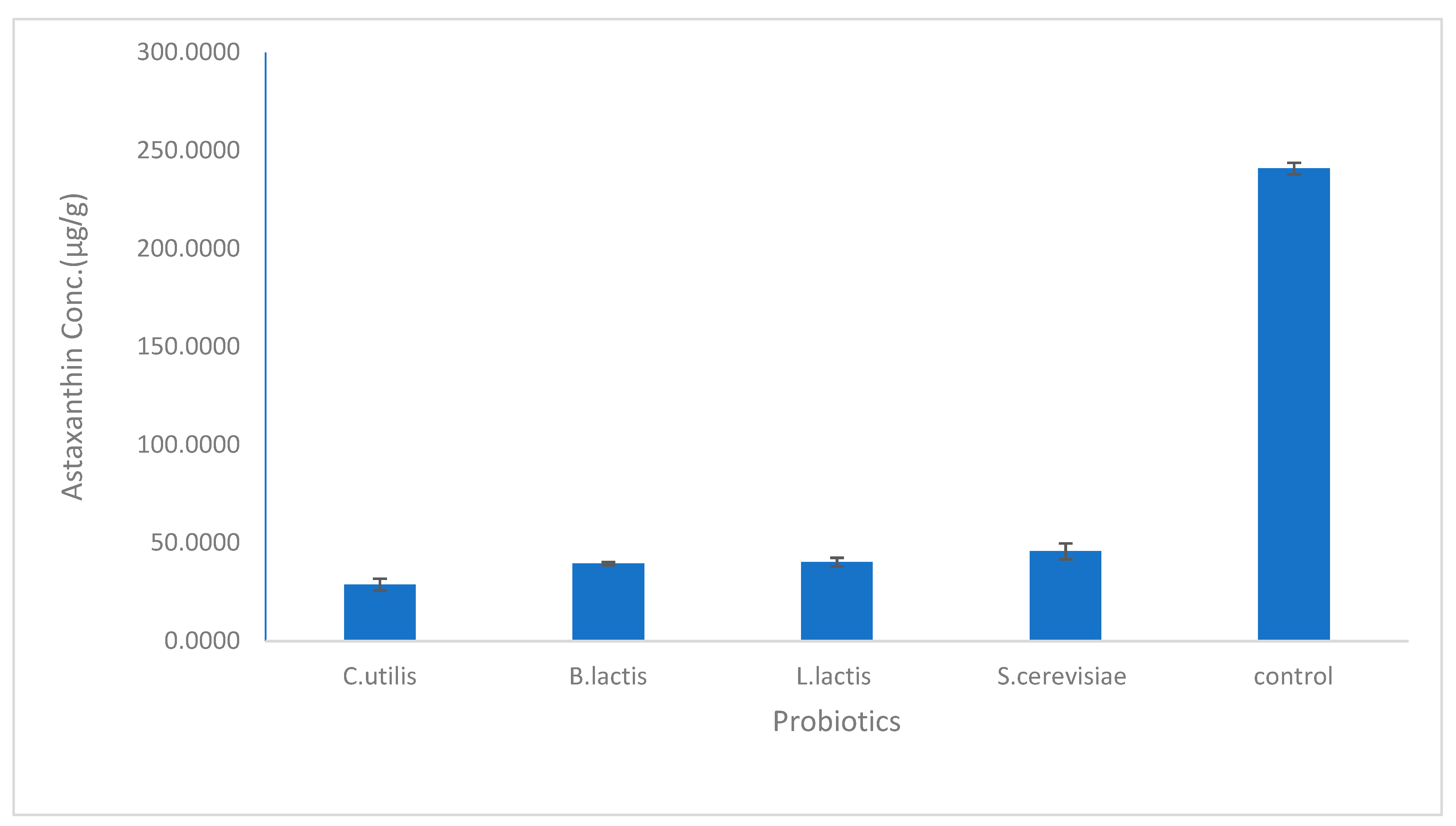
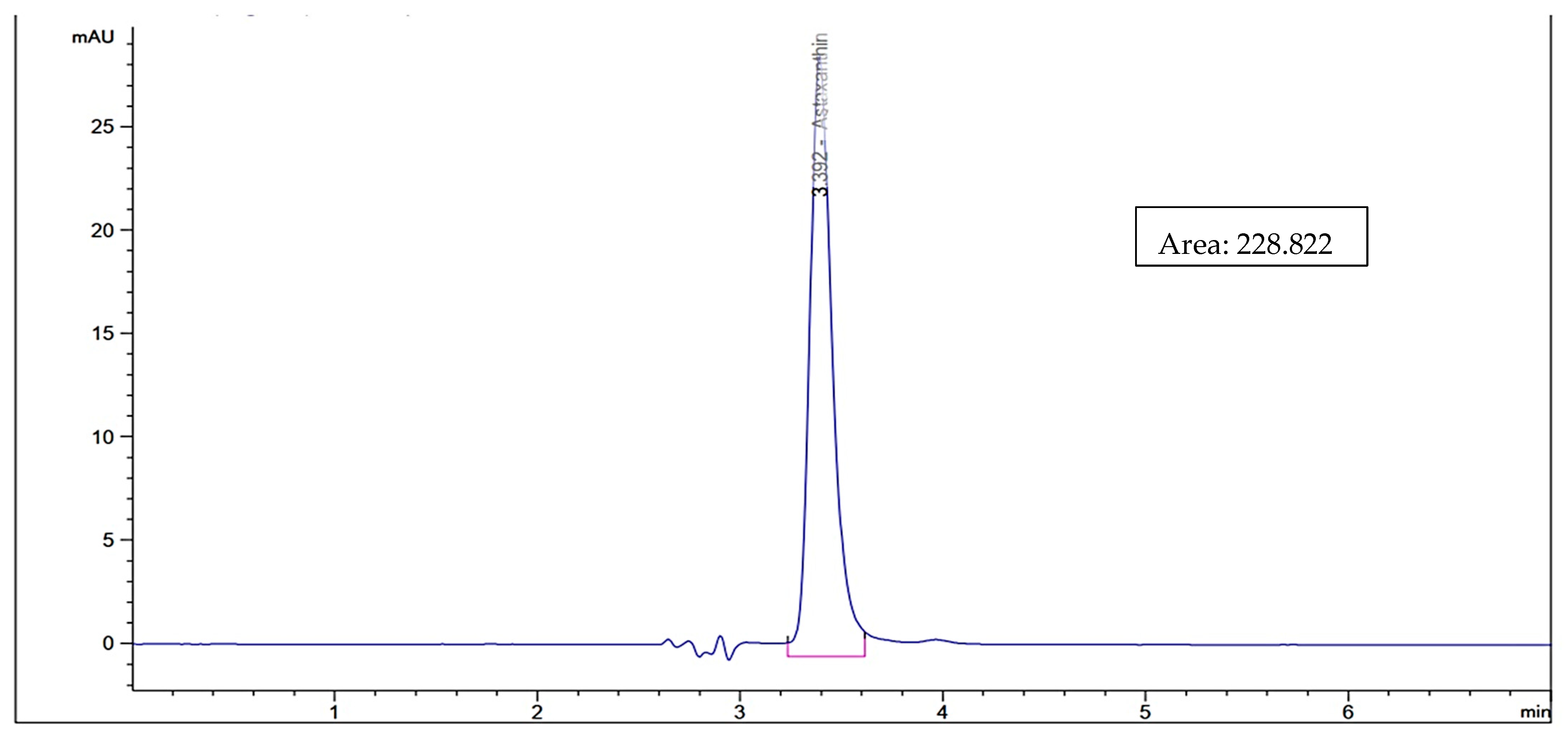
4.1. HPLC Analysis
4.2. Antimicrobial Activity
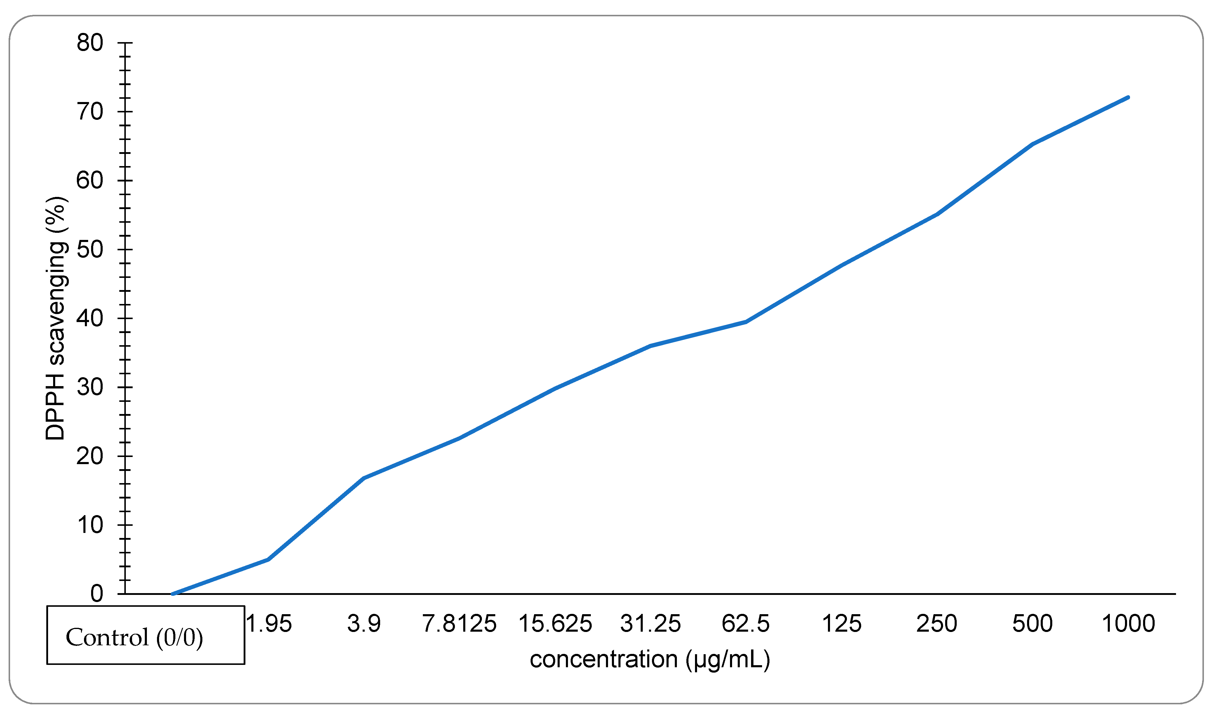
4.3. Antioxidant Activity
4.4. Anti-Inflammatory Activity
4.5. Anticancer Activity
5. Discussion
6. Conclusions
Author Contributions
Funding
Institutional Review Board Statement
Informed Consent Statement
Data Availability Statement
Conflicts of Interest
References
- Martin, J.W.; Davis, G.E. An Updated Classification of the Recent Crustacea; Natural History Museum of Los Angeles County: Los Angeles, CA, USA, 2001; p. 129. ISBN 1-891276-27-1. [Google Scholar]
- Hobbs, H., III. Investigations of the troglobitic crayfish Orconectes inermis testii (Hay) in Mayfield’s Cave, Monroe County, Indiana. Int. J. Speleol. 1981, 11, 21–32. [Google Scholar] [CrossRef]
- Yan, N.; Chen, X. Sustainability: Don’t waste seafood waste. Nature 2015, 524, 155–157. [Google Scholar] [CrossRef] [PubMed]
- Arbia, W.; Arbia, L.; Adour, L.; Amrane, A. Chitin extraction from crustacean shells using biological methods–A review. Food Technol. Biotechnol. 2013, 51, 12–25. [Google Scholar]
- Fang, S.; Cao, W.; Shao, Q.; Huang, W.; Wang, F.; Cheng, X.; Cao, J.; Luo, J.; Wu, Y. Reutilization of waste crawfish shell and sludge for efficient volatile fatty acids production by synchronously regulating the bioavailable substrates and microbial metabolic traits. J. Clean. Prod. 2022, 349, 131456. [Google Scholar] [CrossRef]
- Cheong, J.Y.; Muskhazli, M. Global Perspectives on Astaxanthin; Putra Malaysia University: Serdang, Malaysia, 2021; Chapter 13; pp. 253–279. ISBN 9780128233054. [Google Scholar]
- Tsuchiya, M.; Scita, G.; Freisleben, H.J.; Kagan, V.E.; Packer, L. [44] Antioxidant radical-scavenging activity of carotenoids and retinoids compared to α-tocopherol. Methods Enzymol. 1992, 213, 460–472. [Google Scholar] [PubMed]
- Zhao, T.; Yan, X.; Sun, L.; Yang, T.; Hu, X.; He, Z.; Liu, F.; Liu, X. Research progress on extraction, biological activities and delivery systems of natural astaxanthin. Trends Food Sci. Technol. 2019, 91, 354–361. [Google Scholar] [CrossRef]
- Eldessouki, E.A.; Diab, A.M.; Selema, T.A.; Sabry, N.M.; Abotaleb, M.M.; Khalil, R.H.; El-Sabbagh, N.; Younis, N.A.; Abdel-Tawwab, M. Dietary astaxanthin modulated the performance, gastrointestinal histology, and antioxidant and immune responses and enhanced the resistance of Litopenaeus vannamei against Vibrio harveyi infection. Aquac Int. 2022, 30, 1–19. [Google Scholar] [CrossRef]
- Chakrabarti, R. Carotenoprotein from shrimp process waste. J. Funct. Foods 2019, 185–202. [Google Scholar]
- Kidd, P. Astaxanthin, cell membrane nutrient with diverse clinical benefits and anti-aging potential. Altern. Med. Rev. 2011, 16, 355–364. [Google Scholar] [PubMed]
- McCall, B.; McPartland, C.K.; Moore, R.; Frank-Kamenetskii, A.; Booth, B.W. Effects of astaxanthin on the proliferation and migration of breast cancer cells in vitro. Antioxidants 2018, 7, 135. [Google Scholar] [CrossRef] [PubMed]
- Al-Tarifi, B.Y.; Mahmood, A.; Sheikh, H.I.; Amir, W.; Ahmad, N.W.; Assaw, S. Inhibitory effects of astaxanthin on lps-induced raw 264.7 inflammation and breast cancer mcf-7 cell lines proliferation. Sustain. Sci. 2022, 17, 55–66. [Google Scholar] [CrossRef]
- Tanaka, T.; Morishita, Y.; Suzui, M.; Kojima, T.; Okumura, A.; Mori, H. Chemoprevention of mouse urinary bladder carcinogenesis by the naturally occurring carotenoid astaxanthin. J. Carcinog. 1994, 15, 15–19. [Google Scholar] [CrossRef] [PubMed]
- Song, X.D.; Zhang, J.J.; Wang, M.R.; Liu, W.B.; Gu, X.B.; Lv, C.J. Astaxanthin induces mitochondria-mediated apoptosis in rat hepatocellular carcinoma CBRH- 7919 cells. Biol. Pharm. Bull. 2011, 34, 839–844. [Google Scholar] [CrossRef] [PubMed]
- Shanmugapriya, K.; Kim, H.; Saravana, P.S.; Chun, B.S.; Kang, H.W. Astaxanthin-alpha tocopherol nanoemulsion formulation by emulsification methods: Investigation on anticancer, wound healing, and antibacterial effects. Colloids Surf. B Biointerfaces 2018, 172, 170–179. [Google Scholar] [CrossRef] [PubMed]
- Bennedsen, M.; Wang, X.; Willén, R.; Wadström, T.; Andersen, L.P. Treatment of H. pylori infected mice with antioxidant astaxanthin reduces gastric inflammation, bacterial load and modulates cytokine release by splenocytes. Immunol. Lett. 2000, 70, 185–189. [Google Scholar] [CrossRef]
- Davinelli, S.; Nielsen, M.E.; Scapagnini, G. Astaxanthin in skin health, repair, and disease: A comprehensive review. Nutrients 2018, 10, 522. [Google Scholar] [CrossRef] [PubMed]
- Radzun, K.A.; Rusmidi, M.H.H.; Norisam, I.; Iran, N.; Pardi, F.; Ismail, A.; Razak, W.R.W.A.; Hafid, S.R.A. Anti-inflammatory Effects of Astaxanthin Extracted from Microalgae Hematococcus pluvialis and Combinations with Palm Tocotrienol Rich-Fraction in RAW 264.7 Macrophages. Pharm. J. 2022, 14, 205–215. [Google Scholar] [CrossRef]
- Terazawa, S.; Nakajima, H.; Shingo, M.; Niwano, T.; Imokawa, G. Astaxanthin attenuates the UVB-induced secretion of prostaglandin E2 and interleukin-8 in human keratinocytes by interrupting MSK1 phosphorylation in a ROS depletion–independent manner. Exp. Dermatol. 2012, 21, 11–17. [Google Scholar] [CrossRef] [PubMed]
- Wade, N.; Goulter, K.C.; Wilson, K.J.; Hall, M.R.; Degnan, B.M. Esterified astaxanthin levels in lobster epithelia correlate with shell colour intensity: Potential role in crustacean shell colour formation. Comp. Biochem. Physiol. Part B Biochem. Mol. Biol. 2005, 141, 307–313. [Google Scholar] [CrossRef]
- López-Cervantes, J.; Sánchez-Machado, D.I.; Gutiérrez-Coronado, M.A.; Ríos-Vázquez, N.J. Quantification of astaxanthin in shrimp waste hydrolysate by HPLC. Biomed. Chromatogr. 2006, 20, 981–984. [Google Scholar] [CrossRef] [PubMed]
- Hu, J.; Lu, W.; Lv, M.; Wang, Y.; Ding, R.; Wang, L. Extraction and purification of astaxanthin from shrimp shells and the effects of different treatments on its content. Rev. Bras. De Farmacogn. 2019, 29, 24–29. [Google Scholar] [CrossRef]
- Khanafari, A.; Saberi, A.; Azar, M.; Vosooghi, G.; Jamili, S.; Sabbaghzadeh, B. Extraction of astaxanthin esters from shrimp waste by chemical and microbial methods. J. Environ. Health Sci. Eng. 2007, 4, 93–98. [Google Scholar]
- Ali, M.; Khalil, N.; Abd El-Ghany, M. Biodegradation of some polycyclic aromatic hydrocarbons by Aspergillus terreus. Afr. J. Microbiol. Res. 2012, 6, 3783–3790. [Google Scholar]
- Wigner, P.; Bijak, M.; Saluk-Bijak, J. Probiotics in the Prevention of the Calcium Oxalate Urolithiasis. Cells 2022, 11, 284. [Google Scholar] [CrossRef] [PubMed]
- Buerth, C.; Heilmann, C.J.; Klis, F.M.; de Koster, C.G.; Ernst, J.F.; Tielker, D. Growth-Dependent Secretome of Candida Utilis. Microbiology 2011, 157, 2493–2503. [Google Scholar] [CrossRef] [PubMed]
- Fialho, M.B.; Toffano, L.; Pedroso, M.P.; Augusto, F.; Pascholati, S.F. Volatile Organic Compounds Produced by Saccharomyces Cerevisiae Inhibit the in Vitro Development of Guignardia Citricarpa, the Causal Agent of Citrus Black Spot. World J. Microbiol. Biotechnol. 2009, 26, 925–932. [Google Scholar] [CrossRef]
- Lu, M.; Zhao, C.; Zhou, P.; Yu, L. Analysis and identification of astaxanthin and its carotenoid precursors from Xanthophyllomyces dendrorhous by high-performance liquid chromatography. Z. Für Nat. C 2010, 65, 489–494. [Google Scholar] [CrossRef] [PubMed]
- Gaydos, J.M.; Harrington, B.J. Agar disk diffusion for the quality control testing of Autobac elution disks. Antimicrob. Agents Chemother. 1982, 21, 516–518. [Google Scholar] [CrossRef]
- Pfaller, M.A.; Burmeister, L.; Bartlett, M.S.; Rinaldi, M.G. Multicenter evaluation of four methods of yeast inoculum preparation. J. Clin. Microbiol. 1988, 26, 1437–1441. [Google Scholar] [CrossRef] [PubMed]
- Matar, M.J.; Ostrosky-Zeichner, L.; Paetznick, V.L.; Rodriguez, J.R.; Chen, E.; Rex, J.H. Correlation between E-test, disk diffusion, and microdilution methods for antifungal susceptibility testing of fluconazole and voriconazole. Antimicrob. Agents Chemother. 2003, 47, 1647–1651. [Google Scholar] [CrossRef]
- Chintong, S.; Phatvej, W.; Rerk-Am, U.; Waiprib, Y.; Klaypradit, W. In vitro antioxidant, antityrosinase, and cytotoxic activities of astaxanthin from shrimp waste. Antioxidants 2019, 8, 128. [Google Scholar] [CrossRef] [PubMed]
- Navarro-Hoyos, M.; Alvarado-Corella, D.; Moreira-Gonzalez, I.; Arnaez-Serrano, E.; Monagas-Juan, M. Polyphenolic composition and antioxidant activity of aqueous and ethanolic extracts from Uncaria tomentosa bark and leaves. Antioxidants 2018, 7, 65. [Google Scholar] [CrossRef] [PubMed]
- Moualek, I.; Aiche, G.I.; Guechaoui, N.M.; Lahcene, S.; Houali, K. Antioxidant and anti-inflammatory activities of Arbutus unedo aqueous extract. Asian Pac. J. Trop. Biomed. 2016, 6, 937–944. [Google Scholar] [CrossRef]
- Ahmed, E.A.; Mohamed, H.E.; Abd El-Salam, H.S. In vitro Antimicrobial Activity of Astaxanthin Crude Extract from Haematococcus pluvialis. Egypt. J. Aquat. Biol. 2022, 26, 95–106. [Google Scholar] [CrossRef]
- Pashkow, F.J.; Watumull, D.G.; Campbell, C.L. Astaxanthin: A novel potential treatment for oxidative stress and inflammation in cardiovascular disease. Am. J. Card. 2008, 101, S58–S68. [Google Scholar] [CrossRef]
- Yamashita, E. The effects of a dietary supplement containing astaxanthin on skin condition. Food Style 2005, 21, 9–72. [Google Scholar]
- Bašković, M.; Ježek, D. Letter to the Editor re ‘Astaxanthin Reduces the Severity of Intestinal Damage in a Neonatal Rat Model of Necrotizing Enterocolitis’. Am. J. Perinatol. 2022. [Google Scholar] [CrossRef]
- Hamdi, S.A.H. Eco-friendly methods for recycling of crayfish “Procambarus clarkii” by-product for astaxanthin extraction and quantification. Egypt. J. Aquat. Biol. 2022, 26, 239–251. [Google Scholar] [CrossRef]
- Yoon, C.H.; Bok, H.S.; Choi, D.K.; Row, K.H. Optimization condition of astaxanthin extract from shrimp waste using response surface methodology. Korean J. Chem. Eng. 2012, 50, 545–550. [Google Scholar] [CrossRef]
- Radzali, S.A.; Baharin, B.S.; Othman, R.; Markom, M.; Rahman, R.A. Cosolvent selection for supercritical fluid extraction of astaxanthin and other carotenoids from Penaeus monodon waste. J. Oleo Sci. 2014, 63, 769–777. [Google Scholar] [CrossRef]
- Santos, C.A.D.; Padilha, C.E.; Damasceno, K.S.; Leite, P.I.; de Araújo, A.C.; Freitas, P.R.; Vieira, É.A.; Cordeiro, A.M.T.M.; de Sousa, F.C., Jr.; Assis, C.F.D. Astaxanthin Recovery from Shrimp Residue by Solvent Ethanol Extraction Using Choline Chloride: Glycerol Deep Eutectic Solvent as Adjuvant. J. Braz. Chem. Soc. 2021, 32, 1030–1039. [Google Scholar]
- Dalei, J.; Sahoo, D. Extraction and characterization of astaxanthin from the crustacean shell waste from shrimp processing industries. In Proceedings of the International Conference on Public Health and Medical Sciences, Suzhou, China, 26–28 May 2022. [Google Scholar]
- Akyön, Y. Effect of antioxidants on the immune response of Helicobacter pylori. Clin. Microbiol. Infect. 2002, 8, 438–441. [Google Scholar] [CrossRef] [PubMed]
- Ngginak, J.; Mangibulude, J.C.; Rondonuwu, F.S. The Identification of Carotenoids and Testing of Carotenoid Antioxidants from Sand Lobster (Panulirus homarus) Egg Extract. Indones. J. Mar. Sci. Ilmu Kelaut. 2017, 22, 3. [Google Scholar] [CrossRef]
- Zhang, X.; Ren, X.; Zhao, X.; Liu, H.; Wang, M.; Zhu, Y. Stability, structure, and antioxidant activity of astaxanthin crystal from Haematococcus pluvialis. JAOCS 2022, 99, 367–377. [Google Scholar] [CrossRef]
- Parvathy, S.; Subramanian, P.; Karthick, S.A.; Subbaiya, R. The structural, optical, antimicrobial and anticancer properties of biocompatible astaxanthin coated ZnO and CeO2 nanoparticles. Mater. Lett. 2022, 312, 131669. [Google Scholar] [CrossRef]


| Microorganism | Inhibition Zone Diameter (mm) | ||
|---|---|---|---|
| Sample | Control | ||
| Positive Control | Negative Control | ||
| Gram positive | Ampicillin | ||
| Staphylococcus aureus (ATCC:13565) | NA | 20 ± 0.1 | 0.0 |
| Staphylococcus aureus (ATCC:25923) | NA | 20 ± 0.1 | 0.0 |
| Streptococcus mutans (ATCC:25175) | NA | 29 ± 0.5 | 0.1 |
| Gram negative | Gentamycin | ||
| Escherichia coli (ATCC:10536) | NA | 27 ± 0.5 | 0.0 |
| Escherichia coli (ATCC:25955) | NA | 27 ± 0.5 | 0.0 |
| Klebsiella pneumonia (ATCC:10031) | NA | 25 ± 0.5 | 0.0 |
| Pseudomonas aeruginosa (ATCC:27853) | NA | 28 ± 0.3 | 0.0 |
| Fungal strains | Nystatin | ||
| Candida albicans (ATCC:10231) | 12.3 ± 0.5 | 21 ± 0.5 | 0.0 |
| Aspergillus ochraceous (ATCC:22947) | NA | 18 ± 0.5 | 0.0 |
| Aspergillus parasiticus (ATCC:26690) | NA | 18 ± 0.5 | 0.1 |
| Aspergillus niger (ATCC:16404) | NA | 19 ± 0.5 | 0.0 |
| Aspergillus niger (ATCC: MH368137) | NA | 19 ± 0.5 | 0.0 |
Publisher’s Note: MDPI stays neutral with regard to jurisdictional claims in published maps and institutional affiliations. |
© 2022 by the authors. Licensee MDPI, Basel, Switzerland. This article is an open access article distributed under the terms and conditions of the Creative Commons Attribution (CC BY) license (https://creativecommons.org/licenses/by/4.0/).
Share and Cite
Hamdi, S.; Elsayed, N.; Algayar, M.; Ishak, V.; Ahmed, M.; Ahmed, S.; Kamal, M.; Abd El-Ghany, M. Biological Extraction, HPLC Quantification and Medical Applications of Astaxanthin Extracted from Crawfish “Procambarus clarkii” Exoskeleton By-Product. Biology 2022, 11, 1215. https://0-doi-org.brum.beds.ac.uk/10.3390/biology11081215
Hamdi S, Elsayed N, Algayar M, Ishak V, Ahmed M, Ahmed S, Kamal M, Abd El-Ghany M. Biological Extraction, HPLC Quantification and Medical Applications of Astaxanthin Extracted from Crawfish “Procambarus clarkii” Exoskeleton By-Product. Biology. 2022; 11(8):1215. https://0-doi-org.brum.beds.ac.uk/10.3390/biology11081215
Chicago/Turabian StyleHamdi, Salwa, Nour Elsayed, Mohamed Algayar, Verina Ishak, Mariam Ahmed, Sara Ahmed, Mohamed Kamal, and Mohamed Abd El-Ghany. 2022. "Biological Extraction, HPLC Quantification and Medical Applications of Astaxanthin Extracted from Crawfish “Procambarus clarkii” Exoskeleton By-Product" Biology 11, no. 8: 1215. https://0-doi-org.brum.beds.ac.uk/10.3390/biology11081215