Article

Phenolic Composition and Wound Healing Potential Assessment of Moroccan Henna (Lawsonia inermis) Aqueous Extracts

by
Soukaina El Massoudi
1,
Abdellah Zinedine
2,
João Miguel Rocha
3,4,5,*,
Meryem Benidir
1,
Ilham Najjari
1,
Lahsen El Ghadraoui
1,
Meryem Benjelloun
1 and
Faouzi Errachidi
1
1
Functional Ecology and Environmental Engineering Laboratory, Faculty of Science and Technology, Sidi Mohamed Ben Abdellah University, P.O. Box 2202, Fez 30050, Morocco
2
BIOMARE Laboratory, Faculty of Sciences, Chouaib Doukkali University, P.O. Box 20, EL Jadida 24000, Morocco
3
CBQF-Centro de Biotecnologia e Química Fina–Laboratório Associado, Escola Superior de Biotecnologia, Universidade Católica Portuguesa, Rua Diogo Botelho 1327, 4169-005 Porto, Portugal
4
LEPABE—Laboratory for Process Engineering, Environment, Biotechnology and Energy, Faculty of Engineering, University of Porto, Rua Dr. Roberto Frias, s/n, 4200-465 Porto, Portugal
5
ALiCE—Associate Laboratory in Chemical Engineering, Faculty of Engineering, University of Porto, Rua Dr. Roberto Frias, s/n, 4200-465 Porto, Portugal
*
Author to whom correspondence should be addressed.
Submission received: 18 May 2023 / Revised: 1 June 2023 / Accepted: 13 June 2023 / Published: 15 June 2023
(This article belongs to the Collection Feature Papers in Cosmetics in 2023)

Abstract

:
The present study aims at valorizing Moroccan Henna (Lawsonia inermis) by developing healing formulations for cosmetic and therapeutic uses. For such a goal, the plant was collected from three locations in southeastern Morocco (Alnif, Tafraoute Sidi Ali and Tazarine). Phytochemical analysis of Henna leaves was performed by determining phenolic compound contents, and flavonoids and tannins in it, through its aqueous extracts. Then, specific formulations were prepared using aqueous extracts of L. inermis to assess their in vivo wound healing potential in Swiss albino mice used as animal models. Results disclosed that phenolic compounds (13.48%), as well as flavonoid (9.25%) and tannin (2.57%) contents are higher in Henna leaf extracts from Alnif, while Tazarine Henna aqueous extract was found to be richer in saponins (0.32%). Exclusion chromatographic analysis on Sephadex G50 gel corroborates the obtained results and shows that Lawsone levels (Henna coloring agent) are higher in Henna collected from Alnif. Aqueous Henna leaf extracts, at a dose of 10% in petroleum jelly, have been assessed for their ability to heal induced burns in mice. Healing monitoring, carried-out with Henna extracts on mice batches and those of two control batches (mice batch treated with petroleum jelly alone and batch treated with petroleum jelly containing 1% (flamazine), showed a great reduction in burnt surface with an accentuated contraction percentage (CP) and complete re-epithelialization duration (CRD) at 21 days in the 3 studied Henna-based formulations. These findings suggest the interest of potential development of Henna-based formulations, as a source of phenolic compounds, for further dermatological, cosmetic and therapeutic applications.

1. Introduction

Morocco is recognized for its vegetal biodiversity due to its variation of bioclimatic stages from its desertic south to humid north. Such diversity offers unequivocal potential benefits in terms of medicinal plants, which nowadays remain insufficiently exploited. These natural plants are frequently used for various applications—including primary healthcare, cultural and traditional practices—due to their low cost and safety in use.
Henna (Lawsonia inermis) is a plant grown in tropical regions commonly used for cosmetic purposes. Henna powder is also frequently used for its healing properties attributed to its active principles, which may have beneficial implications in wound healing.
Plants have always aroused high interest by civilizations around the world, where people traditionally used natural products for medicinal and cosmetic purposes to improve their well-being [1,2]. Thus, certain archaeological sites in Egypt have revealed the presence of cosmetic products of vegetable or mineral origin, where the inventory included Henna and dental powder [3,4].
A cosmetic product is a substance intended to come into contact with the human body to clean, perfume, modify and preserve it in good conditions. Natural cosmetic products are becoming common because of the continuing discovery of their therapeutic uses. Nowadays, over 60,000 plant varieties exist in Africa, and around 8000 of them are being used in herbal medicine, particularly cosmetic products [5,6,7], thus making apparent their potential socioeconomic and cultural interest, as well as their environmentally sustainable features. In Morocco, the regulatory context for cosmetic products is covered by Ministry of Health circular No. 48 DMP/20, which relates to the registration of cosmetic products and defines a cosmetic product as any substance or preparation intended to be brought into contact with various surface parts of the human body.
Primary healthcare improved security margins and the low cost of traditional medicines make plants crucial to human daily life and the treatment against numerous diseases. The application of these plants is also closely linked to the lives of Moroccans for cultural, folkloric, prophetic or religious reasons [8,9]. L. inermis, commonly referred to as Henna in Arab countries, is the most popular and widespread in tropical areas [10]. L. inermis belongs to the family Lythraceae, whose leaves play a particularly significant component. They are used in a wide range of industries, including drugs and cosmetics, due to their abundance of the active Lawsone ingredients (a red–orange dye (2-hydroxy-1,4-naphthoquinone), also known as hennotannic acid) used in hair, nail, hand and textile dyeing segments of the cosmetic market for antibacterial, antifungal, antioxidant, analgesic and anti-inflammatory agents [11,12,13]. Despite all these benefits, L. inermis-based cosmetic sector remains in an incipient stage, and Morocco is one of the richest countries for the potential advantages of Henna on a world scale. Plantation is still only present in only a few precarious areas, has little added-value, and still has not been industrialized, as it is used in other countries such as Iran and India.
Despite the existing research and knowledge on chemical and phytochemical properties of Moroccan Henna, its cosmetic and functional (biological activity and technological functionality) properties (and other traits, especially its beneficial effects on human skin) remain poorly known [14].
Overall, this plant possesses curative properties attributed to its active components, which may potentially have therapeutic implications in wound healing. In this regard, the current research effort contributes towards in vivo examining our ointment formulations based on an aqueous extract of L. inermis, collected in three Moroccan areas, to the wound healing against silver sulfadiazine.
Indeed, provided that Henna is traditionally known for its healing properties attributed to its active components, it can potentially have therapeutic applications in wound healing. Therefore, this study aimed at studying the phenolic composition of Moroccan Henna, collected in three different origins of the country, to prepare ointment formulations based on Moroccan Henna leaf aqueous extracts, and finally, to assess their in vivo wound healing potential using mice as an animal laboratory model.

2. Material and Methods

2.1. Leaves of Lawsonia inermis

Fresh leaves of L. inermis were collected in July 2019 from 3 different areas in southeastern Morocco—chiefly, Alnif (LI1), Tafraoute Sidi Ali (LI2) and Tazarine (LI3). Leaves were washed, air-dried for 21 days at room temperature and then ground into powder.

2.2. Aqueous Extract (AQ) Obtention

To obtain the aqueous extract (AQ), leaf powders (10 g) from the 3 studied Henna were dissolved in distilled water (100 mL). The mixture was then stirred under magnetic agitation at 700 rpm for 72 h at room temperature before being filtered. Filtrate was collected and concentrated using a rotary evaporator under vacuum at a temperature of 40 °C. Obtained extract was stored in a clean airtight container, labeled and kept at a temperature of 4 °C until further use.

2.3. Ointment Formulation

The formulations were made using a galenic basis that contains white soft paraffin and a 5% aqueous extract of L. inermis. Thus, 9.5 g of paraffin and 0.5 g of extract were combined to create 10 g of simple ointment base. The ointment mixtures were individually transferred to a clean container. Three distinct Henna aqueous extracts from Alnif, Tafraoute Sidi Ali and Tazarine were used to make distinct ointments.
To ensure a good dispersion of the aqueous extracts in the hydrophobic matrix (Vaseline: soft paraffin), we exploited, on one hand, the natural carbohydrate polymers presence in extracts with considerable quantities and the high viscosity of soft paraffin endowed with a delicate texture fibrous, which, on the other hand, improves the dispersion of the particles even hydrophilic. Therefore, an entanglement can be created mechanically and appear in the case of particles having a non-spherical geometric structure (fibrillar, lamellar, dendritic). With this in mind, we called on an RW 20 Digital propeller stirrer—IKA designed to fit into the most complex stirring applications, for stirring and high viscosity mixtures. The success of the dispersion was controlled by monitoring ointments produced stability over a long period.

2.4. Total Phenolic Compounds Content

Total phenolic content in extracts was determined by the Folin–Ciocalteu reagent following the method described by Singleton and Rossi (1965) [15] with adaptations. Distilled water was used as a blank, and gallic acid was used to produce a standard calibration curve obtained by spectrophotometry at a wavelength of 765 nm. Results were expressed in grams of gallic acid equivalent (GAE) per 100 g dry matter of extract (g GAE/100 g DM.

2.5. Total Flavonoid Content

Flavonoid content was quantified using the spectrophotometric method described by Zhishen et al. [16] based on the aluminum trichloride (AlCl3) reagent. Absorbance was read at 510 nm against the blank reagent containing water. Results were expressed in grams of quercetin equivalents (QE) per 100 g of dry matter (g QE/100 g DM).

2.6. Total Tannin Content

Tannin content was determined by the colorimetric method of Folin–Ciocalteu, described by Ba et al. (2010) [17]. Distilled water was used as a blank. Standard tannic acid solutions were prepared as described above. The total tannin content (TTC) was expressed in mg of equivalent tannic acid TAE per 100 g of dry matter (g TAE/100 g DM).

2.7. Total Saponin Content

Total saponin content was determined by the spectrophotometric method as described by Hiai et al. (1976) with modifications [18]. The absorbance was measured at 544 nm, relative to a blank containing 30% aqueous methanol in place of the sample extract. Total Saponin concentrations (TSC) were expressed as diosgenin equivalents (DE µg/100 g DM) calculated from a standard calibration curve.

2.8. Gel Filtration Chromatography

Since the crude aqueous extract of L. inermis is a complex matrix, it is necessary to proceed with the fractionations and purification of most of molecules in order to further characterization. Crude extract fractionation was performed in an open column of Sephadex G50. This method is a size-exclusion chromatography allowing molecules to separate based on their molecular size. Sephadex gel is made up of highly porous microbeads, where molecules with highest molecular weights only diffuse outside pores beads and exit the column first. In contrast, smaller molecules diffuse inside all microbeads, are delayed and exit the column afterwards [19].
A column with a diameter of 2.5 cm and a length of 50 cm was used with a flowrate set at 1 mL/min, based on the method of Siddiqui et al., with some modifications [20]. An amount of 20 grams of Sephadex G50 was mixed with 150 mL of lithium chloride buffer solution (5 mM NaOH, 2.5 mM LiCl). Half mg/mL of each extract were fractionated on Sephadex gel, and separated fractions were collected in test tubes at a volume of 2 mL and analyzed with a spectrophotometer at 380 (for phenolic compounds) and 490 nm (for Lawsone) [21].

2.9. Wound Healing Activity Assessment

Swiss albino mice are often chosen for wound healing tests due to their physiological characteristics rather than experimental reasons. This animal model displays faster regeneration of epidermis and production of matrix compared to humans, enabling the study of all wound healing stages, including dermal reconstruction, as well as genetic modifications that imitate certain human pathologies, such as diabetes, immunodeficiency and obesity [22]. The objective of this research is to investigate wound healing effect of a cosmetic formulation containing L. inermis leaf extracts and compare it to the control treatment for burn care units (1% silver sulfadiazine). The technique used to induce wounds on mice’s backs allows wound healing phases monitoring, from re-epithelialization to wound contraction.
-
Ethics statement
Swiss albino mice of both sexes were provided by the animal facility of the Faculty of Sciences and Techniques of Fes. Upon receipt, the animals were housed in groups of five individuals in standard polystyrene cages for a two-week adaptation period before being used in biological activities. The animals had free access to water and food and were maintained at a constant temperature of 23 ± 1 °C. An ethics statement was obtained for all activities involving animals.
-
Animal preparation
We used groups of mice consisting of 10 males and 15 females as an experimental model to induce thermal burns, with a weight range between 40 and 41.4 g. Each animal was housed in an individual polystyrene cage with ad libitum access to food and water. The animals were divided into 5 groups, each containing 5 mice:
Negative control group (NC): mice were left untreated and treated with white soft paraffin;
Positive control group (PC): mice were treated with 1% silver sulfadiazine, also known as Flamazine;
LI1 group: mice were treated with the Alnif ointment formulation;
LI2 group: mice were treated with the Tafraoute Sidi Ali ointment formulation;
LI3 group: mice were treated with the Tazarine ointment formulation.
-
Performance of experimental burns
To follow their weight variation during treatment, we weighed the mice of each group at the beginning of the experiment. The animals underwent general anesthesia with ether, and the burns were performed on the previously shaved back of each animal, chosen for ease of access. Burns were induced using a round metal bar with a diameter of 10 mm heated to a temperature of 100 °C using boiling water [23]. After reaching 100 °C, the object was quickly applied without pressure for 20 s to the study area.
-
Treatment application
Burned mice were treated once a day with a cream corresponding to their respective lot, while mice in NC group received no treatment. The mice’s wounds were left uncovered, but photos were taken daily to monitor the healing process. The photos were taken at the same angle to avoid wound size underestimation.
-
Study of scar evolution using digital planimetry
The evolution of burn wound healing was studied using digital planimetry, which involves photographing wounds and analyzing their surface area using ImageJ software [24]. To enable the software to measure wounds in square millimeters, a ruler was used during photography.
-
Percentage of contraction
The wound contraction percentage (WCP) was determined by calculating the average wound size of five mice from the same group and comparing it with the initial burn surface area [25] using the following Equation (1):
W C P = Initial   wound   size   D 0 Wound   size   at   Dn Initial   wound   size × 100
where:
-
WCP: Wound contraction percentage;
-
D0: Day 0;
-
Dn: Day n.
-
Epithelialization period
The epithelialization period, characterized by the proliferation of epithelial stem cells, was evaluated by the number of days required for the dermal damage and complete closure of wound leaving no remaining injury [26,27].

2.10. Statistical Analyses

The obtained results were statistically analyzed to determine intra-variety and inter-variety variability in three different regions. The data were processed using Minitab version 20.2 software. Two-factor ANOVA was applied to compare means, and Pearson’s correlation test was used for quantitative variables.

3. Results and Discussion

3.1. Phenolic Compounds Analysis

Yield of extraction, total phenolic contentment, flavonoids contents, tannins and saponins results are illustrated in the table below (Table 1). Data in Table 1 show that Henna from Alnif region is very rich in water-soluble compounds, phenolic compounds, flavonoids and tannin, followed by Henna from Tazarine region and, lastly, we found the Henna from Tafraoute Sidi Ali region. These families of molecules are targeted by this study because they play a very important role in healing phenomenon.

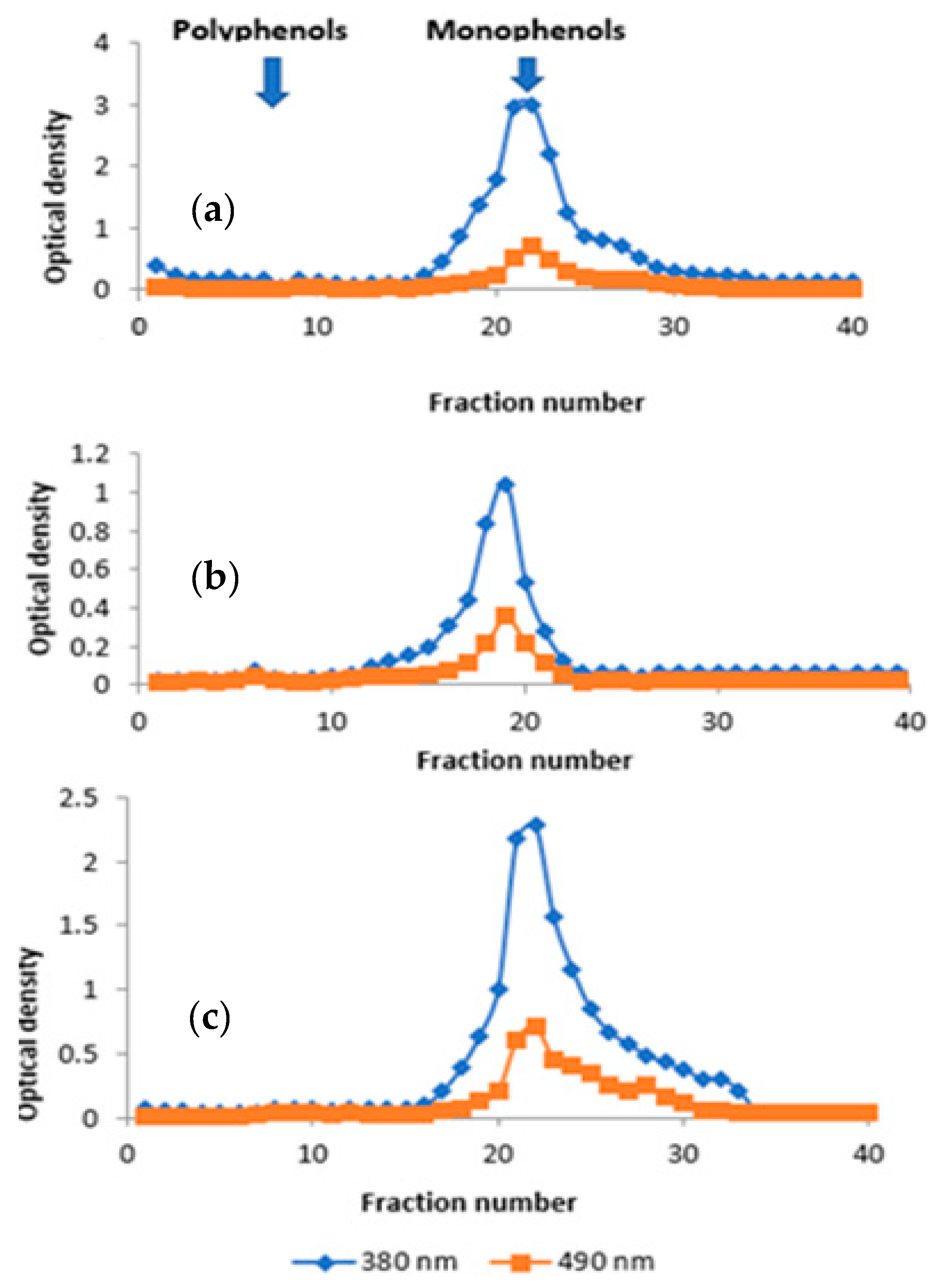
3.2. Gel Filtration Chromatography

Gel filtration chromatography, also known as exclusion chromatography or gel permeation chromatography, allows a simple and rapid separation of molecules that are highly sensitive to denaturation. Chromatographic results obtained after spectrophotometric analysis of Henna from Alnif, Tafraoute Sidi Ali and Tazarine are shown in the graphics below (Figure 1).
Chromatogram of L. inermis from Alnif (Figure 1a) reveals a single major peak with an optical density of 3 for the aqueous extract and 0.7 for Lawsone after an elution time of 20 min from the 15th to the 35th sample tube. The yield of the 2 curves is 32.7%. The area between the 2 curves represents the distribution of 67.3% of the other chemical compounds forming the total phenolic compounds.
Chromatogram of Henna from Tafraoute Sidi Ali (Figure 1b) shows a major peak with an optical density of 1.046 for the aqueous extract and 0.355 for Lawsone, after an elution time of 24 min from the 12th to the 24th sample tube. The yield of the 2 curves is 33.94%. The other chemical compounds forming the phenolic compounds are represented by 66.06% of the area between the 2 curves.
The results of Figure 1c show that the aqueous extract of L. inermis from Tazarine is mainly composed of a peak at 380 nm with Lawsone after an elution time of 32 min from the 18th to the 34th sample tube. The yield of the 2 curves is 31.41% with a maximum optical density of 2.289 and 0.719, respectively, for the aqueous extract and Lawsone. The area between the 2 curves represents the distribution of 68.59% of the other chemical compounds forming the phenolic compounds. Based on the results in Figure 1a–c, it can be concluded that the fraction of Lawsone is perfectly separated by distilled water, and this fraction is composed of a single monophenol peak observed at 490 nm absorbance.
From Figure 1a–c, it is observed that the aqueous extract of Henna from Tazarine represents the richest extract in Lawsone, followed by Tafraoute Sidi Ali and then Alnif. Henna from Tazarine could be valorized within the framework of potential industrialization.

3.3. Evaluation of In Vivo Wound Healing Activity

-
Epithelialization period
According to obtained results, the average total healing durations of burns from LI1, LI2 and LI3 groups, compared to those of NC and PC groups, showed significant differences in favor of the treatments with L. inermis ointment formulations (Table 2).
The latter recorded average durations of complete epithelialization were 19.33 ± 0.05, 20.33 ± 4.65 and 21.67 ± 5.10 days for Tazarine LI, Alnif LI and Tafraoute Sidi Ali LI, respectively, while the PC group recorded an average duration of 30.67 ± 3.63 days. Finally, the untreated mice group (NC) took 42.33 ± 24.54 days to completely re-epithelialize the burns. A highly statistically significant difference was recorded between the different groups of LI1, LI2, LI3, PC and NC (ANOVA: F = 185.53; df = 4; p ≤ 0.001).
-
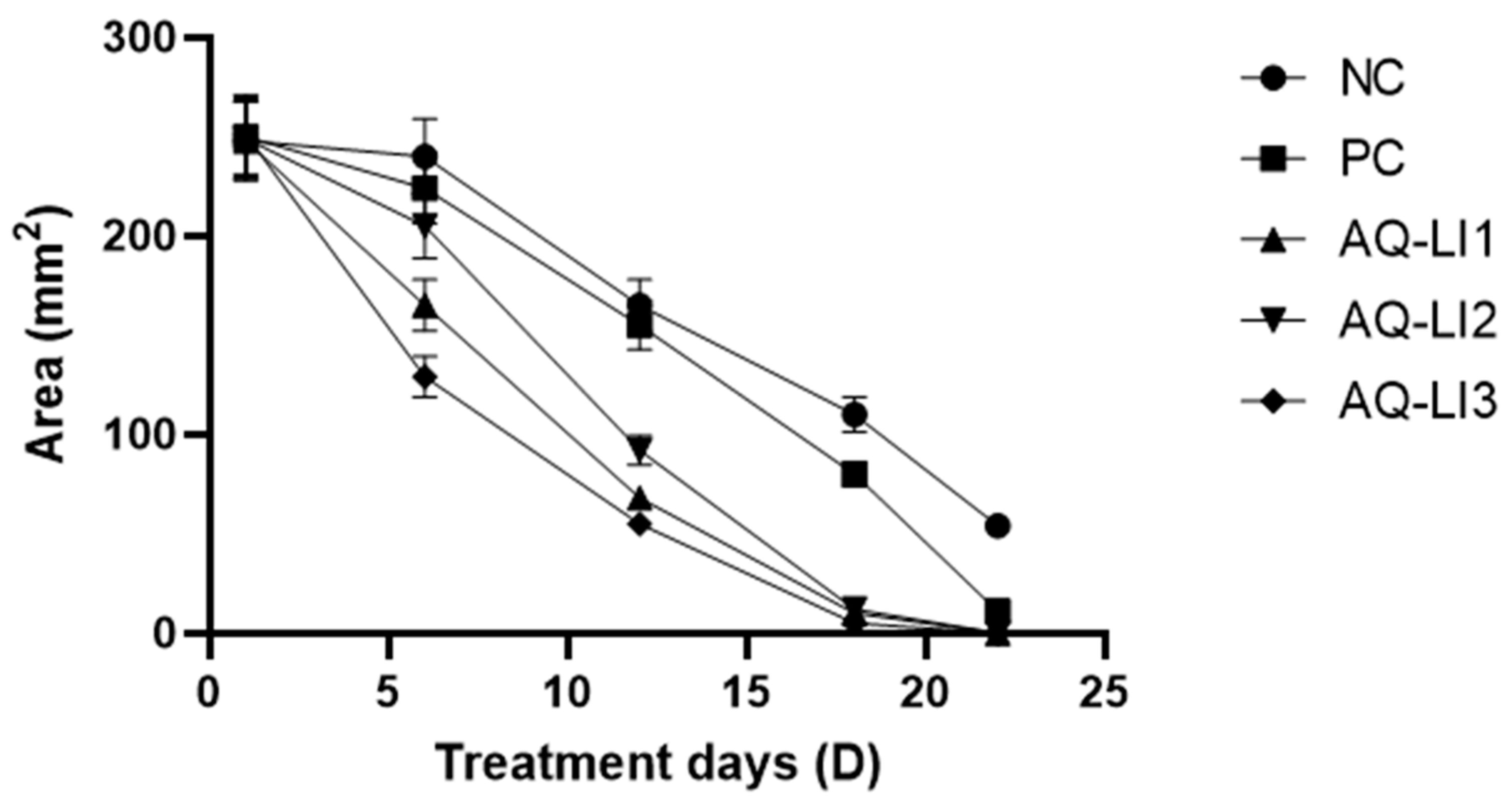
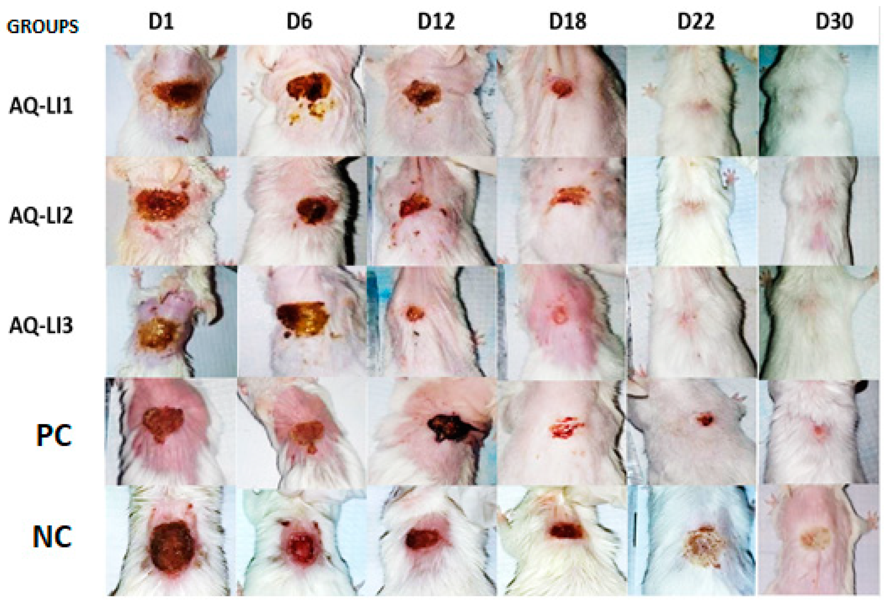
Average surface and mean percentage of burn contraction
Variations in burning surface and contraction percentage are presented in Figure 2; Figure 3 below.
According to the obtained results, we noticed that burning surface decreased during treatments, and this surface evolution varied from one group to another. During the first days from D1 to D6 post-burn, which included the healing inflammatory phase, burns of the Tazarine AQ-LI group recorded the highest reductions in their surface from 240.77 ± 2.13 to 126.93 ± 2.04 mm2, with a percentage of contraction of 33.13%, followed by Alnif AQ-LI from 242.63 ± 2.34 to 164.70 ± 2.15 mm2, with a percentage of contraction equal to 22.77%.
Subsequently, the variety Tafraoute Sidi Ali AQ-LI showed values ranging from 241 ± 2.51 to 201.53 ± 2.27 mm2. However, the lowest reductions burn surface were recorded in NC group, ranging from 241.33 ± 2.86 to 239.73 ± 3.07 mm2, with a low percentage of contraction of 14.83%. Statistically, the percentage of contraction revealed a highly significant effect at the AQ-LI1, AQ-LI2, AQ-LI3, PC, and NC groups level.
In one hand, starting from day 12, the difference in size between the burns of the groups became more pronounced, and the groups treated with our formulations maintained significant differences when compared to PC and NC groups, specifically the Tazarine variety group (AQ-LI3). Thus, all burns in the AQ-LI1, AQ-LI2 and AQ-LI3 groups have been completely healed after 22 days.
On the other hand, mice treated with Flamazine (PC) healed after only 24 days. The NC group had the largest average surface area of 163.43 ± 2.73 mm2 with a contraction percentage of 21.88%, and the first cases of complete burn healing were observed on the 35th day. The results obtained (Figure 4) confirm the traditional uses of the leaves of L. inermis species reported by our study. These results are also in agreement with other studies that have shown that this species is used for the healing of wounds [28,29] and burns [30].
A research group evaluated the analgesic and anti-inflammatory effects of Lawsone isolated from Henna leaves (L. inermis) [31]. The results of this study unfolded that the bioactive molecule Lawsone induced a strong analgesic effect when compared to the positive control with aspirin and they concluded that Lawsone has analgesic and anti-inflammatory effects, confirming the practical medical importance of the L. inermis species. This latter is widely used traditionally for its profitability and safety purposes; however, further studies are necessary to determine the systemic safety of Lawsone. It should be noted, however, that this is the first time that the healing effect of a galenic formulation of L. inermis leaf extracts has been studied and compared with silver sulfadiazine (SSD), the conventional treatment for burns. Silver sulfadiazine (Flamazine) is used against underlying infections due to its antibacterial activity, but prophylaxis is essential for better wound repair [32]. Its ease of application is associated with minimal pain and median cutaneous infiltration [33].
Despite these attributes, numerous side effects of SSD have been reported, such as cases of renal toxicity, which mark its use during long treatment durations and on large wounds [34]. Late cases of healing and allergic reactions to Flamazine have been reported, limiting its use in certain patients [35] cited by [36].
To this end, scientists have focused in recent years on studies evaluating the anti-inflammatory and healing effects of plant extracts. The healing effect of Crocus sativus (commonly known as saffron) is significantly superior to that of Flamazine [37]. Similarly, the research group conducted a comparative study of the healing effects of aloe vera and Flamazine, demonstrating that aloe vera allows for good re-epithelialization of burns compared to Flamazine [36].
The healing effect of our formulated L. inermis was more pronounced during the inflammatory phase of healing, where wounds from batches AQ-LI1, AQ-LI2 and AQ-LI3 showed very significant decreases in size when compared to PC and NC batches. This could be explained by its anti-inflammatory activity. Likewise, in natural products, the healing action of these formulations is due to the different chemical constituents of their composition. A study revealed that the healing effect of these natural blends is due to mechanisms such as cell multiplication, activation of collagen synthesis, antioxidant power and antimicrobial and anti-inflammatory effects [38]. In the case of L. inermis, its richness in bioactive molecules such as fatty acids, triglycerides, saponins, tannins, flavonoids, polyphenols and others [39,40] has been observed. These compounds contain a hydroxyl function that allows them to trap free radicals, giving them significant antioxidant power. This may explain the complete healing of the groups treated with L. inermis formulations in a shorter period than those treated with NC and PC. Additionally, the abundance of saponins in the extracts of L. inermis leaves (see biochemical study) could explain the faster healing of burns compared to controls. These results are consistent with those of Ankita et al. [41], who evaluated some properties of the Ficus religiosa fruit and developed a healing ointment—and with their findings, concluded the importance of saponins which are combined with the powder of Ficus benghalensis leaves in stimulating the healing effect as well as reducing wounds over a period of 18 days. Moreover, the addition of antioxidants to the list of treatments for burned mice leads to beneficial effects such as a reduction in the impact of wound damage, and a decrease in the total recovery time compared to other mice treated without the addition of antioxidants [42].
Indeed, the structure of the epidermis (the upper layers of the skin) is damaged and the protective hydrolipidic film of the skin is lost under the effect of the burn. Therefore, the skin does not act as a barrier, but rather, as an entry point for infection and ultimately, becomes dehydrated [43]. The protective effect of our formulated treatments on burned skin is directly related to their lipid content. This has been revealed by El Massoudi et al., who showed that the extracts of L. inermis leaves are very rich in total lipids [39].
-
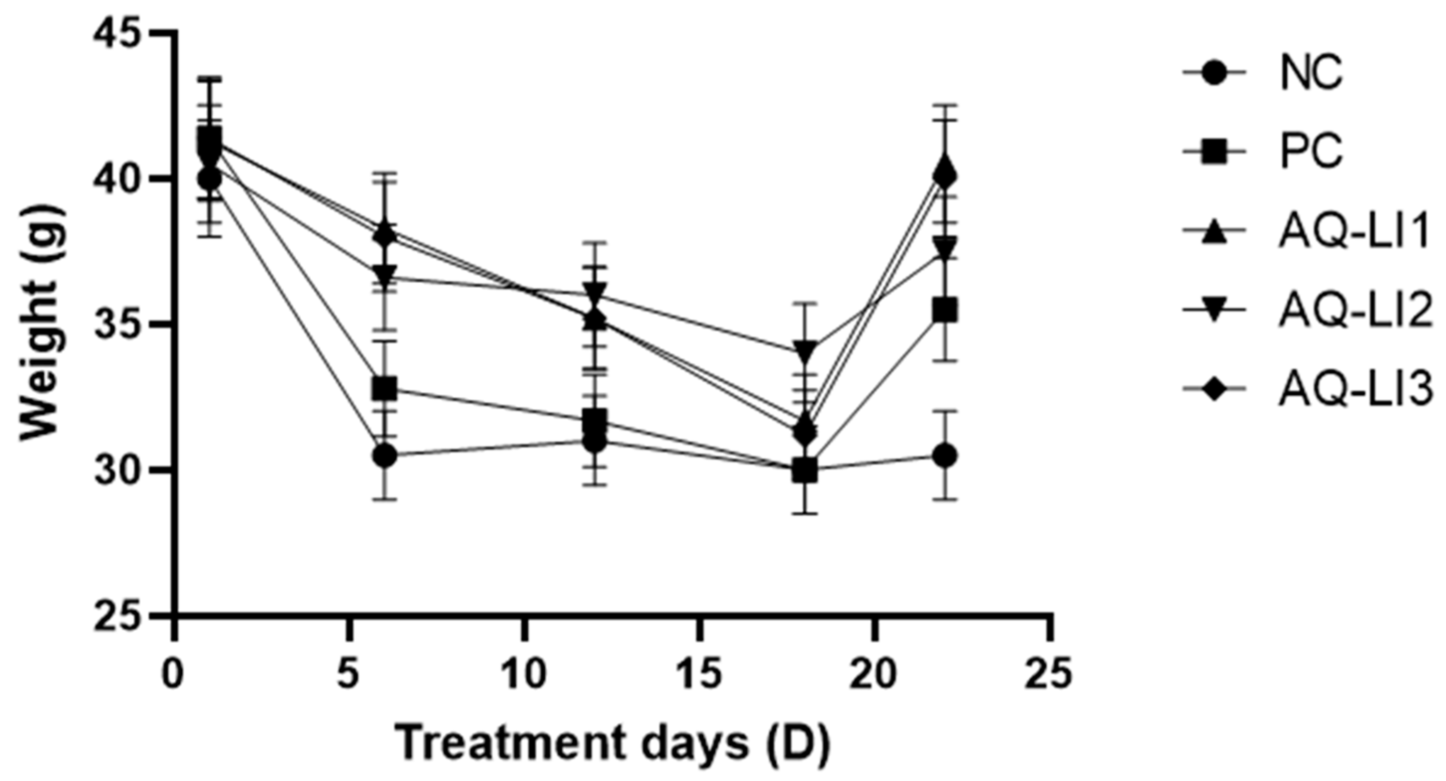
Variation in weight of mice during treatment
The results shown in Figure 5 illustrate the mice weight variation from each group AQ-LI1, AQ-LI2, AQ-LI3, NC and PC during treatment.
These results (Figure 5) showed that weights decreased during healing for all groups. The most significant decreases were recorded during the first 12 days. The groups treated with our formulations lost 6 g, while the NC and PC groups lost 9 g. This difference, noted in treated group with Flamazine, may be explained by negative effect of included chemical molecules in this product.
These results also show that just after total healing of burns in groups AQ-LI1, AQ-LI2 and AQ-LI3 at 22 days, mice were able to return to their initial weight. The analysis of variance related to the weight of the mice showed a significant difference between the 3 groups studied (ANOVA: df = 4; F = 2.63; p ≤ 0.001).
Weight is among the factors that correlate with wound healing process. Following burn, neuro–hormonal factors—notably, catecholamines, glucocorticoids and humoral mediators such as cytokines (IL1, IL6, TNF)—are generated. This results in a state of hyper catabolism in the patient’s body, characterized mainly by the breakdown of protein and carbohydrate nutritional stores by reducing insulin secretion and lipid stores requiring increased energy expenditure. These metabolic consequences are essential to provide the hyperactivated cells with the necessary nutritional elements (glucose, fatty acids, amino acids) for tissue recovery. The intensity of these processes is related to the severity of the injury. The results have led to the deduction that L. inermis leaves are an interesting plant whose leaf extracts could be used in burn treatment. However, pharmacodynamic and phytochemical studies need to be carried out to establish the mechanism of action of the developed formulations and recognize the active principles responsible for the healing effect.

4. Conclusions

In conclusion, this study demonstrated that the formulations of L. inermis showed a statistically significant effect on burn total healing durations and contraction percentage when compared to control groups (PC and NC). Burns treated with the formulations showed a decrease in burn surface and a variation in the percentage of contraction, with Tazarine variety (AQ-LI3) showing the most significant reduction in burn surface area. The traditional uses of L. inermis leaves were confirmed by the results obtained in the current research study, which is consistent with other studies that have shown that this species is used for healing wounds and burns.

Author Contributions

Conceptualization, F.E. and M.B. (Meryem Benjelloun); methodology, S.E.M. and L.E.G.; validation, F.E. and A.Z.; investigation, S.E.M., I.N. and M.B. (Meryem Benidir); resources, S.E.M. and M.B. (Meryem Benidir); writing—original draft preparation, S.E.M.; writing—review and editing, F.E., A.Z. and J.M.R.; and supervision, F.E. and L.E.G. All authors have read and agreed to the published version of the manuscript.

Funding

The research was supported by Functional Ecology and Environmental Engineering Laboratory funds of the Sidi Mohammed Ben Abdellah University, Morocco.

Institutional Review Board Statement

The animal study protocol was approved by the Institutional Review Board (or Ethics Committee) of the Fes Hospital-University Ethics Committee (CEHUF) protocol (code 001, approval date 2020) for studies involving animals.

Informed Consent Statement

Informed consent was obtained from all subjects involved in the study.

Data Availability Statement

Data are available from the corresponding author upon request.

Acknowledgments

The authors would like to acknowledge the COST Action 18101 SOURDOMICS—Sourdough biotechnology network towards novel, healthier and sustainable food and bioprocesses (https://sourdomics.com/; https://www.cost.eu/actions/CA18101/, accessed on 15 May 2023), where the author Z.A. is member of the working groups 2, 3, 4, 5, 7 and 8, and the author J.M.R. is the Chair and Grant Holder Scientific Representative and is supported by COST (European Co-operation in Science and Technology) (https://www.cost.eu/, accessed on 25 May 2023). COST is a funding agency for research and innovation networks. Author J.M.R. also acknowledges the Universidade Católica Portuguesa, CBQF—Centro de Biotecnologia e Química Fina—Laboratório Associado, Escola Superior de Biotecnologia, Porto, Portugal, as well as the support made by LA/P/0045/2020 (ALiCE) and UIDB/00511/2020-UIDP/00511/2020 (LEPABE) funded by national funds through FCT/MCTES (PIDDAC).

Conflicts of Interest

The authors declare no conflict of interest.

References

  1. Lydia, B.Y. Petite Histoire Du Maquillage; Stock: Paris, France, 1999. [Google Scholar]
  2. Hughes, K.; Ho, R.; Butaud, J.-F.; Filaire, E.; Ranouille, E.; Berthon, J.-Y.; Raharivelomanana, P. A selection of eleven plants used as traditional Polynesian cosmetics and their development potential as anti-aging ingredients, hair growth promoters and whitening products. J. Ethnopharmacol. 2019, 245, 112159. [Google Scholar] [CrossRef] [PubMed]
  3. Walter, P.; Martinetto, P.; Tsoucaris, G.; Brniaux, R.; Lefebvre, M.A.; Richard, G.; Talabot, J.; Dooryhee, E. Making make-up in Ancient Egypt. Nature 1999, 397, 483–484. [Google Scholar] [CrossRef]
  4. González-Minero, F.J.; Bravo-Díaz, L. The Use of Plants in Skin-Care Products, Cosmetics and Fragrances: Past and Present. Cosmetics 2018, 5, 50. [Google Scholar] [CrossRef] [Green Version]
  5. Bouhlal, T.; Benkhnigue, O.; Zidane, L.; Sobh, M.; Fadli, M. Vasculat Plants Used in Tradional Cosmetic by the Human Population in the Plain of the Gharb (Morocco) Full Paper. Nat. Prod. Indian J. 2013, 9, 326–330. [Google Scholar]
  6. Zrira, S. Some Important Aromatic and Medicinal Plants of Morocco. Med. Aromat. Plants World 2017, 3, 91–125. [Google Scholar] [CrossRef]
  7. Saive, M.; Frederich, M.; Fauconnier, M.-L. Plants Used in Traditional Medicine and Cosmetics in Mayotte Island (France): An Ethnobotanical Study. Indian J. Tradit. Knowl. 2018, 17, 645–653. [Google Scholar]
  8. Semwal, R.B.; Semwal, D.K.; Combrinck, S.; Cartwright-Jones, C.; Viljoen, A. Lawsonia inermis L. (henna): Ethnobotanical, phytochemical and pharmacological aspects. J. Ethnopharmacol. 2014, 155, 80–103. [Google Scholar] [CrossRef]
  9. El Ghanjaoui Mohammed Cervera, M.L.; El Rhazi, M.; De La Guardia, M. Assessment of Trace Elements in Traditional Moroccan Cosmetics by Inductively Coupled Plasma Atomic Emission Spectroscopy. Int. J. Sci. Technol. Res. 2014, 3, 104–112. [Google Scholar]
  10. Bafghi, M.F.; Salary, S.; Mirzaei, F.; Mahmoodian, H.; Meftahizade, H.; Zareshahi, R. Antibacterial and anti-trichomunas characteristics of local landraces of Lawsonia inermis L. BMC Complement. Med. Ther. 2022, 22, 203. [Google Scholar] [CrossRef]
  11. Al-Snafi, A.E. A review on Lawsonia inermis: A potential medicinal PLANT. Int. J. Curr. Pharm. Res. 2019, 11, 1–13. [Google Scholar] [CrossRef] [Green Version]
  12. Tang, Y.; He, W.; Yang, S.; Liu, L. Stabilisation and detoxification of henna (Lawsonia inermis L.) extract for hair dye cosmetics by spray-drying encapsulation. Color. Technol. 2019, 135, 439–450. [Google Scholar] [CrossRef]
  13. Obat, R.; Bosire, C. Formulation and evaluation of herbal lipstick using beta vulgaris and Lawsonia inermis as natural colorants. J. Phys. Appl. Sci. (JPAS) 2022, 1, 28–37. [Google Scholar] [CrossRef]
  14. Benzeid, H.; Gouaz, F.; Touré, A.H.; Bouatia, M.; Idrissi, M.O.B.; Draoui, M. Inventory of Toxic Plants in Morocco: An Overview of the Botanical, Biogeography, and Phytochemistry Studies. J. Toxicol. 2018, 2018, 4563735. [Google Scholar] [CrossRef] [Green Version]
  15. Singleton, V.L.; Rossi, J.A. Colorimetry of Total Phenolics with Phosphomolybdic-Phosphotungstic Acid Reagents. Am. J. Enol. Vitic. 1965, 16, 144–158. [Google Scholar] [CrossRef]
  16. Zhishen, J.; Mengcheng, T.; Jianming, W. The determination of flavonoid contents in mulberry and their scavenging effects on superoxide radicals. Food Chem. 1999, 64, 555–559. [Google Scholar] [CrossRef]
  17. Ba, K.; Tine, E.; Destain, J.; Cissé, N.; Thonart, P. Étude Comparative Des Composés Phénoliques, Du Pouvoir Antioxydant De Différentes Variétés De Sorgho Sénégalais Et Des Enzymes Amylolytiques De Leur Malt. Biotechnol. Agron. Soc. Environ. 2010, 14, 131–139. [Google Scholar]
  18. Hiai, S.; Oura, H.; Nakajima, T. Color reaction of some sapogenins and saponins with vanillin and sulfur1c acid. Planta Medica 1976, 29, 116–122. [Google Scholar] [CrossRef] [PubMed]
  19. Mottaghipisheh, J.; Iriti, M. Sephadex® LH-20, Isolation, and Purification of Flavonoids from Plant Species: A Comprehensive Review. Molecules 2020, 25, 4146. [Google Scholar] [CrossRef]
  20. Uddin, N.; Siddiqui, B.S.; Begum, S.; Ali, M.I.; Marasini, B.P.; Khan, A.; Choudhary, M.I. Bioassay-guided isolation of urease and α-chymotrypsin inhibitory constituents from the stems of Lawsonia alba Lam. (Henna). Fitoterapia 2013, 84, 202–207. [Google Scholar] [CrossRef]
  21. Ettayebi, K.; Errachidi, F.; Jamai, L.; Tahri-Jouti, M.A.; Sendide, K.; Ettayebi, M. Biodegradation of polyphenols with immobilized Candida tropicalis under metabolic induction. FEMS Microbiol. Lett. 2003, 223, 215–219. [Google Scholar] [CrossRef] [Green Version]
  22. Davidson, J.M. Animal Models for Wound Repair. Arch. Dermatol. Res. 1998, 290, S1–S11. [Google Scholar] [CrossRef] [PubMed]
  23. Cai, E.Z.; Ang, C.H.; Raju, A.; Tan, K.B.; Hing, E.C.H.; Loo, Y.; Wong, Y.C.; Lee, H.; Lim, J.; Moochhala, S.M.; et al. Creation of Consistent Burn Wounds: A Rat Model. Arch. Plast. Surg. 2014, 41, 317–324. [Google Scholar] [CrossRef] [PubMed] [Green Version]
  24. Nunes, J.P.S.; Dias, A.A.M. ImageJ macros for the user-friendly analysis of soft-agar and wound-healing assays. Biotechniques 2017, 62, 175–179. [Google Scholar] [CrossRef] [PubMed] [Green Version]
  25. Abeje, B.A.; Bekele, T.; Getahun, K.A.; Asrie, A.B. Evaluation of Wound Healing Activity of 80% Hydromethanolic Crude Extract and Solvent Fractions of the Leaves of Urtica simensis in Mice. J. Exp. Pharmacol. 2022, 14, 221–241. [Google Scholar] [CrossRef]
  26. Shivananda Nayak, B.; Isitor, G.; Davis, E.M.; Pillai, G.K. Wound healing activity of Lawsonia inermis linn. 827 The Evidence Based Wound Healing Activity of Lawsonia Inermis Linn. Phytother. Res 2007, 21, 827–831. [Google Scholar] [CrossRef]
  27. Dev, S.K.; Choudhury, P.; Srivastava, R.; Sharma, M. Antimicrobial, anti-inflammatory and wound healing activity of polyherbal formulation. Biomed. Pharmacother. 2019, 111, 555–567. [Google Scholar] [CrossRef]
  28. Rekik, D.M.; Ben Khedir, S.; Daoud, A.; Moalla, K.K.; Rebai, T.; Sahnoun, Z. Wound Healing Effect of Lawsonia inermis. Ski. Pharmacol. Physiol. 2019, 32, 295–306. [Google Scholar] [CrossRef]
  29. Mssillou, I.; Bakour, M.; Slighoua, M.; Laaroussi, H.; Saghrouchni, H.; Amrati, F.E.-Z.; Lyoussi, B.; Derwich, E. Investigation on wound healing effect of Mediterranean medicinal plants and some related phenolic compounds: A review. J. Ethnopharmacol. 2022, 298, 115663. [Google Scholar] [CrossRef]
  30. Hadisi, Z.; Nourmohammadi, J.; Nassiri, S.M. The antibacterial and anti-inflammatory investigation of Lawsonia inermis-gelatin-starch nano-fibrous dressing in burn wound. Int. J. Biol. Macromol. 2018, 107, 2008–2019. [Google Scholar] [CrossRef]
  31. Al-Snafi, A.E.; Talab, T.A.; Alfuraiji, N. The analgesic and anti-inflammatory effect of lawsone isolated from Lawsonia inermis. Sci. Pharm. Sci. 2022, 1, 77–84. [Google Scholar] [CrossRef]
  32. Goldenheim, P.D. An Appraisal of Povidone-Iodine and Wound Healing. Postgrad Med. J. 1993, 69, 97–105. [Google Scholar]
  33. De Francesco, F.; Riccio, M.; Jimi, S. Contribution of Topical Agents such as Hyaluronic Acid and Silver Sulfadiazine to Wound Healing and Management of Bacterial Biofilm. Medicina 2022, 58, 835. [Google Scholar] [CrossRef] [PubMed]
  34. Atiyeh, B.S.; Costagliola, M.; Hayek, S.N.; Dibo, S.A. Effect of silver on burn wound infection control and healing: Review of the literature. Burns 2007, 33, 139–148. [Google Scholar] [CrossRef] [PubMed]
  35. Klasen, H.J. A historical review of the use of silver in the treatment of burns. II. Renewed interest for silver. Burns 2000, 26, 131–138. [Google Scholar] [CrossRef]
  36. Seyed, J.H.; Ghasemali, K.; Azadbakht, M.; Peyman, Z.; Ghasemi, M.; Amirhossein, A. Effect of Aloe Cream versus Silver Sulfadiazine for Healing Burn Wounds in Rats. Acta Dermatovenerol. Croat. 2010, 18, 2–7. [Google Scholar]
  37. Khorasani, G.; Hosseinimehr, S.J.; Zamani, P.; Ghasemi, M.; Ahmadi, A. The Effect of Saffron (Crocus sativus) Extract for Healing of Second-degree Burn Wounds in Rats. Keio J. Med. 2008, 57, 190–195. [Google Scholar] [CrossRef] [Green Version]
  38. Bahramsoltani, R.; Farzaei, M.H.; Rahimi, R. Medicinal plants and their natural components as future drugs for the treatment of burn wounds: An integrative review. Arch. Dermatol. Res. 2014, 306, 601–617. [Google Scholar] [CrossRef]
  39. El Massoudi, S.; Benidir, M.; Chabir, R.; Benjelloun, M.; El Ghadraoui, L.; Errachidi, F. Morphological, Biochemical, and Climatological Analysis of Three Moroccan Henna Verities. Sci. World J. 2019, 2019, 1418456. [Google Scholar] [CrossRef] [Green Version]
  40. Manuja, A.; Rathore, N.; Choudhary, S.; Kumar, B. Phytochemical Screening, Cytotoxicity and Anti-inflammatory Activities of the Leaf Extracts from Lawsonia inermis of Indian Origin to Explore their Potential for Medicinal Uses. Med. Chem. 2020, 17, 576–586. [Google Scholar] [CrossRef]
  41. Raisagar, A.; Kaur, C.D.; Sawarkar, H.A.; Kumar, L.; Raisagar, A.; Karmakar, A.; Sahu, M. Comparative Study of Wound-Healing Effect of Bark Extracts of Ficus religiosa & Ficus benghalensis by Mice Model. J. Pharmacogn. Phytochem. 2019, 8, 1815–1821. [Google Scholar]
  42. Sahib, A.; Al-Jawad, F.; Alkaisy, A. Effect of Antioxidants on the Incidence of Wound Infection in Burn Patients. Ann. Burn. Fire Disasters 2010, 23, 199–205. [Google Scholar]
  43. Platon, J.F. Lipids in Cosmetology. Ol. Corps Gras Lipides 1997, 4, 275–281. [Google Scholar]
Figure 1. Molecular weight distribution of phenolic compounds and Lawsone in Alnif (a), Tafraoute Sidi Ali (b) and Tazarine (c) extracts.
Cosmetics 10 00092 g001
Figure 2. Evolution of the mean values and standard errors of burn surfaces of AQ-LI1, AQ-LI2, AQ-LI3, PC, and NC groups. Note: AQ-LI1: L. inermis from Alnif; AQ-LI2: L. inermis from Tafraoute Sidi Ali; AQ-LI3: L. inermis from Tazarine; PC: positive control; NC: negative control; D: days. Values expressed as mean ± SE to the mean, SE = Standard error; significance threshold at p ≤ 0.05 versus control (n = 5).
Cosmetics 10 00092 g002
Figure 3. Evolution of the mean values and standard errors of burn contraction of all groups. Note: AQ-LI1: L. inermis from Alnif; AQ-LI2: L. inermis from Tafraoute Sidi Ali; AQ-LI3: L. inermis from Tazarine; PC: positive control; NC: negative control; D: days. Values expressed as mean ± SE to the mean, SE = Standard error; significance threshold at p ≤ 0.05 versus control (n = 5).
Cosmetics 10 00092 g003
Figure 4. Observations of the burns treated with AQ-LI1, AQ-LI2 and AQ-LI3 and controls (TP and TN, where TP: positive control and TN: negative control) during 30 days. Note: AQ-LI1: L. inermis from Alnif; AQ-LI2: L. inermis from Tafraoute Sidi Ali; AQ-LI3: L. inermis from Tazarine; PC: positive control; NC: negative control; D: days. Values expressed as mean ± SE to the mean, SE = Standard error; significance threshold at p ≤ 0.05 versus control (n = 5).
Cosmetics 10 00092 g004
Figure 5. Variation (mean values and standard errors) in batch mouse weight of studied groups during treatment. Note: AQ-LI1: L. inermis from Alnif; AQ-LI2: L. inermis from Tafraoute Sidi Ali; AQ-LI3: L. inermis from Tazarine; PC: positive control; NC: negative control; D: days. Values expressed as mean ± SE to the mean, SE = Standard error; significance threshold at p ≤ 0.05 versus control (n = 5).
Cosmetics 10 00092 g005
Table 1. Phytochemical characteristics (mean values and standard errors) of studied Henna aqueous extract (AQ).
ExtractsAQ-LI 1AQ-LI 2AQ-LI 3
Aqueous extract (AQ) Yields (%)50.89 ± 3.0524.80 ± 1.7437.41 ± 1.90
Total phenolic compounds (g GAE)/100 g DM)13.48 ± 0.816.46 ± 0.508.34 ± 0.42
Total flavonoids (g QE/100 g DM)9.25 ± 0.554.42 ± 0.405.54 ± 0.28
Total tannin (g TAE/100 g DM)2.57 ± 0.151.43 ± 0.150.39 ± 0.05
Saponins (mg/100 g DM)291.70 ± 17.5083.38 ± 5.84321.50 ± 16.10
Note: AQ-LI 1: Alnif Aqueous Extract; AQ-LI 2: Tafraoute Sidi Ali Aqueous Extract; and AQ-LI 3: Tazarine Aqueous Extract.
Table 2. Mean values and standard errors of the epithelialization time periods.
Ointment FormulationAQ-LI1AQ-LI2AQ-LI3PCNC
Epithelialization period (day)20.33 ± 4.6521.67 ± 5.119.33 ± 0.0530.67 ± 3.6342.33 ± 2.45
Note: AQ-LI1: L. inermis from Alnif; AQ-LI2: L. inermis from Tafraoute Sidi Ali; AQ-LI3: L. inermis from Tazarine; PC: positive control; NC: negative control. Values expressed as mean ± SE, SE = Standard error, (n = 5) for each extract.
Disclaimer/Publisher’s Note: The statements, opinions and data contained in all publications are solely those of the individual author(s) and contributor(s) and not of MDPI and/or the editor(s). MDPI and/or the editor(s) disclaim responsibility for any injury to people or property resulting from any ideas, methods, instructions or products referred to in the content.

