
Nanogels Based on Hyaluronic Acid as Potential Active Carriers for Dermatological and Cosmetic Applications


1
Department of Chemistry, Materials and Chemical Engineering “G. Natta”, Politecnico di Milano, Piazza Leonardo da Vinci 32, 20133 Milan, Italy
2
R&D Center, Nanotechnology Unit, Academy of History of Healthcare Art, Lungotevere in Sassia 3, 00186 Rome, Italy
3
National Institute of Chemical Physics and Biophysics, Akadeemia tee 23, 12618 Tallinn, Estonia
4
Department of Science and Technology for Sustainable Development and One Health, Università Campus Bio-Medico di Roma, Via Á. del Portillo 21, 00128 Rome, Italy
*
Author to whom correspondence should be addressed.
Cosmetics 2023, 10(4), 113; https://0-doi-org.brum.beds.ac.uk/10.3390/cosmetics10040113
Submission received: 19 June 2023
/
Revised: 1 August 2023
/
Accepted: 3 August 2023
/
Published: 7 August 2023
(This article belongs to the Special Issue Advanced Cosmetic Sciences: Sustainability in Materials and Processes)
Abstract
:Nanogels are a prominent research topic in biomedical and drug delivery applications. The versatility of their chemistry allows them to be tailored both to carry and release a wide range of active molecules, and to target specific tissues or cell types. Within a vast field of possible chemical designs, nanogels based on hyaluronic acid seem particularly interesting from the standpoint of dermatological and cosmetic applications, due to the well-known involvement of hyaluronic acid in several fundamental processes related to skin health and ageing. In spite of this, relatively few studies about these nanocarriers and their potential skin-related benefits have appeared so far in the literature. With the aim to stimulate further interest in the topic, in this review, we provide information on hyaluronic acid-based nanogels, including their key physicochemical properties, their typical drug release behavior, and the main synthetic methodologies. The latter include: approaches based on spontaneous self-assembly of polymer molecules; approaches based on chemical cross-linking, where nanogel formation is promoted by covalent bonds between polymer chains; and hybrid approaches that leverage a combination of the above two mechanisms. We believe this body of information, which we collected by going through the relevant literature from the past 10–15 years, offers cosmetic formulators plenty of options to design innovative products.
1. Introduction
In recent years, nanogels have been a key biomedical research topic, particularly in the context of drug delivery applications [1,2,3]. These materials have the ability to behave like spongy nanoparticles, which can absorb a broad range of pharmaceutical actives and can slowly release them in the physiological environment, where a certain therapeutic effect is desired. Their name—nanogels—is linked to the fact that they share several properties with macroscopic hydrogels, both in terms of absorption/drug loading capacity and from a microscopic structure standpoint. However, a key difference versus bulk hydrogels is the huge surface area deriving from their nanoparticle nature, which makes mass transfer processes much more efficient and enables the systemic administration of nanogels, whereas the applicability of bulk hydrogels as drug carriers is limited to local treatment.
The choice of the building blocks is a key issue to fine-tune the formation and the physicochemical features of the polymeric networks. Both natural and synthetic polymers are eligible for nanogel synthesis: the former are generally proteins (such as albumin, fibroin, collagen, gelatin, soy-derived proteins), oligosaccharides, or polysaccharides (including hyaluronic acid, chitosan, alginate, chondroitin sulfate, agarose, cellulose, and heparin), whereas the synthetic counterparts can be polyethylene glycol (PEG), polyamines, polylactic acid (PLA), poly(-caprolactone), or customized polymers, synthesized through different radical polymerization strategies. Examples are controlled radical polymerization (CRP), reversible addition–fragmentation chain transfer (RAFT) polymerization, and atom transfer radical polymerization (ATRP), which ensure the conjugation of monomers/oligomers characterized by different chemical motifs to obtain specific physicochemical features, such as amphiphilic, zwitterionic, and stimuli-resposive behavior, or to meet biocompatibility criteria by grafting biomolecules on the polymer backbone [4,5,6].
Beyond a wide range of building blocks and synthetic strategies, several manufacturing procedures have been developed or reapplied to produce nanogels, including emulsion/evaporation [7,8], nanoprecipitation [9,10], and microfluidics [11,12,13].
Additionally, nanogels can be designed in order to target specific cell types, for instance, cancer cells. This has led to the synthesis of nanogel particles based on the cross-linking of a relatively broad range of polymers, as well as to the so called “decoration” of the nanogel surface with suitable functional groups. For example, nanoscaffolds based on hyaluronic acid, either alone or copolymerized, have been synthesized with the objective to selectively target the hyaluronan receptor CD44, which is overexpressed on the surface of several tumor cells.
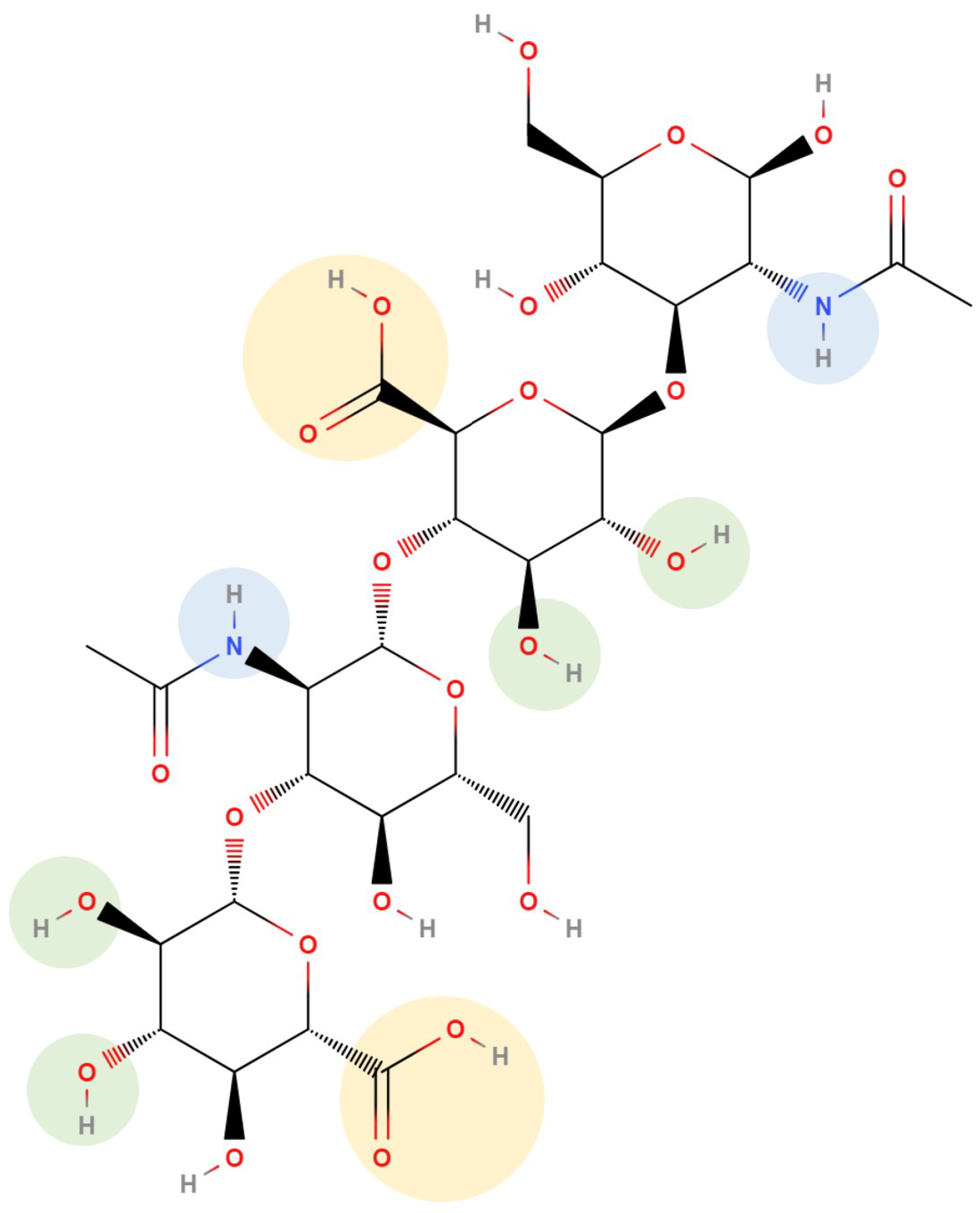
From the standpoint of potential dermatological and cosmetic applications, nanogels based on hyaluronic acid are particularly interesting, due to the involvement of this ubiquitous mucopolysaccharide in several physiological processes, many of which are highly relevant to the skin. As is well-known, hyaluronic acid (chemical structure shown in Figure 1) is a predominant component of the skin extracellular matrix and plays a fundamental role in skin hydration. Its progressive decrease in the epithelial tissues of elderly people has been shown to correlate with a loss of elasticity, wrinkle appearance, and other visible signs of skin ageing [14]. Therefore, the use of nanogels based on hyaluronic acid as drug carriers in a dermatological or cosmetic context offers an opportunity to combine the advantages of sustained drug release with the known biocompatibility, non-immunogenicity [15], and skin health benefits of hyaluronic acid. As a consequence, the latter, from a formulation efficiency point of view, has the potential ability to perform several functions at the same time.
In what follows, we will review the key physicochemical properties of these materials, the main synthetic routes described in the literature from the past 10–15 years, and the most studied drug release applications, with the objective to help assess their potential applicability in the dermatological and cosmetic field. While a few studies on dermatological applications of hyaluronic acid-based nanogels have been published over the past 2–3 years and will be reviewed in a dedicated section of the present work, the topic seems to still be relatively unexplored compared to the large amount of papers concerning both the use of “plain” hyaluronic acid in skin care, and the evaluation of nanogels obtained from a variety of polymer systems for systemic drug delivery.
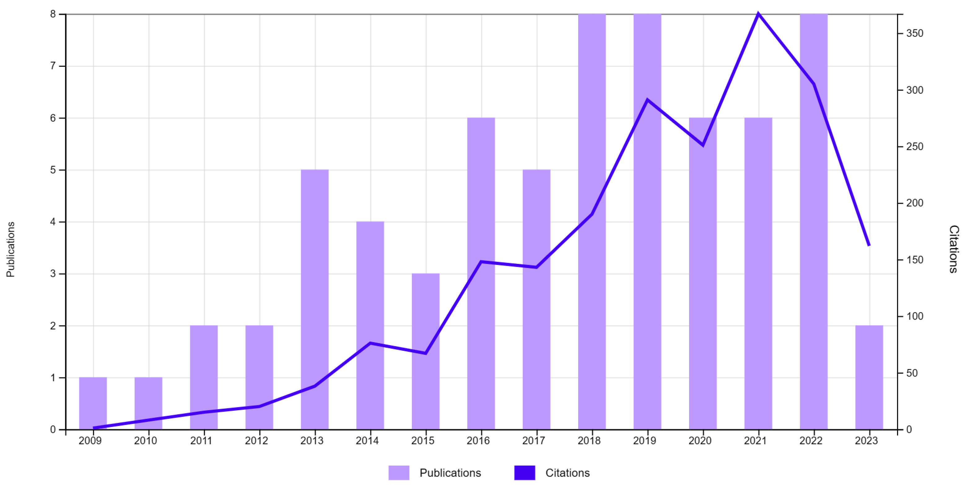
Consequently, among a multitude of nanogel systems described in the literature, we will focus on nanogels that are based on hyaluronic acid only or a combination of hyaluronic acid with other polymers. Therefore, even when using the term “nanogel” without specifying its chemical composition, it is intended that our discussion and cited references will mainly concern the above mentioned subclass of the materials. In addition, we will focus on hyaluronic acid-containing nanogels that have been designed and studied with the main objective to obtain a drug delivery system, as opposed to other potential therapeutic or diagnostic applications. Finally, since our aim is to highlight hyaluronic acid-based nanogels that can be used in both dermatological and cosmetic products, we will disregard nanogel formulations that do not lend themselves well to either application, for instance, due to toxicity or other limitations, or where hyaluronic acid is present but is not one of the main constituents. Figure 2 shows the evolution of the number of publications and citations over the past 15 years on the topic of nanogels based on hyaluronic acid.
2. Physicochemical Properties of Nanogels
Nanogels are made of a 3D network obtained from the physical or chemical cross-linking of one or several polymers [2,16]. Nanogel particles usually have a spherical shape; however, recent nanoparticle fabrication techniques make it possible to obtain nanogels with specific shapes [17,18,19,20]. The average size of nanogel particles can range from about 100 nm up to about 500 nm, depending on the nanogel composition and synthetic procedure.
A peculiar feature of these 3D networks, containing hydrophilic polymers, is the swelling behavior, which is magnified in the transition from a dry to a hydrated state. This has been documented for several nanogel systems. For instance, Fasiku et al. [21] compared the weight of a completely dried hyaluronic acid-based nanogel sample to that of the same sample after equilibration with a PBS solution at room (25 °C) and body temperature (37 °C). They found a sample weight increase after hydration of up to more than at RT, and up to more than at 37 °C. Nanogel swelling has also been quantified by particle size measures in the dry versus hydrated state. Limiti et al. [7] demonstrated this in the case of a hyaluronic acid/polyethyleneimine nanogel by determining the nanogel particle diameter in the dry state through AFM and SEM measures, and comparing this data to DLS particle size results after dispersing the nanogel in a PBS solution. They found their nanogel particles to exhibit an average hydrodynamic diameter of about 220 nm when fully hydrated (measured by DLS), whereas the same particles had an average diameter of only about 114 nm in completely dry conditions, measured by TEM.
This swelling behavior can be advantageously exploited as a release trigger: in dry or anhydrous conditions, the 3D polymer network is more shrunk and has a smaller mesh size, which tends to keep payload molecules trapped inside the nanoparticle. Instead, when the polymer chains constituting the nanogel become hydrated and, therefore, more elongated, the internal mesh size of the nanoparticle increases, favoring the payload release.
Thanks to their hydrophilic character, nanogels based on hyaluronic acid have been used to deliver hydrophobic therapeutic agents, for example, curcumin [22,23,24] and asiatic acid [25], which otherwise tend to have a rather poor water solubility and bioavailability.
Release kinetics data obtained from various nanogel systems shows that the release mechanism is essentially Fickian diffusion. In most cases, drug release curves in a PBS solution or water can be satisfactorily modeled by Fick’s second law. For spherical particles, the corresponding equations can be analytically solved under the assumption of “perfect sink” initial and boundary conditions, resulting in an exponential time dependence of the released payload fraction [26,27]:
where is the total mass of drug released at time t, is the total mass of drug released at infinite time, n is a dummy variable, D is the diffusion coefficient, and r is the nanoparticle radius. It has been shown that Equation (1) can be approximated by the empirical expression [26]:
Here, k is a constant determined by the nanoparticle composition and n is related to particle geometry and the transport mechanism. For Fickian diffusion and spherical particles, by setting , Equation (2) provides a good approximation of Equation (1) for the first 60% of the release curve [26] and has been successfully used to interpolate an experimental drug release data [7,13].
Another significant feature of nanogels, which is of relevance for dermal applications, is their softness. As recently discussed by Scotti et al. [28], nanogel softness plays a crucial role in the adsorption of nanoscaffolds to cells, the production of platelet-like structures for the augmentation of haemostasis [29], and the controlled drug delivery performance. Softness is defined by multiple parameters linked to the energetic variation of deformation, swelling, osmotic deswelling, faceting, and interpenetration. Each parameter provides a specific equation to quantify softness, as reported in Table 1.
The most common approach to quantify nanogel softness is by comparing the elastic free energy of deformation with the thermal energy of a particle, as described in Equation (3). In this case, can be estimated by the Young’s modulus (Y) and the radius (R) of the particle, according to the following equation:
depends on the nanogel concentration c. Furthermore, the nanogel softness can be modulated by the synthetic route: the crosslinker content affects the spatial distribution and entanglement of the polymeric chains leading to a more or less packed nanoscaffold. The lower the cross-linker amount, the higher the swelling capability of the polymeric network. Additionally, the use of linear or branched polymers can play a key role in determining the final physical features of the nanoscaffold: generally, polymer backbones with pendant groups or side chains are characterized by a distinct steric hindrance, which decreases the ability of the chains to be tightly packed and reduces the flexibility and elasticity of the final structure [30]. In particular, a branching unit structure containing an “X”-type crosslinkage with four chains radiating out gives rise to a more rigid nanogel with a lower swelling ratio, compared to a “T”-shaped linkage enabling a more deformable structure [31,32]. Recently, the fabrication of nanogels using branched hyaluronic acid, i.e., through conjugation of the polysaccaride with oligomeric hyaluronic acid or with biomolecules, glycosidic structures, or polymers, is proposed for topical dermal delivery targeting skin dendritic cells [33] or therapeutic activity [34].
The variables involved in the aforementioned equations can be experimentally estimated through different techniques. Generally, dynamic or static light scattering (DLS or SLS), and small-angle neutron or X-ray scattering (SANS or SAXS) are selected to collect data on the characteristic lengths (e.g., hydrodynamic diameter, radius of gyration, molecular weight, thickness of the external layer in a core-shell configuration) and atom force microscopy (AFM) for compressibility/deformation studies.
Additionally, due to their high surface-to-volume ratio, nanogels can be classified as highly interfacial-active nanomaterials. When deposited at a liquid–liquid or air–liquid interface, they deform but preserve their mobility, which can lead to agglomeration or the formation of monolayers, whereas at a solid–liquid interface, nanogels tend to stretch at the interfacial plane. In both cases, the nanoscaffolds are characterized by elastic modulus gradients. For these reasons, a further definition of nanogel softness at the interface is provided by Scotti et al. [28]:
where is the radius of the nanogel at the interface and the hydrodynamic radius in solution. The higher the value of , the softer the nanogels, i.e., the higher the deformation upon adsorption. Computer simulations are developed to study in-depth and predict nanogel behavior at the interface [35,36].
A third important feature of nanogels for topical applications is their degradation [37]. The hydrolysis of ester bonds, chemically labile crosslinkers, and enzymatic cleavage are the main mechanisms of erosion of the nanoscaffolds. Furthermore, a controlled degradation can be introduced through the rational choice of chemical crosslinking between the polymeric chains and the modification of the polymer backbone. Hyaluronan-based nanogels can be degraded by hyaluronidase after binding to specific cell membrane receptors [38]; however, the presence of ester, amide, or sulphide linkages between the polymer chains (both as neat hyaluronic acid or a combination of the polysaccharide with other polymers) promotes a modulated degradation of the network, driven by the surrounding environment (i.e., pH variations, enzymes, aminoacids, oxidants) [39,40,41].
Additionally, the choice of the molecular weight (MW) of the hyaluronic acid can affect nanogel performances, in particular, adhesive, rheological, and stability properties, and transdermal drug delivery [42,43]. The two most common production processes for hyaluronic acid, i.e., extraction from animal tissues and bacterial fermentation, both provide different chain lengths of the biopolymer. Hyaluronic acid characterized by a MW in the range 10–500 kDa exhibits good viscoelasticity, moisture retention, mucoadhesion, and anti-angiogenic activity, which are desirable features in cosmetics, wound healing, ophthalmology, and orthopedic applications. Below 4 kDa, the polysaccaride promotes angiogenesis, inducing the expression of inflammatory mediators and inhibiting tumor growth; instead, a MW higher than 500 kDa corresponds to hyaluronic acid that has anti-angiogenic activity, natural immunodepressant properties, and is also eligible as a inert space-filler for dermal application and tissue regeneration [44,45,46].
The shear-thinning behavior and softness of hyaluronic acid are related to intramolecular hydrogen bond breaking and chain deformation, which trigger a hydrophobic effect from the aliphatic rings; a higher MW leads to an increase of the zero-shear viscosity of the biopolymer in solution [47].
The MW of hyaluronic acid also impacts the cytotoxicity of nanogels and their time-dependent cellular uptake, as mentioned by Zhong et al. [48]. Nanoscaffolds synthesized with lower MW hyaluronic acid are internalized in larger quantities compared to those prepared with a higher MW biopolymer. On the other hand, a higher MW hyaluronic acid exhibits a greater affinity to CD44 receptors thanks to the multivalent binding interactions.
Skin penetration and transdermal delivery are affected by hyaluronic acid MW: the use of longer biopolymer chains in the nanogel formulation results in less effective skin penetration, which may be due to a lower mechanical strength of the nanoscaffold, insufficient to cross the dermal layer. In terms of sustained drug release, a clear trend associated to the increase/decrease of hyaluronic acid MW is not detectable; however, at intermediate MW values (order of magnitude 100 kDa), drug release seems to be longer lasting due to an optimal balance between the mechanical properties of the nanogel and the polysaccharide-drug physicochemical interactions [42].
The aforementioned properties are, as a matter of fact, parameters that can be tuned to produce new nanogel formulations eligible for skin applications.
3. Highlights in the Nanogel-Mediated Release of Therapeutics
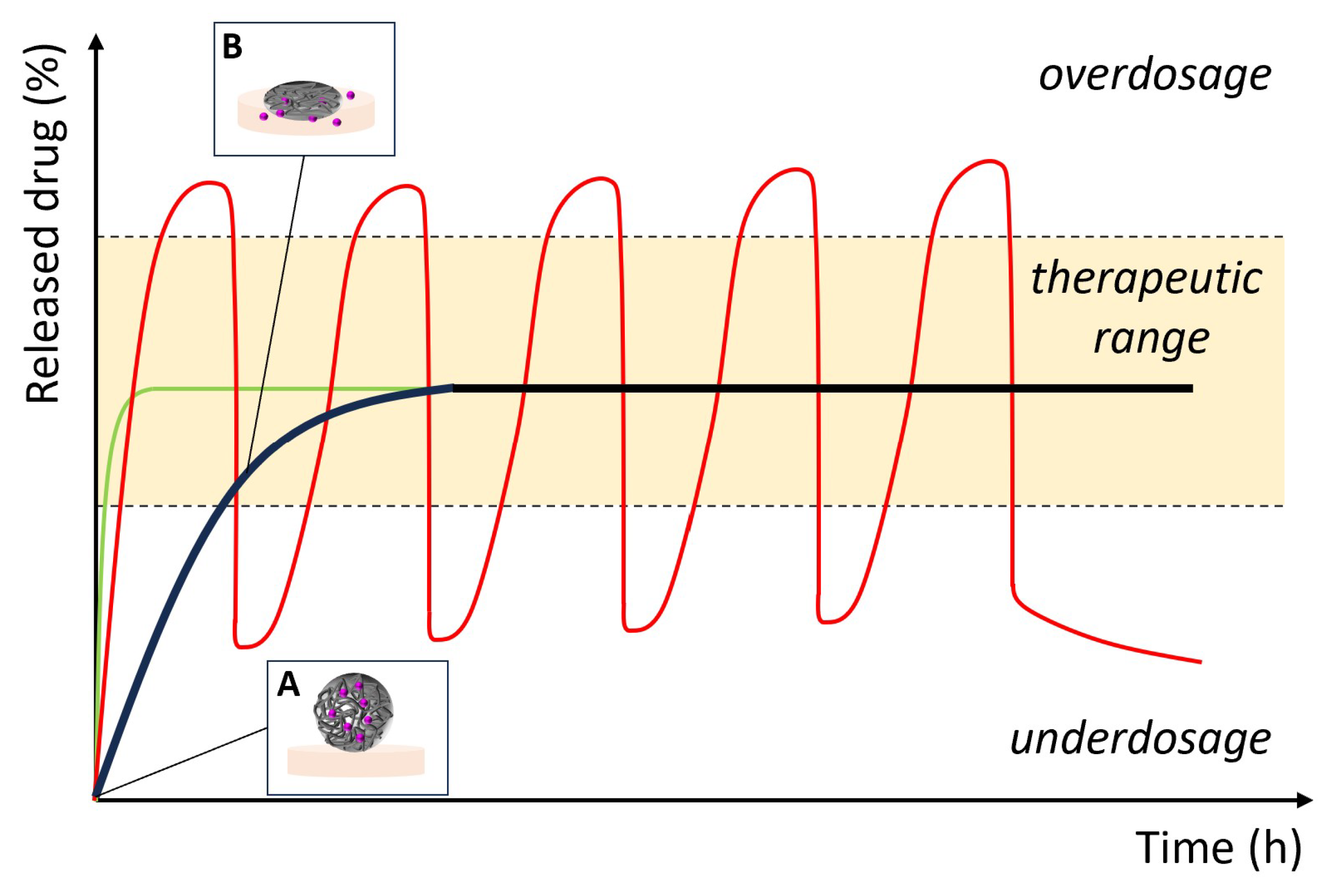
The principles of controlled drug release can also be applied in the dermatological and cosmetic field. Minor scrapes, burn wounds, aging, and skin diseases require the use of different types of therapeutics to avoid worsening of the condition and prevent the spread of infections. Anti-inflammatory drugs, coenzymes, antioxidants, and polyphenols represent the main classes of used therapeutics [49,50,51]. The delivery of the nanoencapsulated molecules and their controlled release is aimed to (Figure 3):
- provide a continuous and sustained availability of the curative effect on the target area;
- maintain the drug concentration within therapeutic values, avoiding under- and overdosing, for a prolonged period of time;
- reduce the number of administrations, increasing the patient’s compliance.
Drug delivery by topical route is challenging due to the multi-layered structure of the skin. In particular, the stratum corneum of the epidermis, due to its composition in proteins and lipids, limits the penetration of therapeutics up to the dermis and hypodermis and controls the exchange of water, oxygen, and chemicals. For these reasons, the formulation of nanocarriers is of great interest [52]. Nanogels can be used to momentarily overcome the skin-barrier functions and enhance drug delivery. In particular, tuning the polarity and the hydrophilic–hydrophobic balance of the nanonetwork can be carried out thanks to the conjugation of hyaluronic acid with other motifs. Additionally, in this context, a significant plus of nanogels is related to their sponge-like behavior, which makes it possible to decouple their synthesis from the drug encapsulation phase. Indeed, nanogels (in dry state) can be stored for several months, preserving their structural integrity, and picked up when needed, obtaining a ready-to-use drug delivery system.
Not only therapeutics but also pigments and inorganic compounds can be encapsulated in nanogels, extending the considerations made above to cosmetic applications. In this case, any systemic absorption that may occur is in principle undesirable, to avoid the risk of systemic effects. Temperature gradients, skin pH, enzymatic concentrations, or redox potentials are the studied parameters to trigger the release of the payload in the desired dermal region [52].
4. Nanogel Synthetic Approaches
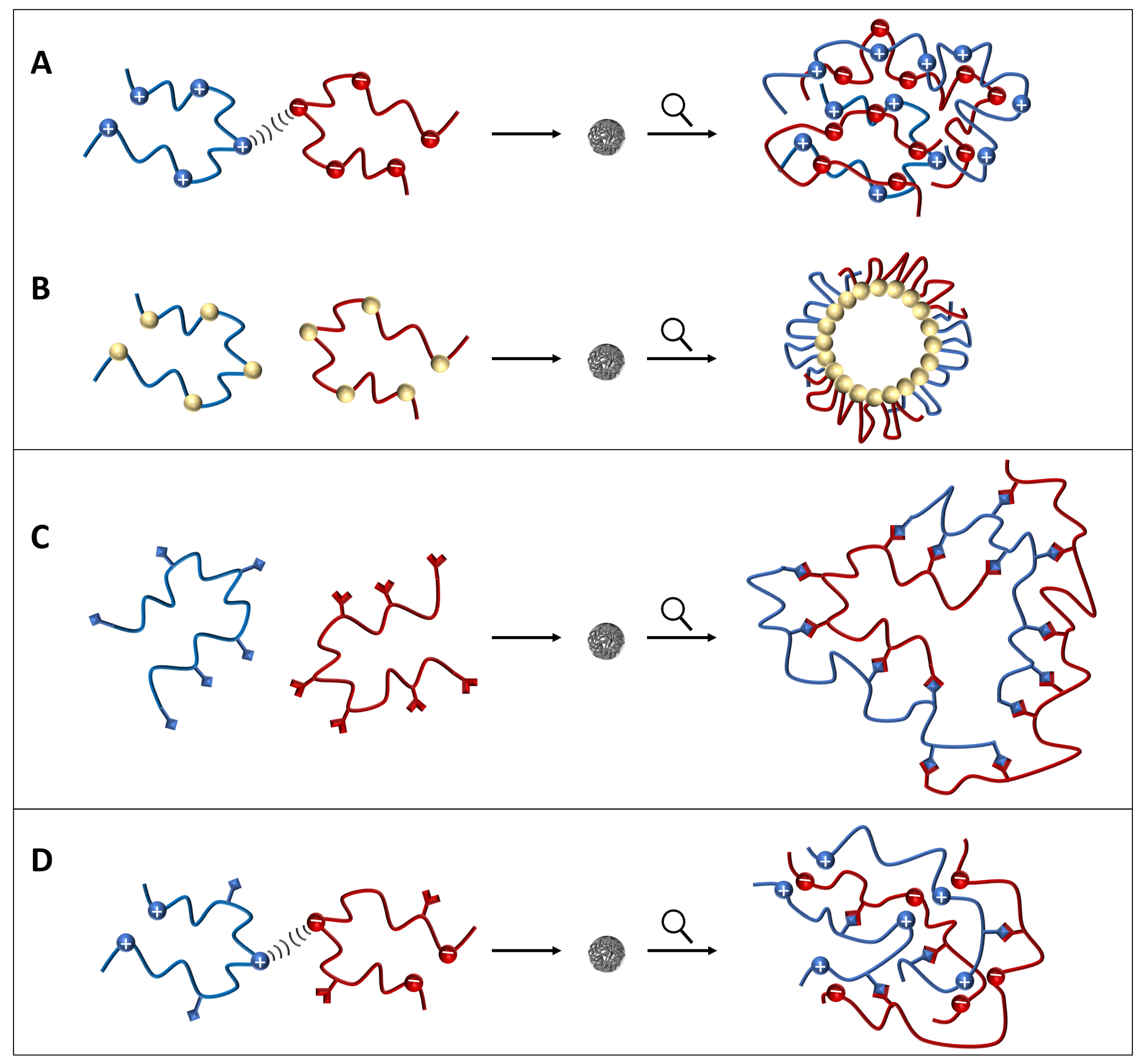
The synthetic approaches used to make nanogels can be divided into three large categories (Figure 4):
- 1.
- Approaches based on self-assembly, where nanogel formation is driven by physical interactions (ionic or van der Waals’ forces) between polymer molecules that self-aggregate spontaneously. Such polymer molecules may all have the same chemical structure or, more frequently, two or several different polymers may participate in nanogel particle formation.
- 2.
- Approaches based on chemical cross-linking, where nanogel particle formation is promoted by covalent bonding between the constituting polymer molecules.
- 3.
- Hybrid approaches, where a combination of the above two methods is used.
4.1. Approaches Based on Self-Assembly
One of the earliest works describing nanogel synthesis concerns a core-shell nanogel system, where liposomal vesicles are coated with alternating layers of hyaluronic acid and chitosan using a purely physical process, which provides nanogels of <300 nm diameter [53]. Another approach leading to core-shell nanogel particles is based on hyaluronic acid grafted with PEG, which is used as a shell for deacetyl mycoepoxydience (DM) nanocrystals [54].
Various methods based on physical self-assembly utilize the modification of hyaluronic acid with hydrophobic groups. For instance, hyaluronic acid can be modified with colesteryl groups, obtaining a derivative that in water or PBS solution spontaneously produces nanogel particles with a diameter ranging from about 50 to about 150 nm, depending on the degree of substitution of the cholesteryl groups [55]. The topic of hyaluronic acid modification through the introduction of various types of steryl groups, to obtain derivatives that can spontaneously form complexes with various drugs, is also discussed in some recent patents [46,51]. The self-assembly properties of cholesteryl–hyaluronic acid can be exploited by further functionalizing the modified polymer through its esterification with hydrophobic drug molecules, such as curcumin, which carries hydroxyl functionalities able to react with the carboxylic groups of hyaluronic acid [56,57].
The self-assembly of hyaluronic acid has also been induced by its modification with relatively long hydrophobic alkyl chains, such as C11 or C16, resulting in the formation of amphiphilic hyaluronic acid conjugates that spontaneously aggregate into nanogels [23,58].
Another interesting example is the one-pot synthesis of a conjugate between PVA grafted with doxorubicin and hyaluronic acid, modified with pyrene ligands, which is enabled by the functionalization of hyaluronic acid with orthogonally reactive aldehyde and thiol groups. The resulting conjugate self-assembles in an aqueous medium, yielding nanoparticles with an average diameter of ≈200 nm [59].
An approach where hyaluronic acid is used without chemically modifying it consists in obtaining ionic complexes of the mucopolysaccharide with a PEG-polylysine–polyalanine (PEG-PK-PA) triblock copolymer. By varying the weight ratio between hyaluronan and the PEG-PK-PA copolymer, one can obtain self-assembled nanogels with zeta potential values ranging from −47 mV to +47 mV, which exhibit different cell internalization efficiencies and different degrees of cytotoxicity [60].
Water and coworkers describe a nanogel system where the self-assembly, facilitated by the use of microfluidics, occurs between the active species to be released, which is the peptide novicidin, and hyaluronic acid modified with octenyl succinic anhydride. This allows the decrease of the systemic toxicity and improves the chemical stability of novicidin [61]. It is also possible to make “ternary” nanogels based on the self-assembly of three different molecules, one of which is a chemotherapeutic agent [62].
Agnello et al. use microfluidics to make self-assembled nanogels from a hyaluronic acid derivative, functionalized with octadecyl and ethylene diamine groups, which is precipitated by the ionic strength of a DPBS (Dulbecco’s phosphate-buffered saline) solution [63].
An approach that presents interesting features also from a manufacturing process standpoint, is described by Simonson et al., who obtain non-chemically cross-linked nanogels by spraying nanodroplets of hyaluronic acid onto a solution containing -poly-L-lysine (PLL). The hyaluronic acid nanodroplets are produced by electrospray ionization. In these conditions, hyaluronic acid and PLL form nanogel particles by self-assembly due to electrostatic interactions. The incorporation of small molecules and proteins into the nanogel phase is achieved by simply co-dissolving the above actives into the PLL solution where the nanogels are formed [64].
Win and coworkers synthesize hyaluronic acid grafted with poly(N-isopropylacrylamide) (pNIPAM), which then self-assembles in water, generating nanogels under sonication. The resulting nanoparticles can be easily loaded with a natural triperpene like asiatic acid [25].
Xu et al. make a conjugate between hyaluronic acid and selenocystamine, which is then used to obtain a nanogel via physical crosslinking through a water-in-oil emulsification approach. The resulting nanogel particles are characterized by a CD44-targeting ability, thanks to hyaluronic acid, and free radical scavenging/anti-inflammatory properties, thanks to the presence of diselenide bonds (Se-Se) [65].
Duan and coworkers describe a nanogel obtained from the self-assembly of polyethylenimine with -cyclodextrin-conjugated hyaluronic acid, which forms the nanoscaffold skeleton through eletrostatic interactions. This is then followed by the addition of cisplatin molecules, which provide crosslinking junctions inside the nanogel via coordination bonds between Pt and the carboxylic groups in the -cyclodextrin-hyaluronic acid conjugate [66]. Another approach based on -cyclodextrin-conjugated hyaluronic acid is presented by Kaewruethai et al., who make a coupling reaction between amine-terminated poly(N-isopropylacrylamide) and -cyclodextrin-functionalized hyaluronic acid. The resulting polymer is used to prepare a self-assembled nanogel by sonication in water, where -cyclodextrin provides an additional drug loading capacity [67].
4.2. Approaches Based on Chemical Crosslinking
Among synthetic approaches that do not rely on self-assembly, Messager et al. describe nanogels obtained from photoinduced polymerization of hyaluronic acid modified with polymerizable methacrylate groups. The polymerization is conducted by dissolving the modified hyaluronic acid in the aqueous phase of a water-in-oil nanoemulsion, followed by irradiation. By varying the nanoemulsion droplet size, irradiation time, and degree of methacrylation, one can control both the final nanogel particle size and swelling behavior, which is a consequence of the degree of crosslinking [71]. Methacrylated hyaluronic acid can be copolymerized with di(ethylene glycol) diacrylate to provide enzyme-sensitive, crosslinked nanogels, using potassium dipersulfate as a radical initiator [40]. Methacrylated hyaluronic acid can also be used to prepare pH-sensitive, acid-degradable nanogels, by employing 2,2-dimethacroyloxy-1-ethoxypropane (DMAEP) as a pH-labile crosslinker [72].
Yu and coworkers present a method to make nanogel particles that are synthesized inside cancer cells by the in situ crosslinking of methacrylated hyaluronic acid, which has been adsorbed on inorganic nanoparticles containing a crosslinker and a photoinitiator. The crosslinking occurs by irradiation, after internalization of the inorganic nanoparticles into cancer cells. The obtained nanogels show a significantly better internalization efficiency versus hyaluronic acid nanogels prepared by other methods [73].
To further strengthen the tumor targeting ability of hyaluronic acid, in some works, the latter is combined with other molecules that are also overexpressed in cancer cells. For instance, Lin et al. make a conjugate of hyaluronic acid with doxorubicin via a legumain substrate peptide bridge. Legumain is upregulated in several solid tumors. This conjugate is subsequently crosslinked to obtain nanogel particles with a polygonal shape [74].
It is also possible to make pH-responsive nanogels, which exhibit a slow release at an acidic pH (e.g., pH = 5) and a minimal release at a neutral pH. This behavior is shown, for instance, by nanogels obtained through a Michael addition-type reaction between thiol-modified hyaluronic acid and a diacrylated EO-PO-EO block copolymer [75]; by nanogels synthesized from the reaction between methacrylated hyaluronic acid and a crosslinker containing ortho ester groups [76]; and by nanogels obtained from the crosslinking between (a) boronic acid-conjugated lactose-modified chitosan and (b) dopamine- and nitric oxide-conjugated partially carbonized hyaluronic acid [77]. The mild acidity of the cancer cell microenvironment is also exploited as a release trigger in Zhu et al. Using cholesteryl methacrylated hyaluronic acid, they synthesize crosslinked nanogels that entrap acid-activatable hyaluronidase. The latter enzyme promotes the payload release, by degrading hyaluronic acid in response to a pH shift towards the acidic range [78].
In some works, the same conjugating agent has been used both to bind a certain compound to the hyaluronic acid backbone, and to cause crosslinking of the hyaluronic acid chains. This is done, for instance, in Choi et al., where the compound conjugated to hyaluronic acid is dihydroxyflavone, a polyphenolic species with strong antioxidant activity, and the conjugating agent is 4-(4,6-dimethoxy-1,3,5-triazin-2-yl)-4-methylmorpholinium chloride (DMTMM) [79].
Tian and coworkers fabricate nanogels through a first crosslinking between a poly(ethylene glycol) diglycidyl ether and hyaluronic acid, followed by a second crosslinking that results from the introduction of cystamine in the nanogel network [80]. The glycerol diglycidyl ether is used as a crosslinker by Suner et al. to make nanogels based on both plain hyaluronic acid, and a hyaluronic acid:sucrose copolymer [81]. Like in the work by Tian et al., cystamine is also used as a crosslinker in a study by Chen et al. However, in this case, the polymer that becomes conjugated with hyaluronic acid is poly(N-isopropylacrylamide) (pNIPAM), allowing nanogels to be synthesized that are at the same time thermoresponsive and capable of glutathione-triggered drug release [82]. Thermoresponsiveness is achieved by Xu and coworkers by making cysteine-modified hyaluronic acid, which is then grafted with pNIPAM [83].
Cao and coworkers make a crosslinked nanogel based on hyaluronic acid and -cyclodextrin, which are linked together by amide bonds, exploiting the hydrophobic cyclodextrin cavity to encapsulate a pharmaceutical active like auraptene, while hyaluronic acid performs the “usual” function of promoting targeted delivery [84].
Yang et al. bind folic acid and hyaluronic acid onto the surface of peptide dendrimers’ nanogels, obtaining an anti-inflammatory drug for osteoarthritis treatment. Suitable modifications of this approach may have potential for other applications, e.g., in the dermatological field [85].
Zhang and coworkers synthesize a PEGylated hyaluronic acid nanogel, showing hypoxia-responsive drug release [86].
Limiti et al. make nanogels from the crosslinking of hyaluronic acid with polyethylenimine, obtained via a water-in-oil, mixed emulsion approach. The mixed emulsion strategy has the advantage, versus previous emulsion/evaporation methods, of enabling the construction of nanoscaffolds based on two (or more) polymers that are both hydrophilic [7]. The same approach can also be implemented by leveraging microfluidics for more precise particle size control [13].
Fu and coworkers prepare nanogels by conjugating hyaluronic acid with monomethoxy poly(ethylene glycol). This conjugate is then reticulated by a cross-linker based on 4,4’-(diazene-1,2-diyl)dibenzoic acid (AzDC) and 1,1’-carbonyldiimidazole (CDI), which is claimed to make drug release from these nanogel particles hypoxia-responsive [87].
In a study by Gao et al., nanogels based on cholesterol-grafted methacrylated hyaluronic acid are surface-decorated with biotin, to obtain nanoscaffolds that have the ability to target both CD44 and biotin receptors [88].
Liwinska and coworkers synthesize a nanogel based on di(ethylene glycol) methyl ether methacrylate, poly(ethylene glycol) methyl ether methacrylate, and hyaluronic acid methacrylate, crosslinked with di(ethylene glycol) diacrylate using a precipitation polymerization method [89].
4.3. Hybrid Approaches
A synthetic strategy that has been adopted by several investigators consists of two steps: first, a self-assembled nanogel is obtained from a suitably modified version of hyaluronic acid or from the association of hyaluronic acid with another polymer. Then, a crosslinking reaction is used to increase the stability of the nanogel particles previously formed by self-assembly.
In line with the above scheme, Yang et al. promote the self-assembly of hyaluronic acid by hydrophobically modifying the polymer with the attachment of pyrene moieties to its backbone. This enables the formation of physically associated nanoparticles, which are then stabilized via chemical cross-linking, resulting in the synthesis of hydrophobic core–hydrophilic shell nanoparticles with a ≈400 nm size [90].
Novoa-Carballal et al. describe a synthetic approach where a block copolymer of hyaluronic acid with polyethylene glycol spontaneously forms nanogel complexes with poly-L-lysine. This is followed by crosslinking with carbodiimide to produce highly stable nanogel particles [91].
An example of the association of unmodified hyaluronic acid with another polymer is presented by Aswinkumar et al., who make a composite nanogel based on hyaluronic acid and chitin, which is then crosslinked with cystamine dihydrochloride [92].
In a few papers by Pedrosa and various coworkers, a modified amphiphilic hyaluronic acid, obtained from its conjugation with a thiolated hydrophobic molecule, provides self-assembled nanoparticles in water with an average diameter of ≈80 nm. The nanogel particles in this system are then reticulated by a suitable crosslinker, with the result that the crosslinked nanogel shows better stability upon dilution as well as higher drug payload capacity than the parent, non-crosslinked material [23,93,94].
Ohta et al. and Amano et al. show the possibility to conjugate hyaluronic acid with chelating ligands, such as iminodiacetic acid or malonic acid. These conjugates can then be mixed with transition metals (cisplatin in the cited papers) to obtain the spontaneous formation of a nanogel that is crosslinked by the coordination of the above ligands with the metal [95,96,97].
5. Existing Studies on Hyaluronic Acid Nanogels for Skin Treatment
In the previous section, we reviewed the literature about nanogel systems based on hyaluronic acid and their synthetic methods, regardless of the specific applications discussed in each study. In most cases, the applications concern drug delivery for the systemic treatment of cancer or a range of inflammatory diseases.
On the other hand, as mentioned in the Introduction, a few studies on dermatological applications of hyaluronic acid-based nanogels have appeared over the past 2–3 years (see Table 2) and, given their relevance to the present work, they will be separately reviewed in this dedicated section. Clearly, the main objective of these studies is to use hyaluronan-based nanocarriers to enhance the skin penetration of some drug or active ingredient and, at the same time, maximize its retention in the skin.
Wei et al. make nanocrystals of baicalin, which is a naturally occurring flavonoid with anti-inflammatory, antimicrobial, and antifungal properties. Being lipophilic and poorly water soluble, it is difficult to effectively use baicalin in topical formulations. By dispersing baicalin nanocrystals into a hyaluronic acid hydrogel, the authors show very good in vitro transdermal permeation data and also the evidence that the hyaluronic acid hydrogel prevents the aggregation of baicalin nanocrystals, which would decrease their ability to penetrate skin [98].
Chen and coworkers prepare nanoethosomes (these are nanoscopic liposomes made using both water and ethanol as solvents) containing 5-aminolevulinic acid, which are embedded into hyaluronic acid meshes. The resulting drug vehicle system exhibits very good in vitro and in vivo transdermal delivery performance, showing potential as a therapeutic strategy for hypertrophic scars and other skin diseases [99]. This may also be an interesting concept to explore in the cosmetic field.
Soriano et al. make a nanogel based on poloxamer 407, chitosan, and hyaluronic acid as the main ingredients, which is loaded with melatonin and shown to deliver significant wound healing benefits [100].
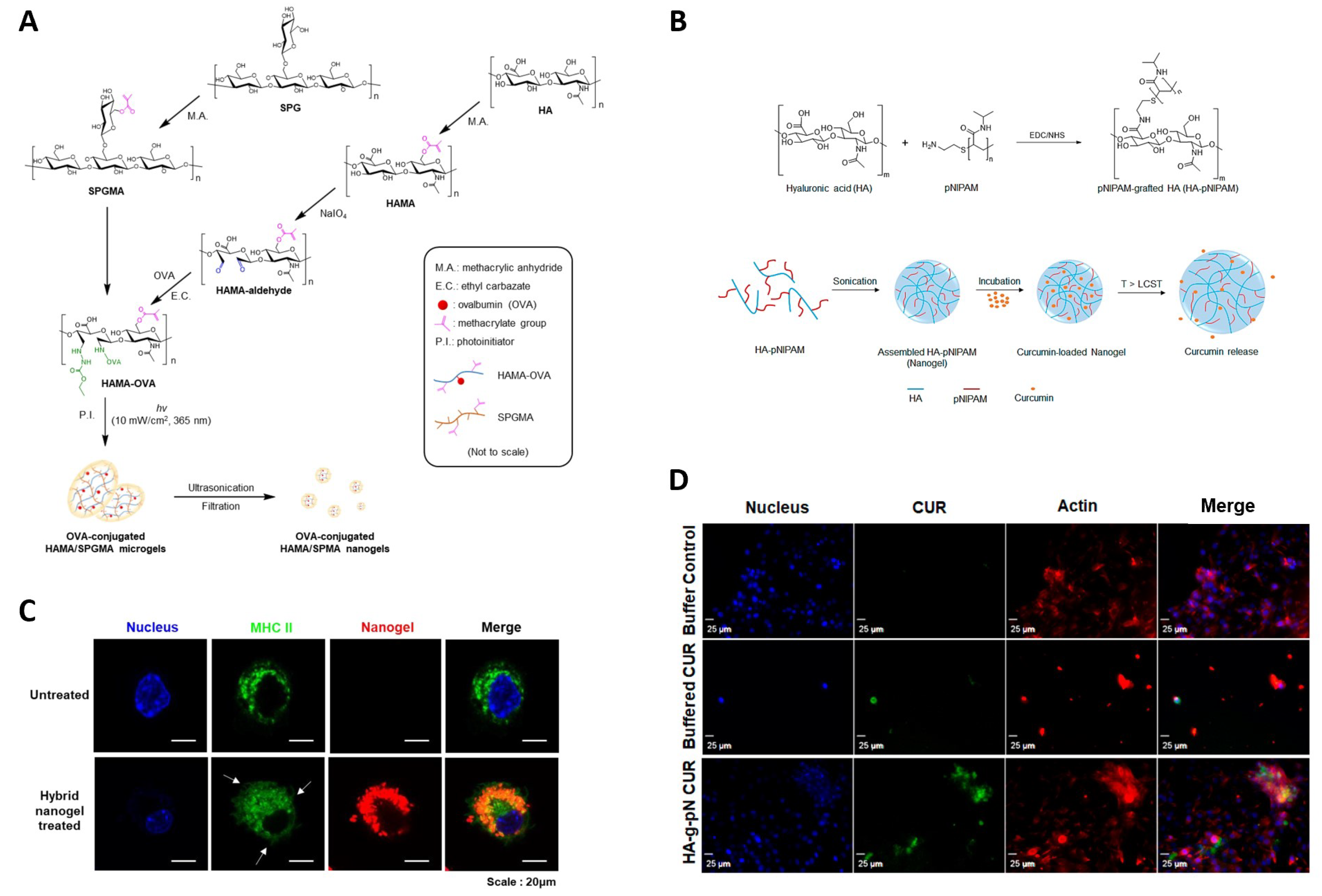
Kim et al. [33] proposed the chemical crosslinking between ovalbumin-conjugated hyaluronic acid-methacrylate and schizophyllan-methacrylate via photopolymerization and ultrasonication. The resulting HA--glucan nanogels exhibited improved skin penetration, thanks to the amphiphilicity and hydrating properties of HA [101], and were internalized in dendritic cells. The nanocarriers were suitable for the delivery of the immunomodulator (i.e., ovalbumin) for the definition of an innovative transdermal immune therapy (Figure 5A,C).

Table 2.
Summary of key literature references concerning skin applications of hyaluronic acid nanogels.
Table 2.
Summary of key literature references concerning skin applications of hyaluronic acid nanogels.
| Authors | Composition | Hyaluronic Acid Molecular Weight | Key Features/Benefits |
|---|---|---|---|
| Wei et al. (2018) [98] | Baicalin nanocrystals obtained by homogenization and spray-drying, then dispersed in hyaluronic acid hydrogel | 800–1000 kDa | Effective transdermal delivery of poorly soluble drugs |
| Chen et al. (2020) [99] | Nanoethosomes containing 5-aminolevulinic acid (ALA), embedded into hyaluronic acid meshes | 10 kDa | Effective transdermal delivery of ALA |
| Soriano et al. (2020) [100] | Melatonin-loaded nanogels made of poloxamer 407, chitosan, and hyaluronic acid | 1.46 MDa | Wound healing |
| Kim et al. (2020) [33] | Ovalbumin-conjugated hyaluronic acid-methacrylate (HAMA-OVA) and schizophyllan-methacrylate (SPGMA) hybrid nanogels | 29 kDa | Topical delivery carrier for immunomodulation |
| Luckanagul et al. (2021) [22] | Hyaluronic acid-grafted poly(N-isopropylacrylamide) nanogel | 47 kDa | Improved delivery/bioavailability of curcumin and other hydrophobic drugs |
| Niu et al. (2022) [24] | Silk peptide-hyaluronic acid based nanogels | <10 kDa | Enhanced topical delivery of curcumin |
| van Gent et al. (2023) [102] | Octenyl succinic anhydride-modified hyaluronic acid nanogels, loaded with antibacterial peptide | 50 kDa | Improved treatment of skin wound infections |
A hyaluronic acid-grafted poly(N-isopropylacrylamide) nanogel synthesized by Luckanagul et al. [22] shows good potential for the delivery of hydrophobic bioactive compounds such as curcumin. In addition, using an artificial skin model, the authors were able to demonstrate a slight skin protection effect (Figure 5B,D).
Nanogels made of octenyl succinic anhydride-modified hyaluronic acid (OSA-HA) are prepared by van Gent and coworkers to encapsulate a synthetic antibacterial and antibiofilm peptide (SAAP)-148 for the treatment of skin wound infections. The nanogels are produced by a microfluidic chip, where a OSA-HA solution and a SAAP-148 solution are injected in different microfluidic streams [102].
In another recent work, hyaluronic acid conjugated with octadecylamine is further conjugated with a silk peptide, to produce a nanogel loaded with curcumin. Evaluation of the in vitro transdermal penetration and skin retention shows a strong improvement compared to the results obtained with free curcumin. This results in significant analgesic and anti-inflammatory benefits [24].
6. Conclusions
In this review, after providing some background information about hyaluronic acid and nanogels, we summarized the literature specifically concerning nanogel systems based on hyaluronic acid as a key constituent.
Our analysis shows that hyaluronic acid is, chemically, a very versatile biopolymer, which can be functionalized to yield a wide range of derivatives that allow various nanogel synthetic strategies to be pursued, based on either physical self-assembly or chemical cross-linking, or a combination of both.
Most of the existing studies about hyaluronic acid nanogels are focused on drug delivery applications and are mainly targeted at cancer therapy, by exploiting the biopolymer affinity for the CD44 receptor. Very few papers are aimed at exploring the potential of these nanocarriers in a dermatological or cosmetic context, although there seems to be a recent, growing trend over the past 2–3 years to look more closely into these applications.
However, it should appear from our analysis that the richness of synthetic methods and nanogel particle compositions described in the literature is such that, in principle, numerous options would be available to the cosmetic formulator who wanted to build on this approach to design innovative products. It should also be clear that nanogel carriers originally synthesized for the delivery of a certain therapeutic agent can work equally well for the release of different types of actives, provided that the latter have sufficient affinity with the nanogel pore environment.
Therefore, we hope this review will stimulate further interest in assessing the potential of this versatile chemistry in the dermatological and cosmetic field, providing formulators with useful perspectives through a structured overview of some of the most relevant literature.
Author Contributions
Conceptualization, E.M. and S.S.; writing—original draft preparation, E.M. and S.S.; writing—review and editing, E.M. and S.S. All authors have read and agreed to the published version of the manuscript.
Funding
S.S. acknowledges support from the Estonian Research Council through Grant PRG1059.
Institutional Review Board Statement
Not applicable.
Informed Consent Statement
Not applicable.
Data Availability Statement
Not applicable.
Conflicts of Interest
The authors declare no conflict of interest.
References
- Maya, S.; Sarmento, B.; Nair, A.; Rejinold, N.S.; Nair, S.V.; Jayakumar, R. Smart stimuli sensitive nanogels in cancer drug delivery and imaging: A review. Curr. Pharm. Des. 2013, 19, 7203–7218. [Google Scholar] [CrossRef] [PubMed]
- Mauri, E.; Giannitelli, S.M.; Trombetta, M.; Rainer, A. Synthesis of Nanogels: Current Trends and Future Outlook. Gels 2021, 7, 36. [Google Scholar] [CrossRef] [PubMed]
- Papa, S.; Veneruso, V.; Mauri, E.; Cremonesi, G.; Mingaj, X.; Mariani, A.; De Paola, M.; Rossetti, A.; Sacchetti, A.; Rossi, F.; et al. Functionalized nanogel for treating activated astrocytes in spinal cord injury. J. Control. Release 2021, 330, 218–228. [Google Scholar] [CrossRef]
- Hajebi, S.; Rabiee, N.; Bagherzadeh, M.; Ahmadi, S.; Rabiee, M.; Roghani-Mamaqani, H.; Tahriri, M.; Tayebi, L.; Hamblin, M.R. Stimulus-responsive polymeric nanogels as smart drug delivery systems. Acta Biomater. 2019, 92, 1–18. [Google Scholar] [CrossRef] [PubMed]
- Du, X.; Gao, Y.; Kang, Q.; Xing, J. Design and Applications of Tumor Microenvironment-Responsive Nanogels as Drug Carriers. Front. Bioeng. Biotechnol. 2021, 9, 771851. [Google Scholar] [CrossRef] [PubMed]
- Moad, G. RAFT (Reversible addition–fragmentation chain transfer) crosslinking (co)polymerization of multi-olefinic monomers to form polymer networks. Polym. Int. 2015, 64, 15–24. [Google Scholar] [CrossRef]
- Limiti, E.; Mozetic, P.; Giannitelli, S.M.; Pinelli, F.; Han, X.; Del Rio, D.; Abbruzzese, F.; Basoli, F.; Rosanò, L.; Scialla, S.; et al. Hyaluronic Acid–Polyethyleneimine Nanogels for Controlled Drug Delivery in Cancer Treatment. ACS Appl. Nano Mater. 2022, 5, 5544–5557. [Google Scholar] [CrossRef]
- Elzayat, A.; Adam-Cervera, I.; Álvarez Bermúdez, O.; Muñoz-Espí, R. Nanoemulsions for synthesis of biomedical nanocarriers. Colloids Surf. Biointerfaces 2021, 203, 111764. [Google Scholar] [CrossRef]
- Oehrl, A.; Schötz, S.; Haag, R. Systematic Screening of Different Polyglycerin-Based Dienophile Macromonomers for Efficient Nanogel Formation through IEDDA Inverse Nanoprecipitation. Macromol. Rapid Commun. 2020, 41, 1900510. [Google Scholar] [CrossRef] [Green Version]
- Giulbudagian, M.; Asadian-Birjand, M.; Steinhilber, D.; Achazi, K.; Molina, M.; Calderón, M. Fabrication of thermoresponsive nanogels by thermo-nanoprecipitation and in situ encapsulation of bioactives. Polym. Chem. 2014, 5, 6909–6913. [Google Scholar] [CrossRef] [Green Version]
- Yang, H.; Huang, D. Method for Continuous Fabrication of Multi-Functional Nanogels. U.S. Patent Application 20230080475, 16 March 2023. [Google Scholar]
- Toprakcioglu, Z.; Challa, P.K.; Morse, D.B.; Knowles, T. Attoliter protein nanogels from droplet nanofluidics for intracellular delivery. Sci. Adv. 2020, 6, eaay7952. [Google Scholar] [CrossRef] [Green Version]
- Giannitelli, S.M.; Limiti, E.; Mozetic, P.; Pinelli, F.; Han, X.; Abbruzzese, F.; Basoli, F.; Rio, D.D.; Scialla, S.; Rossi, F.; et al. Droplet-based microfluidic synthesis of nanogels for controlled drug delivery: Tailoring nanomaterial properties via pneumatically actuated flow-focusing junction. Nanoscale 2022, 14, 11415–11428. [Google Scholar] [CrossRef]
- Papakonstantinou, E.; Roth, M.; Karakiulakis, G. Hyaluronic acid: A key molecule in skin aging. Dermato-Endocrinology 2012, 4, 253–258. [Google Scholar] [CrossRef] [Green Version]
- Choi, K.Y.; Saravanakumar, G.; Park, J.H.; Park, K. Hyaluronic acid-based nanocarriers for intracellular targeting: Interfacial interactions with proteins in cancer. Colloids Surf. Biointerfaces 2012, 99, 82–94. [Google Scholar] [CrossRef] [Green Version]
- Varshosaz, J.; Taymouri, S.; Ghassami, E. Supramolecular Self-Assembled Nanogels a New Platform for Anticancer Drug Delivery. Curr. Pharm. Des. 2018, 23, 5242–5260. [Google Scholar] [CrossRef]
- Rolland, J.P.; Maynor, B.W.; Euliss, L.E.; Exner, A.E.; Denison, G.M.; DeSimone, J.M. Direct Fabrication and Harvesting of Monodisperse, Shape-Specific Nanobiomaterials. J. Am. Chem. Soc. 2005, 127, 10096–10100. [Google Scholar] [CrossRef]
- Kersey, F.R.; Merkel, T.J.; Perry, J.L.; Napier, M.E.; DeSimone, J.M. Effect of Aspect Ratio and Deformability on Nanoparticle Extravasation through Nanopores. Langmuir 2012, 28, 8773–8781. [Google Scholar] [CrossRef] [Green Version]
- Vasudevan, M.; Buse, E.; Lu, D.; Krishna, H.; Kalyanaraman, R.; Shen, A.Q.; Khomami, B.; Sureshkumar, R. Irreversible nanogel formation in surfactant solutions by microporous flow. Nat. Mater. 2010, 9, 436–441. [Google Scholar] [CrossRef]
- Mauri, E.; Perale, G.; Rossi, F. Nanogel Functionalization: A Versatile Approach To Meet the Challenges of Drug and Gene Delivery. ACS Appl. Nano Mater. 2018, 1, 6525–6541. [Google Scholar] [CrossRef]
- Fasiku, V.O.; Omolo, C.A.; Kiruri, L.W.; Devnarain, N.; Faya, M.; Mocktar, C.; Govender, T. A hyaluronic acid-based nanogel for the co-delivery of nitric oxide (NO) and a novel antimicrobial peptide (AMP) against bacterial biofilms. Int. J. Biol. Macromol. 2022, 206, 381–397. [Google Scholar] [CrossRef]
- Luckanagul, J.A.; Bhuket, P.R.N.; Muangnoi, C.; Rojsitthisak, P.; Wang, Q.; Rojsitthisak, P. Self-Assembled Thermoresponsive Nanogel from Grafted Hyaluronic Acid as a Biocompatible Delivery Platform for Curcumin with Enhanced Drug Loading and Biological Activities. Polymers 2021, 13, 194. [Google Scholar] [CrossRef]
- Pedrosa, S.S.; Gonçalves, C.; David, L.; Gama, M. A Novel Crosslinked Hyaluronic Acid Nanogel for Drug Delivery. Macromol. Biosci. 2014, 14, 1556–1568. [Google Scholar] [CrossRef] [Green Version]
- Niu, J.; Yuan, M.; Liu, Y.; Wang, L.; Tang, Z.; Wang, Y.; Qi, Y.; Zhang, Y.; Ya, H.; Fan, Y. Silk peptide-hyaluronic acid based nanogels for the enhancement of the topical administration of curcumin. Front. Chem. 2022, 10, 1028372. [Google Scholar] [CrossRef]
- Win, Y.Y.; Charoenkanburkang, P.; Limprasutr, V.; Rodsiri, R.; Pan, Y.; Buranasudja, V.; Luckanagul, J.A. In Vivo Biocompatible Self-Assembled Nanogel Based on Hyaluronic Acid for Aqueous Solubility and Stability Enhancement of Asiatic Acid. Polymers 2021, 13, 4071. [Google Scholar] [CrossRef]
- Ritger, P.L.; Peppas, N.A. A simple equation for description of solute release I. Fickian and non-fickian release from non-swellable devices in the form of slabs, spheres, cylinders or discs. J. Control. Release 1987, 5, 23–36. [Google Scholar] [CrossRef]
- Crank, J. The Mathematics of Diffusion; Clarendon Press: Oxford, UK, 1975. [Google Scholar]
- Scotti, A.; Schulte, M.F.; Lopez, C.G.; Crassous, J.J.; Bochenek, S.; Richtering, W. How Softness Matters in Soft Nanogels and Nanogel Assemblies. Chem. Rev. 2022, 122, 11675–11700. [Google Scholar] [CrossRef]
- Brown, A.; Stabenfeldt, S.; Ahn, B.; Hannan, R.; Dhada, K.; Herman, E.; Stefanelli, V.; Guzzetta, N.; Alexeev, A.; Lam, W.; et al. Ultrasoft microgels displaying emergent, platelet-like, behaviors. Nat. Mater. 2014, 13, 1108–1114. [Google Scholar] [CrossRef] [Green Version]
- Myerson, J.W.; McPherson, O.; DeFrates, K.G.; Towslee, J.H.; Marcos-Contreras, O.A.; Shuvaev, V.V.; Braender, B.; Composto, R.J.; Muzykantov, V.R.; Eckmann, D.M. Cross-linker-Modulated Nanogel Flexibility Correlates with Tunable Targeting to a Sterically Impeded Endothelial Marker. ACS Nano 2019, 13, 11409–11421. [Google Scholar] [CrossRef]
- Graff, R.W.; Shi, Y.; Wang, X.; Gao, H. Comparison of Loading Efficiency between Hyperbranched Polymers and Cross-Linked Nanogels at Various Branching Densities. Macromol. Rapid Commun. 2015, 36, 2076–2082. [Google Scholar] [CrossRef]
- Cuneo, T.; Gao, H. Recent advances on synthesis and biomaterials applications of hyperbranched polymers. WIREs Nanomed. Nanobiotechnol. 2020, 12, e1640. [Google Scholar] [CrossRef]
- Kim, H.; Lee, S.; Ki, C.S. Modular formation of hyaluronic acid/β-glucan hybrid nanogels for topical dermal delivery targeting skin dendritic cells. Carbohydr. Polym. 2021, 252, 117132. [Google Scholar] [CrossRef] [PubMed]
- Buffa, R.; Nešporová, K.; Basarabová, I.; Halamková, P.; Svozil, V.; Velebný, V. Synthesis and study of branched hyaluronic acid with potential anticancer activity. Carbohydr. Polym. 2019, 223, 115047. [Google Scholar] [CrossRef] [PubMed]
- Camerin, F.; Fernández-Rodríguez, M.; Rovigatti, L.; Antonopoulou, M.N.; Gnan, N.; Ninarello, A.; Isa, L.; Zaccarelli, E. Microgels Adsorbed at Liquid–Liquid Interfaces: A Joint Numerical and Experimental Study. ACS Nano 2019, 13, 4548–4559. [Google Scholar] [CrossRef] [PubMed] [Green Version]
- Ramos, J.; Imaz, A.; Callejas-Fernández, J.; Barbosa-Barros, L.; Estelrich, J.; Quesada-Pérez, M.; Forcada, J. Soft nanoparticles (thermo-responsive nanogels and bicelles) with biotechnological applications: From synthesis to simulation through colloidal characterization. Soft Matter 2011, 7, 5067–5082. [Google Scholar] [CrossRef]
- Palkar, V.; Thakar, D.; Kuksenok, O. Nanogel Degradation at Soft Interfaces and in Bulk: Tracking Shape Changes and Interfacial Spreading. Macromolecules 2023, 56, 1289–1302. [Google Scholar] [CrossRef]
- Misra, S.; Hascall, V.C.; Markwald, R.R.; Ghatak, S. Interactions between Hyaluronan and Its Receptors (CD44, RHAMM) Regulate the Activities of Inflammation and Cancer. Front. Immunol. 2015, 6, 201. [Google Scholar] [CrossRef] [Green Version]
- Griesser, J.; Hetényi, G.; Bernkop-Schnürch, A. Thiolated Hyaluronic Acid as Versatile Mucoadhesive Polymer: From the Chemistry Behind to Product Developments—What Are the Capabilities? Polymers 2018, 10, 243. [Google Scholar] [CrossRef] [Green Version]
- Yang, C.; Wang, X.; Yao, X.; Zhang, Y.; Wu, W.; Jiang, X. Hyaluronic acid nanogels with enzyme-sensitive cross-linking group for drug delivery. J. Control. Release 2015, 205, 206–217. [Google Scholar] [CrossRef]
- Zhang, X.; Malhotra, S.; Molina, M.; Haag, R. Micro- and nanogels with labile crosslinks – from synthesis to biomedical applications. Chem. Soc. Rev. 2015, 44, 1948–1973. [Google Scholar] [CrossRef] [Green Version]
- Chi, Y.; Huang, Y.; Kang, Y.; Dai, G.; Liu, Z.; Xu, K.; Zhong, W. The effects of molecular weight of hyaluronic acid on transdermal delivery efficiencies of dissolving microneedles. Eur. J. Pharm. Sci. 2022, 168, 106075. [Google Scholar] [CrossRef]
- Snetkov, P.; Zakharova, K.; Morozkina, S.; Olekhnovich, R.; Uspenskaya, M. Hyaluronic Acid: The Influence of Molecular Weight on Structural, Physical, Physico-Chemical, and Degradable Properties of Biopolymer. Polymers 2020, 12, 1800. [Google Scholar] [CrossRef]
- Liu, L.; Liu, Y.; Li, J.; Du, G.; Chen, J. Microbial production of hyaluronic acid: Current state, challenges, and perspectives. Microb. Cell Factories 2011, 10, 99. [Google Scholar] [CrossRef] [Green Version]
- Chistyakov, D.V.; Astakhova, A.A.; Azbukina, N.V.; Goriainov, S.V.; Chistyakov, V.V.; Sergeeva, M.G. High and Low Molecular Weight Hyaluronic Acid Differentially Influences Oxylipins Synthesis in Course of Neuroinflammation. Int. J. Mol. Sci. 2019, 20, 3894. [Google Scholar] [CrossRef] [Green Version]
- Nakagawa, Y.; Yabuuchi, K.; Fukumoto, K.; Katsumata, T.; Yang, S. Hyaluronic Acid Derivative Composition, Pharmaceutical Composition and Hyaluronic Acid Derivative-Drug Conjugate Composition. U.S. Patent Application 20230085879, 23 March 2023. [Google Scholar]
- Falcone, S.J.; Palmeri, D.M.; Berg, R.A. Rheological and cohesive properties of hyaluronic acid. J. Biomed. Mater. Res. Part A 2006, 76A, 721–728. [Google Scholar] [CrossRef] [PubMed]
- Zhong, L.; Liu, Y.; Xu, L.; Li, Q.; Zhao, D.; Li, Z.; Zhang, H.; Zhang, H.; Kan, Q.; Sun, J.; et al. Exploring the relationship of hyaluronic acid molecular weight and active targeting efficiency for designing hyaluronic acid-modified nanoparticles. Asian J. Pharm. Sci. 2019, 14, 521–530. [Google Scholar] [CrossRef]
- Wadhwa, K.; Kadian, V.; Puri, V.; Bhardwaj, B.Y.; Sharma, A.; Pahwa, R.; Rao, R.; Gupta, M.; Singh, I. New insights into quercetin nanoformulations for topical delivery. Phytomed. Plus 2022, 2, 100257. [Google Scholar] [CrossRef]
- Arroyo, E.; Valdez, R.; Cornejo-Bravo, J.; Armenta, M.; Olivas, A. Nanogels as controlled drug release systems for Coenzyme Q10 and Resveratrol for cosmetic application. J. Nanoparticle Res. 2021, 23, 163. [Google Scholar] [CrossRef]
- Yabuuchi, K.; Miyamoto, Y.; Fukumoto, K.; Katsumata, T.; Nakagawa, Y. Hyaluronic Acid Derivative, Pharmaceutical Composition, and Hyaluronic Acid Derivative-Drug Complex. U.S. Patent Application 20230066990, 2 March 2023. [Google Scholar]
- Van Gheluwe, L.; Chourpa, I.; Gaigne, C.; Munnier, E. Polymer-Based Smart Drug Delivery Systems for Skin Application and Demonstration of Stimuli-Responsiveness. Polymers 2021, 13, 1285. [Google Scholar] [CrossRef]
- Joo, V.; Ramasamy, T.; Haidar, Z.S. A Novel Self-Assembled Liposome-Based Polymeric Hydrogel for Cranio-Maxillofacial Applications: Preliminary Findings. Polymers 2011, 3, 967–974. [Google Scholar] [CrossRef]
- Zhao, J.; Wang, Y.; Ma, Y.; Liu, Y.; Yan, B.; Wang, L. Smart nanocarrier based on PEGylated hyaluronic acid for deacetyl mycoepoxydience: High stability with enhanced bioavailability and efficiency. Carbohydr. Polym. 2019, 203, 356–368. [Google Scholar] [CrossRef]
- Nakai, T.; Hirakura, T.; Sakurai, Y.; Shimoboji, T.; Ishigai, M.; Akiyoshi, K. Injectable hydrogel for sustained protein release by salt-induced association of hyaluronic acid nanogel. Macromol. Biosci. 2012, 12, 475–483. [Google Scholar] [CrossRef]
- Wei, X.; Senanayake, T.H.; Warren, G.; Vinogradov, S.V. Hyaluronic acid-based nanogel-drug conjugates with enhanced anticancer activity designed for the targeting of CD44-positive and drug-resistant tumors. Bioconjugate Chem. 2013, 24, 658–668. [Google Scholar] [CrossRef] [PubMed] [Green Version]
- Wei, X.; Senanayake, T.H.; Bohling, A.; Vinogradov, S.V. Targeted Nanogel Conjugate for Improved Stability and Cellular Permeability of Curcumin: Synthesis, Pharmacokinetics, and Tumor Growth Inhibition. Mol. Pharm. 2014, 11, 3112–3122. [Google Scholar] [CrossRef] [Green Version]
- Xavier, M.; García-Hevia, L.; Amado, I.R.; Pastrana, L.; Gonçalves, C. In Vitro Intestinal Uptake And Permeability Of Fluorescently-Labelled Hyaluronic Acid Nanogels. Int. J. Nanomed. 2019, 14, 9077–9088. [Google Scholar] [CrossRef] [Green Version]
- Ossipov, D.; Kootala, S.; Yi, Z.; Yang, X.; Hilborn, J. Orthogonal Chemoselective Assembly of Hyaluronic Acid Networks and Nanogels for Drug Delivery. Macromolecules 2013, 46, 4105–4113. [Google Scholar] [CrossRef]
- Ko, D.Y.; Moon, H.J.; Jeong, B. Temperature-sensitive polypeptide nanogels for intracellular delivery of a biomacromolecular drug. J. Mater. Chem. B 2015, 3, 3525–3530. [Google Scholar] [CrossRef]
- Water, J.J.; Kim, Y.; Maltesen, M.J.; Franzyk, H.; Foged, C.; Nielsen, H.M. Hyaluronic Acid-Based Nanogels Produced by Microfluidics-Facilitated Self-Assembly Improves the Safety Profile of the Cationic Host Defense Peptide Novicidin. Pharm. Res. 2015, 32, 2727–2735. [Google Scholar] [CrossRef] [PubMed]
- Liang, K.; Ng, S.; Lee, F.; Lim, J.; Chung, J.E.; Lee, S.S.; Kurisawa, M. Targeted intracellular protein delivery based on hyaluronic acid-green tea catechin nanogels. Acta Biomater. 2016, 33, 142–152. [Google Scholar] [CrossRef]
- Agnello, S.; Bongiovì, F.; Fiorica, C.; Pitarresi, G.; Palumbo, F.S.; Bella, M.A.D.; Giammona, G. Microfluidic Fabrication of Physically Assembled Nanogels and Micrometric Fibers by Using a Hyaluronic Acid Derivative. Macromol. Mater. Eng. 2017, 302, 1700265. [Google Scholar] [CrossRef]
- Simonson, A.W.; Lawanprasert, A.; Goralski, T.D.P.; Keiler, K.C.; Medina, S.H. Bioresponsive peptide-polysaccharide nanogels—A versatile delivery system to augment the utility of bioactive cargo. Nanomed. Nanotechnol. Biol. Med. 2019, 17, 391–400. [Google Scholar] [CrossRef]
- Xu, J.; Chu, T.; Yu, T.; Li, N.; Wang, C.; Li, C.; Zhang, Y.; Meng, H.; Nie, G. Design of Diselenide-Bridged Hyaluronic Acid Nano-antioxidant for Efficient ROS Scavenging to Relieve Colitis. ACS Nano 2022, 16, 13037–13048. [Google Scholar] [CrossRef] [PubMed]
- Duan, Q.Y.; Zhu, Y.X.; Jia, H.R.; Guo, Y.; Zhang, X.; Gu, R.; Li, C.; Wu, F.G. Platinum-Coordinated Dual-Responsive Nanogels for Universal Drug Delivery and Combination Cancer Therapy. Small 2022, 18, e2203260. [Google Scholar] [CrossRef]
- Kaewruethai, T.; Lin, Y.; Wang, Q.; Luckanagul, J.A. The Dual Modification of PNIPAM and β-Cyclodextrin Grafted on Hyaluronic Acid as Self-Assembled Nanogel for Curcumin Delivery. Polymers 2022, 15, 116. [Google Scholar] [CrossRef]
- Ładniak, A.; Jurak, M.; Wiącek, A.E. The effect of chitosan/TiO2/hyaluronic acid subphase on the behaviour of 1,2-dioleoyl-sn-glycero-3-phosphocholine membrane. Biomater. Adv. 2022, 138, 212934. [Google Scholar] [CrossRef] [PubMed]
- Sakulwech, S.; Lourith, N.; Ruktanonchai, U.; Kanlayavattanakul, M. Preparation and characterization of nanoparticles from quaternized cyclodextrin-grafted chitosan associated with hyaluronic acid for cosmetics. Asian J. Pharm. Sci. 2018, 13, 498–504. [Google Scholar] [CrossRef] [PubMed]
- Gennari, A.; de la Rosa, J.M.R.; Hohn, E.; Pelliccia, M.; Lallana, E.; Donno, R.; Tirella, A.; Tirelli, N. The different ways to chitosan/hyaluronic acid nanoparticles: Templated vs direct complexation. Influence of particle preparation on morphology, cell uptake and silencing efficiency. Beilstein J. Nanotechnol. 2019, 10, 2594–2608. [Google Scholar] [CrossRef] [Green Version]
- Messager, L.; Portecop, N.; Hachet, E.; Lapeyre, V.; Pignot-Paintrand, I.; Catargi, B.; Auzély-Velty, R.; Ravaine, V. Photochemical crosslinking of hyaluronic acid confined in nanoemulsions: Towards nanogels with a controlled structure. J. Mater. Chem. B 2013, 1, 3369–3379. [Google Scholar] [CrossRef]
- Luan, S.; Zhu, Y.; Wu, X.; Wang, Y.; Liang, F.; Song, S. Hyaluronic-Acid-Based pH-Sensitive Nanogels for Tumor-Targeted Drug Delivery. ACS Biomater. Sci. Eng. 2017, 3, 2410–2419. [Google Scholar] [CrossRef]
- Yu, J.; Zhang, Y.; Sun, W.; Wang, C.; Ranson, D.; Ye, Y.; Weng, Y.; Gu, Z. Internalized compartments encapsulated nanogels for targeted drug delivery. Nanoscale 2016, 8, 9178–9184. [Google Scholar] [CrossRef] [Green Version]
- Lin, S.; Li, T.; Xie, P.; Li, Q.; Wang, B.; Wang, L.; Li, L.; Wang, Y.; Chen, H.; Nan, K. Targeted delivery of doxorubicin to tumour tissues by a novel legumain sensitive polygonal nanogel. Nanoscale 2016, 8, 18400–18411. [Google Scholar] [CrossRef]
- Gurav, D.D.; Kulkarni, A.S.; Khan, A.; Shinde, V.S. pH-responsive targeted and controlled doxorubicin delivery using hyaluronic acid nanocarriers. Colloids Surf. Biointerfaces 2016, 143, 352–358. [Google Scholar] [CrossRef]
- Yang, G.; Fu, S.; Yao, W.; Wang, X.; Zha, Q.; Tang, R. Hyaluronic acid nanogels prepared via ortho ester linkages show pH-triggered behavior, enhanced penetration and antitumor efficacy in 3-D tumor spheroids. J. Colloid Interface Sci. 2017, 504, 25–38. [Google Scholar] [CrossRef]
- Kang, E.B.; Lee, G.B.; In, I.; Park, S.Y. pH-sensitive fluorescent hyaluronic acid nanogels for tumor-targeting and controlled delivery of doxorubicin and nitric oxide. Eur. Polym. J. 2018, 101, 96–104. [Google Scholar] [CrossRef]
- Zhu, Q.; Chen, X.; Xu, X.; Zhang, Y.; Zhang, C.; Mo, R. Tumor-Specific Self-Degradable Nanogels as Potential Carriers for Systemic Delivery of Anticancer Proteins. Adv. Funct. Mater. 2018, 28, 1707371. [Google Scholar] [CrossRef]
- Choi, Y.R.; Kim, H.J.; Ahn, G.Y.; Lee, M.J.; Park, J.R.; Jun, D.R.; Ryu, T.K.; Park, J.W.; Shin, E.; Choi, S.W. Fabrication of dihydroxyflavone-conjugated hyaluronic acid nanogels for targeted antitumoral effect. Colloids Surf. Biointerfaces 2018, 171, 690–697. [Google Scholar] [CrossRef]
- Tian, Y.; Tian, R.; Chen, L.; Jin, R.; Feng, Y.; Bai, Y.; Chen, X. Redox-Responsive Nanogel with Intracellular Reconstruction and Programmable Drug Release for Targeted Tumor Therapy. Macromol. Rapid Commun. 2019, 40, e1800824. [Google Scholar] [CrossRef]
- Suner, S.S.; Ari, B.; Onder, F.C.; Ozpolat, B.; Ay, M.; Sahiner, N. Hyaluronic acid and hyaluronic acid: Sucrose nanogels for hydrophobic cancer drug delivery. Int. J. Biol. Macromol. 2019, 126, 1150–1157. [Google Scholar] [CrossRef] [PubMed]
- Chen, J.; Wu, M.; Veroniaina, H.; Mukhopadhyay, S.; Li, J.; Wu, Z.; Wu, Z.; Qi, X. Poly(N-Isopropylacrylamide) derived nanogels demonstrated thermosensitive self-assembly and GSH-triggered drug release for efficient tumor Therapy. Polym. Chem. 2019, 10, 4031–4041. [Google Scholar] [CrossRef]
- Xu, L.; Zhong, S.; Gao, Y.; Cui, X. Thermo-responsive poly(N-isopropylacrylamide)-hyaluronic acid nano-hydrogel and its multiple applications. Int. J. Biol. Macromol. 2022, 194, 811–818. [Google Scholar] [CrossRef]
- Cao, Z.; Li, W.; Liu, R.; Li, C.; Song, Y.; Liu, G.; Chen, Y.; Lu, C.; Lu, A.; Liu, Y. pH-Responsive Fluorescence Enhanced Nanogel for Targeted Delivery of AUR and CDDP Against Breast Cancer. Int. J. Nanomed. 2020, 15, 8369–8382. [Google Scholar] [CrossRef] [PubMed]
- Yang, G.; Fan, M.; Zhu, J.; Ling, C.; Wu, L.; Zhang, X.; Zhang, M.; Li, J.; Yao, Q.; Gu, Z.; et al. A multifunctional anti-inflammatory drug that can specifically target activated macrophages, massively deplete intracellular H2O2, and produce large amounts CO for a highly efficient treatment of osteoarthritis. Biomaterials 2020, 255, 120155. [Google Scholar] [CrossRef] [PubMed]
- Zhang, C.; Li, Q.; Wu, C.; Wang, J.; Su, M.; Deng, J. Hypoxia-responsive nanogel as IL-12 carrier for anti-cancer therapy. Nanotechnology 2021, 32, 095107. [Google Scholar] [CrossRef] [PubMed]
- Fu, Y.; Jang, M.S.; Liu, C.; Lee, J.H.; Li, Y.; Yang, H.Y. Hypoxia-responsive hyaluronic acid nanogels with improved endo/lysosomal escape ability for tumor-targeted cytochrome c delivery. Eur. Polym. J. 2022, 173, 111259. [Google Scholar] [CrossRef]
- Gao, D.; Asghar, S.; Ye, J.; Zhang, M.; Hu, R.; Wang, Y.; Huang, L.; Yuan, C.; Chen, Z.; Xiao, Y. Dual-targeted enzyme-sensitive hyaluronic acid nanogels loading paclitaxel for the therapy of breast cancer. Carbohydr. Polym. 2022, 294, 119785. [Google Scholar] [CrossRef]
- Liwinska, W.; Waleka-Bagiel, E.; Stojek, Z.; Karbarz, M.; Zabost, E. Enzyme-triggered- and tumor-targeted delivery with tunable, methacrylated poly(ethylene glycols) and hyaluronic acid hybrid nanogels. Drug Deliv. 2022, 29, 2561–2578. [Google Scholar] [CrossRef]
- Yang, X.; Kootala, S.; Hilborn, J.; Ossipov, D.A. Preparation of hyaluronic acid nanoparticles via hydrophobic association assisted chemical cross-linking—An orthogonal modular approach. Soft Matter 2011, 7, 7517. [Google Scholar] [CrossRef]
- Novoa-Carballal, R.; Pergushov, D.V.; Müller, A.H.E. Interpolyelectrolyte complexes based on hyaluronic acid-block-poly(ethylene glycol) and poly-l-lysine. Soft Matter 2013, 9, 4297. [Google Scholar] [CrossRef]
- Ashwinkumar, N.; Maya, S.; Jayakumar, R. Redox-responsive cystamine conjugated chitin-hyaluronic acid composite nanogels. RSC Adv. 2014, 4, 49547–49555. [Google Scholar] [CrossRef]
- Pedrosa, S.S.; Pereira, P.; Correia, A.; Moreira, S.; Rocha, H.; Gama, F.M. Biocompatibility of a Self-Assembled Crosslinkable Hyaluronic Acid Nanogel. Macromol. Biosci. 2016, 16, 1610–1620. [Google Scholar] [CrossRef] [Green Version]
- Pedrosa, S.S.; Pereira, P.; Correia, A.; Gama, F.M. Targetability of hyaluronic acid nanogel to cancer cells: In vitro and in vivo studies. Eur. J. Pharm. Sci. 2017, 104, 102–113. [Google Scholar] [CrossRef] [Green Version]
- Ohta, S.; Hiramoto, S.; Amano, Y.; Sato, M.; Suzuki, Y.; Shinohara, M.; Emoto, S.; Yamaguchi, H.; Ishigami, H.; Sakai, Y.; et al. Production of Cisplatin-Incorporating Hyaluronan Nanogels via Chelating Ligand-Metal Coordination. Bioconjugate Chem. 2016, 27, 504–508. [Google Scholar] [CrossRef]
- Ohta, S.; Hiramoto, S.; Amano, Y.; Emoto, S.; Yamaguchi, H.; Ishigami, H.; Kitayama, J.; Ito, T. Intraperitoneal Delivery of Cisplatin via a Hyaluronan-Based Nanogel/In Situ Cross-Linkable Hydrogel Hybrid System for Peritoneal Dissemination of Gastric Cancer. Mol. Pharm. 2017, 14, 3105–3113. [Google Scholar] [CrossRef]
- Amano, Y.; Sakura, K.L.; Ohta, S.; Ito, T. Cisplatin-Chelated Iminodiacetic Acid-Conjugated Hyaluronic Acid Nanogels for the Treatment of Malignant Pleural Mesothelioma in Mice. Mol. Pharm. 2022, 19, 853–861. [Google Scholar] [CrossRef] [PubMed]
- Wei, S.; Xie, J.; Luo, Y.; Ma, Y.; Tang, S.; Yue, P.; Yang, M. Hyaluronic acid based nanocrystals hydrogels for enhanced topical delivery of drug: A case study. Carbohydr. Polym. 2018, 202, 64–71. [Google Scholar] [CrossRef] [PubMed]
- Chen, Y.; Zhang, Z.; Xin, Y.; Zhou, R.; Jiang, K.; Sun, X.; He, D.; Song, J.; Zhang, Y. Synergistic transdermal delivery of nanoethosomes embedded in hyaluronic acid nanogels for enhancing photodynamic therapy. Nanoscale 2020, 12, 15435–15442. [Google Scholar] [CrossRef]
- Soriano, J.L.; Calpena, A.C.; Rincón, M.; Pérez, N.; Halbaut, L.; Rodríguez-Lagunas, M.J.; Clares, B. Melatonin nanogel promotes skin healing response in burn wounds of rats. Nanomedicine 2020, 15, 2133–2147. [Google Scholar] [CrossRef] [PubMed]
- Kim, H.; Jeong, H.; Han, S.; Beack, S.; Hwang, B.W.; Shin, M.; Oh, S.S.; Hahn, S.K. Hyaluronate and its derivatives for customized biomedical applications. Biomaterials 2017, 123, 155–171. [Google Scholar] [CrossRef]
- van Gent, M.E.; van Baaren, T.; Kłodzińska, S.N.; Ali, M.; Dolezal, N.; van Doodewaerd, B.R.; Bos, E.; de Waal, A.M.; Koning, R.I.; Drijfhout, J.W.; et al. Encapsulation of SAAP-148 in Octenyl Succinic Anhydride-Modified Hyaluronic Acid Nanogels for Treatment of Skin Wound Infections. Pharmaceutics 2023, 15, 429. [Google Scholar] [CrossRef]
Figure 1.
Chemical structure of hyaluronic acid, showing two disaccharide repeating units. Each disaccharide unit is made of 4-glucuronic acid and 3-N-acetylglucosamine, which are linked together by an ether bond. The repeating units are connected to one another by an ether bond. The carboxyl, hydroxyl, and amine groups eligible for chemical reactions with other polymers or molecules are highlighted.

Figure 2.
Yearly number of publications and citations regarding the topic of nanogels based on hyaluronic acid between January 2009 and June 2023. [Data from Web of Science, provided by Clarivate. Web of Science and Clarivate are trademarks of their respective owners and used herein with permission].

Figure 3.
Scheme of the main drug release profiles: a controlled and sustained release over time (black line) meets the criteria of a prolonged therapeutic effect, resulting in the best option compared to a fast release (green line) and a pulsatile administration of the drug in free form (red line). Putative representation of nanogel adhesion and controlled release of the encapsulated drug over a dermal layer is reported in A and B, respectively.

Figure 4.
Main approaches in nanogel synthesis. (A) Self-assembly via ionic interactions. (B) Self-assembly through hydrophilic (red/blue lines) and hydrophobic (yellow motifs) interactions. (C) Chemical crosslinking. (D) Hybrid strategy.
Figure 4.
Main approaches in nanogel synthesis. (A) Self-assembly via ionic interactions. (B) Self-assembly through hydrophilic (red/blue lines) and hydrophobic (yellow motifs) interactions. (C) Chemical crosslinking. (D) Hybrid strategy.

Figure 5.
Representation of two different strategies to synthesize hyaluronan-based nanogels and examples of their cell internalization. (A) Functionalization of HA hydroxyl groups with aldehyde motifs and subsequent conjugation to ovalbumin; (B) Conjugation of HA with pNIPAM (poly(N-isopropylacrylamide)); (C) Internalization of ovalbumin-conjugated nanogels in JAWSII cells to achieve transdermal immunomodulation; the arrows indicate the formation of dendrites, implying cell maturation, after nanogel treatment (D) Internalization of HA-pNIPAM nanogel in NIH-3T3 cells for controlled release of curcumin. [(A,C): Reprinted from Kim et al. [33] with permission from Elsevier, copyright (2021). (B,D): Readapted and reprinted from Luckanagul et al. [22] with permission from MDPI, copyright (2021)].

Table 1.
Definition of different kinds of softness linked to nanogels and corresponding equations. For a generic variable x, the expression x(c) means that the variable is a function of the nanogel concentration c.
Table 1.
Definition of different kinds of softness linked to nanogels and corresponding equations. For a generic variable x, the expression x(c) means that the variable is a function of the nanogel concentration c.
| Softness Evaluated as: | Variables | Equation |
|---|---|---|
| Deformation | elastic free energy of deformation (), thermal energy of a nanogel () | |
| Deswelling | nanogel size at infinite dilution (R) and in a reference state () | |
| Interpenetrability | radius (R) of colloid (sphere) in concentrated sample and the corresponding nearest neighbor distance () | |
| Faceting | nanogel sphericity (), volume (V), surface area (A) |
Disclaimer/Publisher’s Note: The statements, opinions and data contained in all publications are solely those of the individual author(s) and contributor(s) and not of MDPI and/or the editor(s). MDPI and/or the editor(s) disclaim responsibility for any injury to people or property resulting from any ideas, methods, instructions or products referred to in the content. |
© 2023 by the authors. Licensee MDPI, Basel, Switzerland. This article is an open access article distributed under the terms and conditions of the Creative Commons Attribution (CC BY) license (https://creativecommons.org/licenses/by/4.0/).
Share and Cite
MDPI and ACS Style
Mauri, E.; Scialla, S. Nanogels Based on Hyaluronic Acid as Potential Active Carriers for Dermatological and Cosmetic Applications. Cosmetics 2023, 10, 113. https://0-doi-org.brum.beds.ac.uk/10.3390/cosmetics10040113
AMA Style
Mauri E, Scialla S. Nanogels Based on Hyaluronic Acid as Potential Active Carriers for Dermatological and Cosmetic Applications. Cosmetics. 2023; 10(4):113. https://0-doi-org.brum.beds.ac.uk/10.3390/cosmetics10040113
Chicago/Turabian StyleMauri, Emanuele, and Stefano Scialla. 2023. "Nanogels Based on Hyaluronic Acid as Potential Active Carriers for Dermatological and Cosmetic Applications" Cosmetics 10, no. 4: 113. https://0-doi-org.brum.beds.ac.uk/10.3390/cosmetics10040113
Note that from the first issue of 2016, this journal uses article numbers instead of page numbers. See further details here.