Article

Multifunctional Biological Properties and Topical Film Forming Spray Base on Auricularia polytricha as a Natural Polysaccharide Containing Brown Agaricus bisporus Extract for Skin Hydration

by
Nichcha Nitthikan
1,
Pimporn Leelapornpisid
2,
Ornchuma Naksuriya
3,
Nutjeera Intasai
4 and
Kanokwan Kiattisin
2,5,*
1
PhD Degree Program in Pharmacy, Faculty of Pharmacy, Chiang Mai University, Chiang Mai 50200, Thailand
2
Department of Pharmaceutical Sciences, Faculty of Pharmacy, Chiang Mai University, Chiang Mai 50200, Thailand
3
Sun Herb Thai Chinese Manufacturing, College of Pharmacy, Rangsit University, Pathum Thani 12000, Thailand
4
Division of Clinical Microscopy, Department of Medical Technology, Faculty of Associated Medical Sciences, Chiang Mai University, Chiang Mai 50200, Thailand
5
Innovation Center for Holistic Health, Nutraceuticals and Cosmeceuticals, Faculty of Pharmacy, Chiang Mai University, Chiang Mai 50200, Thailand
*
Author to whom correspondence should be addressed.
Submission received: 13 September 2023 / Revised: 11 October 2023 / Accepted: 18 October 2023 / Published: 20 October 2023

Abstract

:
Mushrooms are edible fungi containing valuable nutrients. They provide attractive bio-active properties, which have confirmed anti-oxidants, anti-aging, and anti-inflammatory properties. Mushrooms possess abundant natural polymers affecting skin hydration and acting as moisturizers supporting skin barrier function. In this study, cloud ear mushroom (Auricularia polytricha) water extract (CW) was produced as a natural polymer to evaluate a new film-forming spray (FFS) containing CW to increase skin hydration and protect transepidermal water loss. CW contained polysaccharides as 748.2 ± 0.02 mg glucose/g extract. CW significantly inhibited the secretion of IL-6 and TNF-α and enhanced skin hydration by increasing aquaporin-3 (AQP3) and filaggrin (FLG) in HaCaT cells. The FFS was formulated using CW, sodium polystyrene sulfonate, and glycerin. The selected formulation contained brown Agaricus bisporus (BE-FFS) evaluated physical appearance, spray angle, spray pattern, and in vitro skin permeation. The BE-FFS has a transparent thin film with suitable occlusive properties, drying time, and physical appearance. Afterward, in vitro skin permeation and human hydration property studies presented the long-lasting effects and provided safety and hydration potential after 4 weeks of use. Overall, all results indicate that the BE-FFS is a natural film-forming spray for skin hydration improvement.

Graphical Abstract

1. Introduction

The skin is a barrier to protect the body against external factors such as microorganisms, chemicals, and environmental harm. Moisturizers are essential factors for skin barrier function related to the stratum corneum (SC), playing a vital role in transepidermal water loss (TEWL) [1]. The main components in the SC are natural moisturizing factors (NMFs), which are present in corneocytes and are highly efficient humectants absorbing hydration in the skin. NMFs contain amino acids or their derivatives, such as pyrrolidone carboxylic acid (PCA), urocanic acid, lactic acid, urea, and sugars [2]. These water-soluble compounds are formed by filaggrin (FLG) breakdown in keratinocytes [3]. Aquaporin-3 (AQP3) is a protein that works as a water or glycerol transport channel and is involved in keratinocyte proliferation and TEWL in the epidermis [4]. Moreover, FLG and AQP3 are related to skin hydration and enhance skin barrier function. When the skin barrier is damaged, it can lead to skin problems such as aging, dryness, and inflammation. Therefore, moisturizers are key to maintaining skin barrier function with their properties including humectant, emollient, and occlusive effects.
Film forming spray (FFS) is a topical spray formulation applied in multiple fields such as cosmetics and pharmaceuticals, useful for skin hydration and maintaining skin water balance. FFS must be prepared using typical polymers that form film properties as a thin transparent film. Applying FFS to adhere to the skin after being sprayed is comfortable and helps distribute the drug. The properties of the polymer act as sustained release to the skin. In addition, a thin film can increase the active compounds’ contact time and permeability and prevent the loss of water vapor from the skin surface [5,6]. The three types of polymers are synthetic, semi-synthetic, and natural. Synthetic polymers are derived from petroleum oil and are made of nylon, polyethylene, and polyester. Semi-synthetic polymers are prepared by modifying the properties of natural polymers, such as cellulose nitrate and cellulose acetate. In contrast, natural polymers are produced by the microbial action of organisms such as polysaccharides biopolymers [7]. Polysaccharides derived from edible mushrooms are gaining attention for their several bio-activities including anti-oxidant, anti-inflammatory, anti-aging, and photoprotective effects [8]. Accordingly, edible mushroom polysaccharides are potential sources of natural bioactive polymers exhibiting a hydration effect due to hyaluronic acid and collagen [9].
Auricularia polytricha (A. polytricha), also known as Cloud Ear, is an edible mushroom highly cultivated in East Asia. It has been reported for its bio-active compounds, including flavonoids and phenolic acids, contributing to bio-activities such as hypoglycemic effect, antitumor activity, and anti-oxidant activity [10]. Moreover, A. polytricha is highly nutritious, containing proteins, carbohydrates, and polysaccharides. Increasing attention has been given to developing transparent thin film with different therapeutics for human skin, including skin hydration and maintenance of skin water balance. However, our previous study showed brown A. bisporus extract their biological activities, including antioxidant, anti-aging, and anti-inflammation, and more potential associated with healing the skin barrier function and skin hydration [11]. In this study, brown A. bisporus extract was continuously selected as an active compound for FFS, which is helpful for cosmeceutical function.
Thus, this study aims to evaluate the effects of a topical FFS base on A. polytricha as a natural polysaccharide containing brown A. bisporus extract for improving skin hydration and transepidermal water loss in a clinical study as an alternative skin hydration product.

2. Materials and Methods

2.1. Materials

Lipopolysaccharide (LPS), ergothioneine, gallic acid, retinoic acid, anthrone, copper sulfate, sodium potassium tartrate, sodium carbonate and Folin–Ciocalteu reagent were purchased from Sigma-Aldrich, Germany. Sodium lauryl sulfate (SLS), sodium polystyrene sulfonate (SPS), and glycerin were purchased from Chanjao Longevity Co., Ltd., Bangkok, Thailand. Dimethyl sulfoxide (DMSO) and ethanol were purchased from Labscan, Dublin, Ireland. Dulbecco’s modified Eagle’s medium (DMEM), fetal bovine serum, 3-(4,5-dimethylthiazol-2-yl)-2,5-diphenyltetrazolium bromide (MTT), penicillin–streptomycin, and trypan blue were purchased from Gibco, Thailand. The human immortalized keratinocyte (HaCaT) cells were purchased from Pacific Science, Thailand (Catalog No. EP-CL-0090, Lot No. 8300I142008). Human IL-6 and TNF-α were purchased from Elabscience Biotechnology, Houston, TX, USA. Human FLG and AQP3 were purchased from MyBiosource™, San Diego, CA, USA.

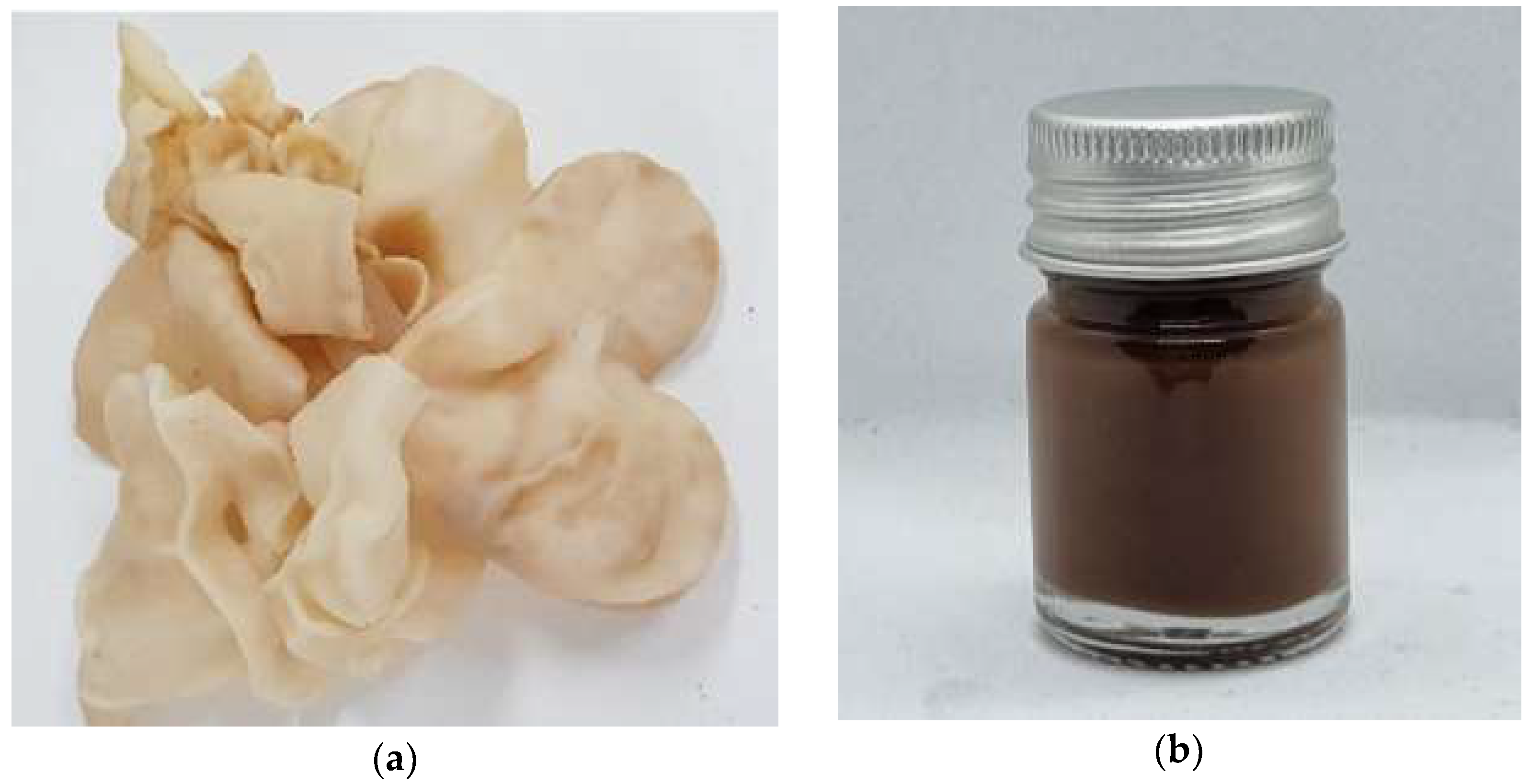
2.2. Cloud Ear Mushroom Extraction

Cloud ear mushrooms were purchased from the Royal Project Foundation Thailand between 2020 and 2021. The freshly fruiting body of mushrooms was dried in a hot air oven (UN 55 Memmert, Schwabach, Germany) at 50 ± 2 °C for an hour. Dried mushrooms were ground to powder using a blender (600 W, Viva Collection Blender, Philips, Bangkok, Thailand) and were extracted with ethanol followed by hot water at 85 °C for 4 h. After that, the extract was filtered through Whatman® filter paper No.1. The solvent was removed using a rotary evaporator (Buchi R-300, Essen, Germany) until the concentrated extract was obtained. The cloud ear mushroom extract (CW) was kept in an amber glass at 2 ± 2 °C for further use.

2.3. Determination of Total Polysaccharides Content

The total polysaccharide content was evaluated using an anthrone method with some modifications [12]. The anthrone reagent was prepared with 0.2% w/v anthrone in sulfuric acid. Briefly, the sample (50 μL) was mixed with anthrone reagent (100 μL) and incubated at 80 °C for 5 min in a water bath (Memmert Waterbath WNB, Schwabach, Germany). The absorbance was measured at 625 nm using a UV-vis spectrophotometer (Shimadzu, Kyoto, Japan). The analytical curve was plotted using standard D-glucose with different concentrations (y = 3.9027x + 0.4686), where y is the absorbance value and x is the standard glucose content (mg). The results were expressed as milligrams of glucose equivalent per gram of extract using the equation below.
Total polysaccharides content (mg glucose/g extract) = (c × V × D)/N
where c is the concentration of glucose (mg), V is the sample volume (mL), D is the dilution factor and N is the weight of the sample (g).

2.4. Cell Culture

2.4.1. Determination of Cytotoxicity

The HaCaT cells in 96-well plates were seeded at 1 × 10⁴ cells/well with the medium (DMEM with 10% v/v fetal bovine serum, 1% v/v penicillin, and streptomycin solution) and incubated at 37 °C and 5% CO2 incubator. After 24 h, the medium was then removed, and the sample (100 µL) was added and incubated at 37 °C and 5% CO2 incubator for 48 h. 15 µL of MTT reagent (5 mg/mL) was added and incubated at 37 °C and 5% CO2 incubator for 4 h. The solution was removed and replaced by DMSO. The absorbance was measured using a microplate reader at a wavelength of 570 nm with a reference wavelength of 630 nm. The results were calculated using the equation below.
% Cell viability = (OD of sample well/OD of vehicle control) × 100
where the OD of the sample is the absorbance of the treated cells, and the OD of the vehicle control is the absorbance of the untreated cells.

2.4.2. Quantification of IL-6 and TNF-α Secretions by ELISA

The culture supernatant was used to determine IL-6 and TNF-α secretions. HaCaT cells were seeded at 1 × 10⁴ cells/well in 96-well plates and incubated at 37 °C at 5% CO2 for 24 h. The cells were pretreated with the sample (50 µL) for 2 h in a 5% CO2 incubator and the medium (DMEM with 10% v/v fetal bovine serum, 1% v/v penicillin, and streptomycin solution) was removed. LPS was added at a final concentration of 1 µg/mL and incubated at 37 °C and 5% CO2 incubator for 24 h. The collected supernatant was centrifuged at 10,000× g for 5 min. Pro-inflammatory cytokines including IL-6 and TNF-α were determined using the sandwich enzyme-linked immunosorbent (ELISA) assay following the manufacturer protocol (Elabscience, Houston, TX, USA).

2.4.3. Expression of FLG and AQP3 by ELISA

HaCaT cells were used to study the expression of FLG and AQP3. HaCaT cells were seeded at 1 × 10⁴ cells/well in 24-well plates. Cells were treated with the sample for 96 h at 37 °C, in a 5% CO2 incubator. After incubation and harvesting, cells were lysed using freeze-thaw cycles to break up the cell membranes. The technique involves freezing a cell suspension in a dry ice bath and then thawing the material in a water bath at 37 °C with multiple cycles. Protein concentrations after the cells were lysed were measured using the Lowry’s method. 20 µL of the test solution was mixed with 2.5 mL of copper reagent (1% w/v copper sulfate, 2% w/v sodium potassium tartrate, and 2% w/v sodium carbonate) at room temperature. After 10 min, 1 M Folin-Ciocalteu reagent with 250 µL was rapidly mixed and incubated for 30 min. The absorbance was measured at 600 nm using a microplate reader. Cell lysed was measured using a Human FLG ELISA Kit (MyBioSource, San Diego, CA, USA) following the manufacturer’s instructions. FLG was measured at 450 nm using a microplate reader. Retinoic acid was used as a positive control, and the assays were performed in triplicate.
HaCaT cells were treated for 24 h at 37 °C in a 5% CO2 incubator. After that, the cell culture supernatant was centrifuged at 10,000× g for 5 min and collected. The concentration of AQP3 was detected using a Human AQP3 ELISA Kit (MyBiosource, San Diego, CA, USA) following manufacturer instructions. AQP3 was measured at 450 nm using a microplate reader. Retinoic acid was used as a positive control, and the assays were performed in triplicate.

2.5. Optimization of Film Forming Spray Base

To optimize the film-forming spray base, the ratio of CW/SPS/glycerin was optimized to identify the mixture with a high percentage of the occlusive properties and minimize the drying time of film forming spray. The optimization was performed using the Design-Expert® (Version 7, Stat-Ease Inc., Minneapolis, MN, USA). All components of the formulation were dissolved in water and mixed well. The active compound was mixed after the film-forming spray base was obtained. The range values of each ingredient for the experimental run are shown in Table 1.

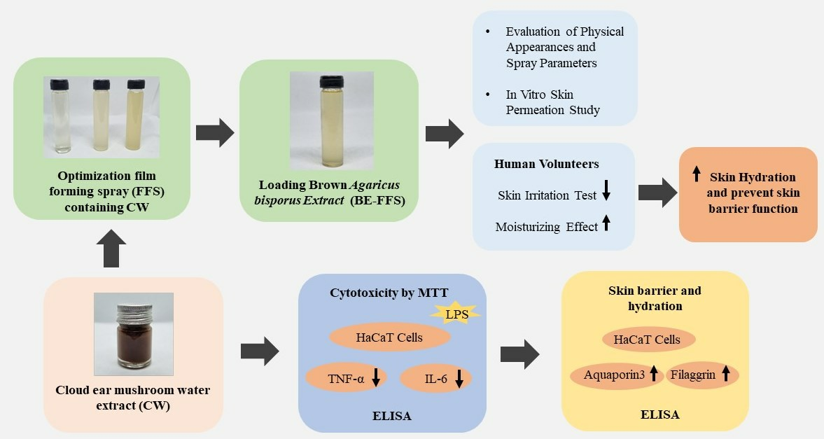
2.6. Preparation of Film Forming Spray Containing Brown A. bisporus Extract

FFS base was prepared using a simple solution method with the combination of SPS, CW, glycerin, and active compound. First, SPS as a synthetic polymer was dissolved in water and stirred using a magnetic stirrer. CW as a natural polymer was added to a solution. Then glycerin was added as a plasticizer and adjusted up to final weight with water. After that, brown Agaricus bisporus extract (BE) as the active ingredient was added to the film-forming spray and mixed well to completely formulate BE-FFS. The concentration of 0.5% w/w BE related to our previous studies was chosen as an active ingredient because it revealed excellent properties including anti-aging, anti-oxidant, and skin inflammation, preventing abnormal epidermal skin barrier function [11].

2.7. Evaluation of the BE-FFS

2.7.1. Film Forming Characteristics

Physical appearance: The clarity and color of film forming spray were evaluated by visual inspection. In addition, the physical appearance of formation after spray on the skin surface was also evaluated by visual inspection.
Viscosity: The viscosity of the film-forming spray was measured using a viscometer (Brookfield Engineering Laboratories, Middleboro, MA, USA). BE-FFS with a volume of 100 mL was evaluated viscosity at 20.0 ± 0.5 °C for 5 min. The viscosity of each sample was recorded every 30 s.
pH: The pH of the film-forming spray was measured using a digital pH meter (Seven-Compact™ S220, Greifensee, Switzerland).

2.7.2. Drying Time

The formulation was evaluated for drying time with some modifications [13]. BE-FFS was sprayed on the human forearm. After a fixed period of time, a glass plate was placed on the skin without pressure. Investigation by the naked eye, BE-FFS is considered as dry when liquid is not found on the glass plate after removal. If the liquid remains on the skin, the experiment was repeated with an increase in drying time. A good film-forming spray should have a minimum drying time to avoid a long waiting time. The time required for completely drying the film was recorded.

2.7.3. Occlusive Factor

The water vapor permeability of the formulation was also vital to determine the occlusive factor with some modifications [14] because it affects skin hydration. The film-forming spray was sprayed over a cellulose filter paper having a surface area of 18 cm2. The cellulose filter paper was placed on the beaker containing 30 mL of water. A similar beaker with water was covered with cellulose filter paper without the formulation as a control. The total weight of the beaker and water was recorded. The beaker was incubated in a hot air oven at 40 °C for 24 h with 50 to 55 %RH. The total weight was recorded after incubation for the volume of water loss. The occlusive factor was determined based on the weight of water loss in the beaker and calculated using the following equation.
F = (A − B)/A × 100
where A is the weight of water lost without spraying, and B is the volume of water lost with spraying.

2.7.4. Spray Pattern and Spray Angle

The study of the spray pattern and spray angle was performed using methylene blue with some modifications [15]. Methylene blue at a concentration of 0.1% w/w was added to the film-forming spray and mixed until homogeneous. The formulation was sprayed on a sheet of bagasse paper (15 × 15 cm2) 7 cm away from the paper. The obtained two-dimensional images of the spray patterns were then analyzed using Image J Software and characterized in terms of the area. Spray angle (θ) was calculated using the following equation.
Spray angle (θ) = tan−1 (l/r)
where l is the distance between the sprayer and the paper, and r is the radius of the sprayed circle.

2.8. In Vitro Skin Permeation Study of BE-FFS

An in vitro skin permeation study was conducted using Franz diffusion cell (Logan instruments, Somerset, NJ, USA) with some modifications [16]. This study was to investigate the permeation of active ingredients from film forming through a synthetic membrane (300 μm, Strat-M® EMD Millipore, Burlington, MA, USA). The structure of the Strat-M® membrane creates a morphology like that of different layers of human skin as the epidermis, dermis, and subcutaneous tissue [17]. Briefly, a membrane was placed on the receptor chamber and closed with a donor chamber with a permeation area of 3.14 cm2. The receiver solution contained PBS (pH 7) with volume of 7.5 mL and incubated at 37 °C with a magnetic stirrer at 300 rpm. BE-FFS (1 mL) was added to the membrane. The receiver solution was withdrawn from the receptor chamber at 0.5, 1, 2, 4, 6, 8, 10, and 12 h and then replaced by a fresh medium with an equal volume. The receptor solution at each time interval was collected to determine the amount of ergothioneine and gallic acid permeated through a membrane because those are active compounds found in BE. The formulation remained on the membrane and was removed by deionized water. Then the membrane was cut in pieces and extracted with methanol by sonicate bath for 10 min. The concentration of ergothioneine and gallic acid in the membrane was analyzed by HPLC.
The cumulative amount of ERT and GA permeated after 12 h (Q12) was directly determined from the concentration of the agent in the receptor compartment per unit area. The steady-state flux (J) was determined from the slope of the steady-state portion of a plot of Q versus time. The permeability coefficient (Kp) was calculated from steady-state flux/donor concentration.

2.9. Human Skin Irritation Test

The skin irritation test was evaluated on the upper arm of volunteers using a standard 8 mm Scanpor® Fin chamber (Dr. Ebeling & Assoc. GmbH, Hamburg, Germany). All subjects provided signed informed consent. Thirty volunteers with dry skin, male and female aged between 45 and 70 years were enrolled in the study. The dry skin condition was evaluated using a Corneometer (Corneometer® CM 825; Courage & Khazaka Electronic GmbH, Cologne, Germany) on a scale ranging from 0 to 100 arbitrary units (a.u.), whereby values higher than 40 a.u. represent normal skin hydration, values between 30 and 40 a.u. constitute dry skin and values lower than 30 a.u. characterize extremely dry skin [18]. Thus, volunteers exhibiting skin hydration values lower than 40 a.u. were included as volunteers.
According to European Society of Contact Dermatitis (ESCD) guidelines for diagnostic patch testing [19], The BE-FFS, 2% w/w SLS (positive control), and deionized water (negative control) were applied on the upper arm of human volunteers using a Fin chamber and left for 48 h. After patch removal, the irritating reactions (erythema and edema) were observed at 24, 48, and 72 h. The identification of irritating scores was based on the Draize scoring system [20] and primary irritation index (PII value) [21].

2.10. Moisturizing Effect of BE-FFS in Human Volunteers

The study was conducted in accordance with the Faculty of Pharmacy, Chiang Mai University, and approved by the Research Ethics Committee (REC) of the Faculty of Pharmacy, Chiang Mai University operates in compliance with ethical principles stated in the Declaration of Helsinki, ICH-GCP and international ethical guidelines (protocol code 002/2020). The moisturizing effect of BE-FFS was conducted with 30 dry skin volunteers (n = 30). The volunteers sprayed the BE-FFS on their upper arm to cover a surface (5 × 15 cm) twice daily; morning and evening for 4 weeks. The parameters related to skin dryness including skin surface hydration and TEWL of all volunteers were evaluated as baselines before applying BE-FFS. The hydration level of the skin was represented in terms of skin water content measured by a Corneometer. TEWL was evaluated using a Tewameter (Tewameter® TM 300; Courage & Khazaka Electronic, Cologne, Germany), measuring the amount of water loss by the skin over a certain period expressed in g/m2/h. During the study, all volunteers were allowed to wash their skin normally but they were restricted to applying moisturizers on the testing areas. The percentage of skin moisturizing and TEWL protection efficacy are calculated using the following equation.
% Efficacy = (T(4weeks) − T(0))/T(0) × 100
where T(4 weeks) is the skin hydration and TEWL level after using the BE-FFS for four weeks and T(0) is the initial skin hydration and TEWL level before using the BE-FFS.

2.11. Statistical Analysis

All results are expressed as the mean ± SD. The results were statistically compared using one-way ANOVA with multiple comparisons using Tukey’s test (GraphPad, Version 7.0). Significant differences were considered at p < 0.05.

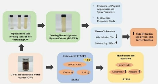
3. Results and Discussion

3.1. Cloud Ear Mushroom Extraction

The fruiting body of cloud ear mushroom and the physical appearance of CW are shown in Figure 1. The fruiting body was light yellow with a gelatinous texture and ear-shaped form. The physical appearance of CW by hot water extraction was a dark brown semisolid extract. The percentage yield of CW was 20.99 ± 1.65%. Hot water extraction can promote the breakdown of chitin in the mushroom cell walls essentially resulting in high molecular weight compounds including polysaccharides, proteins, and free amino acids [22].

3.2. Determination of Total Polysaccharides Content

The polysaccharides in the extract are hydrolyzed into glucose when boiled in an acidic medium, resulting in a green–blue color complex [23]. The total polysaccharides content of CW was 748.2 ± 0.02 mg glucose/g extract. The extract showed high total polysaccharide content because hot water extraction can effectively extract water-soluble polysaccharides [24].

3.3. Cell Culture

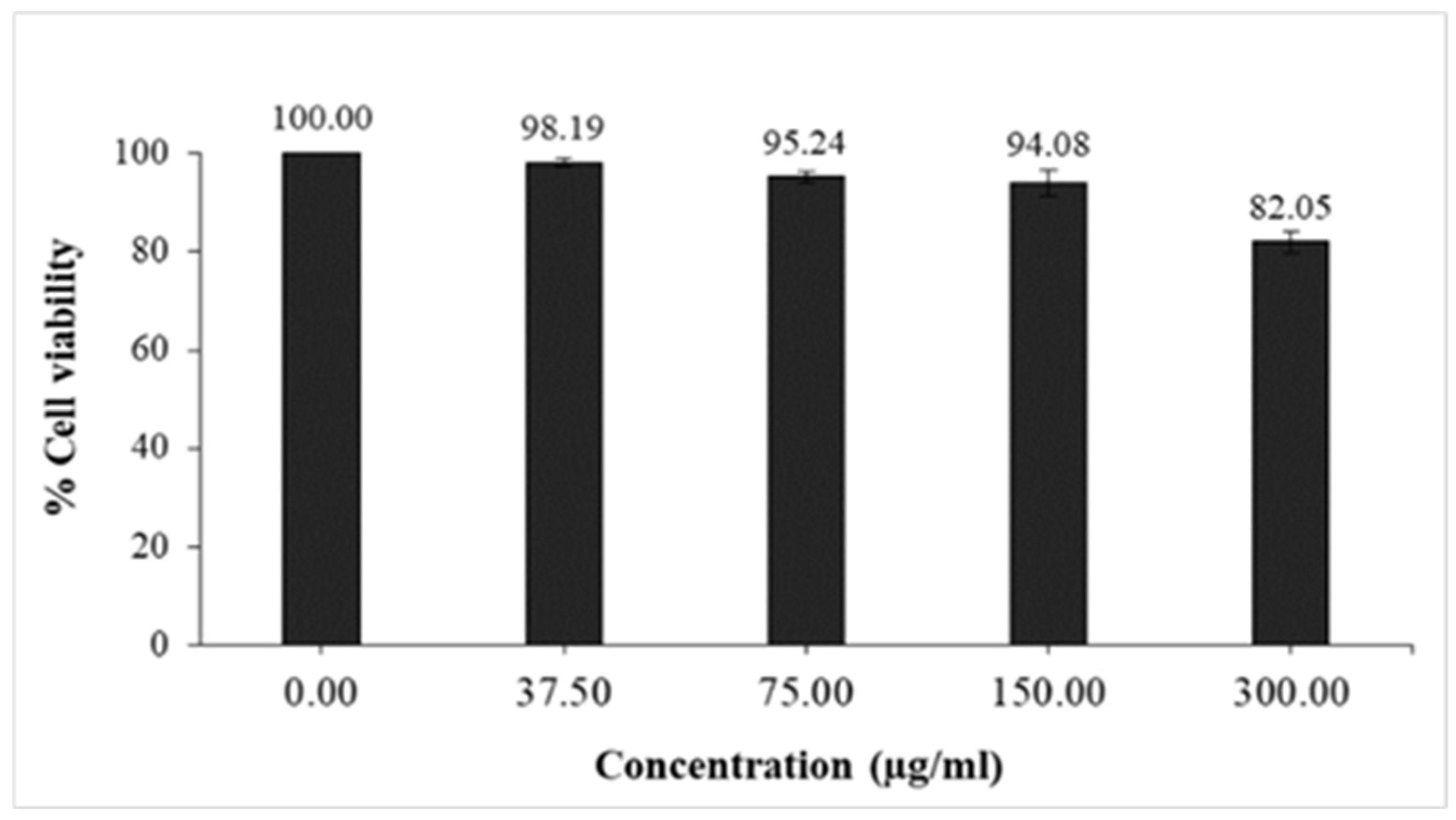
3.3.1. Cytotoxicity Test by MTT Assay

The cytotoxic effect of the CW on the viability of HaCaT cells is presented in Figure 2. The cell viability of the control sample was expressed as 100%. The CW was tested at a concentration range of 37.5 to 300 µg/mL. To identify the non-cytotoxic concentration, the result showed that the IC20 of CW was 300 µg/mL. Thus, the non-toxic concentration was 300 µg/mL which was chosen for the further anti-inflammatory activity study and the expression of FLG and AQP3 tests.

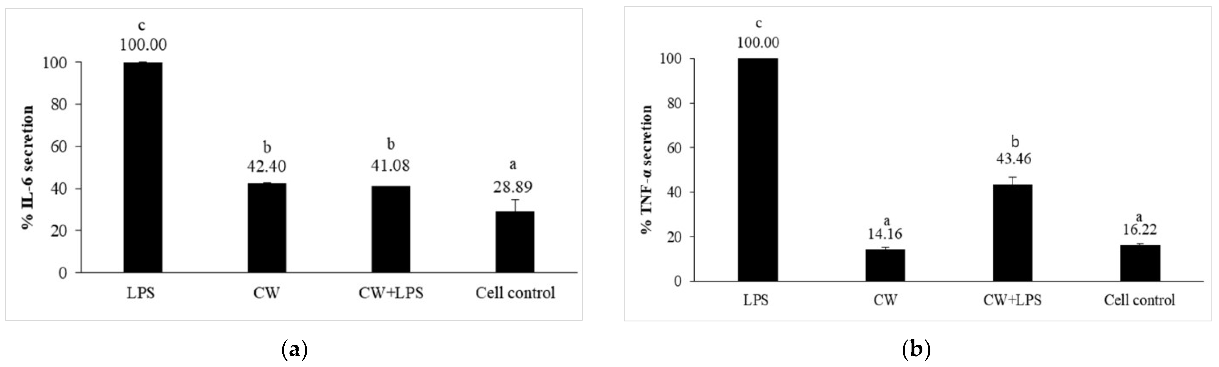
3.3.2. Preventive Effect of Cloud Ear Mushroom Extract against Inflammation in LPS-Induced HaCaT Cells

The present study investigated CW’s anti-inflammatory activity and skin protective effects using HaCaT cell lines as the model system of human skin. The LPS-induced HaCaT cells for the secretions of IL-6 and TNF-α were defined as 100%, as shown in Figure 3. Inflammatory cytokines, including IL-6 and TNF-α, were activated with LPS and incubated for 24 h. The pre-treatment of the extract (CW+LPS) strongly protected HaCaT cells against IL-6 secretion similar to the treatment of CW alone. For TNF-α secretion, the pretreatment extract (CW+LPS) moderately decreased cytokine production compared with that of CW alone. However, CW significantly inhibited pro-inflammation cytokines including IL-6 and TNF-α, compared with LPS alone. The results showed that CW could reduce LPS-induced inflammation and decrease IL-6 and TNF-α secretions in HaCaT cells. In addition, IL-6 levels were involved in the skin growth and differentiation of dermal and epidermal layers [25]. The increase of these cytokines was an immediate response to SC permeability impairment, increased TEWL, and decreased SC hydration [26]. Moreover, IL-6 cytokine has been indicated in a wide range of inflammatory conditions, especially those affecting the skin due to abnormal epidermal skin barriers, thickening of the epidermis, and promoting dryness [27]. The release of inflammatory cytokines could activate mast cells and encourage the production of histamines and MMP-1 that caused the breakdown of skin collagen type 1 and contributed to skin dryness [28]. Therefore, the ability of CW was an anti-inflammatory agent and a skin protector from TEWL. Additionally, it could improve skin barrier function, significantly affecting skin hydration.

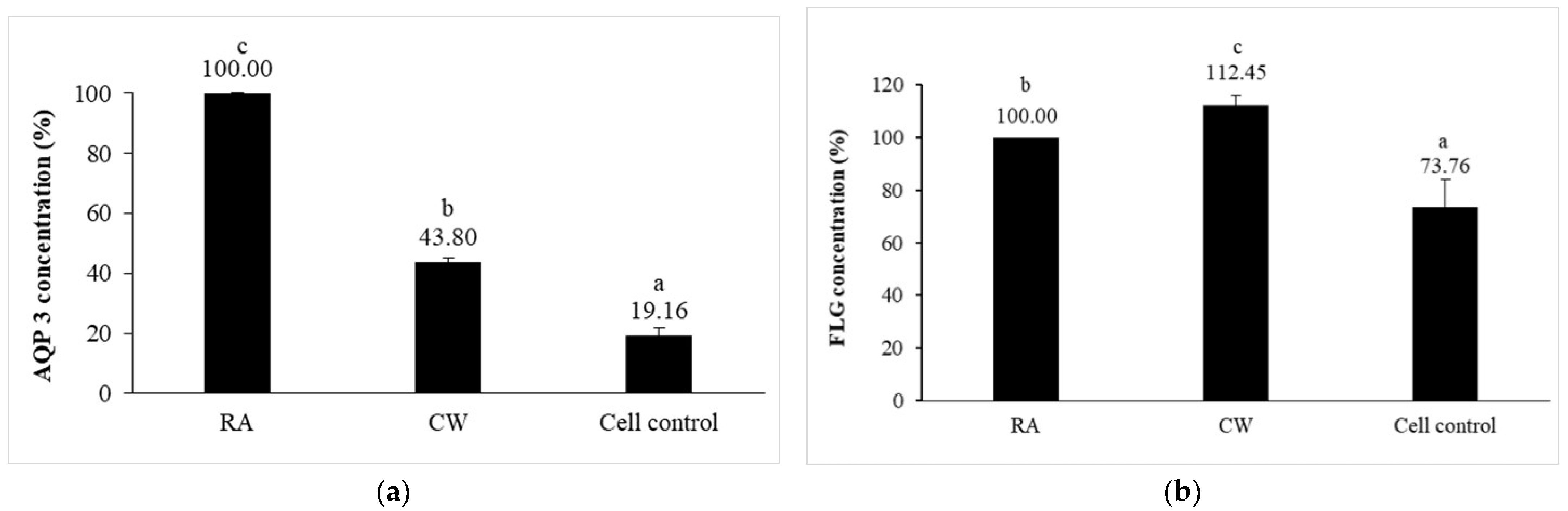
3.3.3. Expression of AQP3 and FLG

Skin hydration relies on many proteins and humectants in the epidermis. AQP3 and FLG are the proteins expressed by keratinocytes and play important roles in hydration due to their moisturizing and barrier-enhancing activities [29]. The important role of aquaporin is directly involved in water transport. AQP3 is an aquaglyceroporin that transports water and glycerol, mediating skin hydration mechanisms. FLG is a key protein that acts as an adhesive to hold keratin together, increasing the strength of the skin barrier in keratinocytes [30]. When HaCaT cells were treated with CW (300 µg/mL) for 96 h, cells were lysed, and the soluble proteins were measured using Lowry’s method and correlated to the protein concentration as 40 µg/mL for FLG expression [31]. In this study, the expression of AQP3 and FLG was observed in CW-treated HaCaT cells, as shown in Figure 4. The results showed that the expression of AQP3 and FLG was significantly increased when treated with CW compared with cell control. It could be confirmed that CW could enhance the capability of the skin barrier and hydration through upregulating AQP3 and FLG.

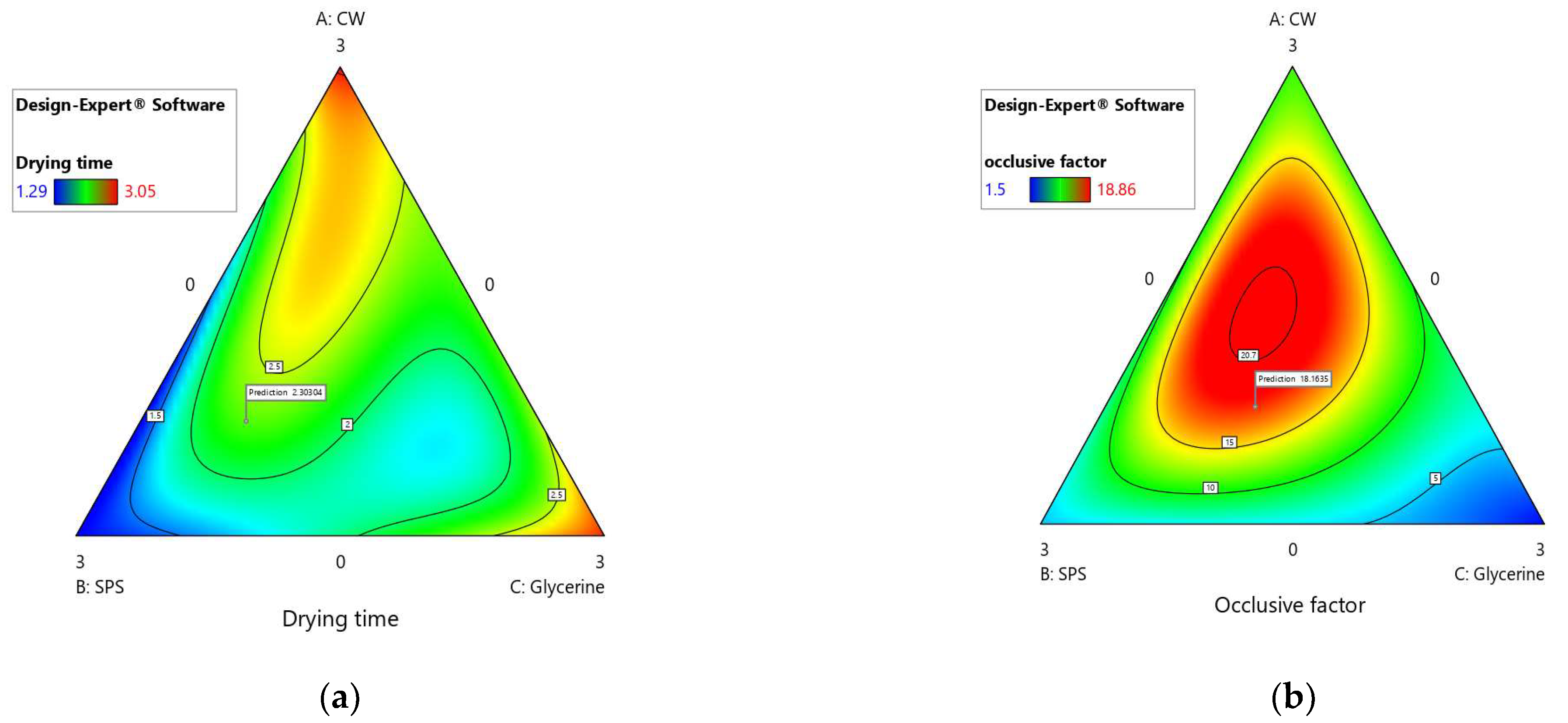
3.4. Optimization of Film Forming Spray Base

A pseudo-components simplex mixture design was carried out to optimize the ratios among FFS-based components and the respective minimum and maximum limits of the following mixture variables: (A) CW (0 to 3% w/w), (B) SPS (0 to 3% w/w) and (C) glycerin (0 to 3% w/w) for variable responses of drying time (min) and occlusive factor (%), Figure 5. The regression equations obtained after applying the mixture design represented the relationship between the main factors and variable responses. The quadratic model best fit the drying time, which was determined to be significant (p < 0.05) with an R2 value of 0.998. The optimization predicted drying time was 2.30 min. CW had a more significant effect on increasing the drying time of FFS-based formulations. The drying time tended to increase when the amount of glycerin was higher. To obtain FFS-based formulations with minimized drying time, using low amounts of CW and glycerin was essential. The regression equation for drying time is stated in the equation. Where A represents CW, B represents SDS, and C represents GLY.
y = 3.05A + 1.29B + 2.99C − 2.77AB − 2.91AC − 0.7946BC
The optimization predicted on the occlusive factor was 18.16%. The occlusive factor of FFS-based formulations was dependent on natural and synthesized polymers. As it can be seen from the results an increase in CW had a more significant effect on improving the occlusive factor, while an increase in glycerin had a decreasing effect. The quadratic model was the best fit for determining the significant occlusive factor (p < 0.05) with an R2 value of 0.986. The regression equation for the occlusive factor was obtained from the mixture design in the equation below.
y = 11.25A + 4.88B + 1.53C + 5.41AB + 13.61AC + 9.87BC
Therefore, the best formulation optimized by the simplex lattice mixture design analysis was 0.74% w/w CW, 1.38% w/w SPS, and 0.87% w/w glycerin, resulting in a drying time of 2.21 min, and an occlusive factor of 17.67 matching the theoretical predictions values. The coefficient of variation (%CV) was 1.91 and the low value indicated that the mixture design sufficiently fit the experimental data and could be used as a tool to optimize desirable formulations. The final formulation containing BE was packed in a spray container and further evaluated for physical appearance.

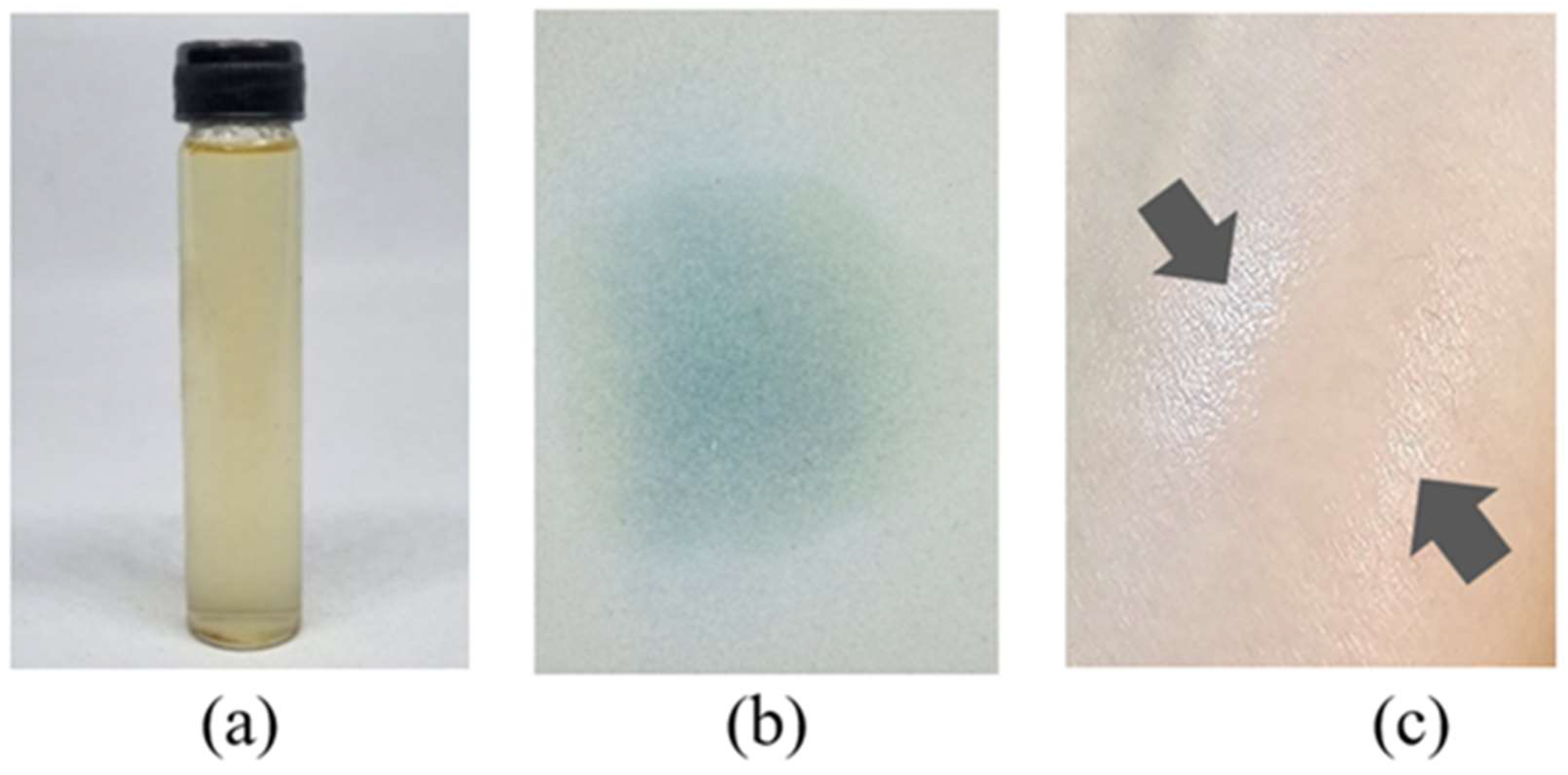
3.5. Evaluation of Physical Appearances and Spray Parameters of BE-FFS

The physical characteristics of BE-FFS were evaluated using pH, viscosity, drying time, occlusive factor, spray pattern, and spray angle. The BE-FFS was brown with no phase separation. When sprayed on the skin, The solution was found to be a thin transparent film, as shown in Figure 6. The BE-FFS had a pH value of 5, which is appropriate in the range of human skin pH, between 5.0 and 6.0 [32]. Viscosity plays an important critical role in affecting droplet spread. The formulation would be un-sprayable throughout the target skin surface when the viscosity was too high. The viscosity of BE-FFS was 10 cps. The drying time of BE-FFS was 2.21 ± 1.55 min. BE-FFS consists of synthetic and natural polymers that are soluble in water, so the drying time was directly correlated to the concentration of the natural polymer [13]. The BE-FFS remained on the skin’s surface without dripping until it dried, efficiently providing spray ability. The spray angle of BE-FFS was also found to be 82.46 ± 0.39. The spray pattern was uniform and exhibited spherical spots indicating a good spray effect. The occlusive factor of BE-FFS was 17.67 ± 2.13% because the mixed polymers showed a suitable occlusive property to cover the skin surface. The occlusive property of film forming is important to improve skin hydration, prevent water loss, and increase skin penetration of active agents through the skin.

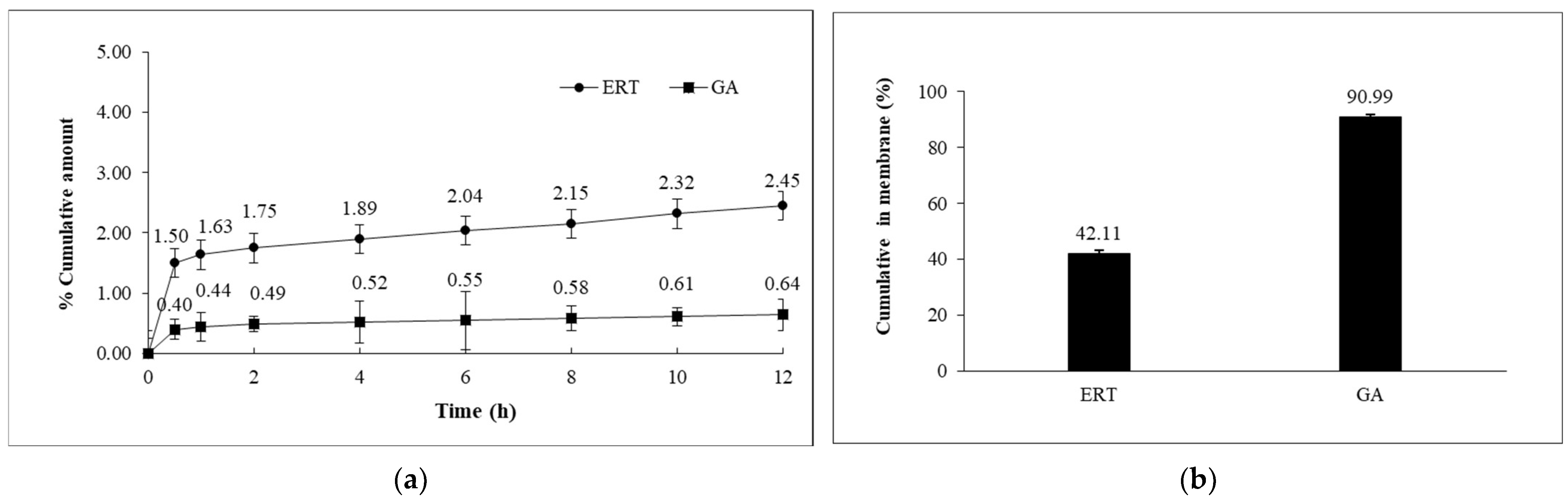
3.6. In Vitro Skin Permeation Study of BE-FFS

Skin permeation of BE-FFS was tested by Franz diffusion cells using Strat-M® synthetic membrane as skin. Our previous study showed that ergothioneine (ERT) and gallic acid (GA) were active compounds in brown A. bisporus extract [11]. Therefore, ERT and GA were chosen as chemical markers for the skin permeation study of BE-FFS detected by HPLC. The Skin permeation parameters of ERT and GA release from BE-FSS were assessed over 12 h, presented in Table 2. Data on skin permeation from BE-FFS through the membrane are illustrated in Figure 7a. After 0.5 h, it became evident that the BE-FFS showed a steady state of skin permeation of ERT and GA, as seen in the linear portion of the plot. The results showed that the BE-FFS had a moderate amount of ERT and GA in the receptor medium. This indicated that the BE-FFS had smoothly penetrated the membrane. After the end of the permeation experiment, the amount of ERT and GA had highly accumulated on the skin, as shown in Figure 7b. ERT and GA were found to have highly accumulated on the membrane, indicating that BE-FFS remained on the skin. This was due to the property of film-forming polymers. Mushroom polysaccharides promote drug penetration, serve as a permeation enhancer, and possess occlusive properties, providing long-term release in the skin [33]. Moreover, the unique advantage of polysaccharides promotes a hydration effect [34]. The BE-FFS consisted of natural polysaccharide and synthetic polymer as a thin film polymer, resulting in accumulating the active compound in the skin, resulting in an effect on the skin barrier with an increase in skin hydration and protection against TEWL.

3.7. Skin Irritation Test in Human Volunteers

The skin irritation reaction of the BE-FFS was evaluated after 48 h using Finn chamber®. The dermal irritancy potential of the test substances is shown in Table 3. The results showed that deionized water and the BE-FFS were nonirritating with a low Primary Dermal Irritation Index value (PDII < 0.5), whereas 2% w/v SLS was slightly irritating (PDII range from 0.5 to 2.0). The results revealed that the BE-FFS was nonirritating to the skin and could be further used for skin hydration tests among human volunteers.

3.8. Moisturizing Effect of BE-FFS in Human Volunteers

The results of the moisturizing property evaluation of BE-FFS on human skin after four weeks are shown in Table 4. The results indicated that BE-FFS can increase skin hydration from 38.47 ± 6.01 a.u. at week 0 to 48.43 ± 8.03 at the end. A pairwise comparison showed that the skin hydration level on week four was significantly higher than the initial level (p < 0.05). The results of skin moisturizing efficacy of BE-FFS application after four weeks were 27.3 ± 9.55%.
TEWL is the amount of water evaporated per unit of time/area. The higher the amount of TEWL, the more skin water loss occurs, indicating that the skin barrier is damaged. In this study, TEWL significantly decreased after four weeks at 9.60 ± 2.30 compared with the initial level (p < 0.05). TEWL protection efficacy was presented at −17.9 ± 5.60%. It can be assumed that the BE-FFS can protect water loss from the skin surface. As mentioned above, the BE-FFS demonstrated an excellent potential effect on hydrating skin and preventing skin water loss. The likely explanation is that the mixed CW and synthetic polymer as a thin film polymer produced an occlusive effect. In addition, CW is composed of many polysaccharides according to the results of total polysaccharides content. Natural polysaccharides function as a moisturizing agent and natural film formers that could retain prolonged skin hydration and protect the skin barrier [35,36,37]. In addition, one related study found that GA and ERT, found in the BE, could repair the skin barrier by adding skin moisturizer and covering skin water loss [11].

4. Conclusions

CW possesses properties of a natural polymer that is nontoxic and can protect against IL-6 and TNF-α secretion on HaCaT cells. Moreover, it significantly increased AQP3 and FLG expression, which can improve skin hydration and prevent skin barrier damage on HaCaT cells. The BE-FFS was successfully prepared using CW and SPS mixing as the film-forming polymers in the aqueous solution. The BE-FFS was demonstrated as a clear, transparent spray, generating a fast, thin film after spraying on the skin surface. The BE-FFS was formulated to increase skin permeation efficacy with an occlusive factor. The BE-FFS can improve skin hydration and moisturizing effectiveness in clinical studies and protect skin from TEWL and non-irritation. Thus, the BE-FFS may prove a potential topical film-forming spray for topical skin hydration application in cosmeceutical products.

Author Contributions

Conceptualization, K.K.; methodology, K.K., P.L., O.N., N.I. and N.N.; software, N.N. and K.K.; validation, K.K., N.N. and N.I.; formal analysis, N.N., K.K. and N.I.; investigation, K.K., N.N. and N.I.; resources, K.K. and N.I.; data curation, K.K. and N.N.; writing—original draft preparation, N.N. and K.K.; writing—review and editing, K.K., P.L., O.N., N.I. and N.N.; visualization, N.N. and K.K.; supervision, K.K.; project administration, K.K.; funding acquisition, K.K. All authors have read and agreed to the published version of the manuscript.

Funding

This research project was partially supported by Chiang Mai University, Faculty of Pharmacy, Chiang Mai University, and Teaching Assistant and Research Assistant Scholarships (TA/RA) academic year 2020 from Chiang Mai University. In addition, this research project was financially supported by the Agricultural Research Development Agency (Public Organization) of Thailand (ARDA).

Institutional Review Board Statement

The study was conducted in accordance with the Faculty of Pharmacy, Chiang Mai University, and approved by the Research Ethics Committee (REC) of the Faculty of Pharmacy, Chiang Mai University operates in compliance with ethical principles stated in the Declaration of Helsinki, ICH-GCP and international ethical guidelines (protocol code 002/2020 and 12/01/2020).

Informed Consent Statement

Informed consent was obtained from all subjects involved in the study.

Data Availability Statement

The data presented in this study are available from the corresponding author upon reasonable request.

Acknowledgments

The authors would like to acknowledge Chiang Mai University and the ARDA of Thailand for their financial support. The authors would also like to acknowledge the Faculty of Pharmacy, Chiang Mai University for the research grant and facilities used in the project. In addition, the authors would also like to acknowledge the Division of Clinical Microscopy, Department of Medical Technology, Faculty of Associated Medical Sciences, Chiang Mai University, Chiang Mai, Thailand for the facilities used in the project. Thank you to Thomus Mcmanamon for proofreading and language formatting the article.

Conflicts of Interest

The authors declare no conflict of interest.

References

  1. Rawlings, A.V.; Scott, I.R.; Harding, C.R.; Bowser, P.A. Stratum corneum moisturization at the molecular level. J. Investig. Dermatol. 1994, 103, 731–740. [Google Scholar] [CrossRef] [PubMed]
  2. Fowler, J. Understanding the role of natural moisturizing factor in skin hydration. Pract. Dermatol. 2012, 9, 36–40. [Google Scholar]
  3. Rawlings, A.V.; Matts, P.J. Stratum corneum moisturization at the molecular level: An update in relation to the dry skin cycle. J. Investig. Dermatol. 2005, 124, 1099–1110. [Google Scholar] [CrossRef] [PubMed]
  4. Ma, T.; Hara, M.; Sougrat, R.; Verbavatz, J.-M.; Verkman, A. Impaired stratum corneum hydration in mice lacking epidermal water channel aquaporin-3. J. Biol. Chem. 2002, 277, 17147–17153. [Google Scholar] [CrossRef]
  5. Kathe, K.; Kathpalia, H. Film forming systems for topical and transdermal drug delivery. Asian J. Pharm. Sci. 2017, 12, 487–497. [Google Scholar] [CrossRef]
  6. Umar, A.K.; Butarbutar, M.; Sriwidodo, S.; Wathoni, N. Film forming sprays for topical drug delivery. Drug Des. Dev. Ther. 2020, 14, 2909–2925. [Google Scholar] [CrossRef]
  7. Mohd, T.A.T.; Manaf, S.F.A.; Abd Naim, M.; Shayuti, M.S.M.; Jaafar, M.Z. Properties of Biodegradable Polymer from Terrestrial Mushroom for Potential Enhanced Oil Recovery. Indones. J. Chem. 2020, 20, 1382–1391. [Google Scholar] [CrossRef]
  8. Sillapachaiyaporn, C.; Chuchawankul, S.; Nilkhet, S.; Moungkote, N.; Sarachana, T.; Ung, A.T.; Baek, S.J.; Tencomnao, T. Ergosterol isolated from cloud ear mushroom (Auricularia polytricha) attenuates bisphenol A-induced BV2 microglial cell inflammation. Food Res. Int. 2022, 157, 111433. [Google Scholar] [CrossRef]
  9. Ngwuluka, N.C.; Ochekpe, N.A.; Aruoma, O.I. Functions of bioactive and intelligent natural polymers in the optimization of drug delivery. Ind. Appl. Intell. Polym. Coat. 2016, 6, 165–184. [Google Scholar]
  10. Packialakshmi, B.; Sudha, G.; Charumathy, M. Studies on phytochemical compounds and antioxidant potential of Auricularia auricula-judae. Int. J. Pharm. Sci. Res. 2017, 8, 3508–3515. [Google Scholar]
  11. Nitthikan, N.; Leelapornpisid, P.; Naksuriya, O.; Intasai, N.; Kiattisin, K. Potential and alternative bioactive compounds from brown Agaricus bisporus mushroom extracts for xerosis treatment. Sci. Pharm. 2022, 90, 59. [Google Scholar] [CrossRef]
  12. Hassan, M.J.; Karim, M.M.; Islam, M.A.; Pramanik, M.H.R.; Hossain, M.A. Changes in root porosity and water-soluble carbohydrates in rice (Oryza sativa L.) under submergence stress. J. Bangladesh Agric. Univ. 2019, 17, 539–544. [Google Scholar] [CrossRef]
  13. Paradkar, M.; Thakkar, V.; Soni, T.; Gandhi, T.; Gohel, M. Formulation, and evaluation of clotrimazole transdermal spray. Drug Dev. Ind. Pharm. 2015, 41, 1718–1725. [Google Scholar] [CrossRef] [PubMed]
  14. Ranade, S.; Bajaj, A.; Londhe, V.; Babul, N.; Kao, D. Fabrication of topical metered dose film forming sprays for pain management. Eur. J. Pharm. Sci. 2017, 100, 132–141. [Google Scholar] [CrossRef] [PubMed]
  15. Zhong, Y.; Zhuang, C.; Gu, W.; Zhao, Y. Effect of molecular weight on the properties of chitosan films prepared using electrostatic spraying technique. Carbohydr. Polym. 2019, 212, 197–205. [Google Scholar] [CrossRef]
  16. Nitthikan, N.; Leelapornpisid, P.; Natakankitkul, S.; Chaiyana, W.; Mueller, M.; Viernstein, H.; Kiattisin, K. Improvement of stability and transdermal delivery of bioactive compounds in green robusta coffee beans extract loaded nanostructured lipid carriers. J. Nanotechnol. 2018. [Google Scholar] [CrossRef]
  17. Haq, A.; Goodyear, B.; Ameen, D.; Joshi, V.; Michniak-Kohn, B. Strat-M® synthetic membrane Permeability comparison to human cadaver skin. Int. J. Pharm. 2018, 547, 432–437. [Google Scholar] [CrossRef]
  18. Clarys, P.; Clijsen, R.; Taeymans, J.; Barel, A.O. Hydration measurements of the stratum corneum comparison between the capacitance method (digital version of the Corneometer CM 825®) and the impedance method (S kicon-200 EX®). Ski. Res. Technol. 2012, 18, 316–323. [Google Scholar] [CrossRef]
  19. Johansen, J.D.; Aalto-Korte, K.; Agner, T.; Andersen, K.E.; Bircher, A.; Bruze, M.; Cannavó, A.; Giménez-Arnau, A.; Gonçalo, M.; Goossens, A. European Society of Contact Dermatitis guideline for diagnostic patch testing–recommendations on best practice. Contact Dermat. 2015, 73, 195–221. [Google Scholar] [CrossRef]
  20. Farage, M.A.; Maibach, H.I.; Andersen, K.E.; Lachapelle, J.M.; Kern, P.; Ryan, C.; Ely, J.; Kanti, A. Historical perspective on the use of visual grading scales in evaluating skin irritation and sensitization. Contact Dermat. 2011, 65, 65–75. [Google Scholar] [CrossRef]
  21. Leelapornpisid, P.; Mungmai, L.; Sirithunyalug, B.; Jiranusornkul, S.; Peerapornpisal, Y. A novel moisturizer extracted from freshwater macroalga [Rhizoclonium hieroglyphicum (C. Agardh) Kützing] for skin care cosmetic. Chiang Mai J. Sci. 2014, 41, 1195–1207. [Google Scholar]
  22. Leong, Y.K.; Yang, F.C.; Chang, J.S. Extraction of polysaccharides from edible mushrooms emerging technologies and recent advances. Carbohydr. Polym. 2021, 251, 117006. [Google Scholar] [CrossRef] [PubMed]
  23. Merdivan, S.; Bettin, P.; Preisitsch, M.; Lindequist, U. Quality control of medicinal mushrooms: Comparison of different methods for the quantification of polysaccharides/β-glucans. Planta Medica 2014, 80, P1N22. [Google Scholar] [CrossRef]
  24. Liu, Y.; Huang, G. Extraction and derivatisation of active polysaccharides. J. Enzym. Inhib. Med. Chem. 2019, 34, 1690–1696. [Google Scholar] [CrossRef] [PubMed]
  25. Gallucci, R.M.; Sloan, D.K.; Heck, J.M.; Murray, A.R.; O’Dell, S.J. Interleukin 6 indirectly induces keratinocyte migration. J. Investig. Dermatol. 2004, 122, 764–772. [Google Scholar] [CrossRef]
  26. Lu, B.; Elias, P.M.; Feingold, K.R. The Role of the Primary Cytokines, TNF, IL-1, and IL-6, in Permeability Barrier Homeostasis. Ski. Barrier 2005, 1, 325–338. [Google Scholar]
  27. Zhu, X.; Jiang, L.; Zhong, Q.; Kong, X.; Zhang, R.; Zhu, L.; Liu, Q.; Wu, W.; Tan, Y.; Wang, J. Abnormal expression of interleukin-6 is associated with epidermal alternations in localized scleroderma. Clin. Rheumatol. 2022, 41, 2179–2187. [Google Scholar] [CrossRef]
  28. Hiramoto, K.; Goto, K.; Tanaka, S.; Horikawa, T.; Ooi, K. Skin liver and kidney interactions contribute to skin dryness in aging KK-Ay/Tajcl mice. Biomedicines 2022, 10, 2648. [Google Scholar] [CrossRef]
  29. Shen, X.; Guo, M.; Yu, H.; Liu, D.; Lu, Z.; Lu, Y. Propionibacterium acnes related anti-inflammation and skin hydration activities of madecassoside, a pentacyclic triterpene saponin from Centella asiatica. Biosci. Biotechnol. Biochem. 2019, 83, 561–568. [Google Scholar] [CrossRef]
  30. Sandilands, A.; Sutherland, C.; Irvine, A.D.; McLean, W.I. Filaggrin in the frontline: Role in skin barrier function and disease. J. Cell Sci. 2009, 122, 1285–1294. [Google Scholar] [CrossRef]
  31. Sarkar, S.; Mondal, M.; Ghosh, P.; Saha, M.; Chatterjee, S. Quantification of total protein content from some traditionally used edible plant leaves: A comparative study. J. Med. Plant Stud. 2020, 8, 166–170. [Google Scholar] [CrossRef]
  32. Mori, N.M.; Patel, P.; Sheth, N.R.; Rathod, L.V.; Ashara, K.C. Fabrication and characterization of film-forming voriconazole transdermal spray for the treatment of fungal infection. Bull. Fac. Pharm. Cairo Univ. 2017, 55, 41–51. [Google Scholar] [CrossRef]
  33. Luo, C.; Xu, X.; Wei, X.; Feng, W.; Huang, H.; Liu, H.; Xu, R.; Lin, J.; Han, L.; Zhang, D. Natural medicines for the treatment of fatigue bioactive components pharmacology and mechanisms. Pharmacol. Res. 2019, 148, 104409. [Google Scholar] [CrossRef] [PubMed]
  34. Li, J.; Xiang, H.; Zhang, Q.; Miao, X. Polysaccharide-Based Transdermal Drug Delivery. Pharmaceuticals 2022, 15, 602. [Google Scholar] [CrossRef] [PubMed]
  35. Fodil-Bourahla, I.; Bizbiz, L.; Schoevaert, D.; Robert, A.; Robert, L. Effect of L-fucose and fucose-rich oligo-and polysaccharides (FROP-s) on skin aging penetration skin tissue production and fibrillogenesis. Biomed. Pharmacother. 2003, 57, 209–215. [Google Scholar] [CrossRef] [PubMed]
  36. Katekawa, E.; Caverzan, J.; Mussi, L.; Camargo-Junior, F.B.; Sufi, B.; Padovani, G.; Nazato, L.; Nogueira, C.; Magalhães, W.V.; Di Stasi, L.C. Novel topical skin hydration agent containing Anadenanthera colubrina polysaccharide-standardized herbal preparation. J. Cosmet. Dermatol. 2020, 19, 1691–1698. [Google Scholar] [CrossRef]
  37. Pacheco, M.S.; Barbieri, D.; da Silva, C.F.; de Moraes, M.A. A review on orally disintegrating films (ODFs) made from natural polymers such as pullulan, maltodextrin, starch, and others. Int. J. Biol. Macromol. 2021, 178, 504–513. [Google Scholar] [CrossRef]
Figure 1. The physical appearance of (a) fruiting body of cloud ear mushroom and (b) cloud ear mushroom water extract.
Cosmetics 10 00145 g001
Figure 2. Viability of HaCaT cells treated with the different concentrations of CW by MTT assay.
Cosmetics 10 00145 g002
Figure 3. Effects of the CW on (a) IL-6 and (b) TNF-α secretion on HaCaT cells. For each study, cells were treated with LPS alone (LPS), extract alone (CW), pre-treatment (CW+LPS), and non-treated (cell control). Different letters above the bars indicate a statistically significant difference (p < 0.05).
Cosmetics 10 00145 g003
Figure 4. Effect of CW on (a) AQP3 and (b) FLG expression in HaCaT cells. Different letters above the bars indicate statistically significant differences (p < 0.05).
Cosmetics 10 00145 g004
Figure 5. The triangle of response surface plot predicted for (a) drying time (min) and (b) occlusive factor (%). The letters A (CW), B (SPS), and C (Glycerin) are pseudo components represented by the mixture of reagents.
Cosmetics 10 00145 g005
Figure 6. Physical appearance of (a) BE-FFS, (b) spray pattern, and (c) an arrow pointing to polymeric film on a human upper arm.
Cosmetics 10 00145 g006
Figure 7. Skin permeation study of ERT and GA from BE-FFS (a) percentage cumulative in receiving compartment and (b) percentage cumulative on the membrane per unit surface area of BE-FFS after 12 h.
Cosmetics 10 00145 g007
Table 1. Independent variables and mixture design of FFS base.
RUNIndependent Variable (%)
A (CW)B (SDS)C (GLY)
1003
220.50.5
3300
41.51.50
5111
601.51.5
70.520.5
80.50.52
91.501.5
10030
11030
12300
Table 2. Skin permeation parameters of ERT and GA release from BE-FSS.
BE-FFSFlux
(µg/cm2/h)
Permeability Coefficient, Kp (cm/h) × 10−6Cumulative Amount, Q12 (μg/cm2)
ERT0.042 ± 0.118.34 ± 0.2749.78 ± 0.62
GA0.011 ± 0.151.30 ± 0.3113.07 ± 0.12
Table 3. Primary Dermal Irritation Index (PDII) and skin irritation reaction were observed among 30 volunteers.
Test SubstancesPDII ValueClassification of Skin Reaction
BE-FFS0.03No irritation
Positive (2% w/v SLS)0.50Slight irritation
Negative (DI water)0.03No irritation
Table 4. Skin hydration and TEWL before and after using formulation for 4 weeks (N = 30). Different letters in the column indicate statistically significant differences (p < 0.05).
WeeksSkin Hydration
(a.u.)
Skin Moisturizing
Efficacy (%)
TEWL
(g/m2/h)
TEWL Protection Efficacy (%)
038.47 ± 6.01 a27.3 ± 9.5514.20 ± 8.10 b−17.9 ± 5.60
448.43 ± 8.03 b9.60 ± 2.30 a
Disclaimer/Publisher’s Note: The statements, opinions and data contained in all publications are solely those of the individual author(s) and contributor(s) and not of MDPI and/or the editor(s). MDPI and/or the editor(s) disclaim responsibility for any injury to people or property resulting from any ideas, methods, instructions or products referred to in the content.

