
Design, Preparation, and Characterization of Effective Dermal and Transdermal Lipid Nanoparticles: A Review

 , , , , , and
Abstract
:1. Introduction
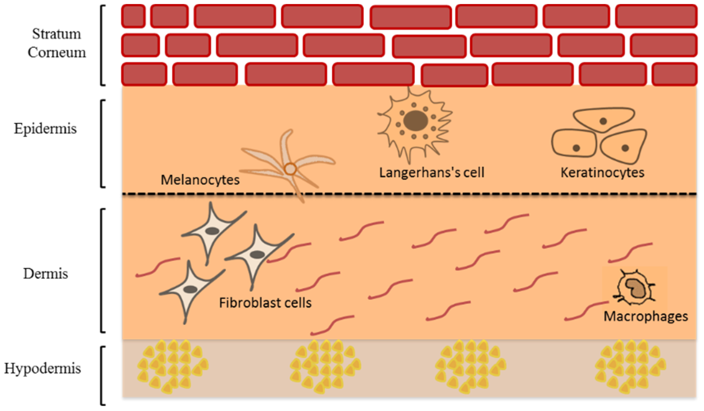
2. Skin
2.1. Skin Structure
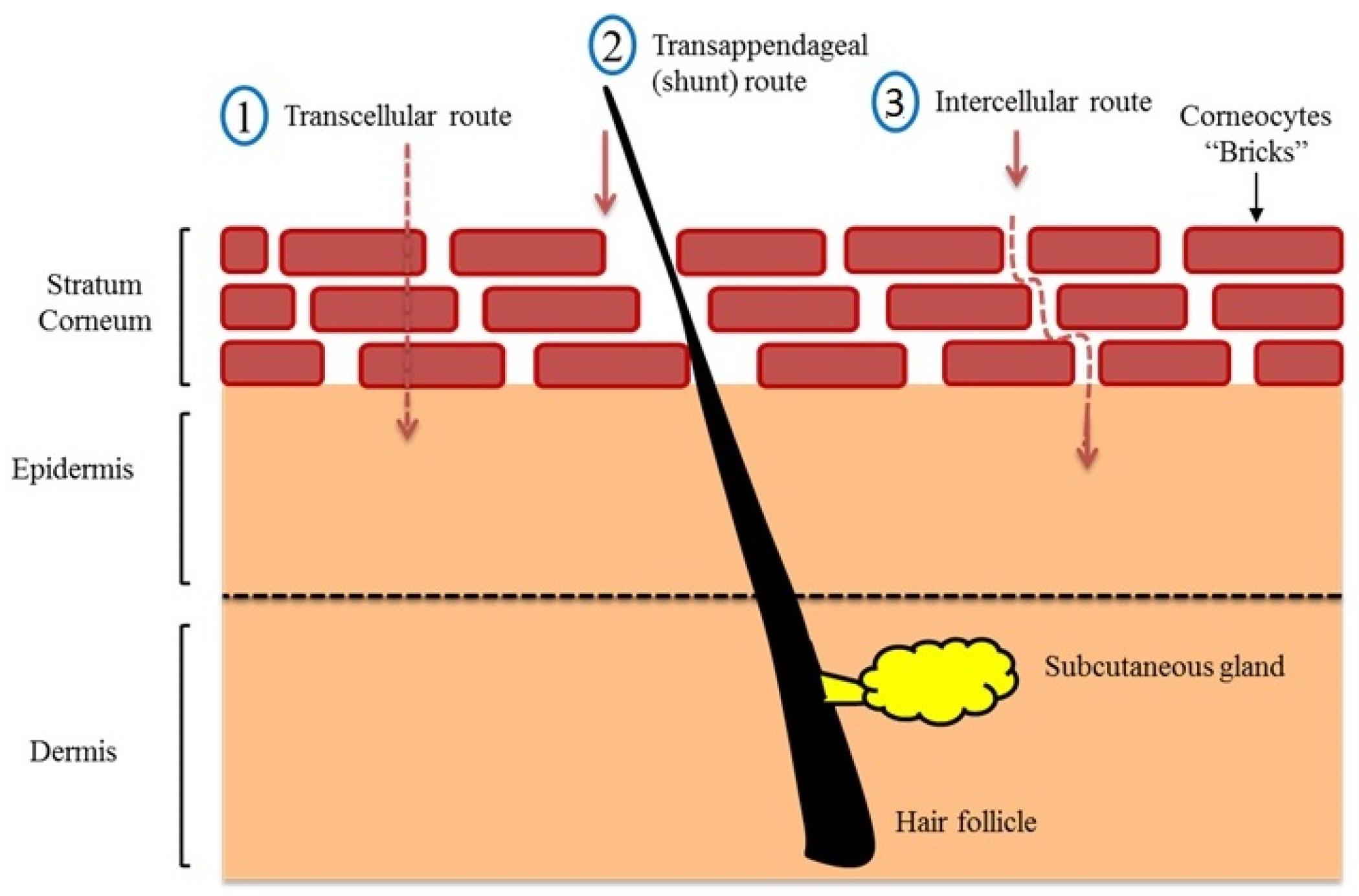
2.2. Skin Transport Mechanism
2.3. Skin Cosmetics
2.4. Skin Therapy
3. Lipid Drug Delivery Systems
3.1. Dermal and Transdermal Drug Delivery
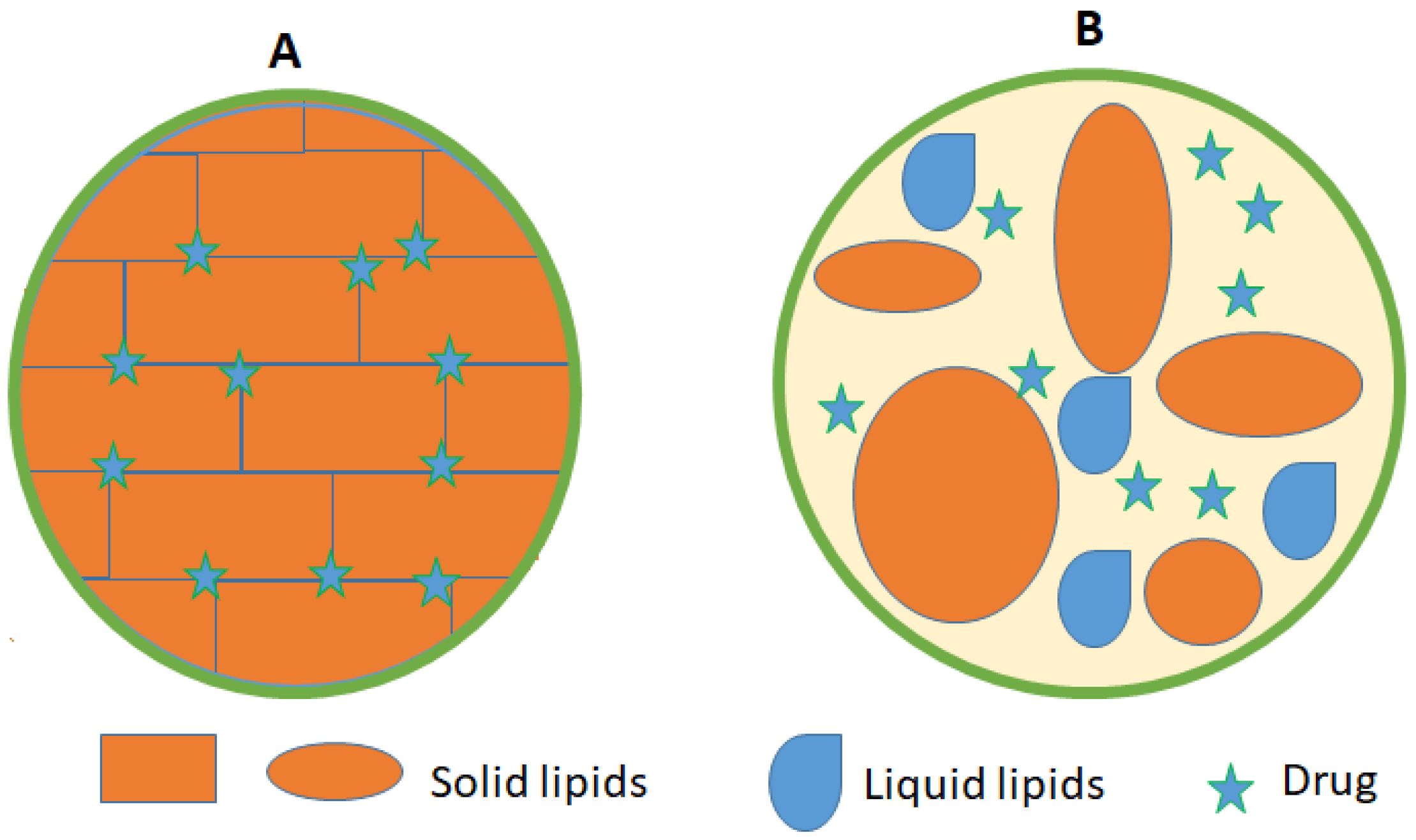
3.2. Solid Lipid Nanoparticles (SLNs)
3.3. Nanostructured Lipid Carriers (NLCs)
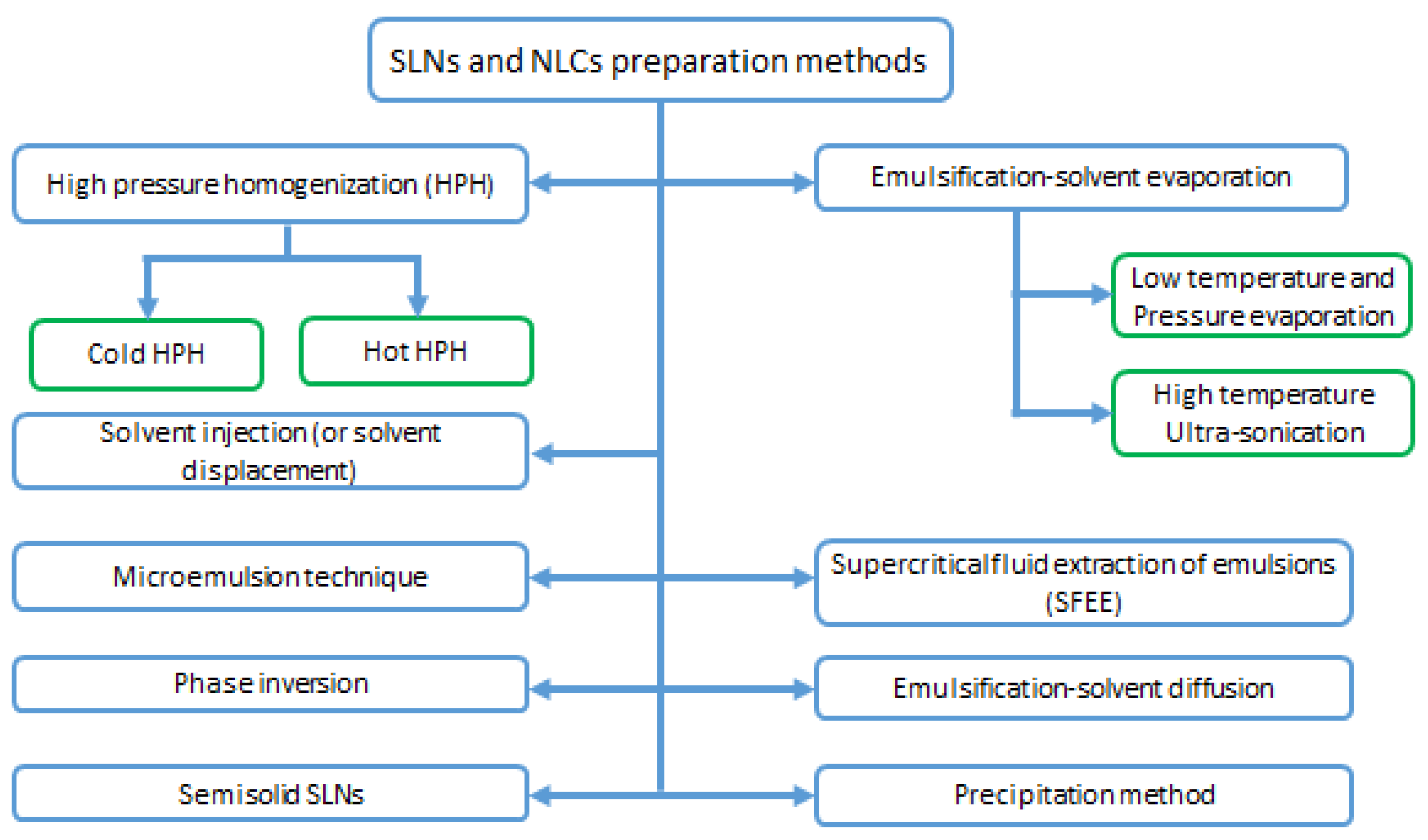
3.4. SLNs and NLCs Preparation Methods
3.4.1. High Pressure Homogenization (HPH) Methods
3.4.2. Microemulsion Technique
3.4.3. Emulsification-Solvent Evaporation
3.4.4. Supercritical Fluid Extraction of Emulsions (SFEE)
3.4.5. Emulsification-Solvent Diffusion Method
3.4.6. Solvent Injection (or Solvent Displacement) Method
3.4.7. Phase Inversion, Multiple Emulsion Technique
3.4.8. Precipitation Method
3.4.9. Semisolid Solid Lipid Nanoparticles
3.5. Nanoemulsions (NEs)
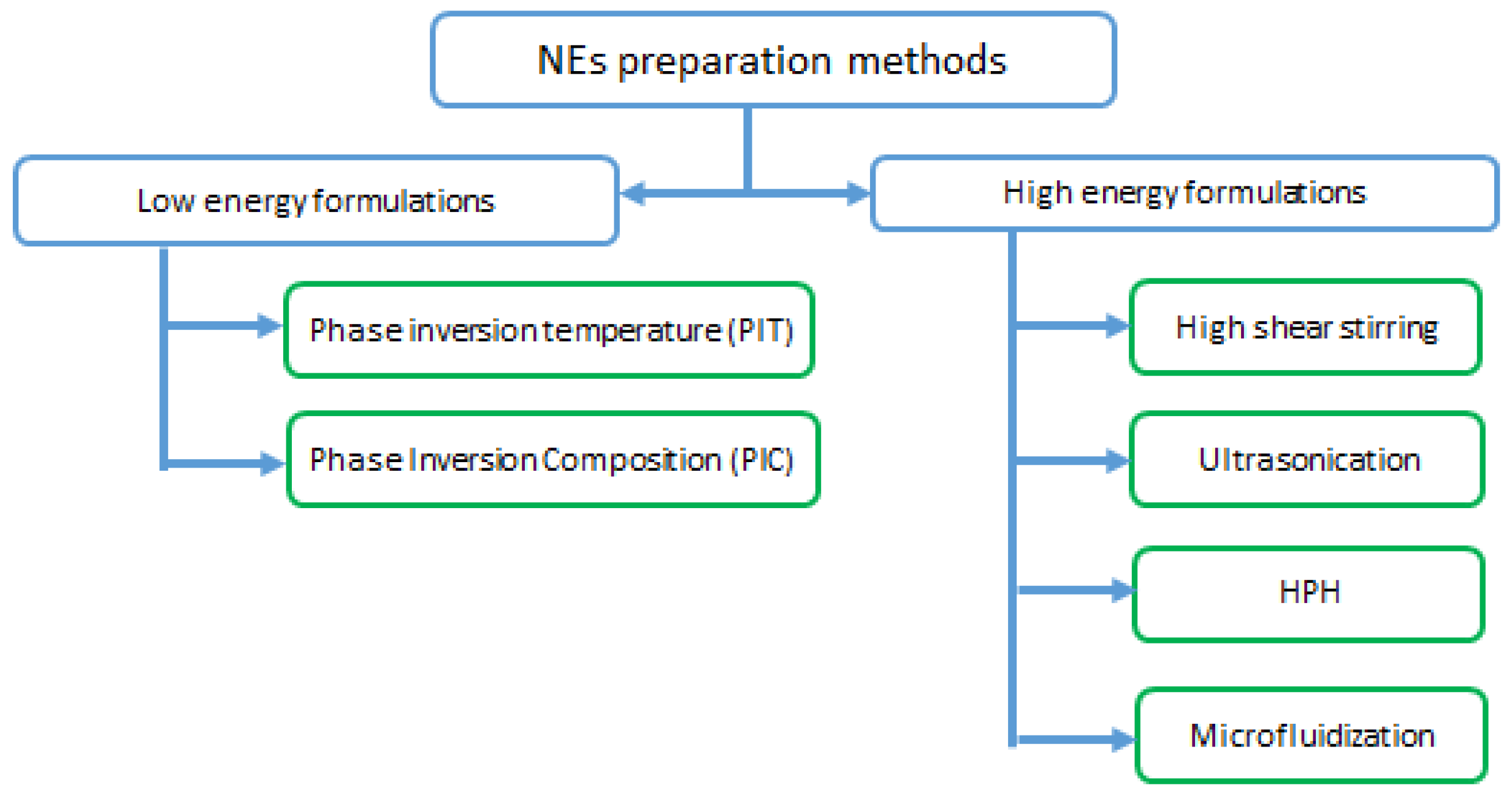
3.6. NEs Preparation Methods
3.6.1. High Energy Formulations
High Shear Stirring
Ultrasonication
High Pressure Homogenization (HPH)
Microfluidization
3.6.2. Low Energy Formulations
Phase Inversion Temperature (PIT) Method
Phase Inversion Composition (PIC) Method
3.7. LNs Characterization
3.7.1. Particle Size and Distribution
3.7.2. Surface Charge
3.7.3. Encapsulation Efficiency (EE)
3.7.4. Crystallinity and Polymorphism
4. LNs Mechanism of Skin Penetration
5. Nanocarriers for Transdermal and Dermal Drug Delivery
5.1. Vitamins and Vitamin Derivatives Delivery
5.2. Sunscreens Delivery
5.3. Psoralen Plus Ultraviolet Light A Therapy (PUVA)
5.4. Wound Healing and Skin Burns Products
5.5. Anti-Aging and Skincare Products
5.6. Topical Glucocorticoids
5.7. Non-Steroidal Anti-Inflammatorty Drugs
6. Conclusions and Future Prospective
Author Contributions
Funding
Institutional Review Board Statement
Informed Consent Statement
Data Availability Statement
Conflicts of Interest
References
- Babamiri, K.; Nassab, R.J.A.S.J. Cosmeceuticals: The evidence behind the retinoids. Aesthetic Surg. J. 2010, 30, 74–77. [Google Scholar] [CrossRef] [PubMed] [Green Version]
- d’Agostino, S.; Azzali, A.; Casali, L.; Taddei, P.; Grepioni, F. Environmentally Friendly Sunscreens: Mechanochemical Synthesis and Characterization of β-CD Inclusion Complexes of Avobenzone and Octinoxate with Improved Photostability. ACS Sustain. Chem. Eng. 2020, 8, 13215–13225. [Google Scholar] [CrossRef]
- Ahmad, A.; Ahsan, H. Lipid-based formulations in cosmeceuticals and biopharmaceuticals. Biomed. Dermatol. 2020, 4, 1–10. [Google Scholar] [CrossRef]
- Barros, C.; Barros, R.B.G. Natural and Organic Cosmetics: Definition and Concepts. Preprints 2020. [Google Scholar] [CrossRef]
- Goodarzi, F.; Zendehboudi, S. A comprehensive review on emulsions and emulsion stability in chemical and energy industries. Can. J. Chem. Eng. 2019, 97, 281–309. [Google Scholar] [CrossRef] [Green Version]
- Van Tran, V.; Moon, J.-Y.; Lee, Y.-C. Liposomes for delivery of antioxidants in cosmeceuticals: Challenges and development strategies. J. Control. Release 2019, 300, 114–140. [Google Scholar] [CrossRef]
- Müller, R.H.; Staufenbiel, S.; Keck, C.M. Lipid Nanoparticles (SLN, NLC) for innovative consumer care & household products. Househ. Pers. Care Today 2014, 9, 18–24. [Google Scholar]
- Jia, Q.; Nash, J.F. Pathology of Aging Skin; Springer: Berlin/Heidelberg, Germany, 2010; pp. 277–291. [Google Scholar]
- Lai-Cheong, J.E.; McGrath, J.A. Structure and function of skin, hair and nails. Medicine 2017, 45, 347–351. [Google Scholar] [CrossRef]
- Saleh, M.M.; Woods, A.; Harvey, R.D.; Young, A.R.; Jones, S.A. Nanomaterials fusing with the skin: Alpha-tocopherol phosphate delivery into the viable epidermis to protect against ultraviolet radiation damage. Int. J. Pharm. 2021, 594, 120000. [Google Scholar] [CrossRef] [PubMed]
- Benson, H.A.; Watkinson, A.C. Topical and Transdermal Drug Delivery: Principles and Practice; John Wiley & Sons: Hoboken, NJ, USA, 2012; pp. 357–366. [Google Scholar]
- McGrath, J.A.; Uitto, J. Structure and Function of the Skin, 9th ed.; John Wiley & Sons: Chichester, UK, 2016; pp. 1–52. [Google Scholar]
- Koster, M.I. Making an epidermis. Ann. N. Y. Acad. Sci. 2009, 1170, 7–10. [Google Scholar] [CrossRef]
- Bergstresser, P.R.; Richard Taylor, J. Epidermal ‘turnover time’—A new examination. Br. J. Dermatol. 1977, 96, 503–506. [Google Scholar] [CrossRef]
- Takahashi, M.; Machida, Y.; Marks, R. Measurement of turnover time of stratum corneum using dansyl chloride fluorescence. J. Soc. Cosmet. Chem. 1987, 38, 321–331. [Google Scholar]
- Bouwstra, J.A.; Groenink, H.W.W.; Kempenaar, J.A.; Romeijn, S.G.; Ponec, M. Water distribution and natural moisturizer factor content in human skin equivalents are regulated by environmental relative humidity. J. Investig. Dermatol. 2008, 128, 378–388. [Google Scholar] [CrossRef] [PubMed] [Green Version]
- Ito, S.; Wakamatsu, K. Quantitative analysis of eumelanin and pheomelanin in humans, mice, and other animals: A comparative review. Pigment. Cell Res. 2003, 16, 523–531. [Google Scholar] [CrossRef] [PubMed]
- Seiberg, M. Keratinocyte–melanocyte interactions during melanosome transfer. Pigment. Cell Res. 2001, 14, 236–242. [Google Scholar] [CrossRef] [PubMed]
- Park, H.; Kosmadaki, M.; Yaar, M.; Gilchrest, B.A. Cellular mechanisms regulating human melanogenesis. Cell. Mol. Life Sci. 2009, 66, 1493–1506. [Google Scholar] [CrossRef] [PubMed]
- Hunger, R.E.; Sieling, P.A.; Ochoa, M.T.; Sugaya, M.; Burdick, A.E.; Rea, T.H.; Brennan, P.J.; Belisle, J.T.; Blauvelt, A.; Porcelli, S.A.; et al. Langerhans cells utilize CD1a and langerin to efficiently present nonpeptide antigens to T cells. J. Clin. Investig. 2004, 113, 701–708. [Google Scholar] [CrossRef] [PubMed] [Green Version]
- Valladeau, J.; Ravel, O.; Dezutter-Dambuyant, C.; Moore, K.; Kleijmeer, M.; Liu, Y.; Duvert-Frances, V.; Vincent, C.; Schmitt, D.; Davoust, J.; et al. Langerin, a novel C-type lectin specific to Langerhans cells, is an endocytic receptor that induces the formation of Birbeck granules. Immunity 2000, 12, 71–81. [Google Scholar] [CrossRef] [Green Version]
- Yan, B.; Liu, N.; Li, J.; Li, J.; Zhu, W.; Kuang, Y.; Chen, X.; Peng, C. The role of Langerhans cells in epidermal homeostasis and pathogenesis of psoriasis. J. Cell. Mol. Med. 2020, 24, 11646–11655. [Google Scholar] [CrossRef] [PubMed]
- Tachibana, T.; Nawa, T. Recent progress in studies on Merkel cell biology. Anat. Sci. Int. 2002, 77, 26–33. [Google Scholar] [CrossRef]
- Moss, G.P.; Gullick, D.R.; Wilkinson, S.C. Predictive Methods in Percutaneous Absorption; Springer: Berlin/Heidelberg, Germany, 2015. [Google Scholar]
- Rawlings, A.V. Trends in stratum corneum research and the management of dry skin conditions. Int. J. Cosmet. Sci. 2003, 25, 63–95. [Google Scholar] [CrossRef] [PubMed]
- Michaels, A.; Chandrasekaran, S.; Shaw, J.E. Drug permeation through human skin: Theory and in vitro experimental measurement. Am. Inst. Chem. Eng. 1975, 21, 985–996. [Google Scholar] [CrossRef]
- Elias, P.M.; Cooper, E.R.; Korc, A.; Brown, B.E. Percutaneous transport in relation to stratum corneum structure and lipid composition. J. Investig. Dermatol. 1981, 76, 297–301. [Google Scholar] [CrossRef] [Green Version]
- Nemes, Z.; Steinert, P.M. Bricks and mortar of the epidermal barrier. Exp. Mol. Med. 1999, 31, 5–19. [Google Scholar] [CrossRef] [PubMed]
- Weerheim, A.; Ponec, M. Determination of stratum corneum lipid profile by tape stripping in combination with high-performance thin-layer chromatography. Arch. Dermatol. Res. 2001, 293, 191–199. [Google Scholar] [CrossRef]
- Hadgraft, J.; Lane, M.E. Skin: The ultimate interface. Phys. Chem. Chem. Phys. 2011, 13, 5215–5222. [Google Scholar] [CrossRef] [PubMed]
- Kattou, P.; Lian, G.; Glavin, S.; Sorrell, I.; Chen, T. Development of a two-dimensional model for predicting transdermal permeation with the follicular pathway: Demonstration with a caffeine study. Pharm. Res. 2017, 34, 2036–2048. [Google Scholar] [CrossRef]
- Nastiti, C.M.; Ponto, T.; Abd, E.; Grice, J.E.; Benson, H.A.; Roberts, M.S. Topical nano and microemulsions for skin delivery. Pharmaceutics 2017, 9, 37. [Google Scholar] [CrossRef]
- Zeb, A.; Arif, S.T.; Malik, M.; Shah, F.A.; Din, F.U.; Qureshi, O.S.; Lee, E.-S.; Lee, G.-Y.; Kim, J.-K. Potential of nanoparticulate carriers for improved drug delivery via skin. J. Pharm. Investig. 2019, 49, 485–517. [Google Scholar] [CrossRef] [Green Version]
- Morganti, P. Use and potential of nanotechnology in cosmetic dermatology. Clin. Cosmet. Investig. Dermatol. CCID 2010, 3, 5. [Google Scholar] [CrossRef] [PubMed] [Green Version]
- Förster, M.; Bolzinger, M.-A.; Fessi, H.; Briançon, S. Topical delivery of cosmetics and drugs. Molecular aspects of percutaneous absorption and delivery. Eur. J. Dermatol. 2009, 19, 309–323. [Google Scholar] [CrossRef] [PubMed]
- Berenson, G.S.; Burch, G.E. Studies of diffusion of water through dead human skin: The effect of different environmental states and of chemical alterations of the Epidermis1. Am. J. Trop. Med. Hyg. 1951, 1, 842–853. [Google Scholar] [CrossRef] [PubMed]
- Sweeney, T.M.; Downing, D.T. The role of lipids in the epidermal barrier to water diffusion. J. Investig. Dermatol. 1970, 55, 135–140. [Google Scholar] [CrossRef] [PubMed] [Green Version]
- Kalia, Y.N.; Guy, R.H. Modeling transdermal drug release. Adv. Drug Deliv. Rev. 2001, 48, 159–172. [Google Scholar] [CrossRef]
- Martin, K.I.; Glaser, D.A. Cosmeceuticals: The new medicine of beauty. Mo. Med. 2011, 108, 60. [Google Scholar]
- Stiefel, C.; Schwack, W. Photoprotection in changing times–UV filter efficacy and safety, sensitization processes and regulatory aspects. Int. J. Cosmet. Sci. 2015, 37, 2–30. [Google Scholar] [CrossRef] [PubMed] [Green Version]
- Young, A.R.; Claveau, J.; Rossi, A.B. Ultraviolet radiation and the skin: Photobiology and sunscreen photoprotection. J. Am. Acad. Dermatol. 2017, 76, S100–S109. [Google Scholar] [CrossRef] [Green Version]
- Schalka, S.; Steiner, D.; Ravelli, F.N.; Steiner, T.; Terena, A.C.; Marçon, C.R.; Ayres, E.L.; Addor, F.A.S.; Miot, H.A.; Ponzio, H.; et al. Brazilian consensus on photoprotection. An. Bras. Dermatol. 2014, 89, 1–74. [Google Scholar] [CrossRef] [Green Version]
- Ueda, C.T.; Shah, V.P.; Derdzinski, K.; Ewing, G.; Flynn, G.; Maibach, H.; Marques, M.; Rytting, H.; Shaw, S.; Thakker, K.; et al. Topical and transdermal drug products. Pharmacop. Forum 2009, 35, 750–764. [Google Scholar] [CrossRef]
- Gupta, R.K.; Soni, P.; Shrivastava, J.; Rajput, P.; Parashar, S. Cosmeceutical role of Medicinal plants/Herbs: A Review on commercially available Cosmetic ingredients. Int. J. Innov. Sci. Technol. 2018, 70–73. [Google Scholar] [CrossRef]
- Draelos, Z.D. Cosmeceuticals: Undefined, unclassified, and unregulated. Clin. Dermatol. 2009, 27, 431–434. [Google Scholar] [CrossRef] [PubMed]
- Lintner, K.; Mas-Chamberlin, C.; Mondon, P.; Peschard, O.; Lamy, L. Cosmeceuticals and active ingredients. Clin. Dermatol. 2009, 27, 461–468. [Google Scholar] [CrossRef] [PubMed]
- Roy, A.; Sahu, R.K.; Matlam, M.; Deshmukh, V.K.; Dwivedi, J.; Jha, A.K. In vitro techniques to assess the proficiency of skin care cosmetic formulations. Pharmacogn. Rev. 2013, 7, 97. [Google Scholar]
- Mukherjee, S.; Ray, S.; Thakur, R. Solid lipid nanoparticles: A modern formulation approach in drug delivery system. Indian J. Pharm. Sci. 2009, 71, 349–358. [Google Scholar] [CrossRef] [Green Version]
- Mu, L.; Sprando, R.L. Application of nanotechnology in cosmetics. Pharm. Res. 2010, 27, 1746–1749. [Google Scholar] [CrossRef] [PubMed]
- Niculae, G.; Lacatusu, I.; Bors, A.; Stan, R. Photostability enhancement by encapsulation of α-tocopherol into lipid-based nanoparticles loaded with a UV filter. Comptes Rendus Chim. 2014, 17, 1028–1033. [Google Scholar] [CrossRef]
- Damiani, E.; Puglia, C. Nanocarriers and Microcarriers for Enhancing the UV Protection of Sunscreens: An Overview. J. Pharm. Sci. 2019, 108, 3769–3780. [Google Scholar] [CrossRef]
- Saleh, M.M.U. Delivery of Tocopherol Phosphate Nanomaterials into the Skin to Protect against Ultraviolet Radiation. Ph.D. Thesis, King’s College London, London, UK, 2019. [Google Scholar]
- Golmohammadzadeh, S.; Mortezania, S.; Jaafari, M.R. Improved photostability, reduced skin permeation and irritation of isotretinoin by solid lipid nanoparticles. Acta Pharm. 2012, 62, 547–562. [Google Scholar]
- Amer, S.S.; Nasr, M.; Mamdouh, W.; Sammour, O. Insights on the use of nanocarriers for acne alleviation. Curr. Drug Deliv. 2019, 16, 18–25. [Google Scholar] [CrossRef]
- Dias, M.; Farinha, A.; Faustino, E.; Hadgraft, J.; Pais, J.; Toscano, C. Topical delivery of caffeine from some commercial formulations. Int. J. Pharm. 1999, 182, 41–47. [Google Scholar] [CrossRef]
- Puglia, C.; Bonina, F.; Rizza, L.; Blasi, P.; Schoubben, A.; Perrotta, R.; Tarico, M.S.; Damiani, E. Lipid nanoparticles as carrier for octyl-methoxycinnamate: In vitro percutaneous absorption and photostability studies. J. Pharm. Sci. 2012, 101, 301–311. [Google Scholar] [CrossRef] [PubMed]
- Dingler, A.; Blum, R.; Niehus, H.; Muller, R.; Gohla, S. Solid lipid nanoparticles (SLNTM/LipopearlsTM) a pharmaceutical and cosmetic carrier for the application of vitamin E in dermal products. J. Microencapsul. 1999, 16, 751–767. [Google Scholar] [PubMed]
- Ng, K.W. Penetration Enhancement of Topical Formulations; Multidisciplinary Digital Publishing Institute: Basel, Switzerland, 2018. [Google Scholar]
- Neubert, R.H. Potentials of new nanocarriers for dermal and transdermal drug delivery. Eur. J. Pharm. Biopharm. 2011, 77, 1–2. [Google Scholar] [CrossRef] [PubMed]
- Duan, Y.; Dhar, A.; Patel, C.; Khimani, M.; Neogi, S.; Sharma, P.; Kumar, N.S.; Vekariya, R.L. A brief review on solid lipid nanoparticles: Part and parcel of contemporary drug delivery systems. RSC Adv. 2020, 10, 26777–26791. [Google Scholar] [CrossRef]
- Ghasemiyeh, P.; Mohammadi-Samani, S. Solid lipid nanoparticles and nanostructured lipid carriers as novel drug delivery systems: Applications, advantages and disadvantages. Res. Pharm. Sci. 2018, 13, 288. [Google Scholar]
- Lauterbach, A.; Müller-Goymann, C.C. Applications and limitations of lipid nanoparticles in dermal and transdermal drug delivery via the follicular route. Eur. J. Pharm. Biopharm. 2015, 97, 152–163. [Google Scholar] [CrossRef]
- Gorzelanny, C.; Mess, C.; Schneider, S.W.; Huck, V.; Brandner, J.M. Skin Barriers in Dermal Drug Delivery: Which Barriers Have to Be Overcome and How Can We Measure Them? Pharmaceutics 2020, 12, 684. [Google Scholar] [CrossRef]
- Mishra, V.; Bansal, K.K.; Verma, A.; Yadav, N.; Thakur, S.; Sudhakar, K.; Rosenholm, J.M. Solid lipid nanoparticles: Emerging colloidal nano drug delivery systems. Pharmaceutics 2018, 10, 191. [Google Scholar] [CrossRef] [Green Version]
- Naseri, N.; Valizadeh, H.; Zakeri-Milani, P. Solid lipid nanoparticles and nanostructured lipid carriers: Structure, preparation and application. Adv. Pharmaceut. Bull. 2015, 5, 305. [Google Scholar] [CrossRef] [Green Version]
- Müller, R.H.; Mäder, K.; Gohla, S. Solid lipid nanoparticles (SLN) for controlled drug delivery–a review of the state of the art. Eur. J. Pharm. Biopharm. 2000, 50, 160–177. [Google Scholar] [CrossRef]
- Kaur, J.; Singh, G.; Saini, S.; Rana, A.C. Innovative Growth in Developing New Methods for Formulating Solid Lipid Nanoparticles and Microparticles. J. Drug Deliv. Ther. 2012, 2, 146–150. [Google Scholar] [CrossRef]
- Shende, P.; Patel, D.; Takke, A. Nanomaterial-Based Cosmeceuticals. In Handbook of Functionalized Nanomaterials for Industrial Applications; Elsevier: Amsterdam, The Netherlands, 2020; pp. 775–791. [Google Scholar]
- Müller, R.; Petersen, R.; Hommoss, A.; Pardeike, J. Nanostructured lipid carriers (NLC) in cosmetic dermal products. Adv. Drug Deliv. Rev. 2007, 59, 522–530. [Google Scholar] [CrossRef] [PubMed]
- Pardeike, J.; Hommoss, A.; Müller, R.H. Lipid nanoparticles (SLN, NLC) in cosmetic and pharmaceutical dermal products. Int. J. Pharm. 2009, 366, 170–184. [Google Scholar] [CrossRef] [PubMed]
- Pandey, V.S.R.; Garg, A.; Kori, M.L.; Rai, G. Nanoemulsion in Cosmetic: From Laboratory to Market. In Nanocosmetics; Nanda, A., Nanda, S., Nguyen, T.A., Rajendran, S., Slimani, Y., Eds.; Elsevier: Amsterdam, The Netherlands, 2020; pp. 327–347. [Google Scholar]
- Abdel-Mottaleb, M. Nanoparticles for Treatment of Atopic Dermatitis. In Nanoscience in Dermatology; Elsevier: Amsterdam, The Netherlands, 2016; pp. 167–175. [Google Scholar]
- Duong, V.-A.; Nguyen, T.-T.-L.; Maeng, H.-J. Preparation of Solid Lipid Nanoparticles and Nanostructured Lipid Carriers for Drug Delivery and the Effects of Preparation Parameters of Solvent Injection Method. Molecules 2020, 25, 4781. [Google Scholar] [CrossRef] [PubMed]
- Garud, A.; Singh, D.; Garud, N. Solid lipid nanoparticles (SLN): Method, characterization and applications. Int. Curr. Pharm. J. 2012, 1, 384–393. [Google Scholar] [CrossRef] [Green Version]
- Bayón-Cordero, L.; Alkorta, I.; Arana, L. Application of solid lipid nanoparticles to improve the efficiency of anticancer drugs. Nanomaterials 2019, 9, 474. [Google Scholar] [CrossRef] [Green Version]
- Azar, F.A.N.; Pezeshki, A.; Ghanbarzadeh, B.; Hamishehkar, H.; Mohammadi, M. Nanostructured lipid carriers: Promising delivery systems for encapsulation of food ingredients. J. Agric. Food Res. 2020, 2, 100084. [Google Scholar] [CrossRef]
- Poonia, N.; Kharb, R.; Lather, V.; Pandita, D. Nanostructured lipid carriers: Versatile oral delivery vehicle. Futur. Sci. OA 2016, 2, FSO135. [Google Scholar] [CrossRef] [Green Version]
- Chauhan, I.; Yasir, M.; Verma, M.; Singh, A.P. Nanostructured lipid carriers: A groundbreaking approach for transdermal drug delivery. Adv. Pharm. Bull. 2020, 10, 150–165. [Google Scholar] [CrossRef]
- Borges, A.; De Freitas, V.; Mateus, N.; Fernandes, I.; Oliveira, J. Solid Lipid Nanoparticles as Carriers of Natural Phenolic Compounds. Antioxidants 2020, 9, 998. [Google Scholar] [CrossRef]
- Trombino, S.; Mellace, S.; Cassano, R. Solid lipid nanoparticles for antifungal drugs delivery for topical applications. Ther. Deliv. 2016, 7, 639–647. [Google Scholar] [CrossRef] [PubMed]
- Floury, J.; Desrumaux, A.; Lardières, J. Effect of high-pressure homogenization on droplet size distributions and rheological properties of model oil-in-water emulsions. Innov. Food Sci. Emerg. Technol. 2000, 1, 127–134. [Google Scholar] [CrossRef]
- Parhi, R.; Suresh, P. Preparation and characterization of solid lipid nanoparticles-a review. Curr. Drug Discov. Technol. 2012, 9, 2–16. [Google Scholar] [CrossRef] [PubMed]
- Gora, S.; Mustafa, G.; Sahni, J.K.; Ali, J.; Baboota, S. Nanosizing of valsartan by high pressure homogenization to produce dissolution enhanced nanosuspension: Pharmacokinetics and pharmacodyanamic study. Drug Deliv. 2016, 23, 930–940. [Google Scholar] [CrossRef] [PubMed] [Green Version]
- Müller, R.H.; Radtke, M.; Wissing, S. Solid lipid nanoparticles (SLN) and nanostructured lipid carriers (NLC) in cosmetic and dermatological preparations. Adv. Drug Deliv. Rev. 2002, 54, S131–S155. [Google Scholar] [CrossRef]
- McClements, D.J. Nanoemulsions versus microemulsions: Clarification of critical differences. Soft Matter 2012, 8, 1719–1729. [Google Scholar] [CrossRef]
- Shah, R.M.; Malherbe, F.; Eldridge, D.; Palombo, E.A.; Harding, I.H. Physicochemical characterization of solid lipid nanoparticles (SLNs) prepared by a novel microemulsion technique. J. Colloid Interface Sci. 2014, 428, 286–294. [Google Scholar] [CrossRef]
- Carneiro, S.P. Nanostructured Lipid Carrier-Based Drug Delivery Systems for Tuberculosis Treatment. In Nanotechnology Based Approaches for Tuberculosis Treatment; Kesharwani, P., Ed.; Academic Press: New Delhi, India, 2020; pp. 193–205. [Google Scholar]
- Mehnert, W.; Mäder, K. Solid lipid nanoparticles: Production, characterization and applications. Adv. Drug Deliv. Rev. 2012, 64, 83–101. [Google Scholar] [CrossRef]
- Pooja, D.; Tunki, L.; Kulhari, H.; Reddy, B.B.; Sistla, R. Optimization of solid lipid nanoparticles prepared by a single emulsification-solvent evaporation method. Data Brief 2016, 6, 15–19. [Google Scholar] [CrossRef] [Green Version]
- Luo, Y.; Chen, D.; Ren, L.; Zhao, X.; Qin, J. Solid lipid nanoparticles for enhancing vinpocetine’s oral bioavailability. J. Control. Release 2006, 114, 53–59. [Google Scholar] [CrossRef]
- Chattopadhyay, P.; Shekunov, B.; Yim, D.; Cipolla, D.; Boyd, B.; Farr, S. Production of solid lipid nanoparticle suspensions using supercritical fluid extraction of emulsions (SFEE) for pulmonary delivery using the AERx system. Adv. Drug Deliv. Rev. 2007, 59, 444–453. [Google Scholar] [CrossRef] [PubMed]
- Schubert, M.; Müller-Goymann, C. Solvent injection as a new approach for manufacturing lipid nanoparticles–evaluation of the method and process parameters. Eur. J. Pharm. Biopharm. 2003, 55, 125–131. [Google Scholar] [CrossRef]
- Heurtault, B.; Saulnier, P.; Pech, B.; Proust, J.E.; Benoit, J.P. A novel phase inversion-based process for the preparation of lipid nanocarriers. Pharm. Res. 2002, 19, 875–880. [Google Scholar] [CrossRef]
- Lippacher, A.; Müller, R.H.; Mäder, K. Semisolid SLN™ dispersions for topical application: Influence of formulation and production parameters on viscoelastic properties. Eur. J. Pharm. Biopharm. 2002, 53, 155–160. [Google Scholar] [CrossRef]
- Gupta, A.; Eral, H.B.; Hatton, T.A.; Doyle, P.S. Nanoemulsions: Formation, properties and applications. Soft Matter 2016, 12, 2826–2841. [Google Scholar] [CrossRef] [Green Version]
- Attwood, D.; Mallon, C.; Ktistis, G.; Taylor, C. A study on factors influencing the droplet size in nonionic oil-in-water microemulsions. Int. J. Pharm. 1992, 88, 417–422. [Google Scholar] [CrossRef]
- Yamashita, Y.; Miyahara, R.; Sakamoto, K. Emulsion and emulsification technology. In Cosmetic Science and Technology: Theoretical Principles and Applications; Elsevier Inc.: Amsterdam, The Netherlands, 2017; pp. 489–506. [Google Scholar]
- Prow, T.W.; Grice, J.E.; Lin, L.L.; Faye, R.; Butler, M.; Becker, W.; Wurm, E.M.; Yoong, C.; Robertson, T.A.; Soyer, H.P.; et al. Nanoparticles and microparticles for skin drug delivery. Adv. Drug Deliv. Rev. 2011, 63, 470–491. [Google Scholar] [CrossRef] [PubMed]
- Hua, S. Lipid-based nano-delivery systems for skin delivery of drugs and bioactives. Front. Pharmacol. 2015, 6, 219. [Google Scholar] [CrossRef]
- Yukuyama, M.N.; Ghisleni, D.D.M.; Pinto, T.I.A.; Bou-Chacra, N.A. Nanoemulsion: Process selection and application in cosmetics—A review. Int. J. Cosmet. Sci. 2016, 38, 13–24. [Google Scholar] [CrossRef] [Green Version]
- Rao, J.; McClements, D.J. Stabilization of phase inversion temperature nanoemulsions by surfactant displacement. J. Agric. Food Chem. 2010, 58, 7059–7066. [Google Scholar] [CrossRef]
- Gonçalves, A.; Nikmaram, N.; Roohinejad, S.; Estevinho, B.N.; Rocha, F.; Greiner, R.; McClements, D.J. Production, properties, and applications of solid self-emulsifying delivery systems (S-SEDS) in the food and pharmaceutical industries. Colloids Surf. A Physicochem. Eng. Asp. 2018, 538, 108–126. [Google Scholar] [CrossRef]
- Chen, Y.; Narayan, S.; Dutcher, C.S. Phase-Dependent Surfactant Transport on the Microscale: Interfacial Tension and Droplet Coalescence. Langmuir 2020, 36, 14904–14923. [Google Scholar] [CrossRef]
- Scholz, P.; Keck, C.M. Nanoemulsions produced by rotor—Stator high speed stirring. Int. J. Pharm. 2015, 482, 110–117. [Google Scholar] [CrossRef]
- Roselan, M.A.; Ashari, S.E.; Faujan, N.H.; Mohd Faudzi, S.M.; Mohamad, R. An Improved Nanoemulsion Formulation Containing Kojic Monooleate: Optimization, Characterization and In Vitro Studies. Molecules 2020, 25, 2616. [Google Scholar] [CrossRef] [PubMed]
- Leong, T.S.H.; Manickam, S.; Martin, G.J.; Li, W.; Ashokkumar, M. Ultrasonic Production of Nano-Emulsions for Bioactive Delivery in Drug and Food Applications; Springer: Berlin/Heidelberg, Germany, 2018. [Google Scholar]
- Gharibzahedi, S.M.; Jafari, S.M. Fabrication of Nanoemulsions by Ultrasonication. In Nanoemulsions; Elsevier: Amsterdam, The Netherlands, 2018; pp. 233–285. [Google Scholar]
- Calligaris, S.; Plazzotta, S.; Valoppi, F.; Anese, M. Combined high-power ultrasound and high-pressure homogenization nanoemulsification: The effect of energy density, oil content and emulsifier type and content. Food Res. Int. 2018, 107, 700–707. [Google Scholar] [CrossRef] [Green Version]
- Ruiz-Montañez, G.; Ragazzo-Sanchez, J.A.; Picart-Palmade, L.; Calderón-Santoyo, M.; Chevalier-Lucia, D. Optimization of nanoemulsions processed by high-pressure homogenization to protect a bioactive extract of jackfruit (Artocarpus heterophyllus Lam). Innov. Food Sci. Emerg. Technol. 2017, 40, 35–41. [Google Scholar] [CrossRef]
- Che Marzuki, N.H.; Wahab, R.A.; Abdul Hamid, M. An overview of nanoemulsion: Concepts of development and cosmeceutical applications. Biotechnol. Biotechnol. Equip. 2019, 33, 779–797. [Google Scholar] [CrossRef] [Green Version]
- Villalobos-Castillejos, F.; Granillo-Guerrero, V.G.; Leyva-Daniel, D.E.; Alamilla-Beltrán, L.; Gutiérrez-López, G.F.; Monroy-Villagrana, A.; Jafari, S.M. Fabrication of Nanoemulsions by Microfluidization. In Nanoemulsions; Jafari, S.M., Ed.; McCl: Toronto, ON, Canada, 2018; pp. 207–232. [Google Scholar]
- Uluata, S.; Decker, E.A.; McClements, D.J. Optimization of nanoemulsion fabrication using microfluidization: Role of surfactant concentration on formation and stability. Food Biophys. 2016, 11, 52–59. [Google Scholar] [CrossRef]
- Muñoz, J.; Alfaro, M.C.; Trujillo-Cayado, L.A.; Santos, J.; Martín-Piñero, M.J. Production of Food Bioactive-Loaded Nanostructures by Microfluidization. In Nanoencapsulation of Food Ingredients by Specialized Equipment; Elsevier: Amsterdam, The Netherlands, 2019; pp. 341–390. [Google Scholar]
- Jafari, S.M.; McClements, D.J. Nanoemulsions: Formulation, Applications, and Characterization; Academic Press: New Delhi, India, 2018. [Google Scholar]
- Jasmina, H.; Džana, O.; Alisa, E.; Edina, V.; Ognjenka, R. Preparation of Nanoemulsions by High-Energy and Lowenergy Emulsification Methods. In CMBEBIH 2017; Springer: Berlin/Heidelberg, Germany, 2017; pp. 317–322. [Google Scholar]
- Sonneville-Aubrun, O.; Yukuyama, M.N.; Pizzino, A. Application of Nanoemulsions in Cosmetics. In Nanoemulsions; Elsevier: Amsterdam, The Netherlands, 2018; pp. 435–475. [Google Scholar]
- Yu, L.; Li, C.; Xu, J.; Hao, J.; Sun, D. Highly stable concentrated nanoemulsions by the phase inversion composition method at elevated temperature. Langmuir 2012, 28, 14547–14552. [Google Scholar] [CrossRef] [PubMed]
- Feng, J.; Rodríguez-Abreu, C.; Esquena, J.; Solans, C. A Concise Review on Nano-emulsion Formation by the Phase Inversion Composition (PIC) Method. J. Surfactants Deterg. 2020, 23, 677–685. [Google Scholar] [CrossRef]
- Teulon, J.-M.; Godon, C.; Chantalat, L.; Moriscot, C.; Cambedouzou, J.; Odorico, M.; Ravaux, J.; Podor, R.; Gerdil, A.; Habert, A.; et al. On the operational aspects of measuring nanoparticle sizes. Nanomaterials 2019, 9, 18. [Google Scholar] [CrossRef] [PubMed] [Green Version]
- Rice, S.B.; Chan, C.; Brown, S.C.; Eschbach, P.; Han, L.; Ensor, D.S.; Stefaniak, A.B.; Bonevich, J.; Vladár, A.E.; Walker, A.R.H.; et al. Particle size distributions by transmission electron microscopy: An interlaboratory comparison case study. Metrologia 2013, 50, 663–678. [Google Scholar] [CrossRef] [PubMed] [Green Version]
- Loo, C.; Basri, M.; Ismail, R.; Lau, H.; Tejo, B.; Kanthimathi, M.; Hassan, H.; Choo, Y. Effect of compositions in nanostructured lipid carriers (NLC) on skin hydration and occlusion. Int. J. Nanomed. 2013, 8, 13–22. [Google Scholar] [CrossRef] [PubMed] [Green Version]
- Danaei, M.; Dehghankhold, M.; Ataei, S.; Davarani, F.H.; Javanmard, R.; Dokhani, A.; Khorasani, S.; Mozafari, M.R. Impact of particle size and polydispersity index on the clinical applications of lipidic nanocarrier systems. Pharmaceutics 2018, 10, 57. [Google Scholar] [CrossRef] [Green Version]
- Midekessa, G.; Godakumara, K.; Ord, J.; Viil, J.; Lättekivi, F.; Dissanayake, K.; Kopanchuk, S.; Rinken, A.; Andronowska, A.; Bhattacharjee, S.; et al. Zeta Potential of Extracellular Vesicles: Toward Understanding the Attributes that Determine Colloidal Stability. ACS Omega 2020, 5, 16701–16710. [Google Scholar] [CrossRef]
- Necula, B.; Apachitei, I.; Fratila-Apachitei, L.; Teodosiu, C.; Duszczyk, J. Stability of nano-/microsized particles in deionized water and electroless nickel solutions. J. Colloid Interface Sci. 2007, 314, 514–522. [Google Scholar] [CrossRef]
- Bahari, L.A.S.; Hamishehkar, H. The impact of variables on particle size of solid lipid nanoparticles and nanostructured lipid carriers; a comparative literature review. Adv. Pharm. Bull. 2016, 6, 143. [Google Scholar] [CrossRef]
- Gupta, S.; Kesarla, R.; Chotai, N.; Misra, A.; Omri, A. Systematic approach for the formulation and optimization of solid lipid nanoparticles of efavirenz by high pressure homogenization using design of experiments for brain targeting and enhanced bioavailability. BioMed Res. Int. 2017, 2017, 1–18. [Google Scholar] [CrossRef] [PubMed]
- Argimón, M.; Romero, M.; Miranda, P.; Mombrú, Á.W.; Miraballes, I.; Zimet, P.; Pardo, H. Development and characterization of vitamin a-loaded solid lipid nanoparticles for topical application. J. Braz. Chem. Soc. 2017, 28, 1177–1184. [Google Scholar] [CrossRef]
- Tantra, R.; Schulze, P.; Quincey, P. Effect of nanoparticle concentration on zeta-potential measurement results and reproducibility. Particuology 2010, 8, 279–285. [Google Scholar] [CrossRef]
- Wang, N.; Cheng, X.; Li, N.; Wang, H.; Chen, H. Nanocarriers and their loading strategies. Adv. Healthc. Mater. 2019, 8, e1801002. [Google Scholar] [CrossRef] [PubMed]
- Doktorovova, S.; Souto, E.B. Nanostructured lipid carrier-based hydrogel formulations for drug delivery: A comprehensive review. Expert Opin. Drug Deliv. 2009, 6, 165–176. [Google Scholar] [CrossRef] [PubMed]
- Censi, R.; Di Martino, P. Polymorph impact on the bioavailability and stability of poorly soluble drugs. Molecules 2015, 20, 18759–18776. [Google Scholar] [CrossRef] [Green Version]
- Nazila, K.; Yameen, B.; Wu, J.; Farokhzad, O.C. Nanoparticles: Mechanisms of Controlling Drug Release Nazila. Chem. Rev. 2016, 116, 2602–2663. [Google Scholar]
- Gil-González, E.; Perejón, A.; Sánchez-Jiménez, P.E.; Medina-Carrasco, S.; Kupčík, J.; Šubrt, J.; Criado, J.M.; Pérez-Maqueda, L.A. Crystallization kinetics of nanocrystalline materials by combined X-ray diffraction and differential scanning calorimetry experiments. Cryst. Growth Des. 2018, 18, 3107–3116. [Google Scholar] [CrossRef] [Green Version]
- Gill, P.; Moghadam, T.T.; Ranjbar, B. Differential scanning calorimetry techniques: Applications in biology and nanoscience. J. Biomol. Tech. 2010, 21, 167. [Google Scholar]
- Ribeiro, A.P.B.; Masuchi, M.H.; Miyasaki, E.K.; Domingues, M.A.F.; Stroppa, V.L.Z.; de Oliveira, G.M.; Kieckbusch, T.G. Crystallization modifiers in lipid systems. J. Food Sci. Technol. 2015, 52, 3925–3946. [Google Scholar] [CrossRef] [Green Version]
- Tyler, A.I.; Law, R.V.; Seddon, J.M. X-Ray Diffraction of Lipid Model Membranes. In Methods in Membrane Lipids; Springer: Berlin/Heidelberg, Germany, 2015; pp. 199–225. [Google Scholar]
- Baroli, B. Penetration of nanoparticles and nanomaterials in the skin: Fiction or reality? J. Pharm. Sci. 2010, 99, 21–50. [Google Scholar] [CrossRef]
- Rahbari, R.; Ichim, I.; Bamsey, R.; Burridge, J.; Guy, O.J.; Bolodeoku, J.; Graz, M. Characterisation of Drug Delivery Efficacy Using Microstructure-Assisted Application of a Range of APIs. Pharmaceutics 2020, 12, 1213. [Google Scholar] [CrossRef] [PubMed]
- Guimarães, K.L.; Ré, M.I. Lipid nanoparticles as carriers for cosmetic ingredients: The first (SLN) and the Second Generation (NLC). In Nanocosmetics and Nanomedicines; Springer: Berlin/Heidelberg, Germany, 2011; pp. 101–122. [Google Scholar]
- Ghasemiyeh, P.; Mohammadi-Samani, S. Potential of Nanoparticles as Permeation Enhancers and Targeted Delivery Options for Skin: Advantages and Disadvantages. Drug Des. Dev. Ther. 2020, 14, 3271. [Google Scholar] [CrossRef]
- Kakadia, P.G.; Conway, B.R. Lipid nanoparticles for dermal drug delivery. Curr. Pharm. Des. 2015, 21, 2823–2829. [Google Scholar] [CrossRef] [PubMed] [Green Version]
- Li, B.; Ge, Z.-Q. Nanostructured lipid carriers improve skin permeation and chemical stability of idebenone. AAPS Pharmscitech 2012, 13, 276–283. [Google Scholar] [CrossRef] [Green Version]
- Souto, E.B.; Baldim, I.; Oliveira, W.P.; Rao, R.; Yadav, N.; Gama, F.M.; Mahant, S. SLN and NLC for topical, dermal, and transdermal drug delivery. Expert Opin. Drug Deliv. 2020, 17, 357–377. [Google Scholar] [CrossRef] [PubMed]
- Borgia, S.L.; Regehly, M.; Sivaramakrishnan, R.; Mehnert, W.; Korting, H.; Danker, K.; Röder, B.; Kramer, K.; Schäfer-Korting, M. Lipid nanoparticles for skin penetration enhancement—Correlation to drug localization within the particle matrix as determined by fluorescence and parelectric spectroscopy. J. Control. Release 2005, 110, 151–163. [Google Scholar] [CrossRef]
- Arora, R.; Katiyar, S.S.; Kushwah, V.; Jain, S. Solid lipid nanoparticles and nanostructured lipid carrier-based nanotherapeutics in treatment of psoriasis: A comparative study. Expert Opin. Drug Deliv. 2017, 14, 165–177. [Google Scholar] [CrossRef] [PubMed]
- Schwarz, J.C.; Baisaeng, N.; Hoppel, M.; Löw, M.; Keck, C.M.; Valenta, C. Ultra-small NLC for improved dermal delivery of coenyzme Q10. Int. J. Pharm. 2013, 447, 213–217. [Google Scholar] [CrossRef] [PubMed]
- Ghasemiyeh, P.; Azadi, A.; Daneshamouz, S.; Heidari, R.; Azarpira, N.; Mohammadi-Samani, S. Cyproterone acetate-loaded nanostructured lipid carriers: Effect of particle size on skin penetration and follicular targeting. Pharm. Dev. Technol. 2019, 24, 812–823. [Google Scholar] [CrossRef]
- Patzelt, A.; Mak, W.C.; Jung, S.; Knorr, F.; Meinke, M.C.; Richter, H.; Rühl, E.; Cheung, K.Y.; Tran, N.B.N.N.; Lademann, J. Do nanoparticles have a future in dermal drug delivery? J. Control. Release 2017, 246, 174–182. [Google Scholar] [CrossRef]
- Blume-Peytavi, U.; Vogt, A. Human hair follicle: Reservoir function and selective targeting. Br. J. Dermatol. 2011, 165, 13–17. [Google Scholar] [CrossRef]
- Verma, A.; Jain, A.; Hurkat, P.; Jain, S.K. Transfollicular drug delivery: Current perspectives. Res. Rep. Transdermal Drug Deliv. 2016, 5, 1–17. [Google Scholar]
- van der Maaden, K.; Jiskoot, W.; Bouwstra, J. Microneedle technologies for (trans) dermal drug and vaccine delivery. J. Control. Release 2012, 160, 645–655. [Google Scholar] [CrossRef] [PubMed]
- Kesarwani, A.; Yadav, A.K.; Singh, S.; Gautam, H.; Singh, H.N.; Sharma, A.; Yadav, C. Theoretical aspects of transdermal drug delivery system. Bull. Pharm. Res. 2013, 3, 78–89. [Google Scholar]
- Marchetti, J.M.; de Souza, M.C.; Marotta-Oliveira, S.S. Nanocarriers and cancer therapy: Approaches to Topical and Transdermal Delivery. In Nanocosmetics and Nanomedicines; Springer: Berlin/Heidelberg, Germany, 2011; pp. 269–286. [Google Scholar]
- Das, S.; Chaudhury, A. Recent advances in lipid nanoparticle formulations with solid matrix for oral drug delivery. AAPS Pharmscitech 2011, 12, 62–76. [Google Scholar] [CrossRef] [PubMed] [Green Version]
- Kakadia, P.G.; Conway, B.R. Solid lipid nanoparticles: A potential approach for dermal drug delivery. Am. J. Pharmacol. Sci. 2014, 2, 4–5. [Google Scholar] [CrossRef] [Green Version]
- Jenning, V.; Gohla, S.H. Encapsulation of retinoids in solid lipid nanoparticles (SLN). J. Microencapsul. 2001, 18, 149–158. [Google Scholar] [PubMed]
- Castro, G.A.; Coelho, A.L.L.; Oliveira, C.A.; Mahecha, G.A.; Oréfice, R.L.; Ferreira, L.A. Formation of ion pairing as an alternative to improve encapsulation and stability and to reduce skin irritation of retinoic acid loaded in solid lipid nanoparticles. Int. J. Pharm. 2009, 381, 77–83. [Google Scholar] [CrossRef] [PubMed]
- Gokce, E.H.; Korkmaz, E.; Dellera, E.; Sandri, G.; Bonferoni, M.C.; Ozer, O. Resveratrol-loaded solid lipid nanoparticles versus nanostructured lipid carriers: Evaluation of antioxidant potential for dermal applications. Int. J. Nanomed. 2012, 7, 1841. [Google Scholar] [CrossRef] [Green Version]
- Wissing, S.A.; Müller, R.H. A novel sunscreen system based on tocopherol acetate incorporated into solid lipid nanoparticles. Int. J. Cosmet. Sci. 2001, 23, 233–243. [Google Scholar] [CrossRef] [PubMed]
- Gupta, S.; Bansal, R.; Gupta, S.; Jindal, N.; Jindal, A. Nanocarriers and nanoparticles for skin care and dermatological treatments. Indian Dermatol. Online J. 2013, 4, 267. [Google Scholar] [CrossRef]
- Cavalli, R.; Morel, S.; Gasco, M.R.; Chetoni, P. Preparation and evaluation in vitro of colloidal lipospheres containing pilocarpine as ion pair. Int. J. Pharm. 1995, 117, 243–246. [Google Scholar] [CrossRef]
- Souto, E.B.; Müller, R.H. 14 Lipid Nanoparticles (Solid Lipid Nanoparticles and Nanostructured Lipid Carriers) for Cosmetic, Dermal, and Transdermal Applications. Nanoparticulate Drug Deliv. Syst. 2007, 166, 213. [Google Scholar]
- Montenegro, L. Nanocarriers for skin delivery of cosmetic antioxidants. J. Pharm. Pharmacogn. Res. 2014, 2, 73–92. [Google Scholar]
- Ratnam, D.V.; Ankola, D.; Bhardwaj, V.; Sahana, D.K.; Kumar, M.R. Role of antioxidants in prophylaxis and therapy: A pharmaceutical perspective. J. Control. Release 2006, 113, 189–207. [Google Scholar] [CrossRef] [PubMed]
- Black, H.S. Potential involvement of free radical reactions in ultraviolet light-mediated cutaneous damage. Photochem. Photobiol. 1987, 46, 213–221. [Google Scholar] [CrossRef] [PubMed]
- Morales, J.O.; Valdés, K.; Morales, J.; Oyarzun-Ampuero, F. Lipid nanoparticles for the topical delivery of retinoids and derivatives. Nanomedicine 2015, 10, 253–269. [Google Scholar] [CrossRef] [PubMed] [Green Version]
- Sundram, K.; Khor, H.; Ong, A.S.; Pathmanathan, R. Effect of dietary palm oils on mammary carcinogenesis in female rats induced by 7, 12-dimethylbenz (a) anthracene. Cancer Res. 1989, 49, 1447–1451. [Google Scholar]
- Pace, A.; Savarese, A.; Picardo, M.; Maresca, V.; Pacetti, U.; Del Monte, G.; Biroccio, A.; Leonetti, C.; Jandolo, B.; Cognetti, F. Neuroprotective effect of vitamin E supplementation in patients treated with cisplatin chemotherapy. J. Clin. Oncol. 2003, 21, 927–931. [Google Scholar] [CrossRef] [PubMed]
- Werninghaus, K.; Meydani, M.; Bhawan, J.; Margolis, R.; Blumberg, J.B.; Gilchrest, B.A. Evaluation of the photoprotective effect of oral vitamin E supplementation. Arch. Dermatol. 1994, 130, 1257–1261. [Google Scholar] [CrossRef]
- Thiele, J.J.; Ekanayake-Mudiyanselage, S. Vitamin E in human skin: Organ-specific physiology and considerations for its use in dermatology. Mol. Asp. Med. 2007, 28, 646–667. [Google Scholar] [CrossRef]
- Harrison, F.E.; May, J.M. Vitamin C function in the brain: Vital role of the ascorbate transporter SVCT2. Free Radic. Biol. Med. 2009, 46, 719–730. [Google Scholar] [CrossRef] [Green Version]
- Gašperlin, M.; Gosenca, M. Main approaches for delivering antioxidant vitamins through the skin to prevent skin ageing. Expert Opin. Drug Deliv. 2011, 8, 905–919. [Google Scholar] [CrossRef]
- AlZahabi, S.; Sakr, O.S.; Ramadan, A.A. Nanostructured lipid carriers incorporating prickly pear seed oil for the encapsulation of vitamin A. J. Cosmet. Dermatol. 2019, 18, 1875–1884. [Google Scholar] [CrossRef] [PubMed]
- Agrawal, Y.; Petkar, K.C.; Sawant, K.K. Development, evaluation and clinical studies of Acitretin loaded nanostructured lipid carriers for topical treatment of psoriasis. Int. J. Pharm. 2010, 401, 93–102. [Google Scholar] [CrossRef] [PubMed]
- Sabouri, M.; Samadi, A.; Nasrollahi, S.A.; Farboud, E.S.; Mirrahimi, B.; Hassanzadeh, H.; Kashani, M.N.; Dinarvand, R.; Firooz, A. Tretinoin loaded nanoemulsion for acne vulgaris: Fabrication, physicochemical and clinical efficacy assessments. Pharmaceutics 2018, 31, 316–323. [Google Scholar] [CrossRef] [PubMed]
- Harde, H.; Agrawal, A.K.; Katariya, M.; Kale, D.; Jain, S. Development of a topical adapalene-solid lipid nanoparticle loaded gel with enhanced efficacy and improved skin tolerability. RSC Adv. 2015, 5, 43917–43929. [Google Scholar] [CrossRef]
- Jain, A.K.; Jain, A.; Garg, N.K.; Agarwal, A.; Jain, A.; Jain, S.A.; Tyagi, R.K.; Jain, R.K.; Agrawal, H.; Agrawal, G.P.; et al. Adapalene loaded solid lipid nanoparticles gel: An effective approach for acne treatment. Colloids Surf. B Biointerfaces 2014, 121, 222–229. [Google Scholar] [CrossRef] [PubMed]
- Jain, A.; Garg, N.K.; Jain, A.; Kesharwani, P.; Jain, A.K.; Nirbhavane, P.; Tyagi, R.K. A synergistic approach of adapalene-loaded nanostructured lipid carriers, and vitamin C co-administration for treating acne. Drug Dev. Ind. Pharm. 2016, 42, 897–905. [Google Scholar] [CrossRef]
- Jeon, H.S.; Seo, J.E.; Kim, M.S.; Kang, M.H.; Oh, D.H.; Jeon, S.O.; Jeong, S.H.; Choi, Y.W.; Lee, S. A retinyl palmitate-loaded solid lipid nanoparticle system: Effect of surface modification with dicetyl phosphate on skin permeation in vitro and anti-wrinkle effect in vivo. Int. J. Pharm. 2013, 452, 311–320. [Google Scholar] [CrossRef]
- Pinto, F.; de Barros, D.P.; Fonseca, L.P. Design of multifunctional nanostructured lipid carriers enriched with α-tocopherol using vegetable oils. Pharmaceutics 2018, 118, 149–159. [Google Scholar] [CrossRef]
- de Souza, I.D.; Saez, V.; de Campos, V.E.; Mansur, C.R. Size and Vitamin E Release of Nanostructured Lipid Carriers with Different Liquid Lipids, Surfactants and Preparation Methods. Macromol. Symp. 2019, 388, 1800011. [Google Scholar] [CrossRef] [Green Version]
- Chen, J.; Wei, N.; Lopez-Garcia, M.; Ambrose, D.; Lee, J.; Annelin, C.; Peterson, T. Development and evaluation of resveratrol, Vitamin E, and epigallocatechin gallate loaded lipid nanoparticles for skin care applications. Eur. J. Pharm. Biopharm. 2017, 117, 286–291. [Google Scholar] [CrossRef] [PubMed]
- Harun, M.S.; Wong, T.W.; Fong, C.W. Advancing skin delivery of α-tocopherol and γ-tocotrienol for dermatitis treatment via nanotechnology and microwave technology. Biology 2021, 593, 120099. [Google Scholar]
- Prasertpol, T.; Tiyaboonchai, W. Nanostructured lipid carriers: A novel hair protective product preventing hair damage and discoloration from UV radiation and thermal treatment. Photochem. Photobiol. B 2020, 204, 111769. [Google Scholar] [CrossRef]
- Brownlow, B.; Nagaraj, V.J.; Nayel, A.; Joshi, M.; Elbayoumi, T. Development and in vitro evaluation of vitamin E-enriched nanoemulsion vehicles loaded with genistein for chemoprevention against UVB-induced skin damage. Pharm. Nanotechnol. 2015, 104, 3510–3523. [Google Scholar] [CrossRef]
- Nasiri, F.; Faghfouri, L.; Hamidi, M. Preparation, optimization, and in-vitro characterization of α-tocopherol-loaded solid lipid nanoparticles (SLNs). Molecules 2020, 46, 159–171. [Google Scholar] [CrossRef] [PubMed]
- Campani, V.; Biondi, M.; Mayol, L.; Cilurzo, F.; Pitaro, M.; De Rosa, G. Development of nanoemulsions for topical delivery of vitamin K1. Int. J. Pharm. 2016, 511, 170–177. [Google Scholar] [CrossRef]
- Pradhan, M.; Singh, D.; Singh, M.R. Fabrication, optimization and characterization of Triamcinolone acetonide loaded nanostructured lipid carriers for topical treatment of psoriasis: Application of Box Behnken design, in vitro and ex vivo studies. J. Drug Deliv. Sci. Technol. 2017, 41, 325–333. [Google Scholar] [CrossRef]
- Teeranachaideekul, V.; Souto, E.B.; Müller, R.H.; Junyaprasert, V.B. Physicochemical characterization and in vitro release studies of ascorbyl palmitate-loaded semi-solid nanostructured lipid carriers (NLC gels). J. Microencapsul. 2008, 25, 111–120. [Google Scholar] [CrossRef]
- Wissing, S.; Müller, R. Solid lipid nanoparticles (SLN)—A novel carrier for UV blockers. Pharmazie 2001, 56, 783–786. [Google Scholar]
- Nesseem, D. Formulation of sunscreens with enhancement sun protection factor response based on solid lipid nanoparticles. Int. J. Cosmet. Sci. 2011, 33, 70–79. [Google Scholar] [CrossRef]
- Netto MPharm, G.; Jose, J. Development, characterization, and evaluation of sunscreen cream containing solid lipid nanoparticles of silymarin. J. Cosmet. Dermatol. 2018, 17, 1073–1083. [Google Scholar] [CrossRef]
- Puglia, C.; Damiani, E.; Offerta, A.; Rizza, L.; Tirendi, G.G.; Tarico, M.S.; Curreri, S.; Bonina, F.; Perrotta, R.E. Evaluation of nanostructured lipid carriers (NLC) and nanoemulsions as carriers for UV-filters: Characterization, in vitro penetration and photostability studies. Eur. J. Pharm. Sci. 2014, 51, 211–217. [Google Scholar] [CrossRef] [PubMed]
- Nikolić, S.; Keck, C.; Anselmi, C.; Müller, R. Skin photoprotection improvement: Synergistic interaction between lipid nanoparticles and organic UV filters. Eur. J. Pharm. Sci. 2011, 414, 276–284. [Google Scholar] [CrossRef]
- Sanad, R.A.; AbdelMalak, N.S.; Badawi, A.A. Formulation of a novel oxybenzone-loaded nanostructured lipid carriers (NLCs). AAPS PharmSciTech 2010, 11, 1684–1694. [Google Scholar] [CrossRef] [PubMed]
- Niculae, G.; Lacatusu, I.; Badea, N.; Meghea, A. Lipid nanoparticles based on butyl-methoxydibenzoylmethane: In vitro UVA blocking effect. Nanotechnology 2012, 23, 315704. [Google Scholar] [CrossRef] [PubMed]
- Niculae, G.; Lacatusu, I.; Badea, N.; Stan, R.; Vasile, B.S.; Meghea, A. Rice bran and raspberry seed oil-based nanocarriers with self-antioxidative properties as safe photoprotective formulations. Photochem. Photobiol. Sci. 2014, 13, 703–716. [Google Scholar] [CrossRef]
- Lacatusu, I.; Niculae, G.; Badea, N.; Stan, R.; Popa, O.; Oprea, O.; Meghea, A. Design of soft lipid nanocarriers based on bioactive vegetable oils with multiple health benefits. Chem. Eng. J. 2014, 246, 311–321. [Google Scholar] [CrossRef]
- Dario, M.F.; Oliveira, F.F.; Marins, D.S.; Baby, A.R.; Velasco, M.V.; Löbenberg, R.; Bou-Chacra, N.A. Synergistic photoprotective activity of nanocarrier containing oil of Acrocomia aculeata (Jacq.) Lodd. Ex. Martius—Arecaceae. Colloids Interfaces 2018, 112, 305–312. [Google Scholar] [CrossRef]
- Andreo-Filho, N.; Bim, A.V.K.; Kaneko, T.M.; Kitice, N.A.; Haridass, I.N.; Abd, E.; Lopes, P.S.; Thakur, S.S.; Parekh, H.S.; Roberts, M.S.; et al. Development and evaluation of lipid nanoparticles containing natural botanical oil for sun protection: Characterization and in vitro and in vivo human skin permeation and toxicity. Skin Pharmacol. Physiol. 2018, 31, 1–9. [Google Scholar] [CrossRef]
- Badea, G.; Lăcătuşu, I.; Badea, N.; Ott, C.; Meghea, A. Use of various vegetable oils in designing photoprotective nanostructured formulations for UV protection and antioxidant activity. Chem. Eng. Sci. 2015, 67, 18–24. [Google Scholar] [CrossRef]
- Badea, G.; Badea, N.; Brasoveanu, L.I.; Mihaila, M.; Stan, R.; Istrati, D.; Balaci, T.; Lacatusu, I. Naringenin improves the sunscreen performance of vegetable nanocarriers. NJC 2017, 41, 480–492. [Google Scholar] [CrossRef]
- Chu, C.C.; Tan, C.P.; Nyam, K.L. Development of nanostructured lipid carriers (NLCs) using pumpkin and kenaf seed oils with potential photoprotective and antioxidative properties. Eur. J. Lipid Sci. Technol. 2019, 121, 1900082. [Google Scholar] [CrossRef]
- Salunkhe, S.S.; Bhatia, N.M.; Pokharkar, V.B.; Thorat, J.D.; Bhatia, M.S. Topical delivery of Idebenone using nanostructured lipid carriers: Evaluations of sun-protection and anti-oxidant effects. Int. J. Pharm Investig. 2013, 43, 287–303. [Google Scholar] [CrossRef]
- Asfour, M.H.; Kassem, A.A.; Salama, A. Topical nanostructured lipid carriers/inorganic sunscreen combination for alleviation of all-trans retinoic acid-induced photosensitivity: Box-Behnken design optimization, in vitro and in vivo evaluation. Eur. J. Pharm. Sci. 2019, 134, 219–232. [Google Scholar] [CrossRef] [PubMed]
- Pivetta, T.P.; Silva, L.B.; Kawakami, C.M.; Araujo, M.M.; Del Lama, M.P.F.; Naal, R.M.Z.; Maria-Engler, S.S.; Gaspar, L.R.; Marcato, P.D. Topical formulation of quercetin encapsulated in natural lipid nanocarriers: Evaluation of biological properties and phototoxic effect. J. Drug Del. Sci. Tech. 2019, 53, 101148. [Google Scholar] [CrossRef]
- Abdel-Salam, F.S.; Ammar, H.O.; Elkheshen, S.A.; Mahmoud, A.A. Anti-inflammatory sunscreen nanostructured lipid carrier formulations. Colloids Interfaces 2017, 37, 13–19. [Google Scholar] [CrossRef]
- Medeiros, T.S.; Moreira, L.M.; Oliveira, T.M.; Melo, D.F.; Azevedo, E.P.; Gadelha, A.E.; Fook, M.V.; Oshiro-Júnior, J.A.; Damasceno, B.P. Bemotrizinol-Loaded Carnauba Wax-Based Nanostructured Lipid Carriers for Sunscreen: Optimization, Characterization, and In vitro Evaluation. AAPS PharmSciTech 2020, 21, 1–13. [Google Scholar] [CrossRef] [PubMed]
- Parrish, J.A.; Fitzpatrick, T.B.; Tanenbaum, L.; Pathak, M.A. Photochemotherapy of psoriasis with oral methoxsalen and longwave ultraviolet light. Engl. J. Med. 1974, 291, 1207–1211. [Google Scholar] [CrossRef]
- Gasparro, F.P. The role of PUVA in the treatment of psoriasis. Am. J. Clin. Dermatol. 2000, 1, 337–348. [Google Scholar] [CrossRef] [PubMed]
- Cadet, J.; Voituriez, L.; Nardin, R.; Viari, A.; Vigny, P. A new class of psoralen photoadducts to DNA components: Isolation and characterization of 8-MOP adducts to the osidic moiety of 2′-deoxyadenosine. Am. J. Clin. Dermatol. 1988, 2, 321–339. [Google Scholar] [CrossRef]
- Doppalapudi, S.; Jain, A.; Chopra, D.K.; Khan, W. Psoralen loaded liposomal nanocarriers for improved skin penetration and efficacy of topical PUVA in psoriasis. Eur. Pharm. Sci. 2017, 96, 515–529. [Google Scholar] [CrossRef]
- Schäfer-Korting, M.; Mehnert, W.; Korting, H.-C. Lipid nanoparticles for improved topical application of drugs for skin diseases. Adv. Drug Deliv. Rev. 2007, 59, 427–443. [Google Scholar] [CrossRef] [PubMed]
- Lai, F.; Sinico, C.; Valenti, D.; Manca, M.L.; Fadda, A.M. Nanoemulsions as vehicle for topical 8-methoxypsoralen delivery. Colloids Surf. B Biointerfaces 2008, 4, 326–330. [Google Scholar] [CrossRef]
- Oliveira, C.A.; Gouvêa, M.M.; Antunes, G.R.; de Freitas, Z.M.F.; de Carvalho Marques, F.F.; Ricci-Junior, E. Nanoemulsion containing 8-methoxypsoralen for topical treatment of dermatoses: Development, characterization and ex vivo permeation in porcine skin. Int. J. Pharm. 2018, 547, 1–9. [Google Scholar] [CrossRef] [PubMed]
- Fang, J.-Y.; Fang, C.-L.; Liu, C.-H.; Su, Y.-H. Lipid nanoparticles as vehicles for topical psoralen delivery: Solid lipid nanoparticles (SLN) versus nanostructured lipid carriers (NLC). Eur. J. Pharm. Biopharm. 2008, 70, 633–640. [Google Scholar] [CrossRef]
- Pitzanti, G.; Rosa, A.; Nieddu, M.; Valenti, D.; Pireddu, R.; Lai, F.; Cardia, M.C.; Fadda, A.M.; Sinico, C. Transcutol® P Containing SLNs for Improving 8-Methoxypsoralen Skin Delivery. Pharmaceutics 2020, 12, 973. [Google Scholar] [CrossRef] [PubMed]
- Battaglia, L.; Peira, E.; Sapino, S.; Gallarate, M. Lipid nanosystems in topical PUVA therapy. J. Dispers. Sci. Technol. 2012, 33, 565–569. [Google Scholar] [CrossRef]
- Gad, H.A.; Abd El-Rahman, F.A.; Hamdy, G.M. Chamomile oil loaded solid lipid nanoparticles: A naturally formulated remedy to enhance the wound healing. J. Drug Deliv. Sci. Technol. 2019, 50, 329–338. [Google Scholar] [CrossRef]
- Saporito, F.; Sandri, G.; Bonferoni, M.C.; Rossi, S.; Boselli, C.; Cornaglia, A.I.; Mannucci, B.; Grisoli, P.; Vigani, B.; Ferrari, F. Essential oil-loaded lipid nanoparticles for wound healing. Int. J. Nanomed. 2018, 13, 175. [Google Scholar] [CrossRef] [Green Version]
- Ghaffari, S.; Alihosseini, F.; Sorkhabadi, S.M.R.; Bidgoli, S.A.; Mousavi, S.E.; Haghighat, S.; Nasab, A.A.; Kianvash, N. Nanotechnology in wound healing; semisolid dosage forms containing curcumin-ampicillin solid lipid nanoparticles, in-vitro, ex-vivo and in-vivo characteristics. Adv. Pharm. Bull. 2018, 8, 395. [Google Scholar] [CrossRef]
- Karami, M.A.; Sharif Makhmal Zadeh, B.; Koochak, M.; Moghimipur, E. Superoxide dismutase-loaded solid lipid nanoparticles prepared by cold homogenization method: Characterization and permeation study through burned rat skin. Jundishapur J. Nat. Pharm. Prod. 2016, 11, e33968. [Google Scholar] [CrossRef] [Green Version]
- Junyaprasert, V.B.; Teeranachaideekul, V.; Souto, E.B.; Boonme, P.; Müller, R.H. Q10-loaded NLC versus nanoemulsions: Stability, rheology and in vitro skin permeation. Int. J. Pharm. 2009, 377, 207–214. [Google Scholar] [CrossRef] [PubMed]
- Pardeike, J.; Schwabe, K.; Müller, R.H. Influence of nanostructured lipid carriers (NLC) on the physical properties of the Cutanova Nanorepair Q10 cream and the in vivo skin hydration effect. Adv. Drug Deliv. Rev. 2010, 396, 166–173. [Google Scholar] [CrossRef] [PubMed]
- Nayak, K.; Katiyar, S.S.; Kushwah, V.; Jain, S. Coenzyme Q10 and retinaldehyde co-loaded nanostructured lipid carriers for efficacy evaluation in wrinkles. J. Drug Target. 2018, 26, 333–344. [Google Scholar] [CrossRef] [PubMed]
- Shrotriya, S.; Ranpise, N.; Vidhate, B. Skin targeting of resveratrol utilizing solid lipid nanoparticle-engrossed gel for chemically induced irritant contact dermatitis. Drug Deliv. Transl. Res. 2017, 7, 37–52. [Google Scholar] [CrossRef] [PubMed]
- Puglia, C.; Offerta, A.; Tirendi, G.G.; Tarico, M.S.; Curreri, S.; Bonina, F.; Perrotta, R.E. Design of solid lipid nanoparticles for caffeine topical administration. Drug Deliv. 2016, 23, 36–40. [Google Scholar] [CrossRef]
- Singh Hallan, S.; Sguizzato, M.; Pavoni, G.; Baldisserotto, A.; Drechsler, M.; Mariani, P.; Esposito, E.; Cortesi, R. Ellagic acid containing nanostructured lipid carriers for topical application: A preliminary study. Molecules 2020, 25, 1449. [Google Scholar] [CrossRef] [Green Version]
- Vijayan, V.; Aafreen, S.; Sakthivel, S.; Reddy, K.R. Formulation and characterization of solid lipid nanoparticles loaded Neem oil for topical treatment of acne. J. Acute Dis. 2013, 2, 282–286. [Google Scholar] [CrossRef] [Green Version]
- Lacatusu, I.; Badea, N.; Ovidiu, O.; Bojin, D.; Meghea, A. Highly antioxidant carotene-lipid nanocarriers: Synthesis and antibacterial activity. J. Nanoparticle Res. 2012, 14, 1–16. [Google Scholar] [CrossRef]
- Nozaki, O. Steroid analysis for medical diagnosis. J. Chromatogr. A 2001, 935, 267–278. [Google Scholar] [CrossRef]
- van der Goes, M.C.; Jacobs, J.W.; Bijlsma, J.W. The value of glucocorticoid co-therapy in different rheumatic diseases-positive and adverse effects. Arthritis Res. Ther. 2014, 16, 1–13. [Google Scholar] [CrossRef] [PubMed] [Green Version]
- Oray, M.; Abu Samra, K.; Ebrahimiadib, N.; Meese, H.; Foster, C.S. Long-term side effects of glucocorticoids. Expert Opin. Drug Saf. 2016, 15, 457–465. [Google Scholar] [CrossRef] [PubMed]
- Howard, M.D.; Hood, E.D.; Zern, B.; Shuvaev, V.V.; Grosser, T.; Muzykantov, V.R. Nanocarriers for vascular delivery of anti-inflammatory agents. Annu. Rev. Pharmacol. Toxicol. 2014, 54, 205–226. [Google Scholar] [CrossRef] [Green Version]
- Beloqui, A.; Coco, R.; Alhouayek, M.; Solinís, M.Á.; Rodríguez-Gascón, A.; Muccioli, G.G.; Préat, V. Budesonide-loaded nanostructured lipid carriers reduce inflammation in murine DSS-induced colitis. Int. J. Pharm. 2013, 454, 775–783. [Google Scholar] [CrossRef] [PubMed]
- Zhang, J.; Smith, E. Percutaneous permeation of betamethasone 17-valerate incorporated in lipid nanoparticles. J. Pharm. Sci. 2011, 100, 896–903. [Google Scholar] [CrossRef]
- Kong, X.; Zhao, Y.; Quan, P.; Fang, L. Development of a topical ointment of betamethasone dipropionate loaded nanostructured lipid carrier. Asian J. Pharm. Sci. 2016, 11, 248–254. [Google Scholar] [CrossRef] [Green Version]
- Hanna, P.A.; Ghorab, M.M.; Gad, S. Development of betamethasone dipropionate-loaded nanostructured lipid carriers for topical and transdermal delivery. Antiinflamm Antiallergy Agents Med Chem. 2019, 18, 26–44. [Google Scholar] [CrossRef]
- Nagaich, U.; Gulati, N. Nanostructured lipid carriers (NLC) based controlled release topical gel of clobetasol propionate: Design and in vivo characterization. Drug Deliv. Res. 2016, 6, 289–298. [Google Scholar] [CrossRef]
- Silva, L.A.D.; Taveira, S.F.; Lima, E.M.; Marreto, R.N. In vitro skin penetration of clobetasol from lipid nanoparticles: Drug extraction and quantitation in different skin layers. Braz. J. Pharm. Sci. 2012, 48, 811–817. [Google Scholar] [CrossRef] [Green Version]
- Silva, L.A.D.; Andrade, L.M.; de Sá, F.A.P.; Marreto, R.N.; Lima, E.M.; Gratieri, T.; Taveira, S.F. Clobetasol-loaded nanostructured lipid carriers for epidermal targeting. J. Pharm. Pharmacol. 2016, 68, 742–750. [Google Scholar] [CrossRef]
- Andrade, L.M.; Silva, L.A.D.; Krawczyk-Santos, A.P.; de SM Amorim, I.C.; da Rocha, P.B.R.; Lima, E.M.; Anjos, J.L.V.; Alonso, A.; Marreto, R.N.; Taveira, S.F.; et al. Improved tacrolimus skin permeation by co-encapsulation with clobetasol in lipid nanoparticles: Study of drug effects in lipid matrix by electron paramagnetic resonance. Eur. J. Pharm. Biopharm 2017, 119, 142–149. [Google Scholar] [CrossRef] [PubMed]
- Jain, S.; Addan, R.; Kushwah, V.; Harde, H.; Mahajan, R.R. Comparative assessment of efficacy and safety potential of multifarious lipid based Tacrolimus loaded nanoformulations. Int. J. Pharm. 2019, 562, 96–104. [Google Scholar] [CrossRef]
- Pradhan, M.; Singh, D.; Murthy, S.N.; Singh, M.R. Design, characterization and skin permeating potential of Fluocinolone acetonide loaded nanostructured lipid carriers for topical treatment of psoriasis. Steroids 2015, 101, 56–63. [Google Scholar] [CrossRef] [PubMed]
- Doktorovová, S.; Araújo, J.; Garcia, M.L.; Rakovský, E.; Souto, E.B. Formulating fluticasone propionate in novel PEG-containing nanostructured lipid carriers (PEG-NLC). Pharmaceutics 2010, 75, 538–542. [Google Scholar] [CrossRef]
- Kumar, R.; Siril, P.F.; Javid, F. Unusual anti-leukemia activity of nanoformulated naproxen and other non-steroidal anti-inflammatory drugs. Mater. Sci. Eng. C Mater. Biol. Appl. 2016, 69, 1335–1344. [Google Scholar] [CrossRef]
- Altman, R.D.; Barthel, H.R. Topical therapies for osteoarthritis. Drugs 2011, 71, 1259–1279. [Google Scholar] [CrossRef] [PubMed]
- Saino, V.; Monti, D.; Burgalassi, S.; Tampucci, S.; Palma, S.; Allemandi, D.; Chetoni, P. Optimization of skin permeation and distribution of ibuprofen by using nanostructures (coagels) based on alkyl vitamin C derivatives. Eur. J. Pharm. Biopharm. 2010, 76, 443–449. [Google Scholar] [CrossRef]
- Bhalekar, M.R. Solid lipid nanoparticles incorporated transdermal patch for improving the permeation of Piroxicam. AJP 2016, 10. [Google Scholar] [CrossRef]
- Peng, L.-H.; Wei, W.; Shan, Y.-H.; Chong, Y.-S.; Yu, L.; Gao, J.-Q. Sustained release of piroxicam from solid lipid nanoparticle as an effective anti-inflammatory therapeutics in vivo. Drug Dev. Ind. Pharm. 2017, 43, 55–66. [Google Scholar] [CrossRef] [PubMed]
- Mohammadi-Samani, S.; Zojaji, S.; Entezar-Almahdi, E. Piroxicam loaded solid lipid nanoparticles for topical delivery: Preparation, characterization and in vitro permeation assessment. Int. J. Nanomed. 2018, 47, 427–433. [Google Scholar] [CrossRef]
- Dasgupta, S.; Ray, S.; Dey, S.; Pal, P.; Mazumder, B. Transdermal Lipid Nanocarriers: A Potential Delivery System for Lornoxicam. Pharm. Nanotechnol. 2017, 5, 32–43. [Google Scholar] [CrossRef] [PubMed]
- Gao, S.; Tian, B.; Han, J.; Zhang, J.; Shi, Y.; Lv, Q.; Li, K. Enhanced transdermal delivery of lornoxicam by nanostructured lipid carrier gels modified with polyarginine peptide for treatment of carrageenan-induced rat paw edema. Int. J. Nanomed. 2019, 14, 6135. [Google Scholar] [CrossRef] [PubMed] [Green Version]
- Elkomy, M.H.; Elmenshawe, S.F.; Eid, H.M.; Ali, A.M. Topical ketoprofen nanogel: Artificial neural network optimization, clustered bootstrap validation, and in vivo activity evaluation based on longitudinal dose response modeling. Drug Deliv. 2016, 23, 3294–3306. [Google Scholar] [CrossRef] [PubMed]
- Pham, C.V.; Van, M.C.; Thi, H.P.; Thanh, C.Đ.; Ngoc, B.T.; Van, B.N.; Le Thien, G.; Van, B.N.; Nguyen, C.N. Development of ibuprofen-loaded solid lipid nanoparticle-based hydrogels for enhanced in vitro dermal permeation and in vivo topical anti-inflammatory activity. J. Drug Deliv. Sci. Technol. 2020, 57, 101758. [Google Scholar] [CrossRef]
- Mennini, N.; Cirri, M.; Maestrelli, F.; Mura, P. Comparison of liposomal and NLC (nanostructured lipid carrier) formulations for improving the transdermal delivery of oxaprozin: Effect of cyclodextrin complexation. Int. J. Pharm. 2016, 515, 684–691. [Google Scholar] [CrossRef]
- Garg, N.K.; Sharma, G.; Singh, B.; Nirbhavane, P.; Tyagi, R.K.; Shukla, R.; Katare, O. Quality by Design (QbD)-enabled development of aceclofenac loaded-nano structured lipid carriers (NLCs): An improved dermatokinetic profile for inflammatory disorder (s). Int. J. Pharm. 2017, 517, 413–431. [Google Scholar] [CrossRef] [PubMed]
- Sinha, P.; Srivastava, N.; Rai, V.K.; Mishra, R.; Ajayakumar, P.; Yadav, N.P. A novel approach for dermal controlled release of salicylic acid for improved anti-inflammatory action: Combination of hydrophilic-lipophilic balance and response surface methodology. AAPS PharmSciTech 2019, 52, 870–884. [Google Scholar] [CrossRef]
- Khurana, S.; Bedi, P.; Jain, N. Preparation and evaluation of solid lipid nanoparticles based nanogel for dermal delivery of meloxicam. Chem. Phys. Lipids. 2013, 175, 65–72. [Google Scholar] [CrossRef]
- Khurana, S.; Jain, N.; Bedi, P. Nanoemulsion based gel for transdermal delivery of meloxicam: Physico-chemical, mechanistic investigation. Life Sci. 2013, 92, 383–392. [Google Scholar] [CrossRef] [PubMed]


| LNs Characterization | |
|---|---|
| Investigated Characteristics | Characterization Method |
| Particle size and distribution | Photon correlation spectrometry (PCS) |
| Dynamic light scattering (DLS) | |
| Laser diffraction (LD) | |
| Transmission electron microscopy (TEM) | |
| Scanning electron microscopy (SEM) | |
| Surface charge | Laser Doppler electrophoresis |
| Dynamic light scattering (DLS) | |
| Encapsulation efficiency (EE) | Ultrafiltration, Ultracentrifugation, dialysis or solid-phase extraction |
| RP-HPLC | |
| Crystallinity and polymorphism | Differential scanning calorimetry (DSC) |
| X-ray diffraction (XRD) | |
| X-ray scattering techniques (WAXS and SAXS) | |
| Excipient | Bioactive Molecule | NC | Preparation Method | Model | Comment | Ref. |
|---|---|---|---|---|---|---|
| Solid lipid: Emulium® κ2 Oil: Prickly Pear Seed oil Surfactant: Brij 721 | Vitamin A | NLC | Hot-homogenization | Rat skin | Enhanced efficacy | [173] |
| Solid lipid: Cetyl alcohol Surfactant/Stabilizer: Gelucire 44/14® Tween 80 | Vitamin A | SLN | Hot-homogenization | - | Vitamin E protection | [127] |
| Solid lipid: Precirol ATO 5 Oil: Oleic acid Surfactant: Tween 80 | Acitretin | NLC | Solvent diffusion | Psoriasis patients | Enhanced efficacy | [174] |
| Oil: Caprylic/Capric Triglyceride Surfactant: Tween 80 | Tretinoin | NE | High energy emulsification | Split-face pilot study | Enhanced treatment of Acne vulgaris | [175] |
| Solid lipid: Glyceryl monostearate Surfactant: Pluronic F68 | Adapalene | SLN | Hot Homogenization technique | - | Enhanced efficacy | [176] |
| Solid lipid: Tristearin Surfactant/Stabilizer: Soya lecithin Tween 80 | Adapalene | SLN | Solvent injection | Skin of Wistar rats | Enhanced antiacne potential with minimized irritation | [177] |
| Solid lipid: Tristearin Phospholipid-90NG Oil: Labrasol Surfactant: Tween 80 | Adapalene and Vitamin C | NLC | High pressure homogenization | Human skin and porcine ear skin | Enhanced therapeutic efficacy | [178] |
| Solid lipid: Precirol ATO5 Surfactant: Gelucire 50/13® | Retinyl palmitate | SLN | High shear homogenization | Mice skin | Enhanced efficacy | [179] |
| Solid lipid: Lauric acid, Myristic acid Palmitic acid, Stearic acid Oil (Vegetable): Sunflower, Sweet Almond, Olive, Coconut Surfactant: Tween 80 | α-tocopherol | NLC | Miniemulsion with ultrasoniation | - | Good antioxidant and scavenging activity | [180] |
| Solid lipid: Stearic acid Oil: Avocado oil Coconut oil Medium chain triglycerides Surfactant/Stabilizer: Tween 80, Pluronic F-127 Cremophor RH40 | Vitamin E | NLC | Emulsion-solidification | - | Improved delivery of vitamin E | [181] |
| Solid lipid: Cetyl palmitate Compritol® 888 Oil: Sesame Surfactant/Stabilizer: Tween 80 Phospholipon 80 | Resveratrol and Vitamin E and Epigallocatechin Gallate | SLN + NLC | High pressure microfluidization | - | Long lasting antioxidant effect | [182] |
| Oil: Sun flower Surfactant/Stabilizer: Tween 80 Span 20 Ethanol | α-tocopherol γ-tocotrienol | NE | - | Sprague dawley rats | Enhanced vitamin E delivery | [183] |
| Solid lipid: Glyceryl monostearate Stearic acid Myristic acid Surfactant/Stabilizer: Tween 80 Soybean phospholipids | Vitamin E acetate | NLC | Hot high pressure homogenization | Virgin hair and color dye hair | Promoted photoprotective effect | [184] |
| Lipid: Hydroxystearate Isopropyl myristate Tocomin Stabilizer: Polyethyleneglycol | Genistein and Vitamin E | NE | Hybrid homogenization | - | Improved efficacy as chemotherapeutic and preventive. | [185] |
| Solid lipid: Stearic acid Surfactant/Stabilizer: Phosphatidylcholine | α-Tocopherol acetate | SLN | Solvent injection-homogenization | - | Enhanced efficacy | [186] |
| Oil: α-tocopherol Surfactant/Stabilizer: tween 80, Ethanol | Vitamin K | NE | Low energy | Porcine ears | Enhanced stability | [187] |
| Solid lipid: Softisan® 154 Oil: Olive Surfactant/Stabilizer: Poloxamer 188 and Lipoid S100 | Calcipotriol | NLC | Emulsification–ultrasonication | Porcine ear skin | Enhanced efficacy | [188] |
| Solid lipid: Glyceryl monostearate Cetyl alcohol White beeswax Oil: Labrafil M1944 | Ascorbyl palmitate | NLC | High pressure homogenization | Virgin hair and color dye hair | Enhanced efficacy | [189] |
| Excipient | Chemical UV Filter | NC | Preparation Method | Model | Comment | Ref. |
|---|---|---|---|---|---|---|
| Solid lipid: Compritol 888 ATO Liquid lipid: Miglyol812 Surfactant: Lutrol F68 | Ethyl hexyltriazone + DHHB + Bemotrizinol + OMC + Avobenzone | NE + NLC | Ultrasonication Method | Adult human skin | Enhanced stability | [193] |
| Solid lipid: Compritol 888 ATO Liquid lipid: Miglyol 812 Surfactant: Lutrol F68; | Octyl methoxycinnamate | SLN NLC | Ultrasonication Method | Adult human skin | Enhanced stability | [56] |
| Solid lipid Carnauba wax Dynasan® 118 Bees wax; Atowa Compritol Softisan Phytowax Syncrowax Apifil® Liquid lipid: Miglyol® 812 Surfactant/Stabilizer: PlantaCare® 2000 UP | Ethylhexyl triazone Bis-ethylhexyloxyphenol methoxyphenyl triazine (Tinosorb® S) Ethylhexyl methoxycinnamat | NLC | Hot high pressure homogenization | - | Variable synergistic effect | [194] |
| Solid lipid: Glyceryl monostearate Liquid lipid: Miglyol 812/Oleic Acid Stabilizer: Polyviny alchol | Oxybenzone | NLC | Solvent Diffusion | Franz diffusion cells | Reduced irritation effect | [195] |
| Solid lipid: n-hexadecyl palmitate glyceryl stearate Liquid lipid: Medium chain triglycerides (Myritol 812) Surfactant mixture: Tween 20/Tween 80, Poloxamer Phosphatidylcholine | BMDBM (Avobenzone) + | NLC SLN | Melt emulsification method coupled with high shear homogenization | - | Stability improvement | [196] |
| Solid lipid: n-hexadecyl Palmitate Emulgade SE/PF Liquid lipid: bran oil Red raspberry seed oil Surfactant/Stabilizer: Tween 80 Lecithin Synperonic F68 | BMDBM (Avobenzone) + Octocrylene | SLN NLC | Melt emulsification method coupled with high shear homogenization | - | Improved antioxidant and photo-protective activity | [197] |
| Solid lipid: n-Hexadecyl palmitate Glyceryl stearate Liquid lipid: medium chain triglycerides (Myritol 812) Surfactant mixture: Tween 20/Tween 80, poloxamer phosphatidylcholine | BMDBM (Avobenzone) and α-tocopherol | NLC SLN | Melt emulsification method coupled with high shear homogenization | - | Improved Stability | [50] |
| Solid lipid: Glycerol monostearate Cetyl palmitate OR Emulgade SE/PF and Carnauba wax Liquid lipid: Pumpkin Amaranth Squalene Stabilizers: Sodium Colate Tween20 Poloxamer | BMDBM (Avobenzone) Octocrylene | NLC | High shear and high pressure homogenization | Prolonged release and improved stability | [198] | |
| Solid lipid: Cetyl palmitate Triglyceride of Capric/Caprylic acid Soybean lecithin liquid oil: Bocaiúva almond oil Pulb oil Surfactant mixture: Sodium lauryl sulfate Poloxamer sorbitan monooleate | BMDBM (Avobenzone) Octocrylene | NLC | High pressure homogenization | - | Improved Photo-protective activity due to synergistic effect | [199] |
| Solid lipid: Glyceryl monostearate Stabilizer: Hydroxyethylcellulose Sodium lauryl sulfate | Octyl methoxycinnamate + Urucum oil | SLN | Melt emulsification method coupled with high shear homogenization | Human abdominal skin | Reduced amount of UV filter | [200] |
| Solid lipid: Glycerol monostearate Emulgade SE/PF Vegetable and fruit oils: Pomegranate seed, Wheat germ, Blackcurrant seed, Sesame seed, Carrot root, Raspberry seed, and Rice bran Surfactant: Sodium cholate Tween 20 Poloxamer | Diethylamino hydroxybenzoyl hexyl benzoate | NLC | Ultrasonication | - | Increased photo-protective | [201] |
| Solid lipid: Glycerol monostearate Cetyl palmitate Liquid oil: Pomegranate seed oil Wheat germ oil Surfactant: Sodium cholate Tween 20 Poloxamer | UVA filter DHHB and Naringenin | NLC | High pressure homogenization | MTS cells and Franz diffusion cells | Effective antioxidant and Enhanced SPF | [202] |
| Solid lipid: Beeswax Carnauba wax liquid oil: Pumpkin seed oil Kenaf seed oil Emulsifier: Tween 20 Poloxamer 188 Soy lecithin | Uvinul T150 and Uvinul A Plus Granular | NLC | Ultrasonication | Franz diffusion cells | Broad spectrum effectiveness with fewer side effects. | [203] |
| Solid lipid: Compritol 888 ATO Liquid lipid: Captex 500 P Surfactant: Labrasol Co-surfactant: Transcutol P | Idebenone | NLC | Solvent precipitation | Human skin | Enhanced efficacy | [204] |
| Solid lipid: Palmitic Acid Liquid lipid: Oleic Acid Surfactant/ Stabilizer: Tween 80 Lecithin | BMDBM (Avobenzone) Octocrylene + α-tocopherol | NLC | High pressure homogenization method | Swiss albino mice | Enhanced Photostability | [205] |
| Solid lipid: Illipe butter Liquid lipid: Calendula oil Surfactant: Pluronic F68 | Quercetin | NLC | Utrasonication | Pig ear skin RBL-2H3 Cells | Modified penetration | [206] |
| Solid lipid: Glyceryl monostearate Surfactant: Tween 80 | Silymarin | SLN | Microemulsion | Albino rat’s skin | Enhanced efficacy | [192] |
| Solid lipid: Precriol®ATO5/ or Tristearin Liquid lipid: Capryol™ or Isopropyl myristate Surfactant: Poloxamer® 407 Labrafil ® M1944CS | Diflucortolone valerate + Titanium dioxide | NLC | High shear homogenization and ultrasonication | - | Synergistic Effect | [207] |
| Solid lipid: carnauba wax Liquid lipid: Caprylic/Capric triglycerides Surfactant: Polysorbate 80 Span® 80 | Bemotrizinol | NLC | Utrasonication | - | Improved efficacy | [208] |
| Excipient | Photoactive Compound | NC | Preparation Method | Model | Comment | Ref. |
|---|---|---|---|---|---|---|
| Oil: Soy bean oil Stabilizer: Soy phosphatidylcholine or Hydrogenated soy phosphatidylcholine or Soy lecithin | 8-Methoxypsoralen | NE | High sonication | Newborn pig skin | Enhanced efficacy | [214] |
| Oil: Clove essential Oil Surfactant: Poloxamer 407 | 8- Methoxypsoralen | NE | High sonication | Porcine ear skin | Enhanced efficacy | [215] |
| Solid lipid: Precirol ATO 5 Oil: Precirol squalene Surfactants/stabilizers: Soybean phospholipids/ MyverolPF68 Tween 80 | 8- Methoxypsoralen and 5-methoxypsoralen, and 4,5,8-trimethylpsoralen | NLC SLN | High shear homogenization | Nude mice | Minimizing the permeation differentiation between normal and hyperproliferative skin | [216] |
| Solid lipid: Compritol 888 Surfactants/stabilizers: Poloxamer 188 and Transcutol® P | 8- Methoxypsoralen | SLN | Hot homogenization and ultrasonication | 3T3 cells | higher diffusion accumulation of the drug | [217] |
| Solid lipid: Stearyl alcohol Mygliol 818 Oil: Octyl octanoate Surfactants/stabilizers: Pluronic F68 Tween 80 α-tocopherol | 8-Methoxsalen | NE and SLN | Hot homogenization | - | Reduced photooxidation | [218] |
| Excipient | Bioactive Molecule | NC | Preparation Method | Model | Comment | Ref. |
|---|---|---|---|---|---|---|
| Solid lipid: Stearic acid Surfactant: Tween 80 | Chamomile oil | SLN | Hot homogenization | Rats | Enhanced efficacy | [219] |
| Solid lipid: Cocoa butter Oil: Olive oil Surfactant: Lecithin | Eucalyptus or Rosemary | SLN NLC | High shear homogenization followed by ultrasonication | Fibroblasts Cells + Staphylococcus aureus + Streptococcus pyogenes. | Wound repair promotion. | [220] |
| Solid lipid: Stearic acid Cholesterol Surfactant: Poloxamer Tween 80 | Curcumin and Ampicillin | SLN | High pressure homogenization | Wistar rats Rabbits | Improved Healing | [221] |
| Solid lipid: Compritol®888 Cholesterol Oil: Tween 80 Span 20 Soybean Lecithin | Superoxide dismutase | SLN | Cold homogenization | Burned Rats | Enhanced permeability | [222] |
| Excipient | Bioactive Molecules | NC | Preparation Method | Model | Comment | Ref. |
|---|---|---|---|---|---|---|
| Solid lipid: Cetyl palmitate Medium chain triglyceride Oil: Miglyol® 812 Stabilizer: TegoCare 450 | Coenzyme Q10 | NE NLC | High pressure homogenization | Healthy skin | Deep penetration of Q10 | [223] |
| Solid lipid: Cetyl palmitate Oil: Miglyol® 812 Surfactant: Labrasol | Coenzyme Q10 | NE NLC | High pressure homogenization | Healthy skin | Deep penetration of Q10 | [224] |
| Solid lipid: Cetyl palmitate Oil: Cetiol® OE Miglyol® 812. Surfactant: Span® 20 Tween® 80 | Coenzyme Q10 | Ultra -small NLC | Hot high pressure homogenization | Franz diffusion cells | High efficacy for Ultra small NLC | [146] |
| Solid lipid: Isopropyl myristate Glycerol Monostearate Compritol-888-ATO, Precirol-AT5 Cetyl Palmitate Oil: Cremophor®RH-40 Surfactant: Poloxamer® F68 | coenzyme Q10 (CoQ10) and Retinaldehyde | NLC | High shear homogenization | HaCaT Cell and Pig ear skin | Enhanced efficacy | [225] |
| Solid lipid: Compritol 888ATO Oil: Poloxamer188 Surfactant: Tween80 | Resveratrol | NLC SLN | High shear homogenization | Rat skin and Fibroblast cell | Enhanced efficacy | [158] |
| Solid lipid: Precirol ATO 5 Surfactant: Tween 20 Span 80 | Resveratrol | SLN | Ultrasonication | Human cadaver skin Rabbits | Enhanced efficacy | [226] |
| Solid lipid: Softisan 100 Surfactant: Pluronic F68 | Caffeine | SLN | Quasi-emulsion solvent diffusion | Human Skin | Enhanced efficacy | [227] |
| Solid lipid: Tristearin/tricaprylin Oil: Labrasol | Ellagic acid | NLC | Hot homogenization and Ultrasonication | Franz diffusion cells | Enhanced efficacy | [228] |
| Solid lipid: Lecithin Cholesterol Surfactant/ stabilizer Tween 80 PVA | Neem oil | SLN | Double emulsification | Franz diffusion cells | Prolonged treatment of membrane Acne | [229] |
| Solid lipid: n-Hexadecyl palmitate Glyceril Stearate Oil: grape seed oil squalene Surfactant/stabilizer lecithin Tween 20 Synperonic F68 | Carotene | NLC | High shear homogenization | Escherichia coli | Antioxidant | [230] |
| Excipient | Bioactive Molecule | NC | Preparation Method | Model | Comment | Ref. |
|---|---|---|---|---|---|---|
| Solid lipid: Precirol ATO®5 Oil: Miglyol 812 Surfactant: Tween 80 Poloxamer 188 | Budesonide | NLC | High-pressure homogenization | Enhanced Efficacy | [235] | |
| Solid lipid: Monostearin/ Beeswax Stabilizer: Lecithin | Betamethasone- 17-valerate | SLN | Solvent injection | Human thigh skin | Enhanced skin permeation | [236] |
| Solid lipid: Precirol ATO 5 Oleic oil Tween 80 | Betamethasone dipropionate | NLC | Melt emulsification combined with ultra-sonication | Rabbit Skin | High retention in skin tissue without irritation | [237] |
| Solid lipid: Glycerylmonostearate Oil: Oleic acid Surfactant: Tween 80 | Betamethasone dipropionate | NLC | High shear homogenization and sonication | Westar rat Franz cells | Deep skin penetration | [238] |
| Solid lipid: Compritol® ATO 888 Oil: Oleic acid Surfactant/Stabilizer: Poloxamer 188 Sodium taurocholate | Clobetasol propionate | NLC | Hot high pressure homogenization | (K-C) cell | Prolonged and fast anti-flammatory activity | [239] |
| Solid lipid: Stearic acid Oil: Oleic acid Surfactant/Stabilizer: Lecithin Sodium taurodeoxycholate | Clobetasol propionate | NLC | Microemulsion | Porcine ear skin | Enhanced permeation | [240,241] |
| Solid lipid: Stearic acid, Oil: Oleic acid Surfactant/Stabilizer: Lecithin Sodium taurodeoxycholate | tacrolimus and clobetasol | NLC | Microemulsion | Porcine ear skin | Enhanced efficacy | [242] |
| Solid lipid: Compritol® ATO 888 Pluronic® F-68 Phospholipon® 90 G Phospholipon® 90 G Glyceryl mono oleate Oil: Capmul® MCM C8 Surfactant: Pluronic® F-68 Pluronic F-127 | Tacrolimus | LC SLN NLC | SLN+ NLC: High pressure homogenization liposome: thin film hydration LC: Hydrotope method | Pig ear skin | Increased bioavaibility without skin irritation | [243] |
| Solid lipid: Compritol 888 ATO Oil: Miglyol 812 Surfactant: Poloxamar | Triamcinolone acetonide | NLC | Microemulsion | - | Enhanced efficacy | [244] |
| Solid lipid: Precirol ATO 5 Oil: Labrasol Softigen 767 Surfactant/stabilizer: Soybean lecithin Tween 80 | Fluticasone propionate | NLC | Microemulsion | - | Modified stability | [245] |
| Excipient | Bioactive Molecule | NC | Preparation Method | Model | Comment | Ref. |
|---|---|---|---|---|---|---|
| Solid lipid: ATO 188 Surfactant: Polysorbate 0 | Piroxicam | SLN | Pre-emulsion Sonication | Enhanced bioavailability and transdermal permeation | [249] | |
| Solid lipid: Lecithin Glycerol Monostearate Surfactant: Tween 80 | Piroxicam | SLN | Solvent evaporation | Edema model in rat | Enhanced transdermal permeation | [250] |
| Solid lipid: Stearic acid Cholesterol Surfactant: Brij35 Brij72 | Piroxicam | SLN | Solvent emulsification/ evaporation | Guinea pig skin | Enhanced transdermal permeation | [251] |
| Solid lipid: Phosphatidyl choline Stearic acid Oil: Oleic acid Surfactant/stabilizers Phosphatidyl choline Pluronic F68 Phosphatidyl choline | Lornoxicam | ME SLN NLC | Hot homogenization followed by Sonication | Porcine ear and Wister albino rat | Enhanced bioavailability efficacy | [252] |
| Solid lipid: Glyceryl monostearate Triglycerides Soy lecithin DOGS-NTA-Ni Surfactant: Tween 80 Poloxamer 188 | Lornoxicam | NLC gel | Emulsion-evaporation | (HaCaT) cell | Enhanced efficacy | [253] |
| Solid lipid: Cetostearyl alcohol Surfactant: Tween 80 | Iibuprofen | SLN | Hot homogenization | Dorsal skin of Sprague–Dawley | Enhanced anti-inflammatory and analgesic activities | [255] |
| Solid lipid: Precirol ATO5 Phosphatidylcholine Cholesterol stearylamine Oil: Labrasol Surfactant: Tween 80 Liposome sodium cholate | Oxaprozin | NLC + Lipo-some | Hot homogenization followed by ultrasonication | Human skin | Enhanced topical delivery | [256] |
| Solid lipid: Cetyl alcohol Oil: Transcutol®P Surfactant: Tween 80 | Aceclofenac | NLC | Microemulsion | - | Enhanced efficacy | [257] |
| Oil: Lemongrass oil Surfactant: Tween 20 Labrasol Carbitol | Salicylic acid | NE | Low energy spontaneous emulsification | Mice Ears | Deep penetration Enhanced anti-inflammatory | [258] |
| Solid lipid: Cetyl palmitate Surfactant: Tween 80 Propylene glycol | Meloxicam | SLN | Microemulsion | Mice | Enhanced anti-inflammatory activity penetration | [259] |
| Solid lipid: Caprylic acid Surfactant/Stabilizer: Tween 80 Propylene glycol | Meloxicam | NE- | Microemulsion | Albino rats | Enhanced anti-inflammatory and penetration | [260] |
Publisher’s Note: MDPI stays neutral with regard to jurisdictional claims in published maps and institutional affiliations. |
© 2021 by the authors. Licensee MDPI, Basel, Switzerland. This article is an open access article distributed under the terms and conditions of the Creative Commons Attribution (CC BY) license (https://creativecommons.org/licenses/by/4.0/).
Share and Cite
Khater, D.; Nsairat, H.; Odeh, F.; Saleh, M.; Jaber, A.; Alshaer, W.; Al Bawab, A.; Mubarak, M.S. Design, Preparation, and Characterization of Effective Dermal and Transdermal Lipid Nanoparticles: A Review. Cosmetics 2021, 8, 39. https://0-doi-org.brum.beds.ac.uk/10.3390/cosmetics8020039
Khater D, Nsairat H, Odeh F, Saleh M, Jaber A, Alshaer W, Al Bawab A, Mubarak MS. Design, Preparation, and Characterization of Effective Dermal and Transdermal Lipid Nanoparticles: A Review. Cosmetics. 2021; 8(2):39. https://0-doi-org.brum.beds.ac.uk/10.3390/cosmetics8020039
Chicago/Turabian StyleKhater, Dima, Hamdi Nsairat, Fadwa Odeh, Mais Saleh, Areej Jaber, Walhan Alshaer, Abeer Al Bawab, and Mohammad S. Mubarak. 2021. "Design, Preparation, and Characterization of Effective Dermal and Transdermal Lipid Nanoparticles: A Review" Cosmetics 8, no. 2: 39. https://0-doi-org.brum.beds.ac.uk/10.3390/cosmetics8020039