Article

A Promising Cutaneous Leishmaniasis Treatment with a Nanoemulsion-Based Cream with a Generic Pentavalent Antimony (Ulamina) as the Active Ingredient

1
Laboratorio de Formulación, Interfases, Reología y Procesos (FIRP), Facultad de Ingeniería, Universidad de Los Andes, Merida 5101, Venezuela
2
Laboratorio de Síntesis y Extracción de Principios Activos, Instituto de Investigaciones “José Witremundo Torrealba”, Núcleo Universitario Rafael Rangel, Trujillo, Universidad de Los Andes, Merida 5101, Venezuela
3
Laboratorio de Resonancia Magnética Nuclear, Grupo de Productos Naturales, Departamento de Química, Facultad de Ciencias, Universidad de Los Andes, Merida 5101, Venezuela
*
Author to whom correspondence should be addressed.
Submission received: 31 October 2021 / Revised: 25 November 2021 / Accepted: 26 November 2021 / Published: 3 December 2021
(This article belongs to the Special Issue Feature Papers in Cosmetics in 2021)

Abstract

:
Leishmania parasites are the etiological agents of Leishmaniasis, a tropical disease that affects around 15 million people in about 90 countries. The chosen therapy for this disease is based on antimony V compounds, such as meglumine antimoniate. It can be administered as a parenteral, subcutaneous or perilesional form as successive infiltrations with pre-established doses localized in the border of the granuloma that characterizes the wound of Cutaneous Leishmaniasis (CL). Herein, a topical pharmaceutical recipe, such as an emulsion, is proposed to eliminate the trauma caused by administering the medicine in parenteral form to the face or other difficult access zones. The evaluation of this vehicle was performed by analyzing parameters such as pH, viscosity, homogeneity and droplet size distribution. Furthermore, the effectiveness of the emulsion was proved by in vitro experiments using Strat-M synthetic membranes, showing that the transdermal passage of the antimonial complex is guaranteed. Moreover, complete healing of the wound has been attained in patients with CL, as shown with two clinical cases in this article.

1. Introduction

The skin is the most extensive organ of the body [1,2]. Formulations to heal the skin have been used since the Pharaoh’s time when soaps made of vegetable oils or fats saponified by ashes were used [3]. There are several examples of formulations in the form of ointments, nanoemulsions or gel phases to heal the skin [4,5,6]. Nevertheless, bacteria and parasitic diseases are usually healed with parenteral administration, as is the case of Leishmaniasis [7]. The development of a formulation that could heal the skin, such as an ointment or a cream, would provide an easier administration, with less pain and active agents more readily available [8,9]. Leishmaniasis is a parasitic disease caused by about twenty protozoa of the genus Leishmania, transmitted by a vector, especially in regions of extreme poverty where it can become endemic [10]. Clinical manifestations range from skin ulcers to diverse visceral severe forms, with Cutaneous Leishmaniasis being the most common [11,12]. Leishmaniasis has become a public health problem with about 12 million people affected, with nearly 2 million new cases and 70,000 deaths per year in tropical and subtropical countries [13,14,15].
Ninety percent of the world’s Leishmaniasis occurs in the Middle East [9,16]. In America, the disease can be found from Argentina to Mexico. In Venezuela, there are different clinical forms of American tegumentary Leishmaniasis, with an average of 2000 cases per year and about 143 cases per 100,000 inhabitants [17]. The highest tolls are found in the western part of the country, especially in Lara, Trujillo, Táchira and Mérida states [18].
The World Health Organization (WHO) recommends the systemic application of pentavalent antimonial compounds as a Leishmaniasis treatment, mainly in sodium stibogluconate (Pentostam) and meglumine antimoniate (Glucantime) forms, via parenteral administration on a daily basis for three weeks [19]. These antimonial compounds have been used for more than 70 years, considered equivalent in clinical efficiency, secondary effects, pharmacokinetics and action mechanisms [7,20,21]. They are administered as intramuscular or intravenous injections in high concentrations but are cardio-toxic [22] and nephrotoxic [23], although the kidneys excrete them in a few hours. The application of these compounds is very painful and administered in several doses; thus, the treatment is of high cost and difficult to implement in rural communities. On the other hand, because of the intolerance and/or resistance generated in some endemic regions of India [24] and Nepal [25], and, following the recommendations of the WHO [26], new active molecules and new administration methods have been tested to minimize adverse effects. Amphotericin B, pentadimine and paromycin (synonymous of aminosidine) can be used as first-line treatments with high toxicity and parenteral administration as limitations [27,28,29,30,31].
More recently, photodynamic therapy with 5-aminolevulinic acid and immunotherapy with imiquimod in a cream vehicle have been tested [21,32]. Very few topical treatments with a good effectiveness have been reported; among them are antiseptics such as paromomycin sulfate, an aminoglycoside in cream used with 12% of methyl benzethonium chloride [33]. Some authors have proposed the use of locally vehiculated rifamycin, thanks to the use of a CO2 fractional laser with good results [34].
Better effectiveness has been found with injections of meglumine antimoniate (20 mg/Kg/day for 20 days) with a 7.5% Imiquimod cream [21,35]. Arevalo and coauthors indicated that after three months of this combined treatment, a therapeutic improvement of the infection was observed (microbiological criteria) of 100% vs. 57% of meglumine antimoniate as a sole drug [32].
Another alternative for local treatment of CL is the use of perilesional injections, some with bleomycin sulfate 1% and others with amphotericin B22, but the best results have been obtained with intralesional injections of antimonial derivatives [9,21].
Nevertheless, after all the efforts during the past few decades to avoid drug resistance and secondary effects of the antimonial compounds, the best treatment to combat CL is still the use of pentavalent antimonial (SbV) compounds in the form of meglumine antimoniate. Its pharmacokinetics, bio-disposability, elimination, tolerance and adverse effects are well known [35,36]. That is the reason why a generic antimonial formulation, by the name of Ulamina, was synthesizedbetween the Chemotherapy and Control laboratory of the Jose Witremundo Torrealba Research Institute (JWTRI) and the Science Faculty of the Universidad de Los Andes in Venezuela (SFULA), which is under the clinical evaluation and registration process by the Venezuelan Ministry of Health [7,18,37].
Ulamina is a pentavalent antimonial compound (SbV) in the form of meglumine antimoniate, a potential leishmanicidal, that has been pharmacologically and toxicologically evaluated in different animal models and behaves pharmacologically as other patented drugs of common use in America. Moreover, cardiotoxicity and histopathological studies have shown similar results in experimental rabbit models [18,35,38].
Since 1987, the JWTRI has treated around 4000 patients diagnosed with CL with this drug. The clinical trials were done by a perilesional infiltration with an Ulamina solution [12,18]. Results showed 95% healing in a setting of 800 volunteers diagnosed with CL.
The treating scheme mentioned above is associated with a traumatic effect due to the type of administration and the location of the injury to infiltrate, as is the case of genital and face injuries, especially with small children [9,39]. These conditions promote treatment dropout, especially in regions of difficult access. Therefore, it is necessary to find painless alternative low cost treatments with fewer side effects and reduced toxicity using the same active compounds in actual use, guaranteeing treatment eficiency by a non-invasive route [40].
The transdermal application of drugs has many advantages in absorption terms. Therefore, studies of skin absorption have been a priority in pharmacy. An alternative drug administration route has as several advantages, including the elimination of the first step of the hepatic metabolism, continuous drug liberation, frequency reduction of administration and collateral effects reduction, among others [41,42]. Nevertheless, the range of available compounds for the transdermal route is limited to small (<500 Da), neutral and relatively lipophilic molecules capable of trespassing the skin by passive diffusion [43,44,45].
The formulation of disperse systems with surfactants and their behavior in transdermal passage has been well studied [46,47,48,49]. As vehicles for the topical administration of different active ingredients, micellar solutions, organogels, microemulsions and nanoemulsions have been used. The absorption of active ingredients and their therapeutic effect concerning the formulation’s composition and the activity of other additives that improve or promote absorption through the stratum corneum and epidermis have been studied [50,51].
Emulsions are a pharmacological resource that improve skin absorption. They are systems characterized by oil droplet dispersions in water (or also dispersions of water in oil), sometimes with the dispersed droplets in the nanometric range, known as nanoemulsions. Nanoemulsions can be obtained by very high-energy or low-energy methods, such as spontaneous emulsification or phase inversion [52,53]. The small size of the droplets in a nanoemulsion (20–500 nm) gives them characteristics of great value in the pharmaceutical and cosmetic fields, especially for skin absorption and the organoleptic character of the formulations [53,54,55,56]. On the other hand, bicontinuous systems or microemulsions compared to conventional vehicles show a much better solubility of the active ingredients due to their hydrophilic–lipophilic duality that characterizes their structures [49,53,57,58].
Until now, few works have been reported on developing topical formulations of antimonial compounds or any other active ingredients to treat Leishmania sp. [12,21]. In 2015, a patent was registered to the Universidad Nacional de Colombia on a polymeric film based on chitosan to release meglumine antomoniate [59]. In 2020, Horoiwa et al., studied sugar-based colloidal nanocarriers for topical meglumine antimoniate applications, showing promising results [60].
This study is motivated by an increasing need to deploy a leishmaniasis-treating formulation accessible to rural and isolated areas, with a vehicle tailored to be applied on the skin without pain. A glucose-derived compound, “meglumine” functionalized with SbV, has been developed in our university with high effectivity against CL. Moreover, advances in nanoemulsion formulations allow the use of active substances for their release on skin. Therefore, we present the first results of the formulation and the transdermal passage through a synthetic membrane of a topical nanoemulsion-based cream, which contains Ulamina (meglumine antimoniate) at a concentration of 10 wt% as its active ingredient. The results shown in this study are from the formulation that attained the best performance. Furthermore, the formulated cream was used in several patients with outstanding results, two of which are shown in this article as preliminary information.

2. Materials and Methods
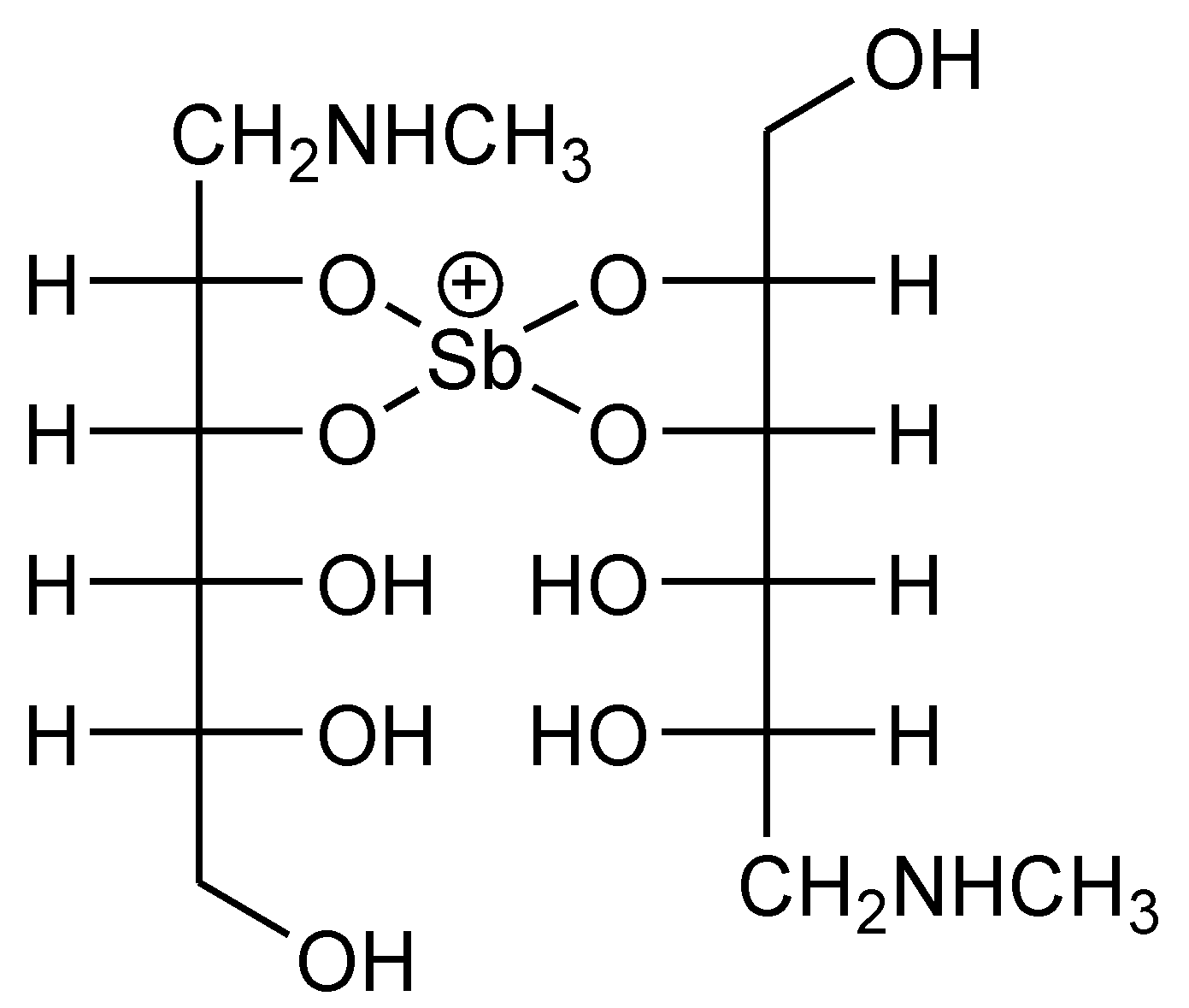
2.1. Reagents

2.1.1. Nanoemulsion Formulation

The molecule used as the active principle is an antimonial pentavalent compound (SbV) in a meglumine antimoniate form, known as Ulamina (Figure 1). Surfactants are of the non-ionic type: Sorbitan Monolaurate of HLB = 8.6 and polisorbate 80 of HLB = 15, which are known to be biocompatible with skin and were kindly supplied by Oxiteno, Sao Paulo, Brasil. Other additives to prepare the nanoemulsion-based cream are Carbopol Acrylamer 941 (total concentration in the formulation of 1.5 wt%), methylparaben (0.18 wt% in the formulation) and propylparaben (0.02 wt% in the formulation). These compounds were supplied by Merck, Darmstadt, Germany with 99.5% purity. A low molecular weight paraffin was used as the oil phase. The total surfactant concentration in the cream was 4.5 wt%.

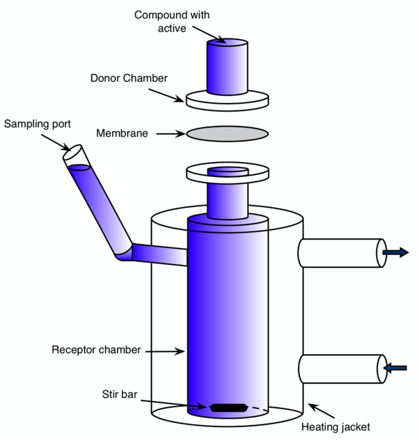
2.1.2. Transdermal Passage Tests

The transdermal passage of the antimonial active principle was carried out through Franz cells (described in Section 2.3). Strat-MTM membranes (Merck-Millipore, Darmstadt, Germany) were used for the tests, and a salty buffer was used as a receptor agent. Strat-MTM membranes have been proven to resemble skin, because both membranes exhibit similar permeabilities to chemicals.
The saline buffer was composed of 1.5125 g Na2HPO4 and 2.0875 g KH2PO4 (100% purity, Riedel-de Haën, Seelze, Germany) added into a 250 mL volumetric flask, and 80% of the required volume of distilled water was added with continuous mixing.
Fick’s laws of diffusion describes the transdermal permeation experiment [1,61,62]. Flow and apparent permeability are described by the following equation:
Flux = Papp Cd
where Flux is the mass flow of the solute (µg/cm2/min), Papp is the apparent permeability of the membrane (cm/min) and Cd (µg/cm3) is the concentration of the active ingredient in the cream.
The transdermal passage tests were carried out during 8 h. Samples were taken from the receptor compartment every 15 min, and the antimony content was analyzed. The antimony flux (µg/cm2/min) and the accumulated mass (mg) over time were determined.

2.1.3. Antimony Analysis

Antimonium (III) chloride (99.7% Sigma-Aldrich, St. Louis, MO, USA), HCl (37% Riedel-de Haën, Seelze, Germany), potassium tartrate (99.5% Merck, Darmstadt, Germany), potassium hydrogen phosphate (99.5% Riedel-de Haën, Seelze, Germany), potassium iodide (99% Sigma-Aldrich, St. Louis, MO, USA), pyrogallol bromine red (Sigma-Aldrich, St. Louis, MO, USA), ethanol (99.8% Riedel-de Haën, Seelze, Germany), ascorbic acid (99.7% Sigma-Aldrich, St. Louis, MO, USA) and sodium hydroxide (99% Merck, Darmstadt, Germany) were used as received.

2.2. Ulamina Nanoemulsion Preparation (O/W) and Cream Fabrication

Several formulations of oil in water (O/W = 20/80) emulsions were made with a mixture of non-ionic surfactants following the low energy method proposed by Forgiarini et al. [52,63,64]. The concentration of the active ingredient was fixed to 10% of the antimonial pentavalent compound that was obtained from a 30% Ulamina stock solution. Initially, the HLB of the surfactant mixture was varied to choose the formulation that would produce the nanoemulsion with the smallest droplet size, maintaining a monodisperse distribution and maximum kinetic stability.
A Eurostar mixer of IKA Labotechnik (Staufen, Germany) was used to prepare the nanoemulsion with a mixing speed of 450× g. Water with the Ulamina active compound was added drop by drop to the paraffin–surfactant mixture at a constant temperature (T = 30 °C). In the procedure, a liquid crystalline phase was formed, which has been argued to be essential for the nanoemulsion formation mechanism [65,66,67].
The Carbopol was solubilized in an amount of water corresponding to 20% of the total cream water, and its final concetration in the cream was 1.0 wt%.
Likewise, the parabens, as preservative agents, were solubilized before adding the Carbopol to the indicated water fraction (20%). Once the pH has been adjusted, this last portion of water was added to the nanoemulsion, and the system was stirred for 2 min at 1200× g to guaratee the homogenization of the final cream.
A Carbopol Acrilamer 941 polymer was added to the chosen nanoemulsion to increase the viscosity of the O/W nanoemulsion and achieve not only an adequate texture and rheological behavior of the cream, but also to guarantee greater kinetic stability. Methylparaben and propylparaben were used as preserving agents for the cream.
The HLB calculation of the surfactant mixture (HLBM) was made using the following equation:
HLBM = X1HLB1 + X2HLB2
where Xi and HLBi are surfactant weight fraction and HLB, respectively.

2.3. Cream Evaluation

2.3.1. Human Sensory Testing

The sensory tests were done with ten evaluators who carried out the following protocol [68,69,70]: handwashing with tap water and drying with paper towels was done for 5 min. Subsequently, 0.1 g of cream was applied, which was rubbed gently on the back of the hand with the index finger. This rubbing was done 5 times in a circular motion. Then, the evaluators left each cream for 5 min and used an assessment sheet to evaluate the aspects of the cream. Subsequently, tap water was used to rinse the area where the cream was applied. The cream did not contain Ulamina and 3 parameters were assessed: homogeneity, extensibility and elimination after rinsing. The parameters were rated on a 3-point scale (1: bad, 2: fair, 3: good).
Homogeneity: homogeneity of the cream was checked by its visual appearance (color, aspect, crystallinity, amount of waste) and its sensation to touch.
Type of film and extensibility: this sensorial characteristic was qualitatively pondered by the type of film formed in the skin when the cream was applied.
Elimination: the ease of removal of the cream after its application was examined by washing the part of the skin where it was applied with tap water.
This stage was essential in the formulation process. Consumer perception is becoming more important every day for final products’ acceptance, as shown in multiple reports [71,72,73].
Once the final cream texture was chosen, physicochemical characteristics, such as pH, droplet size, rheology, cream stability over time and transdermal passage, were evaluated.

2.3.2. Microscopy, Droplet Size and pH

A polarized light Nikon microscope, model Eclipse E600POL, with ACT-1 software was used to observe and capture microphotographs of the formulated cream. The droplet size of nanoemulsion and cream samples was measured by dispersing the emulsion in a sodium pyrophosphate solution at 1%. The droplet size distributions were measured using a laser diffraction particle size analyzer, MasterSizer, model 2000 of Malvern Ltd. (Malvern, UK).
The pH was measured using a solution of 0.5 g of cream dissolved in 50 mL of distilled water using a pH-meter, Metrohm 691 (Metrohm AG, Herisau, Switzerland).

2.3.3. Rheological Studies

The sensory characteristics of the nanoemulsion-based cream were estimated with rheological properties such as consistency and flow index. These properties can be evaluated using a rheological model such as the power law (Ostwald Law) model. This was done by measuring either viscosity versus shear speed or the shear stress versus shear rate curve, within the range of 0.1 to 100 s−1, with an AR-G2 rheometer of controlled tension (TA Instruments, New Castle, DE, USA) using a plate–plate geometry (plate diameter 40 mm) at 25 °C.
The power law model, written as τ = kγn, was proposed to study the cream rheology characteristics, where: τ is the shear stress expressed in Pascals (Pa); k is the consistency index, i.e., the viscosity at 1 s−1 shear rate, describing how thick (viscous) a material is when low shear is applied to it [74,75]; n is the flow index; and γ is the shear rate at which a shear strain is applied, expressed in reciprocal seconds (s−1). The approximate flow index and consistency index of the cream can be obtained by plotting the natural logarithm of the shear speed and shear stress according to the power law model [74,76].

2.3.4. Cream Stability Tests

Accelerated stability tests of the creams were carried out as a function of temperature. In the first analysis, several control samples were prepared that were stored at ambient temperature for 7 days [68]. In addition, 20 g of the developed formulation was stored in a stove at 40 °C for 40 days, following the procedure of Grimm [70] and Bjerregaard [77]. The stability protocol of the samples consisted in the following storage conditions (Table 1).

2.4. In Vitro Tests

2.4.1. Transdermal Passage

The transdermal passage through kinetic tests of in vitro permeation were performed in Franz cells. Figure 2 shows a Franz cell, which consists of an upper compartment (giver) where the sample under study was placed (vehicle with the active ingredient). Moreover, an inferior compartment (reception) was placed within a liquid, which, in this case, was a saline buffer solution at pH = 7.4, very similar to the mammal’s extracellular liquid [39,78]. A membrane, which in this case was the Strat-M membrane, was placed between both compartments.
The Strat-M membrane’s composition tends to emulate human skin structure. It consists of two layers of polyether sulfone and poliolefins, with a mixture of synthetic lipids that produce certain resistance to the passage of any substance [79]. The contact area of each cell is 0.7854 cm2, while the volume of the receptor chamber is 10 mL.
The inferior compartment has a volume of 10 mL and has a sampling arm from where samples are taken to analyze the concentration of the active ingredients that have crossed the membrane. The cell has an outer jacket and a thermostatic bath with recirculation to keep a constant temperature during the experiments. The cells were maintained at 37 ± 0.5 °C in all the experiments, since this is the generally accepted temperature of the skin’s surface.
It is essential that while the cream samples are being studied, the membrane and the solution in the receptor compartment must be in permanent and total contact between them so that the transfer area is always constant. Ten cells were assembled in a series with a magnetic plate, IKA-Werke, with 10 agitation points. All the experiments were performed in triplicate, and the mean results of them are reported.

2.4.2. Antimony Analysis

Determination of antimony concentration of the samples collected in the transdermal passage experiments was performed by the pyrogallol bromine red method, developed by Christopher [80] and adapted by Rath [81]. The samples were analyzed with a UV spectrometer, Shimadzu model mini 1240 (Shimadzu Corporation, Kyoto, Japan).

2.5. Preliminar In Vivo Test

A phase 2 prospective study to evaluate the therapeutic efficacy of the topical antimonial formulation was perfomed by the Control and Chemotherapy Laboratory of the JWTRI. The patients included in this trial were volunteers of legal age of both sexes, from endemic areas of Sucre state, who were treated by the staff of the Chemotherapy and Control Laboratory of JWTRI for Leishmaniasis diagnosis and treatment. These patients had ulcerated skin lesions without mucosal alterations. Of these patients, only the results of two cases have been chosen for this article.
Volunteer patients from municipalities of the Sucre state who presented flattened lesions with irregular but defined edges and base hardening, a product of the formation of a granuloma, were diagnosed parasitologically through microscopic studies of imprints and biopsies that determine lesions caused by infections of Leishmania sp. These studies were immunologically confirmed by positive reactions against antigens of Leishmaniasis braziliensis sub genus Viannia [82] and characterized by PCR studies [83].
In all cases, a comprehensive analysis was performed using:
  • Epidemiological Diagnosis: The origin of the patient is determined and is related to known areas of transmission.
  • Clinical Diagnosis: Presence of flattened skin lesions with irregular but well-defined edges, which present base hardening as a result of the development of a granuloma, which are characteristics of lesions caused by Leishmaniasis.
  • Parasitological Diagnosis: It was carried out by microscopic observation of smears of aspirates and biopsies and stained with Giemsa stain at 10% in phosphate buffer pH 7.2, where macrophages infected with amastigotes were observed, confirming the diagnosis of infection by Leishmania sp.
  • Immune Diagnosis: Their immune responses were confirmed, using the Montenegro Test [84] as a test for Leishmania, by intradermal reaction with the figurative antigen from the promastigotes form of Leishmania (Viannia) braziliensis [82].

Treatment

The treatment for the two cases reported in this study consisted of two daily topical applications of the nanoemulsion cream.
The patients gave their consent in writing.

3. Results

3.1. Nanoemulsion Formation

The nanoemulsions formulated in the present research have a composition of 4.5 wt% of the surfactant mixture and a constant water/oil ratio equal to 80/20. The best combination of non-ionic surfactants was studied by measuring the droplet size as a function of the HLB of the surfactant mixture.
In all the experiments, the low-energy emulsification method was applied. As the previous literature indicates [53,85], a liquid crystalline phase is usually formed when nanoemulsions are obtained. The nanoemulsification method by “persuasion” or phase transition has been argued to be intrinsically linked to the dissolution of the liquid crystalline phase, which protects the droplet from coalescence [53,86].

Nanoemulsion Droplet Size Distribution

Figure 3 presents the droplet sizes of the nanoemulsions as a function of the HLB of the surfactant mixture. The formulation with an HLB = 12.25 was chosen due to its smaller droplet size. In addition, the droplet size of the emulsion (Figure 4) shows a homogeneous distribution of the nanoemulsion with droplets ranging between 90–130 nm and a mean of 115 nm.

3.2. Cream Sensory Tests

3.2.1. Homogeneity

After the emulsions were formed, the Carbopol Acrilamer 941 polymer was added to attain the viscosity and microstructure necessary to obtain a stable dispersion. In the present research, all the formulations had uniformity and homogeneity. This was confirmed by visual appearance, as shown in Figure 5, and its feel to touch. It had no residues and was crystalline and emollient.
After applying the cream to the skin, most of the evaluators concluded that the cream was easy to apply, left a non-greasy feeling after application and was easy to remove with tap water. The results of the attributes of the sensory tests are presented in Table 2.
The organoleptic characteristics were evaluated as a function of time. The observations provided a first impression of the product’s quality. The cream presented a homogeneous appearance and color and an acceptable and smooth texture after topical application, as outlined in Table 3.

3.2.2. Topical Cream pH

The topical cream pH was between 6 and 7, which is considered a compatible value with the pH of skin, sometimes referred to as an optimal pH [87,88].

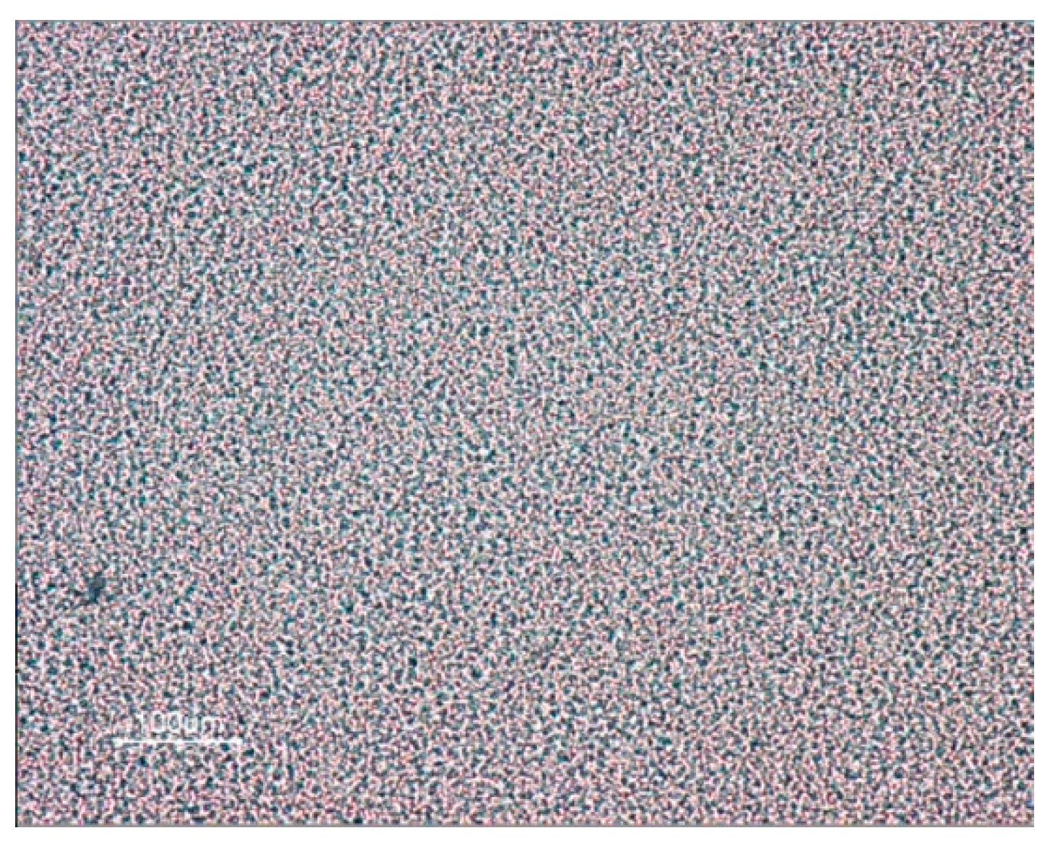
3.2.3. Optical Microscopy

Several images were taken of each formulation to ensure the homogeneity and uniformity on the emulsions. The microscopic image in Figure 6 shows a uniform droplet size that demonstrates the cream’s homogeneity and low polydispersity.

3.3. Rheological Studies

3.3.1. Viscosity Measurements

Skin creams generally exhibit shear-thinning behavior. This means that the viscosity is not a constant value but depends on the intensity of the shear. This relationship is described by plotting viscosity versus shear rate (Figure 7). A cream to be applied to damaged skin by a disease, for example, should be easily applied to wounds and should be able to be easily spread at shear rates; in this study, it was from 0.1 to 50 s−1.
The rheogram was performed on the freshly prepared cream and on the dispersion after 60 days of preparation, and both had a flow index between 0 and 1 (n = 0.19 and 0.21, respectively), as shown in Figure 7; therefore, they were shear-thinning fluids. The values indicate how smoothly the ointment flows when the shear is applied [74,76,89]. Creams with a high degree of shear-thinning tend to feel softer and spread more smoothly. This characteristic did not change in the time frame studied. However, the lower consistency index of cream aged for 60 days (which decreased from 55.7 to 38.4), would make the cream easier to dispense and give a smooth and “less sticky” feeling on the skin [71].
On the other hand, the stabilization effect of the polymer by increasing the viscosity of the continuous phase guaranteed the stability of the cream during the required storage time of up to 1 year from its manufacture, as required in this study.

3.3.2. Cream Stability Tests

Samples of the cream with 10% Ulamina had great kinetic stability at ambient temperature and at 40 °C. It is important to point out that in both cases, the possible changes in the cream were observed during a year, and no change (creaming, sedimentation) was noticed nor was its stability altered. This is attained due to two effects: the small size of the nanodroplet, stabilized by the non-ionic surfactant mixture, and by the polymer stabilization effect.
No modifications were observed; the maintenance of all the initial characteristics of appearance of the cream were stable during approximately one year.
The droplet size obtained in this formulation directly correlates with the high stability observed in the cream with Carbopol Acrilamer 941. In Figure 8, a scheme of the microstructure of the cream is presented.

3.4. In Vitro Tests

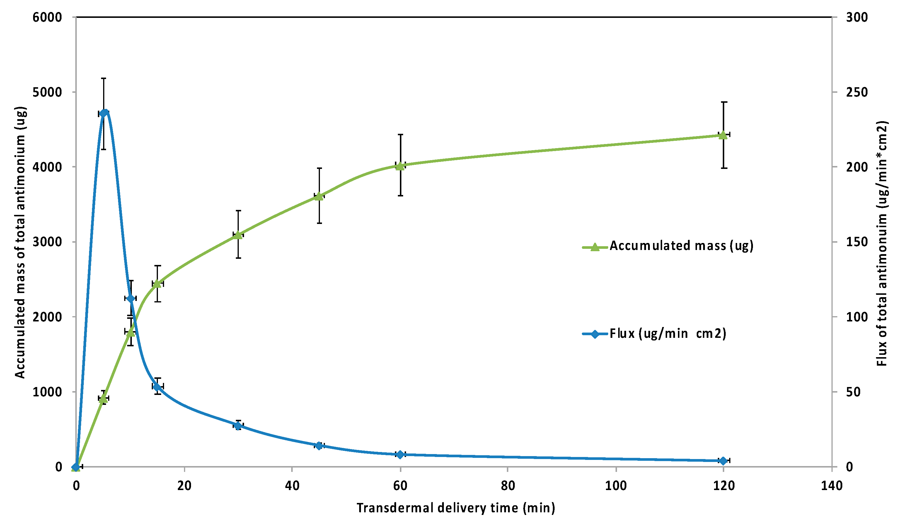
3.4.1. Transdermal Experiments

Figure 9 shows the in vitro tests made during 8 h, indicating that the majority of the active ingredient, Ulamina, was transferred in the first 15 min of contact until a plateau was reached. The analysis of the collected mass transfer samples indicated that the active ingredient passage reached up to 50% in the first 15 min of contact time. Each point is the average of three measurements. This was similar to other effective transdermal drug delivery systems [90].

3.4.2. Antimony Analysis

In Figure 9, the antimony flow rate and its total accumulated mass in the receiving cell are also shown.
The total transferred mass after 120 min of transdermal passage was 4.44 mg. Figure 9 shows that the maximum antimony flow was at 5 min, and, at this point, the total accumulated mass was 0.93 mg, representing 20.90% of the total mass in 120 min. At approximately 30 min, a constant regime was reached, and, at this point, 3.11 mg of total antimony had been transferred.

3.5. Clinical Cases and In Vivo Tests

Table 4 shows the results of two patients with CL treated with the Ulamina cream in a preliminary study. A 19-year-old woman and a 67-year-old man were selected among several patients of the phase 2 prospective study. The former presented a lesion in the leg and the latter in the hand. The Ulamina topical nanoemulsion was applied over the skin every 12 h for 7 and 6 weeks, respectively. Both patients showed healing of the infection and cicatrization of the infected lesion. This would indicate that this type of meglumine antimoniate active component solubilized in the aqueous phase of the nanoemulsion has the potential to be used in the short term for the treatment of CL worldwide.

4. Conclusions

The measurement and analysis of the emulsion’s properties indicate that Ulamina vehiculized as a nanodroplet cream is stable from the physicochemical point of view and guarantees transdermal passage. Therefore, Ulamina can be considered an active ingredient with the proper characteristics to be administered topically with favorable therapeutic response in skin injuries caused by Leishmaniasis. Its effectiveness in vitro has been studied via transdermal passage using a Strat-M synthetic membrane. The active compound’s availability due to the transdermal passage effectiveness indicates that deploying the cream for extended use by patients in low-income regions of Latin America and Asian tropical countries could be feasible. Furthermore, as an alternative and less invasive treatment, the cream has also been used in two patients, showing total healing of the bacterial infection and wound. In the near future, a phase 2 prospective study will be completed, which actually shows promising results. As a final note, the process was a very low-energy procedure that generated emulsions with high stability, in contrast with high-energy processes as microfluidizers that required high-energy input, representing high CO2-equivalent emissions.

Author Contributions

Conceptualization: L.M., A.F. and C.S.; methodology: J.R., A.C. and J.A.F.; validation: F.V. and J.V.S.; formal analysis: J.A.F. and J.V.S.; writing—original draft preparation: J.B. and S.K.-K.; writing—review and editing: J.B. and A.F.; supervision: J.B.; project administration: J.B.; funding acquisition: J.B. All authors have read and agreed to the published version of the manuscript.

Funding

Procter & Gamble of Venezuela, Oxiteno and FIRP Laboratory.

Institutional Review Board Statement

No. 2007-000-960: The study conducted was approved by the Institutional Review Board of the Instituto de Investigaciones “José Witremundo Torrealba”, Núcleo Universitario Rafael Rangel, Trujillo, Universidad de Los Andes, Venezuela.

Informed Consent Statement

Informed consent was obtained from all subjects involved in the study. The agreed study protocol, as well as the consent forms and the procedures, were reviewed and approved by the Ethics Committee of the JWTRI.

Data Availability Statement

The data presented in this study are available in the article.

Acknowledgments

The authors acknowledge Procter & Gamble of Venezuela for financially contributing from 2010 in the development of the cream and also with the purchase of a vehicle used by the Parasitological Research Institute “Jose Witremundo Torrealba” for field work. Oxiteno and and Corporación Technipore SA are also recognized for kindly providing surfactants and STRAT-M membranes, respectively. We thank Jeny Sayago, José Alejandro Dapena, Ronald Márquez and J.L. Salager for their collaboration in this study.

Conflicts of Interest

The authors declare no conflict of interest.

References

  1. Bouwstra, J.A. The skin barrier, a well-organized membrane. Colloids Surf. A Physicochem. Eng. Asp. 1997, 124, 403–413. [Google Scholar] [CrossRef]
  2. Burgess, C.M. Cosmetic Dermatology, 1st ed.; Springer: Berlin/Heidelberg, Germany, 2005. [Google Scholar]
  3. Neto, A.M.F.; Salinas, S.R.A. The Physics of Lyotropic Liquid Crystals: Phase Transitions and Structural Properties. In The Physics of Lyotropic Liquid Crystals: Phase Transitions and Structural Properties; Oxford University Press on Demand: Oxford, UK, 2010; pp. 1–320. [Google Scholar] [CrossRef]
  4. Prieto-Blanco, M.C.; Fernández-Amado, M.; López-Mahía, P.; Muniategui-Lorenzo, S.; Prada-Rodríguez, D. Surfactants in Cosmetics: Regulatory Aspects and Analytical Methods. Anal. Cosmet. Prod. 2018, 249–287. [Google Scholar]
  5. Vaughan, C.D. Using solubility parameters in cosmetics formulation. J. Soc. Cosmet. Chem. Jpn. 1985, 36, 319–333. [Google Scholar]
  6. Marti-Mestres, G. Pharmaceutical Emulsions and Suspensions; CRC Press: Boca Raton, FL, USA, 2000. [Google Scholar] [CrossRef]
  7. Frézard, F.; Demicheli, C.; Ribeiro, R.R. Pentavalent Antimonials: New Perspectives for Old Drugs. Molecules 2009, 14, 2317–2336. [Google Scholar] [CrossRef] [Green Version]
  8. Wallen-Russell, C.; Wallen-Russell, S. Meta Analysis of Skin Microbiome: New Link between Skin Microbiota Diversity and Skin Health with Proposal to Use This as a Future Mechanism to Determine Whether Cosmetic Products Damage the Skin. Cosmetics 2017, 4, 14. [Google Scholar] [CrossRef] [Green Version]
  9. Heras-Mosteiro, J.; Monge-Maillo, B.; Pinart, M.; Pereira, P.L.; Garcia-Carrasco, E.; Cuadrado, P.C.; Royuela, A.; Roman, I.M.; López-Vélez, R. Interventions for Old World cutaneous leishmaniasis. Cochrane Database Syst. Rev. 2017, 11, CD005067. [Google Scholar] [CrossRef]
  10. Torres-Guerrero, E.; Quintanilla-Cedillo, M.R.; Ruiz-Esmenjaud, J.; Arenas, R. Leishmaniasis: A review. F1000Research 2017, 6, 750. [Google Scholar] [CrossRef]
  11. De Lima, H.; Borges, R.H.; Escobar, J.; Convit, J. Leishmaniasis cutánea americana en Venezuela: Un análisis clínico epidemiológico a nivel nacional y por entidad federal, 1988–2007. Bol. Malariol. Salud Amb. 2010, 50, 283–300. [Google Scholar]
  12. Añez, N.; Rojas, A.; Scorza-Dagert, J.V.; Morales, C. Successful treatment against American cutaneous leishmaniasis by intralesional infiltration of a generic antimonial compound-lidocaine combination. A follow up study. Acta Trop. 2018, 185, 261–266. [Google Scholar] [CrossRef]
  13. Añez, N.; Rojas, A.; Crisante, G. Evaluation of conventional serological tests for the diagnosis of American cutaneous leishmaniasis. Boletín De Malariol. Y Salud Ambient. 2007, 47, 55–62. [Google Scholar]
  14. WHO Expert Committee on the Control of the Leishmaniases & World Health Organization. Control of the leishmaniases: Report of a Meeting of the WHO Expert Commitee on the Control of Leishmaniases, Geneva, Switzerland, 22–26 March 2010; World Health Organization: Geneva, Switzerland, 2010. [Google Scholar]
  15. Freitas-Junior, L.H.; Chatelain, E.; Kim, H.A.; Siqueira-Neto, J.L. Visceral leishmaniasis treatment: What do we have, what do we need and how to deliver it? Int. J. Parasitol. Drugs Drug Resist. 2012, 2, 11–19. [Google Scholar] [CrossRef] [PubMed] [Green Version]
  16. López, L.; Vélez, I.; Asela, C.; Cruz, C.; Alves, F.; Robledo, S.; Arana, B. A phase II study to evaluate the safety and efficacy of topical 3% amphotericin B cream (Anfoleish) for the treatment of uncomplicated cutaneous leishmaniasis in Colombia. PLoS Negl. Trop. Dis. 2018, 12, e0006653. [Google Scholar] [CrossRef]
  17. Bonfante-Garrido, R. Leishmanias y leishmaniasis tegumentaria en América Latina. Bol. La Of. Sanit. Panam. 1983, 95, 418–426. [Google Scholar]
  18. Scorza Dagert, J.V.; Morales, C.; Petit de Peña, Y.; Vásquez, L.; Rojas, E.; Scorza, J.V.B. Síntesis de un complejo antimonial pentavalente (Ulamina) y su aplicación experimental para el tratamiento de leishmaniasis cutánea localizada en Venezuela. Boletín De Malariol. Y Salud Ambient. 2006, 46, 59–65. [Google Scholar]
  19. World Health Assembly. Control of Leishmaniasis: Report by the Secretariat; World Health Organization: Ginebra, Switzerland, 2007; Available online: https://apps.who.int/iris/handle/10665/25315 (accessed on 3 December 2007).
  20. Henao, H.H.; Osorio, Y.; Saravia, N.G.; Gomez, A.; Travi, B. Eficacia y toxicidad de los antimoniales pentavalentes (Glucantime y Pentostam) en un modelo animal de leishmaniasis cutánea americana: Aplicación de la luminometría. Biomedica 2004, 24, 393–402. [Google Scholar] [CrossRef] [PubMed] [Green Version]
  21. Pinart, M.; Rueda, J.-R.; Romero, G.A.; Pinzón-Flórez, C.E.; Osorio-Arango, K.; Maia-Elkhoury, A.N.S.; Reveiz, L.; Elias, V.M.; A Tweed, J. Interventions for American cutaneous and mucocutaneous leishmaniasis. Cochrane Database Syst. Rev. 2020, 8, CD004834. [Google Scholar] [CrossRef]
  22. Matoussi, N.; Ameur, H.; Amor, S.; Fitouri, Z.; Becher, S. Toxicité cardiaque de l’antimoniate de méglumine (Glucantime®). À propos d’une observation. Médecine Mal. Infect. 2007, 37, S257–S259. [Google Scholar] [CrossRef] [PubMed]
  23. Lindoso, J.A.L.; Costa, J.M.L.; Queiroz, I.T.; Goto, H. Review of the current treatments for leishmaniases. Res. Rep. Trop. Med. 2012, 3, 69–77. [Google Scholar] [CrossRef] [PubMed] [Green Version]
  24. Murray, H.W.; Berman, J.D.; Davies, C.R.; Saravia, N.G. Advances in leishmaniasis. Lancet 2005, 366, 1561–1577. [Google Scholar] [CrossRef]
  25. Pandey, B.D.; Pun, S.B.; Kaneko, O.; Pandey, K.; Hirayama, K. Case Report: Expansion of Visceral Leishmaniasis to the Western Hilly Part of Nepal. Am. J. Trop. Med. Hyg. 2011, 84, 107–108. [Google Scholar] [CrossRef] [Green Version]
  26. Schofield, C.J. UNDP/World Bank/WHO special programme for research and training in tropical diseases (TDR). Parasitol. Today 1989, 5, 235–238. [Google Scholar] [CrossRef]
  27. Soto, J.; Grogl, M.; Berman, J.; Olliaro, P. Limited efficacy of injectable aminosidine as single-agent therapy for Colombian cutaneous leishmaniasis. Trans. R. Soc. Trop. Med. Hyg. 1994, 88, 695–698. [Google Scholar] [CrossRef]
  28. Buffet, P.A.; Morizot, G. Cutaneous leishmaniasis in France: Towards the end of injectable therapy? Bull. Soc. Pathol. Exot. 2003, 96, 383–388. [Google Scholar] [PubMed]
  29. Sundar, S.; Jha, T.K.; Singh, V.R.; Mishra, M.; Buffels, R.; Thakur, C.P. Low-dose liposomal amphotericin B in refractory Indian visceral leishmaniasis: A multicenter study. Am. J. Trop. Med. Hyg. 2002, 66, 143–146. [Google Scholar] [CrossRef] [Green Version]
  30. Hepburn, N.C.; Tidman, M.J.; Hunter, J.A. Aminosidine (paromomycin) versus sodium stibogluconate for the treatment of American cutaneous leishmaniasis. Trans. R. Soc. Trop. Med. Hyg. 1994, 88, 700–703. [Google Scholar] [CrossRef]
  31. Mushtaq, S.; Dogra, D.; Dogra, N. Clinical Response with intralesional Amphotericin B in the treatment of old world cutaneous leishmaniasis: A preliminary report. Dermatol. Ther. 2016, 29, 398–405. [Google Scholar] [CrossRef] [PubMed]
  32. Arevalo, I.; Tulliano, G.; Quispe, A.; Spaeth, G.; Matlashewski, G.; Llanos-Cuentas, A.; Pollack, H. Role of Imiquimod and Parenteral Meglumine Antimoniate in the Initial Treatment of Cutaneous Leishmaniasis. Clin. Infect. Dis. 2007, 44, 1549–1554. [Google Scholar] [CrossRef] [PubMed]
  33. Kim, D.H.; Chung, H.J.; Bleys, J.; Ghohestani, R.F. Is Paromomycin an Effective and Safe Treatment against Cutaneous Leishmaniasis? A Meta-Analysis of 14 Randomized Controlled Trials. PLOS Negl. Trop. Dis. 2009, 3, e381. [Google Scholar] [CrossRef] [PubMed] [Green Version]
  34. Lodi, G.; Sannino, M.; Caterino, P.; Cannarozzo, G.; Bennardo, L.; Nisticò, S.P. Fractional CO2 laser-assisted topical rifamycin drug delivery in the treatment of pediatric cutaneous leishmaniasis. Pediatric Dermatol. 2021, 38, 717–720. [Google Scholar] [CrossRef] [PubMed]
  35. Vásquez, L.; Dagert, J.V.S.; Scorza, J.V.; Vicuña-Fernández, N.; De Peña, Y.P.; Lopez, S.; Bendezu, H.; Rojas, E.; Pérez, B. Pharmacokinetics of experimental pentavalent antimony after intramuscular administration in adult volunteers. Curr. Ther. Res. 2006, 67, 193–203. [Google Scholar] [CrossRef] [PubMed] [Green Version]
  36. Vásquez de Ricciardi, L.; Vicuña-Fernández, N.; de Peña, Y.P.; López, S.; Scorza, J.V.; Scorza-Dage, J.V.; Villegas, E.; Pérez, B. Disposición farmacocinética de las especies de antimonio en perros después de una dosis de antimoniato de meglumina (Glucantime®). Boletín De Malariol. Y Salud Ambient. 2008, 48, 27–33. [Google Scholar]
  37. Gallignani, M.; Ayala, C.; Brunetto, M.D.R.; Burguera, M.; Burguera, J. Flow analysis–hydride generation–Fourier transform infrared spectrometric determination of antimony in pharmaceuticals. Talanta 2003, 59, 923–934. [Google Scholar] [CrossRef]
  38. Finnin, B.C.; Morgan, T.M. Transdermal penetration enhancers: Applications, limitations, and potential. J. Pharm. Sci. 1999, 88, 955–958. [Google Scholar] [CrossRef] [PubMed]
  39. Van Bocxlaer, K.; McArthur, K.-N.; Harris, A.; Alavijeh, M.; Braillard, S.; Mowbray, C.; Croft, S. Film-Forming Systems for the Delivery of DNDI-0690 to Treat Cutaneous Leishmaniasis. Pharmaceutics 2021, 13, 516. [Google Scholar] [CrossRef] [PubMed]
  40. Rodrigues, L.N.; Zanluchi, J.M.; Grebogi, I.H. Percutaneous absorption enhancers: Mechanisms and potential. Braz. Arch. Biol. Technol. 2007, 50, 949–961. [Google Scholar] [CrossRef] [Green Version]
  41. Roberts, M.; Cross, S.; Pellett, M. Skin Transport. In Dermatological and Transdermal Formulations, 1st ed.; Walters, K.A., Ed.; CRC Press, Taylor & Francis: New York, NY, USA, 2002. [Google Scholar]
  42. Magnusson, B.M.; Anissimov, Y.; Cross, S.E.; Roberts, M. Molecular Size as the Main Determinant of Solute Maximum Flux Across the Skin. J. Investig. Dermatol. 2004, 122, 993–999. [Google Scholar] [CrossRef] [PubMed] [Green Version]
  43. Leite-Silva, V.R.; De Almeida, M.M.; Fradin, A.; Grice, J.; Roberts, M. Delivery of drugs applied topically to the skin. Expert Rev. Dermatol. 2012, 7, 383–397. [Google Scholar] [CrossRef]
  44. Hendradi, E.; Obata, Y.; Isowa, K.; Nagai, T.; Takayama, K. Effect of Mixed Micelle Formulations Including Terpenes on the Transdermal Delivery of Diclofenac. Biol. Pharm. Bull. 2003, 26, 1739–1743. [Google Scholar] [CrossRef] [PubMed] [Green Version]
  45. Monzote, L.; Herrera, I.; Satyal, P.; Setzer, W.N. In-Vitro Evaluation of 52 Commercially-Available Essential Oils Against Leishmania amazonensis. Molecules 2019, 24, 1248. [Google Scholar] [CrossRef] [PubMed] [Green Version]
  46. Sonneville-Aubrun, O.; Simonnet, J.-T.; L’Alloret, F. Nanoemulsions: A new vehicle for skincare products. Adv. Colloid Interface Sci. 2004, 108–109, 145–149. [Google Scholar] [CrossRef] [PubMed]
  47. Yuan, J.S.; Ansari, M.; Samaan, M.; Acosta, E.J. Linker-based lecithin microemulsions for transdermal delivery of lidocaine. Int. J. Pharm. 2008, 349, 130–143. [Google Scholar] [CrossRef]
  48. Lim, P.F.C.; Liu, X.Y.; Kang, L.; Ho, P.C.L.; Chan, S.Y. Physicochemical effects of terpenes on organogel for transdermal drug delivery. Int. J. Pharm. 2008, 358, 102–107. [Google Scholar] [CrossRef] [PubMed]
  49. Pouton, C.W. Formulation of self-emulsifying drug delivery systems. Adv. Drug Deliv. Rev. 1997, 25, 47–58. [Google Scholar] [CrossRef]
  50. Alenius, H.; Roberts, D.W.; Tokura, Y.; Lauerma, A.; Patlewicz, G.; Roberts, M. Skin, drug and chemical reactions. Drug Discov. Today Dis. Mech. 2008, 5, e211–e220. [Google Scholar] [CrossRef]
  51. Buck, P. Skin barrier function: Effect of age, race and inflammatory disease. Int. J. Aromather. 2004, 14, 70–76. [Google Scholar] [CrossRef]
  52. Forgiarini, A.; Esquena, J.; González, C.; Solans, C. Studies of the Relation between Phase Behavior and Emulsification Methods with Nanoemulsion Formation. Prog. Colloid Polym. Sci. 2000, 115, 36. [Google Scholar]
  53. Forgiarini, A.; Esquena, J.; González, C.; Solans, C. Formation of Nano-emulsions by Low-Energy Emulsification Methods at Constant Temperature. Langmuir 2001, 17, 2076. [Google Scholar] [CrossRef]
  54. Tadros, T.; Izquierdo, P.; Esquena, J.; Solans, C. Formation and stability of nano-emulsions. Adv. Colloid Interface Sci. 2004, 108–109, 303–318. [Google Scholar] [CrossRef] [PubMed]
  55. Shakeel, F.; Baboota, S.; Ahuja, A.; Ali, J.; Shafiq, S. Skin permeation mechanism and bioavailability enhancement of celecoxib from transdermally applied nanoemulsion. J. Nanobiotechnology 2008, 6, 8. [Google Scholar] [CrossRef] [PubMed] [Green Version]
  56. Zhou, H.; Yue, Y.; Liu, G.; Li, Y.; Zhang, J.; Gong, Q.; Yan, Z.; Duan, M. Preparation and Characterization of a Lecithin Nanoemulsion as a Topical Delivery System. Nanoscale Res. Lett. 2009, 5, 224–230. [Google Scholar] [CrossRef] [PubMed] [Green Version]
  57. Villarreal, A.M.; Fernandez, C.; Forgiarini, A.; Marquez, L.; Nielloud, F.; Salager, J. Nanoencapsulation de filtres solaires via nanoémulsions. In Procédés et Formulations au Service de la Santé; Durand, A., Canselier, J.-P., Eds.; EDP Sciences: Les Ulis, France, 2011; pp. 1–14. [Google Scholar]
  58. Salager, J.-L.; Antón, R.; Bullón, J.; Forgiarini, A.; Marquez, R. How to Use the Normalized Hydrophilic-Lipophilic Deviation (HLDN) Concept for the Formulation of Equilibrated and Emulsified Surfactant-Oil-Water Systems for Cosmetics and Pharmaceutical Products. Cosmetics 2020, 7, 57. [Google Scholar] [CrossRef]
  59. Clares, B.; Pujol, A.; Hernández, P.; Riera, C.; Calpena, A.-C. Evaluación de la cinética de liberación de antimoniato de meglumina desde geles para su aplicación tópica. Cosmetics 2020, 7, 57. [Google Scholar] [CrossRef]
  60. Horoiwa, T.A.; Cortez, M.; Sauter, I.P.; Migotto, A.; Bandeira, C.L.; Cerize, N.N.P.; de Oliveira, A. Sugar-based colloidal nanocarriers for topical meglumine antimoniate application to cutaneous leishmaniasis treatmente: Ex vivo cutaneous retention and in vivo evaluation. Eur. J. Pharm. Sci. 2020, 147, 105295. [Google Scholar] [CrossRef] [PubMed]
  61. Uchida, T.; Kadhum, W.R.; Kanai, S.; Todo, H.; Oshizaka, T.; Sugibayashi, K. Prediction of skin permeation by chemical compounds using the artificial membrane, Strat-M™. Eur. J. Pharm. Sci. 2014, 67, 113–118. [Google Scholar] [CrossRef] [PubMed] [Green Version]
  62. Peña-Juárez, M.C.; Guadarrama-Escobar, O.R.; Escobar-Chávez, J.J. Transdermal Delivery Systems for Biomolecules. J. Pharm. Innov. 2021, 1–14. [Google Scholar] [CrossRef]
  63. Forgiarini, A.; Esquena, J.; González, C.; Solans, C. Formation and stability of nano-emulsions in mixed nonionic surfactant systems. Prog. Colloid Polym. Sci. 2007, 118, 184–189. [Google Scholar] [CrossRef]
  64. Salager, J.-L.; Forgiarini, A.; Lopez, J.C.; Marfisi, S.; Alvarez, G. Dynamics of Near-zero Energy Emulsification. In Proceedings of the 6th World Surfactant Congress CESIO, Berlin, Germany, 21–23 June 2004; pp. 1–11. [Google Scholar]
  65. Mayer, S.; Weiss, J.; McClements, D.J. Vitamin E-enriched nanoemulsions formed by emulsion phase inversion: Factors influencing droplet size and stability. J. Colloid Interface Sci. 2013, 402, 122–130. [Google Scholar] [CrossRef]
  66. Marquez, R.; Bullón, J.; Cárdenas, A.; Briceño, M.I.; Forgiarini, A. Rheological Changes of Parenteral Emulsions During Phase-Inversion Emulsification. J. Dispers. Sci. Technol. 2008, 29, 621–627. [Google Scholar] [CrossRef]
  67. Perazzo, A.; Preziosi, V.; Guido, S. Phase inversion emulsification: Current understanding and applications. Adv. Colloid Interface Sci. 2015, 222, 581–599. [Google Scholar] [CrossRef]
  68. Maruno, M.; Da Rocha-Filho, P.A. O/W Nanoemulsion after 15 Years of Preparation: A Suitable Vehicle for Pharmaceutical and Cosmetic Applications. J. Dispers. Sci. Technol. 2009, 31, 17–22. [Google Scholar] [CrossRef]
  69. Singh, M.; Sharma, S.; Khokra, S.L.; Sahu, R.K.; Jangde, R. Preparation and evaluation of herbal cosmetic cream. Pharmacologyonline 2011, 2, 1258–1264. [Google Scholar]
  70. Grimm, W. Extension of the International Conference on Harmonization Tripartite Guideline for Stability Testing of New Drug Substances and Products to Countries of Climatic Zones III and IV. Drug Dev. Ind. Pharm. 1998, 24, 313–325. [Google Scholar] [CrossRef] [PubMed]
  71. Adejokun, D.A.; Dodou, K. Quantitative Sensory Interpretation of Rheological Parameters of a Cream Formulation. Cosmetics 2020, 7, 2. [Google Scholar] [CrossRef] [Green Version]
  72. Xing, H.; Krogmann, A.R.; Vaught, C.; Iv, E.C. Understanding the Global Sensory Landscape for Facial Cleansing/Makeup Remover Wipes. Cosmetics 2019, 6, 44. [Google Scholar] [CrossRef] [Green Version]
  73. Sousa, G.D.; de Souza Dantas, I.M.; de Santana, D.P.; Leal, L.B. New Oils for Cosmetic O/W Emulsions: In Vitro/In Vivo Evaluation. Cosmetics 2018, 5, 6. [Google Scholar] [CrossRef] [Green Version]
  74. Gallegos, C.; Franco, J. Rheology of food, cosmetics and pharmaceuticals. Curr. Opin. Colloid Interface Sci. 1999, 4, 288–293. [Google Scholar] [CrossRef]
  75. Brummer, R.; Godersky, S. Rheological studies to objectify sensations occurring when cosmetic emulsions are applied to the skin. Colloids Surf. A Physicochem. Eng. Asp. 1999, 152, 89–94. [Google Scholar] [CrossRef]
  76. Tadros, T.F. Rheology of Dispersions: Principles and Applications; John Wiley & Sons: Hoboken, NY, USA, 2010. [Google Scholar]
  77. Bjerregaard, S.; Vermehren, C.; Söderberg, I.; Frokjaer, S. Accelerated Stability Testing of a Water-in-Oil Emulsion. J. Dispers. Sci. Technol. 2001, 22, 23–31. [Google Scholar] [CrossRef]
  78. Willimann, H.; Walde, P.; Luisi, P.; Gazzaniga, A.; Stroppolo, F. Lecithin Organogel as Matrix for Transdermal Transport of Drugs. J. Pharm. Sci. 1992, 81, 871–874. [Google Scholar] [CrossRef]
  79. Haq, A.; Dorrani, M.; Goodyear, B.; Joshi, V.; Michniak-Kohn, B. Membrane properties for permeability testing: Skin versus synthetic membranes. Int. J. Pharm. 2018, 539, 58–64. [Google Scholar] [CrossRef]
  80. Christopher, D.; West, T.S. Spectrophotometric determination of antimony with bromopyrogallol red. Talanta 1966, 13, 507–513. [Google Scholar] [CrossRef]
  81. Rath, S.; Jardim, W.F.; Dórea, J.G. A simple spectrophotometric procedure for the determination of antimony (III) and (V) in antileishmanial drugs. Anal. Bioanal. Chem. 1997, 358, 548–550. [Google Scholar] [CrossRef]
  82. Espinoza, A.; Rojas, E.; Scorza, J.V. Reacción intradérmica de un antígeno monovalente de Leishmania (Viannia) brasiliensis en casos de leishmaniasis cutánea en el estado Trujillo, Venezuela. Rev. Soc. Ven. Microbiol. 2002, 22, 174–181. [Google Scholar]
  83. Jorquera, A.; González, R.; Marchán-Marcano, E.; Oviedo, M.; Matos, M. Multiplex-PCR for detection of natural Leishmania infection in Lutzomyia spp. captured in an endemic region for cutaneous leishmaniasis in state of Sucre, Venezuela. Mem. Inst. Oswaldo Cruz 2005, 100, 45–48. [Google Scholar] [CrossRef] [Green Version]
  84. Carstens-Kass, J.; Paulini, K.; Lypaczewski, P.; Matlashewski, G. A review of the leishmanin skin test: A neglected test for a neglected disease. PLOS Negl. Trop. Dis. 2021, 15, e0009531. [Google Scholar] [CrossRef] [PubMed]
  85. Solè, I.; Pey, C.M.; Maestro, A.; Gonzalez, C.; Porras, M.; Solans, C.; Gutierrez, J.M. Nano-emulsions prepared by the phase inversion composition method: Preparation variables and scale up. J. Colloid Interface Sci. 2009, 344, 417–423. [Google Scholar] [CrossRef] [PubMed]
  86. López-Montilla, J.C.; Herrera-Morales, P.E.; Pandey, S.; Shah, D.O. Spontaneous Emulsification: Mechanisms, Physicochemical Aspects, Modeling, and Applications. J. Dispers. Sci. Technol. 2002, 23, 219–268. [Google Scholar] [CrossRef]
  87. Ali, S.M.; Yosipovitch, G. Skin pH: From Basic SciencE to Basic Skin Care. Acta Derm. Venereol. 2013, 93, 261–267. [Google Scholar] [CrossRef] [Green Version]
  88. Wohlrab, J.; Gebert, A. pH and Buffer Capacity of Topical Formulations. Curr. Probl. Dermatol. 2018, 54, 123–131. [Google Scholar] [CrossRef]
  89. Lequeux, F. Emulsion rheology. Curr. Opin. Colloid Interface Sci. 1998, 3, 408–411. [Google Scholar] [CrossRef]
  90. Prausnitz, M.R.; Langer, R. Transdermal drug delivery. Nat. Biotechnol. 2008, 26, 1261–1268. [Google Scholar] [CrossRef] [PubMed]
Figure 1. Pentavalent antimonial structure (Ulamine).
Cosmetics 08 00115 g001
Figure 2. Franz diffusion cell and direction of the flows.
Cosmetics 08 00115 g002
Figure 3. Nanoemulsions droplet size as a function of the HLB of the surfactant blend.
Cosmetics 08 00115 g003
Figure 4. Droplet size distribution of the emulsion without a polymer, HLB = 12.25.
Cosmetics 08 00115 g004
Figure 5. Appearance of the cream obtained with 10 wt% of Ulamina.
Cosmetics 08 00115 g005
Figure 6. Microphotograph of Ulamina cream at 10 wt% with 100× magnification. Polarized light Nikon microscope, model Eclipse E600POL.
Cosmetics 08 00115 g006
Figure 7. Rheological behavior of Ulamina cream at 10 wt%. ARG2 rheometer, TA instruments. Plate–plate geometry. Gap 2000 microns. Temperature 25 °C.
Cosmetics 08 00115 g007
Figure 8. Scheme of the proposed microstructure and mechanism of stabilization of the cream.
Cosmetics 08 00115 g008
Figure 9. Flow and accumulated total mass of antimony during the transdermal passage tests.
Cosmetics 08 00115 g009
Table 1. Variables used for the cream stability tests.
VariableValue
Ambient temperatureAverage temperature on the shelf without the use of air conditioners or air conditioning (25 ± 2 °C)
Refrigeration temperature(5 ± 3 °C)
Accelerated temperature(40 ± 2 °C) Sampling and analysis time
Table 2. Human sensory test results on 10 evaluators, following the procedure outlined in the experimental section.
Sensorial
Attribute
DescriptionMeasurement CharacteristicsAverage of 10 Evaluators
HomogeneityAspect of the cream, e.g., homogeneus, heterogeneous, separationVisual appearance (color, aspect, crystallinity, amount of waste) and its sensation to touchGood
Film extensibilityCharacteristics of the film formed over the skinQualitatively pondered by the type of film formed in the skin when the cream was appliedGood
EliminationAbility of the cream to be washed or eliminated from the skinEase of removal from the skinGood
Table 3. Organoleptic characteristics of the cream.
ParameterCharacteristics
AppearanceHomogeneous
Texture after topical applicationSmooth
ColorWhite
OdorPleasant smell, acceptable
Table 4. Clinical cases characteristics, treatment and results of treatment.
Patient
Sex
AgeOriginWoundUlcerIndicated
Treatment
Before TreatmentAfter Treatment
Patient 1
Sex: F
19Sanguijuela de los Blancos, Bermúdez Municipality, Sucre state8 × 10 cm ulcer on the right thigh+leishmania with infected macrophages of amastigot formUlamina topical nanoemulsion every 12 h for 7 weeks Cosmetics 08 00115 i001 Cosmetics 08 00115 i002
Patient 2
Sex: M
67Rio Caribe, Mpio. Arismendi Municipality, Sucre state15 × 40 mm ulcer on the right wrist+leishmania confirmed parasitological diagnosisUlamina topical nanoemulsion every 12 h for 6 weeks Cosmetics 08 00115 i003 Cosmetics 08 00115 i004
Publisher’s Note: MDPI stays neutral with regard to jurisdictional claims in published maps and institutional affiliations.

