Article

Involvement of Aquaporin 1 in the Motility and in the Production of Fibrillin 1 and Type I Collagen of Cultured Human Dermal Fibroblasts

1
School of Bioscience and Biotechnology, Tokyo University of Technology, Tokyo 192-0982, Japan
2
Bionics Program, Tokyo University of Technology Graduate School, Tokyo 192-0982, Japan
*
Author to whom correspondence should be addressed.
Submission received: 17 October 2022 / Revised: 5 November 2022 / Accepted: 8 November 2022 / Published: 10 November 2022
(This article belongs to the Special Issue Feature Papers in Cosmetics in 2022)

Abstract

:
Aminocarbonyl proteins increase with age in the dermal layer. Gene Chip analysis of mRNA expression in human dermal fibroblasts cultured on collagen gels treated with glyceraldehyde as an aminocarbonyl protein and on untreated collagen gels showed a decrease in the amount of aquaporin 1 (AQP1) mRNA. In this study, we clarified the involvement of AQP1 in collagen gel contraction and the production of fibrillin 1 and type I collagen in cultured human dermal fibroblasts. In the experiment, AQP1 siRNA was transfected into cultured human dermal fibroblasts to deplete AQP1, and the cell motility and contractile activity of the collagen gel were assessed. The production of fibrillin 1 and type I collagen was also examined by RT–qPCR and antibody staining. AQP1 depletion decreased the collagen gel contractile activity, and both the amount of mRNA and the antibody staining of fibrillin 1 and type I collagen decreased. Furthermore, the depletion of AQP1 reduced the levels of F-actin and phosphorylated myosin light chain 2, suggesting their involvement in reductions of the motility and collagen gel contractile activity of fibroblasts. These findings suggest that AQP1 is an important biomolecule for cell motility in human dermal fibroblasts and that decreased motility results in decreased expression of extracellular matrix proteins such as fibrillin 1 and type I collagen.

1. Introduction

The skin is composed of the epidermis, dermis, and subcutaneous tissue, and the dermis is composed of collagen fibers, which are extremely important in maintaining the skin structure, account for more than 70% of the total content, and form flexible and elastic dermal tissue. The proteins that make up collagen fibers are produced by fibroblasts, which interact with collagen fibers to form flexible and elastic dermal tissue. The types of collagen produced by cultured human fibroblasts are type I, type V, type VII, and type XV [1]. Type I collagen accounts for approximately 80% of the collagen in the dermis and is an essential protein for skin elasticity. Type I collagen is synthesized when procollagen, a precursor of collagen, is secreted out of fibroblasts. The N- and C-termini of procollagen are cleaved by proteases to synthesize collagen molecules [2], and the collagen molecules polymerize to form collagen fibrils. This fibrillar structure maintains the structure of the dermis. Fibrillin is a 350 kDa glycoprotein involved in the synthesis of microfibrils and elastic fibers in connective tissue [3] and is produced by fibroblasts [4]. Fibrillins 1, 2, and 3 are present in humans, and fibrillin 1 exhibits the highest expression [3]. Elastic fibers in the dermis are formed by the deposition of tropoelastin, fibrin 5, and other substances on the microfibrils of fibrillin 1. Since fibrillin 1 mRNA expression is decreased in photoaged skin [4], it is thought to affect elastic fibers, leading to the formation of wrinkles and sagging.
Collagen gel contraction is a phenomenon in which fibroblasts pull and contract collagen fibers. When collagen extracted from tissue is placed under neutral and physiological ionic strength at 37 °C, collagen fibers are reconstituted into collagen gel. When fibroblasts are cultured in collagen gel, the fibroblasts pull and contract the collagen fibers, causing them to orientate, and the reconstituted collagen gel becomes more flexible, elastic, and strong to form a dermis-like structure. Thus, the contracted gel obtained by fibroblast collagen gel contraction is considered a model for dermal tissue [5]. Fibroblasts derived from elderly individuals have lower collagen gel contractile activity than fibroblasts derived from young people, and aging is thought to decrease the gel contractile activity of fibroblasts [6]. This finding suggests that when the contractile activity of collagen gel decreases due to aging, the dermal tissue cannot be contracted and maintained in a tightened state as it was when the skin was young. In other words, wrinkles, sagging, and a lack of firmness in skin associated with aging are caused by a decrease in tissue contractility of dermal fibroblasts and a loss of flexibility, elasticity, and strength; thus, increasing the collagen fiber contractile activity of dermal fibroblasts is expected to improve wrinkles, sagging, and the lack of firmness in the skin. It is also known that phosphorylation of myosin light chain 2 is involved in collagen gel contraction [7].
The aminocarbonylation of lysine and arginine residues in dermal collagen cross-links collagen fibers, causing skin stiffening and wrinkling [8]. The aminocarbonylation of epidermal basement membranes causes reduced moisturizing and enhanced melanin production [9]. Skin aminocarbonylation is caused by protein denaturation by peroxides and other agents [10].
Aquaporins (AQPs) are a family of small transmembrane proteins that facilitate the transport of water across cell membranes, primarily through water pores [11]. These proteins are ubiquitously expressed in humans, vertebrates, invertebrates, plants, and microorganisms [12]. Currently, 13 AQPs have been identified in humans and are classified into water-selective AQPs (AQP 0–6, 8), aquaglyceroporins (AQP 3, 7, 9, 10), and super-aquaporins (AQP 11, 12) based primarily on the genetic sequence [13]. As in a previous study, we analyzed the mRNA expression of human dermal fibroblasts cultured on untreated and glyceraldehyde-treated collagen gels using Gene Chip and confirmed a decrease in the amount of AQP1 mRNA. AQP2 mRNA expression was also observed in cultured human dermal fibroblasts, but AQP2 expression was about one-twentieth that of AQP1. AQP1 mRNA expression was decreased when cultured on glyceraldehyde-treated collagen, but AQP2 mRNA expression was not. This finding suggests that the decrease in AQP1 may be related to the skin problems that occur with aging.
Cell migration takes place by the polymerization of actin monomers (G-actin) toward the direction of travel to form actin filaments (F-actin). Actin fibers contribute to forward movement by interacting with myosin and causing contraction. At the same time, cells transmit forces at adhesion points to the substrate, and actin fibers are involved in such adhesion structures. Actin-binding LIM protein 1 (abLIM1) co-occurs with F-actin, promotes F-actin formation, and plays an important role in the formation of actin fiber networks [14]. Consistent with its actin-binding properties, abLIM proteins reportedly exhibit stress fiber-like localization upon overexpression and are important for cell migration [15,16,17]. Furthermore, the depletion of abLIM1 reduces the number of stress fibers in NIH3T3 cells, whereas its overexpression increases intracellular F-actin [18].
This study elucidated the involvement of AQP1 in the motility and collagen gel contraction activity of cultured human dermal fibroblasts and the production of fibrillin 1 and type I collagen.

2. Materials and Methods

2.1. Depletion Operation

Normal diploid fibroblasts (JCRB0541) collected from 34-year-old skin purchased from the National Institutes of Biomedical Innovation, Health, and Nutrition in Japan were used in this study. Cultured human dermal fibroblasts were grown in 6-well plates (AGC Techno Glass Co., Ltd., Shizuoka, Japan), 35 mm dishes with 14 mm φ glass bottoms (Matsunami Glass Ind., Ltd., Osaka, Japan), or 35 mm dishes (AGC Techno Glass Co., Ltd.) in 10% FBS (Serana Europe GmbH, Brandenburg, Germany)-DMEM (Thermo Fisher Scientific Inc., Waltham, MA, USA) and incubated in an incubator (37 °C, 5% CO2) for 1 day. Then, 18 μL of Lipofectamine® RNAiMAX transfection reagent (Thermo Fisher Scientific Inc.) was diluted in 300 μL of Opti-MEM® serum-free medium (Thermo Fisher Scientific Inc.), and 3 μL of Silencer® Select AQP1 siRNA (s1515; Thermo Fisher Scientific Inc.) was diluted in 150 μL of serum-free medium. Transfection reagent diluted in serum-free medium and AQP1 siRNA were mixed at 150 μL and allowed to stand for 5 min at room temperature. This resulting mixture was used as the AQP1 siRNA solution. Equal amounts of transfection reagent and AQP1 siRNA solution diluted in serum-free medium were added to each well or dish in which fibroblasts were cultured and cultured in an incubator (37 °C, 5% CO2) for 2 or 3 days. In the fibrillin 1 and type I collagen staining experiments, Silencer® Select negative control siRNA (4390843; Thermo Fisher Scientific Inc.) was used.

2.2. Observation of the Motility of Cultured Human Dermal Fibroblasts in Which AQP1 Was Depleted by AQP1 siRNA

Fibroblasts cultured in untreated medium (control), medium with the transfection reagent, medium with AQP1 siRNA, and medium with the negative control siRNA were time-lapse photographed for 12 h at 37 °C in the presence of 5% CO2 using an inverted phase contrast microscope (IX70, Olympus, Tokyo, Japan). Images taken at 0, 3, 6, and 12 h were used to measure the migration distance of the cells.

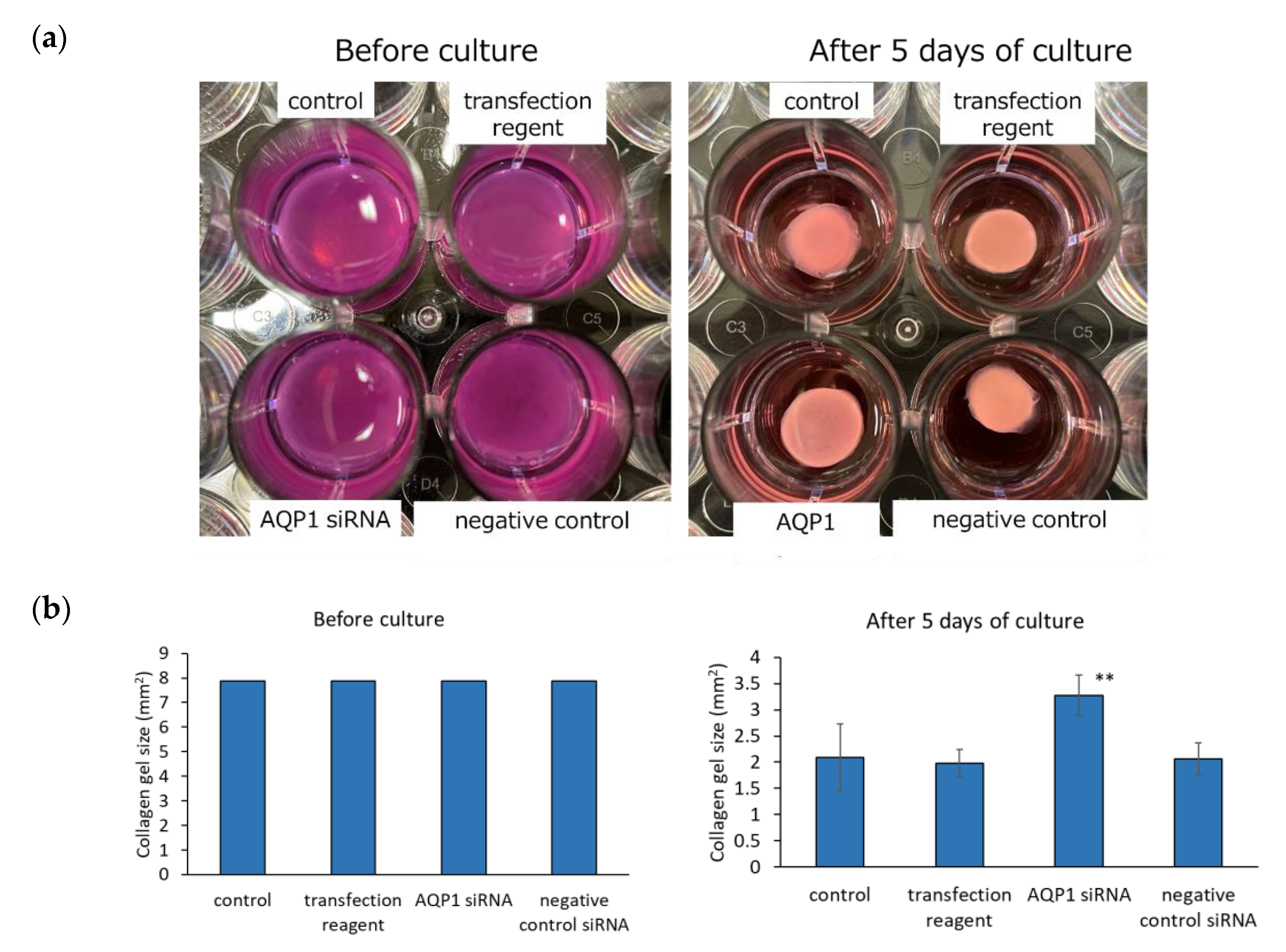
2.3. Observation of the Collagen Gel Contraction of Human Dermal Fibroblasts in Which AQP1 Was Depleted by AQP1 siRNA

Fibroblasts cultured in untreated medium (control), medium with the transfection reagent, or medium with AQP1 siRNA were collected by 0.05% trypsin-EDTA treatment. The collected cell suspension and 10% FBS-DMEM were added at a ratio of 1:1 to a centrifuge tube and centrifuged at 1100 rpm for 5 min, and the cells were collected by decanting. To a 24-well plate on ice, 100 μL of 5 × MEM, 5 μL of 100 × Gluta MAX, 25 μL of FBS, 350 μL of 3 mg/mL of Native collagen (Koken Co., Ltd., Tokyo, Japan), 10 μL of 1 mol/L of NaHCO3, and 20 μL of cultured fibroblasts (2 million cells/mL) were added. The plate was placed in an incubator (37 °C, 5% CO2) for 1 h. The gel attached to the wall of the plate was detached with a syringe needle, and 500 μL of one of the three types of media was added to each well, and the plate was incubated in an incubator (37 °C, 5% CO2) for 5 days. Relative cell counts were obtained using a Cell Counting Kit-8 (Dojindo Chemical Research Institute, Kumamoto, Japan). Cell Counting Kit-8 solution was added and the absorbance of the water-soluble formazan dye after 0 and 2 h was measured at 450 nm using a multi-detection microplate reader (Multi-Detection Microplate POWERSAN HT; BioTek, Winooski, VT, USA). Cell viability was expressed as the difference between the two absorbance values.

2.4. Measurement of AQP1, Phosphorylated Myosin Light Chain 2, F-Actin, abLIM1, Fibrillin 1, and Type I Collagen Staining after AQP1 Depletion by AQP1 siRNA

Cultured human dermal fibroblasts were subjected to depletion for 3 days in Matsunami glass-bottom dishes, as described in Section 2.1. After removing the medium from the depleted cells and two washes with sterile PBS, 1500 μL of 4% paraformaldehyde-phosphate buffer was added, and the cells were fixed for 15 min at 4 °C. After two washes with sterile PBS, the cells were blocked by treatment with 1 mL of 10% goat serum-PBS for 20 min at room temperature. After two washes with sterile PBS, anti-AQP1 antibody (200-fold dilution; Proteintech Group, Inc., Rosemont, IL, USA), anti-phospho-myosin light chain 2 (Ser19) antibody (50-fold dilution; Cell Signaling Technology, Inc., Danvers, MA, USA), phalloidin-iFluor 555 conjugate (1000-fold dilution; Cayman Chemical Company, Ann Arbor, MI, USA), anti-abLIM1 antibody (100-fold dilution; Proteintech Group, Inc.), anti-fibrillin 1 antibody (200-fold dilution; Millipore Sigma, Burlington, MA, USA), and anti-type I collagen antibody (500-fold dilution; Rockland Immunochemicals, Inc., Limerick, PA, USA) were added to the glass sections and allowed to stand for 1.5 h at room temperature. After washing twice with sterile PBS, Alexa Fluor 647 goat anti-rabbit IgG (H + L) (Thermo Fisher Scientific Inc.) diluted 1000-fold, Alexa Fluor 488 goat anti-rabbit IgG (H + L) (Thermo Fisher Scientific Inc.), Alexa Fluor 488 goat anti-mouse IgG (H + L) (Thermo Fisher Scientific Inc.) diluted 1000-fold, and Alexa Fluor 568 goat anti-rabbit IgG (H + L) (Thermo Fisher Scientific Inc.) diluted 1000-fold were added to the glass sections as secondary antibodies and allowed to stand for 40 min at room temperature. The samples were washed twice with sterile PBS, and 0.2% DAPI (Dojindo Chemical Research Institute)-PBS was added to the glass section and then allowed to stand for 10 min at room temperature. After two washes with sterile PBS, 2 mL of sterile PBS was added, and images were taken by confocal laser scanning microscopy. The captured images were analyzed using ImageJ (National Institutes of Health, Bethesda, MD, USA).

2.5. Measurement of the mRNA Levels of AQP1, Fibrillin 1, and Type I Collagen after AQP1 Depletion by AQP1 siRNA

Collagen contraction was performed in 6-well plates for 2 days using cultured human dermal fibroblasts in which AQP1 was depleted by AQP1 siRNA, as described in Section 2.1. A QIA shredder (50) (QIAGEN N.V., Venlo, Netherlands) and RNeasy® Protect Mini Kit (50) (QIAGEN N.V., Venlo, Netherlands) were used for the extraction of RNA from the contracted collagen gel. The extracted RNA was added to 96-well plates, and RT–qPCR reagent (Takara Bio Inc., Shiga, Japan) was added. mRNA quantification was performed with QuantStudio 5 (Thermo Fisher Scientific Inc.) using the relative calibration curve method. AQP1 (QIAGEN N.V.), COL1A2 (QIAGEN N.V.), and FBN1 (QIAGEN N.V.) were used as primers, and ACTB (Thermo Fisher Scientific Inc.) and GAPDH (Thermo Fisher Scientific Inc.) as housekeeping genes.

2.6. Western Blotting of AQP1, Phosphorylated Myosin Light Chain 2, abLIM1, Fibrillin 1, and Type I Collagen after AQP1 Depletion by AQP1 siRNA

After culturing, 300 µL of SDS sample buffer (62.5 mM Tris-HCl (pH 6.8), 2% w/v SDS, 10% glycerol, 50 mM DTT, and 0.01% w/v bromophenol blue) was added and heated at 95 °C for 5 min. Two 4–20% SDS–PAGE plates were applied and electrophoresed at 54 mA for 1.5 h. Transfer was performed at 125 mA using PVDF membranes (Millipore Sigma) by the wet method. The membranes were washed 3 times for 5 min, incubated in blocking buffer (Tris-buffered saline (TBS) with 0.1% Tween-20 (TBS-T) and 5% w/v nonfat dry milk) for 30 min and incubated with primary antibody for 1 h. The primary antibodies used were: anti-AQP1 antibody (1000-fold dilution in TBS-T), anti-phosphomyosin light chain 2 (Ser19) antibody (1000-fold dilution in TBS-T), anti-abLIM1 antibody (1000-fold dilution in TBS-T; Proteintech Group), anti-fibrillin 1 antibody (500-fold dilution in TBS-T; GeneTex, Irvine, CA, USA), anti-type I collagen antibody (2000-fold dilution in TBS-T; Proteintech Group, Inc.), and anti-β-actin mouse antibody (10,000-fold dilution in TBS-T; Proteintech Group, Inc.). After washing with TBS-T, the membranes were incubated with anti-rabbit IgG, HRP, or anti-mouse IgG, HRP (5000-fold dilution; Proteintech Group, Inc.), incubated in TBS-T for 30 min, washed 4 times (2 h) in TBS-T, and immersed in Western Blot Hyper HRP Substrate solution (Takara Biotechnology, Inc., Siga, Japan), and images were recorded by LuminoGraph I (ATTO Corporation, Tokyo, Japan).

2.7. Statistical Analysis

The numerical data were recorded in Excel (Microsoft, Redmond, WA, USA) and the means and standard deviations were calculated. The data shown in the figures were subjected to two-way ANOVA, conducted using BellCurve Excel Statistics (Social Survey Research Information Co., Ltd., Tokyo, Japan). Tukey’s test was used for multiple comparisons, and p < 0.05 was considered to indicate statistical significance.

3. Results

3.1. Efficiency of AQP1 Knockdown

Figure 1a,b show the AQP1 mRNA and protein levels in human dermal fibroblasts cultured in untreated medium (control), medium with the transfection reagent, medium with AQP1 siRNA, or medium with the negative control siRNA for 3 days. The human dermal fibroblasts cultured with AQP1 siRNA showed lower amounts of AQP1 mRNA and its protein than those cultured with the control medium, the cells with the transfection reagent, and the cells cultured with the negative control siRNA. The efficiency of AQP1 knockdown was approximately 80% or higher.
Figure 1c shows the results from the measurement of the amount of AQP1 in the cells after culturing with anti-AQP1 antibody using confocal laser scanning microscopy. Figure 1d shows the image analysis of the amount of immunoreactivity per cell using ImageJ. Human dermal fibroblasts were cultured in control medium, medium with the transfection reagent, or medium with AQP1 siRNA for 3 days. The human dermal fibroblasts cultured with AQP1 siRNA showed lower amounts of AQP1 in the pericellular space than the control cells or the human dermal fibroblasts cultured with the transfection reagent.

3.2. Cell Motility Activity of Human Dermal Fibroblasts in Which AQP1 Was Depleted by AQP1 siRNA

Figure 2 shows the results from measuring the migration distance of human fibroblasts when cultured in untreated medium (control), medium with the transfection reagent, medium with AQP1 siRNA, or medium with the negative control siRNA. The results obtained after incubation for 3, 6, 9, and 12 h showed that human dermal fibroblasts cultured with AQP1 siRNA had a shorter migration distance and reduced motility activity compared with those of the cells cultured under the control, the transfection reagent condition, and the negative control siRNA.

3.3. Collagen Gel Contractive Activity of Human Dermal Fibroblasts in Which AQP1 Was Depleted by AQP1 siRNA

Human dermal fibroblasts cultured in untreated medium (control), medium with the transfection reagent, or medium with AQP1 siRNA were seeded in collagen gels and cultured for 5 days. Images of the collagen gel are shown in Figure 3a. The degree of collagen gel contraction by human dermal fibroblasts was lower in the medium with AQP1 siRNA than in the control medium, the medium with the transfection reagent, and in the medium with the negative control siRNA. Figure 3b shows the collagen gel area before and after 5 days of culture. Collagen gel contraction was inhibited in the AQP1 siRNA-treated cells compared with the cells cultured in the control medium, the medium with the transfection reagent, or the medium with the negative control siRNA. Significant differences were observed between AQP1 siRNA-treated cells and transfection reagent-treated cells. The relative cell counts after 5 days of culture, as measured by the absorbance at 450 nm using a cell counting kit-8, showed no significant difference among the control cells, the cells cultured with the transfection reagent, the cells cultured with AQP1 siRNA, and the cells cultured with negative control siRNA (Figure 3c).

3.4. Amount of Staining of Phosphorylated Myosin Light Chain 2 and F-Actin in Human Dermal Fibroblasts in Which AQP1 Was Depleted by AQP1 siRNA

Human dermal fibroblasts were cultured in untreated medium (control), medium with the transfection reagent, or medium with AQP1 siRNA for 3 days. Human dermal fibroblasts cultured with AQP1 siRNA showed lower amounts of both phosphorylated myosin light chain 2 and F-actin than those cultured in the control medium, those cultured in medium with the transfection reagent, or those cultured in the negative control siRNA (Figure 4a). Figure 4b shows the images of cultured human dermal fibroblasts stained with the anti-phosphorylated myosin light chain 2 antibody and phalloidin-iFluor 555 conjugate obtained by confocal laser scanning microscopy. Cultured human skin fibroblasts transfected with AQP1 siRNA showed reduced levels of both phosphorylated myosin light chain 2 and F-actin. Furthermore, the amount of antibody staining per cell was determined using ImageJ, and the levels of both phosphorylated myosin light chain 2 and F-actin were significantly reduced in the cells treated with AQP1 siRNA compared with those cultured in the medium with the transfection reagent (Figure 4c).

3.5. Antibody Staining of abLIM1 and F-Actin in Human Dermal Fibroblasts in Which AQP1 Was Depleted by AQP1 siRNA

Human dermal fibroblasts were cultured in untreated medium (control), medium with the transfection reagent, or medium with AQP1 siRNA for 3 days. Human dermal fibroblasts cultured with AQP1 siRNA showed lower amounts of abLIM1 than the control cells, those cultured with the transfection reagent, or those cultured with the negative control siRNA (Figure 5a).
Cultured human dermal fibroblasts stained with the anti-abLIM1 antibody and phalloidin-iFluor 555 conjugate were observed by confocal laser scanning microscopy. Both abLIM1 and F-actin were stained at the same location in the cells cultured in medium with AQP1 siRNA. Cells transfected with AQP1 siRNA showed reduced levels of both abLIM1 and F-actin (Figure 5b). Furthermore, an analysis using ImageJ showed that the amount of antibody staining for both abLIM1 and F-actin per number of cells was significantly reduced in the cells treated with AQP1 siRNA compared with those cultured in the medium with the transfection reagent (Figure 5c).

3.6. Amount of Antibody Staining of Fibrillin 1 and Type I Collagen of Human Dermal Fibroblasts in Which AQP1 Was Depleted by AQP1 siRNA

Human dermal fibroblasts were cultured in untreated medium (control), medium with the transfection reagent, or medium with AQP1 siRNA for 3 days. Human dermal fibroblasts cultured with AQP1 siRNA showed lower amounts of both fibrillin 1 and type I collagen than those cultured in the control medium, those cultured in medium with the transfection reagent, or those cultured in the negative control siRNA (Figure 6a). Confocal laser scanning microscopy of cultured human dermal fibroblasts after 3 days of AQP1 depletion showed that both fibrillin 1 and type I collagen were reduced in the AQP1-depleted samples (Figure 6b). Furthermore, using ImageJ, the amount of antibody staining per number of cells was found to be significantly reduced in the AQP1-depleted cells compared with those cultured in the medium with the transfection reagent (Figure 6c).

3.7. mRNA Levels of Fibrillin 1 and Type I Collagen in Human Dermal Fibroblasts Depleted of AQP1 by AQP1 siRNA

RNA extracted from collagen gels containing cultured human dermal fibroblasts in which AQP1 was depleted was analyzed by real-time PCR. AQP1 siRNA significantly decreased the amount of AQP1 mRNA, indicating that the depletion was properly performed.
We also found that the mRNA levels of fibrillin 1 and type I collagen were significantly decreased in cultured human dermal fibroblasts in which AQP1 was depleted (Figure 7).

4. Discussion

In skin, mice deficient in AQP3, which transports both water and glycerol, exhibit dry skin [19,20], and the mRNA expression levels of AQP1, AQP3, AQP4, AQP7, and AQP9 in skin are significantly decreased in aging mice [21]. AQP1 is also highly expressed in dermal fibroblasts of systemic sclerosis lesional skin, and a positive correlation exists between the degree of AQP1 expression and the degree of tissue fibrosis [22]. It has been reported that AQP1 mRNA expression is decreased by oxidative stress and UVB irradiation in human dermal fibroblasts [23,24]. However, there are no reports on what happens to normal dermal fibroblasts after AQP1 is depleted. In this study, AQP1 was found to be highly expressed in human dermal fibroblasts, suggesting that the water permeability of the cell membrane promoted by AQP1 is involved in cell movement. Previous studies have suggested that AQP1 is located at the front end of migrating cells and promotes migration by facilitating turnover of cell membrane protrusions (lamellipodia) [25]. AQP1 was universally present in the cell periphery in this experiment. In addition, the depletion of AQP1 by AQP1 siRNA reduced AQP1 in the pericellular space. Since the depletion of AQP1 suppressed cell motility, the turnover of lamellipodia in the pericellular area and the influx of water into the cytoplasmic space by AQP1 are thought to be involved in cell motility.
It has also been reported that depletion of abLIM1 decreases the number of stress fibers in NIH3T3 cells, whereas its overexpression increases intracellular F-actin [18]. The depletion of AQP1 by AQP1 siRNA in cultured human dermal fibroblasts results in reduced motility activity. AbLIM1 co-occurs with actin filaments and promotes actin filament formation. In cultured human dermal fibroblasts grown in medium supplemented with AQP1 siRNA, the staining of anti-abLIM1 antibodies and actin filaments was decreased, suggesting that the depletion of AQP1 reduced the function of abLIM1 and the formation of actin filaments, resulting in the suppression of cell motility. In vitro assays showed that abLIM1 cross-links and binds F-actin, inducing the formation of dense F-actin networks [14]. The involvement of abLIM1 in the formation of the F-actin network has been previously suggested [14].
The organization of the cytoskeleton requires the presence of membrane-associated proteins, including the PDZ domain, which is involved in scaffolding [26]. Proteins containing PDZ domains play an important role in anchoring receptor proteins at the membrane to cytoskeletal components. For example, Lin-7, a PDZ protein, forms a complex with CASK/Lin-2, which interacts with cytoskeletal proteins to retain and organize signaling complexes at the cell membrane and is important for maintaining and organizing signaling complexes at the cell membrane [27]. The knockdown of AQP1 markedly affects the organization of the actin cytoskeleton through Lin-7/β-catenin interactions [28]. The loss of AQP1 leads to the destabilization of F-actin reorganization and has been shown to be associated with the formation of a new cellular cytoskeleton [29], which reportedly results in reduced cell motility and invasion in melanoma and human endothelial cell lines [28]. Our Gene Chip analysis of human dermal fibroblasts cultured on aminocarbonylated collagen revealed a decrease in LIN7B and AQP1 mRNA, but not LIN7A or CASK/LIN2, compared with the levels in human dermal fibroblasts cultured on non-aminocarbonylated collagen. The expression level of LIN7B was low, and whether the depletion of AQP1 in human dermal fibroblasts markedly affects the organization of the actin cytoskeleton through the Lin-7B/β-catenin interaction remains unclear. Furthermore, no effect of aminocarbonylation was observed on ITGA2 and ITGB1, which are molecules that adhere to collagen.
When human dermal fibroblasts are cultured in reconstituted collagen gel, the collagen gel contracts and becomes flexible. The contracted collagen gel exhibits increased flexibility, elasticity, and strength. It has been reported that increased phosphorylation of myosin light chain 2 in fibroblasts increases the collagen gel contractile activity [7]. Since AQP1 depletion did not affect the number of cells after five days of culture, it appears that AQP1 depletion, not the cell growth rate, is dependent on the cell’s ability to contract collagen. Cultured human dermal fibroblasts grown in medium supplemented with AQP1 siRNA showed decreased levels of anti-phosphorylated myosin light chain 2 antibody-positive protein and F-actin, suggesting that the depletion of AQP1 reduces the amount of F-actin and the formation of phosphorylated myosin light chain, resulting in suppression of the collagen gel contractive activity of fibroblasts.
Wrinkles and sagging are some of the most common skin problems associated with aging and are caused by degeneration of the extracellular matrix, which makes up most of the dermis [30]. This extracellular matrix contains elastic fibers and collagen fibers. Fibrillin 1 is one of the components of elastic fibers in the dermis and exhibits a similar distribution to that of elastic fibers but is particularly expressed in oxytalan fibers and micro-fibrils (fine fibers approximately 10 nm in diameter) oriented from the epidermal basement membrane to the lower dermis. Ultraviolet (UV) B (UVB) and UVA-II reduce the antioxidant function, inducing the formation of aging cross-links in collagen and the aminocarbonylation of proteins [9]. UV-absorbing molecules (chromophores) present in the skin are involved in the formation of UV-induced aging cross-links. For example, UV radiation absorbed by the collagen protein tyrosine forms a photoaging cross-link called di-tyrosine, which stiffens collagen [31,32] and facilitates wrinkle formation. The half-life of collagen turnover in skin is as long as 14.8 years [33], and when collagen fibers are damaged by UV light, the damaged collagen fibers remain in the dermis for a long time. Since collagen is a protein with a slow metabolic turnover, long-term exposure to sunlight causes accumulation of damage to collagen, resulting in a loss of elasticity. In contrast, aminocarbonylation modifies proteins with aldehydes produced by oxidative reactions of lipids and sugars to produce aminocarbonylated proteins. Aminocarbonylation is caused by external influences such as ultraviolet radiation, the decrease in antioxidants in the skin caused by aging, the formation of lipid peroxides from sebum, and oxidative reactions of sugars. Aminocarbonylation of the dermis causes a decrease in transparency, and aminocarbonylation of the basement membrane affects epidermal keratinization and reduces the moisture retention and barrier function of the stratum corneum [9].
Cultured human dermal fibroblasts depleted of AQP1 by the addition of AQP1 siRNA show decreased mRNA and protein levels of type I collagen and fibrillin 1, which suggests that AQP1 depletion causes decreases in the production of type I collagen and fibrillin 1 in cultured human dermal fibroblasts. In fibroblasts, water molecules are essential for activities such as the production of the intercellular matrix.

5. Conclusions

AQP1, which plays a role in the cellular uptake of water molecules, decreases in expression with aging. It is also known that cell motility and the expression of extracellular matrix proteins decrease with aging. In this study, we provided the first demonstration that AQP1 is a key biomolecule for cell motility and the expression of extracellular matrix proteins. Increases in AQP1, the expression of which declines with aging, are expected to improve the motility of dermal fibroblasts, increase the levels of type 1 collagen and fibrillin 1, and improve wrinkles and sagging, which increase with aging.

Author Contributions

Conceptualization, K.M.; methodology, K.M.; investigation, S.Y. and K.M.; data curation, S.Y.; writing—original draft preparation, S.Y.; writing—review and editing, K.M.; visualization, S.Y.; supervision, K.M. All authors have read and agreed to the published version of the manuscript.

Funding

This research received no external funding.

Institutional Review Board Statement

Not applicable.

Informed Consent Statement

Not applicable.

Data Availability Statement

Not applicable.

Conflicts of Interest

The authors declare no conflict of interest.

References

  1. Maeda, K.; Koizumi, S.; Sano, T.; Yoshimura, A.; Kiriyama, H.; Mimura, S. Utility of the Teslar facial massager for skin elasticity and the mechanism of its effects. Cosmetics 2018, 5, 49. [Google Scholar] [CrossRef] [Green Version]
  2. Ricard-Blum, S.; Ruggiero, F. The collagen superfamily: From the extracellular matrix to the cell membrane. Pathol. Biol. 2005, 53, 430–442. [Google Scholar] [CrossRef] [PubMed]
  3. Godwin, A.R.F.; Singh, M.; Lockhart-Cairns, M.P.; Alanazi, Y.F.; Cain, S.A.; Baldock, C. The role of fibrillin and microfibril binding proteins in elastin and elastic fibre assembly. Matrix Biol. 2019, 84, 17–30. [Google Scholar] [CrossRef] [PubMed]
  4. Watson, R.E.; Griffiths, C.E.; Craven, N.M.; Shuttleworth, C.A.; Kielty, C.M. Fibrillin-rich microfibrils are reduced in photoaged skin. Distribution at the dermal-epidermal junction. J. Investig. Dermatol. 1999, 112, 782–787. [Google Scholar] [CrossRef]
  5. Bell, E.; Sher, S.; Hull, B.; Merrill, C.; Rosen, S.; Chamson, A.; Asselineau, D.; Dubertret, L.; Coulomb, B.; Lapiere, C.; et al. The reconstitution of living skin. J. Investig. Dermatol. 1983, 81 (Suppl. 1), 2s–10s. [Google Scholar] [CrossRef] [Green Version]
  6. Yamato, M.; Yamamoto, K.; Hayashi, T. Age-related changes in collagen gel contraction by cultured human lung fibroblasts resulting in cross-over of contraction curves between young and aged cells. Mech. Ageing Dev. 1993, 67, 149–158. [Google Scholar] [CrossRef]
  7. Takayama, Y.; Mizumachi, K. Effects of lactoferrin on collagen gel contractile activity and myosin light chain phosphorylation in human fibroblasts. FEBS Lett. 2001, 508, 111–116. [Google Scholar] [CrossRef] [Green Version]
  8. Pageon, H.; Asselineau, D. An in vitro approach to the chronological aging of skin by glycation of the collagen: The biological effect of glycation on the reconstructed skin model. Ann. N. Y. Acad. Sci. 2005, 1043, 529–532. [Google Scholar] [CrossRef]
  9. Morimoto, H.; Gu, L.; Zeng, H.; Maeda, K. Amino Carbonylation of epidermal basement membrane inhibits epidermal cell function and is suppressed by methylparaben. Cosmetics 2017, 4, 38. [Google Scholar] [CrossRef] [Green Version]
  10. Ogura, Y.; Kuwahara, T.; Akiyama, M.; Tajima, S.; Hattori, K.; Okamoto, K.; Okawa, S.; Yamada, Y.; Tagami, H.; Takahashi, M.; et al. Dermal carbonyl modification is related to the yellowish color change of photo-aged Japanese facial skin. J. Dermatol. Sci. 2011, 64, 45–52. [Google Scholar] [CrossRef]
  11. Verkman, A.S.; Ruiz-Ederra, J.; Levin, M.H. Functions of aquaporins in the eye. Prog. Retin. Eye Res. 2008, 27, 420–433. [Google Scholar] [CrossRef] [PubMed] [Green Version]
  12. Agre, P.; King, L.S.; Yasui, M.; Guggino, W.B.; Ottersen, O.P.; Fujiyoshi, Y.; Engel, A.; Nielsen, S. Aquaporin water channels--from atomic structure to clinical medicine. J. Physiol. 2002, 542 Pt. 1, 3–16. [Google Scholar] [CrossRef]
  13. Ishibashi, K.; Hara, S.; Kondo, S. Aquaporin water channels in mammals. Clin. Exp. Nephrol. 2009, 13, 107–117. [Google Scholar] [CrossRef] [PubMed]
  14. Li, G.; Huang, S.; Yang, S.; Wang, J.; Cao, J.; Czajkowsky, D.M.; Shao, Z.; Zhu, X. abLIM1 constructs non-erythroid cortical actin networks to prevent mechanical tension-induced blebbing. Cell Discov. 2018, 4, 42. [Google Scholar] [CrossRef] [PubMed] [Green Version]
  15. Roof, D.J.; Hayes, A.; Adamian, M.; Chishti, A.H.; Li, T. Molecular characterization of abLIM, a novel actin-binding and double zinc finger protein. J. Cell Biol. 1997, 138, 575–588. [Google Scholar] [CrossRef]
  16. Barrientos, T.; Frank, D.; Kuwahara, K.; Bezprozvannaya, S.; Pipes, G.C.; Bassel-Duby, R.; Richardson, J.A.; Katus, H.A.; Olson, E.N.; Frey, N. Two novel members of the ABLIM protein family, ABLIM-2 and -3, associate with STARS and directly bind F-actin. J. Biol. Chem. 2007, 282, 8393–8403. [Google Scholar] [CrossRef] [Green Version]
  17. Frank, D.; Frey, N. Cardiac Z-disc signaling network. J. Biol. Chem. 2011, 286, 9897–9904. [Google Scholar] [CrossRef] [Green Version]
  18. Schneider, P.; Bayo-Fina, J.M.; Singh, R.; Kumar Dhanyamraju, P.; Holz, P.; Baier, A.; Fendrich, V.; Ramaswamy, A.; Baumeister, S.; Martinez, E.D.; et al. Identification of a novel actin-dependent signal transducing module allows for the targeted degradation of GLI1. Nat. Commun. 2015, 6, 8023. [Google Scholar] [CrossRef] [Green Version]
  19. Ma, T.; Hara, M.; Sougrat, R.; Verbavatz, J.M.; Verkman, A.S. Impaired stratum corneum hydration in mice lacking epidermal water channel aquaporin-3. J. Biol. Chem. 2002, 277, 17147–17153. [Google Scholar] [CrossRef] [Green Version]
  20. Hara, M.; Ma, T.; Verkman, A.S. Selectively reduced glycerol in skin of aquaporin-3-deficient mice may account for impaired skin hydration, elasticity, and barrier recovery. J. Biol. Chem. 2002, 277, 46616–46621. [Google Scholar] [CrossRef]
  21. Ikarashi, N.; Kon, R.; Kaneko, M.; Mizukami, N.; Kusunoki, Y.; Sugiyama, K. Relationship between aging-related skin dryness and aquaporins. Int. J. Mol. Sci. 2017, 18, 1559. [Google Scholar] [CrossRef] [PubMed] [Green Version]
  22. Yamashita, T.; Asano, Y.; Saigusa, R.; Taniguchi, T.; Nakamura, K.; Miura, S.; Toyama, T.; Takahashi, T.; Ichimura, Y.; Hirabayashi, M.; et al. Increased expression of aquaporin-1 in dermal fibroblasts and dermal microvascular endothelial cells possibly contributes to skin fibrosis and edema in patients with systemic sclerosis. J. Dermatol. Sci. 2019, 93, 24–32. [Google Scholar] [CrossRef] [Green Version]
  23. Fujinami, K.; Dan, K.; Tanaka-Kagawa, T.; Kawamura, I. Anti-aging effects of polyoxometalates on skin. Appl. Sci. 2021, 11, 11948. [Google Scholar] [CrossRef]
  24. Kim, W.O.; Kim, S.A.; Jung, Y.A.; Suh, S.I.; Ryoo, Y.W. Ultraviolet B downregulated aquaporin 1 expression via the MEK/ERK pathway in the dermal fibroblasts. Ann. Dermatol. 2020, 32, 213–222. [Google Scholar] [CrossRef] [PubMed]
  25. Verkman, A.S. Mammalian aquaporins: Diverse physiological roles and potential clinical significance. Expert. Rev. Mol. Med. 2008, 10, e13. [Google Scholar] [CrossRef] [Green Version]
  26. Craven, S.E.; Bredt, D.S. PDZ proteins organize synaptic signaling pathways. Cell 1998, 93, 495–498. [Google Scholar] [CrossRef] [Green Version]
  27. Lee, H.J.; Zheng, J.J. PDZ domains and their binding partners: Structure, specificity, and modification. Cell Commun. Signal. 2010, 8, 8. [Google Scholar] [CrossRef] [Green Version]
  28. Monzani, E.; Bazzotti, R.; Perego, C.; La Porta, C.A. AQP1 is not only a water channel: It contributes to cell migration through Lin7/beta-catenin. PLoS ONE 2009, 4, e6167. [Google Scholar] [CrossRef] [Green Version]
  29. Ponting, C.P. Evidence for PDZ domains in bacteria, yeast, and plants. Protein Sci. 1997, 6, 464–468. [Google Scholar] [CrossRef] [Green Version]
  30. Rittié, L.; Fisher, G.J. Natural and sun-induced aging of human skin. Cold Spring Harb. Perspect. Med. 2015, 5, a015370. [Google Scholar] [CrossRef]
  31. Maeda, K. Analysis of ultraviolet radiation wavelengths causing hardening and reduced elasticity of collagen gels in vitro. Cosmetics 2018, 5, 14. [Google Scholar] [CrossRef] [Green Version]
  32. Waykole, P.; Heidemann, E. Dityrosine in collagen. Connect. Tissue Res. 1976, 4, 219–222. [Google Scholar] [CrossRef] [PubMed]
  33. Verzijl, N.; DeGroot, J.; Thorpe, S.R.; Bank, R.A.; Shaw, J.N.; Lyons, T.J.; Bijlsma, J.W.; Lafeber, F.P.; Baynes, J.W.; TeKoppele, J.M. Effect of collagen turnover on the accumulation of advanced glycation end products. J. Biol. Chem. 2000, 275, 39027–39031. [Google Scholar] [CrossRef] [PubMed]
Figure 1. (a) AQP1 mRNA expression levels in cultured human dermal fibroblasts after AQP1 depletion for 3 days by AQP1 siRNA in cultured human dermal fibroblasts. (b)Western blotting for AQP1 in cultured human dermal fibroblasts following AQP1 depletion by AQP1 siRNA. (c) Antibody staining for AQP1 in cultured human dermal fibroblasts following AQP1 depletion by AQP1 siRNA. (d) Image analysis of the amount of immunoreactivity per cell using ImageJ. ***: p < 0.001 vs. the transfection reagent. **: p < 0.01 vs. the transfection reagent. The figure shows the means ± standard deviations (n = 3).
Cosmetics 09 00117 g001aCosmetics 09 00117 g001b
Figure 2. Motility activity of human dermal fibroblasts cultured with the addition of AQP1 siRNA. *: p < 0.05 vs. the transfection reagent. **: p < 0.01 vs. the transfection reagent. The figures show the means ± standard deviations (n = 7).
Cosmetics 09 00117 g002aCosmetics 09 00117 g002b
Figure 3. Collagen gel contraction of cultured human dermal fibroblasts. (a) Images of collagen gel before and after 5 days of culture. (b) Area of collagen gel after 5 days of culture. **: p < 0.01 vs. the transfection reagent. The figures show the means ± standard deviations (n = 3). (c) Relative cell count after 5 days of culture.
Cosmetics 09 00117 g003aCosmetics 09 00117 g003b
Figure 4. (a) Western blotting for phosphorylated myosin light chain 2 in cultured human dermal fibroblasts following AQP1 depletion by AQP1 siRNA. (b) Antibody staining for phosphorylated myosin light chain 2 and F-actin in cultured human dermal fibroblasts following AQP1 depletion by AQP1 siRNA. (c) Image analysis of the amount of immunoreactivity per cell using ImageJ. *: p < 0.05 vs. the transfection reagent. The figure shows the means ± standard deviations (n = 3).
Cosmetics 09 00117 g004aCosmetics 09 00117 g004b
Figure 5. (a) Western blotting for abLIM1 in cultured human dermal fibroblasts following AQP1 depletion by AQP1 siRNA. (b) Antibody staining for the abLIM1 chain and F-actin in cultured human dermal fibroblasts after AQP1 depletion by AQP siRNA. (c) Image analysis of the amount of immunoreactivity per cell using ImageJ. *: p < 0.05 vs. the transfection reagent. **: p < 0.01 vs. the transfection reagent. The figure shows the means ± standard deviations (n = 3).
Cosmetics 09 00117 g005
Figure 6. (a) Western blotting for fibrillin 1 and type I collagen in cultured human dermal fibroblasts following AQP1 depletion by AQP1 siRNA. (b) Antibody staining for fibrillin 1 and type I collagen in cultured human dermal fibroblasts after AQP1 depletion by AQP1 siRNA. (c) Image analysis of the amount of immunoreactivity per cell using ImageJ. *: p < 0.05 vs. the transfection reagent. The figure shows the means ± standard deviations (n = 3).
Cosmetics 09 00117 g006aCosmetics 09 00117 g006b
Figure 7. mRNA expression levels in cultured human dermal fibroblasts after AQP1 depletion for 2 days by AQP1 siRNA. ***: p < 0.001 vs. the transfection reagent. The figure shows the means ± standard deviations (n = 3).
Cosmetics 09 00117 g007
Publisher’s Note: MDPI stays neutral with regard to jurisdictional claims in published maps and institutional affiliations.

