Article

Rosmarinic Acid Multifunctional Sunscreen: Comet Assay and In Vivo Establishment of Cutaneous Attributes

by
Thalita Marcílio Cândido
1,
Maíra Bueno Ariede
1,
Claudinéia Aparecida Sales de Oliveira Pinto
1,
Fabiana Vieira Lima
2,
Wagner Vidal Magalhães
3,
Natália Mencacci Esteves Pedro
3,
Giovana Padovani
3,
Bianca da Silva Sufi
3,
Patrícia Rijo
4,
Maria Valéria Robles Velasco
1,
Catarina Rosado
4,† and
André Rolim Baby
1,*,†
1
Department of Pharmacy, Faculty of Pharmaceutical Sciences, University of São Paulo, São Paulo 05508-900, Brazil
2
Division of Pharmacy, Department of Health Science, Federal University of Espírito Santo, São Mateus 29932-540, Brazil
3
Research and Development Department, Chemyunion Ltd., Sorocaba 18087-101, Brazil
4
CBIOS, Universidade Lusófona’s Research Center for Biosciences & Health Technologies, 1749-024 Lisbon, Portugal
*
Author to whom correspondence should be addressed.
These authors contributed equally to this work.
Submission received: 3 October 2022 / Revised: 24 November 2022 / Accepted: 28 November 2022 / Published: 12 December 2022
(This article belongs to the Special Issue Feature Papers in Cosmetics in 2022)

Abstract

:
The skin acts as a protective barrier, guarding the body against microorganisms, chemicals, and several environmental factors. Accordingly, this all-important organ must be kept healthy to maintain its optimal functionality. One approach to maintain skin health is the application of multifunction bioactive sunscreens containing antioxidant molecule(s). Rosmarinic acid (RA), a phenolic compound, is known for its antioxidant activity. Herein, the safety and efficacy of a multifunction prototype sunscreen were investigated, aiming to evaluate the performance of this polyphenol with two known and widely used UV filters (bemotrizinol and octyl p-methoxycinnamate). Samples protected the DNA fragmentation compared to UV control, by the comet assay, and showed good skin compatibility in subjects. Formulations F1 and F3 were able to increase skin hydration, and, possibly, the RA interfered with this attribute. An increase in transepidermal water loss was observed for formulations F1, F2, and F4, which may be related to the vehicle, containing the RA or not. No decreases were observed in the inflammatory reaction caused by the ethyl nicotinate with any of the samples. As a perspective, we suggest trials with a greater number of subjects or protocol modifications. Altering the vehicle qualitative and quantitative composition is also a pertinent perspective.

1. Introduction

It is known that reactive species are naturally originated in biochemical processes such as energy production, inflammation, phagocytosis, and cell growth regulation, among others. External factors are also able to provide the production of species such as exposure to ultraviolet (UV) radiation, ozone, X-rays, and air pollutants [1,2,3,4]. In the skin, free radicals are constantly generated by cutaneous cells such as fibroblasts and keratinocytes. Nevertheless, healthy skin can eradicate these reactive species with nonenzymatic (glutathione, ubiquinol, vitamins C and E) and enzymatic (glutathione peroxidase, glutathione reductase catalase, superoxide dismutase, and thioredoxin reductase) antioxidants [5,6,7].
When the number of free radicals produced in the tissues exceeds the antioxidant defenses, what is known as oxidative stress occurs. As a result, the peroxidation of lipid membranes, damage to DNA, protein, and enzymes appear, and these harms, in turn, worsen in cognitive dysfunction, degenerative cardiovascular diseases, and cancer, among other illnesses [8].
Energetic photons of UV light can be transmitted through the layers of the skin and after that, can be absorbed by cellular chromophores. DNA bases are cellular chromophores; thus, they can directly absorb UV photons, initiating photo-induced reactions. Furthermore, damage in the cells may occur by photosensitization processes, where endogenous or exogenous sensitizers absorb UV photons. Depending on the type, the distribution and concentration of skin chromophores, epidermal thickness, and its functional condition, photobiochemical reactions occur, provoking changes in the cell and tissue biology due to the formation of reactive oxygen species (ROS). The interactions between ROS and cellular biomolecules provoke a final biological response, as illustrated in Figure 1 [5,6,9,10].
Although the formation of these reactive species is inevitable, it is possible, with the administration of endogenous and exogenous antioxidants, to minimize their formation, thus avoiding injurious effects to the organisms. Molecules must perform one or more of the following actions to act as antioxidants: oxygen depletion; single oxygen extinction; chelation of metal ions that would otherwise catalyze reactions to form reactive oxygen species; elimination of reactive oxygen species or termination of the oxidation propagation chain reaction; and the prevention of oxidative damage. Hydroxyls attached to aromatic rings such as in polyphenols can react with free radicals, as shown in Figure 2 [8].
Rosmarinic acid (RA) is a polyphenol whose molecular structure was first elucidated in 1958. Its molecule was first isolated from the leaves of Rosmarinus officinalis L. and later from plants belonging to the families Lamiaceae (e.g., lavender, sage and mint) and Boraginaceae (e.g., cordia and echium). The name “rosmarinic acid” was given precisely since it was first extracted from rosemary (R. officinalis L.). It is an ester derived from caffeic and 3,4-dihydroxyphenylacetic acids. The RA molecule has a carboxylic group, two aromatic rings A and A′ with ortho-catechol structures, the unsaturated C=C bond and the ester portion [11]. Different studies have shown that RA can act biologically in several ways, being an antiviral, antibiotic, anti-inflammatory, and antioxidant [12,13,14,15].
The daily topical application of sunscreens can protect the skin from UV rays and thus delay or reduce the occurrence of skin harm such as the appearance of melanomas, spots, and wrinkles. Considering that antioxidants are capable of helping to minimize the occurrence of ROS that are related to the damages previously mentioned, the association of such molecules to the photoprotective formulations optimizes the beneficial effects of these products over the skin [9,14,15,16,17].
Considering the photoprotective and antioxidant potential of RA, in this research work, the safety and efficacy of a multifunction prototype sunscreen were investigated, aiming to evaluate the performance of this polyphenol with two known and widely used UV filters, bemotrizinol and octyl p-methoxycinnamate.

2. Materials and Methods

2.1. Materials

Rosmarinic acid (96%), agarose ethylenediaminetetraacetic acid disodium salt dihydrate, sodium chloride, Trizma, Triton X-100, dimethyl sulfoxide, phosphate buffer, methyl nicotinic, and sodium hydroxide were purchased from Sigma-Aldrich (St Louis, MO, USA). Octyl p-methoxycinnamate and bemotrizinol were purchased from Mapric (São Paulo, Brazil) and Brasquim (São Paulo, Brazil), respectively. The water was purified in a Milli-Q-plus System (Merck Milipore, USA). Long-lived, spontaneously immortalized human keratinocyte cells (HaCaT) were obtained from the Cell Bank of Rio de Janeiro. DMEM supplemented with 10% fetal bovine serum, trypsin, and SYBR Gold (1:10,000, Invitrogen—Cat S11494) were purchased from Thermo Fisher Scientific (Waltham, MA, USA).

2.2. Methods

2.2.1. Composition of the Formulations

Four formulations containing commercially available octyl p-methoxycinnamate (UVB filter), bemotrizinol (broad spectrum filter), plus 1.0% (w/w) of the bioactive compound rosmarinic acid were used in the study [17]. The active ingredients of the formulations are presented in Table 1.

2.2.2. In Vitro Assay

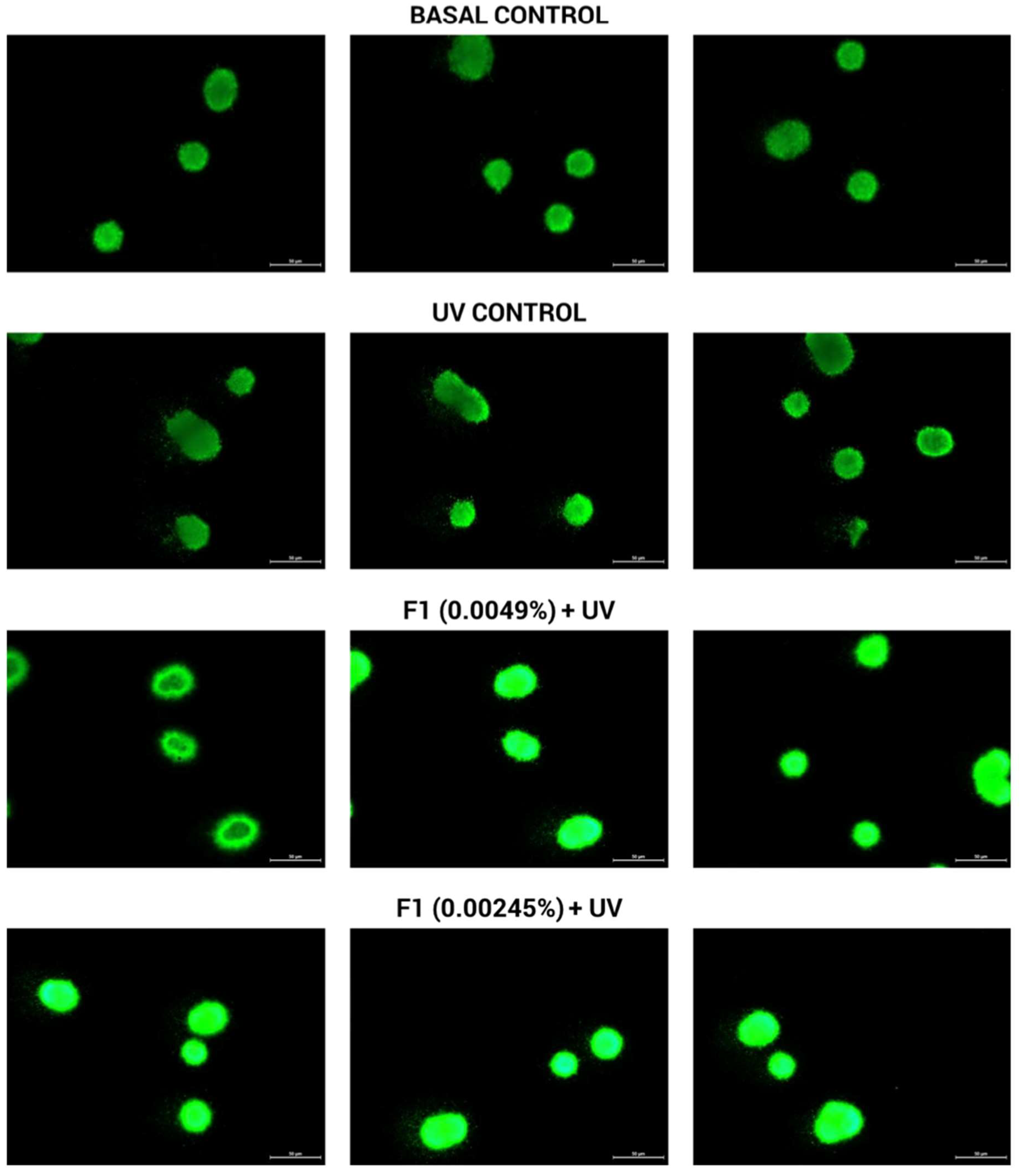
In Vitro Evaluation of DNA Fragmentation of Human Cells Exposed to UV Radiation by the Comet Assay Method

Human Keratinocytes Cell Culture
Human keratinocyte (HaCaT) cells were seeded in 75 cm2 bottles, cultivated, and expanded in a humid atmosphere, in the presence of 5% CO2 at 37 °C using DMEM supplemented with 10% fetal bovine serum. Upon reaching 70% confluence, cells were trypsinized and seeded in 6-well plates for later treatments and the further evaluation of DNA fragmentation.
Incubation with the Formulations and Cells Irradiation
Cells were incubated with the samples at two non-cytotoxic concentrations, 0.0049 and 0.00245% (w/v), for 24 h for subsequent exposure to UV radiation. After the incubation time, cells were submitted to 3 J/cm2 of UVA/UVB in a sun simulation chamber (Suntest CPS+, Atlas, Linsengericht, Germany). Then, cells were kept in suitable culture conditions for an additional 24 h. Cell lysates were collected and proceeded with the comet assay.
Comet Assay
Cell lysates were centrifuged for 5 min at 1500 rpm. Afterward, the cell pellet was mixed with 1000 µL of 0.75% low melting point agarose. Immediately, 100 µL of this suspension was spread onto slides previously coated with 1% agarose. After solidification, the slides were immersed in a freshly prepared lysing solution [2.5 M NaCl, 100 mM ethylenediaminetetraacetic acid disodium salt dihydrate (EDTA Na2S), and 10 mM Trizma Base, pH 10], with the addition of 1% Triton X-100 and 10% dimethyl sulfoxide, for at least 1 h. After lysis, slides were incubated for 5 min in cold PBS (free Ca2+ and Mg2+) and for 40 min in alkaline solution (pH >13). Cells on the slides were submitted to electrophoresis (300 mA, 25 V) for 30 min. Slides were neutralized by three washes with neutralization buffer (pH 7.5) and the DNA was precipitated. DNA was stained with SYBR Gold (1:10,000, Invitrogen—Cat S11494). A number of 150 cells was randomly selected on each slide and analyzed using an optical fluorescence microscope (Leica, DM 6000 B, Wetzlar, Germany) coupled with a camera of 2.8 MP (Leica, DFC7000 T, Wetzlar, Germany).
The comet tail length was classified into four classes (scores): 1 = low damage; 2 = medium damage; 3 = high damage; and 4 = almost all DNA in the tail. The final score of each experimental group was performed by multiplying the number that represented the class of the comet and the total number of comets found in this class. The sum of the results from the multiplications was carried out, corresponding to the final score [18].

2.2.3. In Vivo Assays

Subjects

The protocol was concluded with 12 healthy subjects with phototypes II to VI, after oral information and written consent. The procedures were in accordance with the ethical standards on human experimentation (Ethics Committee of Universidade Lusófona’s Research Center for Biosciences & Health Technologies) and the Declaration of Helsinki. Exclusion criteria were the presence of dermatitis or other skin or allergic disease and smoking. Subjects were instructed not to apply any topical products to the test sites during the study but were allowed to wash normally. The mean age was 32 ± 12 years old (20 to 44 years).

Skin Biocompatibility

Aliquots of the formulations were applied to predefined sites on both forearms (two formulations per forearm of each subject, in a 9.0 cm2 area). The application site of the samples was randomized to minimize the occurrence of possible biases. Measurements were taken before and after 30 min of application with a corneometer (stratum corneum hydration), a Tewameter TM300 (skin barrier quantified through the transepidermal water loss, TEWL), a pH meter (pH value of the skin surface) (C&K Electronics GmbH, Germany), and a chromameter (red color, a *, according to CIE Lab system) (Minolta Camera Co., Osaka, Japan). Afterward, the subjects were instructed to apply the formulations twice a day for 7 days. On the eighth day, new measurements were performed. The non-invasive measurements were conducted according to the specialized literature and guidelines (n = 3) [19,20,21,22]. To reduce the interindividual variability, the results were treated as the ratio between the response and the basal/control value.

Anti-Inflammatory Activity

Aliquots of the samples were applied to the forearm of each volunteer in a 9.0 cm2 area. The study was conducted in two phases: a pre-treatment, which consisted in the application of the samples twice a day for 7 days. On the eighth day, the second phase began, where erythema formation was induced in each pre-treated area by applying filter paper (2.25 cm2) saturated with an aqueous solution of methyl nicotinate (0.5% w/w) for 60 s. Measurements of the skin microcirculation of individual test sites were recorded continuously for 15 min using laser doppler flowmetry equipment (PeriFlux System 5000, Perimed, Stockholm, Sweden). Therefore, the in vivo evaluation of the anti-inflammatory effect was based on the ability of the samples to decrease the extent of an erythema-induced response. The area under the curve of the perfusion profile obtained at each site was measured for all subjects, together with the slope of the tangent line in the hyperemia stage [23]. Results were analyzed as the ratio between the values obtained at each sample site and the control values for the volunteers, in the selected parameters.

2.3. Statistical Analysis

Experiments were conducted randomly, in triplicate, and with a significance level of 5% (p ≤ 0.05), conducted by Minitab software, version 18 (Minitab, Inc., State College, PA, USA).

3. Results

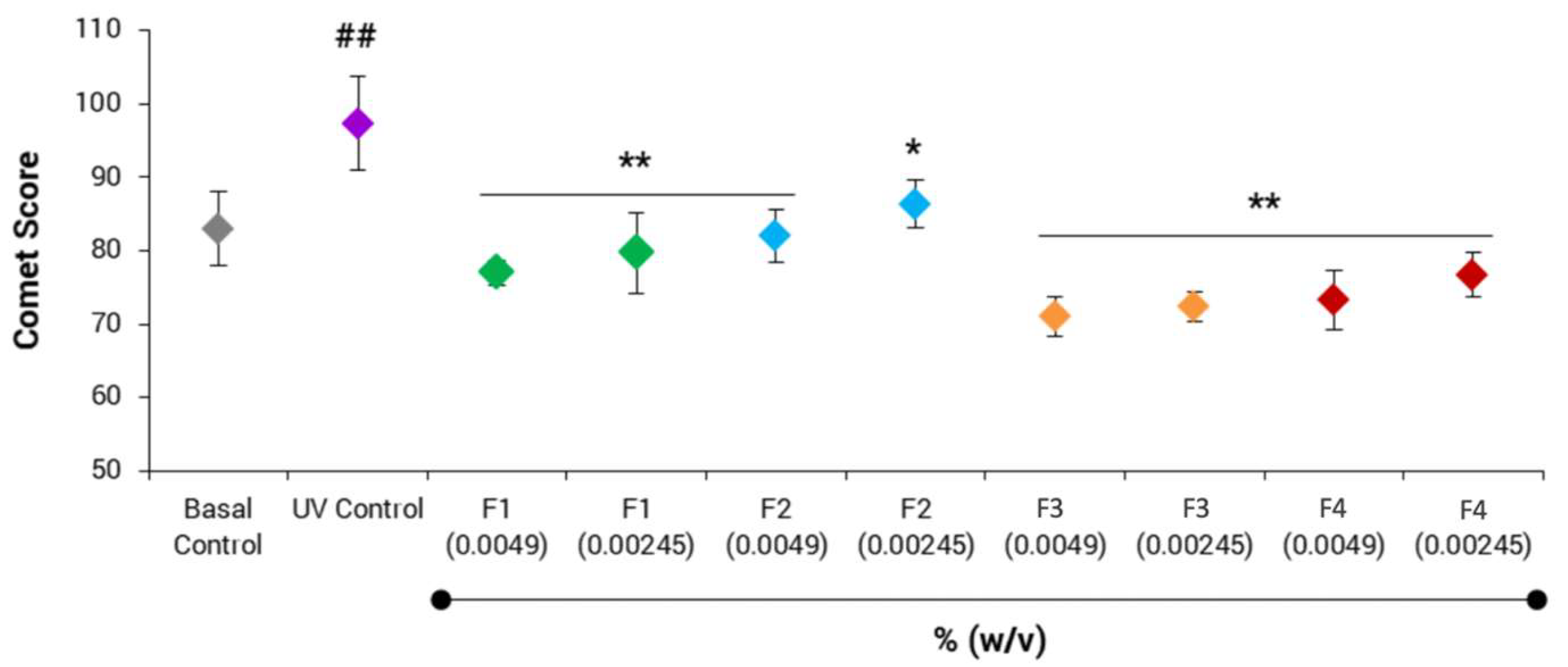
3.1. In Vitro Evaluation of DNA Fragmentation of Human Cells Exposed to Ultraviolet Radiation by the Comet Assay Method

The efficacy of the samples was evaluated in vitro by the comet assay, observing the fragmentation of the treated and untreated human keratinocyte (HaCaT) cells after exposure to UV radiation. Results are shown in Figure 3 and Figure 4.
It was possible to observe the protective behavior of F1, F2, F3, and F4. The UV control induced 17% of DNA fragmentation in comparison to the basal one. All tested samples under exposure to UV radiation in both concentrations promoted a significant decrease in the DNA fragmentation of 21 and 18%, 16 and 11%, 27 and 26%, 25 and 21%, respectively, in comparison to the UV control.

3.2. In Vivo Skin Biocompatibility

To evaluate the skin biocompatibility of the formulations, measurements were performed with a corneometer, Tewameter TM 300, pH meter, and chromameter equipment. The results, as the ratio between the response and basal/control, are shown in Table 2.

3.3. In Vivo Anti-Inflammatory Activity

The anti-inflammatory potential of the formulations consisted in their ability to decrease the extent of an induced erythema response in subjects, being the results shown in Table 3.

4. Discussion

Formulations, at both concentrations, into the cell cultures, under the UV stress condition, protected the DNA fragmentation compared to UV control. Although we noticed the decrease in DNA fragmentation, we could not exclusively attribute this finding to the presence of RA in the samples, since F1 and F3 were absent in this phenolic compound. Possibly, the RA concentration at 1.0% was not completely enough to develop a distinctive performance among the samples, since all of them presented some level of DNA protection ranging from 11 to 27% including the blank sample (F1) [14,24,25]. Additionally, the test could have a limitation when complex samples are performed. Furthermore, our protocol similarly indicated that the blank sample and the sunscreen one (without RA) protected the cells from DNA fragmentation under UV stress, and specialized literature has reported that RA was able to reduce the extant of DNA injury by other mechanisms through doxorubicin or sepsis, for instance [26,27]. Although investigations point to RA as a molecule capable of protecting DNA fragmentation, studies that have investigated its influence in a multifunctional photoprotective formulation are restricted. For this reason, we used a complex multifunctional sample, compatible with the available commercial products, to study DNA fragmentation by the comet assay method. The effect of RA, as an isolated compound, in the protection of DNA damage against UV radiation is reported in the specialized literature. Pérez-Sánchez and coworkers evaluated the RA protective effects in human keratinocytes and reported an increased survival of the cells upon UVB radiation [28]. In similar research, Psotova and coworkers demonstrated that RA exhibited the ability to reduce the decrease in the cell viability of keratinocytes exposed to UVA radiation [29]. Although these results are promising, complex dermocosmetic formulations must be tested in scenarios more adjusted to consumer use.
F1 and F3 provided an increase in skin hydration 30 min after their application and after daily application (p-value < 0.05), twice a day. F2 and F4 had the same effect, however, only after 30 min of application (p-value < 0.05). The increase in skin hydration could be related to the composition of the dermocosmetic sample composed of emollients and humectants such as glycerin, isopropyl myristate, and silicones (cyclopentasiloxane, dimeticone, and trimethylsiloxysilicate). These ingredients could have contributed to the skin moisture by humectancy and occlusion [30,31]. We noticed that samples containing RA, independently of the presence of UV filters, were not able to maintain the cutaneous superficial hydration effect on the seventh day, however, all samples positively interacted with this skin attribute after 30 min. Tomazelli and coworkers and Ruscinc and coworkers observed compatible results from their multifunction bioactive sunscreens containing 0.1% rutin and 5.0% Vaccinium myrtillus L. extract, respectively, when in vivo skin hydration was not improved after the application of those products [32,33].
Treatments with F1 and F2 resulted in an increase in the TEWL after 30 min of application (p-value < 0.05). Treatment with the F4 resulted in an increase in TEWL at both times of analysis (30 min and 7 days) and F3 did not interfere in this cutaneous tissue attribute. Considering the scenario of our results, we may infer that our vehicle, containing RA or not, provided some level of disturbance over the barrier attribute of the skin by elevating the TEWL; nonetheless, the presence of UV filters in F3 (absence of RA) may have inhibited such an effect. These findings may also be related to the presence of diethylene glycol monoethyl ether in all samples, which can increase the permeation of substances when applied over the skin as well as cause an increase in cutaneous water loss [32,33]. Additionally, in the pH and a * parameters, no significant differences were found (p-value > 0.05) compared to the control, which indicated that, despite the increase in TEWL, the samples developed a safe profile for topical application as they did not cause redness nor changes on the skin pH value [23].
It has been extensively reported that topical application of nicotinic acid and its esters causes local erythema [34,35] by vasodilation of peripheral blood capillaries in the dermal papillae of dermis through prostaglandin D2 and E2 releases. The prostaglandin D2 liberation also occurs by the incidence of UV radiation on the skin by the generation of ROS [36]. Considering this phenomenon, the association of antioxidants in photoprotective formulations can optimize skin protection. RA is known for its antioxidant potential by free radical scavenging action proven by several experimental protocols such as DPPH (2,2-diphenyl-1-picrylhydrazyl) free radical scavenging and TBARS (malonyl dialdehyde formation) [14,17,37]. Its molecule presents two catechol structures conjugated with a carboxylic acid group as an important structural element in the antioxidant activity of this compound [38]. Its activity as a photoprotective adjuvant ingredient was evaluated with successful results in the elevation of the sun protection factor (SPF) in vivo of a sunscreen system and RA also increased the tyrosinase activity and its expression level in B16 melanoma cells [15,39,40].
Regarding the area under the curve, no significant differences were reached when comparing the sites pretreated with the samples and the control (p-value > 0.05). The slope of the tangent line in the hyperemia results followed a similar trend, and F2 slightly accelerated the formation of the erythema caused by ethyl nicotinate when compared with the control sample. Thus, RA formulations, divergent to that expected, were not able to decrease the onset and erythema intensity according to our protocol. Matwiejczuk and coworkers researched a RA protective effect against the influence of methylparaben and propylparaben on collagen in fibroblasts. Parabens can inhibit the biosynthesis of collagen and reduce the proliferation and viability of human skin fibroblasts [41]. In this investigation, RA provided protection against these changes, being the findings related to the RA antioxidant capacity [42]. Fernando and coworkers demonstrated the antioxidant capacity of RA by a reduction in UVB-induced intracellular ROS and weakening oxidative damage to protein and DNA [14]. Pattananandecha and coworkers studied the effects of an extract containing RA on UVA-irradiated human skin fibroblasts. The extract was able to inhibit ROS and matrix metalloproteinase-1 [43]. Several studies involving RA in modified systems has also been reported [44,45,46,47,48]. For instance, Perra and colleagues developed hyalurosomes loaded with an extract rich in RA and proved, in vitro, its protection against oxidative stress in skin fibroblasts [49].

5. Conclusions

The samples in vitro protected the DNA fragmentation compared to the UV control and showed in vivo good skin compatibility. Formulations F1 and F3 were able to increase skin hydration, and, possibly, the RA interfered with this attribute. An increase in TEWL was observed for formulations F1, F2 and F4, which may be related to vehicle composition, containing or not the RA. Regarding the in vivo anti-inflammatory efficacy, no decreases were observed in the inflammatory reaction caused by the ethyl nicotinate with any of the evaluated formulations. As a perspective, we suggest trials with a greater number of subjects or protocol modifications such as the application of a monitored sample, a greater number of applications per day, or even the occlusion of the application site. Altering the qualitative and quantitative composition of the vehicle is also a pertinent perspective.

Supplementary Materials

The following supporting information can be downloaded at: https://0-www-mdpi-com.brum.beds.ac.uk/article/10.3390/cosmetics9060141/s1, Table S1: Qualitative and quantitative composition of the formulations.

Author Contributions

Conceptualization, T.M.C., A.R.B. and C.R.; Methodology, T.M.C., C.A.S.d.O.P., W.V.M., G.P. and B.d.S.S.; Investigation, T.M.C., N.M.E.P., G.P. and B.d.S.S.; Resources, F.V.L., M.V.R.V. and A.R.B.; Writing—original draft preparation, T.M.C., M.B.A., N.M.E.P., G.P., B.d.S.S., W.V.M., P.R., C.R. and A.R.B.; Writing—review and editing, T.M.C., F.V.L., P.R. and A.R.B.; Supervision, A.R.B. and C.R.; Project administration, T.M.C., C.A.S.d.O.P., W.V.M., A.R.B. and C.R.; Funding acquisition, A.R.B., P.R., M.V.R.V., W.V.M. and C.R. All authors have read and agreed to the published version of the manuscript.

Funding

Fundação de Amparo à Pesquisa do Estado de São Paulo (FAPESP, Process 2019/16169-0); Conselho Nacional de Desenvolvimento Científico e Tecnológico (CNPq, Process 305250/2019-1); Coordenação de Aperfeiçoamento de Pessoal de Nível Superior—Brasil (CAPES, Finance Code 001 and 88887.371188/2019-00); and FCT-Foundation for Science and Technology, UIDB/04567/2020 and UIDP/04567/2020.

Institutional Review Board Statement

The study was conducted in accordance with the Declaration of Helsinki, and approved by the Ethics Committee of Universidade Lusófona’s Research Center for Biosciences & Health Technologies (protocol code CE.ECTS/P11.21 approved on 16 November 2021) for studies involving humans.

Informed Consent Statement

Informed consent was obtained from all subjects involved in the study.

Data Availability Statement

Not applicable.

Acknowledgments

Auhtors thank Fundação de Amparo à Pesquisa do Estado de São Paulo, Conselho Nacional de Desenvolvimento Científico e Tecnológico, and Coordenação de Aperfeiçoamento de Pessoal de Nível Superior—Brasil, and FCT-Foundation for Science and Technology. Authors also thank CAPES Print for their financial support.

Conflicts of Interest

The authors declare no conflict of interest.

References

  1. Bar-Or, D.; Bar-Or, R.; Rael, L.T.; Brody, E.N. Oxidative Stress in Severe Acute Illness. Redox Biol. 2015, 4, 340–345. [Google Scholar] [CrossRef] [PubMed] [Green Version]
  2. Lobo, V.; Patil, A.; Phatak, A.; Chandra, N. Free Radicals, Antioxidants and Functional Foods: Impact on Human Health. Pharmacogn. Rev. 2010, 4, 118–126. [Google Scholar] [CrossRef] [PubMed] [Green Version]
  3. Sime, S.; Reeve, V.E. Protection from Inflammation, Immunosuppression and Carcinogenesis Induced by UV Radiation in Mice by Topical Pycnogenol®. Photochem. Photobiol. 2004, 79, 193. [Google Scholar] [CrossRef] [PubMed]
  4. Liang, R.; Liu, Y.; Fu, L.-M.; Ai, X.-C.; Zhang, J.-P.; Skibsted, L.H. Antioxidants and Physical Integrity of Lipid Bilayers under Oxidative Stress. J. Agric. Food Chem. 2012, 60, 10331–10336. [Google Scholar] [CrossRef] [PubMed]
  5. Svobodova, A.; Walterova, D.; Vostalova, J. Ultraviolet Light Induced Alteration to the Skin. Biomed. Pap. Med. Fac. Univ. Palacky Olomouc Czech Repub. 2006, 150, 25–38. [Google Scholar] [CrossRef] [Green Version]
  6. Pinnell, S.R.; Carolina, N. Cutaneous Photodamage, Oxidative Stress, and Topical Antioxidant Protection. J. Am. Acad. Dermatol. 2003, 48, 1–22. [Google Scholar] [CrossRef]
  7. Sleem, F.; Afaq, F.; Mukhtar, H. Photochemoprevention of Skin Cancer by Botanical Agents. Photodermatol. Photoimmunol. Photomed. 2003, 19, 56–72. [Google Scholar]
  8. Han, R.M.; Zhang, J.P.; Skibsted, L.H. Reaction Dynamics of Flavonoids and Carotenoids as Antioxidants. Molecules 2012, 17, 2140–2160. [Google Scholar] [CrossRef] [Green Version]
  9. Svobodová, A.; Psotová, J.; Walterová, D. Natural Phenolics in the Prevention of UV-Induced Skin Damage. A Review. Biomed. Pap. 2003, 147, 137–145. [Google Scholar] [CrossRef] [Green Version]
  10. Seité, S.; Fourtanier, A.M.A. The Benefit of Daily Photoprotection. J. Am. Acad. Dermatol. 2008, 58, S160–S166. [Google Scholar] [CrossRef]
  11. Swisłocka, R.; Regulska, E.; Karpinska, J.; Swiderski, G.; Lewandowski, W. Molecular Structure and Antioxidant Properties of Alkali Metal Salts of Rosmarinic Acid. Experimental and DFT Studies. Molecules 2019, 24, 2645. [Google Scholar] [CrossRef] [PubMed]
  12. Petersen, M. Rosmarinic Acid: New Aspects. Phytochem. Rev. 2013, 12, 207–227. [Google Scholar] [CrossRef]
  13. Abdullah Al-Dhabi, N.; Valan Arasu, M.; Ha Park, C.; Un Park, S. Guest Editorial: Recent studies on rosmarinic acid and its biological and pharmacological activities. Excli J. 2014, 13, 1192–1195. [Google Scholar]
  14. Fernando, P.M.D.J.; Piao, M.J.; Kang, K.A.; Ryu, Y.S.; Hewage, S.R.K.M.; Chae, S.W.; Hyun, J.W. Rosmarinic Acid Attenuates Cell Damage against UVB Radiation-Induced Oxidative Stress via Enhancing Antioxidant Effects in Human HaCaT Cells. Biomol. Ther. 2016, 24, 75–84. [Google Scholar] [CrossRef] [PubMed] [Green Version]
  15. Sánchez-Campillo, M.; Gabaldon, J.A.; Castillo, J.; Benavente-García, O.; Del Baño, M.J.; Alcaraz, M.; Vicente, V.; Alvarez, N.; Lozano, J.A. Rosmarinic Acid, a Photo-Protective Agent against UV and Other Ionizing Radiations. Food Chem. Toxicol. 2009, 47, 386–392. [Google Scholar] [CrossRef]
  16. Cândido, T.M.; Ariede, M.B.; Lima, F.V.; Guedes, L.D.S.; Velasco, M.V.R.; Baby, A.R.; Rosado, C. Dietary Supplements and the Skin: Focus on Photoprotection and Antioxidant Activity—A Review. Nutrients 2022, 14, 1–15. [Google Scholar]
  17. Candido, T.M.; Ariede, M.B.; Pinto, C.A.S.d.O.; Lourenço, F.R.; Rosado, C.; Velasco, M.V.R.; Baby, A.R. Prospecting In Vitro Antioxidant and Photoprotective Properties of Rosmarinic Acid in a Sunscreen System Developed by QbD Containing Octyl p -Methoxycinnamate. Cosmetics 2022, 9, 29. [Google Scholar] [CrossRef]
  18. Ladeira, M.S.P.; Rodrigues, M.A.M.; Freire-Maia, D.V.; Salvadori, D.M.F. Use of Comet Assay to Assess DNA Damage in Patients Infected by Helicobacter Pylori: Comparisons between Visual and Image Analyses. Mutat. Res. Toxicol. Environ. Mutagen. 2005, 586, 76–86. [Google Scholar] [CrossRef]
  19. Berardesca, E.; Loden, M.; Serup, J.; Masson, P.; Rodrigues, L.M. The Revised EEMCO Guidance for the in Vivo Measurement of Water in the Skin. Ski. Res. Technol. 2018, 24, 351–358. [Google Scholar] [CrossRef]
  20. Elias, P.M. The Skin Barrier as an Innate Immune Element. Semin. Immunopathol. 2007, 29, 3–14. [Google Scholar] [CrossRef]
  21. Oliveira, C.A.d.; Peres, D.D.A.; Graziola, F.; Chacra, N.A.B.; de Araújo, G.L.B.; Flórido, A.C.; Mota, J.; Rosado, C.; Velasco, M.V.R.; Rodrigues, L.M.; et al. Cutaneous Biocompatible Rutin-Loaded Gelatin-Based Nanoparticles Increase the SPF of the Association of UVA and UVB Filters. Eur. J. Pharm. Sci. 2016, 81, 1–9. [Google Scholar] [CrossRef] [PubMed]
  22. Pinnagoda, J.; Tupkek, R.A.; Agner, T.; Serup, J. Guidelines for Transepidermal Water Loss (TEWL) Measurement: A Report from the Standardization Group of the European Society of Contact Dermatitis. Contact Dermat. 1990, 22, 164–178. [Google Scholar] [CrossRef] [PubMed]
  23. Viciolle, E.; Castilho, P.; Rosado, C. In Vitro and in Vivo Assessment of the Effect of Laurus Novocanariensis Oil and Essential Oil in Human Skin. Int. J. Cosmet. Sci. 2012, 34, 546–550. [Google Scholar] [CrossRef]
  24. Pérez-Sánchez, A.; Barrajón-Catalán, E.; Caturla, N.; Castillo, J.; Benavente-García, O.; Alcaraz, M.; Micol, V. Protective Effects of Citrus and Rosemary Extracts on UV-Induced Damage in Skin Cell Model and Human Volunteers. J. Photochem. Photobiol. B Biol. 2014, 136, 12–18. [Google Scholar] [CrossRef] [PubMed]
  25. Amoah, S.K.S.; Sandjo, L.P.; Kratz, J.M.; Biavatti, M.W. Rosmarinic Acid—Pharmaceutical and Clinical Aspects. Planta Med. 2016, 82, 388–406. [Google Scholar] [CrossRef] [PubMed] [Green Version]
  26. Furtado, R.A.; de Araújo, F.R.R.; Resende, F.A.; Cunha, W.R.; Tavares, D.C. Protective Effect of Rosmarinic Acid on V79 Cells Evaluated by the Micronucleus and Comet Assays. J. Appl. Toxicol. 2009, 30, 254–259. [Google Scholar] [CrossRef] [PubMed]
  27. Bacanlı, M.; Aydın, S.; Taner, G.; Göktaş, H.; Şahin, T.; Başaran, A.; Başaran, N. Does Rosmarinic Acid Treatment Have Protective Role against Sepsis-Induced Oxidative Damage in Wistar Albino Rats? Hum. Exp. Toxicol. 2016, 35, 877–886. [Google Scholar] [CrossRef]
  28. Pérez-Sánchez, A.; Barrajón-Catalán, E.; Herranz-López, M.; Castillo, J.; Micol, V. Lemon Balm Extract (Melissa officinalis, L.) Promotes Melanogenesis and Prevents UVB-Induced Oxidative Stress and DNA Damage in a Skin Cell Model. J. Dermatol. Sci. 2016, 84, 169–177. [Google Scholar] [CrossRef]
  29. Psotova, J.; Svobodova, A.; Kolarova, H.; Walterova, D. Photoprotective Properties of Prunella Vulgaris and Rosmarinic Acid on Human Keratinocytes. J. Photochem. Photobiol. B 2006, 84, 167–174. [Google Scholar] [CrossRef]
  30. Leite e Silva, V.R.; Schulman, M.A.; Ferelli, C.; Gimenis, J.M.; Ruas, G.W.; Baby, A.R.; Velasco, M.V.R.; Taqueda, M.E.; Kaneko, T.M. Hydrating Effects of Moisturizer Active Compounds Incorporated into Hydrogels: In Vivo Assessment and Comparison between Devices. J. Cosmet. Dermatol. 2009, 8, 32–39. [Google Scholar] [CrossRef]
  31. Lodén, M. The Clinical Benefit of Moisturizers. J. Eur. Acad. Dermatol. Venereol. 2005, 19, 672–688. [Google Scholar] [CrossRef] [PubMed]
  32. Tomazelli, L.C.; de Assis Ramos, M.M.; Sauce, R.; Cândido, T.M.; Sarruf, F.D.; de Oliveira Pinto, C.A.S.; de Oliveira, C.A.; Rosado, C.; Velasco, M.V.R.; Baby, A.R. SPF Enhancement Provided by Rutin in a Multifunctional Sunscreen. Int. J. Pharm. 2018, 552, 401–406. [Google Scholar] [CrossRef] [PubMed]
  33. Ruscinc, N.; Morocho-Jácome, A.L.; Martinez, R.M.; Magalhães, W.V.; Escudeiro, C.C.; Giarolla, J.; Rosado, C.; Velasco, M.V.R.; Baby, A.R. Vaccinium Myrtillus L. Extract Associated with Octocrylene, Bisoctrizole, and Titanium Dioxide: In Vitro and in Vivo Tests to Evaluate Safety and Efficacy. J. Cosmet. Dermatol. 2022, 21, 4765–4774. [Google Scholar] [CrossRef] [PubMed]
  34. Sauce, R.; Pinto, C.A.S.d.O.; Velasco, M.V.R.; Rosado, C.; Baby, A.R. Ex Vivo Penetration Analysis and Anti-Inflammatory Efficacy of the Association of Ferulic Acid and UV Filters. Eur. J. Pharm. Sci. 2021, 156, 105578. [Google Scholar] [CrossRef]
  35. Sauce, R.; Pinto, C.A.S.d.O.; Ayala-Jara, C.; Prieto, Z.A.; Velasco, M.V.R.; Baby, A.R. Preliminary Protocol Development of a Hplc-Tbars-Evsc (Ex Vivo Stratum Corneum) Assay for Skin Research: Application in a Sunscreen System. Sci. Pharm. 2021, 89, 17. [Google Scholar] [CrossRef]
  36. de Oliveira, C.A.; Dario, M.F.; Sarruf, F.D.; Mariz, I.F.A.; Velasco, M.V.R.; Rosado, C.; Baby, A.R. Safety and Efficacy Evaluation of Gelatin-Based Nanoparticles Associated with UV Filters. Colloids Surf. B Biointerfaces 2016, 140, 531–537. [Google Scholar] [CrossRef]
  37. Erkan, N.; Ayranci, G.; Ayranci, E. Antioxidant Activities of Rosemary (Rosmarinus Officinalis L.) Extract, Blackseed (Nigella Sativa L.) Essential Oil, Carnosic Acid, Rosmarinic Acid and Sesamol. Food Chem. 2008, 110, 76–82. [Google Scholar] [CrossRef]
  38. Hitl, M.; Kladar, N.; Gavarić, N.; Božin, B. Rosmarinic Acid-Human Pharmacokinetics and Health Benefits. Planta Med. 2021, 87, 273–282. [Google Scholar] [CrossRef]
  39. Marcílio Cândido, T.; Bueno Ariede, M.; Vieira Lima, F.; de Souza Guedes, L.; Robles Velasco, M.V.; Rolim Baby, A.; Rosado, C. Dietary Supplements and the Skin: Focus on Photoprotection and Antioxidant Activity—A Review. Nutrients 2022, 14, 1248. [Google Scholar] [CrossRef]
  40. Bispo, M.D.O. Eficácia Fotoprotetora Do Ácido Rosmarínico Associado a Filtros UVA e UVB; Faculty of Pharmaceutical Sciences, University of São Paulo: São Paulo, Brazil, 2021. [Google Scholar]
  41. Majewska, N.; Zaręba, I.; Surażyński, A.; Galicka, A. Methylparaben-Induced Decrease in Collagen Production and Viability of Cultured Human Dermal Fibroblasts. J. Appl. Toxicol. 2017, 37, 1117–1124. [Google Scholar] [CrossRef]
  42. Matwiejczuk, N.; Galicka, A.; Zareba, I.; Brzóska, M.M. The Protective Effect of Rosmarinic Acid against Unfavorable Influence of Methylparaben and Propylparaben on Collagen in Human Skin Fibroblasts. Nutrients 2020, 12, 1282. [Google Scholar] [CrossRef] [PubMed]
  43. Pattananandecha, T.; Apichai, S.; Julsrigival, J.; Ungsurungsie, M.; Kawasaki, N.; Saenjum, C. UVA-Irradiated Human Fibroblasts of Rosmarinic Acid Enriched Extract Prepared from Thunbergia laurifolia Leaves. Plants 2021, 10, 1648. [Google Scholar] [CrossRef]
  44. Sethi, B.; Sahdev, A.K.; Purwar, S. Development, Optimization Characterization and in Vitro Study of Rosmarinic Acid Phytovesicles. Res. J. Pharm. Technol. 2019, 12, 5231–5239. [Google Scholar] [CrossRef]
  45. Rodríguez-Luna, A.; Talero, E.; Ávila-Román, J.; Romero, A.M.F.; Rabasco, A.M.; Motilva, V.; González-Rodríguez, M.L. Preparation and In Vivo Evaluation of Rosmarinic Acid-Loaded Transethosomes After Percutaneous Application on a Psoriasis Animal Model. AAPS PharmSciTech 2021, 22, 103. [Google Scholar] [CrossRef]
  46. Campos, D.A.; Madureira, A.R.; Gomes, A.M.; Sarmento, B.; Pintado, M.M. Optimization of the Production of Solid Witepsol Nanoparticles Loaded with Rosmarinic Acid. Colloids Surf. B Biointerfaces 2014, 115, 109–117. [Google Scholar] [CrossRef]
  47. Veras, K.S.; Fachel, F.N.S.; Teixeira, H.F.; Koester, L.S. Technological Strategies Applied for Rosmarinic Acid Delivery through Different Routes—A Review. J. Drug Deliv. Sci. Technol. 2022, 68, 103054. [Google Scholar] [CrossRef]
  48. Casanova, F.; Estevinho, B.N.; Santos, L. Preliminary Studies of Rosmarinic Acid Microencapsulation with Chitosan and Modified Chitosan for Topical Delivery. Powder Technol. 2016, 297, 44–49. [Google Scholar] [CrossRef]
  49. Perra, M.; Fancello, L.; Castangia, I.; Allaw, M.; Escribano-Ferrer, E.; Peris, J.E.; Usach, I.; Manca, M.L.; Koycheva, I.K.; Georgiev, M.I.; et al. Formulation and Testing of Antioxidant and Protective Effect of Hyalurosomes Loading Extract Rich in Rosmarinic Acid Biotechnologically Produced from Lavandula angustifolia Miller. Molecules 2022, 27, 2423. [Google Scholar] [CrossRef]
Figure 1. Action of UV radiation on the cellular biomolecules (modified from [5]).
Cosmetics 09 00141 g001
Figure 2. Scheme of reactions between the phenolic compounds and free radicals (modified from [8]).
Cosmetics 09 00141 g002
Figure 3. Evaluation of DNA protection potential of samples F1, F2, F3 and F4 in human keratinocytes after exposure to UV radiation. Data represent the mean ± standard deviation of three independent experiments. * p < 0.01 compared to the UV control group; ## p < 0.01 compared to the basal control group; ** p < 0.01 compared to the UV control group (ANOVA, Dunnet, Scotland).
Cosmetics 09 00141 g003
Figure 4. The comet assay method, microscope images at 1000× magnification. Cell DNA damage in human keratinocytes, being basal control (cell culture in regular condition); UV control (cell culture submitted to UV radiation); and the test products F1, F2, F3, and F4 in cell cultures exposed to UV radiation.
Cosmetics 09 00141 g004aCosmetics 09 00141 g004bCosmetics 09 00141 g004c
Table 1. Composition of the active ingredients of the samples.
Compounds (% w/w)
FormulationRosmarinic AcidOctyl p-methoxycinnamateBemotrizinol
F1---
F21.0--
F3-7.510.0
F41.07.510.0
Complete composition of the samples can be found in Table S1 (Supplementary Materials).
Table 2. In vivo skin biocompatibility by the corneometer, Tewameter TM 300, pH meter, and chromameter.
AssaySampleTime after First
Application
Mean ± Standard Deviation
(Ratio)
p-Value
Superficial hydration
(Corneometer)
F130 min1.405 ± 0.0700.000
7 days1.356 ± 0.2660.022
F230 min1.285 ± 0.2420.034
7 days1.363 ± 0.4010.077
F330 min1.557 ± 0.3060.007
7 days1.435 ± 0.3320.024
F430 min1.312 ± 0.1410.003
7 days1.328 ± 0.3810.089
TEWL/Transepidermal water loss (Tewameter)F130 min1.554 ± 0.3370.010
7 days1.702 ± 0.7270.065
F230 min1.692 ± 0.6350.044
7 days1.168 ± 0.5160.462
F330 min1.591 ± 0.5980.060
7 days1.493 ± 0.4940.058
F430 min2.213 ± 1.1370.048
7 days1.891 ± 0.7270.030
a* (redness)
(Chromameter)
F130 min1.007 ± 0.1430.904
7 days1.045 ± 0.1530.502
F230 min1.001 ± 0.0790.967
7 days1.044 ± 0.0630.150
F330 min0.986 ± 0.0370.386
7 days1.056 ± 0.0790.140
F430 min0.993 ± 0.0460.709
7 days1.054 ± 0.0780.153
Skin pH value
(pH meter)
F130 min0.994 ± 0.0630.819
7 days0.977 ± 0.0890.560
F230 min0.993 ± 0.0430.703
7 days0.983 ± 0.0790.617
F330 min1.028 ± 0.0820.446
7 days1.010 ± 0.1020.813
F430 min1.040 ± 0.0620.179
7 days1.028 ± 0.0770.414
Comparisons were made before and after the application.
Table 3. The ratio between the values obtained at each sample site and the control values for the parameters of the area under curve and slope of the tangent line in the hyperemia.
ParameterSampleMean ± Standard Deviation
(Ratio)
p-Value
Area under the curveF21.572 ± 1.2800.169
F31.020 ± 0.5750.910
F41.235 ± 0.9930.451
TangentF21.918 ± 1.2730.038
F31.280 ± 0.8620.307
F41.741 ± 1.6000.155
Comparisons were between the control and each applied sample.
Publisher’s Note: MDPI stays neutral with regard to jurisdictional claims in published maps and institutional affiliations.

