Review

Differences in Biofilm Formation by Methicillin-Resistant and Methicillin-Susceptible Staphylococcus aureus Strains

by
Eduardo Hernández-Cuellar
1,*,
Kohsuke Tsuchiya
2,
Ricardo Valle-Ríos
3,4 and
Oscar Medina-Contreras
5
1
Laboratorio de Biología Celular y Tisular, Departamento de Morfología, Universidad Autónoma de Aguascalientes, Aguascalientes 20100, C.P., México
2
Division of Immunology and Molecular Biology, Cancer Research Institute, Kanazawa University, Kakuma-machi, Kanazawa 920-1192, Japan
3
Research Division, Faculty of Medicine, Universidad Nacional Autónoma de México (UNAM), Mexico City 04360, C.P., México
4
Laboratory of Research in Immunology and Proteomics, Federico Gómez Children’s Hospital of Mexico, Mexico City 06720, C.P., México
5
Epidemiology, Endocrinology & Nutrition Research Unit, Mexico Children’s Hospital (HIMFG), Mexico City 06720, C.P., México
*
Author to whom correspondence should be addressed.
Submission received: 16 September 2023 / Revised: 27 October 2023 / Accepted: 2 November 2023 / Published: 5 November 2023
(This article belongs to the Section Infectious Disease)

Abstract

:
Staphylococcus aureus (S. aureus) is a common pathogen involved in community- and hospital-acquired infections. Its biofilm formation ability predisposes it to device-related infections. Methicillin-resistant S. aureus (MRSA) strains are associated with more serious infections and higher mortality rates and are more complex in terms of antibiotic resistance. It is still controversial whether MRSA are indeed more virulent than methicillin-susceptible S. aureus (MSSA) strains. A difference in biofilm formation by both types of bacteria has been suggested, but how only the presence of the SCCmec cassette or mecA influences this phenotype remains unclear. In this review, we have searched for literature studying the difference in biofilm formation by MRSA and MSSA. We highlighted the relevance of the icaADBC operon in the PIA-dependent biofilms generated by MSSA under osmotic stress conditions, and the role of extracellular DNA and surface proteins in the PIA-independent biofilms generated by MRSA. We described the prominent role of surface proteins with the LPXTG motif and hydrolases for the release of extracellular DNA in the MRSA biofilm formation. Finally, we explained the main regulatory systems in S. aureus involved in virulence and biofilm formation, such as the SarA and Agr systems. As most of the studies were in vitro using inert surfaces, it will be necessary in the future to focus on biofilm formation on extracellular matrix components and its relevance in the pathogenesis of infection by both types of strains using in vivo animal models.

Graphical Abstract

1. Introduction

Staphylococcus aureus is a common human pathogen capable of producing community- and hospital-acquired infections. The severity of infections varies from mild skin and soft tissue infections to more complicated ones, such as bacteremia, osteomyelitis, and endocarditis [1]. Methicillin, a β-lactam antibiotic that targets the biosynthesis of bacterial cell walls, was used as an alternative to replace penicillin, as it was known in the 1950s that most S. aureus isolates were resistant to this antibiotic. However, the first cases of methicillin resistance were detected as early as 1 year after its introduction in 1959 [2]. Even though methicillin is no longer a commercially available antibiotic, the concept of methicillin-resistant S. aureus (MRSA) is still in use and refers to virtually all the S. aureus strains resistant to β-lactam antibiotics, except for the latest generation of cephalosporines [1]. MRSA strains have been a serious worldwide human threat as their infections are more difficult to treat in comparison with those of methicillin-susceptible S. aureus (MSSA). Methicillin resistance in S. aureus is associated with the presence of the mecA gene that encodes the protein PBP2a (penicillin-binding protein 2a) with lower affinity to β-lactam antibiotics. This type of resistance is not acquired due to the presence of a plasmid containing an antibiotic resistance gene but due to the incorporation of a foreign mobile genetic element containing mecA called a staphylococcal cassette chromosome (SCCmec) into the bacterial chromosome of MRSA strains [3].
Since the discovery of the mecA gene as responsible for the resistance to most β-lactam antibiotics in MRSA isolates, and its association with higher mortality rates [2,4], interest in the virulence factors, resistance to other antibiotics, and the capacity of biofilm formation by these strains has emerged. Even though there is not a direct association of the mecA gene or SCCmec element with other genes conferring resistance to other antibiotics or biofilm-related genes, it may be possible that MRSA isolates possess a more complex genetic background in comparison with MSSA. However, there are still controversial results in terms of the difference in the virulence between both types of bacteria, and it may be due to the diversity of clones, making this analysis not that simple [5,6].
We have searched in the databases PubMed, Scopus, and Web of Science for publications analyzing the differences in biofilm formation by MRSA and MSSA isolates using similar conditions. We found that from 20 research articles [7,8,9,10,11,12,13,14,15,16,17,18,19,20,21,22,23,24,25,26], 10 (50%) mentioned that MRSA isolates were better at biofilm formation than MSSA, 9 (45%) found no differences, and only 1 (5%) reported that MSSA were better than MRSA (Table 1). For biofilm formation analysis, the microtiter method was used with crystal violet (CV) staining or a similar method (65%), or the Congo red method was used together with the microtiter method (35%) (Supplementary Table S1). The Congo red method is a qualitative assay for the detection of biofilm-producing bacteria. The method uses Congo red dye and sucrose in BHI agar. Bacteria forming black colonies with a dry and crystalline appearance are considered biofilm producers, while those with pinkish-red colonies are consider non-biofilm producers. In case of the microtiter plate assay, the bacteria are allowed to form biofilms in 96-well polystyrene microplates, and then the biofilm is stained with crystal violet dye. Finally, the biofilm can be quantified by dissolving it with acetic acid and checking the optical density at around 600 nm in a plate reader [27]. It is worth mentioning that there are many factors that may have influenced the results: for example, for the microtiter method, Tryptic Soy Broth was the predominant medium used; however, supplementation with glucose varied in concentration, from no supplementation to 0.25, 0.5, 1, or 2% %) (Supplementary Table S1). Also, there was variation related to the way of classifying the ability to form biofilms; for example, in several publications, it was reported as a classification from non-producers to weak, moderate, or strong biofilm producers. In other cases, it was only mentioned if the isolates were able to produce biofilm or not, or a total quantification of the biofilm biomass was undertaken by measuring the optical density. The percentage of correspondence between the Congo red method and the microtiter method with CV staining was only mentioned in three publications, with an agreement between both methods of 96%, 98%, and 28.2% [10,17,26]. As expected, in the work with the lowest percentage of agreement, it was found that MRSA isolates were better biofilm producers than MSSA isolates only for the Congo red assay but not for the microtiter method [10]. Furthermore, another form of variation in the results may have come from the statistics analysis used; for example, it was shown that for MSSA, 21 out of 36 (58.33%) isolates were able to form biofilm, while for MRSA, it was 46 out of 56 (82.14%). However, the differences were reported as not statistically significant [23]. Finally, there were also differences in the way of classifying the S. aureus isolates as MRSA or MSSA in the 20 publications. In 8 (40%), disk diffusion or a similar method to analyze the susceptibility to cefoxitin or oxacillin in accordance with the Clinical and Laboratory Standards Institute (CLSI) guidelines was mentioned, and in 12 (60%), the disk diffusion method was used together with the detection of the mecA gene (Table 1). For the antibiotic susceptibility analysis, nine publications mentioned the detection of cefoxitin (45%), four oxacillin (20%), two both antibiotics (10%), and five did not specify which antibiotic was used (25%) (Table 1). It is worth noting that in the nine publications that reported no differences between the MRSA and MSSA isolates in terms of biofilm formation, six used only the disk diffusion method to classify the S. aureus isolates as MRSA, while in the cases where MRSA was better at biofilm formation, 8 out of 10 used mecA detection and antibiotic susceptibility analysis. We suggest that confirmation with mecA detection is important in the classification of S. aureus as MRSA. In this regard, it was reported that from 15 MRSA isolates, only 8 (53%) were positive for the presence of mecA, while all the MSSA isolates analyzed were negative [7]. It is generally accepted that methicillin resistance is acquired due to the presence of mecA; however, there are descriptions of methicillin resistance independent of mecA, such as borderline oxacillin-resistant S. aureus (BORSA) strains and modified methicillin-resistant S. aureus (MODSA) strains, but their frequency and clinical relevance is uncertain [28,29].
Based on this analysis, there is not enough evidence to support any difference in biofilm formation between the MRSA and MSSA isolates. Also, the results may be validated with more precise lineage classification of the isolates, as a dominant clone may have been present during the time of the study in a specific region. For example, there have been described for S. aureus several clonal complexes and endemic or global distributions based on the differences in the sequence of seven housekeeping genes (arcC, aroE, glpF, gmk, pta, tpi, and yqiL), as discovered using multi-locus sequence typing (MLST) (S. aureus clonal complex designation|PubMLST) [30]. These clonal complexes, depending on the type, may include community-associated MRSA (CA-MRSA), hospital-associated MRSA (HA-MRSA), or MSSA clones. In addition, 14 types of SCCmec elements are currently recognized by the International Working Group on the Classification of Staphylococcal Cassette Chromosomal Elements based on the genetic variation of the two main components, the ccr gene complex and the mec gene [31]. Some SCCmec types have been associated with HA-MRSA (SCCmec type I, II, and III), while others with CA-MRSA (SCCmec type IV and V) [32]. Therefore, the association analysis between MRSA and MSSA isolates in relation to biofilm formation capacity, must include in the future a better characterization of the isolates in order to provide more information related to their clonal origin.

1.1. Differences in Biofilm Formation by MRSA and MSSA

PIA (Polysaccharide intercellular adhesin), also called PNAG (poly-N-acetylglucosamine), is a glycan with repeats of β-1,6-linked 2-acetamido-2-deoxy-D-glucopyranosyl residues, involved in intercellular adhesion, aggregation, and attachment to abiotic surfaces for S. aureus. PIA synthesis depends on the ica(intercellular adhesin)-ADBC operon, whose expression products are IcaA, IcaD, IcaB, and IcaC [33]. IcaA is a transmembrane N-acetyl-glucosaminyltransferase that, together with IcaD, synthesizes N-acetyl-glucosamine oligomers of more than 20 residues. Then, IcaC translocates these polysaccharides out of the cell surface. Finally, IcaB deacetylates the poly-N-acetylglucosamine molecules, which is important for the adherence of these polymers to the outer cell surface and biofilm formation (Figure 1) [33]. The role of PIA and the ica operon in biofilm formation was first described in Staphylococcus epidermidis (S. epidermidis) [34]. Later, Crampton et al. showed for the first time the presence of the ica operon in all the strains of S. aureus analyzed. Also, the mutation of the ica locus in the S. aureus strain ATCC 35556 resulted in reduced biofilm formation [35]. Since then, PIA has been considered the main extracellular matrix component involved in biofilm formation by S. aureus. However, it is worth noting that not all the strains analyzed by Crampton were able to form biofilm in vitro [35]. Likewise, it was tested the ability to form biofilm by 128 S. aureus isolates, and only 57.1% showed a biofilm-positive phenotype, even though all of them were icaA-positive [36]. We now know that icaADBC genes are present in most of the S. aureus clinical isolates, and that the expression of those genes is tightly regulated in vitro; for example, CO2 levels, anaerobicity, glucose, and osmotic stress have been shown to influence ica operon expression and biofilm formation [19,37]. This may explain, at least in part, why not all the S. aureus isolates containing those genes can form biofilms.
Fitzpatrick et al. showed differences in biofilm formation by MRSA and MSSA clinical isolates using BHI medium alone or supplemented with 1% glucose or 4% NaCl. A higher percentage of MSSA isolates were able to form a biofilm in a NaCl medium in comparison with MRSA, while the proportion was similar in BHI glucose. It was suggested a possible association between methicillin susceptibility and biofilm formation depending on the specific environmental conditions [37]. It is worth mentioning that the association of biofilm production, methicillin susceptibility, and mecA expression was first analyzed in S. epidermidis [38,39]. Also, the transcription of the ica operon and biofilm formation induced by NaCl was first reported in S. epidermidis [40,41]. It is now well accepted that the ica operon is expressed under osmotic stress conditions, and this correlates with biofilm formation in MSSA strains [42]. In case of MRSA, it was shown an increase in biofilm formation by four ica-positive MRSA strains in a BHI medium supplemented with glucose but not in a BHI or BHI-NaCl medium [37]. Also, only in one MRSA strain was the expression of icaA induced in BHI-NaCl, but the deletion of the ica operon in this strain did not affect the biofilm formation in BHI-glucose. The expression of ica operon at the transcriptional level was not dependent on the presence of NaCl for the others three MRSA strains. The authors concluded that the ability to form biofilm only in BHI-glucose by these MRSA strains was independent of the ica-ADBC operon [37].
Later, the ability of MRSA and MSSA strains isolated from device-related infections to form biofilms was tested. Again, it was found that biofilm formation in BHI-NaCl was more frequent in MSSA strains, in comparison with MRSA, which preferentially formed biofilms in a medium containing glucose. For most of the MSSA strains tested, as expected, the addition of NaCl to the BHI medium was associated with the expression of icaA, PIA production, and biofilm formation. Even though the icaA expression was also increased in most MRSA strains in the presence of NaCl, PIA production was not observed [19], suggesting post-transcriptional regulation of PIA synthesis. Furthermore, deletion of the ica operon in the MSSA isolates affected their ability to form biofilms in BHI NaCl, while deletion of the ica operon in the MRSA isolates did not affect their ability to form biofilms in BHI glucose. Interestingly, glucose-induced MRSA biofilms were susceptible to treatment with proteinase K, while NaCl-induced MSSA biofilms were susceptible to sodium periodate, a reagent used to destabilize β-1,6-linked polysaccharides [19]. Thus, the chemical composition of the biofilms generated by both types of strains under those conditions seems to be different.

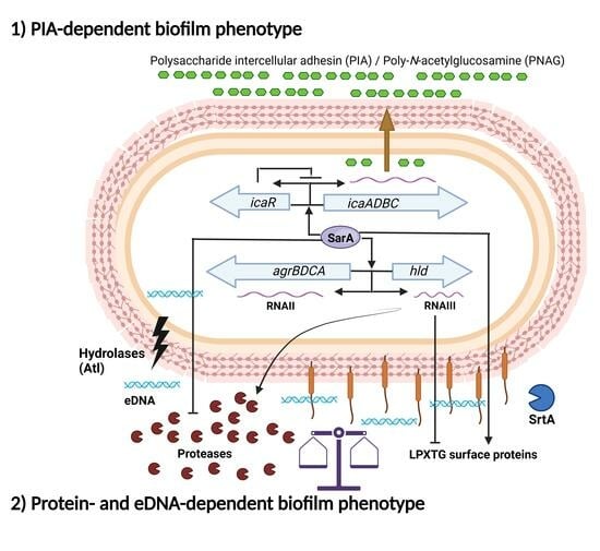
1.2. Biofilm Formation Independent of the icaADBC Operon

In addition to the aforementioned cases of biofilm formation independent of the ica operon in MRSA strains in a medium containing glucose, one of the first reports of an ica-independent biofilm formation in S. aureus was from a clinical isolate named UAMS-1 (MSSA). It was shown that a mutation in the ica locus had little impact on its biofilm formation capacity in vitro and in vivo [43]. Also, Lim et al. identified using mutagenesis analysis a gene called rbf involved in biofilm formation. The sequence suggested that it was a transcriptional regulator, and it did not affect the expression of the ica operon. Thus, it was considered an ica-independent mechanism [44]. Upstream of the ica operon is the icaR gene; Conlon et al. showed that icaR is a repressor of PIA production and biofilm formation by inhibiting the expression of the ica operon in S. epidermidis [40]. It was later shown that Rbf promotes biofilm formation by repressing the transcription of icaR in S. aureus [45].
Also found using mutagenesis analysis was a gene called bap (biofilm-associated protein) involved in attachment to inert surfaces, intercellular adhesion, and biofilm formation. Bap was described as a surface protein consisting of 2276 amino acids. However, it was only found in a small fraction of the S. aureus isolates from bovine mastitis lesions (5%), while it was absent in the clinical S. aureus isolates analyzed from humans [46]. In the S. aureus strain SA113, whose biofilm formation ability has been associated with the ica operon, complementation with the bap gene in the mutant strain SA113 Δica resulted in biofilm formation, suggesting the role of bap in biofilm formation independent of the ica operon [46]. In fact, bap is a surface protein containing the LPXTG motif. Since biofilms from MRSA strains in BHI-glucose were susceptible to proteinase K treatment, it is reasonable to believe that surface proteins such as bap must be responsible for the ica-independent biofilm formation in these strains. In this regard, a genomic analysis from MRSA strains revealed the presence of several LPXTG surface proteins presumed to be involved in the adherence to host tissues such as clumping factor A and B (Clf A/B), fibronectin binding protein A and B (Fnb A/B), collagen adhesin (Cna), protein A (Spa), methicillin resistance surface protein (Pls), SdrC/D/E, and several cell wall surface anchor proteins (Sas A/B/C/D/E/F/G/H/I/J/K). There were also found other non-LPXTG surface proteins such as autolysin (Atl), elastin binding protein (Ebp), and the fibrinogen binding proteins Fib and Efb [47].
Likewise, a mutation of sortase A (SrtA), an enzyme that catalyzes the cell wall anchoring of LPXTG proteins, in an S. aureus strain with an ica-independent biofilm phenotype was deficient in biofilm formation [48,49]. It was also shown using mass spectrometry that SdrD and protein A (Spa) were key components of the cell wall of an S. aureus strain able to form ica-independent biofilms, and deletion of the spa gene resulted in reduced biofilm formation [48]. SasC and SasG were also shown to promote biofilm formation in S. aureus [50,51]. Interestingly, it was found that MRSA strains in a BHI medium with glucose lowered the pH to mildly acidic conditions, and this was associated with the expression of fnbA and fnbB, and biofilm formation [49]. Mutations of fnbA and fnbB in several MRSA strains reduced biofilm formation. However, these mutations did not affect the biofilm formation in MSSA strains with a PIA-dependent biofilm phenotype. It was proposed that the effect on biofilm formation of fnbA and fnbB was due to intercellular aggregation rather than initial attachment [49]. Furthermore, carriage of both the fnbA and fnbB genes correlated with higher biofilm formation in MRSA strains in comparison with those strains carrying only one gene. It is worth mentioning that the proportion of bacteria expressing both genes was even higher in the MSSA clinical isolates, 69% (68/99) for MSSA, and 33% (40/118) for MRSA [52]. Thus, LPXTG proteins seem to be important in the proteinaceous biofilms formed by MRSA strains.
Also described was extracellular DNA (eDNA) as an important component of the S. aureus biofilms, as addition of DNase I to the medium or to the preformed biofilms resulted in their inhibition or destabilization, respectively [53,54]. CidA, a hydrolase involved in cell lysis, was shown to be important for DNA release and biofilm regulation in S. aureus [54]. Later, the major autolysin of S. aureus called Atl was found to mediate cell lysis and eDNA release, contributing to biofilm formation [55]. It was also shown that Atl was important for attachment and biofilm formation in MRSA strains with a FnbA/B biofilm phenotype, but not for MSSA strains with a PIA-dependent biofilm phenotype [56]. Furthermore, mutation of the protease ClpP in the S. aureus Newman strain resulted in an increase in biofilm formation, and it was a PIA-independent but protein- and eDNA-dependent biofilm [57]. Transcription of the agrA and agrC genes was reduced in the clpP-mutant strain, and it was associated with biofilm formation and decreased levels of protease activity. Interestingly, an increase in the expression of the hydrolase sle1 was found in the clpP-mutant strain, together with an increase in cell lysis and eDNA release [57]; however, it was not clear how the Agr system was affected in the clpP-mutant strain. Altogether, these results indicate the relevance of hydrolases in eDNA release and their contribution to biofilm formation by S. aureus. It is now well accepted that the biofilm composition in MRSA strains depends more on surface proteins and eDNA in comparison with the ica-dependent biofilm phenotype in MSSA strains [42]. Most of the studies have used a medium with NaCl for MSSA strains and a medium with glucose for MRSA strains, favoring the mentioned biofilm phenotypes. However, it is worth noting that a high proportion of MSSA isolates can also form biofilms in mediums containing glucose [19,20]. Thus, it is reasonable that depending on the environment, MSSA strains could form biofilms with a PIA-independent phenotype as well. Indeed, the first experiments analyzing the content of eDNA in the biofilms of S. aureus used UAMS-1 and SH1000, two MSSA strains [53,54,55].
Interestingly, a MRSA clinical isolate capable of switching from a PIA-dependent to a PIA-independent biofilm was found, depending on the medium composition. For example, in a TSB medium with glucose, it formed a PIA-independent biofilm, but when NaCl was added to the medium, detection of PIA and biofilm formation was observed. Deletion of ica in this S. aureus strain did not affect its capacity to form a biofilm in a medium with glucose, but it completely lost the capacity of biofilm formation in a medium with NaCl [58]. Sortase A (srtA) mutation revealed that LPXTG proteins were important for biofilm formation in a medium with glucose while they did not affect the generation of a PIA-dependent biofilm in a medium with NaCl. Genomic analysis revealed a unique repertoire of 20 LPXTG proteins in this strain, and deletion of fnbA and fnbB was associated with a complete loss of the PIA-independent biofilm. Also, fnbB was induced at the transcriptional level in the medium with glucose in comparison with the medium containing NaCl, but the icaC mRNA levels were unaffected in both types of media, suggesting again that there must exist post-transcriptional regulatory mechanisms in PIA expression [58].

1.3. Main Regulatory Systems in Biofilm Formation by S. aureus

The accessory gene regulator (agr) operon consists of the agrBDCA genes and is part of a quorum-sensing regulatory system in S. aureus. The expression of many virulence factors in S. aureus is controlled by the Agr system, which consists of two transcripts whose expression are controlled by promoters P2 and P3. The first transcript is an operon that encodes the four agrBDCA genes, and the second is RNAIII, the true effector molecule that regulates the expression of all the genes regulated by the Agr system. The gene encoding the δ-toxin (hld) is in the effector RNAIII regulatory molecule. This system is activated during the transition from the exponential growth phase to the stationary growth phase [59]. The Agr system responds to the extracellular levels of AIP (autoinducing peptide), an eight-residue peptide, with the last five residues forming a thiolactone cyclic ring. AIPs are formed via the action of AgrB, a membrane-bound peptidase that proteolytically processes AgrD, the peptide precursor of AIP. AgrC is a membrane-bound histidine kinase sensor of AIPs that is autophosphorylated, and this signal passes to the response regulator AgrA, which in turn binds to the promoters P2 and P3 to start their transcription (Figure 2) [60].
Analyzing the agr phenotype in terms of δ-toxin production (the product of the RNAIII transcript in the agr operon) and the biofilm formation capacity, it was demonstrated that 78% of the agr-negative S. aureus strains formed a biofilm in comparison with only 6% of the agr-positive strains [59]. It was also shown that agr mutation in S. aureus strains enhanced biofilm formation in comparison with the wild-type strains. Interestingly, the expression of PIA was not regulated by the Agr system [59]. It has been suggested that agr is repressed in a medium containing glucose and it correlates with biofilm formation [42]. Indeed, it was shown that expression of RNAIII was repressed in a medium containing glucose [61]. It was also found that reactivation of the Agr system via AIP addition or glucose depletion triggered detachment, destabilizing biofilms, only in agr-competent strains [61]. It has been shown that the activation of the Agr system results in upregulation of the Aur metalloprotease and the SpIABCDEF serine proteases, together with a downregulation of several surface proteins [62]. In fact, the inhibition of serine proteases and an S. aureus aur mutant, or a double mutant strain deficient in the aur gene and SpI genes, showed reduced detachment and a stable biofilm after the addition of AIPs, indicating that the Agr system destabilizes biofilms via the action of extracellular proteases.
Furthermore, Beenkem et al. described an S. aureus strain RN6390 that was unable to form biofilm in vitro, and deletion of the agr locus resulted in gaining the ability to form biofilm for this strain. However, a second deletion of sarA in the RN6390 agr-deficient strain resulted again in a lack of biofilm formation, suggesting an association between these genes [63]. Indeed, SarA (staphylococcal accessory regulator) is a DNA-binding protein regulating the transcription of genes important for virulence in S. aureus such as agr, hla, spa, and fbnA, indicating agr-dependent and agr-independent regulatory mechanisms in SarA [64]. For example, it was described using a sarA- or agr-mutant strain that the transcription of 104 genes was upregulated and 34 downregulated in an agr-dependent manner, while 76 were upregulated and 44 downregulated in a sarA-dependent manner [62]. It was concluded that sarA transcriptional regulation is not limited only to the agr genes. It was also described that SarA represses the collagen adhesin gene (cna) in an agr-independent manner [65]. Similarly, it was found that SarA positively controlled bap-dependent biofilm formation in an agr-independent mechanism in three S. aureus strains, and it was shown that SarA binds to the bap promoter, regulating its expression [66]. Also, proposed was the agr-independent transcriptional regulation of fbnA by the binding of SarA to the promoter site of this gene [67]. Thus, there is enough evidence of the SarA regulation of genes related to biofilm formation in S. aureus independent of the Agr system.
In addition, it was shown that sarA mutants decreased PIA production and biofilm formation by downregulating ica operon transcription. Furthermore, sarA mutants showed higher protease activity beyond the downregulation of PIA synthesis [68,69,70]. It was also found in S. aureus clinical isolates amenable to genetic manipulation that biofilm formation was abolished for sarA MRSA mutants in a glucose-containing medium and for sarA MSSA mutants in a NaCl-containing medium [19]. This indicates the role of SarA in PIA-dependent and PIA-independent biofilm formation. In comparison with agr mutants, only 5 out of 21 of the agr MRSA mutants resulted in an increase in biofilm formation, highlighting the role of the Agr system in some of the PIA-independent proteinaceous biofilms of MRSA strains, rather than in the PIA-dependent biofilms of MSSA strains [19].
It was also described for S. epidermidis that mutation in σB (a stress response regulator, SigB) or rsbU, an activator of σB, results in reduced ica transcription and biofilm formation in osmotic stress conditions, which favored the PIA-dependent biofilm phenotype. It was found that σB acts as repressor of the negative regulator icaR gene [71]. Interestingly, it was shown in S. aureus that SarA and σB induced the expression of icaR beyond that of the icaADBC operon [72]. As expected, an S. aureus double mutant lacking the expression of sarA and σB showed an enhanced reduction in the transcriptional expression of the ica operon. Surprisingly, the biofilm formation was only partially affected in the double mutant in comparison with the sarA mutant. Also, the σB mutant was not affected in its biofilm formation and PIA production [68]. This unexpected result unveils a complex regulation of biofilm-related genes by SarA and SigB that may be independent of each other.
Finally, LuxS is an enzyme that catalyzes the production of a signaling molecule called autoinducer-2 (AI-2). The LuxS/AI-2 quorum-sensing system is important for bacterial growth, metabolism, virulence, and biofilm formation. It was shown that the LuxS/AI-2 system negatively regulates PIA production and biofilm formation in S. aureus by repressing the transcription of rbf [73]. We have previously mentioned that Rbf represses the transcription of icaR, the negative regulator of the ica operon [45].

1.4. Concluding Remarks and Future Directions

We have addressed in this review the recent literature comparing the ability of biofilm formation by MRSA and MSSA isolates. MRSA isolates are resistant to most of β-lactam antibiotics, but whether MRSA strains possess different virulent factors that may further complicate infections is still controversial. In fact, it seems to be that the presence of virulence factors is associated with the origin of the isolates. In this regard, we explained that the bap protein was found in S. aureus isolates from bovine mastitis lesions while it was absent in isolates from human origin. Furthermore, tracking the origin of clones for epidemiology studies has been accomplished by analyzing the clonal complexes and the type of SCCmec elements in MRSA strains. Even though in some studies, the association of biofilm formation with a specific type of SCCmec element has been proposed, we consider that the origin of the isolates, rather than the mere acquisition of the SCCmec element, is important for this phenotype. Interestingly, the finding of MSSA isolates forming biofilms preferentially via a PIA-dependent mechanism, and MRSA isolates forming them via a PIA-independent but protein- and eDNA-dependent mechanism, suggests differences in biofilm formation by these strains. We highlighted the role of several hydrolases involved in the release of eDNA and their contributions to biofilm formation by MRSA strains, as well as surface proteins containing the LPXTG motif, which are anchored to the cell wall via sortase A. Genetic analysis revealed the presence of a unique repertoire of these types of proteins in MRSA strains, and functional analysis showed that some of these proteins are in fact important for biofilm formation depending on the MRSA strain analyzed. Finally, the gene regulatory protein SarA is not only an important regulator of PIA-dependent biofilms by controlling the expression of the ica operon, but also induces the expression of the Agr system, which in turn regulates PIA-independent biofilm formation by modulating the expression of extracellular proteases and surface proteins. As most of the research analyzing the biofilm formation capacity in S. aureus has been undertaken in vitro on abiotic surfaces, we consider that in the future, the contribution of the relevant genes that have been described in the context of adhesion and biofilm formation on extracellular matrix components should be analyzed, as well as their relevance to pathogenesis, using in vivo infection models.

Supplementary Materials

The following supporting information can be downloaded at https://0-www-mdpi-com.brum.beds.ac.uk/article/10.3390/diseases11040160/s1, Table S1: Source of the S. aureus isolates, and the biofilm detection method (BDM) used in the analysis. CV, crystal violet staining. CR, Congo Red Method.

Author Contributions

Conceptualization, E.H.-C. and K.T.; investigation and formal analysis, E.H.-C., R.V.-R. and O.M.-C.; validation and original draft preparation, E.H.-C. and K.T.; reviewing and editing, R.V.-R. and O.M.-C. All authors have read and agreed to the published version of the manuscript.

Funding

This research received no external funding.

Data Availability Statement

The data presented in this study are available in this article (and Supplementary Material).

Conflicts of Interest

The authors declare no conflict of interest.

References

  1. Peacock, S.J.; Paterson, G.K. Mechanisms of Methicillin Resistance in Staphylococcus aureus. Annu. Rev. Biochem. 2015, 84, 577–601. [Google Scholar] [CrossRef] [PubMed]
  2. Lakhundi, S.; Zhang, K. Methicillin-Resistant Staphylococcus aureus: Molecular Characterization, Evolution, and Epidemiology. Clin. Microbiol. Rev. 2018, 31, e00020-18. [Google Scholar] [CrossRef] [PubMed]
  3. Deurenberg, R.H.; Stobberingh, E.E. The evolution of Staphylococcus aureus. Infect. Genet. Evol. 2008, 8, 747–763. [Google Scholar] [CrossRef] [PubMed]
  4. Gajdács, M. The Continuing Threat of Methicillin-Resistant Staphylococcus aureus. Antibiotics 2019, 8, 52. [Google Scholar] [CrossRef] [PubMed]
  5. Imani Fooladi, A.A.; Ashrafi, E.; Tazandareh, S.G.; Koosha, R.Z.; Rad, H.S.; Amin, M.; Soori, M.; Larki, R.A.; Choopani, A.; Hosseini, H.M. The distribution of pathogenic and toxigenic genes among MRSA and MSSA clinical isolates. Microb. Pathog. 2015, 81, 60–66. [Google Scholar] [CrossRef]
  6. Rozgonyi, F.; Kocsis, E.; Kristóf, K.; Nagy, K. Is MRSA more virulent than MSSA? Clin. Microbiol. Infect. 2007, 13, 843–845. [Google Scholar] [CrossRef] [PubMed]
  7. Gaire, U.; Thapa Shrestha, U.; Adhikari, S.; Adhikari, N.; Bastola, A.; Rijal, K.R.; Ghimire, P.; Banjara, M.R. Antibiotic Susceptibility, Biofilm Production, and Detection of mecA Gene among Staphylococcus aureus Isolates from Different Clinical Specimens. Diseases 2021, 9, 80. [Google Scholar] [CrossRef]
  8. Goudarzi, M.; Mohammadi, A.; Amirpour, A.; Fazeli, M.; Nasiri, M.J.; Hashemi, A.; Goudarzi, H. Genetic diversity and biofilm formation analysis of Staphylococcus aureus causing urinary tract infections in Tehran, Iran. J. Infect. Dev. Ctries. 2019, 13, 777–785. [Google Scholar] [CrossRef]
  9. Kadkhoda, H.; Ghalavand, Z.; Nikmanesh, B.; Kodori, M.; Houri, H.; Taghizadeh Maleki, D.; Karimi Bavandpour, A.; Eslami, G. Characterization of biofilm formation and virulence factors of Staphylococcus aureus isolates from paediatric patients in Tehran, Iran. Iran. J. Basic Med. Sci. 2020, 23, 691–698. [Google Scholar] [CrossRef] [PubMed]
  10. Leshem, T.; Schnall, B.S.; Azrad, M.; Baum, M.; Rokney, A.; Peretz, A. Incidence of biofilm formation among MRSA and MSSA clinical isolates from hospitalized patients in Israel. J. Appl. Microbiol. 2022, 133, 922–929. [Google Scholar] [CrossRef]
  11. Mahmoudi, H.; Pourhajibagher, M.; Chiniforush, N.; Soltanian, A.R.; Alikhani, M.Y.; Bahador, A. Biofilm formation and antibiotic resistance in meticillin-resistant and meticillin-sensitive Staphylococcus aureus isolated from burns. J. Wound Care 2019, 28, 66–73. [Google Scholar] [CrossRef] [PubMed]
  12. Ohadian Moghadam, S.; Pourmand, M.R.; Aminharati, F. Biofilm formation and antimicrobial resistance in methicillin-resistant Staphylococcus aureus isolated from burn patients, Iran. J. Infect. Dev. Ctries. 2014, 8, 1511–1517. [Google Scholar] [CrossRef] [PubMed]
  13. Piechota, M.; Kot, B.; Frankowska-Maciejewska, A.; Grużewska, A.; Woźniak-Kosek, A. Biofilm Formation by Methicillin-Resistant and Methicillin-Sensitive Staphylococcus aureus Strains from Hospitalized Patients in Poland. BioMed Res. Int. 2018, 2018, 4657396. [Google Scholar] [CrossRef]
  14. Tabandeh, M.; Kaboosi, H.; Taghizadeh Armaki, M.; Pournajaf, A.; Peyravii Ghadikolaii, F. New update on molecular diversity of clinical Staphylococcus aureus isolates in Iran: Antimicrobial resistance, adhesion and virulence factors, biofilm formation and SCCmec typing. Mol. Biol. Rep. 2022, 49, 3099–3111. [Google Scholar] [CrossRef]
  15. Ghasemian, A.; Najar Peerayeh, S.; Bakhshi, B.; Mirzaee, M. Comparison of Biofilm Formation between Methicillin-Resistant and Methicillin-Susceptible Isolates of Staphylococcus aureus. Iran. Biomed. J. 2016, 20, 175–181. [Google Scholar] [CrossRef]
  16. Khasawneh, A.I.; Himsawi, N.; Abu-Raideh, J.; Salameh, M.A.; Al-Tamimi, M.; Al Haj Mahmoud, S.; Saleh, T. Status of Biofilm-Forming Genes among Jordanian Nasal Carriers of Methicillin-Sensitive and Methicillin-Resistant Staphylococcus aureus. Iran. Biomed. J. 2020, 24, 386–398. [Google Scholar] [CrossRef]
  17. Grinholc, M.; Wegrzyn, G.; Kurlenda, J. Evaluation of biofilm production and prevalence of the icaD gene in methicillin-resistant and methicillin-susceptible Staphylococcus aureus strains isolated from patients with nosocomial infections and carriers. FEMS Immunol. Med. Microbiol. 2007, 50, 375–379. [Google Scholar] [CrossRef] [PubMed]
  18. Abd El Rahman, A.; El Kholy, Y.; Shash, R.Y. Correlation between mazEF Toxin-Antitoxin System Expression and Methicillin Susceptibility in Staphylococcus aureus and Its Relation to Biofilm-Formation. Microorganisms 2021, 9, 2274. [Google Scholar] [CrossRef] [PubMed]
  19. O’Neill, E.; Pozzi, C.; Houston, P.; Smyth, D.; Humphreys, H.; Robinson, D.A.; O’Gara, J.P. Association between Methicillin Susceptibility and Biofilm Regulation in Staphylococcus aureus Isolates from Device-Related Infections. J. Clin. Microbiol. 2007, 45, 1379–1388. [Google Scholar] [CrossRef] [PubMed]
  20. Fitzpatrick, F.; Humphreys, H.; O’Gara, J.P. Environmental regulation of biofilm development in methicillin-resistant and methicillin-susceptible Staphylococcus aureus clinical isolates. J. Hosp. Infect. 2006, 62, 120–122. [Google Scholar] [CrossRef]
  21. Tahmasebi, H.; Dehbashi, S.; Jahantigh, M.; Arabestani, M.R. Relationship between biofilm gene expression with antimicrobial resistance pattern and clinical specimen type based on sequence types (STs) of methicillin-resistant S. aureus. Mol. Biol. Rep. 2020, 47, 1309–1320. [Google Scholar] [CrossRef] [PubMed]
  22. Belbase, A.; Pant, N.D.; Nepal, K.; Neupane, B.; Baidhya, R.; Baidya, R.; Lekhak, B. Antibiotic resistance and biofilm production among the strains of Staphylococcus aureus isolated from pus/wound swab samples in a tertiary care hospital in Nepal. Ann. Clin. Microbiol. Antimicrob. 2017, 16, 15. [Google Scholar] [CrossRef] [PubMed]
  23. Indrawattana, N.; Sungkhachat, O.; Sookrung, N.; Chongsa-nguan, M.; Tungtrongchitr, A.; Voravuthikunchai, S.P.; Kong-ngoen, T.; Kurazono, H.; Chaicumpa, W. Staphylococcus aureus clinical isolates: Antibiotic susceptibility, molecular characteristics, and ability to form biofilm. BioMed Res. Int. 2013, 2013, 314654. [Google Scholar] [CrossRef] [PubMed]
  24. Maharjan, S.; Ansari, M.; Maharjan, P.; Rai, K.R.; Sabina, K.C.; Kattel, H.P.; Rai, G.; Rai, S.K. Phenotypic detection of methicillin resistance, biofilm production, and inducible clindamycin resistance in Staphylococcus aureus clinical isolates in Kathmandu, Nepal. Trop. Med. Health 2022, 50, 71. [Google Scholar] [CrossRef]
  25. Saud, B.; Khatri, G.; Amatya, N.; Paudel, G.; Shrestha, V. Methicillin-Resistant and Biofilm-Producing Staphylococcus aureus in Nasal Carriage among Health Care Workers and Medical Students. Can. J. Infect. Dis. Med. Microbiol. 2023, 2023, 8424486. [Google Scholar] [CrossRef] [PubMed]
  26. Senobar Tahaei, S.A.; Stájer, A.; Barrak, I.; Ostorházi, E.; Szabó, D.; Gajdács, M. Correlation Between Biofilm-Formation and the Antibiotic Resistant Phenotype in Staphylococcus aureus Isolates: A Laboratory-Based Study in Hungary and a Review of the Literature. Infect. Drug Resist. 2021, 14, 1155–1168. [Google Scholar] [CrossRef]
  27. Kırmusaoğlu, S. The Methods for Detection of Biofilm and Screening Antibiofilm Activity of Agents. In Antimicrobials, Antibiotic Resistance, Antibiofilm Strategies and Activity Methods; IntechOpen: London, UK, 2019. [Google Scholar] [CrossRef]
  28. Hryniewicz, M.M.; Garbacz, K. Borderline oxacillin-resistant Staphylococcus aureus (BORSA)—A more common problem than expected? J. Med. Microbiol. 2017, 66, 1367–1373. [Google Scholar] [CrossRef]
  29. Mlynarczyk-Bonikowska, B.; Kowalewski, C.; Krolak-Ulinska, A.; Marusza, W. Molecular Mechanisms of Drug Resistance in Staphylococcus aureus. Int. J. Mol. Sci. 2022, 23, 8088. [Google Scholar] [CrossRef]
  30. Kumar, S.; Anwer, R.; Yadav, M.; Sehrawat, N.; Singh, M.; Kumar, V. Molecular Typing and Global Epidemiology of Staphylococcus aureus. Curr. Pharmacol. Rep. 2021, 7, 179–186. [Google Scholar] [CrossRef]
  31. Uehara, Y. Current Status of Staphylococcal Cassette Chromosome mec (SCCmec). Antibiotics 2022, 11, 86. [Google Scholar] [CrossRef]
  32. Nagasundaram, N.; Sistla, S. Existence of multiple SCCmec elements in clinical isolates of methicillin-resistant Staphylococcus aureus. J. Med. Microbiol. 2019, 68, 720–727. [Google Scholar] [CrossRef] [PubMed]
  33. Nguyen, H.T.T.; Nguyen, T.H.; Otto, M. The staphylococcal exopolysaccharide PIA—Biosynthesis and role in biofilm formation, colonization, and infection. Comput. Struct. Biotechnol. J. 2020, 18, 3324–3334, Erratum in Comput. Struct. Biotechnol. J. 2023, 21, 2035. [Google Scholar] [CrossRef] [PubMed]
  34. Mack, D.; Nedelmann, M.; Krokotsch, A.; Schwarzkopf, A.; Heesemann, J.; Laufs, R. Characterization of transposon mutants of biofilm-producing Staphylococcus epidermidis impaired in the accumulative phase of biofilm production: Genetic identification of a hexosamine-containing polysaccharide intercellular adhesin. Infect. Immun. 1994, 62, 3244–3253. [Google Scholar] [CrossRef]
  35. Cramton, S.E.; Gerke, C.; Schnell, N.F.; Nichols, W.W.; Goetz, F. The Intercellular Adhesion (ica) Locus Is Present in Staphylococcus aureus and Is Required for Biofilm Formation. Infect. Immun. 1999, 67, 5427–5433. [Google Scholar] [CrossRef]
  36. Knobloch, J.K.; Horstkotte, M.A.; Rohde, H.; Mack, D. Evaluation of different detection methods of biofilm formation in Staphylococcus aureus. Med. Microbiol. Immunol. 2002, 191, 101–106. [Google Scholar] [CrossRef]
  37. Fitzpatrick, F.; Humphreys, H.; O’Gara, J.P. Evidence for icaADBC-Independent Biofilm Development Mechanism in Methicillin-Resistant Staphylococcus aureus Clinical Isolates. J. Clin. Microbiol. 2005, 43, 1973–1976. [Google Scholar] [CrossRef] [PubMed]
  38. Mempel, M.; Feucht, H.; Ziebuhr, W.; Endres, M.; Laufs, R.; Grüter, L. Lack of mecA transcription in slime-negative phase variants of methicillin-resistant Staphylococcus epidermidis. Antimicrob. Agents Chemother. 1994, 38, 1251–1255. [Google Scholar] [CrossRef] [PubMed]
  39. Mempel, M.; Müller, E.; Hoffmann, R.; Feucht, H.; Laufs, R.; Grüter, L. Variable degree of slime production is linked to different levels of beta-lactam susceptibility in Staphylococcus epidermidis phase variants. Med. Microbiol. Immunol. 1995, 184, 109–113. [Google Scholar] [CrossRef]
  40. Conlon, K.M.; Humphreys, H.; O Gara, J.P. icaR Encodes a Transcriptional Repressor Involved in Environmental Regulation of ica Operon Expression and Biofilm Formation in Staphylococcus epidermidis. J. Bacteriol. 2002, 184, 4400–4408. [Google Scholar] [CrossRef]
  41. Rachid, S.; Ohlsen, K.; Witte, W.; Hacker, J.; Ziebuhr, W. Effect of subinhibitory antibiotic concentrations on polysaccharide intercellular adhesin expression in biofilm-forming Staphylococcus epidermidis. Antimicrob. Agents Chemother. 2000, 44, 3357–3363. [Google Scholar] [CrossRef]
  42. McCarthy, H.; Rudkin, J.K.; Black, N.S.; Gallagher, L.; O’Neill, E.; O’Gara, J.P. Methicillin resistance and the biofilm phenotype in Staphylococcus aureus. Front. Cell. Infect. Microbiol. 2015, 5, 1. [Google Scholar] [CrossRef]
  43. Beenken, K.E.; Dunman, P.M.; McAleese, F.; Macapagal, D.; Murphy, E.; Projan, S.J.; Blevins, J.S.; Smeltzer, M.S. Global Gene Expression in Staphylococcus aureus Biofilms. J. Bacteriol. 2004, 186, 4665–4684. [Google Scholar] [CrossRef]
  44. Lim, Y.; Jana, M.; Luong, T.T.; Lee, C.Y. Control of Glucose- and NaCl-Induced Biofilm Formation by rbf in Staphylococcus aureus. J. Bacteriol. 2004, 186, 722–729. [Google Scholar] [CrossRef]
  45. Cue, D.; Lei, M.G.; Luong, T.T.; Kuechenmeister, L.; Dunman, P.M.; O’Donnell, S.; Rowe, S.; O’Gara, J.P.; Lee, C.Y. Rbf promotes biofilm formation by Staphylococcus aureus via repression of icaR, a negative regulator of icaADBC. J. Bacteriol. 2009, 191, 6363–6373. [Google Scholar] [CrossRef]
  46. Cucarella, C.; Solano, C.; Valle, J.; Amorena, B.; Lasa, I.; Penades, J.R. Bap, a Staphylococcus aureus Surface Protein Involved in Biofilm Formation. J. Bacteriol. 2001, 183, 2888–2896. [Google Scholar] [CrossRef] [PubMed]
  47. Gill, S.R.; Fouts, D.E.; Archer, G.L.; Mongodin, E.F.; Deboy, R.T.; Ravel, J.; Paulsen, I.T.; Kolonay, J.F.; Brinkac, L.; Beanan, M.; et al. Insights on evolution of virulence and resistance from the complete genome analysis of an early methicillin-resistant Staphylococcus aureus strain and a biofilm-producing methicillin-resistant Staphylococcus epidermidis strain. J. Bacteriol. 2005, 187, 2426–2438. [Google Scholar] [CrossRef]
  48. Merino, N.; Toledo-Arana, A.; Vergara-Irigaray, M.; Valle, J.; Solano, C.; Calvo, E.; Lopez, J.A.; Foster, T.J.; Penadés, J.R.; Lasa, I. Protein A-mediated multicellular behavior in Staphylococcus aureus. J. Bacteriol. 2009, 191, 832–843. [Google Scholar] [CrossRef]
  49. O’Neill, E.; Pozzi, C.; Houston, P.; Humphreys, H.; Robinson, D.A.; Loughman, A.; Foster, T.J.; O’Gara, J.P. A novel Staphylococcus aureus biofilm phenotype mediated by the fibronectin-binding proteins, FnBPA and FnBPB. J. Bacteriol. 2008, 190, 3835–3850. [Google Scholar] [CrossRef] [PubMed]
  50. Geoghegan, J.A.; Corrigan, R.M.; Gruszka, D.T.; Speziale, P.; O’Gara, J.P.; Potts, J.R.; Foster, T.J. Role of surface protein SasG in biofilm formation by Staphylococcus aureus. J. Bacteriol. 2010, 192, 5663–5673. [Google Scholar] [CrossRef] [PubMed]
  51. Schroeder, K.; Jularic, M.; Horsburgh, S.M.; Hirschhausen, N.; Neumann, C.; Bertling, A.; Schulte, A.; Foster, S.; Kehrel, B.E.; Peters, G.; et al. Molecular characterization of a novel Staphylococcus aureus surface protein (SasC) involved in cell aggregation and biofilm accumulation. PLoS ONE 2009, 4, e7567. [Google Scholar] [CrossRef] [PubMed]
  52. O’Neill, E.; Humphreys, H.; O’Gara, J.P. Carriage of both the fnbA and fnbB genes and growth at 37 degrees C promote FnBP-mediated biofilm development in meticillin-resistant Staphylococcus aureus clinical isolates. J. Med. Microbiol. 2009, 58 Pt 4, 399–402. [Google Scholar] [CrossRef]
  53. Izano, E.A.; Amarante, M.A.; Kher, W.B.; Kaplan, J.B. Differential roles of poly-N-acetylglucosamine surface polysaccharide and extracellular DNA in Staphylococcus aureus and Staphylococcus epidermidis biofilms. Appl. Environ. Microbiol. 2008, 74, 470–476. [Google Scholar] [CrossRef] [PubMed]
  54. Rice, K.C.; Mann, E.E.; Endres, J.L.; Weiss, E.C.; Cassat, J.E.; Smeltzer, M.S.; Bayles, K.W. The cidA murein hydrolase regulator contributes to DNA release and biofilm development in Staphylococcus aureus. Proc. Natl. Acad. Sci. USA 2007, 104, 8113–8118. [Google Scholar] [CrossRef]
  55. Bose, J.L.; Lehman, M.K.; Fey, P.D.; Bayles, K.W. Contribution of the Staphylococcus aureus Atl AM and GL murein hydrolase activities in cell division, autolysis, and biofilm formation. PLoS ONE 2012, 7, e42244. [Google Scholar] [CrossRef]
  56. Houston, P.; Rowe, S.E.; Pozzi, C.; Waters, E.M.; O’Gara, J.P. Essential role for the major autolysin in the fibronectin-binding protein-mediated Staphylococcus aureus biofilm phenotype. Infect. Immun. 2011, 79, 1153–1165. [Google Scholar] [CrossRef]
  57. Liu, Q.; Wang, X.; Qin, J.; Cheng, S.; Yeo, W.S.; He, L.; Ma, X.; Liu, X.; Li, M.; Bae, T. The ATP-Dependent Protease ClpP Inhibits Biofilm Formation by Regulating Agr and Cell Wall Hydrolase Sle1 in Staphylococcus aureus. Front. Cell. Infect. Microbiol. 2017, 7, 181. [Google Scholar] [CrossRef] [PubMed]
  58. Vergara-Irigaray, M.; Valle, J.; Merino, N.; Latasa, C.; García, B.; Ruiz de Los Mozos, I.; Solano, C.; Toledo-Arana, A.; Penadés, J.R.; Lasa, I. Relevant role of fibronectin-binding proteins in Staphylococcus aureus biofilm-associated foreign-body infections. Infect. Immun. 2009, 77, 3978–3991. [Google Scholar] [CrossRef]
  59. Vuong, C.; Saenz, H.L.; Götz, F.; Otto, M. Impact of the agr quorum-sensing system on adherence to polystyrene in Staphylococcus aureus. J. Infect. Dis. 2000, 182, 1688–1693. [Google Scholar] [CrossRef]
  60. Jenul, C.; Horswill, A.R. Regulation of Staphylococcus aureus Virulence. Microbiol. Spectr. 2019, 7. [Google Scholar] [CrossRef] [PubMed]
  61. Boles, B.R.; Horswill, A.R. Agr-mediated dispersal of Staphylococcus aureus biofilms. PLoS Pathog. 2008, 4, e1000052. [Google Scholar] [CrossRef] [PubMed]
  62. Dunman, P.M.; Murphy, E.; Haney, S.; Palacios, D.; Tucker-Kellogg, G.; Wu, S.; Brown, E.L.; Zagursky, R.J.; Shlaes, D.; Projan, S.J. Transcription profiling-based identification of Staphylococcus aureus genes regulated by the agr and/or sarA loci. J. Bacteriol. 2001, 183, 7341–7353. [Google Scholar] [CrossRef] [PubMed]
  63. Beenken, K.E.; Blevins, J.S.; Smeltzer, M.S. Mutation of sarA in Staphylococcus aureus Limits Biofilm Formation. Infect. Immun. 2003, 71, 4206–4211. [Google Scholar] [CrossRef] [PubMed]
  64. Chien, Y.; Manna, A.C.; Cheung, A.L. SarA level is a determinant of agr activation in Staphylococcus aureus. Mol. Microbiol. 1998, 30, 991–1001. [Google Scholar] [CrossRef] [PubMed]
  65. Blevins, J.S.; Beenken, K.E.; Elasri, M.O.; Hurlburt, B.K.; Smeltzer, M.S. Strain-dependent differences in the regulatory roles of sarA and agr in Staphylococcus aureus. Infect. Immun. 2002, 70, 470–480. [Google Scholar] [CrossRef] [PubMed]
  66. Trotonda, M.P.; Manna, A.C.; Cheung, A.L.; Lasa, I.; Penadés, J.R. SarA positively controls bap-dependent biofilm formation in Staphylococcus aureus. J. Bacteriol. 2005, 187, 5790–5798. [Google Scholar] [CrossRef]
  67. Wolz, C.; Pöhlmann-Dietze, P.; Steinhuber, A.; Chien, Y.T.; Manna, A.; van Wamel, W.; Cheung, A. Agr-independent regulation of fibronectin-binding protein(s) by the regulatory locus sar in Staphylococcus aureus. Mol. Microbiol. 2000, 36, 230–243. [Google Scholar] [CrossRef]
  68. Valle, J.; Toledo-Arana, A.; Berasain, C.; Ghigo, J.M.; Amorena, B.; Penadés, J.R.; Lasa, I. SarA and not sigmaB is essential for biofilm development by Staphylococcus aureus. Mol. Microbiol. 2003, 48, 1075–1087. [Google Scholar] [CrossRef]
  69. Campbell, M.J.; Beenken, K.E.; Ramirez, A.M.; Smeltzer, M.S. The major role of sarA in limiting Staphylococcus aureus extracellular protease production in vitro is correlated with decreased virulence in diverse clinical isolates in osteomyelitis. Virulence 2023, 14, 2175496. [Google Scholar] [CrossRef]
  70. Ramirez, A.M.; Beenken, K.E.; Byrum, S.D.; Tackett, A.J.; Shaw, L.N.; Gimza, B.D.; Smeltzer, M.S. SarA plays a predominant role in controlling the production of extracellular proteases in the diverse clinical isolates of Staphylococcus aureus LAC and UAMS-1. Virulence 2020, 11, 1738–1762. [Google Scholar] [CrossRef] [PubMed]
  71. Knobloch, J.K.; Jäger, S.; Horstkotte, M.A.; Rohde, H.; Mack, D. RsbU-dependent regulation of Staphylococcus epidermidis biofilm formation is mediated via the alternative sigma factor sigmaB by repression of the negative regulator gene icaR. Infect. Immun. 2004, 72, 3838–3848. [Google Scholar] [CrossRef]
  72. Cerca, N.; Brooks, J.L.; Jefferson, K.K. Regulation of the intercellular adhesin locus regulator (icaR) by SarA, sigmaB, and IcaR in Staphylococcus aureus. J. Bacteriol. 2008, 190, 6530–6533. [Google Scholar] [CrossRef] [PubMed]
  73. Ma, R.; Qiu, S.; Jiang, Q.; Sun, H.; Xue, T.; Cai, G.; Sun, B. AI-2 quorum sensing negatively regulates rbf expression and biofilm formation in Staphylococcus aureus. Int. J. Med. Microbiol. 2017, 307, 257–267. [Google Scholar] [CrossRef] [PubMed]
Figure 1. PIA (polysaccharide intercellular adhesin) is produced by the icaADBC operon in S. aureus. PIA-dependent biofilm formation has been described mainly for MSSA isolates under osmotic stress conditions. The ica operon is regulated by SarA and SigB. IcaR is part of the operon and acts as a negative regulator of PIA production. Figure was created using BioRender.com.
Diseases 11 00160 g001
Figure 2. The Agr (accessory gene regulator) system is an operon that regulates the expression of several virulence factors in S. aureus. The agrBDCA operon is activated by SarA. In some S. aureus strains, the Agr system regulates PIA-independent biofilm formation by negatively regulating the expression of surface proteins while positively regulating the expression of extracellular proteases. Figure was created using BioRender.com.
Diseases 11 00160 g002
Table 1. Percentage of publications reporting that MRSA isolates had better (>), less (<), or the same (=) biofilm formation capacity in comparison with MSSA. Characterization of MRSA isolates was assessed via susceptibility to cefoxitin and/or oxacillin using the disk diffusion method (DDM) or mecA detection together with the DDM. For the DDM, the antibiotic is indicated as cefoxitin (Cef), oxacillin (Ox), not mentioned (Nm), and not done (Nd).
MRSA > MSSAMRSA = MSSAMRSA < MSSA
mecA + DDM40% (8/20)
REF. [7,8,11,14] (Cef), [9,12] (Ox), [10,13] (Nm)
15% (3/20)
REF. [15] (Ox), [16] (Ox/Cef), [19] (Nd)
5% (1/20)
REF. [17] (Nm)
DDM10% (2/20)
REF. [18,21] (Cef)
30% (6/20)
REF. [24,25,26] (Cef), [22] (Ox), [23] (Ox/Cef), [20] (Nm)
0% (0/20)
Total (100%)50%45%5%
Disclaimer/Publisher’s Note: The statements, opinions and data contained in all publications are solely those of the individual author(s) and contributor(s) and not of MDPI and/or the editor(s). MDPI and/or the editor(s) disclaim responsibility for any injury to people or property resulting from any ideas, methods, instructions or products referred to in the content.

