Review

Nanoemulsion: A Review on Mechanisms for the Transdermal Delivery of Hydrophobic and Hydrophilic Drugs

1
Department of Pharmaceutics &Pharmaceutical Technology, Faculty of Pharmaceutical Sciences and Pharmaceutical industries, Future University in Egypt (FUE), 11835 Cairo, Egypt
2
Department of Pharmaceutics& Industrial Pharmacy, Ain Shams University, 11591 Cairo, Egypt
3
Quality Control Department, Elnajah Medical Services, Benghazi, Libya
*
Author to whom correspondence should be addressed.
Submission received: 1 June 2019 / Revised: 29 June 2019 / Accepted: 9 July 2019 / Published: 12 July 2019

Abstract

:
Nanoemulsions (NEs) are colloidal dispersions of two immiscible liquids, oil and water, in which one is dispersed in the other with the aid of a surfactant/co-surfactant mixture, either forming oil-in-water (o/w) or water-in-oil (w/o) nanodroplets systems, with droplets 20–200 nm in size. NEs are easy to prepare and upscale, and they show high variability in their components. They have proven to be very viable, non-invasive, and cost-effective nanocarriers for the enhanced transdermal delivery of a wide range of active compounds that tend to metabolize heavily or suffer from undesirable side effects when taken orally. In addition, the anti-microbial and anti-viral properties of NE components, leading to preservative-free formulations, make NE a very attractive approach for transdermal drug delivery. This review focuses on how NEs mechanistically deliver both lipophilic and hydrophilic drugs through skin layers to reach the blood stream, exerting the desired therapeutic effect. It highlights the mechanisms and strategies executed to effectively deliver drugs, both with o/w and w/o NE types, through the transdermal way. However, the mechanisms reported in the literature are highly diverse, to the extent that a definite mechanism is not conclusive.

Graphical Abstract

1. Introduction

Nanoemulsions (NEs) are submicron sized and have gained great interest as drug carriers for improving the delivery of therapeutic agents. They are considered an advanced nanodroplet systems for systemic, controlled, and targeted drug delivery. NEs are colloidal dispersions consisting of two immiscible liquids (water and oil), in which one liquid is dispersed in the other by means of an appropriate surfactant mixture, forming a system where the size of droplets falls in the range of 20 to 200 nm, as reported by Takur et al. [1]. Nanoemulsion (NE) in particular is a very cost-effective technique, with high storage stability, as it is easily prepared and does not always require high energy or complicated procedures [2,3]. Despite the similarities between NEs and microemulsions (MEs) in terms of their physical appearance, components, and preparation techniques, NEs are kinetically stable and thermodynamically metastable, while MEs are thermodynamically stable [4].
Transdermal drug delivery is a very established route of drug administration that is known to have enhanced therapeutic efficacy, ease of administration, and an allow an immediate withdrawal of treatment if necessary [5]. This administration route bypasses the hepatic metabolism to reach systemic circulation [6], leading to an increase in drug bioavailability. Moreover, it reduces the adverse effects of some medicines, e.g., non-steroidal anti-inflammatory drugs (NSAIDs), which are usually associated with gastrointestinal tract (GIT) drug administration [7,8]. Drug delivery via skin for systemic circulation is suitable for a number of clinical conditions, such as hypertension, arthritis, diabetes, cancer, and high blood cholesterol level [9]. Because the dose is given transdermally and is sustained over a long period via tailored delivery systems, particularly in the case of NE, it is of utmost importance to improve patient compliance [10].

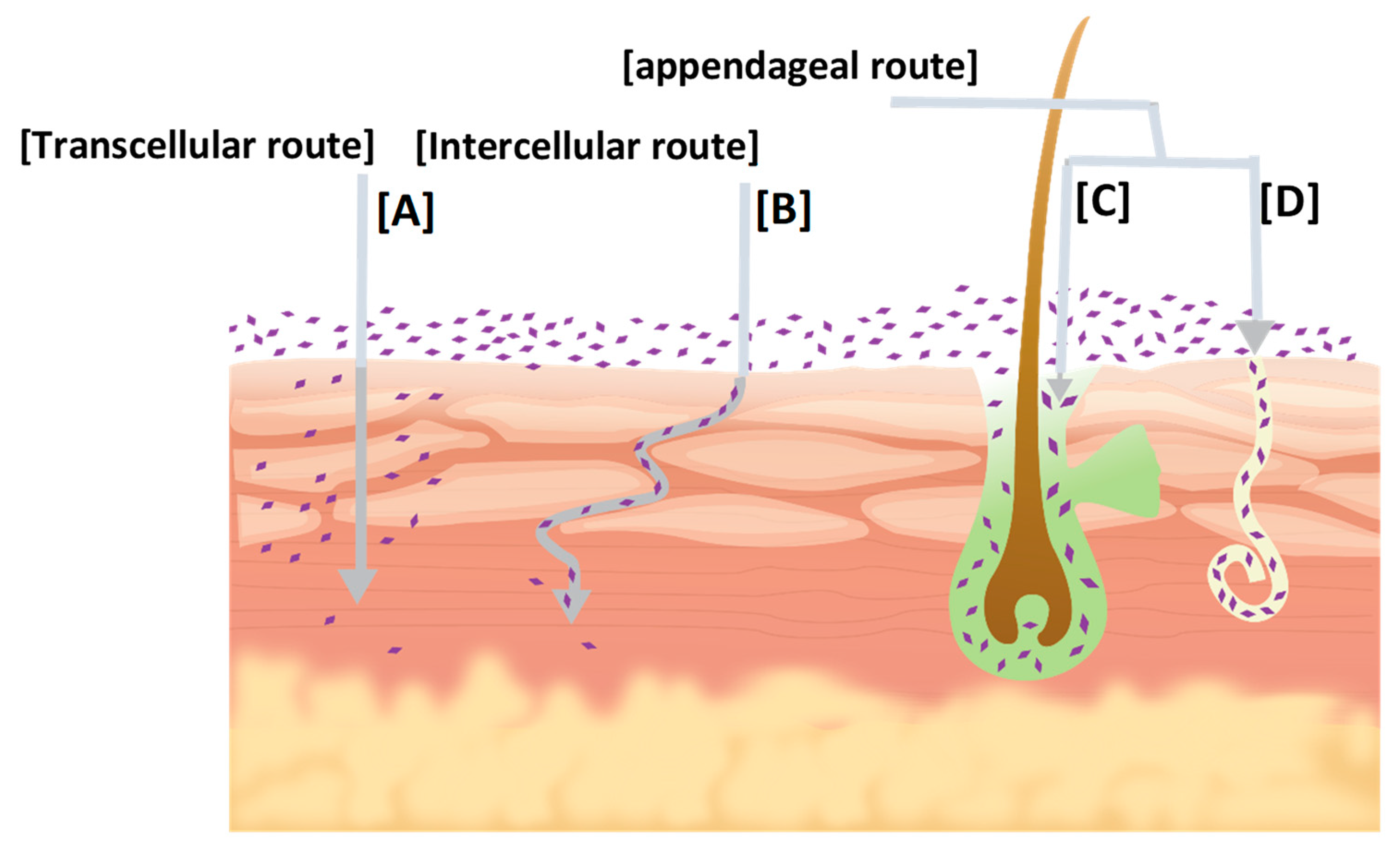
2. Skin Permeation Pathways

The outermost layer of the epidermis, the stratum corneum (SC), forms a hydrophobic barrier that impedes the transport of exogenous chemicals, including drugs [11]. An important requirement for transdermal delivery is that the drug carried by a vehicle is able to reach the skin surface at an adequate rate and in sufficient amounts [12]. The SC is the hardest layer of skin due to its flattered corneocytes which are surrounded by lipid bilayers consisting of ceramides [9]. It is arranged in 10–15 rows that are 10 μm in thickness, composed of highly keratinized cells, known as corneocytes. These cells are surrounded by a continuous lipid phase, known as the intercellular lipid lamellae, and have been said to resemble a “brick and mortar” model [13]. Finally, diffusion through the SC is the sum of a series of segments of lateral diffusion and intramembrane trans-bilayer transport [14].
A permeant applied to the skin has three possible routes across the epidermis, as illustrated in Figure 1: The transcellular route, a lipid domain associated with the proteins inside corneocytes, the intercellular route, and the appendageal route, through hair follicles, via associated sebaceous glands and sweat ducts [15]. The fractional appendage area available for transport is only about 0.1%. This route usually contributes negligibly to the steady state drug flux. This pore pathway may be important for ions and large polar molecules that struggle to cross an intact SC [15]. The transcellular route is a sequence of partitioning and diffusion into alternating hydrophilic and lipophilic domains of cells, respectively, in the extracellular matrix [16,17]. In the intercellular route, the permeant crosses the hard path within the extracellular matrix, without traversing the cells. Small hydrophilic molecules generally prefer the transcellular route over the intercellular route, and the reverse is true for lipophilic molecules. The transcellular and intercellular routes constitute the transepidermal pathway [18].
The combined flux of these two pathways, lipid and pore, determines the overall observed flux across the skin. It is widely accepted that the trans epidermal pathway is usually the predominant pathway of skin permeation and that under sink conditions, diffusion across the SC constitutes the rate-limiting step that determines the overall flux of the permeant.

3. Determination of Transdermal Permeability Coefficient

The total experimental permeability coefficient (P) for full-thickness skin may be expressed as [19]:
P =   1 1 P S C + 1 P D / E
where PSC is the permeability coefficient of the SC and PD/E is the permeability coefficient for the epidermis-dermis combination, which can be estimated from permeation experiments with tape-stripped skin. PSC can be further divided into parallel lipoidal and pore pathway components in the SC via the following equation:
PSC = PL + PP
where PL and PP are the permeability coefficients for the lipoidal and pore pathways, respectively. By substituting Equation (2) into Equation (1), the following expression is obtained:
P =   1 1 P L + P P + 1 P D / E
Highly lipophilic drugs may be retained in the lipophilic SC and resist partitioning into the more hydrophilic viable epidermis [20]. Thus, clearance from, rather than diffusion across, the SC may then become the rate-limiting step for highly lipophilic drugs. Similarly, the appendageal pathway may be more important for highly hydrophilic molecules, electrolytes, and large molecules with low diffusion coefficients [21,22]. For lipophilic permeants, Equation (2) may be well approximated as follows:
PSCPL
In an analysis of experimental data obtained with hairless mice skin in phosphate buffer saline, Equation (4) has been found to be true for lipophilic permeant corticosterone in a PBS, as PP « PL and PD/E » PL [23,24].

4. NE in Transdermal Delivery

Nanosized drug delivery systems significantly enhance the bioavailability and solubility of active constituents by penetrating vital cellular reservoirs [25,26]. Nanoemulsions (NEs) have been reported to improve the transdermal permeation of many drugs when compared to conventional topical formulations [27]. Many formulators have investigated the skin permeation mechanism of many drugs using NEs and microemulsions (MEs) as delivery systems [20,28,29]. NEs and MEs share similar components in different ratios and the same penetration mechanisms. Their main difference lies in their droplet shape, size distribution, and kinetic stability [30]. To the best of our knowledge, although different mechanisms have been suggested to explain the effect of NE on skin permeation in the literature, an answer to the following question is not well understood: Do the NE properties or NE components provide the transdermal permeation enhancement? Thus, this review focuses on the different reported mechanisms by which NE enhanced the transdermal permeation of hydrophobic and hydrophilic drugs.

4.1. Physical Properties of Transdermal NE

NEs are a transparent (translucent) liquid in a liquid colloidal dispersion system which is kinetically stable, with a droplet size <100 nm, as reported by McClements [30]. However, he stated that other NE particle size upper limits, such as 200 and 500 nm, were proposed in recent literature. The long-term physical stability of NEs, with no flocculation or coalescence, provides unique properties and makes them approach thermodynamic stability [31]. NEs consist of oil and aqueous phases, stabilized by a surfactant and co-surfactant in defined ratios [32]. NE, when compared to conventional emulsion, shows superior properties, namely sturdiness against gravitational separation, a droplet size in the nanometer range, improved stability, and larger capacity for drug encapsulation [33]. The dispersibility of NEs is very high compared to MEs, owing to the small droplet size that prevents flocculation and allows dispersion without separation [34]. NEs can be used to deliver either hydrophilic or lipophilic drugs in water-in-oil (w/o) or oil-in-water (o/w) formulations, respectively [8].

4.1.1. Oil-in-Water (o/w) NE

Most of the drugs available in the market today that are developed by the pharmaceutical industry are poorly soluble in water and lipophilic in nature [20]. Recently, scientists have shifted their interest toward nanotechnology-based lipid systems, such as solid dispersions, solid lipid nanoparticles, liposomes, MEs, and NEs, due to their enhanced ability to solubilize drugs, increase bioavailability, and increase drug carrying capacity, which are also the most advanced commercial approaches. NE is a leading trend in transdermal drug delivery systems since it enhances drug skin permeation and improves the bioavailability of poorly soluble drugs when compared to other transdermal dosage forms [35,36].

4.1.2. Water-in-Oil (w/o) NE

NEs that are of the type w/o, issued for hydrophilic compounds, are less common than o/w NEs for transdermal delivery. In w/o NEs, the drug resides in the water phase rather than the oil phase, although its partitioning in the oil phase is inevitable, based on its oil-water partition coefficient. Since the drugs used in this category are water-soluble, the selection of surfactants in these preparations is based on a proper hydrophile-lipophile balance (HLB) value, in order to reduce the tension between the water phase and the oil phase and produce a stable formula [2].

4.2. Components of Transdermal NE

4.2.1. Oil Phase

Oleic acid (OA) is a common oil phase that is employed in the composition of NEs. OA has an inherent penetration enhancing ability, as it causes the SC to absorb more water and swell, and it also compromises some of the structural components of the SC, thus increasing penetration through this limiting and protective barrier [37]. Other oils that possess permeability enhancing abilities, such as capryol 90 [5] and isopropyl myristate [8], have been reported in the literature. The viscous oil α-tocopherol gives a very small droplet size, followed by hexyl laurate [31].

4.2.2. Surfactants

Surfactants are compounds that have the ability to enhance permeation through the skin, which is assumed to be related to their ability to reversibly bind to keratin filaments, which in turn leads to the disruption of corneocytes, thus altering the diffusion coefficient of the SC [38,39]. A study using a NE containing medium-chain monoglycerides and diglycerides as surfactants demonstrated that the concentration of surfactant blend can differentially affect the permeation of the studied hydrophilic and lipophilic drugs across skin [36]. As reported, the transdermal delivery of hydrophilic drugs but not lipophilic drugs was significantly enhanced when the concentration of the surfactant blend was increased [40]. An explanation for this is that the very high concentration of surfactants modified the skin barrier [41].
Surfactants are generally classified into non-ionic, anionic, cationic, or zwitterionic types [42]. Non-ionic surfactants are the most commonly used type of surfactants in transdermal NEs, due to their low toxicity and minimum interference in NEs [43]. These surfactants have the ability to fluidize SC lipids and hence enhance drug absorption [44]. Their rate of enhanced penetration is influenced by two possible mechanisms. At the beginning, the surfactant enters the intercellular regions of the SC, fluidizing, solubilizing, and extracting the lipid components [45]. Following this process, the surfactant penetrates the intercellular matrix, interacting and binding with keratin filaments, hypothetically resulting in the disruption of corneocytes [46,47].
Anionic surfactants, for instance, better enhance the skin penetration ability of target molecules, exerting a more powerful interaction with keratin and lipids [43,48,49,50,51]. Additionally, sodium lauryl sulfate (SLS) alkyl chains were reported to be involved in the hydrophobic interaction with skin structures, exposing the end sulphate group of the surfactant, creating additional bonding sites in the membrane, thus leading to increased skin hydration [52,53].
Cationic surfactants also affect the cornified cells by interacting with the keratin fibrils and disrupting the cell-lipid matrix. They may also change the electronic properties of the SC by interacting with the anionic components there, enhancing the transfer of anionic drugs into the skin [43].
There is no perfect combination of surfactants that can be used in all formulations, since many possibilities need to be explored. For the selection of a surfactant, the greater its degree of ethoxylation, the lower the viscosity conferred to the formulation. This is because when the degree of ethoxylation of the surfactant is higher, its solubility in water is higher, thus decreasing the viscosity of the medium [54].

4.2.3. Co-Surfactants

The addition of a co-surfactant reduces the interfacial tension and increases the fluidity of the liquid-liquid interface by decreasing its bending stress. The interfacial tension continuously decreases with an increasing co-surfactant concentration, until a minimum, beyond which the interfacial tension increases again. The concentration of alcohol necessary to reach this minimum becomes higher as the alkyl chain of the alcohol becomes shorter [55]. NEs prepared with n-butanol, n-hexanol, and n-pentanol, considered as medium-chain alcohols, have reduced interfacial tension between the surfactant and water phase. This ability is reduced with longer alcohol chains [56].
The size and region of NEs can be strongly influenced by the presence of the surfactant and co-surfactant in the system [57], by altering the surfactant film rigidity, increasing its flexibility, and taking up different curvatures required to form NEs over a wide composition range [58]. Short-chain co-surfactants, like ethanol, tend to extend the phase diagram NE area, acting as a hydrotrope that dramatically decreases the surfactant film interfacial tension between the oil and water phases [59].
The effect of the co-surfactant on the NE stability is dependent on its chain length. Accordingly, when long-chain alcohols such as heptanol and hexanol are used as co-surfactants they result in the separation of a closed water domain inside a continuum of a hydrocarbon layer. This leads to a less uniform and less organized micelle system. On the other hand, medium-chain alcohols form stable oil droplets within the system, with less sign of phase separation [60]. The selection of the co-surfactant can control the release of the drug by tailoring the viscosity of the NE to either low or high viscosity, without compromising the stability of the NE [61]. The inclusion of alcohol in the formulation exerts a strong influence on the density and viscosity of the NE. Ethanol, for example, tends to lower the viscosity of the overall preparation [59].
Furthermore, co-surfactants can improve the solubility of the drug loaded into the system [62]. Ethanol also has a low topical toxicity and can be mixed with a wide range of surfactants, increasing the dissolving power of active compounds [63]. Multiple co-surfactants, when optimally selected, can act in conjunction to improve the overall flux of the drug without the need to add a permeation enhancer (PE) [64].

4.2.4. Permeation Enhancers (Accelerants)

Permeation enhancers (PEs) have been employed to temporarily alter the physicochemical nature of the SC, thus reversibly facilitating the passage of the drug. PEs exert reversible modifying action on the connections between corneocytes [16]. A different mode of action has been proposed, through affecting the metabolic activity in the skin [65,66]. Many studies indicate the importance and influence of the chemical structure of PEs (i.e., terpenes, azones, fatty alcohols, and fatty acids) on the permeation enhancement of drugs [42].
Amongst the most commonly used PEs are fatty acids, e.g., OA (which is usually used as an oil phase in NE formulations), esters of fatty acids such as isopropyl palmitate and ethyl oleate, terpenes, for instance limonene, menthol, and cineole, and short-chain alcohols such as ethanol. Many have shown an effective transdermal permeation enhancing ability with low skin irritation [67].
In the past, OA was used as a PE before the emergence of nanoscale systems. A formulation containing 6% OA showed an increase in penetration enhancement of up to 208-fold when compared to a formulation containing no OA [68,69,70,71].
The most commonly used terpene is limonene. It was used to enhance the transdermal permeation of both lipophilic and amphiphilic compounds while found ineffective with hydrophilic compounds [72]. One study compared transdermal drug delivery through rabbit skin using 5% menthol as a PE, emphasizing the impact of the addition of PE to formulations to enhance systemic drug delivery [73].
Other PEs have been employed in enhancing drug transdermal permeation, for example azone, dimethyl sulfoxide (DMSO), and N-methyl pyrrolidone (NMP). Azone (laurocapran) was the first specifically designed as a skin PE, being effective at low concentrations ranging between 0.1 to 5%. It has been reported to enhance the skin transport of a wide variety of drugs, including steroids, antibiotics, and antiviral agents, through partitioning into the skin bilayer lipid and disrupting their packing arrangement [65].
DMSO has been incorporated along with ethanol as a PE for the transdermal delivery of acyclovir [74]. Comparisons of the enhancer potencies based on the free concentration of the enhancers revealed a nearly semi-logarithmic linear relationship between enhancer potency and the carbon number of the alkyl chain length [75]. DMSO interacts with keratin and works as an enhancer by changing the protein conformation and opening up water channels within the corneocyte [76].
N-methyl pyrrolidone (NMP) was found to be a more effective enhancer for the aqueous phase of MEs than the organic phase. Owing to its low isopropyl myristate/water partition, NMP resides exclusively in the water phase of the system, where its enhancing effect from that phase should dominate. In w/o MEs, NMP is sequestered in the encapsulated phase and unable to interact with skin. This might explain the better transdermal transport of both hydrophilic and hydrophobic permeants from o/w MEs [77].
Short-chain alcohols such as ethanol, isopropyl alcohol (IPA), and polyethylene glycol (PEG) have a pronounced permeation enhancement ability compared to medium chain alcohols, such as n-propanol, n-butanol, and n-pentanol, which are more suitable as co-surfactants than PEs [78]. Investigators gave ethanol great attention in transdermal delivery regarding its mechanism of permeation enhancement. Researchers compared the effectiveness of ethanol as a PE with lauric acid and sodium tauroglycocholate to help facilitate the passage of aminophylline through skin. Lauric acid showed the best enhancing ability in the first hour, while after four hours ethanol leaped ahead, resulting in a 60% permeation enhancement, which is explained by ethanol’s ability to temporarily modify the SC to increase permeation [45].
The enhancement ability of ethanol tends to have an optimum range, where in high concentrations drug penetration is reduced [79]. At low levels (<25%), ethanol has little or no effect on the pore pathway. It has been proposed that the ethanol enhancement effect at low levels may be interpreted in terms of increasing fluidity in the transport rate-limiting lipid domains [80]. In other words, low ethanol content (<50%) may be effective in fluidizing the SC lipid bilayer at or near the polar head plane, but not in the bilayer hydrocarbon interiors. The polar/ionic permeants were transported via the pore pathway at all ethanol concentrations. In contrast, at high ethanol levels (>50%), there was a significant increase in new pore formation in the SC, while at very high ethanol levels (≥75%), the pore pathway appeared to dominate the transport of all permeants, including the lipophilic permeants, irrespective of polarity [81].

5. Biocidal Property of NE

Surfactant NEs have been proven to have a broad-spectrum biocidal activity against a variety of microorganisms, including gram-positive and gram-negative bacteria, spores, and enveloped viruses [82]. Soybean oil nanodroplets, when stabilized by a detergent and solvent, selectively fuse with the bacterial membrane or viral envelope, destabilizing lipids and initiating the disruption of the pathogen. Myc et al. demonstrated an effective anti-fungal activity through a novel NE consisting of oil, three non-ionic detergents, solvents, and water [82].
Previously, such a combination of properties against microbes could only be achieved with antibiotics and disinfectants [83]. This capability shows great selectivity as a standalone mode of action and as an added feature to formulations, without the need of preservatives to maintain stability. Moreover, this particular property implies the direct application of the NE on the skin surface, even without the need of disinfecting the area of application. Despite this antimicrobial activity, NEs do not exert any toxicity to skin layers or to normal cells.
As reported, these antimicrobial NEs were composed of detergents, oils, and 20% water, forming a w/o system. Eucalyptus oil, which has a broad-spectrum antibacterial activity, was used as an oil phase in a NE applied to a Listeria monocytogenes strain. Here, the NE showed a sporicidal activity, mediated by both Triton X-100 and tri-n-butyl phosphate components. This unique sporicidal action of the emulsion is interesting because Bacillus spores are generally resistant to most disinfectants, including many commonly used detergents [84,85]. NE also possesses a broad-spectrum activity against a variety of bacterial strains, such as Salmonella, E. coli, S. aureus [76,86]. It has been reported that a novel NE composed of tributyl phosphate, soybean oil, and Triton X-100, has the potential to be used as a topical antimicrobial agent against several pathogens [87]. Although all the previous studies done on antimicrobial NEs were intended for topical use, this property could be exploited for transdermal applications.

6. Methods of NE Preparation

Depending on the desired formulation, the appropriate preparation method should be carefully selected to optimize the droplet size distribution, since it strongly affects the stability behavior of the NE [33]. Below, methods of NE preparation are divided into high and low energy methods.

6.1. High Energy Methods

High energy NE formation involves the use of devices that reduce the size of the inner phase droplet into a unified and reproducible range. This method is sophisticated, consumes large amount of energy, and is not suitable for thermolabile components such as proteins, enzymes, nucleic acid, and retinoids [88].

6.1.1. High-Pressure Homogenization

The high-pressure homogenization method uses many techniques to achieve the required particle size (PS). Among these techniques are hydraulic shear, cavitation, and intense turbulence [89], usually under a pressure of 500 to 5000 psi [90]. The surfactant and co-surfactant are passed through a small opening (orifices) where dilution is performed later.
This method can be done on multiple cycles, passing the liquids through the homogenizer multiple times to unify and reduce the droplet size down to 1–10 nm. Optimally, a NE with an oil content of 20% is used for this method, as a large amount of oil in the formulation reduces the productivity of this method significantly. This is one of the most frequently used methods for the preparation of NEs using a high energy technique [88].

6.1.2. Microfluidization

Microfluidization is a patented mixing technology that involves a high pressure positive displacement pump with a pressure of 500–20,000 psi [89]. This pressure forces the phases to pass through micro-channels. The two phases are mixed and proceed as a coarse emulsion, made to pass through micro-channels, which leads to a massive amount of shear. The liquids are then passed through an interaction chamber microfluidizer unit until the desired droplet size is achieved [88]. This method is expensive and is not suitable to produce NE in large quantities, so up-scaling using this method is difficult and not viable [91].

6.1.3. Ultra-Sonication

A sonicator probe introduces a vibration of a certain wavelength which produces cavitation, producing a small droplet size, hence preventing the NE from coalescing [92]. This method can only be used in a laboratory setting, as large volumes of production are not feasible [90].

6.1.4. Jet Disperser

Similar to microfluidization but with no moving parts, this method is able to handle extreme pressures of up to 400 MPa [88]. Two jets facing each other pour two different liquids through the nozzles, causing them to collide with each other. With the help of laminar elongation flow, the formed NE is collected through an orifice plate that coordinates the energy dispersion. This method is not as efficient as microfluidization, but it is less expensive and more energy efficient [92].

6.2. Low Energy Methods

6.2.1. Phase Inversion Temperature

Also known as the condensation method, the phase inversion temperature method was first carried out in 1968 [93]. They concluded that the increase in temperature results in the chemical changes of polyoxyethylene surfactants by degradation of the polymer chain with temperature [89,94]. This method involves the transitioning of the liquid phases by the spontaneous curvature of the surfactant [93]. This could be achieved by keeping the composition of the emulsion fixed while changing the temperature of the setting (phase inversion temperature, PIT) [95]. It can also be performed by keeping the temperature constant and changing the composition of the system (emulsion inversion point, EIP) [96].

6.2.2. Spontaneous Emulsification

This method is the simplest technique to produce NEs, it does not require any special equipment or devices. The oil phase, surfactant, co-surfactant, and the aqueous phase are added together in a stepwise manner, where gentle stirring is sufficient to produce the NE. The limitation of this method is the rigorous process of selection of the correct ingredients to produce a stable NE with a small droplet size [91,97,98].

6.2.3. Solvent Displacement Method

This method involves the dissolving of the oil phase in a solvent such as ethanol or acetone, then adding them both to the aqueous phase containing the surfactant and co-surfactant. This method does not require energy as it happens spontaneously at room temperature [93]. The viscosity of the emulsion, surfactant type, and concertation are the defined parameters that must be obtained to acquire the required NE [99,100].

7. Characterization of Transdermal NE

The characterization of a NE includes an array of testing steps that guarantee the successful formation of the NE. The droplet size confirmation, stability, and compatibility of all the formulation components, skin irritation testing, and successful transdermal delivery of the drug play an important role in the biological activity of the NE [101,102]. The most common characterization tests are collected and summarized in Table 1.

8. NEs for Transdermal Delivery of Hydrophilic Drugs

8.1. Transdermal w/o NEs Containing Hydrophilic Drugs

From the literature, several experiments have explored the efficacy of w/o NEs on the transdermal permeation of hydrophilic drugs through both animal and human skin (Table 2). However, there is still a surprising lack of data on NEs optimized for the delivery of hydrophilic drugs [125]. When the water-soluble metoprolol was formulated in o/w NE, it exhibited higher in vitro release compared to w/o NE. However, there was no significant difference between the 2 types of NE regarding rat skin permeation and no mechanism was suggested [126].
The loading of ropinirole hydrochloride (RHCL), a selective non-ergoline dopamine D2 receptor agonist used to treat Parkinson’s disease in transdermal NEs, was investigated. RHCL has poor bioavailability (50%) due to the extensive first pass effect. Transdermal w/o NE was compared to the aqueous solution. The results were very promising, where the w/o NE did indeed penetrate rat skin at a rate of 63.23 mg/cm2/h, while the aqueous solution failed to facilitate the transport of RHCL through the skin. These results showed a very promising prospect for w/o NE to be used for the delivery of hydrophilic drugs that show poor bioavailability through conventional routes of administration [127]. Previous studies reported similar results explaining that ME and NE could modify the surface electrical charge of an ionic drug and then enhance the permeability of a hydrophilic drug [128,129].
The potency of w/o NE, to facilitate inulin permeation, a diagnostic aid for renal function that is considered a large and water-soluble molecule, was tested. The ability to permeate hairless and hairy mice skin and also hairy rat skin was investigated. The drug flux was dependent on the HLB of surfactant mixture, rather than any other factors, including the drug molecular weight (Mw) and animal skin characteristics, such as the SC thickness and follicle-type. Using in vitro testing and comparing the penetration results of the NE with a micellar solution and aqueous control, the results revealed that the transdermal permeation of the micellar system was lower than the NE, due to the high viscosity and homogeneity with the follicular sebum [130]. This hypothesis was further supported by the low penetration result of “low-oil” NE that failed to enter the follicular pathway to successfully facilitate the release of inulin from the inner water phase. The combined results suggested that w/o NE, which is compatible with the lipophilic sebum of hair follicle, enhanced the transport of hydrophilic solutes, implying that such transport is transfollicular [130].
The presence of sebum in the hair follicle greatly hinders the delivery of hydrophilic compounds [131,132,133]. Caffeine was detected in blood only 5 min after application on the skin of healthy volunteers for open follicles and 20 min for closed hair follicles, emphasizing the importance of the pore pathway in hydrophilic drug delivery. Experimentally, an in vivo application showed better penetration through the hair follicle than the in vitro experiment regarding the transfollicular route [134,135].
The NE with soybean oil, span 80 as a surfactant, and isopropyl alcohol as a co-surfactant, at concentrations of 46.5, 32.4, and 10%, respectively, was developed for incorporating glycyrrhizin, which is used for a variety of health complications, such as allergies, ulcers, chronic hepatitis, and arthritis. The developed w/o NE has enhanced penetration properties on human cadaver skin, where the optimum formula showed the most linear and sustained release profile due to a low droplet size and reduced viscosity [136]. The topical application of a single dose of w/o NE that was composed of olive oil, span 80 as non-ionic surfactant, and containing plasmid DNA, mediated transgene expression. The deposition of plasmid DNA was primarily in the hair follicles. In contrast, none of cationic liposomes mediated transgenic protein expression. Thus, w/o NE was suggested to be a suitable candidate in gene therapy to facilitate the transfection of follicular keratinocytes [137].
As reported, thiocolchicoside (TCC), with a very poor oral bioavailability due to an extensive first pass metabolism, was incorporated into a w/o NE consisting of linseed and Sefsol as the oil phase, span 80 as the surfactant, and Transcutol P as a co-surfactant, having an average PS of 117 nm. The results showed a 5-fold improvement in transdermal delivery compared to the aqueous solution in vitro using porcine skin. These promising data serves as a good indicator to carry out future in vivo studies for TCC NE to further prove TCC efficiency [138].
Caffeine NE was developed using OA and eucalyptol, which both serve a dual function of being an oil phase and a PE, showing a 51% and 54% passage of caffeine, compared to 27% penetration of a topical caffeine solution when the follicles were opened. On the other hand, the percentage of penetrated drug was only 18.9% through blocked follicles [139]. Follicular delivery is tricky in vitro, since human skin follicles tend to collapse, becoming blocked with dry sputum, and secretion pathways become inactive. In vivo results always show better results in live skin than dead skin [140].

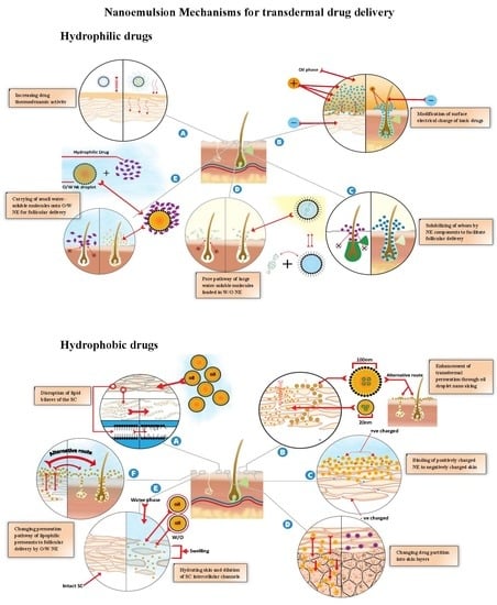
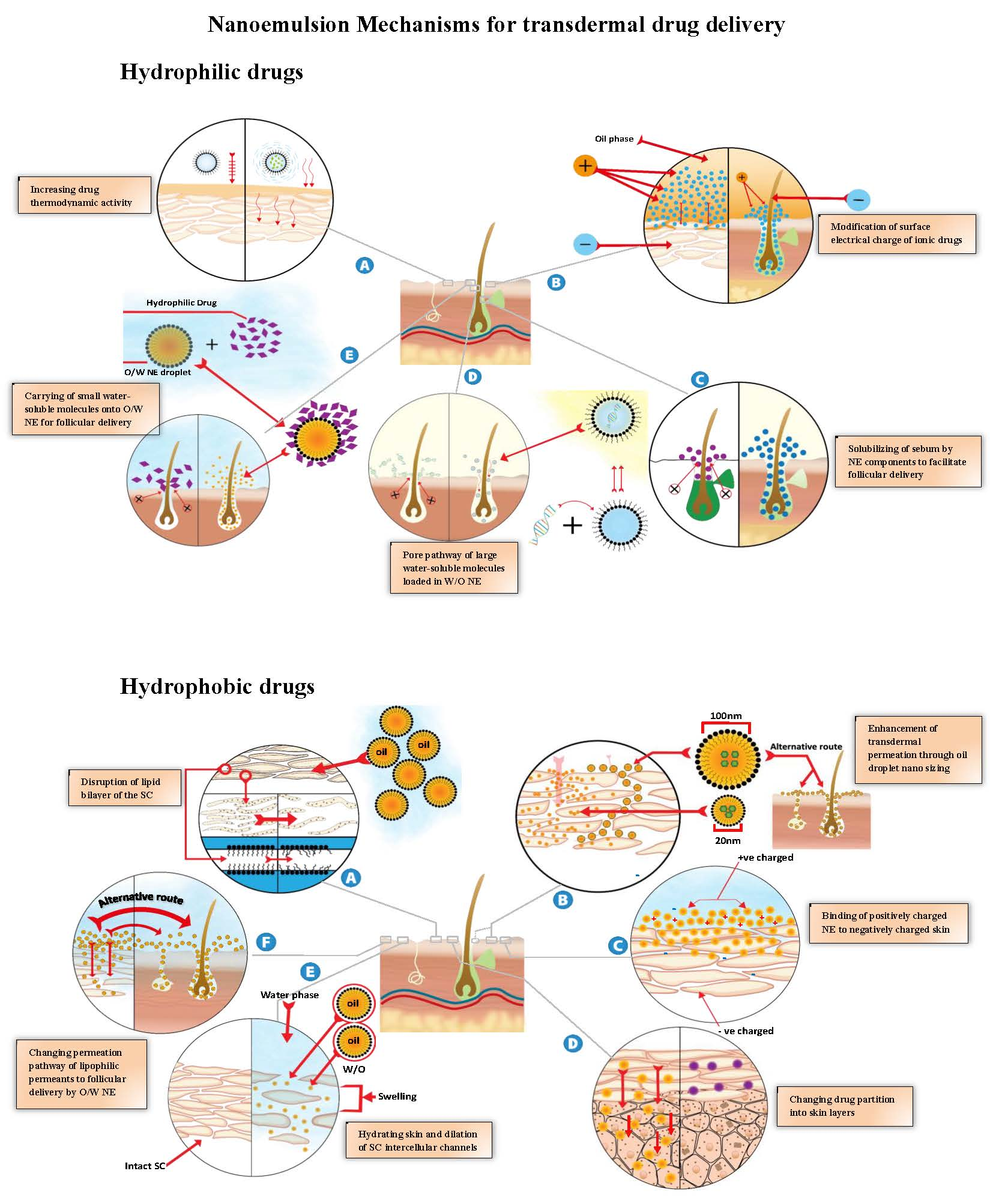
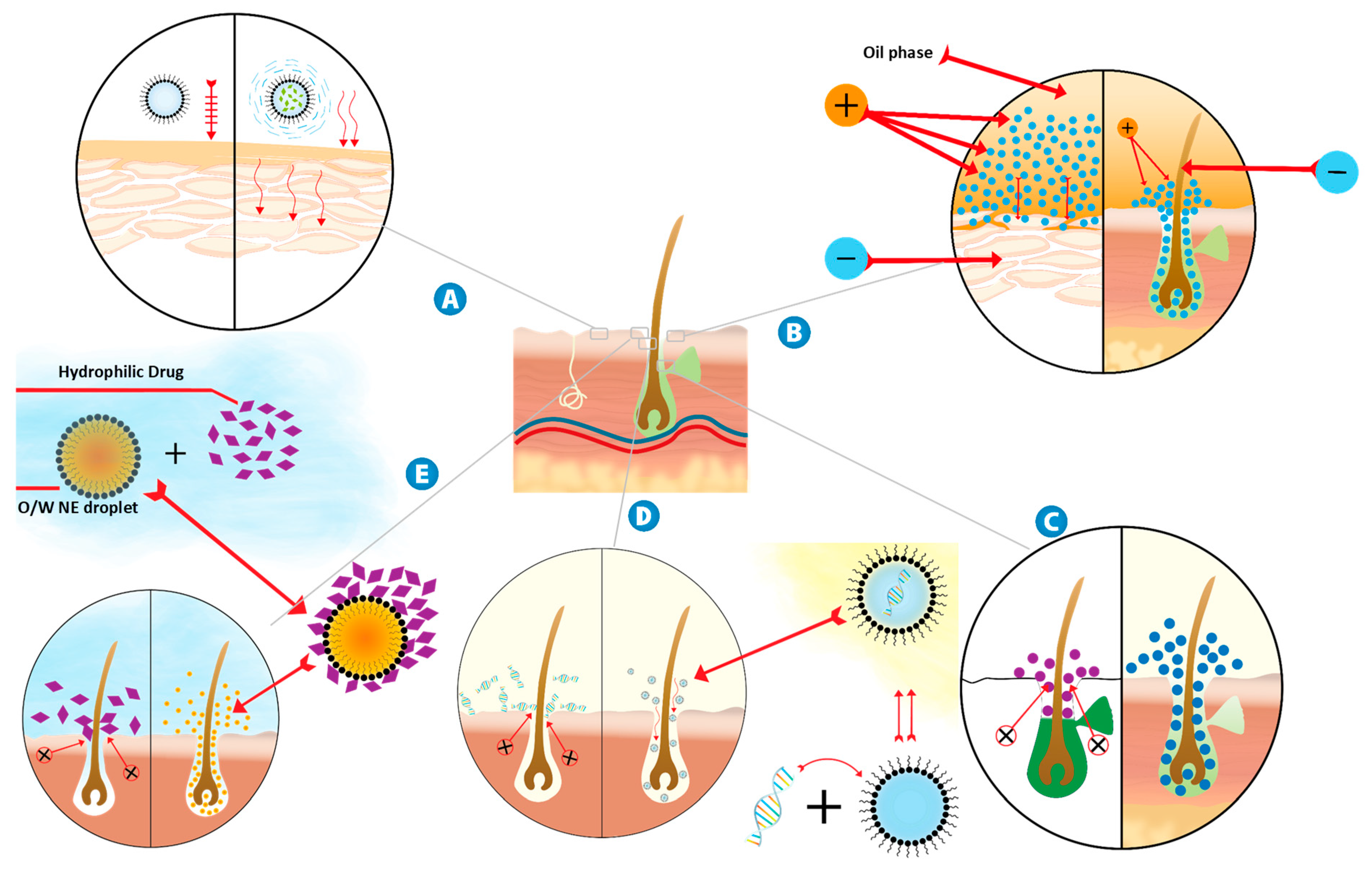
8.2. NE Mechanisms for Enhanced Transdermal Delivery of Hydrophilic Drugs

The mechanism by which a hydrophilic drug is transported through skin from a w/o NE, is indeed different from a hydrophobic drug. As the passage through skin requires an increased lipophilicity [12], it is then important to study the transdermal transport of a hydrophilic compound delivered from NE systems. It was hypothesized that a hydrophilic drug would not be available for percutaneous transport from NEs, unless water from the NE freely permeates transdermally. Therefore, the sufficient mobility of water within NE vehicle and the sufficient percutaneous transport of water across the skin barrier are both required [142]. Moreover, the active compound’s PS and the droplet size play a more important role here than in o/w [143]. Different reported mechanistic theories are discussed as follows and illustrated in Figure 2.

8.2.1. Increasing Drug Thermodynamic Activity

Drug thermodynamic activity is a major factor in skin permeation, where the drug moves from the internal phase to the external phase, then depositing into the skin layers. This provides a continuous process which has a major role on the extent to which the drug can diffuse transdermally, especially for hydrophilic drugs. This phenomenon was observed with glycyrrhizin, which has a large Mw of 822.94 g/mol, where having a low concentration of the surfactant/co-surfactant mixture (Smix) in the formula showed a better release through human skin than with a large concentration, hypothesizing that the low viscosity due to the low Smix content contributed to give the drug molecules more freedom to move through phases and eventually penetrate the SC. Thermodynamic activity is also enhanced with the reversal of altering the skin’s natural barrier resistance and modifying the drug SC partition coefficient by NE components as surfactants and PEs [136]. It has also been suggested that ME, by continuously fluctuating interfaces, may increase the drug mobility, hence aiding its penetration [144]. In addition, the high solubilization capacity of ME increases the thermodynamic activity of permeants, which is one of the driving forces of drug transport [144]. The drug in this energy-rich system can diffuse across the flexible interfacial surfactant film between the phases. This is a thermodynamic process that increases partitioning and diffusion into the SC [145].

8.2.2. Modification of Surface Electrical Charge of Ionic Drugs

The hydrophilic drug RHCL, with low Mw, when topically applied as an aqueous solution, showed no penetration properties, despite the drug’s hydrophilicity and small molecular size. Based on these findings, NE formulation was developed with isopropyl myristate (IPM) as the oil phase (5%), Brij® 35 + Brij® 30 (20–30%) as the surfactant, IPA as the co-surfactant (20–30%), and double-distilled water (34.5–50%).The developed NE exhibited a significant increase in drug permeation rate [105]. It was explained that NEs could modify the surface electrical charge of an ionic drug and then enhance the permeability of a hydrophilic drug, as reported in literature [146,147].

8.2.3. Solubilization of Sebum by NE Components

SC layers tend to be thinner closer to follicular orifices, which are rich in terms of their capillary network. Areas of the human body with higher follicular density like the forehead show good follicular drug delivery compared to other areas like the forearm [136]. However, follicular delivery depends on presence of sebum and the blockage of pores. Otberg et al. [136] reported that the artificial blocking of hair follicles by a varnish wax mixture significantly reduced skin penetration of the hydrophilic permeant caffeine.
It was found that a w/o NE with a low HLB surfactant mixture showed better penetration compared to aqueous and other micellar formulations, where compatibility with the lipophilic nature of the sebum plays a role in solubilizing it by the surfactant mixture, thus clearing the way for the drug to penetrate more easily [130]. There seems to be an ongoing limitation concerning in vitro release studies of the follicular pathway due to follicular shaft collapse and the cessation of perifollicular circulation. Moreover, the animal skin model used to evaluate this route is not always consistent with the release profile when applied to human skin [140]. However, certain in vivo human studies with the hydrophilic molecule caffeine NE showed that the transfollicular route contributed to over a third of the total penetration [139,148,149].

8.2.4. Pore Pathway for the Transport of Large Water-Soluble Molecules Loaded into W/O NEs

Large water-soluble drugs are unable to cross the lipid corneocyte domain. The pore pathway or hair follicle route, a primary path which w/o NEs containing large molecules use to reach the main circulation, is an important transdermal mode of delivering water-soluble permeants. In the gene delivery approach, hair follicles might be a portal toward the entrance of topically applied negatively charged DNA [150].
Recently, scientists researched the need to deliver and transfect plasmid DNA to follicular keratinocytes. They found that transdermal w/o NE allowed DNA expression into therapeutic transgenic proteins [137]. The prepared plain NE had a mean PS of 42.3 nm, while after the addition of a DNA plasmid, a smaller NE with a mean PS of 32 nm was produced. The levels of gene expression after the topical application of the DNA NE were significantly higher than with the plasmid DNA aqueous solution. The authors hypothesized that forming a stable w/o NE may drive the conformation of DNA into a more condensed state. They supported their hypothesis by highlighting the absence of aggregates and large particles during the PS analysis [137].

8.2.5. Carrying of Small Water-Soluble Molecules into O/W NE for Follicular Delivery

The skin permeation of the hydrophilic small molecule 5-aminolevulinic acid (ALA) in both w/o and o/w NE was studied. Low in vitro drug release of w/o NEs was detected and attributed to their high viscosity. Surprisingly, o/w NEs showed the highest permeation flux through nude mice skin, meanwhile, w/o NEs did not show any improvement over the aqueous solution. This result was explained by the required process of drug partitioning from the inner phase to the external phase in w/o NE, while the release from o/w requires diffusion through the external phase [141]. The in vivo high permeation from the o/w NE suggested that other mechanisms than the lipid pathway predominate the delivery. Among these suggested mechanisms are the follicular/sebaceous route, the enhancer effect, and an increase of thermodynamic activity, as suggested by Zhang el al [151].
The importance of hair follicles for o/w NE penetration was verified by in vivo confocal laser scanning microscopy in mice models. Representative fluorescence staining indicated enhanced ALA penetration into deeper skin layers after the application of the o/w nanocarrier systems compared to the control. However, the effect of permeation enhancement for ALA was not detected in the in vitro skin permeation study. The given explanation for this was that, after skin excision, the follicular volume was reduced by the contraction of the elastic fibers, such that the follicles were less receptive to the applied substances [141]. The extended release properties obtained after 48 h address the following question: Is retaining hydrophilic small molecules within skin layers for longer periods a possible mechanism of o/w NEs? From our point of view, w/o NEs, for small hydrophilic molecules, may facilitate their transepidermal permeation. In contrast, the trans-appendageal route is the accepted route of permeation in the case of incorporation in w/o NEs.

9. NEs for the Transdermal Delivery of Hydrophobic Drugs

9.1. Transdermal O/W NEs Containing Hydrophobic Drugs

The incorporation of lipophilic drugs in o/w NEs is reported to be a tool to enhance the transdermal permeation of such drugs, despite the variable explained mechanisms (Table 3).
Aceclofenac was formulated in o/w NE and compared to conventional gel. The optimized formula, consisting of 10% Labrafil, 35.33% Tween 80, 17.6% Transcutol P, 5% Triacetin, and 32% water, showed improved permeation enhancement and acceptable irritation results. The in vivo efficacy showed 40% more anti-inflammatory results in comparison to the aceclofenac gel [151].
The spontaneous emulsification technique was used to prepare the NE formulation containing clozapine, a drug that exhibits only 27% oral bioavailability due to the extensive hepatic metabolism. OA was used as the oil phase, Tween 20 as the surfactant, and Transcutol P as the co-surfactant (Smix 3:1), with 1% clozapine. The NE was incorporated into a gel to extend the contact time with the skin. Overall, 70% of the drug penetrated the skin over a period of 10 h, exhibiting both a rapid release profile over the first 2 h and an extended release profile over the 8 h of skin contact [10].
A tamoxifen o/w NE was investigated for cancer suppression performance in tumor-induced mice. The relative tumor volume after the 23 days testing period showed promising results and increased bioavailability in comparison to the orally administered drug, due to the avoidance of the first pass metabolism related to the transdermal route and consequently a better penetration to the target site [152].
Cumin was loaded in an o/w NE, exploring its capacity to reduce the oxidative damage related to a variety of diseases. The NE formulations showed good permeation results and high zeta potential, which is an indication of good stability. The highest permeation was observed after 6 h. The formulation was able to restore hepatic enzyme plasma levels after the extensive hepatotoxic effect induced by paracetamol after 7 days. Furthermore, a prolonged effect was witnessed, lasting up to 7 days after the single topical application of the NE. This indicated that a weekly application for patients suffering from chronic disease is convenient, with long term health benefits [5].
The secondary effect of tricycle antidepressants (imipramine and doxepin) which possess an analgesic effect on the peripheral nerve endings was investigated in o/w NE formulations. The NE formulation application increased the paw withdrawal time by 13% and 28% for the imipramine NE and the doxepin NE, respectively [153].
A hyaluronic acid-based NE was used as a carrier for vitamin E [154]. The effect on the stability of the NE was investigated by altering the zeta potential, degree of substitution, pH, and crosslinking agents. The encapsulation efficiency results, small size, and improved stability showed that the investigated NE was a potential transdermal carrier for lipophilic drugs [154].
An antifungal agent (amphotericin B) was incorporated into an o/w NE to enhance the transdermal delivery and effectiveness against Candida albicans and Aspergillus niger. The study revealed that the NE’s synergistic ability allows a reduction of the antifungal dose, with increasing therapeutic efficiency [155]. The prepared NE (Capmul PG8 as the oil phase, with Labrasol and PEG-400 as the surfactant and co-surfactant, respectively, and DMSO as the transdermal enhancer) was compared to the commercially available Fungizome® gel and drug solution as well. NE amphotericin B exhibited enhanced transdermal penetration and exhibited a stronger antifungal ability, coupled with longer a shelf life when compared to the conventional amphotericin B topical dose [155].
Singh et al. developed a NE for Carvedilol, a drug that has a short plasma half-life and extensive first pass metabolism. Upon conducting the in vitro and in vivo experimentation, good results were obtained, indicating a great potential for Carvedilol NE to be used in the future [156]. In another study, a significant increase in the Carvedilol permeability coefficient in the NE vehicle was obtained, compared to control film [157].
Finally, human skin permeation studies showed that MEs even enable iodide ions to diffuse through the skin [158]. A high drug amount was released from the NE in comparison to the nano-lipid carrier, which promoted the penetration into the skin, as previously reported [159].

9.2. NE Mechanisms for the Enhanced Transdermal Delivery of Hydrophobic Drugs

In many studies, NEs have proven to be a superior novel drug delivery system, not only due to the advantages mentioned above (ease of preparation, cost effectiveness, and stability) but most importantly due to how efficient and effective NEs are in delivering hydrophobic drugs transdermally to the systemic circulation [168]. Several mechanisms have been proposed by various studies, as mentioned below and presented in Figure 3.

9.2.1. Disruption of the SC Lipid Bilayers

The hydrophobicity of the SC bilayer allowed the entry of PEs and surfactants containing fatty acids. They disrupt the bilayer by creating separate domains, inducing highly permeable pathways in the SC [37]. The enhanced skin permeation and penetration of meloxicam (MLX) from the NE gel was reported, which can be attributed to its compositions in which the ingredients such as caprylic acid (short-chain fatty acid), Tween 80, and PG act as permeation enhancers, potentially significantly reducing the barrier properties of the SC through the extraction of SC lipids [111]. In contrast, the very low permeation rate of MLX from the Tween 80 and propylene glycol solution across hairless mouse skin over 36 h was reported [169]. The difference in the results here may be attributed to the fact that the NE components act synergistically and significantly disrupt the barrier properties of the SC [170]. MLX transport might be attributed to the formation of the pores in the lipid bilayer, therefore creating an increased effective volume inside the SC lipid domain available for drug diffusion [171].
From the improved penetration results of the caffeine and naproxen NEs, it was hypothesized that OA and eucalyptol act as enhancers that disrupt the SC of the skin through the dissolution of lipids layers, allowing the active compound to pass through [164].
The fennel essential oil NE showed high potential for reducing the plasma glucose levels of rats when delivered via the transdermal route, due to the presence of OA and PEG, as both of them act as PEs for dermal delivery, since they increase the fluidity of the liquid portion of the SC. Moreover, Tween 20 (a surfactant) enhanced the flux of the materials permeating through biological membranes, resulting in the better penetration of oil and hence improving its therapeutic activity [172].
An enhanced permeation profile of the lipophilic celecoxib (CXB) in an o/w formulation was reported [20]. This result was explained by the disruption and induction of a temporary denaturation state of the keratin filaments in the SC in its various layers, resulting in a transport pathway in the lipid bilayers. An optimized NE was prepared by dissolving 2% w/w of CXB in a 10% w/w combination of Sefsol 218 and triacetin (1:1). Then, a 50% w/w mixture of Tween 80 and Transcutol P was slowly added in the oil phase.

9.2.2. Enhancement of Transdermal Permeation through the Nano-Sizing of Oil Droplets

In order to investigate the effect of the oil droplet size on transdermal permeation, 1% curcumin was added to an o/w NE. Two homogenization techniques were applied, using either a high-speed homogenizer adjusted at 24,000 rpm or a high-pressure homogenizer accustomed to 1500 bar. The high-speed method resulted in a PS around 600 nm, while the high-pressure technique resulted in a rather smaller size (less than 100 nm). Studies have revealed that anti-inflammatory activity is superior with a smaller NE droplet size [173]. In addition, small droplets have a better chance to adhere to membranes, along with covering a large surface area, transporting bioactive NE compounds in a more controlled way [37]. A larger droplet size with rigid architecture may not readily penetrate. The small droplet size of the NE also provides a very large surface area for drug transfer into the skin [111].
An anti-inflammatory compound, 3,5-dihydroxy-4-isopropylstilbene (DHPS), was integrated into an o/w NE, dramatically improving the transdermal effect of the DHPS. The obtained enhancement was explained by the NE providing better dispersion and a larger contact surface area for the drug molecules, compared to drug suspension, leading to elevated solubility and permeability [166]. Reducing the droplet size from large (5 microns) to sub-micron emulsion (100 nm) increased diazepam transdermal penetration. This submicron size resulted in satisfactory drug delivery for up to 6 h, with only one single topical application of diazepam cream onto mice skin [174]. A possible explanation of the enhanced transdermal delivery of lipophilic progesterone and hydrophilic adenosine compared to the drug solutions was the large surface area of the NE, which is associated with the low interfacial tension and the small droplet size [40].
Glibenclamide was incorporated into a transdermal NE gel, due to its low oral bioavailability and its severe and fatal hypoglycemic potential. A NE gel was formulated successfully and tested for compatibility and release. The results of the study indicated that NE gel formulations enhanced the drug permeation, and this was attributed to nano-sizing [163]. The large surface area was one possible explanation for this, however, the authors agreed that the role of droplet size was less important than previously assumed by other authors [175,176]. In another study, data revealed that a negatively charged NE with a smaller PS (136 nm) had a higher in vitro release and increased penetration when compared to the positively charged formulation (157 nm). The results were explained by the fact that when the particles are smaller, the surface and the solubility pressure of the particles is bigger [177,178].
Su et al. experimented with the effective parameters for the PS of an octyldodecanol NE. They suggested that the water percentage played the biggest part on NE droplet size, and that the more water content, the bigger the droplet size. In their opinion, the effective parameters for droplet size are the mixing rate (higher rotation speeds resulted in a smaller oil droplet size), temperature, and NE surfactants used in the NE [179].

9.2.3. Binding of the Positively Charged NE to Negatively Charged Skin

Aiming to evaluate the effect of charge on skin penetration, fludrocortisone acetate and flumethasone pivalate NEs were experimented with, with a pre-impregnated aqueous phase of either a negative or positive charge. Observing the charge effect through release studies revealed that the positively charged NE was more efficient in transporting the active molecules through skin than the negatively charged one. The explanation for this enhanced ability is the binding of the positive NE onto the negatively charged skin, leading to an increase in contact time which eventually allows the NE oily core to pass steadily successfully pass through the various skin layers [180]. The inclusion of a positive charged carrier in the formulation and the subsequent adherence to the negative charged skin was aimed to increase the NE retention time and consequently the drug bioavailability [163]. The skin accumulation of miconazole (MCZ) from positively charged droplets is higher than that from negatively charged ones. This is attributed to the binding affinity of droplets to negatively charged skin. Researchers proposed that positively charged vehicles appear to be promising for topical drug delivery [181]. NEs containing chitosan were demonstrated to be more adequate as a vehicle for substances intended to act on the skin, such as sunscreens, as photoprotective products are topically administrated and they should not permeate through the skin. Since chitosan is positively charged, with a degree of deacetylation of 79%, it could explain the attractive interaction of the polymer with the skin which is negatively charged, thus preventing skin permeation [182].
Regarding stability, the positive surface charge of nanodroplets induced by phytosphingosine and the pH provided good stability for the NE, as the repulsive forces prevented the collision of NE droplets by a steric shield [183,184]. Regarding skin permeation, it has been shown that a positively charged NE significantly promoted the diffusion rate of both econazole and MCZ nitrate through rat skin when compared to the negatively charged preparations. This was done through increasing the amount of cumulated drug and consequently the transdermal flux [143]. On the other hand, the increased penetration of the positively charged NE is explained by the increased interaction and adsorption of particles with the negatively charged keratinized corneocytes of the SC, the main barrier of the skin. Here, the results showed that there is a clear relation between the NE charge and skin penetration, however, the authors revealed that there was no detectable skin permeation due to drug accumulation in the skin layers [177,178]. Finally, we can conclude from the literature that NE charge influence is more relevant than droplet size regarding skin retention, while the droplet size effect dominates in the context of skin permeation.

9.2.4. Enhancement Transdermal Permeation by Reducing the NE Viscosity

The successful passage of the drug through the SC was related to NE’s low viscosity, along with the presence of an aqueous portion in the formulation. Researchers observed the impact of viscosity when they compared their NE with a gel formulation, hypothesizing that low viscosity, working in conjunction with other factors, such as droplet size, increases the contact area with skin, resulting in easier penetration [163].
The permeation enhancement of celecoxib (CBX) was attributed to the NE’s low viscosity and formulation stability, where both the oil phase and the water phase work in conjunction, leading to more efficient drug permeation compared to conventional gel [160]. A piroxicam NE exhibited better skin flux compared to the NE gel preparation. The obtained results were attributed to the low viscosity of the NE, which facilitates the passage of NE components through the SC [167]. The small globule size of the terbinafine NE and its low viscosity were the main factors for enhanced drug permeation [110]. As the proportion of the Carbopol 934 matrix increased in the terbinafine NE gel, the drug permeation rate declined, along with increased drug deposition in the epidermis and dermis. Here, the authors concluded that increasing the NE viscosity tends to change the formulation from transdermal delivery to topical delivery [185]. Similar results were obtained for valsartan transdermal permeation in Carbopol gel, where the gelling agent reduced drug penetration due to the transformation of the NE to a highly ordered nanostructure [186].
A lower inulin skin permeation rate was detected from micellar formulations, regardless of the surfactant composition. The given explanation for this was that the high viscosity of all micellar systems possibly played a role in lowering the permeation rates of inulin across hairy mouse skin into the Franz cell receiver compartment [130].

9.2.5. Changing the Drug Partition into Skin Layers

Scientists compared the difference in penetration profile between the copaiba oil NE with Tween 20 as the surfactant and the oil alone by measuring the amount of the lipophilic drug β-caryophyllene. Upon conducting permeation and retention studies, it was found that when copaiba oil was used alone, the lipophilic drug was only found in the SC layer, failing to penetrate into the epidermis and dermis, while the copaiba oil NE was able to be detected successfully in the SC, epidermis, and dermis, demonstrating NE’s inherent ability to provide a more efficient penetration and retention in skin layers than the drug alone [187]. A study researched the importance of ME structure on drug uptake into the skin. In particular, cyclosporine skin retention data suggested that the surfactant type, co-surfactant type, water content, and the presence of a rheological modifier could enhance drug mobility and modify its tendency to partition into the SC [188]. Most likely, both o/w and w/o MEs are valuable systems that preferentially retain the drug into skin, but the mechanism still requires more studies and explanations [189].

9.2.6. Hydrating the Skin and Dilating the SC Intercellular Channels

Hydration disrupts the SC structure. Overall, 15% to 20% of the SC weight is composed of water [190]. Skin hydration leads to SC swelling and the opening of its tight brick structures, consequently increasing permeability [38]. It has been reported that extended skin hydration makes the intercellular spaces distend, dilating the lacunar networks that link the connections of the water pool system in SC interstices, which are disconnected under normal conditions [191]. Actually, the existing water in the intercellular lipid region act as water pools and are not continuous toward the deeper layers of the SC. However, small channels are formed when the skin is hydrated, reducing the diffusional resistance through the rough SC structures. Both lipophilic and hydrophilic compounds benefit from this feature. Small hydrophilic molecule penetration enhancers such as azone seem to benefit the most from hydration, regarding absorption in corneocytes at least [192]. Even though such a mechanism seems to increase both the penetration of both hydrophilic and lipophilic compounds, researchers argue that hydrophilic compounds may not benefit greatly from skin hydration, since the formation of such water pool channels in the SC is short lived and can only aid the lipophilic compounds in o/w NEs. They explained that such NEs containing water as an external phase saturate SC layers faster and hence lead to an enhanced permeability into the dermis [65,193]. In addition, the aqueous part of NE enters through the appendageal pathway and enlarges the interlamellar volume of the SC lipid bilayer, allowing the lipophilic drug to pass more easily through the various sheets of the SC [173]. An additional mechanism was suggested, where hydration and increasing water content accelerates permeant thermodynamic activity and consequently penetration through the skin [194].
These observations suggested that the transport properties can be regulated by the water content in the SC [195]. The NE components may change the water gradient in upper skin layers by avoiding evaporation, exhibiting an occlusive effect, and consequently affecting skin permeation [196]. Fourier transform infrared spectroscopy detected that by increasing water concentration in MEs, the ratio of the amide I/II band gradually increases, suggesting an increase in the hydration of the SC [197,198]. In general, increased tissue hydration appears to increase the transdermal delivery of both hydrophilic and lipophilic permeants [199].

9.2.7. Changing the Permeation Pathway of Lipophilic Permeants to Follicular Delivery

Despite follicular delivery not being the usual pathway of hydrophobic molecules, such drugs when formulated in o/w NEs, accumulate near hair follicles. Fluorescent dye (Nile red, 0.1% w/w) was incorporated in a capsaicin o/w NE to study skin penetration. Capsaicin is an interesting lipid soluble active ingredient with multiple therapeutic uses, such as in cancer treatment, inflammation, and treating cardiovascular system (CVS) diseases. Being able to deliver capsaicin while bypassing its first pass effect would definitely be of outmost importance. The optimized NE has shown deep permeation through pig skin layers. Strong fluorescence intensity was witnessed across all of the skin layers, where the tested NE showed red fluorescence deep to the dermis, reaching up to 700 μm. The strongest fluorescent spot was observed near the hair follicles. Capsaicin o/w NEs can penetrate and accumulate in the skin well through the hair follicle [165]. Similar results were obtained from ME and demonstrated that o/w and not w/o ME is a promising vehicle for the transfollicular delivery of the lipophilic adapalene [200].

10. Conclusions

Many hydrophilic and hydrophobic drugs were successfully loaded in w/o or o/w NEs, respectively, demonstrating their ability to pass into general circulation. Accordingly, the NE components have to be optimally selected to suit the drug psychochemical properties and simultaneously predict NE penetration behavior through the skin.
For the first time, different penetration mechanisms of each NE type have been explored and studied. For hydrophilic drugs, different mechanisms have explained their mode of penetration for w/o NEs. The facilitated transport of these permeants was attributed to the effect of the NE in increasing the drug thermodynamic activity or modifying the surface electrical charge of ionic drugs. The same enhancement was sometimes explained by the solubilization of sebum by the NE components, facilitating the follicular delivery of hydrophilic drugs. Most of these mechanisms focus more on the nature of the NE than the physicochemical properties of the drug. However, other transdermal strategies focus on permeant properties, namely either large or small Mw water-soluble molecules loaded into w/o NEs, triggering follicular delivery, the normal dominant pathway of hydrophilic permeants.
On the other hand, the penetration of hydrophobic drugs focuses more on the effect of the NE components on the skin barrier. The disruption of the lipid bilayer of the SC was still the most popular mechanism of transdermal enhancement, despite the reported enhancement through oil droplet nano-sizing and reducing viscosity during the preparation of the NE. The binding of a positively charged NE to negatively charged skin was reported as a controversial mechanism, as some researchers ascribed the acceleration in penetration to this mechanism while others revealed that it causes an increase in drug accumulation and retention in skin layers, and hence a reduction in permeation. Hydrating the skin and dilating the SC intercellular aqueous and lipid channels were reported to enhance the permeation of lipophilic permeants. Moreover, changing the permeation pathway of lipophilic permeants to follicular delivery using o/w NEs was reported, despite the well-known facts that oily drugs pass through the lipid pathway and that the area of the pore pathway is neglected. The question is: Do we need to incorporate lipophilic drugs in o/w NEs? Does changing the pathway to hair follicles reduce the transdermal passage of lipophilic permeants? We suggest that o/w NEs have low potential in enhancing the permeation of hydrophobic molecules, therefore, further studies are needed.
Most likely, the overall combination of such different mechanisms is the real cause of the skin penetration enhancement effect for both types of permeants. It seems that no one specific mechanism could provide a satisfying explanation for the superiority of NEs compared to other formulations. Finally, the mechanisms reported in the literature are highly diverse, to the extent that a definite mechanism is not very conclusive, highlighting the need for further investigation.
The compatibility with a wide range of hydrophobic and hydrophilic drugs, depending on its type (o/w and w/o), makes NE a very encouraging transdermal dosage form. The diverse preparation techniques, high drug encapsulation capacity (either in oil or water droplets), stability, safety, and cost effectiveness motivate researchers to carry out further studies. This review suggests future research should be focused on more understanding of the penetration mechanisms, the development of a ready-to-use NE, and targeting the CNS via a transdermal NE. From the literature, NEs are considered a useful tool for by passing the P-glycoprotein (P-gp) pump through the general ‘masking’ concept of the active agents, where the agents prevent the droplets from being discovered by the transporter while they enter the blood brain barrier. Moreover, surfactants such as polysorbate 80 (P80) in NE droplets are well-known P-gp inhibitors.
Despite all the trials made in understanding the mechanisms of how NEs are able to successfully transport hydrophilic and hydrophobic compounds through skin layers into the systemic circulation and the various proposed mechanisms hypothesized and summarized in this review article, more focused research is needed to fully study, confirm, and understand the exact interaction that happens between the skin layers and NE components through physical observation and real time monitoring of the NE penetration of the skin. In other words, the drug molecule pathway through the SC lipoid or pore domain needs to be fully understood.
Certain NE components seem to show a wide compatibility range with many drugs, such as OA, labrasol, ethanol, and Tween, as seen in the publications shared in this review. This could be developed into a concept of formulating a general-purpose NE carrier that is pre-formulated and manufactured as a ready-to-use NE, allowing formulators to only add the active constituent. The industrial development and production of NEs could be also very promising, being a time-saving and cost-effective process based on the simplicity in NE preparation as well as the cheapness of their constituents. Within a specific HLB range, pharmacists would be able to prepare transdermal NEs for patients who show low compliance or inability to tolerate the side effects associated with taking the drug orally. The low toxicity of many of NE components makes this a very promising concept for future work in transdermal delivery systems.
A lot of research is still required for the development of novel NE formulations, especially in drugs that target the central nervous system. Such drugs are most often very lipophilic in nature, passing through the blood brain barrier to exert their action and usually requiring patients to take them chronically for months or years. This drug group is also extensively metabolized by the liver. Delivering these drugs transdermally, through bypassing the hepatic metabolism, sustains a steady plasma concentration and potentially provides a more localized transdermal application for a fast onset of action.

Funding

This research received no external funding.

Conflicts of Interest

The authors declare no conflict of interest.

Abbreviations

ALA5-aminolevulinic acid
CVScardiovascular system
DHPS3,5-Dihydroxy-4-isopropylstilbene
DMSODimethyl sulfoxide
EIPemulsion inversion point
GITgastrointestinal tract
HLBhydrophile-lipophile balance
IPAIsopropyl alcohol
IPMIsopropyl myristate
MCZMiconazole
MEMicroemulsion
MLXMeloxicam
MPaMega Pascal
Mwmolecular weight
NENanoemulsion
NMPN-methyl pyrrolidone
NSAIDsNon-steroidal anti-inflammatory drugs
o/woil-in-water
OAOleic acid
PEPermeation enhancer
PEGPolyethylene glycol
PITPhase inversion temperature
PSParticle size
psiPounds square inch
RHCLRopinirole hydrochloride
SCStratum corneum
SLSsodium lauryl sulfate
Smixsurfactant/co-surfactant mixture
TCCThiocolchicoside
CBXCelecoxib
w/owater-in-oil

References

  1. Thakur, N.; Garg, G.; Sharma, P.K.; Kumar, N. Nanoemulsions: A Review on Various Pharmaceutical Application. Glob. J. Pharmacol. 2012, 6, 222–225. [Google Scholar]
  2. Shakeel, F.; Ramadan, W. Transdermal delivery of anticancer drug caffeine from water-in-oil nanoemulsions. Colloids Surf. B Biointerfaces 2010, 75, 356–362. [Google Scholar] [CrossRef]
  3. Çinar, K. A Review on Nanoemulsions: Preparation Methods and Stability. Trak. Univ. J. Eng. Sci. 2017, 18, 73–83. [Google Scholar]
  4. Sarker, D. Engineering of Nanoemulsions for Drug Delivery. Curr. Drug Deliv. 2005, 2, 297–310. [Google Scholar] [CrossRef]
  5. Mostafa, D.M.; Kassem, A.A.; Asfour, M.H.; Al Okbi, S.Y.; Mohamed, D.A.; Hamed, T.E.S. Transdermal cumin essential oil nanoemulsions with potent antioxidant and hepatoprotective activities: In-vitro and in-vivo evaluation. J. Mol. Liq. 2015, 212, 6–15. [Google Scholar] [CrossRef]
  6. Khopade, A.J.; Nandakumar, K.S.; Jain, N.K. Lectin-functionalized multiple emulsions for improved cancer therapy. J. Drug Target. 1998, 6, 285–292. [Google Scholar] [CrossRef]
  7. Hildebrand, A.; Schaedlich, A.; Rothe, U.; Neubert, R.H.H. Sensing specific adhesion of liposomal and micellar systems with attached carbohydrate recognition structures at lectin surfaces. J. Colloid Interface Sci. 2002, 249, 274–281. [Google Scholar] [CrossRef]
  8. Barakat, N.; Fouad, E.; Elmedany, A. Formulation Design of Indomethacin-Loaded Nanoemulsion For Transdermal Delivery. Pharm. Anal. Acta 2011, 2, 1–8. [Google Scholar] [CrossRef]
  9. Nastiti, C.M.R.R.; Ponto, T.; Abd, E.; Grice, J.E.; Benson, H.A.E.; Roberts, M.S. Topical nano and microemulsions for skin delivery. Pharmaceutics 2017, 9, 37. [Google Scholar] [CrossRef]
  10. Shafaat, K.; Kumar, B.; Das, S.K.; Ul Hasan, R.; Prajapati, S.K. Novel nanoemulsion as vehicles for transdermal delivery of Clozapine: In vitro and in vivo studies. Int. J. Pharm. Pharm. Sci. 2013, 5, 126–134. [Google Scholar]
  11. Iman, I.S.; Nadia, A.S.; Ebtsam, M.A. Formulation and stability study of chlorpheniramine maleate transdermal patch. Asian J. Pharm. 2010, 4, 17–23. [Google Scholar]
  12. Peira, E.; Scolari, P.; Gasco, M.R. Transdermal permeation of apomorphine through hairless mouse skin from microemulsions. Int. J. Pharm. 2001, 226, 47–51. [Google Scholar] [CrossRef]
  13. Singh, I.; Morris, A.P. Performance of transdermal therapeutic systems: Effects of biological factors. Int. J. Pharm. Investig. 2011, 1, 4–9. [Google Scholar] [CrossRef] [PubMed] [Green Version]
  14. Johnson, M.E.; Blankschtein, D.; Langer, R. Evaluation of solute permeation through the stratum corneum: Lateral bilayer diffusion as the primary transport mechanism. J. Pharm. Sci. 1997, 86, 1162–1172. [Google Scholar] [CrossRef] [PubMed]
  15. Barry, B.W. Novel mechanisms and devices to enable successful transdermal drug delivery. Eur. J. Pharm. Sci. 2001, 14, 101–114. [Google Scholar] [CrossRef]
  16. Barry, B.W. Lipid-Protein-Partitioning theory of skin penetration enhancement. J. Control. Release 1991, 15, 237–248. [Google Scholar] [CrossRef]
  17. Roberts, M.S. Targeted drug delivery to the skin and deeper tissues: Role of physiology, solute structure and disease. Clin. Exp. Pharmacol. Physiol. 1997, 24, 874–879. [Google Scholar] [CrossRef]
  18. Ng, K.W.; Lau, W.M. Skin deep: The basics of human skin structure and drug penetration. In Percutaneous Penetration Enhancers Chemical Methods in Penetration Enhancement: Drug Manipulation Strategies and Vehicle Effects; Maibach, H., Ed.; Springer: Berlin/Heidelberg, Germany, 2015; pp. 3–11. ISBN 9783662450130. [Google Scholar]
  19. Shaker, D.S.; Ghanem, A.H.; Li, S.K.; Warner, K.S.; Hashem, F.M.; Higuchi, W.I. Mechanistic studies of the effect of hydroxypropyl-β-cyclodextrin on in vitro transdermal permeation of corticosterone through hairless mouse skin. Int. J. Pharm. 2003, 253, 1–11. [Google Scholar] [CrossRef]
  20. Shakeel, F.; Baboota, S.; Ahuja, A.; Ali, J.; Shafiq, S. Skin permeation mechanism and bioavailability enhancement of celecoxib from transdermally applied nanoemulsion. J. Nanobiotechnol. 2008, 6, 8. [Google Scholar] [CrossRef]
  21. Scheuplein, R.J. Mechanism of Percutaneous Absorption: II. Transient Diffusion and the Relative Importance of Various Routes of Skin Penetration. J. Investig. Dermatol. 1967, 48, 79–88. [Google Scholar] [CrossRef]
  22. Trauer, S.; Lademann, J.; Knorr, F.; Richter, H.; Liebsch, M.; Rozycki, C.; Balizs, G.; Büttemeyer, R.; Linscheid, M.; Patzelt, A. Development of an in vitro modified skin absorption test for the investigation of the follicular penetration pathway of caffeine. Skin Pharmacol. Physiol. 2010, 23, 320–327. [Google Scholar] [CrossRef] [PubMed]
  23. Warner, K.S.; Kevin Li, S.; Higuchi, W.I. Influences of alkyl group chain length and polar head group on chemical skin permeation enhancement. J. Pharm. Sci. 2001, 90, 1143–1153. [Google Scholar] [CrossRef] [PubMed]
  24. Yoneto, K.; Ghanem, A.-H.; Higuchi, W.I.; Peck, K.D.; Li, S.K. Mechanistic studies of the 1-alkyl-2-pyrrolidones as skin permeation enhancers. J. Pharm. Sci. 1995, 84, 312–317. [Google Scholar] [CrossRef] [PubMed]
  25. Lawrence, M.J.; Rees, G.D. Microemulsion-Based Media as Novel Drug Delivery Systems. Adv. Drug Deliv. Rev. 2000, 45, 89–121. [Google Scholar] [CrossRef]
  26. Valenta, C.; Schultz, K. Influence of carrageenan on the rheology and skin permeation of microemulsion formulations. J. Control. Release 2004, 95, 257–265. [Google Scholar] [CrossRef] [PubMed]
  27. Singh, R.; Chakravorty, A.; Kumar, P.; Chaturvedi, A. NANOEMULSION: An Effective Therapy for Transdermal Drug Delivery. Res. J. Biol. 2013, 3, 33–38. [Google Scholar]
  28. Changez, M.; Varshney, M.; Chander, J.; Dinda, A.K. Effect of the composition of lecithin/n-propanol/isopropyl myristate/water microemulsions on barrier properties of mice skin for transdermal permeation of tetracaine hydrochloride: In vitro. Colloids Surf. B Biointerfaces 2006, 50, 18–25. [Google Scholar] [CrossRef]
  29. Dreher, F.; Walde, P.; Walther, P.; Wehrli, E. Controlled release Interaction of a lecithin microemulsion gel with human. J. Control. Release 1997, 45, 131–140. [Google Scholar] [CrossRef]
  30. McClements, D.J. Nanoemulsions versus microemulsions: Terminology, differences, and similarities. Soft Matter 2012, 8, 1719–1729. [Google Scholar] [CrossRef]
  31. Bouchemal, K.; Briançon, S.; Perrier, E.; Fessi, H. Nano-emulsion formulation using spontaneous emulsification: Solvent, oil and surfactant optimisation. Int. J. Pharm. 2004, 280, 241–251. [Google Scholar] [CrossRef]
  32. Suyal, J.; Ganesh, B. An introductory review article on nanoemulsion. J. Pharm. Pharm. Sci. 2017, 2, 35–40. [Google Scholar]
  33. Montes de Oca-Ávalos, J.M.; Candal, R.J.; Herrera, M.L. Nanoemulsions: Stability and physical properties. Curr. Opin. Food Sci. 2017, 16, 1–6. [Google Scholar] [CrossRef]
  34. Sharma, N.; Bansal, M.; Visht, S.; Sharma, P.; Kulkarni, G. Nanoemulsion: A new concept of delivery system. Chron. Young Sci. 2010, 1, 2–6. [Google Scholar]
  35. Shrestha, H.; Bala, R.; Arora, S. Lipid-Based Drug Delivery Systems. J. Pharm. 2014, 2014, 801820. [Google Scholar] [CrossRef] [PubMed]
  36. Kawakami, K.; Yoshikawa, T.; Moroto, Y.; Kanaoka, E.; Takahashi, K.; Nishihara, Y.; Masuda, K. Microemulsion formulation for enhanced absorption of poorly soluble drugs: I. Prescription design. J. Control. Release 2002, 81, 65–74. [Google Scholar] [CrossRef]
  37. Kogan, A.; Garti, N. Microemulsions as transdermal drug delivery vehicles. Adv. Colloid Interface Sci. 2006, 123, 369–385. [Google Scholar] [CrossRef] [PubMed]
  38. Benson, H. Transdermal Drug Delivery: Penetration Enhancement Techniques. Curr. Drug Deliv. 2005, 2, 23–33. [Google Scholar] [CrossRef] [PubMed]
  39. Gupta, A.; Eral, H.B.; Hatton, T.A.; Doyle, P.S. Nanoemulsions: Formation, properties and applications. Soft Matter 2016, 12, 2826–2841. [Google Scholar] [CrossRef]
  40. Hosmer, J.; Reed, R.; Bentley, M.V.L.B.; Nornoo, A.; Lopes, L.B. Microemulsions Containing Medium-Chain Glycerides as Transdermal Delivery Systems for Hydrophilic and Hydrophobic Drugs. AAPS PharmSciTech 2009, 10, 589–596. [Google Scholar] [CrossRef] [Green Version]
  41. Mei, Z.; Chen, H.; Weng, T.; Yang, Y.; Yang, X. Solid lipid nanoparticle and microemulsion for topical delivery of triptolide. Eur. J. Pharm. Biopharm. 2003, 56, 189–196. [Google Scholar] [CrossRef]
  42. Pandey, A. Role of Surfactants as Penetration Enhancer in Transdermal Drug Delivery System. J. Mol. Pharm. Org. Process Res. 2014, 2, 2–7. [Google Scholar] [CrossRef]
  43. Kitagawa, S.; Kasamaki, M.; Ikarashi, A. Effects of n-alkyltrimethylammonium on skin permeation of benzoic acid through excised guinea pig dorsal skin. Chem. Pharm. Bull. 2000, 48, 1698–1701. [Google Scholar] [CrossRef] [PubMed]
  44. Scheuplein, R.; Ross, L. Effects of surfactants and solvents on the permeability of epidermis. J. Soc. Cosmet. Chem. 1970, 21, 853–873. [Google Scholar]
  45. Kouchak, M.; Handali, S. Effects of Various Penetration Enhancers on Penetration of Aminophylline Through Shed Snake Skin. Jundishapur J. Nat. Pharm. Prod. 2014, 9, 24–29. [Google Scholar] [CrossRef] [PubMed] [Green Version]
  46. Scott, R.; Guy, R.H.; Hadgraft, J. Prediction of Percutaneous Penetration: Methods, Measurements, Modelling. Scott, R.C., Richard, H., Guy, J.H., Eds.; IBC Technical Services: London, UK, 1990; ISBN 1852711175, 9781852711177. [Google Scholar]
  47. Breuer, M.M. The interaction between surfactants and keratinous tissues. J. Soc. Cosmet Chem. 1979, 30, 41–64. [Google Scholar]
  48. Froebe, C.L.; Simion, F.A.; Rhein, L.D.; Cagan, R.H.; Kligman, A. Stratum corneum lipid removal by surfactants: Relation to in vivo irritation. Dermatologica 1990, 181, 277–283. [Google Scholar] [CrossRef] [PubMed]
  49. Lévêque, J.L.; de Rigal, J.; Saint-Léger, D.; Billy, D. How does sodium lauryl sulfate alter the skin barrier function in man? A multiparametric approach. Skin Pharmacol. 1993, 6, 111–115. [Google Scholar] [CrossRef]
  50. Patil, S.; Singh, P.; Maibach, H. Radial Spread of Sodium Lauryl Sulfate After Topical Application. Pharm. Res. 1995, 12, 2018–2023. [Google Scholar] [CrossRef]
  51. Patil, S.; Singh, P.; Sarasour, K.; Maibach, H. Quantification of sodium lauryl sulfate penetration into the skin and underlying tissue after topical application—Pharmacological and toxicological implications. J. Pharm. Sci. 1995, 84, 1240–1244. [Google Scholar] [CrossRef]
  52. Rhein, L.; Robbins, C.; Fernee, K. Surfactant structure effects on swelling of isolated human stratum corneum. J. Soc. Cosmet. Chem. 1986, 139, 125–139. [Google Scholar]
  53. Gibson, W.T.; Teall, M.R. Interactions of C12 surfactants with the skin: Changes in enzymes and visible and histological features of rat skin treated with sodium lauryl sulphate. Food Chem. Toxicol. 1983, 21, 587–594. [Google Scholar] [CrossRef]
  54. Thomas, G. Polefka Handbook of detergents Part A: Properties. In Marcel Dekker; Broze, G., Ed.; Marcel Dekker Inc.: New York, NY, USA, 1999; pp. 433–468. ISBN 0-8247-1417-2. [Google Scholar]
  55. Gradzielski, M. Effect of the Cosurfactant Structure on the Bending Elasticity in Nonionic Oil-in-Water Microemulsions. Langmuir 1998, 14, 6037–6044. [Google Scholar] [CrossRef]
  56. Zana, R. Surfactant solutions: New methods of Investigation. In Surfuctant Solutions: New Methods of Investigation; Marcel Dekker Inc.: New York, NY, USA, 1979; pp. 2–51. ISBN 0824776232. [Google Scholar]
  57. Yadav, S.A.; Singh, D.; Poddar, S. Influence of components of nanoemulsion system for transdermal drug delivery of nimodipine. Asian J. Pharm. Clin. Res. 2012, 5, 209–214. [Google Scholar]
  58. Shafiq, S.; Shakeel, F.; Talegaonkar, S.; Ahmad, F.J.; Khar, R.K.; Ali, M. Development and bioavailability assessment of ramipril nanoemulsion formulation. Eur. J. Pharm. Biopharm. 2007, 66, 227–243. [Google Scholar] [CrossRef] [PubMed]
  59. Tenjarla, S. Microemulsions: An overview and pharmaceutical applications. Crit. Rev. Ther. Drug Carrier Syst. 1999, 16, 461–521. [Google Scholar] [CrossRef] [PubMed]
  60. Kegel, W.K.; Lekkerkerker, H.N.W. Phase behaviour of an ionic microemulsion system as a function of the cosurfactant chain length. Colloids Surf. A Physicochem. Eng. Asp. 1993, 76, 241–248. [Google Scholar] [CrossRef] [Green Version]
  61. Klossek, M.L.; Marcus, J.; Touraud, D.; Kunz, W. The extension of microemulsion regions by combining ethanol with other cosurfactants. Colloids Surf. A Physicochem. Eng. Asp. 2013, 427, 95–100. [Google Scholar] [CrossRef]
  62. Stilbs, P. Fourier transform NMR pulsed-gradient spin-echo (FT-PGSE) self-diffusion measurements of solubilization equilibria in SDS solutions. J. Colloid Interface Sci. 1982, 87, 385–394. [Google Scholar] [CrossRef]
  63. Resende, K.X.; Corrêa, M.A.; Gomes De Oliveira, A.; Scarpa, M.V.; Scarpa, M.V. Effect of cosurfactant on the supramolecular structure and physicochemical properties of non-ionic biocompatible microemulsions. Rev. Bras. Ciências Farm. Braz. J. Pharm. Sci. 2008, 44, 35–42. [Google Scholar] [CrossRef] [Green Version]
  64. Ling, Y.; Yu, M.; Guo, F.; Li, N.; Tan, F.P. Synergistic effect of mixed cosurfactants on transdermal delivery of indomethacin from O/W microemulsion. Chem. Res. Chin. Univ. 2013, 29, 338–343. [Google Scholar] [CrossRef]
  65. Williams, A.C.; Barry, B.W. Penetration enhancers. Adv. Drug Deliv. Rev. 2012, 64, 128–137. [Google Scholar] [CrossRef]
  66. dos Anjos, J.L.V.; Alonso, A. Terpenes increase the partitioning and molecular dynamics of an amphipathic spin label in stratum corneum membranes. Int. J. Pharm. 2008, 350, 103–112. [Google Scholar] [CrossRef] [PubMed]
  67. Abd, E.; Benson, H.A.E.; Roberts, M.S.; Grice, J.E. Minoxidil skin delivery from nanoemulsion formulations containing eucalyptol or oleic acid: Enhanced diffusivity and follicular targeting. Pharmaceutics 2018, 10, 19. [Google Scholar] [CrossRef] [PubMed]
  68. Niazy, E.M. Influence of oleic acid and other permeation promoters on transdermal delivery of dihydroergotamine through rabbit skin. Int. J. Pharm. 1991, 67, 97–100. [Google Scholar] [CrossRef]
  69. Mirejovsky, D.; Takruri, H. Dermal penetration enhancement profile of hexamethylenelauramide and its homologues: In vitro versus in vivo behavior of enhancers in the penetration of hydrocortisone. J. Pharm. Sci. 1986, 75, 1089–1093. [Google Scholar] [CrossRef] [PubMed]
  70. Madison, K.C. Barrier Function of the Skin: ‘“La Raison d’Etre”’ of the Epidermis. J. Investig. Dermatol. 2003, 121, 231–241. [Google Scholar] [CrossRef] [PubMed]
  71. Aungst, B.J.; Nancy, J.R.; Shefter, E. Enhancement of naloxone penetration through human skin in vitro using fatty acids, fatty alcohols, surfactants, sulfoxides and amides. Int. J. Pharm. 1986, 33, 225–234. [Google Scholar] [CrossRef]
  72. Aqil, M.; Ahad, A.; Sultana, Y.; Ali, A. Status of terpenes as skin penetration enhancers. Drug Discov. Today 2007, 12, 1061–1067. [Google Scholar] [CrossRef]
  73. Patel, M.; Joshi, A.; Hassanzadeth, H.; Juluru, R.; Stagni, G. Quantification of dermal and transdermal delivery of meloxicam gels in rabbits. Drug Dev. Ind. Pharm. 2011, 37, 613–617. [Google Scholar] [CrossRef]
  74. Kumar, B.; Jain, S.K.; Prajapati, S.K. Effect of penetration enhancer DMSO on in-vitro skin permeation of acyclovir transdermal microemulsion formulation. Int. J. Drug Deliv. 2011, 3, 83–94. [Google Scholar] [CrossRef]
  75. Warner, K.S.; Shaker, D.S.; Molokhia, S.; Xu, Q.; Hao, J.; Higuchi, W.I.; Li, S.K. Silicone Elastomer Uptake Method for Determination of Free 1-Alkyl-2-Pyrrolidone Concentration in Micelle and Hydroxypropyl-β-Cyclodextrin Systems Used in Skin Transport Studies. J. Pharm. Sci. 2008, 97, 368–380. [Google Scholar] [CrossRef] [PubMed]
  76. Cooper, E.R. Effect of Decylmethyl Sulfoxide on Skin Penetration. In Solution Behavior of Surfactants; Springer: Boston, MA, USA, 1982; pp. 1505–1516. [Google Scholar]
  77. Lee, P.J.; Langer, R.; Shastri, V.P. Novel microemulsion enhancer formulation for simultaneous transdermal delivery of hydrophilic and hydrophobic drugs. Pharm. Res. 2003, 20, 264–269. [Google Scholar] [CrossRef] [PubMed]
  78. Chandra, A.; Sharma, P.; Irchhiaya, R. Effect of alcohols and enhancers on permeation enhancement of ketorolac. Asian J. Pharm. 2009, 3, 37. [Google Scholar] [CrossRef]
  79. Pershing, L.K.; Lambert, L.D.; Knutson, K. Mechanism of ethanol-enhanced estradiol permeation across human skin in vivo. Pharm. Res. 1990, 7, 170–175. [Google Scholar] [CrossRef] [PubMed]
  80. Ghanem, A.H.; Mahmoud, H.; Higuchi, W.L.; Rohr, U.D.; Borsadia, S.; Liu, P.; Fox, J.L.; Good, W.R. The effects of ethanol on the transport of β-estradiol and other permeants in hairless mouse skin. II. A new quantitative approach. J. Control. Release 1987, 6, 75–83. [Google Scholar] [CrossRef]
  81. Ghanem, A.H.; Mahmoud, H.; Higuchi, W.I.; Liu, P.; Good, W.R. The effects of ethanol on the transport of lipophilic and polar permeants across hairless mouse skin: Methods/validation of a novel approach. Int. J. Pharm. 1992, 78, 137–156. [Google Scholar] [CrossRef]
  82. Myc, A.; Vanhecke, T.; Landers, J.J.; Hamouda, T.; Baker, J.R. The fungicidal activity of novel nanoemulsion (X8W60PC) against clinically important yeast and filamentous fungi. Mycopathologia 2002, 155, 195–201. [Google Scholar] [CrossRef]
  83. Rasouli, R.; Alaei-Beirami, M.; Zaaeri, F. Nanobiomaterials Applications in Drug Delivery; Sharma, K.A., Keservani, K.R., Kesharwani, K.R., Eds.; Apple Academic Press: Cambridge, MA, USA, 2018; ISBN 9781351792455. [Google Scholar]
  84. Hamouda, T.; Hayes, M.M.; Cao, Z.; Tonda, R.; Johnson, K.; Wright, D.C.; Brisker, J.; Baker, J.R. A Novel Surfactant Nanoemulsion with Broad-Spectrum Sporicidal Activity against Bacillus Species. J. Infect. Dis. 1999, 180, 1939–1949. [Google Scholar] [CrossRef]
  85. Wright, D.C. Antibacterial Oil-in-Water Emulsions. U.S. Patent 5,618,840, 1997. [Google Scholar]
  86. Shah, P.; Bhalodia, D.; Shelat, P. Nanoemulsion: A pharmaceutical review. Syst. Rev. Pharm. 2010, 1, 24–32. [Google Scholar] [CrossRef]
  87. Hamouda, T.; Myc, A.; Donovan, B.; Shih, A.Y.; Reuter, J.D.; Baker, J.R. A novel surfactant nanoemulsion with a unique non-irritant topical antimicrobial activity against bacteria, enveloped viruses and fungi. Microbiol. Res. 2001, 156, 1–7. [Google Scholar] [CrossRef] [Green Version]
  88. Jasmina, H.; Džana, O.; Alisa, E.; Edina, V.; Ognjenka, R. Preparation of nanoemulsions by high-energy and lowenergy emulsification methods. In CMBEBIH 2017. IFMBE Proceedings; Badnjevic, A., Ed.; Springer: Singapore, 2017; Volume 62, pp. 317–322. [Google Scholar]
  89. Hussan Reza, K. Nanoemulsion as a novel transdermal drug delivery system. Int. J. Pharm. Sci. Res. 2011, 2, 1938–1946. [Google Scholar]
  90. Brewer, E.T. A comparative evaluation of indomethacin, acetaminophen and placebo as antipyretic agents in children. Arthritis Rheum. 1968, 11, 645–651. [Google Scholar] [CrossRef] [PubMed]
  91. Joy, J.; John, A. Nano-Emulsion in Pharmaceuticals: A Review. Curr. Res. Drug Target 2015, 5, 1–4. [Google Scholar]
  92. Leong, T.S.H.; Wooster, T.J.; Kentish, S.E.; Ashokkumar, M. Minimising oil droplet size using ultrasonic emulsification. Ultrason. Sonochem. 2009, 16, 721–727. [Google Scholar] [CrossRef] [PubMed] [Green Version]
  93. Shinoda, K.; Saito, H. The effect of temperature on the phase equilibria and the types of dispersions of the ternary system composed of water, cyclohexane, and nonionic surfactant. J. Colloid Interface Sci. 1968, 26, 70–74. [Google Scholar] [CrossRef]
  94. Maali, A.; Mosavian, M.T.H. Preparation and Application of Nanoemulsions in the Last Decade (2000–2010). J. Dispers. Sci. Technol. 2013, 34, 92–105. [Google Scholar] [CrossRef]
  95. Yukuyama, M.; Kato, E.; Löbenberg, R.; Bou-Chacra, N. Challenges and future prospects of nanoemulsion as a drug delivery system. Curr. Pharm. Des. 2016, 22, 495–508. [Google Scholar] [CrossRef] [PubMed]
  96. Anton, N.; Saulnier, P. Adhesive water-in-oil nano-emulsions generated by the phase inversion temperature method. Soft Matter 2013, 9, 6465–6474. [Google Scholar] [CrossRef] [Green Version]
  97. Forgiarini, A.; Esquena, J.; González, C.; Solans, C. Formation of nano-emulsions by low-energy emulsification methods at constant temperature. Langmuir 2001, 17, 2076–2083. [Google Scholar] [CrossRef]
  98. Anton, N.; Benoit, J.P.; Saulnier, P. Design and production of nanoparticles formulated from nano-emulsion templates-A review. J. Control. Release 2008, 128, 185–199. [Google Scholar] [CrossRef]
  99. Basha, S.P.; Rao, K.P.; Vedantham, C. A brief introduction to methods of preparation, applications and characterization of nanoemulsion drug delivery systems. Indian J. Res. Pharm. Biotechnol. 2013, 1, 25–28. [Google Scholar]
  100. Jaiswal, M.; Dudhe, R.; Sharma, P.K. Nanoemulsion: An advanced mode of drug delivery system. Biotech 2015, 5, 123–127. [Google Scholar] [CrossRef] [PubMed]
  101. Ghosh, P.K.; Majithiya, R.J.; Umrethia, M.L.; Murthy, R.S.R. Design and development of microemulsion drug delivery system of acyclovir for improvement of oral bioavailability. AAPS PharmSciTech 2006, 7, E172. [Google Scholar] [CrossRef] [PubMed]
  102. Craig, D.Q.M.; Barker, S.A.; Banning, D.; Booth, S.W. An investigation into the mechanisms of self-emulsification using particle size analysis and low frequency dielectric spectroscopy. Int. J. Pharm. 1995, 114, 103–110. [Google Scholar] [CrossRef]
  103. Halnor, V.; Pande, V.; Borawake, D.; Nagare, H. Nanoemulsion: A Novel Platform for Drug Delivery System. J. Mater. Sci. Nanotechnol. 2018, 6, 104–115. [Google Scholar]
  104. Chiesa, M.; Garg, J.; Kang, Y.T.; Chen, G. Thermal conductivity and viscosity of water-in-oil nanoemulsions. Colloids Surf. A Physicochem. Eng. Asp. 2008, 326, 67–72. [Google Scholar] [CrossRef]
  105. Nakabayashi, K.; Amemiya, F.; Fuchigami, T.; MacHida, K.; Takeda, S.; Tamamitsu, K.; Atobe, M. Highly clear and transparent nanoemulsion preparation under surfactant-free conditions using tandem acoustic emulsification. Chem. Commun. 2011, 47, 5765–5767. [Google Scholar] [CrossRef] [PubMed]
  106. Kwon, S.S.; Kong, B.J.; Cho, W.G.; Park, S.N. Formation of stable hydrocarbon oil-in-water nanoemulsions by phase inversion composition method at elevated temperature. Korean J. Chem. Eng. 2015, 32, 540–546. [Google Scholar] [CrossRef]
  107. Mason, T.G.; Graves, S.M.; Wilking, J.N.; Lin, M.Y. Extreme emulsification: Formation and structure of nanoemulsions. Condens. Matter Phys. 2006, 9, 193–199. [Google Scholar] [CrossRef]
  108. Samah, N.A.; Williams, N.; Heard, C.M. Nanogel particulates located within diffusion cell receptor phases following topical application demonstrates uptake into and migration across skin. Int. J. Pharm. 2010, 401, 72–78. [Google Scholar] [CrossRef] [PubMed]
  109. Preetz, C.; Hauser, A.; Hause, G.; Kramer, A.; Mäder, K. Application of atomic force microscopy and ultrasonic resonator technology on nanoscale: Distinction of nanoemulsions from nanocapsules. Eur. J. Pharm. Sci. 2010, 39, 141–151. [Google Scholar] [CrossRef] [PubMed]
  110. Li, X.; Anton, N.; Ta, T.M.C.; Zhao, M.; Messaddeq, N.; Vandamme, T.F. Microencapsulation of nanoemulsions: Novel Trojan particles for bioactive lipid molecule delivery. Int. J. Nanomed. 2011, 6, 1313–1325. [Google Scholar]
  111. Khurana, S.; Jain, N.K.; Bedi, P.M.S. Nanoemulsion based gel for transdermal delivery of meloxicam: Physico-chemical, mechanistic investigation. Life Sci. 2013, 92, 383–392. [Google Scholar] [CrossRef] [PubMed]
  112. Saint Ruth, H.; Attwood, D.; Ktistis, G.; Taylor, C.J. Phase studies and particle size analysis of oil-in-water phospholipid microemulsions. Int. J. Pharm. 1995, 116, 253–261. [Google Scholar] [CrossRef]
  113. Bernardi, D.S.; Pereira, T.A.; Maciel, N.R.; Bortoloto, J.; Viera, G.S.; Oliveira, G.C.; Rocha-Filho, P.A. Formation and stability of oil-in-water nanoemulsions containing rice bran oil: In vitro and in vivo assessments. J. Nanobiotechnol. 2011, 9, 44. [Google Scholar] [CrossRef] [PubMed]
  114. Ali, H.H.; Hussein, A.A. Oral nanoemulsions of candesartan cilexetil: Formulation, characterization and in vitro drug release studies. AAPS Open 2017, 3, 4. [Google Scholar] [CrossRef]
  115. Kotta, S.; Khan, A.W.; Ansari, S.H.; Sharma, R.K.; Ali, J. Formulation of nanoemulsion: A comparison between phase inversion composition method and high-pressure homogenization method. Drug Deliv. 2015, 22, 455–466. [Google Scholar] [CrossRef]
  116. Gurpreet, K.; Singh, S.K. Review of Nanoemulsion Formulation and Characterization Techniques. Indian J. Pharm. Sci. 2018, 80, 781–789. [Google Scholar] [CrossRef]
  117. Elsheikh, M.A.; Elnaggar, Y.S.R.; Gohar, E.Y.; Abdallah, O.Y. Nanoemulsion liquid preconcentrates for raloxifene hydrochloride: Optimization and in vivo appraisal. Int. J. Nanomed. 2012, 7, 3787–3802. [Google Scholar]
  118. Debnath, S.; Satayanarayana Kumar, G.V. Nanoemulsion-a method to improve the solubility of lipophilic drugs. Pharmanest 2011, 2, 72–83. [Google Scholar]
  119. Ganta, S.; Sharma, P.; Paxton, J.W.; Baguley, B.C.; Garg, S. Pharmacokinetics and pharmacodynamics of chlorambucil delivered in long-circulating nanoemulsion. J. Drug Target 2010, 18, 125–133. [Google Scholar] [CrossRef] [PubMed]
  120. Yang, M.; Gu, Y.; Yang, D.; Tang, X.; Liu, J. Development of triptolide-nanoemulsion gels for percutaneous administration: Physicochemical, transport, pharmacokinetic and pharmacodynamic characteristics. J. Nanobiotechnol. 2017, 15, 88. [Google Scholar] [CrossRef] [PubMed]
  121. Shahtalebi, M.A.; Sadat-Hosseini, A.; Safaeian, L. Preparation and evaluation of clove oil in emu oil self-emulsion for hair conditioning and hair loss prevention. J. HerbMed Pharmacol. 2016, 5, 72–77. [Google Scholar]
  122. Kurup, N.S.; Joshi, P.R. Formulation and evaluation of herbal microemulsion for controlling hair loss. Int. J. Res. Pharm. Sci. 2013, 4, 420–426. [Google Scholar]
  123. Hu, L.; Hu, Q.; Yang, J. Enhancement of transdermal delivery of ibuprofen using microemulsion vehicle. Iran. J. Basic Med. Sci. 2014, 17, 760–766. [Google Scholar] [PubMed]
  124. Joshi, M.; Pathak, S.; Sharma, S.; Patravale, V. Design and in vivo pharmacodynamic evaluation of nanostructured lipid carriers for parenteral delivery of artemether: Nanoject. Int. J. Pharm. 2008, 364, 119–126. [Google Scholar] [CrossRef] [PubMed]
  125. Schwarz, J.C.; Klang, V.; Karall, S.; Mahrhauser, D.; Resch, G.P.; Valenta, C. Optimisation of multiple W/O/W nanoemulsions for dermal delivery of aciclovir. Int. J. Pharm. 2012, 435, 69–75. [Google Scholar] [CrossRef] [PubMed]
  126. Varshosaz, J.; Andalib, S.; Tabbakhian, M.; Ebrahimzadeh, N. Development of lecithin nanoemulsion based organogels for permeation enhancement of metoprolol through rat skin. J. Nanomater. 2013, 2013, 6. [Google Scholar] [CrossRef]
  127. Tsai, M.J.; Fu, Y.S.; Lin, Y.H.; Huang, Y.B.; Wu, P.C. The effect of nanoemulsion as a carrier of hydrophilic compound for transdermal delivery. PLoS ONE 2014, 9, e102850. [Google Scholar] [CrossRef] [PubMed]
  128. Osborne, D.W.; Ward, A.J.; O'NEILL, K.J. Microemulsions as topical drug delivery vehicles: In-vitro transdermal studies of a model hydrophilic drug. J. Pharm. Pharmacol. 1991, 43, 451–454. [Google Scholar] [CrossRef]
  129. Raza, K.; Negi, P.; Takyar, S.; Shukla, A.; Amarji, B.; Katare, O.P. Novel dithranol phospholipid microemulsion for topical application: Development, characterization and percutaneous absorption studies. J. Microencapsul. 2011, 28, 190–199. [Google Scholar] [CrossRef] [PubMed]
  130. Wu, H.; Ramachandran, C.; Weiner, N.D.; Roessler, B.J. Topical transport of hydrophilic compounds using water-in-oil nanoemulsions. Int. J. Pharm. 2001, 220, 63–75. [Google Scholar] [CrossRef]
  131. Kattou, P.; Lian, G.; Glavin, S.; Sorrell, I.; Chen, T. Development of a Two-Dimensional Model for Predicting Transdermal Permeation with the Follicular Pathway: Demonstration with a Caffeine Study. Pharm. Res. 2017, 34, 2036–2048. [Google Scholar] [CrossRef]
  132. Elmataeeshy, M.E.; Sokar, M.S.; Bahey-El-Din, M.; Shaker, D.S. Enhanced transdermal permeability of Terbinafine through novel nanoemulgel formulation; Development, in vitro and in vivo characterization. Future J. Pharm. Sci. 2018, 4, 18–28. [Google Scholar] [CrossRef]
  133. Meidan, V.M. Methods for quantifying intrafollicular drug delivery: A critical appraisal. Expert Opin. Drug Deliv. 2010, 7, 1095–1108. [Google Scholar] [CrossRef] [PubMed]
  134. Patzelt, A.; Richter, H.; Buettemeyer, R.; Huber, H.J.R.; Blume-Peytavi, U.; Sterry, W.; Lademann, J. Differential stripping demonstrates a significant reduction of the hair follicle reservoir in vitro compared to in vivo. Eur. J. Pharm. Biopharm. 2008, 70, 234–238. [Google Scholar] [CrossRef] [PubMed]
  135. Otberg, N.; Patzelt, A.; Rasulev, U.; Hagemeister, T.; Linscheid, M.; Sinkgraven, R.; Sterry, W.; Lademann, J. The role of hair follicles in the percutaneous absorption of caffeine. Br. J. Clin. Pharmacol. 2008, 65, 488–492. [Google Scholar] [CrossRef] [PubMed]
  136. Harwansh, R.K.; Patra, K.C.; Pareta, S.K.; Singh, J.; Rahman, M.A. Nanoemulsions as vehicles for transdermal delivery of glycyrrhizin. Braz. J. Pharm. Sci. 2011, 47, 769–778. [Google Scholar] [CrossRef] [Green Version]
  137. Wu, H.; Ramachandran, C.; Bielinska, A.U.; Kingzett, K.; Sun, R.; Weiner, N.D.; Roessler, B.J. Topical transfection using plasmid DNA in a water-in-oil nanoemulsion. Int. J. Pharm. 2001, 221, 23–34. [Google Scholar] [CrossRef]
  138. Kumar, D.; Ali, J.; Baboota, S. Omega 3 fatty acid-enriched nanoemulsion of thiocolchicoside for transdermal delivery: Formulation, characterization and absorption studies. Drug Deliv. 2016, 23, 591–600. [Google Scholar] [CrossRef]
  139. Abd, E.; Benson, H.A.E.; Roberts, M.S.; Grice, J.E. Follicular Penetration of Caffeine from Topically Applied Nanoemulsion Formulations Containing Penetration Enhancers: In vitro Human Skin Studies. Skin Pharmacol. Physiol. 2018, 31, 252–260. [Google Scholar] [CrossRef] [PubMed]
  140. Verma, A.; Jain, A.; Hurkat, P.; Jain, S.K. Transfollicular drug delivery: Current perspectives. Res. Rep. Transdermal Drug Deliv. 2016, 5, 1–17. [Google Scholar]
  141. Zhang, L.W.; Al-Suwayeh, S.A.; Hung, C.F.; Chen, C.C.; Fang, J.Y. Oil components modulate the skin delivery of 5-aminolevulinic acid and its ester prodrug from oil-in-water and water-in-oil nanoemulsions. Int. J. Nanomed. 2011, 6, 693–704. [Google Scholar] [Green Version]
  142. Syed Azhar, S.N.A.; Ashari, S.E.; Salim, N. Development of a kojic monooleate-enriched oil-in-water nanoemulsion as a potential carrier for hyperpigmentation treatment. Int. J. Nanomed. 2018, 13, 6465–6479. [Google Scholar] [CrossRef] [PubMed]
  143. Tong, K.; Zhao, C.; Sun, D. Formation of nanoemulsion with long chain oil by W/O microemulsion dilution method. Colloids Surf. A Physicochem. Eng. Asp. 2016, 497, 101–108. [Google Scholar] [CrossRef]
  144. Heuschkel, S.; Goebel, A.; Neubert, R.H.H. Microemulsions—Modern Colloidal Carrier for Dermal and Transdermal Drug Delivery. J. Pharm. Sci. 2008, 97, 603–631. [Google Scholar] [CrossRef] [PubMed]
  145. Sintov, A.C.; Shapiro, L. New microemulsion vehicle facilitates percutaneous penetration in vitro and cutaneous drug bioavailability in vivo. J. Control. Release 2004, 95, 173–183. [Google Scholar] [CrossRef] [PubMed]
  146. Khachane, P.V.; Jain, A.S.; Dhawan, V.V.; Joshi, G.V.; Date, A.A.; Mulherkar, R.; Nagarsenker, M.S. Cationic nanoemulsions as potential carriers for intracellular delivery. Saudi Pharm. J. 2015, 23, 188–194. [Google Scholar] [CrossRef]
  147. Youenang Piemi, M.P.; Korner, D.; Benita, S.; Marty, J.P. Positively and negatively charged submicron emulsions for enhanced topical delivery of antifungal drugs. J. Control. Release 1999, 58, 177–187. [Google Scholar] [CrossRef]
  148. Liu, X.; Grice, J.E.; Lademann, J.; Otberg, N.; Trauer, S.; Patzelt, A.; Roberts, M.S. Hair follicles contribute significantly to penetration through human skin only at times soon after application as a solvent deposited solid in man. Br. J. Clin. Pharmacol. 2011, 72, 768–774. [Google Scholar] [CrossRef] [Green Version]
  149. Af-idah, B.M.; Nurahmanto, D.; Risky, D.D. Formulation and Optimization of Caffeine Nanoemulsion Using Factorial Design Study. In Proceedings of the ICMHS, Jember, Indonesia, 15–17 July 2017; pp. 6–9. [Google Scholar]
  150. Shaker, D.S.; Sloat, B.R.; Uyen, M.L.; Löhr, C.V.; Yanasarn, N.; Fischer, K.A.; Cui, Z. Immunization by application of DNA vaccine onto a skin area wherein the hair follicles have been induced into anagen-onset stage. Mol. Ther. 2007, 15, 2037–2043. [Google Scholar] [CrossRef] [PubMed]
  151. Shakeel, F.; Baboota, S.; Ahuja, A.; Ali, J.; Aqil, M.; Shafiq, S. Nanoemulsions as vehicles for transdermal delivery of aceclofenac. AAPS PharmSciTech 2007, 8, 191–199. [Google Scholar] [CrossRef] [PubMed] [Green Version]
  152. Pathan, I.B.; Mallikarjuna Setty, C. Nanoemulsion system for transdermal delivery of tamoxifen citrate: Design, Characterization, effect of penetration enhancers and in vivo studies. Dig. J. Nanomater. Biostruct. 2012, 7, 1373–1387. [Google Scholar]
  153. Sandig, A.G.; Campmany, A.C.C.; Campos, F.F.; Villena, M.J.M.; Naveros, B.C. Transdermal delivery of imipramine and doxepin from newly oil-in-water nanoemulsions for an analgesic and anti-allodynic activity: Development, characterization and in vivo evaluation. Colloids Surf. B Biointerfaces 2013, 103, 558–565. [Google Scholar] [CrossRef] [PubMed]
  154. Kong, M.; Park, H.J. Stability investigation of hyaluronic acid based nanoemulsion and its potential as transdermal carrier. Carbohydr. Polym. 2011, 83, 1303–1310. [Google Scholar] [CrossRef]
  155. Hussain, A.; Singh, V.K.; Singh, O.P.; Shafaat, K.; Kumar, S.; Ahmad, F.J. Formulation and optimization of nanoemulsion using antifungal lipid and surfactant for accentuated topical delivery of Amphotericin B. Drug Deliv. 2016, 23, 3101–3110. [Google Scholar] [CrossRef] [PubMed] [Green Version]
  156. Pratap, S.B.; Brajesh, K.; Jain, S.K.; Kausar, S. Development and Characterization of A Nanoemulsion Gel formulation for Transdermal delivery of Carvedilol for Transdermal delivery of Carvedilol. Int. J. Drug Dev. Res. 2012, 4, 151–161. [Google Scholar]
  157. Zaid Alkilani, A.; Hamed, R.; Al-Marabeh, S.; Kamal, A.; Abu-Huwaij, R.; Hamad, I. Nanoemulsion-based film formulation for transdermal delivery of carvedilol. J. Drug Deliv. Sci. Technol. 2018, 46, 122–128. [Google Scholar] [CrossRef]
  158. Lou, H.; Qiu, N.; Crill, C.; Helms, R.; Almoazen, H. Development of W/O Microemulsion for Transdermal Delivery of Iodide Ions. AAPS PharmSciTech 2013, 14, 168–176. [Google Scholar] [CrossRef]
  159. Junyaprasert, V.B.; Teeranachaideekul, V.; Souto, E.B.; Boonme, P.; Müller, R.H. Q10-loaded NLC versus nanoemulsions: Stability, rheology and in vitro skin permeation. Int. J. Pharm. 2009, 377, 207–214. [Google Scholar] [CrossRef]
  160. Baboota, S.; Shakeel, F.; Ahuja, A.; Ali, J.; Shafiq, S. Design, development and evaluation of novel nanoemulsion formulations for transdermal potential of celecoxib. Acta Pharm. 2007, 57, 315–332. [Google Scholar] [CrossRef] [PubMed] [Green Version]
  161. Aqil, M.; Kamran, M.; Ahad, A.; Imam, S.S. Development of clove oil based nanoemulsion of olmesartan for transdermal delivery: Box–Behnken design optimization and pharmacokinetic evaluation. J. Mol. Liq. 2016, 214, 238–248. [Google Scholar] [CrossRef]
  162. Arora, R.; Aggarwal, G.; Harikumar, S.L.; Kaur, K. Nanoemulsion Based Hydrogel for Enhanced Transdermal Delivery of Ketoprofen. Adv. Pharm. 2014, 2014, 468456. [Google Scholar] [CrossRef]
  163. Wais, M.; Samad, A.; Nazish, I.; Khale, A.; Aqil, M.; Khan, M. Formulation Development Ex-Vivo and in-Vivo Evaluation of Nanoemulsion for transdermal delivery of glibenclamide. Int. J. Pharm. Pharm. Sci. 2013, 5, 747–754. [Google Scholar]
  164. Abd, E.; Namjoshi, S.; Mohammed, Y.H.; Roberts, M.S.; Grice, J.E. Synergistic Skin Penetration Enhancer and Nanoemulsion Formulations Promote the Human Epidermal Permeation of Caffeine and Naproxen. J. Pharm. Sci. 2016, 105, 212–220. [Google Scholar] [CrossRef] [PubMed]
  165. Kim, J.H.; Ko, J.A.; Kim, J.T.; Cha, D.S.; Cho, J.H.; Park, H.J.; Shin, G.H. Preparation of a Capsaicin-Loaded Nanoemulsion for Improving Skin Penetration. J. Agric. Food Chem. 2014, 62, 725–732. [Google Scholar] [CrossRef] [PubMed]
  166. Zhang, Y.; Gao, J.; Zheng, H.; Zhang, R.; Han, Y. The preparation of 3,5-dihydroxy-4-isopropylstilbene nanoemulsion and in vitro release. Int. J. Nanomed. 2011, 6, 649–657. [Google Scholar] [CrossRef] [Green Version]
  167. Aggarwal, G.; Dhawan, B.; Harikumar, S. Enhanced transdermal permeability of piroxicam through novel nanoemulgel formulation. Int. J. Pharm. Investig. 2014, 4, 65. [Google Scholar] [CrossRef]
  168. Abolmaali, S.S.; Tamaddon, A.M.; Farvadi, F.S.; Daneshamuz, S.; Moghimi, H. Pharmaceutical nanoemulsions and their potential topical and transdermal applications. Iran. J. Pharm. Sci. 2011, 7, 139–150. [Google Scholar]
  169. Ki, H.; Choi, H. The Effect of Meloxicam/Ethanolamine Salt Formation on Per-cutaneous Absorption of Meloxicam. Arch. Pharm. Res. 2007, 30, 215–221. [Google Scholar] [CrossRef]
  170. Williams, A.C.; Barry, B.W. Penetration enhancers. Adv. Drug Deliv. Rev. 2004, 56, 603–618. [Google Scholar] [CrossRef] [PubMed]
  171. Rastogi, S.K.; Singh, J. Lipid extraction and transport of hydrophilic solutes through porcine epidermis. Int. J. Pharm. 2001, 225, 75–82. [Google Scholar] [CrossRef]
  172. Mostafa, D.M.; El-Alim, S.H.A.; Asfour, M.H.; Al-Okbi, S.Y.; Mohamed, D.A.; Awad, G. Transdermal nanoemulsions of Foeniculum vulgare Mill. essential oil: Preparation, characterization and evaluation of antidiabetic potential. J. Drug Deliv. Sci. Technol. 2015, 29, 99–106. [Google Scholar] [CrossRef]
  173. Wang, X.; Jiang, Y.; Wang, Y.W.; Huang, M.T.; Ho, C.T.; Huang, Q. Enhancing anti-inflammation activity of curcumin through O/W nanoemulsions. Food Chem. 2008, 108, 419–424. [Google Scholar] [CrossRef] [PubMed]
  174. Schwarz, J.S.; Weisspapir, M.R.; Friedman, D.I. Enhanced transdermal delivery of diazepam by submicron emulsion (SME) creams. Pharm. Res. 1995, 12, 687–692. [Google Scholar] [CrossRef]
  175. Ammar, H.O.; Ghorab, M.M.; Mostafa, D.M.; Ghoneim, A.M. Self-nanoemulsifying drug delivery system for sertraline hydrochloride: Design, Preparation and characterization. Int. J. Pharm. Pharm. Sci. 2014, 6, 589–595. [Google Scholar]
  176. Di Federico, V.; Longo, S.; King, S.E.; Chiapponi, L.; Petrolo, D.; Ciriello, V. Gravity-driven flow of Herschel-Bulkley fluid in a fracture and in a 2D porous medium. J. Fluid Mech. 2017, 821, 59–84. [Google Scholar] [CrossRef]
  177. Baspinar, Y.; Borchert, H.H. Penetration and release studies of positively and negatively charged nanoemulsions—Is there a benefit of the positive charge? Int. J. Pharm. 2012, 430, 247–252. [Google Scholar] [CrossRef]
  178. Wang, J.-J.; Sung, K.C.; Hu, O.Y.-P.; Yeh, C.-H.; Fang, J.-Y. Submicron lipid emulsion as a drug delivery system for nalbuphine and its prodrugs. J. Control. Release 2006, 115, 140–149. [Google Scholar] [CrossRef]
  179. Su, R.; Yang, L.; Wang, Y.; Yu, S.; Guo, Y.; Deng, J.; Zhao, Q.; Jin, X. Formulation, development, and optimization of a novel octyldodecanol-based nanoemulsion for transdermal delivery of ceramide IIIB. Int. J. Nanomed. 2017, 12, 5203–5221. [Google Scholar] [CrossRef]
  180. Hoeller, S.; Sperger, A.; Valenta, C. Lecithin based nanoemulsions: A comparative study of the influence of non-ionic surfactants and the cationic phytosphingosine on physicochemical behaviour and skin permeation. Int. J. Pharm. 2009, 370, 181–186. [Google Scholar] [CrossRef] [PubMed]
  181. Peira, E.; Carlotti, M.E.; Trotta, C.; Cavalli, R.; Trotta, M. Positively charged microemulsions for topical application. Int. J. Pharm. 2008, 346, 119–123. [Google Scholar] [CrossRef] [PubMed]
  182. Cerqueira-Coutinho, C.; Santos-Oliveira, R.; dos Santos, E.; Mansur, C.R. Development of a photoprotective and antioxidant nanoemulsion containing chitosan as an agent for improving skin retention. Eng. Life Sci. 2015, 15, 593–604. [Google Scholar] [CrossRef]
  183. Yilmaz, E.; Borchert, H.-H. Effect of lipid-containing, positively charged nanoemulsions on skin hydration, elasticity and erythema—An in vivo study. Int. J. Pharm. 2006, 307, 232–238. [Google Scholar] [CrossRef] [PubMed]
  184. Yilmaz, E.; Borchert, H.H. Design of a phytosphingosine-containing, positively-charged nanoemulsion as a colloidal carrier system for dermal application of ceramides. Eur. J. Pharm. Biopharm. 2005, 60, 91–98. [Google Scholar] [CrossRef] [PubMed]
  185. Zheng, Y.; Ouyang, W.-Q.; Wei, Y.-P.; Syed, S.; Hao, C.-S.; Wang, B.-Z.; Shang, Y.-H. Effects of Carbopol® 934 proportion on nanoemulsion gel for topical and transdermal drug delivery: A skin permeation study. Int. J. Nanomed. 2016, 11, 5971–5987. [Google Scholar] [CrossRef] [PubMed]
  186. Malgope, A.; Murthy, P.N.; Ramani, R.; Dey, S. Development of Nanoemulsion as Carrier for Transdermal Delivery of Valsartan. Int. J. Pharm. Chem. Sci. 2013, 2, 1655–1665. [Google Scholar]
  187. Lucca, L.G.; de Matos, S.P.; Borille, B.T.; Dias, D.D.; Teixeira, H.F.; Veiga, V.F., Jr.; Limberger, R.P.; Koester, L.S. Determination of β-caryophyllene skin permeation/retention from crude copaiba oil (Copaifera multijuga Hayne) and respective oil-based nanoemulsion using a novel HS-GC/MS method. J. Pharm. Biomed. Anal. 2015, 104, 144–148. [Google Scholar] [CrossRef]
  188. Benigni, M.; Pescina, S.; Grimaudo, M.A.; Padula, C.; Santi, P.; Nicoli, S. Development of microemulsions of suitable viscosity for cyclosporine skin delivery. Int. J. Pharm. 2018, 545, 197–205. [Google Scholar] [CrossRef]
  189. Baroli, B.; López-Quintela, M.A.; Delgado-Charro, M.B.; Fadda, A.M.; Blanco-Méndez, J. Microemulsions for topical delivery of 8-methoxsalen. J. Control. Release 2000, 69, 209–218. [Google Scholar] [CrossRef]
  190. Warner, R.R.; Stone, K.J.; Boissy, Y.L. Hydration Disrupts Human Stratum Corneum Ultrastructure. J. Investig. Dermatol. 2003, 120, 275–284. [Google Scholar] [CrossRef] [PubMed] [Green Version]
  191. Elias, P.M.; Holleran, W.M.; Feingold, K.R.; Tsai, J.; Menon, G.K. The Potential of Metabolic Interventions to Enhance Transdermal Drug Delivery. J. Investig. Dermatol. Symp. Proc. 2002, 7, 79–85. [Google Scholar] [CrossRef] [PubMed] [Green Version]
  192. Van Hal, D.A.; Jeremiasse, E.; Junginger, H.E.; Spies, F.; Bouwstra, J.A. Structure of fully hydrated human stratum corneum: A freeze-fracture electron microscopy study. J. Investig. Dermatol. 1996, 106, 89–95. [Google Scholar] [CrossRef] [PubMed]
  193. Rai, V.K.; Mishra, N.; Yadav, K.S.; Yadav, N.P. Nanoemulsion as pharmaceutical carrier for dermal and transdermal drug delivery: Formulation development, stability issues, basic considerations and applications. J. Control. Release 2018, 270, 203–225. [Google Scholar] [CrossRef] [PubMed]
  194. Kemken, J.; Ziegler, A.; Müller, B.W. Influence of supersaturation on the pharmacodynamic effect of bupranolol after dermal administration using microemulsions as vehicle. Pharm. Res. 1992, 9, 554–558. [Google Scholar] [CrossRef] [PubMed]
  195. Silva, C.L.; Topgaard, D.; Kocherbitov, V.; Sousa, J.J.S.; Pais, A.A.C.C.; Sparr, E. Stratum corneum hydration: Phase transformations and mobility in stratum corneum, extracted lipids and isolated corneocytes. Biochim. Biophys. Acta Biomembr. 2007, 1768, 2647–2659. [Google Scholar] [CrossRef] [PubMed] [Green Version]
  196. Lopes, L. Overcoming the Cutaneous Barrier with Microemulsions. Pharmaceutics 2014, 6, 52–77. [Google Scholar] [CrossRef] [Green Version]
  197. Gupta, R.R.; Jain, S.K.; Varshney, M. AOT water-in-oil microemulsions as a penetration enhancer in transdermal drug delivery of 5-fluorouracil. Colloids Surf. B Biointerfaces 2005, 41, 25–32. [Google Scholar] [CrossRef]
  198. Hathout, R.M.; Mansour, S.; Mortada, N.D.; Geneidi, A.S.; Guy, R.H. Uptake of Microemulsion Components into the Stratum Corneum and Their Molecular Effects on Skin Barrier Function. Mol. Pharm. 2010, 7, 1266–1273. [Google Scholar] [CrossRef]
  199. Mathur, V.; Satrawala, Y.; Rajput, M.S. Physical and chemical penetration enhancers in transdermal drug delivery system. Asian J. Pharm. 2014, 4. [Google Scholar] [CrossRef]
  200. Bhatia, G.; Zhou, Y.; Banga, A.K. Adapalene Microemulsion for Transfollicular Drug Delivery. J. Pharm. Sci. 2013, 102, 2622–2631. [Google Scholar] [CrossRef] [PubMed]
Figure 1. Permeation pathways in the skin (the stratum corneum, SC, is illustrated). (A) The transcellular route associated with the proteins inside corneocytes. (B) The intercellular route and the appendageal route through (C) hair follicles with associated sebaceous glands (D) via sweat ducts.
Scipharm 87 00017 g001
Figure 2. Transdermal enhancement of hydrophilic drugs from NE: (A) Increasing drug thermodynamic activity. (B) Modification of surface electrical charge of ionic drugs. (C) Solubilizing of sebum by NE components to facilitate follicular delivery. (D) Pore pathway of large water-soluble molecules loaded in w/o NE. (E) Carrying of small water-soluble molecules into o/w NE for follicular delivery.
Scipharm 87 00017 g002
Figure 3. Mechanisms of transdermal enhancement of hydrophobic drugs from NE: (A) Disruption of lipid bilayer of the SC. (B) Enhancement of transdermal permeation through oil droplet nano-sizing. (C) Binding of positively charged NE to negatively charged skin. (D) Changing drug partition into skin layers. (E) Hydrating skin and the dilation of the SC intercellular channels. (F) Changing the permeation pathway of lipophilic permeants to follicular delivery with an o/w NE.
Scipharm 87 00017 g003
Table 1. Characterization tests performed for transdermal nanoemulsion (NE).
TestsEquipmentSignificanceDescriptionRef
Visual inspectionNaked eyeTo determine the successful formation of the NE.Visual observation of sudden turbidity followed by the production of clear and transparent NE.[103]
ViscosityRotational viscometerLow viscosity NE has faster release and rapid skin penetration than high viscosity NE, o/w NE are usually low in viscosity compared to the w/o NE.Measurement of the amount of torque needed to rotate the paddle in the NE.[104,105,106]
MorphologyTransmission electron microscopy (TEM)
Scanning electron microscope (SEM)
Atomic force microscopy (AFM)
Neutron scattering
Ultrasonic resonator technology
Cryo-electron microscope
To verify the droplets fabricated with enough consistency in their shape and size being in the nano-range.The NE sample is negatively stained with a 1% solution of phosphotungstic acid and then applied to the copper or carbon coated grid, depending on the model of the TEM or SEM used. The accelerating voltage used is usually 20 kV, and by using the appropriate software and magnification, a quantitative measurement can be achieved, along with the quality and consistency of the NE drops.[107,108,109]
Particle size
Polydispersity index (PDI)
Zeta potential (ZP)
Photon correlation spectroscopy (PCS)
Dynamic light scattering (DLS)
To quantify the homogeneity and dispersion of the NE as well as to estimate the broadness and range of the droplet size. The lower the PDI value (<0.2) and the higher the ZP, the better NE stability is against onward ripping and other destabilizing forces.Size and size distribution are measured from the collected measurements of the scattering of dynamic light of the NE droplets.
ZP is measured as the potential charge difference between the particles and the continuous phase.
[110,111,112]
Electro-conductivityA conductivity meterAn indication of preliminary change in NE droplet size change although the relationship between electrical conductivity and NE instability is not linear.The meter measures the amount of electrical current or conductance in the NE sample. The meter, equipped with a probe, is placed into the sample to be measured. The meter applies a voltage between two electrodes inside the probe. Electrical resistance from the total dispersed particles in sample causes a drop in voltage, which is read by the meter.[113,114]
Refractive indexA refractometerA strong indication of uniformity and the formation of an isotropic NE.Comparing the refractive index (RI) of the NE with water (RI = 1.333), where the closer the NE value to the water value, the more uniform and transparent the NE is.[115,116]
In vitro skin permeationFranz diffusion cell apparatusTo assess transcutaneous penetration or membrane retention.A sample of NE is placed into the donor compartment after placing a variety of membranes, either synthetic or excised skin from an animal model. The receiver compartment of the Franz diffusion cell is filled with a phosphate buffer saline with a pH of 7.4, which simulates the blood stream. It is then stirred at 100 rpm at a temperature of 37 °C. A sample of 1 mL is taken either manually or automatically, filtered, then analyzed using UV spectroscopy or HPLC. Once the value of the released drug has been determined for every hour, the steady state flux (Jss) is calculated with the formula Jss = P.CD, where P is the permeability coefficient and CD is the donor chamber concentration.[19,117]
In vivo dermato-pharmacokinetics and pharmacodynamicsIntact live animals
HPLC
To establish a plasma drug concentration-time profile or to assess a pharmacological drug effect.Administering NE to a shaved animal skin. Blood samples are withdrawn at different intervals, centrifuged, and then the plasma is analyzed using HPLC to determine the amount of drug that reached circulation. Furthermore, the pharmacodynamics properties of the NE are assessed depending on the pharmacological effect of the drug.[36,84,118,119,120]
Skin irritationLive animals (rats or rabbits)To determine whether NE produced irritation or not.The healthy experimental animals were divided into groups and the formulation was applied on the hair-free skin of animals by uniform spreading within a specific area. The experiment was usually carried out for 7 days and the application sites were graded according to a visual scoring scale. The sites tested were put under observation for 48 h to detect if any erythema or edema was formed after application. Skin irritation was scored following the Draize method. [121,122,123,124]
Table 2. Nanoemulsions (w/o) for the transdermal delivery of hydrophilic drugs.
PermeantNanoemulsion ComponentsUnderlying Mechanism of PenetrationRef.
Oil PhaseSurfactantCo-SurfactantAqueous Phase
Ropinirole hydrochlorideBrij 30Brij 35 + Brij 30Isopropyl alcohol (IPA)WaterEnhanced transdermal delivery is mainly attributed to the thermodynamic activity of the drug along with the low viscosity, resulting in a shortened release lag time from 12 to 2.7 h. Increasing the ethanol content from 20 to 30% showed a slight increase in flux from 20.25 to 25.94 µg/cm2.[127]
InulinOlive oilTween 80No co-surfactant usedWaterUsing a low hydrophile-lipophile balance (HLB) surfactant mixture showed better penetration compared to aqueous and other micellar formulations. This enhancement in follicular penetration is attributed to the solubilization of sebum by NE components.[130]
GlycyrrhizinSoybean oilSpan 80Brij 35 + IPAWaterThe selected optimum formula showed enhanced and sustained release profile on human skin. Results are attributed to low droplet size and viscosity.[136]
DNA plasmidOlive oilTween 80No co-surfactant usedWaterWhen DNA plasmid was loaded into w/o NE, a more condensed state of DNA was formed, resulting in higher gene expression levels due to its deposition in hair follicles.[137]
ThiocolchicosideLinseed oilSpan 80Transcutol PWaterA 5-fold increase in the penetration of thiocolchicoside (TCC) was reported. The small droplet size as well as the NE components acting as penetration enhancers are the main driving factors for transdermal enhancement.[138]
CaffeineOA/EUVolpo-N10EthanolWaterCaffeine loaded NE formulations showed the transport of 51% and 54% of the drug, compared to 27% in case of a topical caffeine solution. This is attributed to the transfollicular route by NE.[139]
5-Aminolevulinic acidSoybean oilSpan 80α-terpineolWaterThe o/w 5-aminolevulinic acid (ALA) showed a high flux rate, but not the w/o NE, which is mostly attributed to NE components. The addition of α-terpineol, which is a penetration enhancer, did not yield any further improvement in penetration. The thermodynamic activity of the drug is also one of the contributing factors.[141]
Table 3. Nanoemulsion (o/w) for the transdermal delivery of hydrophobic drugs.
PermeantNE ComponentsUnderlying Mechanism of PenetrationRef.
Oil PhaseSurfactantCo-SurfactantPEAq. Phase
AceclofenacLabrafilTween 80Transcutol P----WaterCompared to aceclofenac gel, the developed NE formulation showed an increased anti-inflammatory effect and better penetration. The suggested mechanism is the low droplet size and viscosity.[151]
CelecoxibSefsol-218 and TriacetinCremophor-EL/Tween 80Transcutol P----WaterThe optimized formulation showed the highest permeation value, with an inhibition of 70.8% of inflammation area. The permeation efficiency of celecoxib is attributed to the small droplet size, low viscosity, and permeation enhancement factor of both the hydrophilic and hydrophobic domains in the NE phases.[160]
OlmesartanClove oilTween 20PEG ---WaterThe NE formulation exhibited a prolonged Tmax with a large AUC value giving the olmesartan NE great bioavailability compared to its oral counterpart. The small droplet size and NE components enhancing skin penetration were the underlying mechanisms here. [161]
Ketoprofen OATween 80Transcutol P---WaterThe NE formulation showed better permeation results in comparison with the plain drug gel, drug solution, and the marketed formulation, Transcutol P. The permeation enhancing ability played a major role. Incorporating the NE into a gel showed no improvement in flux, indicating the NE components, along with the small droplet size, is what led to such a profound skin permeation ability.[162]
Glibenclamide Labrafac and tricetinTween 80Diethylene glycol monoethyl ether ---WaterThe bioavailability of GLBD was enhanced by 3.92 times compared to the oral formulation. The NE small droplet size and penetration enhancement ability of NE components are the underlying mechanisms.[163]
Clozapine OATween 20Transcutol P---WaterThe combination of OA along with Tween 20 and Transcutol P showed great penetration enhancing ability, allowing a 3-fold improvement in transdermal release compared to the traditional emulsion. Here, the enhancing ability of the NE components is the mechanism that successfully aided the delivery of the drug transdermally. [10]
TamoxifenOAChromophore RH40EthanolDill essential oil WaterGood penetration results were reported with the selected formulation which had the smallest droplet size, and the addition of 5% Dill essential oil showed a profound effect on tamoxifen flux.[152]
Caffeine and NaproxenOA and EUVolpo N10Ethanol----WaterUsing human skin in this study, it was demonstrated that both caffeine and naproxen showed enhanced human epidermal permeation, mostly through the solubilizing of the active elements in the NE components, followed by NE components ability to disrupt the lipid bilayer of the SC cells.[164]
CuminOATween 20Ethanol----WaterThe plasma total antioxidant capacity reached maximum efficiency after 7 days of transdermal application of cumin NE. The enhancement ability of these three components in the NE played a big role in raising the antioxidant activity systemically using cumin.[5]
Imipramine and doxepin OALabrasolPlural oleiqueLimoneneWaterFor both drugs, the resulted NE droplet size was below 28 nm. The selection of OA as an oil phase was due to its ability to fluidize the lipid bilayer of the SC, along with 5% terpene (limonene). This gave promising results, showing both the local and systemic analgesic activity of the drugs. Low droplet size and disruption of SC lipid bilayer was the underlying mechanism of penetration.[153]
Vitamin E Methylene chlorideTween 80 ----Water Droplet size reduction and positive charge NE binding to the skin are important influencers in the successful transdermal delivery of vitamin E from hyaluronic acid-based NE.[154]
CapsaicinOleoresinTween 80 and Span 80------WaterUsing a CLSM to determine the depth of penetration for a capsaicin loaded NE, fluorescence intensity was detected through all skin layers, indicating the successful penetration of the NE formulation. Droplet sizes of 20 to 62 nm and the optimization of the HLB value of Smix were the main contributors to such successful results.[165]
Amphotericin BCPG8LABPEG 400---WaterAntifungal NE was developed and compared to formulation in the market Amphotericin B NE showed greater penetration results than aqueous dye and commercial product using CLSM. Results are attributed to NE components ability to enhance transdermal delivery through SC.[155]
CarvedilolOA and IPMTween20Carbitol---WaterAn improvement of 1.72-fold in the AUC was reported with the carvedilol NE over the oral formulation. Such results were mainly attributed to the optimum droplet size and low viscosity.[156,157]
MeloxicamCaprylic acidTween 80PEG 400---WaterThe synergetic properties of the NE components contributed towards the enhanced penetration of MLX into the skin, altering the tightly packed nature of the SC.[111]
3,5-Dihydroxy-4-isopropylstilbene IPMCremophor EL 40Ethanol,
n-butanol,
n-propanol,
1,2propanediol
---WaterThe usage of four types of co-surfactants and oil droplet nano-sizing were what led to good in vitro drug release results.[166]
PiroxicamOATween 80Ethanol---WaterA piroxicam NE was developed and incorporated into an emulgel. Even though incorporating the NE into a gel form eased applicability to the skin, flux was noticeably reduced compared to normal the NE. By using 35% ethanol, low viscosity of the formulation was the leading mechanism here.[167]
AUC: Area under the curve, CLSM: Confocal laser scanning microscope, CXB: Celecoxib, CPG8: Capmul PG8, EU: Eucalyptol, GLBD: Glibenclamide, HLB: Hydrophilic-lipophilic balance, IPM: Isopropyl myristate, LAB: Labrasol, MLX: Meloxicam, NE: Nanoemulsion, OA: Oleic acid, PE: Permeation enhancer, PEG: Polyethylene glycol, SC: Stratum corneum, Tmax: The time required to reach maximum plasma concentration.

