Article

Crystal Habits and Biological Properties of N-(4-Trifluoromethylphenyl)-4-Hydroxy-2,2-Dioxo-1H-2λ6,1-Benzothiazine-3-Carboxamide

by
Igor V. Ukrainets
1,*,
Lidiya A. Petrushova
1,
Andrii I. Fedosov
2,
Natali I. Voloshchuk
3,
Pavlo S. Bondarenko
3,
Svitlana V. Shishkina
4,5,
Lyudmila V. Sidorenko
1 and
Galina Sim
6
1
Department of Pharmaceutical Chemistry, National University of Pharmacy, 53 Pushkinska st., 61002 Kharkiv, Ukraine
2
Department of Medical Chemistry, National University of Pharmacy, 4 Valentynivska st., 61168 Kharkiv, Ukraine
3
Department of Pharmacology, N.I. Pirogov Vinnitsa National Medical University, 56 Pirogov st., 21018 Vinnitsa, Ukraine
4
STC “Institute for Single Crystals”, National Academy of Sciences of Ukraine, 60 Nauki ave., 61001 Kharkiv, Ukraine
5
Department of Inorganic Chemistry, V.N. Karazin Kharkiv National University, 4 Svobody sq., 61077 Kharkiv, Ukraine
6
Department of Pharmaceutical Chemistry, Far Eastern State Medical University, 35 Murav’eva-Amurskogo st., 680000 Khabarovsk, Russia
*
Author to whom correspondence should be addressed.
Submission received: 13 November 2019 / Revised: 2 December 2019 / Accepted: 6 December 2019 / Published: 18 December 2019

Abstract

:
In order to study polymorphic modifications of N-(4-trifluoromethylphenyl)-4-hydroxy-2,2-dioxo-1H-2λ6,1-benzothiazine-3-carboxamide, which is of interest as a promising analgesic, its three colorless crystal forms with different habitus have been obtained: sticks of ethyl acetate, plates of meta-xylene and blocks of ortho-xylene. However, the X-ray diffraction analysis has shown that all the forms studied have the identical molecular and crystal structure in spite of such significant differences in appearance. Moreover, pharmacological tests have revealed significant differences in the analgesic activity in these samples (a total of five experimental models were used: “acetic-acid-induced writhing”, “hot plate”, “thermal irritation of the tail tip” (tail-flick), “tail electric stimulation” and “neuropathic pain”), acute toxicity and the ability to cause gastric damage. As a result, only the plate crystal form of N-(4-trifluoromethylphenyl)-4-hydroxy-2,2-dioxo-1H-2λ6,1-benzothiazine-3-carboxamide is recommended for further studies. Thus, it has been proven that the habitus of crystals is an important characteristic of the drug substance and is able to have a noticeable effect on its biological properties. Changes in habitus should be considered as a guide to the mandatory verification of at least the basic pharmacological parameters of the new form regardless of whether the molecular and crystal structure changes.

Graphical Abstract

1. Introduction

More recently, polymorphism (i.e., the ability to exist in several crystal modifications), which is inherent in many substances, including pharmaceuticals, was considered by scientists as a funny game of nature and nothing more. However, gradually, with the accumulation of experimental material, a clear understanding has been formed that polymorphism for pharmacy is an extremely serious and important problem that requires close attention and comprehensive study. As it turns out, various crystal modifications, not only of biologically active substances themselves, but also of excipients, are able to radically change (and not always for the better) the technological, pharmaceutical or pharmacological characteristics of drugs already produced, and, therefore, standardized and thoroughly tested [1,2,3,4,5,6,7]. It is for this reason that in modern conditions polymorphism of pharmaceuticals is taken under strict control by the regulatory authorities in many countries of the world [8].
Nevertheless, it should be emphasized that the phenomenon of polymorphism is still largely incomprehensible and unpredictable. Its manifestations are often found accidentally and in rather unexpected forms [9], and the result obtained once is not always possible to reproduce even observing, seemingly, all the necessary conditions [2,10]. Another important point is that new crystal forms of known drugs (provided, naturally, that they have any advantages over their modifications already described) are recognized as intellectual property [9,11] with all the possibilities and privileges for their creator arising from this legal right. Hence, it becomes clear that the desire of drug developers (especially commercially successful ones) is to obtain and study as many crystal modifications of the biologically active substance proposed before it enters the pharmaceutical market.
Taking into account the above data we have devoted this research to obtaining, as well as studying, the structure and biological properties of various crystal forms of N-(4-trifluoromethylphenyl)-4-hydroxy-2,2-dioxo-1H-2λ6,1-benzothiazine-3-carboxamide (Figure 1). The reason for the choice of this compound as the study object was the high analgesic activity demonstrated by it during our earlier screening testing [12].

2. Materials and Methods

2.1. Chemistry

The synthesis of the starting substance of N-(4-trifluoromethylphenyl)-4-hydroxy-2,2-dioxo-1H-2λ6,1-benzothiazine-3-carboxamide was carried out by the method described in [12]. To obtain crystal forms A, B and C, saturated solutions of N-(4-trifluoromethylphenyl)-4-hydroxy-2,2-dioxo-1H-2λ6,1-benzothiazine-3-carboxamide were prepared in boiling ethyl acetate, meta-xylene or ortho-xylene, respectively, and allowed to stay at room temperature for slow cooling. The resulting sticks A, plates B or blocks C were filtered and dried on air. Samples of crystal forms A, B and C with the particle size of 0.177–0.210 mm were selected for pharmacological tests using the appropriate sieves.

2.2. X-ray Structural Analysis of N-(4-Trifluoromethylphenyl)-4-Hydroxy-2,2-Dioxo-1H-2λ6,1-Benzothiazine-3-Carboxamide Crystalline Forms A, B and C

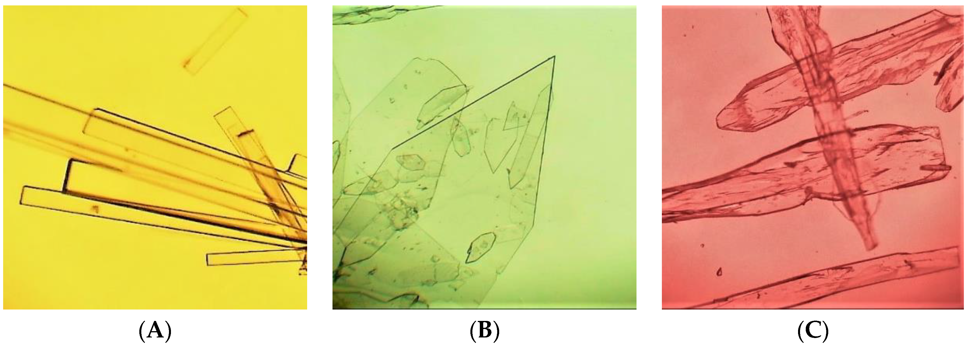
The crystals of form A were monoclinic, colorless sticks, C16H11N2O4F3S at 273 K, a 28.782(2), b 7.3709(4), c 14.933(1) Å, β 96.323(7)°, V 3148.6(4) Å3, Mr 384.33, Z 4, space group C2/c, dcalc 1.622 g/cm3, µ(MoKα) 0.266 mm−1, F(000) 1568.
The crystals of form B were monoclinic, colorless plates, C16H11N2O4F3S, at 100 K, a 28.697(2), b 7.3421(4), c 14.613(9) Å, β 96.112(9)°, V 3061.6(3) Å3, Mr 384.33, Z 4, space group C2/c, dcalc 1.668 g/cm3, µ(MoKα) 0.273 mm−1, F(000) 1568.
The crystals of form C were monoclinic, colorless blocks,·C16H11N2O4F3S, at 273 K, a 28.791(4), b 7.366(1), c 14.929(2) Å, β 96.37(1)°, V 3146.5(8) Å3, Mr 384.33, Z 4, space group C2/c, dcalc 1.623 g/cm3, µ(MoKα) 0.266 mm−1, F(000) 1568.
The unit cell parameters and intensities of 14,701 reflections (4596 independent reflections, Rint 0.041) for A, 15,594 reflections (4463 independent reflections, Rint 0.072) for B and 6611 reflections (2614 independent reflections, Rint 0.085) for C were measured on an Xcalibur-3 diffractometer (Oxford Diffraction Limited, Oxford, UK) using MoKα radiation, a CCD detector, graphite monochromator, and ω-scanning to 2θmax 60°. The structures were solved by the direct method using the SHELXTL program package (Institute of Inorganic Chemistry, Göttingen, Germany) [13]. The positions of the hydrogen atoms were found from the electron density difference maps and refined using the “riding” model with Uiso = 1.2Ueq for the nonhydrogen atom bonded to a given hydrogen atom. The hydrogen atoms involved in O–H…O and N–H…O hydrogen bond formation were refined using isotropic approximation. The structures were refined using F2 full-matrix least-squares analysis in the anisotropic approximation for nonhydrogen atoms to wR2 0.119 for 4577 reflections (R1 0.045 for 3034 reflections with F > 4σ (F), S = 0.965) for A, wR2 0.123 for 4451 reflections (R1 0.051 for 3052 reflections with F > 4σ (F), S = 0.966) for B and wR2 0.169 for 2599 reflections (R1 0.070 for 1694 reflections with F > 4σ (F), S = 0.979) for C. The final atomic coordinates and the crystallographic data for the molecules of N-(pyridin-2-yl)-4-methyl-2,2-dioxo-1H-2λ6,1-benzothiazine-3-carboxamide crystalline forms A, B and C have been deposited with the Cambridge Crystallographic Data Centre, 12 Union Road, CB2 1EZ, UK (Fax: +44-1223-336033; e-mail: [email protected]) and are available on request quoting the deposition numbers CCDC 1964379, and 1964377, respectively [14,15,16].

2.3. Powder Diffraction Study of N-(4-Trifluoromethylphenyl)-4-Hydroxy-2,2-Dioxo-1H-2λ6,1-Benzothiazine-3-Carboxamide Crystalline Forms A, B and C

The powder diffraction study of the N-(4-trifluoromethylphenyl)-4-hydroxy-2,2-dioxo-1H-2λ6,1-benzothiazine-3-carboxamide crystalline forms A, B and C was performed using a Siemens D500 diffractometer (Siemens Analytical X-ray Instruments Inc., Madison, WI, USA) with CuKα radiation (λ = 1.54184 Å), Bragg–Brentano θ/2θ geometry, and a curved graphite monochromator on the counter arm, scanning with Δ2θ = 0.02° and a 40-s accumulation time in each point. The Rietveld refinement was carried out with WinPLOTR and FullProf programs (Institute Laue-Langevin, Grenoble, France) [17,18,19] using the model structures obtained by monocrystal diffraction method.

2.4. Pharmacology

All biological experiments were carried out in full accord with the European Convention on the Protection of Vertebrate Animals Used for Experimental and Other Scientific Purposes and the Ukrainian Law No. 3447-IV “On protection of animals from severe treatment” [20] (project ID 3410U14, approved 15 October 2015). The pharmacological research was carried out with the permission and under the supervision of the Commission on Bioethics (N.I. Pirogov Vinnitsa National Medical University, Vinnitsa, Ukraine).
All pharmacological experiments described in this article were performed on animals from the vivarium of the Institute of Pharmacology and Toxicology of the Academy of Medical Sciences of Ukraine (Kyiv, Ukraine). All animals received standard food for rodents and water. One day before the tests, the animals were transferred to a scientific laboratory for adaptation. At all times they were kept at 20–22 °C, a relative humidity of 40–60% and a 12 h light/12 h dark cycle.
The analgesic action was studied on white Wistar male rats weighing 19–22 g (10 animals for each test substance and way of administration) using the “acetic-acid-induced writhing” model [21]. The nociceptive effect was reproduced by intraperitoneal injection of 0.6% aqueous solution of acetic acid in an amount of 0.1 mL per 10 g of animal weight in 30 min after the preliminary intraperitoneal or oral administration of the substances studied. Animals of the control group were intraperitoneally administered only diluted acetic acid in the specified concentration. After the introduction of acetic acid the animals were placed on a paper substrate and covered with a glass cap with a hole for free access of air. The number of “writhings” was counted from the 5th to the 20th min, inclusively. The analgesic effect was assessed by the ability of the test substances to reduce the number of “writhings” compared to the control and expressed as a percentage.
The analgesic activity was studied on white Wistar male rats weighing 160–190 g (10 animals for each test substance and way of administration) using the “hot plate” model [21]. First, the experimental animals received the test samples intraperitoneally or orally or an equivalent amount of water and Tween-80 (control group). After an hour, the animals were placed on a metal surface surrounded by a cylinder heated to an average of 52 °C (Ugo Basile, Italy). The time from the moment of placement on a hot surface to the appearance of a behavioral response to the nociceptive stimulation was recorded: licking the hind legs (phase 1); jumping and/or the desire to jump out of the cylinder (phase 2). By summing up these indicators, the total duration of latent periods was determined. Their increase indicated the analgesic activity of the compounds studied compared to the control and was expressed as a percentage.
The antinociceptive effect was studied on white Wistar male rats weighing 160–190 g (10 animals for each test substance and way of administration) using the model of “thermal irritation of the tail tip” (tail-flick) [21]. Thermal irritation was caused by a focused beam of light. First, the rats were placed in individual cells with their tails out, and the tail-flick analgesia meter (Ugo Basile, Italy) was used to determine the initial level of the latent period (the time of the attempt to get rid of the pain stimulus). Then, the substances studied were administered intraperitoneally or orally to the experimental animals; in an hour the experiment was repeated. The potency of the analgesic action was assessed by increasing the latency period compared to the baseline level taken as a control.
The analgesic activity was studied on white Wistar male rats weighing 160–190 g (seven animals for each test substance and way of administration) using the “tail electric stimulation” model [21]. The rats were placed in cramped plexiglass cages with a copper plate floor that served as an electrode. Using an RS12 stimulator (TUR, Dresden, Germany), an electric stimulus was supplied in the form of sequences of constant current (rectangular pulses, 50 ms impulse width, 50 Hz, increment of 100 mA/s) on the lateral base of the tail through two needle electrodes made of stainless steel and located at a distance of 1 cm from each other. The minimum current strength that caused vocalization, immersion of the tail or paw flinches from the conductive surface of the floor was taken as the initial pain threshold. After that, the animals were treated with the substances studied (intraperitoneally or orally), and pain responses were measured again in 1, 2, 4 and 6 h after their administration. The analgesic effect was assessed by comparing the initial values of the pain threshold and its changes in 1, 2, 4 and 6 h after a single injection of the test substances.
The analgesic action was studied on white Wistar male rats weighing 180–200 g (seven animals for each test substance and way of administration) using the “neuropathic pain” model [21,22]. This model was reproduced under anesthesia by ligation of the sciatic nerve in the upper third of the thigh at the level of the popliteal fossa above the site of its trifurcation on nerve Tibialis, nerve Peroneus and nerve Suralis. The pathological process development lasted 14 days. The degree of hyperalgesia was determined using von Frey monofilaments from the Semmes Weinstein kit (Stoelting, Dale Wood, IL, USA) covering a wide range of pressure strength from 0.008 to 300 g, as well as a dolorimeter (Baseline, White Plains, NY, USA). To do this, we compared the pain threshold—the minimum pressure on the lower surface of the rat’s foot (g/mm2) that caused a pain response (vocalization and/or paw flinches)—on the operated limb before surgery (baseline) and on the 14th day after surgery (pathology without treatment—control). The analgesic action of crystal forms A, B and C was assessed 2 h after their single intraperitoneal or oral administration by determining changes in the pain threshold compared to the control.
All of the crystal forms (A, B and C), regardless of the experimental model and route of administration, were tested in the dose of 20 mg/kg in the form of aqueous suspensions stabilized with Tween-80.
Acute toxicity of N-(4-trifluoromethylphenyl)-4-hydroxy-2,2-dioxo-1H-2λ6,1-benzothiazine-3-carboxamide crystal forms A, B and C was studied on intact white mice weighing 18–22 g (seven animals with each dose). The substances studied were administered orally in the form of a thin aqueous suspension stabilized with Tween-80 (at high doses in 2–3 doses with an interval of 10 min). The number of surviving animals was recorded every 24 h for 14 days. Doses causing death in 16%, 50%, 84% and 100% of the test animals (LD16, LD50, LD84 and LD100, respectively) were calculated according to the Litchfield–Wilcoxon method [23].
Gastric damage of crystal forms A, B and C was studied on white Wistar male rats weighing 180–200 g (seven animals with each dose). Before the experiments, the animals were starved for 48 h, having access to drinking water ad libitum. During this time, they were housed in cages with raised bottoms of wide wire mesh in order to avoid coprophagy. The test compounds were administered orally through a gastric probe in the form of a thin aqueous suspension stabilized with Tween-80. Further studies of gastric damages were performed in 4 h according to the method described in detail in [24]. Doses causing erosion in 16%, 50%, 84% and 100% of the test animals (UD16, UD50, UD84 and UD100, respectively) were calculated according to the Litchfield-Wilcoxon’s method [23].
All results obtained during pharmacological tests were processed using the STATISTICA 6.1 software package (StatSoft Inc., Tulsa, OK, USA). Descriptive statistics included calculations of the arithmetic means (M) and standard errors of the mean (±m). The significance of differences within one group was assessed using the Wilcoxon nonparametric test. The reliability of intergroup differences was determined using the nonparametric Mann–Whitney U-criteria. The effects were considered statistically significant at p ≤ 0.05.

3. Results and Discussion

3.1. The Molecular and Crystal Structure Study

We have obtained various crystal modifications of N-(4-trifluoromethylphenyl)-4-hydroxy-2,2-dioxo-1H-2λ6,1-benzothiazine-3-carboxamide by simple crystallization from organic solvents that are widely used and necessarily pharmaceutically acceptable [25]. As a result of these simple procedures, it has been found that crystallization of ethyl acetate, meta-xylene or ortho-xylene provides the formation of three visually completely different types of colorless crystals—long sticks with a square cross-section (form A), thin chisel-shaped plates (form B) or elongated blocks without any particular geometric structure and similar to tree branches (form C), respectively (Figure 2).
These significant differences in appearance (habitus) suggested that sticks A, plates B and blocks C were three polymorphic modifications of N-(4-trifluoromethylphenyl)-4-hydroxy-2,2-dioxo-1H-2λ6,1-benzothiazine-3-carboxamide. However, a single-crystal X-ray diffraction study (see Section 2.2) showed that the molecular and crystal structures for all these types of crystals were identical within the error of the experiment, especially if the different shooting temperature was taken into account (the experiment for very thin and therefore weakly reflecting plates B was carried out at 100 K, while the experiments for well-reflecting sticks A and blocks C were carried out at room temperature).
In all samples, regardless of the crystal form, the dihydrothiazine heterocycle is in a conformation that is intermediate between the twist-bath and the sofa conformation (Figure 3). The most indicative geometric characteristics of the molecules studied are some bond lengths, folding parameters [26], deviations of atoms S(1) and C(8) from the mean-square plane of the remaining bicycle atoms, as well as individual torsion angles (Table 1). The cyclic nitrogen atom has a pyramidal configuration; the sum of the valence angles centered on it is 353° in structure A, 351° in structure B and 339° in structure C.
The carbamide groups of anilide fragments at the C(8) atom are coplanar to the C(7)−C(8) endocyclic double bond (torsion angles—C(7)−C(8)−C(9)−O(2) in Table 1); apparently, they are stabilized by O(1)−H…O(2) intramolecular hydrogen bonds (Table 2). The formation of hydrogen bonds also leads to elongation of the C(9)−O(2) and C(7)−C(8) bonds compared to their mean values of 1.210 Å and 1.326 Å, respectively [27], and shortening of the C(7)−O(1) bond (its mean value was 1.362 Å). The 4-trifluoromethylphenyl substituent at the N(2) atom occupies an antiperiplanar position with respect to the C(8)−C(9) bond (torsion angle—C(10)−N(2)−C(9)−C(8)). In this case, the aromatic cycle is somewhat non-coplanar to the plane of the carbamide fragment (torsion angle—C(9)−N(2)−C(10)−C(11)); this is a consequence of the mutual influence of two oppositely directed factors: the formation of the C(11)−H…O(2) intramolecular hydrogen bond (Table 2) and steric repulsion (shortened intramolecular contact H(2N)…H(15) 2.21 Å in A, 2.20 Å in B and 2.28 Å in C). The C(1)−N(1) bond is elongated compared to its mean value of 1.371 Å.
In all three forms of crystals (A, B and C) the molecules of N-(4-trifluoromethylphenyl)-4-hydroxy-2,2-dioxo-1H-2λ6,1-benzothiazine-3-carboxamide form centrally symmetric dimers due to the N(1)−H…O(4)’ intermolecular hydrogen bond (Figure 4).
Powder diffractograms of crystal forms A, B and C of N-(4-trifluoromethylphenyl)-4-hydroxy-2,2-dioxo-1H-2λ6,1-benzothiazine-3-carboxamide (Figure 5) confirmed the phase purity of the samples studied and demonstrated the full compliance with single-crystal data. The Rietveld calculations using models obtained in decoding structures by the single-crystal method have shown that the atomic structure is the same for all three samples, and the differences in the intensity of reflections observed in the experiment (Figure 5) are due to the predominant orientation of powder particles in the samples.

3.2. Evaluation of the Biological Properties

For many years, the particle size of pharmaceutical substances has been considered a key parameter that provides the full range of their properties as drugs [28,29,30,31]. Therefore, it is not surprising that this parameter was carefully controlled at various stages of work with drugs—from their research development to industrial production. However, in recent years, another equally if not more important characteristic of biologically active substances, which also significantly affects their consumer properties, has been the shape of particles or habitus (needles, plates, cubes, prisms, shapeless crystals with different ratios of faces, etc.), which began to assert itself louder and more insistently. As it turned out, this factor is the main cause of many problems, and (judging by the number of publications), most often the technological ones. Thus, drug manufacturers have long noticed that samples of the original substance being different in appearance can vary greatly in wettability, compressibility, abrasiveness, hygroscopicity, stability, solubility and other physical and chemical properties. All these parameters one way or another inevitably affect the quality of the final product. Hence, apparently, there is the increased attention to their study [32,33,34,35,36,37,38]. At the same time, no less interesting, and in some ways even a more important medical aspect of the problem we are considering remains in the background. There is a paradoxical situation—with a very extensive amount of information about the physical and chemical properties and the structure of various crystal modifications of many medicinal substances (see, for example, the Cambridge Crystallographic Data Centre [39]) it is extremely difficult, if not impossible, to find information about their biological activity. In the open press such information gets only in those rare cases when there are legal disputes between developers of different polymorphic forms of commercially successful pharmaceuticals [9], or when a drug that had already entered the market suddenly undergoes spontaneous structural transformation into a new crystal modification—more stable, but much less active and even dangerous [8,9,10,40]. It should be emphasized that we are talking only about real pharmaceuticals that have official permission for medical use. The biological properties of different crystal forms of new substances, which have the prospect of becoming a drug but so far have not, have recently been studied and described rather widely [41,42,43,44,45,46,47]. As a rule, polymorphic modifications of a substance are tested, i.e., samples that differ significantly not only in habitus, but also in the molecular and crystal structure. The crystal forms A, B and C of N-(4-trifluoromethylphenyl)-4-hydroxy-2,2-dioxo-1H-2λ6,1-benzothiazine-3-carboxamide presented in this article are unique in that they differ from each other only in appearance with the identity of all parameters of the internal structure. Due to this, they are unusual and very interesting objects for pharmacological tests. Such a study will allow evaluating the impact on biological effects of crystal habit alone since samples A, B and C are insoluble in water and, therefore, will be tested in the form of an aqueous suspension, i.e., in a solid dispersed phase.

3.2.1. Analgesic Activity

Interesting results were shown by our first biological experiment conducted on the “acetic-acid-induced writhing” classical model in white mice (Table 3). This model is based on the chemical pain stimulation and allows us to form an opinion about the peripheral component in the mechanism of the analgesic effect of the test samples [21]. In intraperitoneal administration (i.p.), all three crystal forms of N-(4-trifluoromethylphenyl)-4-hydroxy-2,2-dioxo-1H-2λ6,1-benzothiazine-3-carboxamide significantly reduce the number of “writhings” compared to the control data. However, the potency of their analgesic effect varies greatly and decreases in the following order: B > C >> A. In other words, plates B eliminate the somatic visceral pain response most effectively (87.5%). Blocks C are a little inferior to them (76.9%) by this indicator, while sticks A reduce the number of “writhings” only by 35.1% which actually turns this crystal form of N-(4-trifluoromethylphenyl)-4-hydroxy-2,2-dioxo-1H-2λ6,1-benzothiazine-3-carboxamide into a moderate analgesic. When changing the method of administration to per os (p.o.), in principle, the same trend remains: B >> C > A, with the only difference being that now blocks C are in the category of moderate analgesics.
When studying the effect of crystal forms A, B and C of N-(4-trifluoromethylphenyl)-4-hydroxy-2,2-dioxo-1H-2λ6,1-benzothiazine-3-carboxamide on the central component of the nociceptive system three experimental models were used. The first of them—“hot plate”—was applied in white Wistar male rats (Table 4). At the same time, it was found that plates B significantly increased both phases of the latent period with intraperitoneal and oral administration and thus retained their leadership as an analgesic. Regarding blocks C, they somewhat lost their position and gave way to sticks A in their activity. The attention is drawn to the fact that the potency of the analgesic effect of crystal forms A and C practically does not depend on the way of entering the animal’s body, whereas in the case of form B in oral administration it naturally decreases.
The second experimental model—thermal irritation of the tail tip (tail-flick) in rats using a focused beam of light—also demonstrated a significant advantage of the plate crystal form B (Table 5). In the ability to block the pain reaction it in this case surpasses forms C and A almost by 2 and 3 times, respectively. It is noteworthy that on this model, when changing the intraperitoneal administration to oral, all three samples lose about 30% of their analgesic activity (in relative terms).
The third model of this series—“tail electric stimulation” in rats—allowed to trace the development of the effect of the analgesic effect provided by crystal forms A, B and C of N-(4-trifluoromethylphenyl)-4-hydroxy-2,2-dioxo-1H-2λ6,1-benzothiazine-3-carboxamide over time. From the data given in Table 6 it follows that plates B exhibit a powerful and rapid analgesic effect, which after the second hour of the experiment begins to decrease markedly. The activity of blocks C is less pronounced although it also reaches its maximum by the second hour and its further decline is not so sharp. Particularly, sticks A should be mentioned. Their initially moderate analgesic action gradually intensifies and reaches its peak only by the fourth hour. Then, there is a gradual decrease in analgesia. However, even after six hours of the experiment, sticks A maintain a fairly high level of activity and remain the most powerful analgesic of the entire test group. The effects observed in this model can be explained by different bioavailabilities of the crystal forms studied. Even without any calculations, it is clear that with the same mass, the surface area (and, obviously, bioavailability) of plate crystals B will be noticeably higher than that of blocks C and much higher than that of sticks A. Hence, the form B has a high and fast, but relatively short, analgesic effect. Compact sticks A apparently are absorbed more slowly and therefore have longer but less pronounced analgesia. By the surface area and the nature of the impact on the pain threshold (Table 6), blocks C occupy an intermediate place between plates B and sticks A.
The effects of the substances A, B and C studied on the peripheral and central component of the nociceptive system were assessed on the “neuropathic pain” model in rats (Table 7). Pain of this type can be caused by various pathological processes in the peripheral and central nervous system; their causes are often diabetes, herpesvirus infections, oncological diseases, spinal cord injuries, etc. The results obtained strongly suggest that plates B unconditionally remain the leading crystal form in the intraperitoneal way of introduction. By their ability to reduce the pain threshold, blocks C (when administered orally) closely approach them. However, sticks A on this model were the least effective.

3.2.2. Acute Toxicity and Gastric Damage

Toxicity and damaging action on the gastric mucosa are the most important characteristics of all drugs without exception. These indicators largely determine the effectiveness and safety of practical use of both official and promising pharmaceuticals. Acute toxicity and gastric damage of crystal forms A, B and C of N-(4-trifluoromethylphenyl)-4-hydroxy-2,2-dioxo-1H-2λ6,1-benzothiazine-3-carboxamide were studied in oral administration in mice and rats, respectively. It was found that the least toxic crystal form was blocks C—their LD50 was 697.2 mg/kg (Table 8). Further in order of increasing toxicity there were plates B (LD50 = 586.4 mg/kg) and sticks A (LD50 = 419.3 mg/kg). At the same time, crystal form C unexpectedly proved to be a very powerful provocateur of multiple lesions of the gastric mucosa, especially in high doses. The same can be said of form A, but in low doses. Interestingly, as the dose increases, the damaging effect of this form compared to blocks C is somewhat reduced. According to the experimental data, plates B should be considered the most successful by this indicator (Table 9). Most likely, the result obtained is also associated with differences in the bioavailability of the samples studied. Apparently, blocks C and sticks A due to their crystal structure are found in the stomachs of the experimental animals much longer than plates B and cause more appreciable local lesions of their mucous membranes.
Summing up our pharmacological tests we can unequivocally state that of three crystal forms of N-(4-trifluoromethylphenyl)-4-hydroxy-2,2-dioxo-1H-2λ6,1-benzothiazine-3-carboxamide studied, only one deserves attention—plates B. The basis for this choice was their higher analgesic activity on different experimental models and, very importantly, relatively low toxicity and the ability to cause gastric lesions.

4. Conclusions

Crystallization from various organic solvents produced three colorless crystal forms of N-(4-trifluoro-methylphenyl)-4-hydroxy-2,2-dioxo-1H-2λ6,1-benzothiazine-3-carboxamide: sticks of ethyl acetate, plates of meta-xylene and blocks of ortho-xylene. Using the single-crystal and powder X-ray diffraction analysis it has been proven that, despite the different habitus, all three forms have exactly the same molecular and crystal structure. At the same time, biological tests have revealed significant differences in their analgesic action, acute toxicity and the ability to damage the gastric mucosa. It is suggested that, having the largest crystal surface area, the plate form has increased bioavailability and, as a consequence, improved pharmacological properties. The results obtained allow us to recommend to scientists who work with drug candidates to involve all available crystal forms of the substances studied in the circle of not only their technological, but also necessarily pharmacological studies. It is advisable to pay attention to this aspect even if the only difference between crystal objects is exclusively their habitus. In conclusion, we note that we in no way encroach on, and certainly do not claim the greatness of the famous alchemist and physician Philippus Aureolus Theophrastus Bombastus von Hohenheim, better known in the world under the name of Paracelsus. It just seems that there are times when in his phrase, which is very popular among pharmacists and physicians: “Any substance can be both a medicine and poison; it all depends on the dose”, it will be quite appropriate to make a short continuation: “… and crystal form”.

Author Contributions

The synthesis of the crystal forms presented in this work and analysis of their characteristics were performed by I.V.U. and L.A.P. The X-ray structural studies were performed by S.V.S., I.V.U. and A.I.F. The pharmacological studies were conducted by N.I.V., P.S.B., L.V.S. and G.S. The manuscript was written by I.V.U., L.A.P. and P.S.B.

Funding

This research received no external funding.

Acknowledgments

We are grateful to Elena Y. Gurko (National University of Pharmacy, Kharkiv, Ukraine) for her language help in preparing this manuscript.

Conflicts of Interest

The authors declare no conflict of interest.

References

  1. Haleblian, J.K. Characterization of habits and crystalline modification of solids and their pharmaceutical applications. J. Pharm. Sci. 1975, 64, 1269–1288. [Google Scholar] [CrossRef] [PubMed]
  2. Bernstein, J. Polymorphism—A perspective. Cryst. Growth Des. 2011, 11, 632–650. [Google Scholar] [CrossRef]
  3. Fandaruff, C.; Silva, M.A.S.; Bedor, D.C.G.; De Santana, D.P.; Rocha, H.V.; Rebuffi, L.; Ricardo, C.L.A.; Scardi, P.; Cuffini, S.L. Correlation between microstructure and bioequivalence in anti-HIV drug Efavirenz. Eur. J. Pharm. Biopharm. 2015, 91, 52–58. [Google Scholar] [CrossRef] [PubMed]
  4. Muresan-Pop, M.; Braga, D.; Pop, M.M.; Borodi, G.; Kacso, I.; Maini, L. Crystal structure and physicochemical characterization of Ambazone monohydrate, anhydrous, and acetate salt solvate. J. Pharm. Sci. 2014, 103, 3594–3601. [Google Scholar] [CrossRef] [PubMed]
  5. Gelbrich, T.; Meischberger, I.; Griesser, U.J. Two polymorphs of 5-cyclohexyl-5-ethylbarbituric acid and their packing relationships with other barbiturates. Acta Crystallogr. Sect. C Struct. Chem. 2015, 71, 204–210. [Google Scholar] [CrossRef] [PubMed]
  6. Lutker, K.M.; Quiñones, R.; Xu, J.; Ramamoorthy, A.; Matzger, A.J. Polymorphs and hydrates of acyclovir. J. Pharm. Sci. 2011, 100, 949–963. [Google Scholar] [CrossRef] [Green Version]
  7. Gunn, E.; Guzei, I.A.; Cai, T.; Yu, L. Polymorphism of Nifedipine: Crystal structure and reversible transition of the metastable beta polymorph. Cryst. Growth Des. 2012, 12, 2037–2043. [Google Scholar] [CrossRef]
  8. Morissette, S.L.; Soukasene, S.; Levinson, D.; Cima, M.J.; Almarsson, Ö. Elucidation of crystal form diversity of the HIV protease inhibitor ritonavir by high-throughput crystallization. Proc. Natl. Acad. Sci. USA 2003, 100, 2180–2184. [Google Scholar] [CrossRef] [Green Version]
  9. Bernstein, J. Polymorphism in Molecular Crystals; Oxford University Press: New York, NY, USA, 2002. [Google Scholar]
  10. Rietveld, I.B.; Céolin, R. Rotigotine: Unexpected polymorphism with predictable overall monotropic behavior. J. Pharm. Sci. 2015, 104, 4117–4122. [Google Scholar] [CrossRef]
  11. Hilfiker, R. Polymorphism: In the Pharmaceutical Industry; John Wiley & Sons: Weinheim, Germany, 2006. [Google Scholar]
  12. Petrushova, L.A.; Ukrainets, I.V.; Dzyubenko, S.P.; Grinevich, L.A. Synthesis and the biological activity of 4-hydroxy-2,2-dioxo-1H-2λ6,1-benzothiazin-3-carboxylic acids trifluoromethyl-substituted anilides. J. Org. Pharm. Chem. 2015, 13, 44–48. [Google Scholar] [CrossRef]
  13. Sheldrick, G.M. A short history of SHELX. Acta Crystallogr. Sect. A Found. Crystallogr. 2008, A64, 112–122. [Google Scholar] [CrossRef] [PubMed] [Green Version]
  14. Cambridge Crystallographic Data Center. Request Quoting via: CCDC 1964379. Available online: www.ccdc.cam.ac.uk/conts/retrieving.html (accessed on 7 November 2019).
  15. Cambridge Crystallographic Data Center. Request Quoting via: CCDC 1964378. Available online: www.ccdc.cam.ac.uk/conts/retrieving.html (accessed on 7 November 2019).
  16. Cambridge Crystallographic Data Center. Request Quoting via: CCDC 1964377. Available online: www.ccdc.cam.ac.uk/conts/retrieving.html (accessed on 7 November 2019).
  17. Rodriguez-Carvajal, J. FullProf; Version 4.80; ILL: Grenoble, France, 2010. [Google Scholar]
  18. Rodriguez-Carvajal, J.; Roisnel, T. WinPLOTR.; Trans Tech Publications: Zurich, Switzerland, 2004; pp. 123–126. [Google Scholar]
  19. Rodriguez-Carvajal, J.; Roisnel, T. FullProf.98 and WinPLOTR New Windows 99/NT Applications for Diffraction, Commission for Powder Diffraction; International Union of Crystallography, Newsletter: Chester, UK, 1998; Volume 20, pp. 35–36. [Google Scholar]
  20. Ukrainian Law No. 3447-IV. On Protection of Animals from Severe Treatment. Available online: http://zakon2.rada.gov.ua/laws/show/3447-15 (accessed on 4 August 2017).
  21. Chapter, H. Analgesic, anti-inflammatory, and anti-pyretic activity. In Drug Discovery and Evaluation: Pharmacological Assays, 2nd ed.; Schleyerbach, R., Weithmann, K.U., Bartlett, R.R., Vogel, H.G., Eds.; Springer: Berlin, Germany, 2008; pp. 983–1116. [Google Scholar]
  22. Chaplan, S.R.; Bach, F.W.; Pogrel, J.W.; Chung, J.M.; Yaksh, T.L. Quantitative assessment of tactile allodynia in the rat paw. J. Neurosci. Methods 1994, 53, 55–63. [Google Scholar] [CrossRef]
  23. Litchfield, J.T., Jr.; Wilcoxon, F. A simplified method of evaluating dose-effect experiments. J. Pharmacol. Exp. Ther. 1949, 96, 99–113. [Google Scholar] [PubMed]
  24. Ozaki, S.; Kobayashi, H.; Mori, H.; Kitano, T.; Kawazura, H.; Okazaki, Y. Benzothiazine Derivative. U.S. Patent 4,376,768, 15 March 1983. Available online: https://worldwide.espacenet.com/publicationDetails/originalDocument?CC=US&NR=4376768A&KC=A&FT=D&ND=3&date=19830315&DB=EPODOC&locale=en_EP (accessed on 2 November 2019).
  25. Council of Europa. Residual solvents. In European Pharmacopoeia, 6th ed.; Council of Europa: Strasbourg, France, 2007; Volume 1, pp. 601–610. [Google Scholar]
  26. Zefirov, N.S.; Palyulin, V.A.; Dashevskaya, E.E. Stereochemical studies. XXXIV. Quantitative description of ring puckering via torsional angles. The case of six-membered rings. J. Phys. Org. Chem. 1990, 3, 147–158. [Google Scholar] [CrossRef]
  27. Orpen, A.G.; Brammer, L.; Allen, F.H.; Kennard, O.; Watson, D.G.; Taylor, R. Typical interatomic distances in organic compounds and organometallic compounds and coordination complexes of the d-and f-block metals. In Structure Correlation; Burgi, H.-B., Dunitz, J.D., Eds.; Wiley-VCH: Weinheim, Germany, 1994; pp. 741–784. [Google Scholar]
  28. Minkov, V.S.; Drebushchak, V.A.; Ogienko, A.G.; Boldyreva, E.V. Decreasing particle size helps to preserve metastable polymorphs. A case study of DL-cysteine. CrystEngComm 2011, 13, 4417–4426. [Google Scholar] [CrossRef]
  29. Emshanova, S.V.; Sadchikova, N.P.; Zuev, A.P. Drug particle shape and size control: A necessary factor for high-quality drug production. Pharm. Chem. J. 2007, 41, 40–49. [Google Scholar] [CrossRef]
  30. Onischuk, A.A.; Tolstikova, T.G.; Sorokina, I.V.; Zhukova, N.A.; Baklanov, A.M.; Karasev, V.V.; Borovkova, O.V.; Dultseva, G.G.; Boldyrev, V.V.; Fomin, V.M. Analgesic effect from Ibuprofen nanoparticles inhaled by male mice. J. Aerosol Med. Pulm. Drug Deliv. 2009, 22, 245–253. [Google Scholar] [CrossRef] [Green Version]
  31. Gratton, S.E.A.; Ropp, P.A.; Pohlhaus, P.D.; Luft, J.; Madden, V.J.; Napier, M.E.; DeSimone, J.M. The effect of particle design on cellular internalization pathways. Proc. Natl. Acad. Sci. USA 2008, 105, 11613–11618. [Google Scholar] [CrossRef] [Green Version]
  32. Leuner, C.; Dressman, J. Improving drug solubility for oral delivery using solid dispersions. Eur. J. Pharm. Biopharm. 2000, 50, 47–60. [Google Scholar] [CrossRef]
  33. Chernyshev, V.V.; Morozov, Y.N.; Bushmarinov, I.S.; Makoed, A.A.; Sergeev, G.B. New polymorph of dehydroepiandrosterone obtained via criomodification. Cryst. Growth Des. 2016, 16, 1088–1095. [Google Scholar] [CrossRef]
  34. Watanabe, A.; Yamaoka, Y.; Takada, K. Crystal habits and dissolution behavior of Aspirin. Chem. Pharm. Bull. 1982, 30, 2958–2963. [Google Scholar] [CrossRef] [Green Version]
  35. Hooper, D.; Clarke, F.C.; Docherty, R.; Mitchell, J.C.; Snowden, M.J. Effects of crystal habit on the sticking propensity of Ibuprofen—A case study. Int. J. Pharm. 2017, 531, 266–275. [Google Scholar] [CrossRef]
  36. Bukovec, P.; Benkič, P.; Smrkolj, M.; Vrečer, F. Effect of crystal habit on the dissolution behavior of simvastatin crystals and its relationship to crystallization solvent properties. Die Pharm. Int. J. Pharm. Sci. 2016, 71, 263–268. [Google Scholar] [CrossRef]
  37. Brittain, H.G. Polymorphism in Pharmaceutical Solids; Marcel Dekker: New York, NY, USA, 1999. [Google Scholar]
  38. Stoica, C.; Verwer, P.; Meekes, H.; Van Hoof, P.J.C.M.; Kaspersen, F.M.; Vlieg, E. Understanding the effect of a solvent on the crystal habit. Cryst. Growth Des. 2004, 4, 765–768. [Google Scholar] [CrossRef]
  39. Groom, C.R.; Bruno, I.J.; Lightfoot, M.P.; Ward, S.C. The Cambridge structural database. Acta Crystallogr. B Struct. Sci. Cryst. Eng. Mater. 2016, B72, 171–179. [Google Scholar] [CrossRef]
  40. Ukrainets, I.V.; Mospanova, O.V.; Bereznyakova, N.L.; Davidenko, O.O. Polymorphism and the analgesic activity of N-(3-pyridylmethyl)-4-hydroxy-2-oxo-1,2,5,6,7,8-hexahydroquinoline-3-carboxamide. J. Org. Pharm. Chem. 2015, 13, 41–46. [Google Scholar] [CrossRef] [Green Version]
  41. Dubey, R.; Singh, P.; Singh, A.K.; Yadav, M.K.; Swati, D.; Vinayak, M.; Puerta, C.; Valerga, P.; Kumar, K.R.; Sridhar, B.; et al. Polymorphic signature of the anti-inflammatory activity of 2,2′-{[1,2-phenylenebis(methylene)]bis(sulfanediyl)}bis(4,6-dimethylnicotinonitrile). Cryst. Growth Des. 2014, 14, 1347–1356. [Google Scholar] [CrossRef]
  42. Ukrainets, I.V.; Shishkina, S.V.; Baumer, V.N.; Gorokhova, O.V.; Petrushova, L.A.; Sim, G. The structure of two pseudo-enantiomeric forms of N-benzyl-4-hydroxy-1-methyl-2,2-dioxo-1H-2λ6,1-benzothiazine-3-carboxamide and their analgesic properties. Acta Crystallogr. Sect. C Struct. Chem. 2016, 72, 411–415. [Google Scholar] [CrossRef]
  43. Ukrainets, I.V.; Burian, A.A.; Baumer, V.N.; Shishkina, S.V.; Sidorenko, L.V.; Tugaibei, I.A.; Voloshchuk, N.I.; Bondarenko, P.S. Synthesis, crystal structure and biological activity of ethyl 4-methyl-2,2-dioxo-1H-2λ6,1-benzothiazine-3-carboxylate polymorphic forms. Sci. Pharm. 2018, 86, 21. [Google Scholar] [CrossRef] [Green Version]
  44. Shishkina, S.V.; Levandovski, I.A.; Ukrainets, I.V.; Sidorenko, L.V.; Grinevich, L.A.; Yanchuk, I.B. Polymorphic modifications of N-(4-methoxyphenyl)-6-hydroxy-4-oxo-2,4-dihydro-1H-pyrrolo[3,2,1-ij]-quinoline-5-carboxamide possessing strong diuretic properties. Acta Crystallogr. Sect. C Struct. Chem. 2018, 74 Pt 12, 1759–1767. [Google Scholar] [CrossRef]
  45. Williford, J.M.; Santos, J.L.; Shyam, R.; Mao, H.Q. Shape control in engineering of polymeric nanoparticles for therapeutic delivery. Biomater. Sci. 2015, 3, 894–907. [Google Scholar] [CrossRef] [PubMed]
  46. Hao, N.; Li, L.; Tang, F. Shape-mediated biological effects of mesoporous silica nanoparticles. J. Biomed. Nanotechnol. 2014, 10, 2508–2538. [Google Scholar] [CrossRef] [PubMed]
  47. Chen, J.; Clay, N.; Kong, H. Non-spherical particles for targeted drug delivery. Chem. Eng. Sci. 2015, 125, 20–24. [Google Scholar] [CrossRef] [PubMed] [Green Version]
Figure 1. N-(4-Trifluoromethylphenyl)-4-hydroxy-2,2-dioxo-1H-2λ6,1-benzothiazine-3-carboxamide, which has high analgesic activity [12].
Scipharm 88 00001 g001
Figure 2. Crystals of N-(4-trifluoromethylphenyl)-4-hydroxy-2,2-dioxo-1H-2λ6,1-benzothiazine-3-carboxamide obtained from (A) ethyl acetate, (B) meta-xylene and (C) ortho-xylene.
Scipharm 88 00001 g002
Figure 3. The molecular structure of 4-trifluoromethylphenylamide B with atoms represented by thermal vibration ellipsoids of 50% probability.
Scipharm 88 00001 g003
Figure 4. Dimers of N-(4-trifluoromethylphenyl)-4-hydroxy-2,2-dioxo-1H-2λ6,1-benzothiazine-3-carboxamide molecules in crystal forms A, B and C.
Scipharm 88 00001 g004
Figure 5. X-ray powder patterns of crystal forms A, B, C and their Rietveld refinement: experimental X-ray diffraction patterns (red); calculated X-ray diffraction patterns (black); Bragg position of diffraction maximum (green); difference between experimental and calculated intensity values in each point (blue).
Scipharm 88 00001 g005
Table 1. Some geometric characteristics for crystal forms A, B and C.
EntryCharacteristicCrystal Form ACrystal Form BCrystal Form C
Bond lengths (Å)
1O(1)−C(7)1.330(2)1.327(2)1.328(5)
2O(2)−C(9)1.243(2)1.246(2)1.240(5)
3N(1)−C(1)1.411(2)1.425(2)1.411(5)
4C(7)−C(8)1.368(2)1.378(3)1.379(5)
Parameters of folding and fluctuation of atoms
5S0.570.570.60
6Θ, degrees54.152.551.4
7Ψ, degrees25.124.927.8
8S(1)−0.80−0.79−0.83
9C(8)−0.27−0.27−0.29
Torsion angles (degrees)
10C(7)−C(8)−C(9)−O(2)−1.6(2)−2.4(3)−2.1(5)
11C(8)−C(9)−N(2)−C(10)−178.8(1)−179.2(2)−178.4(3)
12C(9)−N(2)−C(10)−C(11)16.8(3)15.8(3)15.9(6)
13C(14)−C(13)−C(16)−F(1)165.4(5)168.5(2)166.5(5)
Table 2. Characteristics of intra-and intermolecular hydrogen bonds for crystal forms A, B and C.
EntryInteractionCrystal Form ACrystal Form BCrystal Form C
H…A (Å)D−H…A (degrees)H…A (Å)D−H…A (degrees)H…A (Å)D−H…A (degrees)
1O(1)−H…O(2)1.591541.621551.79147
2N(2)−H…O(4)2.091382.091372.12141
3C(11)−H…O(2)2.321182.311182.33118
4N(1)−H…O(4)2.09169 12.01167 12.18158 2
1 The operation of symmetry 1 − x, −y, 1 − z. 2 The operation of symmetry 1 − x, 2 − y, 1 − z.
Table 3. The analgesic activity of crystalline forms A, B and C on the “acetic-acid-induced writhing” model in mice.
EntryProductAverage Number of “Writhings”Analgesic Activity, Compared to Control (%)
1A (i.p.)46.50 ± 5.74 1−35.1
2A (p.o.)56.49 ± 4.53 1−21.1
3B (i.p.)8.84 ± 1.91 1−87.5
4B (p.o.)23.20 ± 3.63 1−67.6
5C (i.p.)16.33 ± 3.50 1−76.9
6C (p.o.)50.81 ± 5.94 1−29.1
7Control71.62 ± 3.25
1 Differences statistically significant for p ≤ 0.05 vs. control. i.p., intraperitoneal administration; p.o., per os.
Table 4. The analgesic activity of crystalline forms A, B and C on the “hot plate” model in rats.
EntryProductLatent Period
Phase 1 (s)Phase 2 (s)Total Duration (s)Compared to Control (%)
1A (i.p.)11.87 ± 0.65 112.46 ± 0.57 124.33 ± 0.68 1+60.8
2A (p.o.)13.15 ± 0.82 110.42 ± 0.32 123.56 ± 0.97 1+55.7
3B (i.p.)16.87 ± 0.49 115.75 ± 0.63 132.62 ± 0.46 1+115.6
4B (p.o.)12.89 ± 0.45 111.60 ± 0.79 124.49 ± 0.69 1+61.9
5C (i.p.)10.61 ± 0.53 110.60 ± 1.26 121.20 ± 1.59 1+40.1
6C (p.o.)10.21 ± 0.55 111.07 ± 1.51 121.29 ± 1.70 1+40.7
7Control8.05 ± 0.827.08 ± 1.2415.13 ± 1.34
1 Differences statistically significant for p ≤ 0.05 vs. control.
Table 5. The analgesic activity of crystal forms A, B and C on the “tail-flick” model in rats.
EntryProductLatent Period, Initial Level (s)Latent Period in 1 h after Introduction of the Compounds (s)Change of the Latent Period Compared to the Initial Level (%)
1A (i.p.)2.07 ± 0.132.86 ± 0.21 1+37.6
2A (p.o.)2.21 ± 0.102.84 ± 0.28 1+27.9
3B (i.p.)1.86 ± 0.143.71 ± 0.18 1+103.1
4B (p.o.)1.84 ± 0.123.14 ± 0.21 1+70.5
5C (i.p.)1.96 ± 0.233.07 ± 0.44 1+57.1
6C (p.o.)1.93 ± 0.172.71 ± 0.32 1+40.5
1 Differences statistically significant for p ≤ 0.05 vs. the initial level.
Table 6. The analgesic activity of crystal forms A, B and C on the “tail electric stimulation” model in rats.
EntryProductDynamics of the Pain Threshold after the Introduction of Compounds (%)
In 1 hIn 2 hIn 4 hIn 6 h
1A (i.p.)+38.1+40.7+69.1+41.6
2A (p.o.)+29.4+42.1+48.9+42.1
3B (i.p.)+99.5+107.6+41.9+11.3
4B (p.o.)+57.7+68.9+41.4+26.9
5C (i.p.)+40.4+88.6+77.6+22.4
6C (p.o.)+59.1+63.0+44.5+25.9
Table 7. The analgesic activity of crystal forms A, B and C on the “neuropathic pain” model in rats.
EntryProductThe Pain Threshold
Initial Level (g/mm2)In 14 Days after Surgery without Treatment 1After Introduction of the Products 2
g/mm2%g/mm2%
1A (i.p.)458.6 ± 12.8151.4 ± 31.3−67.0174.3 ± 25.2+15.1
2A (p.o.)162.9 ± 30.1+7.5
3B (i.p.)395.7 ± 23.3162.9 ± 28.5−58.8225.7 ± 36.4+38.6
4B (p.o.)196.2 ± 37.0+20.5
5C (i.p.)402.9 ± 24.5197.1 ± 40.8−51.1248.6 ± 24.2+26.1
6C (p.o.)237.1 ± 32.4+20.3
1 Differences statistically significant for p ≤ 0.05 vs. the initial pain threshold before surgery. 2 Differences statistically significant for p ≤ 0.05 vs. the pain threshold in 14 days after surgery without treatment.
Table 8. The acute toxicity of crystal forms A, B and C in mice per os.
EntryProductAcute Toxicity (mg/kg)
LD16LD50LD84LD100
1A (p.o.)263.7419.3574.9652.6
2B (p.o.)362.1586.4811.0923.2
3C (p.o.)499.1697.2898.3996.4
Table 9. The acute gastric damage of crystal forms A, B and C in rats.
EntryProductAcute Gastric Damage (mg/kg)
UD16UD50UD84UD100
1A (p.o.)21.171.4121.8147.1
2B (p.o.)62.8117.9173.1200.7
3C (p.o.)58.987.5116.1130.4

