
Preclinical Safety Profile of an Oral Naringenin/Hesperidin Dosage Form by In Vivo Toxicological Tests

 ,
,  , , ,
, , ,  , ,
, ,  and
and 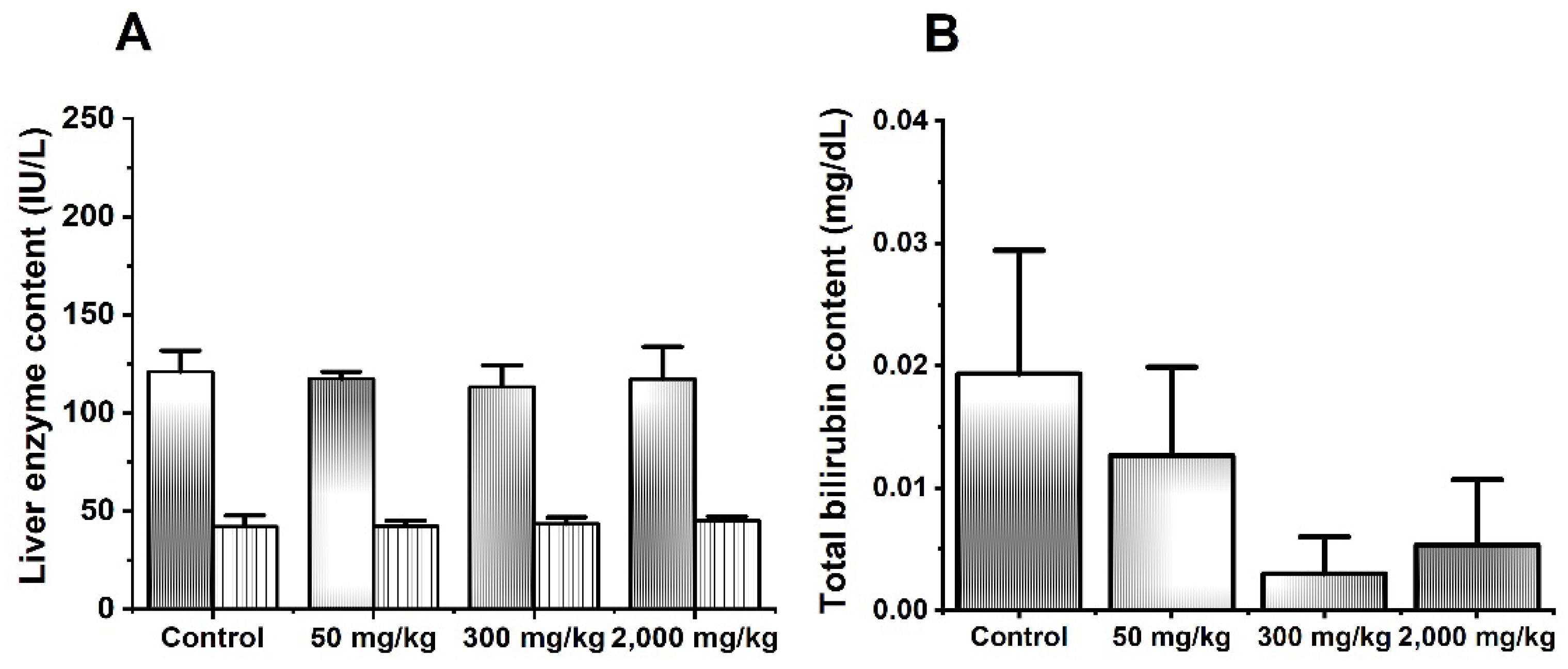
Abstract
:1. Introduction
2. Materials and Methods
2.1. Chemicals and Drugs
2.2. Technopharmaceutical Development of a Solid Dosage Form
2.3. Animals
2.4. Acute Oral Toxicity
2.5. Subchronic Oral Toxicity
2.6. Hematological and Biochemical Components Analysis
2.7. Histopathological Analysis
2.8. Statistical Analysis
3. Results and Discussion
3.1. Acute Oral Toxicity
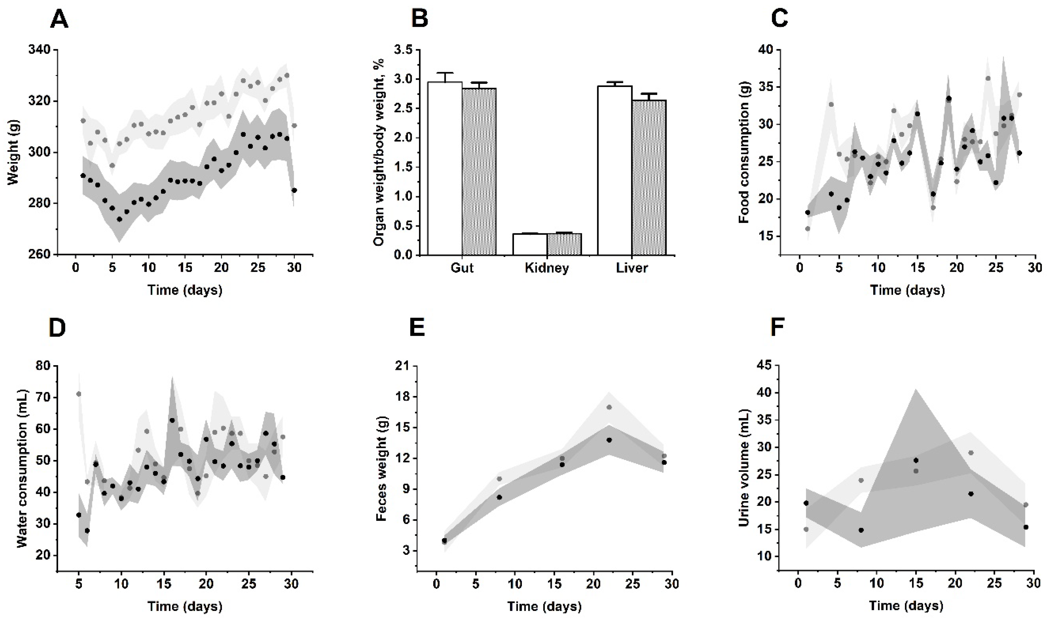
3.2. Subchronic Oral Toxicity
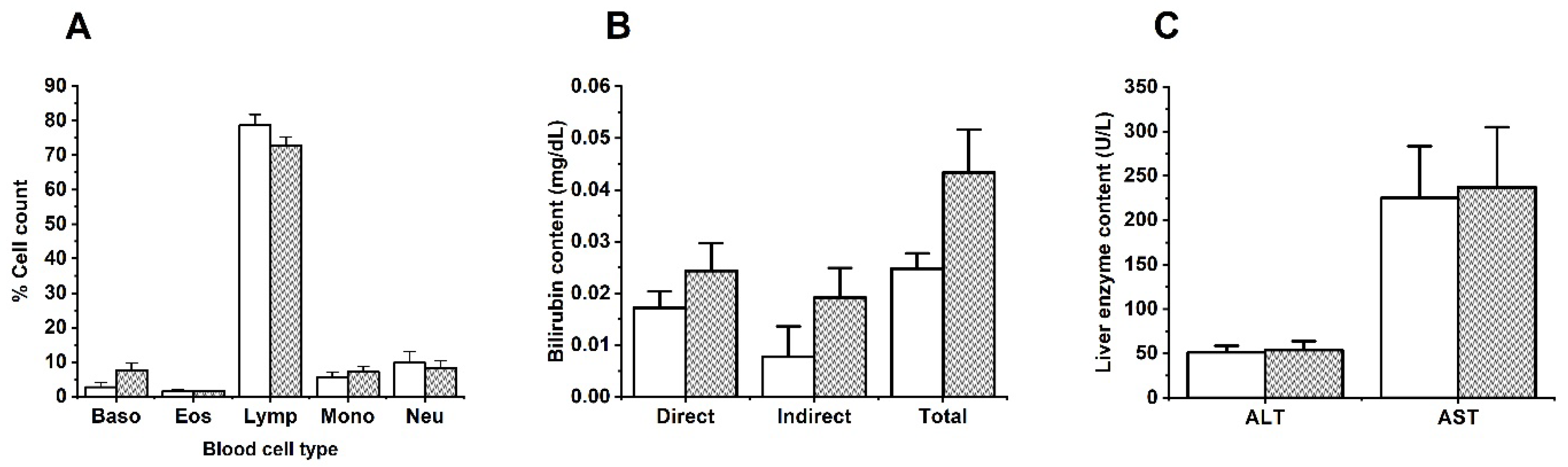
3.3. Changes of Biochemical and Hematological Parameters by MIX–160
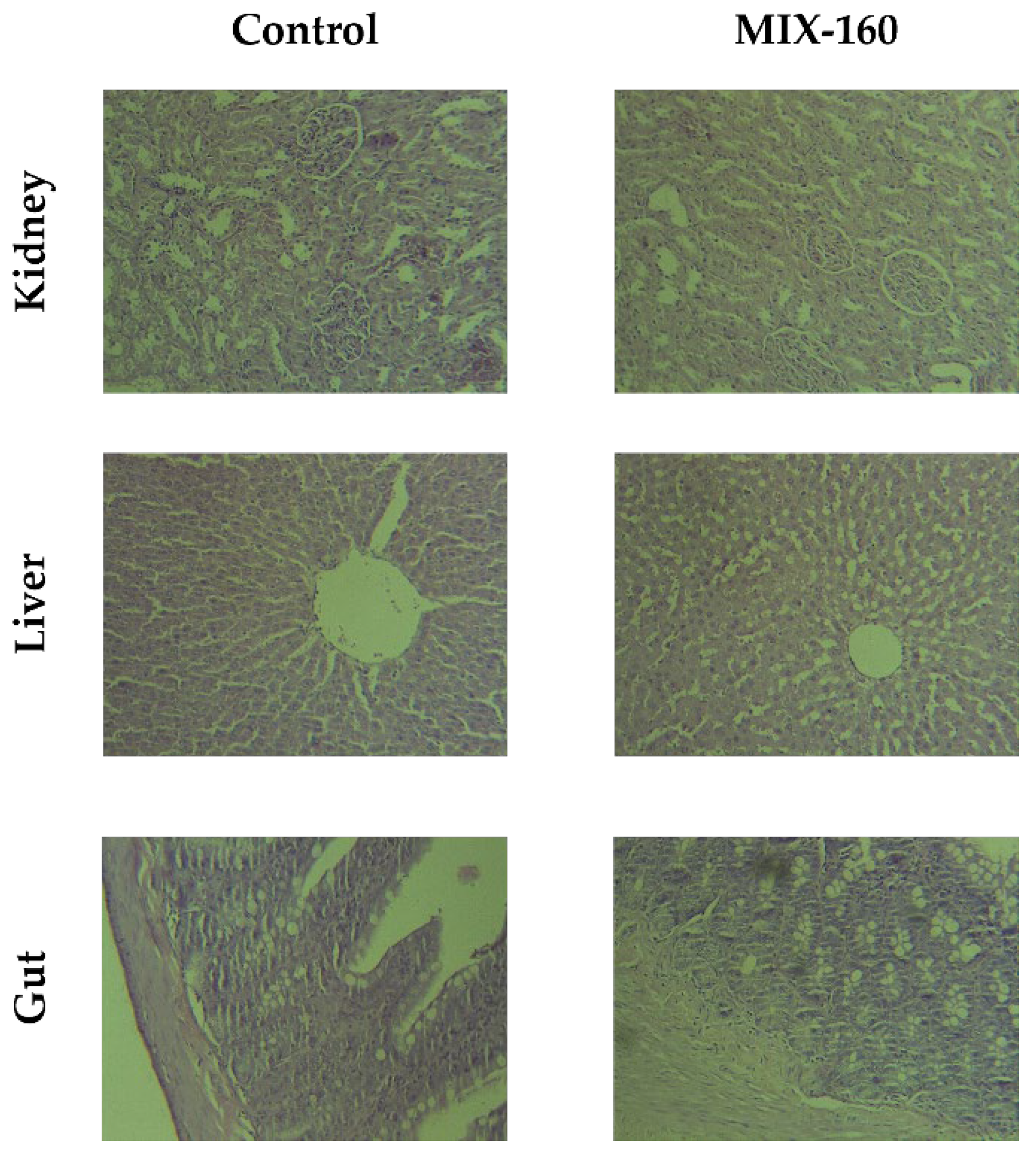
3.4. Histological Findings
4. Conclusions
Author Contributions
Funding
Institutional Review Board Statement
Informed Consent Statement
Acknowledgments
Conflicts of Interest
References
- Pietta, P.G. Flavonoids as antioxidants. J. Nat. Prod. 2000, 63, 1035–1042. [Google Scholar] [CrossRef] [PubMed]
- El-Alfy, T.S.; El-Gohary, H.M.A.; Sokkar, N.M.; Hosny, M.; Al-Mahdy, D.A. A new flavonoid C-glycoside from Celtis australis L. and Celtis occidentalis L. leaves and potential antioxidant and cytotoxic activities. Sci. Pharm. 2011, 79, 963–975. [Google Scholar] [CrossRef] [PubMed] [Green Version]
- Duarte, N.; Lage, H.; Abrantes, M.; Ferreira, M.J.U. Phenolic compounds as selective antineoplasic agents against multidrug-resistant human cancer cells. Planta Med. 2010, 76, 975–980. [Google Scholar] [CrossRef] [PubMed]
- Zhao, H.; Liu, M.; Liu, H.; Suo, R.; Lu, C. Naringin protects endothelial cells from apoptosis and inflammation by regulating the Hippo-YAP Pathway. Biosci. Rep. 2020, 40, BSR20193431. [Google Scholar] [CrossRef] [PubMed] [Green Version]
- Rauter, A.P.; Martins, A.; Borges, C.; Mota-Filipe, H.; Pinto, R.; Sepodes, B.; Justino, J. Antihyperglycaemic and protective effects of flavonoids on streptozotocin-induced diabetic rats. Phyther. Res. 2010, 24, 133–138. [Google Scholar] [CrossRef] [PubMed] [Green Version]
- Abd El-Mawla, A.M.A.; Mohamed, K.M.; Mostafa, A.M. Induction of biologically active flavonoids in cell cultures of Morus nigra and testing their hypoglycemic efficacy. Sci. Pharm. 2011, 79, 951–961. [Google Scholar] [CrossRef] [Green Version]
- Maaliki, D.; Shaito, A.A.; Pintus, G.; El-Yazbi, A.; Eid, A.H. Flavonoids in hypertension: A brief review of the underlying mechanisms. Curr. Opin. Pharmacol. 2019, 45, 57–65. [Google Scholar] [CrossRef]
- Gattuso, G.; Barreca, D.; Gargiulli, C.; Leuzzi, U.; Caristi, C. Flavonoid composition of citrus juices. Molecules 2007, 12, 1641–1673. [Google Scholar] [CrossRef] [Green Version]
- Martínez-Valverde, I.; Periago, M.J.; Provan, G.; Chesson, A. Phenolic compounds, lycopene and antioxidant activity in commercial varieties of tomato (Lycopersicum esculentum). J. Sci. Food Agric. 2002, 82, 323–330. [Google Scholar] [CrossRef]
- Peterson, J.J.; Dwyer, J.T.; Beecher, G.R.; Bhagwat, S.A.; Gebhardt, S.E.; Haytowitz, D.B.; Holden, J.M. Flavanones in oranges, tangerines (mandarins), tangors, and tangelos: A compilation and review of the data from the analytical literature. J. Food Compos. Anal. 2006, 19, 66–73. [Google Scholar] [CrossRef]
- Omoba, O.S.; Obafaye, R.O.; Salawu, S.O.; Boligon, A.A.; Athayde, M.L. HPLC-DAD phenolic characterization and antioxidant activities of ripe and unripe sweet orange peels. Antioxidants 2015, 4, 498–512. [Google Scholar] [CrossRef] [PubMed] [Green Version]
- Ortiz-Andrade, R.R.; Sánchez-Salgado, J.C.; Navarrete-Vázquez, G.; Webster, S.P.; Binnie, M.; García-Jiménez, S.; León-Rivera, I.; Cigarroa-Vázquez, P.; Villalobos-Molina, R.; Estrada-Soto, S. Antidiabetic and toxicological evaluations of naringenin in normoglycaemic and NIDDM rat models and its implications on extra-pancreatic glucose regulation. Diabetes Obes. Metab. 2008, 10, 1097–1104. [Google Scholar] [CrossRef] [PubMed]
- Selvaraj, P.; Pugalendi, K.V. Efficacy of hesperidin on plasma, heart and liver tissue lipids in rats subjected to isoproterenol-induced cardiotoxicity. Exp. Toxicol. Pathol. 2012, 64, 449–452. [Google Scholar] [CrossRef] [PubMed]
- Araujo-León, J.A.; Ortiz-Andrade, R.; Vera-Sánchez, R.A.; Oney-Montalvo, J.E.; Coral-Martínez, T.I.; Cantillo-Ciau, Z. Development and optimization of a high sensitivity LC-MS/MS method for the determination of hesperidin and naringenin in rat plasma: Pharmacokinetic approach. Molecules 2020, 25, 4241. [Google Scholar] [CrossRef]
- Sánchez-Rencillas, A.; González-Rivero, N.; Barrea-Canto, V.; Ibarra-Barajas, M.; Estrada-Soto, S.; Ortiz-Andrade, R. Vasorelaxant and Antihypertensive Activities of Citroflavonoids (Hesperidin/Naringenin Mixture): Potential Prophylactic of Cardiovascular Endothelial Dysfunction. Pharmacogn. Mag. 2019, 15, 84–91. [Google Scholar] [CrossRef]
- Kakkos, S.K.; Allaert, F.A. Efficacy of Ruscus extract, HMC and Vitamin C, constituents of Cyclo 3 fort®, on improving individual venous symptoms and edema: A systematic review and meta-analysis of randomized double-blind placebo-controlled trials. Int. Angiol. 2017, 36, 93–106. [Google Scholar] [CrossRef] [PubMed]
- Kakkos, S.K.; Nicolaides, A.N. Efficacy of micronized purified flavonoid fraction (Daflon®) on improving individual symptoms, signs and quality of life in patients with chronic venous disease: A systematic review and meta-analysis of randomized double-blind placebo-controlled trials. Int. Angiol. 2018, 37, 143–154. [Google Scholar] [CrossRef] [PubMed]
- Sigma Aldrich. (±)-Naringenin ≥ 95%|67604-48-2. Available online: https://www.sigmaaldrich.com/MX/en/product/aldrich/n5893?context=product# (accessed on 5 October 2021).
- Ha, H.; Lee, J.K.; Lee, H.Y.; Seo, C.S.; Kim, J.H.; Lee, M.Y.; Koh, W.S.; Shin, H.K. Evaluation of safety of the herbal formula Ojeok-san: Acute and sub-chronic toxicity studies in rats. J. Ethnopharmacol. 2010, 131, 410–416. [Google Scholar] [CrossRef]
- Kawabe, M.; Tamano, S.; Shibata, M.A.; Hirose, M.; Fukushima, S.; Ito, N. Subchronic toxicity study of methyl hesperidin in mice. Toxicol. Lett. 1993, 69, 37–44. [Google Scholar] [CrossRef]
- Li, P.; Wang, S.; Guan, X.; Liu, B.; Wang, Y.; Xu, K.; Peng, W.; Su, W.; Zhang, K. Acute and 13 weeks subchronic toxicological evaluation of naringin in Sprague-Dawley rats. Food Chem. Toxicol. 2013, 60, 1–9. [Google Scholar] [CrossRef]
- Ortiz-Andrade, R.; Araujo-León, J.A.; Sánchez-Recillas, A.; Navarrete-Vazquez, G.; González-Sánchez, A.A.; Hidalgo-Figueroa, S.; Alonso-Castro, A.J.; Aranda-González, I.; Hernández-Núñez, E.; Coral-Martínez, T.I.; et al. Toxicological Screening of Four Bioactive Citroflavonoids: InVitro, InVivo, and In Silico Approaches. Molecules 2020, 25, 5959. [Google Scholar] [CrossRef] [PubMed]
- Vazquez Garcia, P. Pharmacokinetic Evaluation of MIX-160 (Hesperidin naringenin) in Healthy Wistar Rat; Autonomous University of Yucatán: Mérida, Mexico, 2020. [Google Scholar]
- Li, Y.; Kandhare, A.D.; Mukherjee, A.A.; Bodhankar, S.L. Acute and sub-chronic oral toxicity studies of hesperidin isolated from orange peel extract in Sprague Dawley rats. Regul. Toxicol. Pharmacol. 2019, 105, 77–85. [Google Scholar] [CrossRef] [PubMed]
- Kang, S.; Shin, H.S.; Kim, H.M.; Hong, Y.S.; Yoon, S.; Kang, S.W.; Kim, S.J. Immature Citrus sunki Peel Extract Exhibits Antiobesity Effects by β-Oxidation and Lipolysis in High-Fat Diet-Induced Obese Mice. Transl. Biomed. 2012, 35, 223–230. [Google Scholar] [CrossRef] [Green Version]
- Chen, Q.; Wang, D.; Tan, C.; Hu, Y.; Sundararajan, B.; Zhou, Z. Profiling of Flavonoid and Antioxidant Activity of Fruit Tissues from 27 Chinese Local Citrus Cultivars. Plants 2020, 9, 196. [Google Scholar] [CrossRef] [PubMed] [Green Version]
- Naeini, F.; Namkhah, Z.; Ostadrahimi, A.; Tutunchi, H.; Hosseinzadeh-attar, M.J. A Comprehensive Systematic Review of the Effects of Naringenin, a Citrus-Derived Flavonoid, on Risk Factors for Nonalcoholic Fatty Liver Disease. Adv. Nutr. 2021, 12, 413–428. [Google Scholar] [CrossRef] [PubMed]
- Hacker, M.; Messer, W.; Bachmann, K. Drug Metabolism, 1st ed.; Elsevier Inc.: Burlington, NJ, USA, 2009; pp. 131–173. [Google Scholar] [CrossRef]
- Perazella, M.A. Renal vulnerability to drug toxicity. Clin. J. Am. Soc. Nephrol. 2009, 4, 1275–1283. [Google Scholar] [CrossRef]


Publisher’s Note: MDPI stays neutral with regard to jurisdictional claims in published maps and institutional affiliations. |
© 2022 by the authors. Licensee MDPI, Basel, Switzerland. This article is an open access article distributed under the terms and conditions of the Creative Commons Attribution (CC BY) license (https://creativecommons.org/licenses/by/4.0/).
Share and Cite
Cicero-Sarmiento, C.G.; Ortiz-Andrade, R.; Araujo-León, J.A.; Segura-Campos, M.R.; Vazquez-Garcia, P.; Rubio-Zapata, H.; Hernández-Baltazar, E.; Yañez-Pérez, V.; Sánchez-Recillas, A.; Sánchez-Salgado, J.C.; et al. Preclinical Safety Profile of an Oral Naringenin/Hesperidin Dosage Form by In Vivo Toxicological Tests. Sci. Pharm. 2022, 90, 28. https://0-doi-org.brum.beds.ac.uk/10.3390/scipharm90020028
Cicero-Sarmiento CG, Ortiz-Andrade R, Araujo-León JA, Segura-Campos MR, Vazquez-Garcia P, Rubio-Zapata H, Hernández-Baltazar E, Yañez-Pérez V, Sánchez-Recillas A, Sánchez-Salgado JC, et al. Preclinical Safety Profile of an Oral Naringenin/Hesperidin Dosage Form by In Vivo Toxicological Tests. Scientia Pharmaceutica. 2022; 90(2):28. https://0-doi-org.brum.beds.ac.uk/10.3390/scipharm90020028
Chicago/Turabian StyleCicero-Sarmiento, Carla Georgina, Rolffy Ortiz-Andrade, Jesús Alfredo Araujo-León, Maira Rubí Segura-Campos, Priscila Vazquez-Garcia, Héctor Rubio-Zapata, Efrén Hernández-Baltazar, Victor Yañez-Pérez, Amanda Sánchez-Recillas, Juan Carlos Sánchez-Salgado, and et al. 2022. "Preclinical Safety Profile of an Oral Naringenin/Hesperidin Dosage Form by In Vivo Toxicological Tests" Scientia Pharmaceutica 90, no. 2: 28. https://0-doi-org.brum.beds.ac.uk/10.3390/scipharm90020028