Article

Colorectal Cancer Chemoprevention by S-Allyl Cysteine–Caffeic Acid Hybrids: In Vitro Biological Activity and In Silico Studies

by
Angie Herrera-Ramirez
1,2,*,
Andres F. Yepes-Pérez
1,
Jorge Quintero-Saumeth
1,
Gustavo Moreno-Quintero
1,
Tonny W. Naranjo
2,3 and
Wilson Cardona-Galeano
1,*
1
Chemistry of Colombian Plants Group, Institute of Chemistry, Faculty of Exact and Natural Sciences, University of Antioquia (UdeA), Calle 70 No. 52–21, A.A 1226, Medellin 050010, Colombia
2
Medical and Experimental Mycology Group, Corporación para Investigaciones Biológicas, Medellin 050034, Colombia
3
School of Health Sciences, Pontifical Bolivarian University, Medellin 050034, Colombia
*
Authors to whom correspondence should be addressed.
Submission received: 4 April 2022 / Revised: 10 May 2022 / Accepted: 23 May 2022 / Published: 30 June 2022
(This article belongs to the Special Issue Feature Papers in Scientia Pharmaceutica)

Abstract

:
Conventional chemotherapy for colorectal cancer (CRC) gives only a small increase in patient survival, since it is often diagnosed at late stages, when the tumor has disseminated to other organs. Moreover, it is common to observe that malignant cells may acquire resistance to conventional chemotherapies through different mechanisms, including reducing drug activation or accumulation (by enhancing efflux), inducing alterations in molecular targets, and inhibiting the DNA damage response, among other strategies. Considering these facts, the discovery of new molecules with therapeutic potential has become an invaluable tool in chemoprevention. In this context, we previously evaluated two hybrids (SAC-CAFA-MET and SAC-CAFA-PENT) that exhibited selective cytotoxicity against SW480 cells, with better results than the conventional chemotherapeutic agent (5-fluorouracil; 5-FU). Here, we investigated the possible mechanisms of these molecules in greater depth, to identify whether they could be valuable therapeutic scaffolds in the search for new molecules with chemopreventive potential for the treatment of CRC. Both compounds reduced ROS formation, which could be related to antioxidant effects. Further evaluations showed that SAC-CAFA-MET induces cell death independent of caspases and the tumor-suppressor protein p53, but probably mediated by the negative regulation of the pro-apoptotic Bcl-2. In addition, the lack of activation of caspase-8 and the positive regulation of caspase-3 induced by SAC-CAFA-PENT suggest that this compound acts through an apoptotic mechanism, probably initiated by intrinsic pathways. Furthermore, the downregulation of IL-6 by SAC-CAFA-PENT suggests that it also induces a significant anti-inflammatory process. In addition, docking studies would suggest caspase-3 modulation as the primary mechanism by which SAC-CAFA-PENT elicits apoptosis in SW480human colorectal adenocarcinoma cells. Meanwhile, density functional theory (DFT) calculations suggest that both hybrids would produce effects in the modulation of ROS in SW480 cells via the hydrogen atom transfer (HAT) pathway. The present work notes that SAC-CAFA-MET and SAC-CAFA-PENT could be potential candidates for further investigations in the search for potential chemopreventive agents.

1. Introduction

Colorectal cancer (CRC) is considered one of the main cancers with modifiable causes, and is highly preventable by living a healthy lifestyle, through minimizing exposure to risk factors such as the use of tobacco and alcohol, maintaining a healthy body weight, practicing physical activities, and maintaining a diet low in red and processed meats and high in fiber, vegetables, and fruits [1,2]. Despite this, it is still a leading cancer-related cause of death worldwide, being the second most common, accounting for about 935,173 deaths in 2020—preceded only by lung cancer, with an estimated 1,931,590 new cases in the same year [3,4]. Due to the widespread occurrence of the risk factors and the increase in the statistics, extensive research is ongoing to develop new potential chemopreventive agents against colorectal cancer.
Current treatments for CRC include different regimens with chemotherapeutic agents alone or in combination; for example, FOLFOXIRI (folinic acid/5-FU/oxaliplatin/irinotecan), FOLFIRI (folic acid/5-FU/irinotecan), and FOLFOX (5-FU/leucovorin/oxaliplatin), which are composed of 5-fluorouracil as the backbone of the treatment, as well as XELIRI/CAPIRI (capecitabine with irinotecan) and XELOX/CAPEOX (capecitabine with oxaliplatin), which are composed of an oral form of 5-fluorouracil (capecitabine). Although these conventional treatments are effective, they all cause high-grade toxicity, including neurological disorders, gastrointestinal side effects, myelosuppression, neutropenia, anemia, etc., which often result in dose limitations or cessation of the anticancer therapy [5,6,7]. On the other hand, it has been reported that cancer cells could develop resistance to conventional chemotherapies through many different mechanisms. These include genetic and epigenetic changes in the cancer cell and/or the microenvironment in which the cancer cell resides. These resistance mechanisms include reducing drug activation or accumulation (by enhancing efflux), inducing alterations in molecular targets, and inhibiting the DNA damage response, among other strategies [8]. Considering these elements, the search for new approaches with chemopreventive potential becomes necessary.
Chemoprevention has been explored for the management of different cancers, increasing the interest in understanding the biology of carcinogenesis to identify molecular targets to disturb this process. This strategy is based on the use of natural, synthetic, or biological active compounds to reverse, suppress, or prevent the steps in tumor initiation, promotion, or progression. This strategy can be fulfilled through various mechanisms, such as activation of free-radical-scavenging enzymes, control of chronic inflammation, and downregulation of specific signaling pathways [9,10].
The design and discovery of novel drug candidates represents the initial and, hence, probably the most crucial step in the drug development process, since the identification of hits and subsequent lead structures is a very risky and expensive process. On the other hand, it is well recognized that many diseases are caused by defects in various biological targets, involving a plethora of biochemical and physiological processes that often even occur simultaneously. Moreover, it is common that even the most promising hits will only influence one biological target, and this would probably not be sufficient to effectively combat multifactorial diseases such as cancer. Because of this, the use of agents with different mechanisms of action is one of the methods adopted for treating this disease [11,12].
In this sense, various studies have been developed to explore the potential of natural compounds. For example, S-allyl cysteine (SAC) has exhibited antioxidant properties both in vitro [13,14] and in vivo [15,16,17], antiproliferative effects on neuroblastoma [18] and melanoma [19], and cell-cycle arrest and apoptosis in prostate cancer cells. On the other hand, caffeic acid and its derivatives have shown a broad spectrum of biological properties, including antioxidant [20,21], cytotoxic [22], pro-apoptotic, anti-inflammatory, and anti-angiogenic activity [23]. Because of these biological properties, both compounds have been included in various studies focused on the design of molecules with chemopreventive potential.
Therefore, the use of hybrid compounds has emerged as promising strategy in medicinal chemistry and drug discovery research, since these compounds combine two distinct biologically active molecules into one entity that can act on different targets, displaying a dual effect [24,25,26,27,28]. We have previously reported on the biological activity of different hybrids based on S-allyl cysteine ester–caffeic acid amides, using a colon cancer cell line. According to previous results reported by our group, some of these hybrids induced mitochondrial depolarization and cell-cycle arrest in the G2/M or S phase, suggesting that they could exert either a cytotoxic or cytostatic effect in SW480 cells [29]. Because of these previous findings, in an attempt to investigate the mechanisms of action of the most active molecules in greater depth, we studied the effects of the hybrids on modulating processes related to apoptosis, inflammation, and oxidation, trying to identify whether these S-allyl cysteine–caffeic acid hybrids could be valuable therapeutic scaffolds to conduct additional research in the field of CRC chemoprevention.

2. Results and Discussion

Considering that conventional chemotherapy induces several side effects, which often result in dose limitations or cessation of the anticancer therapy [5,6], the search for new approaches with chemopreventive potential becomes necessary. The aim of the present study was to evaluate the effects of two S-allyl cysteine ester–caffeic acid amide hybrids on the immunomodulation and cell stability of human colon adenocarcinoma cells with respect to changes in ROS production, apoptotic proteins, and inflammation-related biomarkers, as well as the effects on the activity of matrix metalloproteinase 7 (MMP7) and MMP9. In a previous study, we found that the hydroxylated compounds SAC-CAFA-MET and SAC-CAFA-PENT (referred to in the preceding publication as 9a and 9e, respectively) displayed great selective activity (IC50 SW480−48h = 0.12mM for both compounds), with better results than the starting compounds and the reference drug 5-FU [29]. Here, we attempt to determine the possible mechanisms involved in the activity of those hybrids to induce cell death, using human colon cancer cells (SW480).

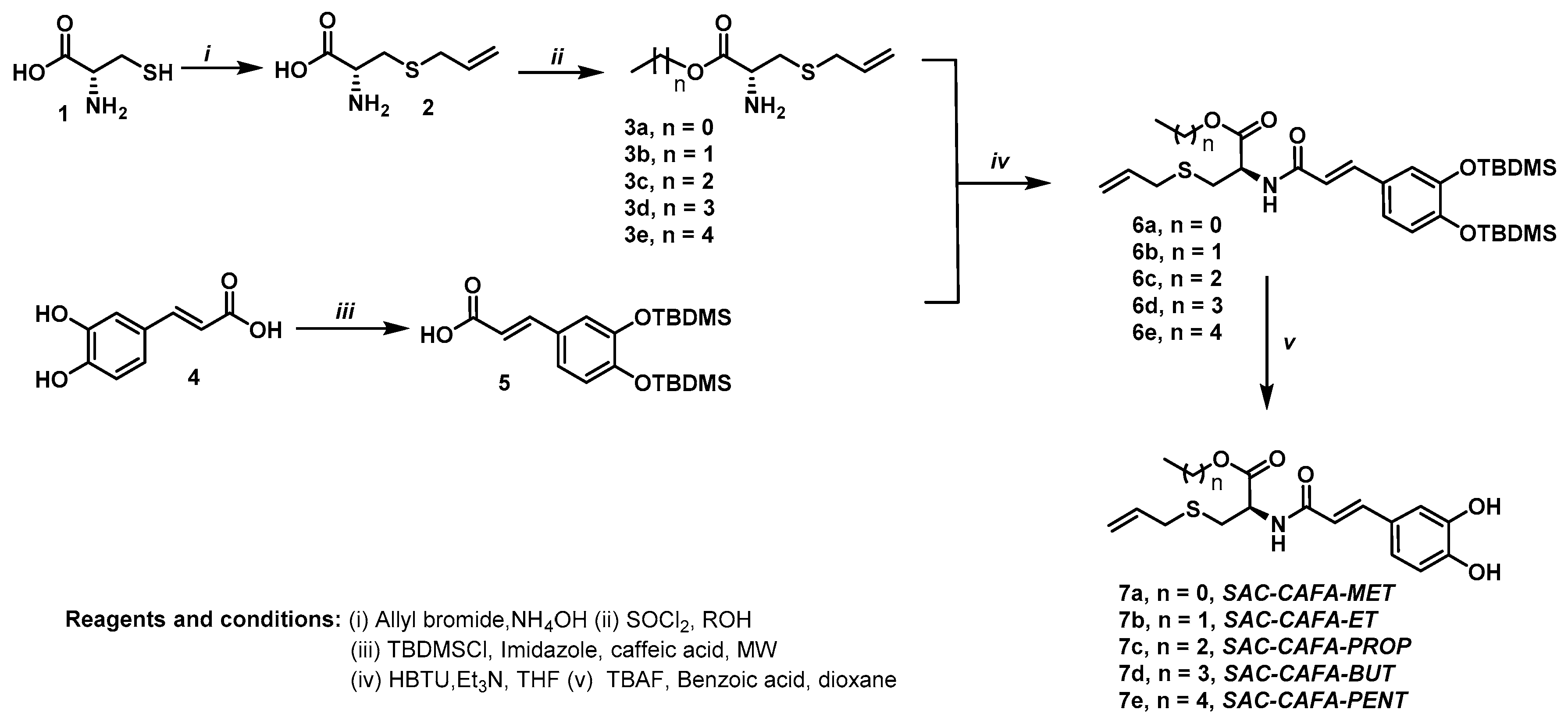
2.1. Source of Hybrid Molecules

The synthesis of the hydroxylated compound SAC-CAFA, together with the full characterization and other biological activities, was previously reported by Castrillon et al. (2019) [29]. Scheme 1 shows a brief description of the route used for the synthesis of the molecules. The reaction of nucleophilic substitution between cysteine (1) and allyl bromide gave the S-allyl cysteine (2), which was esterified with the corresponding alcohol in the presence of thionyl chloride, obtaining compound 3. On the other hand, the phenolic hydroxyl groups of caffeic acid were protected as tert-butyldimethylsilyl (TBDMS), providing compound 5. Then, compounds 3 and 5 were submitted to peptide-type coupling using N,N,N’,N’-tetramethyluronium hexafluorophosphate (HBTU) as an amide bond promoter, yielding compound 6. Finally, the hybrid SAC-CAFA was obtained via the deprotection of compound 7.

2.2. Biological Activity

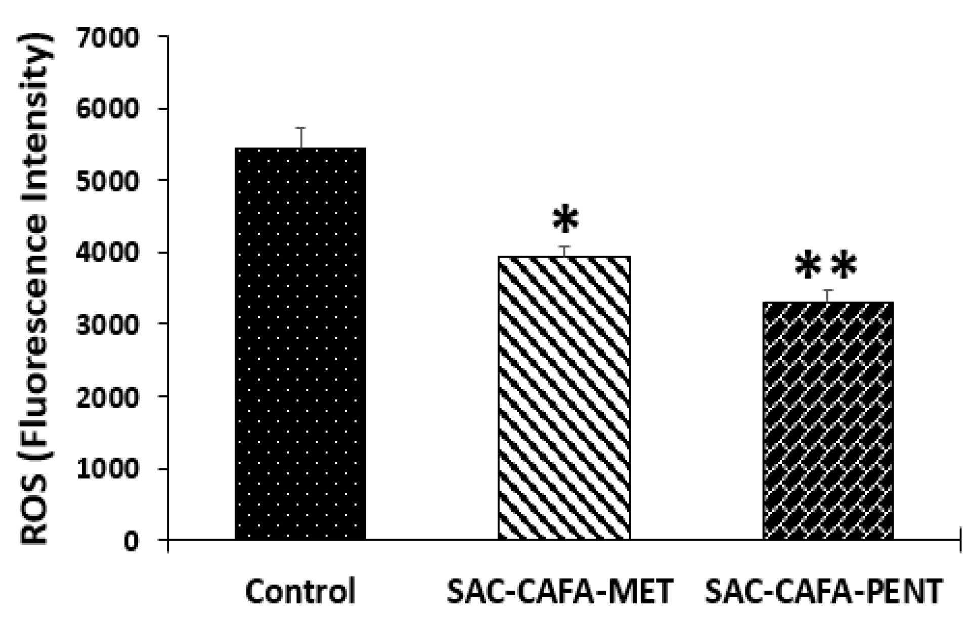
2.2.1. Effect of SAC-CAFA-MET and SAC-CAFA-PENT on Reactive Oxygen Species (ROS)

Reactive oxygen species (ROS) play a pivotal role in biological processes through the regulation of various cellular signaling pathways. Continuous ROS production in normal cells is tightly regulated by antioxidants [30]. ROS exist in two forms: The first comprises free oxygen radicals (e.g., superoxide, hydroxyl radicals, nitric oxide, alkoxyl radicals, and peroxyl radicals). The other form includes non-radical ROS (e.g., hydrogen peroxide, organic hydroperoxides, and hypochlorite) [31]. The intrinsic sources of ROS mostly include mitochondria, inflammatory cells, and several enzymatic cellular complexes. On the other hand, the extrinsic sources of ROS include pro-oxidant environmental toxins, radiation, [32,33], and a variety of chemical compounds, including alcohol, tobacco smoke, and certain drugs [33,34]. The abundant accumulation of ROS may lead to oxidative stress which, in turn, causes oxidative damage to the DNA, RNA, proteins, lipids, and mitochondria, resulting in cell death (apoptosis, autophagy, or necrosis) or the production of pro-inflammatory cytokines [33]. This resulting oxidative damage is the first step involved in mutagenesis and carcinogenesis [35,36]. ROS dynamically influence the tumor microenvironment through a dual role. Thus, at moderate concentrations, ROS activate the cancer cell survival signaling cascade; on the other hand, at high concentrations, ROS can cause cancer cell apoptosis. Hence, the ROS levels are critical, either augmenting tumorigenesis or leading to apoptosis. Considering all of these facts, we evaluated whether SAC-CAFA-MET and SAC-CAFA-PENT could regulate the formation of ROS in a colorectal cancer cell line. To achieve this, we used CM-H2DCFDA—a useful indicator for reactive oxygen species (ROS) in cells. It passively diffuses into cells and, once there, its acetate groups are cleaved by intracellular esterases. Its thiol-reactive chloromethyl group reacts with intracellular glutathione and other thiols. Subsequent oxidation yields a fluorescent adduct that is trapped inside the cell, and is measured by flow cytometry. According to the results, both hybrids significantly reduced ROS formation compared to the control (Figure 1), suggesting that they could act as antioxidants in this model, which could be quite positive considering that this activity is seen as a potential strategy to delay the harmful effects of ROS [35,37]. This is consistent with previous information reported for the parental compounds alone. Thus, several authors have reported that S-allyl cysteine (SAC) possesses strong antioxidant activity by scavenging intracellular ROS [38,39,40]. Similarly, it was reported that SAC reduced oxidative stress in a rat model of focal cerebral ischemia [41], a sporadic Alzheimer’s disease model [42], and a murine model of Parkinson’s disease [43]. Alternatively, Ismail et al. (2020) have also reported that caffeic acid (CA) possesses potential anticancer activity, with well-defined pharmacological mechanisms associated with the inhibition of ROS production [9]. This information supports our findings, suggesting that one possible mechanism of the hybrid molecules SAC-CAFA-MET and SAC-CAFA-PENT could be potentiated by inhibiting the production of ROS in SW80 cells.

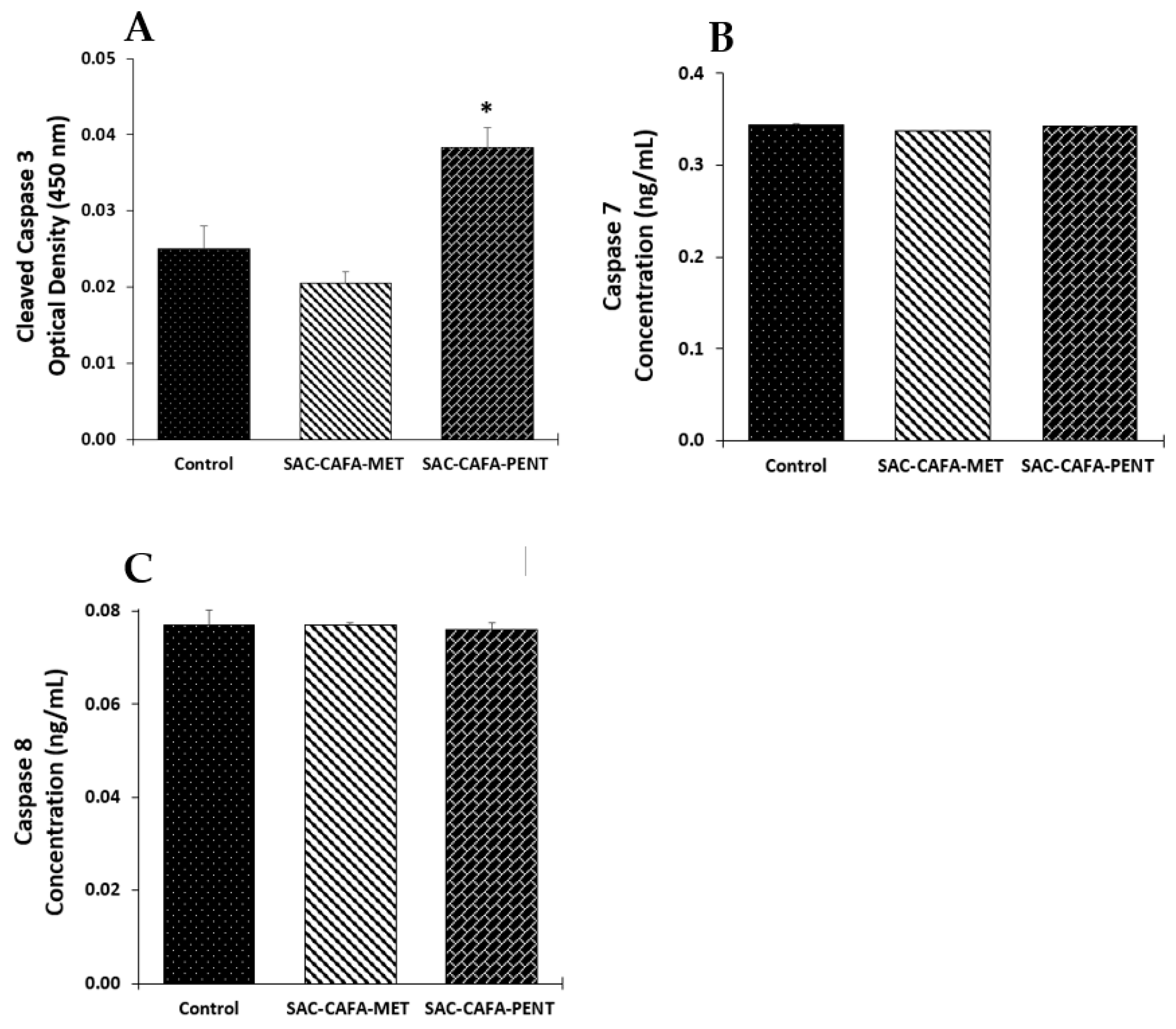
2.2.2. Effects of SAC-CAFA-MET and SAC-CAFA-PENT on the Expression of Caspase-3, -7, and -8

In normal conditions, the apoptotic pathway is a highly regulated process that culminates in the death of a cell, involving various anti-apoptotic and pro-apoptotic proteins together with the sequential activation of proteases called caspases, which are responsible for initiating the hallmarks of the degradation phase of apoptosis, including cell shrinkage, membrane blebbing, and DNA fragmentation [44]. Abnormalities in apoptotic function contribute to both the pathogenesis of colorectal cancer and its resistance to chemotherapeutic drugs. Thus, the design of various anticancer drugs has been focused on the modulation of the different molecules to develop more selective agents with increased efficacy and reduced side effects. To investigate whether the mechanisms of action of SAC-CAFA-MET and SAC-CAFA-PENT are related to apoptosis in SW480 cells, several biomarkers were evaluated.
Caspases can be subclassified as initiators (caspase-8 and -9) or executioners (caspase-3, -6, and -7) [45]. All of these proteases are synthesized as proenzymes, and require a highly regulated process to be activated. Once initiator caspases undergo self-activation, they can activate the executioner caspases to start the hydrolysis of various proteins from the cytoskeleton, nuclear proteins, and other molecules, to initiate the final process of cell death [46]. Because of this, we tested the effects of SAC-CAFA-MET and SAC-CAFA-PENT on the levels of some relevant caspases, such as caspase-3 (Figure 2A), caspase-7 (Figure 2B), and caspase-8 (Figure 2C), to determine their effects on SW480 cells after 48 h of treatment. First, we evaluated the executioner caspase-3, which is considered one of the most important proteins, acting as the primary executioner of apoptotic death, and making this process more efficient [44,47]. According to the results, we found that SAC-CAFA-PENT induced a significant increase in the active form of this protease, suggesting a possible mechanism related to apoptosis in this in vitro model. Furthermore, considering the previous results reported by our group, based on the changes induced by this compound in mitochondrial membrane permeability [29] and the absence in the activation of caspase-8, we hypothesized that apoptosis induced by this hybrid is not triggered by extrinsic pathways. On the other hand, the lack of activation of caspase-3 by SAC-CAFA-MET suggests that this molecule could involve another type of programmed cell death (PCD), which is supported by the previous evidence that reports that PCD can occur in the complete absence of caspase activation [48,49]. In addition, we also observed that SAC-CAFA-MET did not cause changes in the levels of caspase-7 and -8, supporting the idea that it involves a different mechanism unrelated to apoptosis.

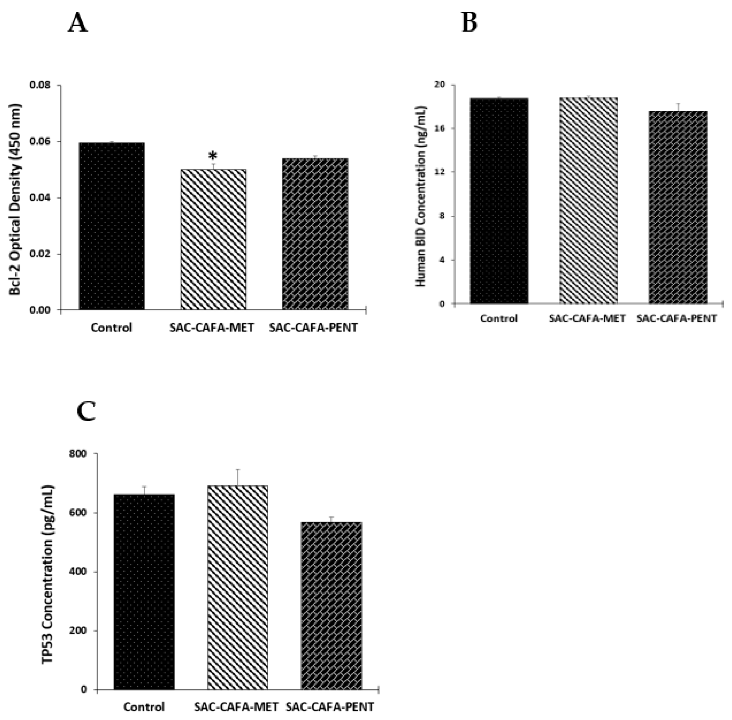
2.2.3. Effects of SAC-CAFA-MET and SAC-CAFA-PENT on the Expression of Apoptotic Biomarkers

Several apoptotic biomarkers play pivotal roles in various cellular processes. The Bcl-2 family is involved in the regulation of apoptosis and, therefore, plays a vital role in protecting against cancer. Among the genes involved in apoptotic pathways, anti-apoptotic Bcl-2 contributes to cancer’s development and progression by promoting the survival of malignant cells. Thus, it is a prime target for novel specific anticancer therapies [50,51]. The effects of SAC-CAFA-MET and SAC-CAFA-PENT were tested in SW480 cells 48 h after treatment, and the results revealed that SAC-CAFA-MET causes significant downregulation of Bcl-2 (Figure 3A). Moreover, considering that this protein suppresses the release of apoptosis-inducing factor (AIF) from the mitochondria, we hypothesized that the negative modulation of this anti-apoptotic biomarker could be implicated in a caspase-independent death process mediated by AIF, causing the loss of mitochondrial membrane potential and the canonical changes in cell death characterized by chromatin condensation and DNA fragmentation [52,53,54,55]. This is also consistent with previous studies reported by our research group, where this hybrid displayed mitochondrial depolarization [29]. Liu et al., 2012 carried out a different study using one of the precursors of the hybrids evaluated (S-allyl cysteine; SAC), and reported similar results, indicating that SAC alone suppressed the proliferation of prostate cancer cells through downregulation of Bcl-2 [56]. In addition, we also evaluated the levels of human bid protein, and the results did not show differences between the treatments with the hybrids and the control (Figure 3B). This result is consistent with the lack of activation of caspase-8 observed in this study, since bid is cleaved by this protease after a death signal [57]. In addition, considering that p53 plays an essential role in regulating cell death [58,59], we tested the effects of SAC-CAFA-MET and SAC-CAFA-PENT (Figure 3C). According to the results, we found that none of the hybrids evaluated caused significant changes in this protein, complementing the information that these hybrids caused tumor cell death through molecular mechanisms independent of p53. Nomura et al., 2001 reported similar results with various caffeic acid esters, suggesting that an active component extracted from honeybee propolis, named caffeic acid phenethyl ester (CAPE), induces apoptosis through both p53-dependent and p53-independent pathways [60].

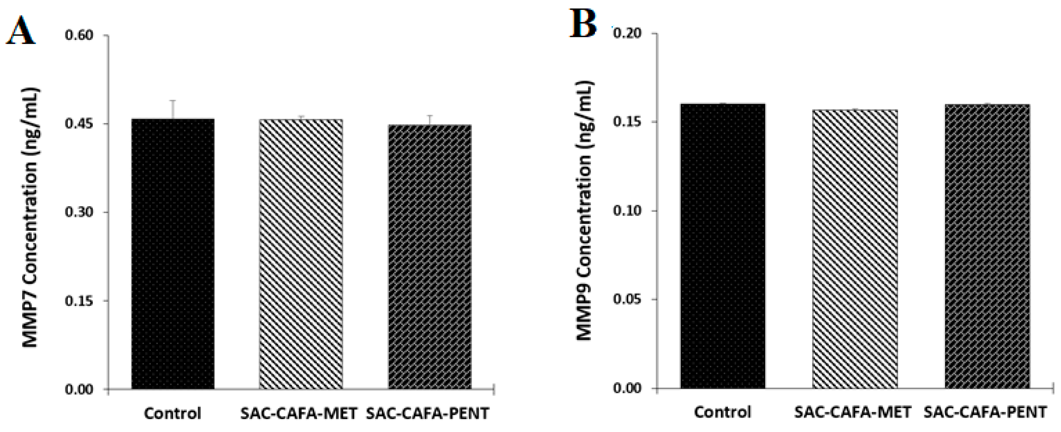
2.2.4. Effects of the Hybrids SAC-CAFA-MET and SAC-CAFA-PENT on the Activity of Matrix Metalloproteinase 7 (MMP7) and MMP9

The extracellular matrix (ECM) is composed of various macromolecules and minerals, providing structural and biochemical support for the cells, and regulating both inter- and intracellular signaling for various processes, such as differentiation, adhesion, and invasion. In cancer, malignant cells interact with the ECM, causing structural remodeling to facilitate migration from a primary tumor site. Various proteins are involved in the remodeling and degradation of the ECM. Among them, matrix metalloproteinases (MMPs) have been extensively investigated for use as proteolytic enzymes, with the ability to degrade all components of the ECM. MMP7 and MMP9 have long been evaluated in colorectal cancer, given the existence of a correlation between an increase in these endopeptidases and CRC invasion [61,62]. Because of that, we decided to evaluate whether the hybrids SAC-CAFA-MET and SAC-CAFA-PENT could regulate the activity of these enzymes in SW480 cells. According to the results, none of the compounds caused changes in the modulation of MMP7/9 (Figure 4A,B), suggesting that the mechanism of action of these molecules is not related to tissue remodeling in these cells.

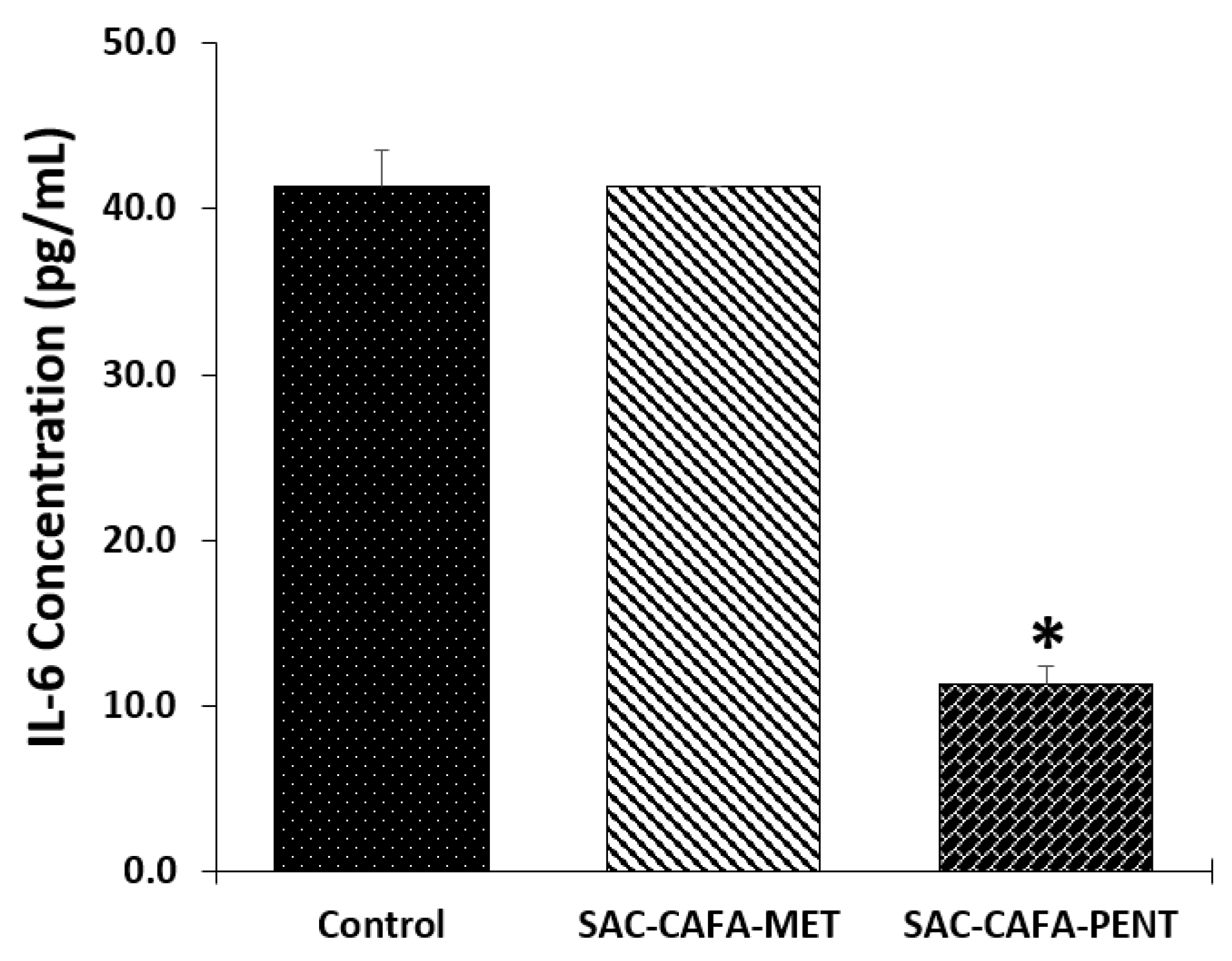
2.2.5. Effects of the hybrids SAC-CAFA-MET and SAC-CAFA-PENT on Expression of Interleukin-6 (IL-6)

It has been observed that inflammation can contribute to the development and progression of CRC [63]. Thus, several studies have focused on the evaluation of a variety of cytokines to observe whether it is possible to modulate their expression. Lin et al. reported in 2020 that an increase in the levels of interleukin-6 (IL-6) in patients with colorectal cancer was related to the size of the tumor, the severity of the pathology, and the survival rate [64]. Moreover, Knüpfer and Preiss (2009) and Galizia et al. (2002) mentioned that downregulation in the expression of this cytokine prevents multiplicity and tumor progression in intestinal cells [65,66]. For these reasons, the study of pro-inflammatory cytokines has become an important target in the discovery of molecules with potential activity against CRC. Considering these facts, we evaluated whether the hybrids SAC-CAFA-MET and SAC-CAFA-PENT could modulate the expression of IL-6 (Figure 5), and our results showed that only the second hybrid mentioned was able to induce a significant negative regulation of this biomarker, suggesting that it can also act through anti-inflammatory mechanisms in this model of colorectal cancer. On the other hand, due to the important role of these cytokines in carcinogenesis, we also evaluated the response to cyclooxygenase (COX) 1 and 2, prostaglandin E2, tumor necrosis factor α (TNF-α), and interleukin-1β (IL-1β). However, we observed that none of the hybrids evaluated could modulate the expression of these biomarkers (data not shown). Other authors have shown similar results when evaluating the precursors used for the synthesis of the hybrids SAC-CAFA-MET and SAC-CAFA-PENT. For example, You et al. (2013) [67] reported that SAC showed no inhibitory effect on COX-2 production using RAW264.7 monocyte/macrophage-like cells. Unlike this study, Kim et al. (2013) [40] evaluated SAC and CA in a skin model, and they concluded that both compounds displayed anti-inflammatory activity through modulation of COX-2. Furthermore, Zarezadeh et al. (2017) [68] reported that SAC caused downregulation of IL-1β in male Wistar albino rats. All of these findings show how different the responses are between models, and complement the possible mechanisms of action associated with the hybrid molecules evaluated in this study.

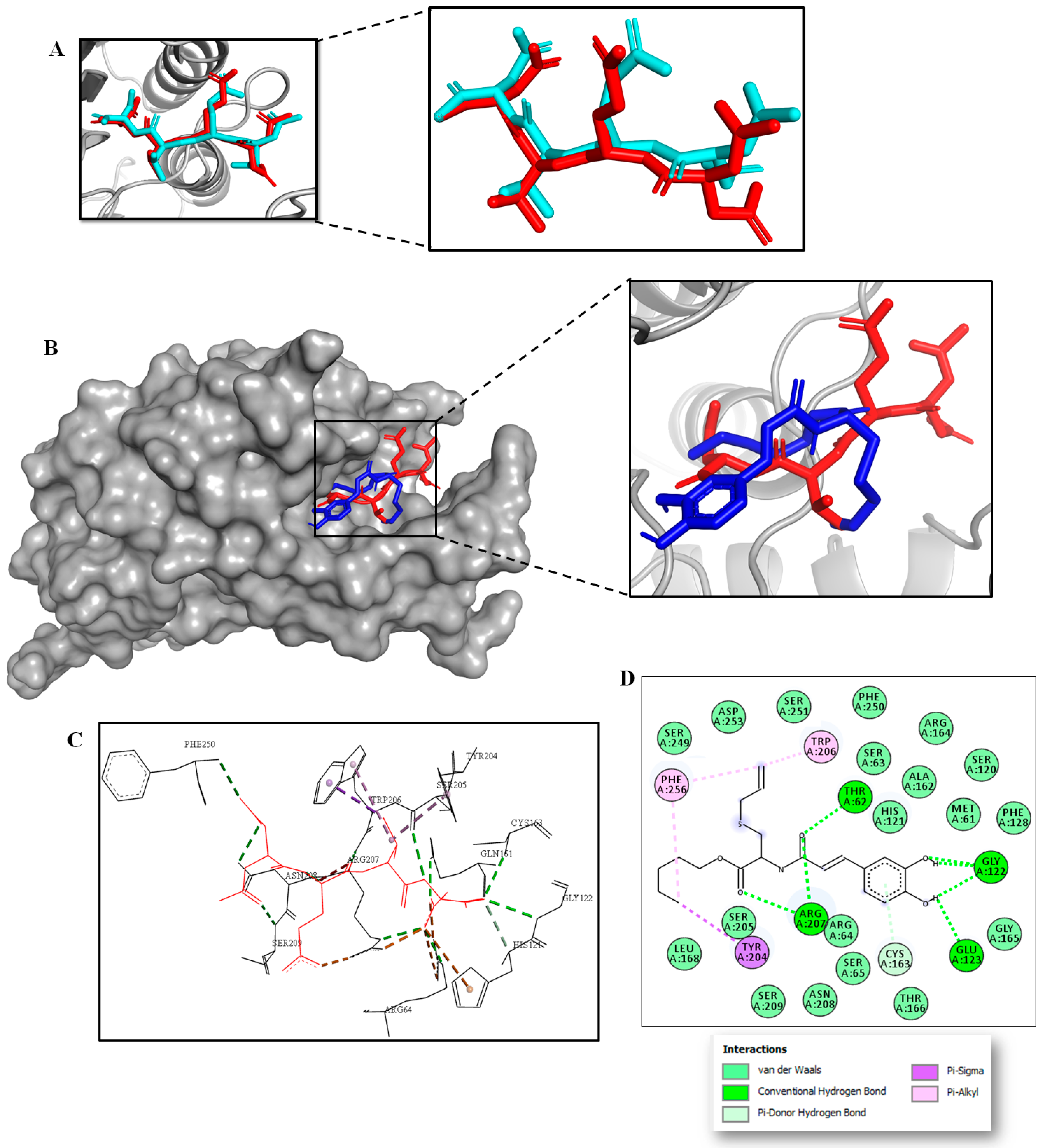
2.3. Multitarget Docking Studies and Prediction of Binding Pose

Considering that these compounds induced changes in different molecular targets, we decided to perform molecular docking experiments to explain the possible interactions. Based on the experimental findings, SAC-CAFA-PENT induced an increase in the levels of caspase-3, which could explain the apoptotic process induced by this compound. Because of this, it was important to understand the coupling between the hybrid and the protease. On the other hand, we performed molecular docking on Bcl-2, since SAC-CAFA-MET caused a significant decrease in this protein, and its modulation could be involved in cell death induction in cancer cells. Finally, we decided to dock IL-6, since SAC-CAFA-PENT caused a significant decrease in this cytokine, which elicits proliferation and anti-apoptotic effects in tumor cells and, thus, negative regulation of this pro-inflammatory cytokine has been contemplated as important strategy in colorectal cancer chemoprevention. In this light, the SAC-CAFA-PENT hybrid was docked into each catalytic domain of human X-ray crystallographic structures of caspase-3 and IL-6, while for SAC-CAFA-MET the three-dimensional (3D) crystal structure of Bcl-2 was used. The docking protocol was performed via grid-based ligand docking with AutoDock Vina, and affinity scores along the best binding pose were estimated (Table 1). Interestingly, among these targeted proteins, SAC-CAFA-PENT displayed greater binding affinity for human caspase-3 (PDB ID: 5I9B) than IL-6 (PDB: 1ALU). In particular, the results indicated that the hit compound SAC-CAFA-PENT binds to caspase-3 with good binding energy—close to −7.3 kcal·mol−1—suggesting that this compound could act on the catalytic domain of the enzyme to significantly activate its function, triggering cell death via apoptosis. Thus, we hypothesized that modulation of the activity of caspase-3 may be the primary biochemical mechanism by which SAC-CAFA-PENT inhibits SW480 cells’ viability. On the other hand, SAC-CAFA-MET did not display significant binding affinity for Bcl-2, suggesting that modulation of Bcl-2 function caused by SAC-CAFA-MET would not be the direct mechanism to explain the significant decrease in this protein.
Caspases are a conserved family of cysteine-dependent proteases that are classically associated in the execution of apoptosis [69]. Among them, caspase-3 plays an important role in regulating and implementing the cell death program during apoptosis events. The caspase-3 substrate-binding region is characterized by the presence of the amino acid residues Arg64, Leu119, Ser120, His121, Gln161, Ala162, Cys163, Ser198, Tyr204, Ser205, Trp206, Asn208, Ser209, Trp214, Ser249, Phe250, Ser251, and Phe252 [70]. Thus, we explored the binding interactions of the promising SAC-CAFA-PENT (in blue) to the caspase-3 receptor (PDB: 5I9B) in that domain. In order to accomplish high throughput, the AutoDock Vina protocol inside the caspase-3 binding pocket was first validated through self-docking. We performed a comparison between the crystallographic binding mode deposited in PDB and the redocked conformation of the Ac-DEVD-CMK ligand. To carry out this validation, the root-mean-square deviation (RMSD) value was calculated to correlate the differences between the atomic distances. As illustrated in Figure 6A, the docked conformation predicted for Ac-DEVD-CMK (in cyan) was spatially close to the crystallographic structure pose (in red), with an optimal RMSD value of 1.075 Å. In addition, the best binding energy calculated for Ac-DEVD-CMK was estimated to be −8.1 kcal·mol−1. These findings indicate a high level of feasibility of our protein–ligand docking protocol, which was able to reproduce the binding pose of the co-crystallized ligand deposited under PDB ID: 5I9B. After the docking protocol was validated, an exhaustive search of the binding pocket was carried out in order to establish key points in the binding site when SAC-CAFA-PENT (blue) was docked into the caspase-3 binding site.
The docking results showed that in addition to the hybrid binding to caspase-3 with a good binding affinity of about −7.3 kcal·mol−1, it also fit well in the active pocket of caspase-3, as illustrated in Figure 6B. Indeed, the best docking conformation obtained for this hybrid was in good agreement with the X-ray crystallographic pose of the peptide-based inhibitor Ac-DEVD-CMK (in red). It was found that similar to the co-crystallized inhibitor (in red), the hybrid SAC-CAFA-PENT (blue) also interacted with those key amino acid residues surrounding the catalytic domain of caspase-3, mainly via the side chains of six amino acids: His121, Gly122, Tyr204, Ser205, Trp206, and Arg207 (Figure 6C,D). In this regard, a visual inspection of the 2D protein–ligand interaction plot (Figure 6D) after the docking run revealed that SAC-CAFA-PENT formed key hydrogen bond interactions with the His121, Glu123, Arg207, Thr62, and Gly122 residues, which play an important role in the function of caspase-3. Furthermore, the hybrids showed interactions via π contacts with the residuals around the catalytic domain of the enzyme, such as Tyr204, Trp206, and Phe256. Finally, multiple hydrophobic interactions surrounded by side chains in the binding cleft were observed between the hybrids and caspase-3, potentially stabilizing the protein–ligand complex upon the binding event. These computational findings suggest that a probable modulation of caspase-3 would be the primary mechanism of action for explaining the in vitro cytotoxic response registered for the SAC-CAFA-PENT hybrid in SW480 colon cancer cells, which was in good agreement with the experimental data.

2.4. DFT Analysis of the Radical-Scavenging Activity of SAC-CAFA-MET and SAC-CAFA-PENT

Excess formation of reactive free radicals by various enzymatic and non-enzymatic processes in the body has been found to be associated the initiation and progression of various cancer types [71]. In neoplastic disorders, overproduction of reactive oxygen species (ROS) contributes to different stages of carcinogenesis events, such as cancer development and progression [72]. Therefore, investigations addressing new compounds expressing antioxidant and free-radical-scavenging effects play a key role in cancer drug development. In this paper, we found that SAC-CAFA-MET and SAC-CAFA-PENT could have effects on the modulation of ROS in SW480 cells in vitro. These findings might suggest that, apart from caspase-3 modulation, a second possible mechanism for the cytotoxic response of hybrids via interrupting cells’ antioxidant effects could be also implicated. Thus, to further understand the abovementioned experimental antioxidant properties for hit hybrids, quantum chemical calculations based on the density functional theory (DFT) approach were employed to investigate the energetic behavior of their free-radical-scavenging reactions. First of all, three of the most favorable antioxidant mechanisms were investigated, as follows: hydrogen atom transfer (HAT), sequential proton transfer–electron transfer (SPT–ET), and sequential electron transfer–proton transfer (SET–PT), in aqueous media and in the gas phase.
(i)
Hydrogen atom transfer (HAT):
R1OH + R2 → R1O + R2H
(ii)
Sequential proton transfer–electron transfer (SPT–ET):
Step 1 R1OH + R2 → R1O + R2H+
Step 2 R1O + R2H+→ R1O + R2H
(iii)
Sequential electron transfer–proton transfer (SET–PT):
Step 1 R1OH + R2 → R1OH+ + R2
Step 2 R1OH+ + R2 → R1O + R2H
As shown in Table 2, DFT analysis of SAC-CAFA-MET and SAC-CAFA-PENT suggested that these compounds would react with free radicals via hydrogen atom transfer (HAT) which, in turn, may be used to explain the antioxidant response to the hybrids in vitro. Thus, when the hydroxyl radical (•OH) was used as a radical target in an aqueous medium, HAT results showed a negative Gibbs free energy value of about −39.0 kcal·mol−1. The highly reactive oxidative free radicals (•R) are strongly implicated in cancer progression. Free radicals (•R) can stimulate lipid peroxidation, oxidative damage to proteins, or induced DNA damage, leading to fatal lesions in the cell that contribute to the initiation, promotion, and progression of cancer. Based on DFT calculations, SAC-CAFA-MET and SAC-CAFA-PENT could interrupt cells’ oxidative stress through a hydrogen atom transfer pathway (HAT), which could explain their effects on the modulation of ROS in SW480 cells.

3. Materials and Methods

3.1. In Vitro Biological Assays

3.1.1. Cell Line, Culture Medium, and Treatment

SW480 (human colon cancer) cells were obtained from The European Collection of Authenticated Cell Cultures (ECACC, Salisbury, UK). Cells were grown in 25 cm2 Falcon flasks containing Dulbecco’s Modified Eagle Medium, supplemented with 10% heat-inactivated (56 °C) horse serum, 1% non-essential amino acids, and 1% penicillin/streptomycin procured from Gibco Invitrogen (Carlsbad, CA, USA). Cells were incubated at 37 °C in a humidified atmosphere of 5% CO2. For all experiments, horse serum was reduced to 3%, and the medium was supplemented with 5 mg/mL transferrin, 5 ng/mL selenium, and 10 mg/mL insulin (ITS-defined medium; Gibco, Invitrogen, Carlsbad, CA, USA) [73,74,75]. The compounds were dissolved in 1% DMSO before exposing the cells for 48 h. For all experiments, the cells were treated using the IC50 value (SAC-CAFA-MET = 124.2 µM; SAC-CAFA-PENT = 118 µM). These values were obtained from dose–response curves that were previously published by our group [29].

3.1.2. Determination of ROS

Intracellular levels of ROS were determined as previously reported [76]. SW480 cells were seeded at a density of 2.5 × 10⁵ cells/well in 6-well tissue culture plates, allowing them to grow for 24 h; afterwards, they were treated for 48 h with either 1% DMSO (control) or the hybrids SAC-CAFA-MET (124.2 µM) and SAC-CAFA-PENT (118 µM). CM-H2DCFDA was then added at a final concentration of 8 μM and incubated for 30 min at 37 °C, protected from light. The fluorescence of 10,000 events was read through flow cytometry using a FACSCanto II flow cytometer and the software BD FACSDiva 6.1.3. (BD Biosciences, San Jose, CA, USA). Data were analyzed using the software FlowJo v. 7.6.2 (FlowJo, Ashland, OR, USA). Cell clumps were excluded using the height and area signals on the forward scatter (FSC-H vs. FSC-A). ROS production was expressed as the percentage (%) increase in fluorescence relative to untreated control cells.

3.1.3. Determination of Inflammatory Cytokines and Apoptotic Proteins

Tumor cells (SW480) were cultured as previously described and then treated with the hybrids SAC-CAFA-MET (124.2 µM) and SAC-CAFA-PENT (118 µM) for 48 h. Afterwards, cells were collected by scraping and lysed with cell lysis buffer (1X, Ref. #9803). The supernatant was used to determine the effects of the hybrids on the modulation of inflammatory cytokines, apoptotic biomarkers, and enzymes. The kits for detecting COX-1, COX-2, IL-1β, IL-6, and TNF-α were provided by Cayman Chemical Company (Ann Arbor, MI, USA); PGE2, caspase-7, caspase-8, MMP7, and MMP9 were obtained from Elabscience Biotechnology Co., (Wuhan, China); the determination of Bid was carried out by G-Biosciences, Inc. (St. Louis, MO, USA), whereas cleaved caspase-3, p53, and Bcl-2 were obtained from Cell-Signaling Technology (Danvers, MA, USA). The assays were performed according to the manufacturers’ instructions.

3.1.4. Statistical Analysis

All experiments were performed at least three times. Data are reported as the mean ± SE (standard error). Cells treated with the vehicle used to solubilize the hybrids (1% DMSO) were used as controls. Statistical differences between the control group and treated cells were evaluated via one-way ANOVA followed by Dunnett’s test. Values with p  ≤  0.05 were considered significant. Data were analyzed with GraphPad Prism version 7.04 for Windows (Graph Pad Software, San Diego, CA, USA).

3.2. Computational Methods

The chemical structure of the SAC-CAFA-PENT hybrid, as well as the inhibitors venetoclax and Ac-DEVD-CMK, were the ligands used in these computational approaches. Their 2D structures were drawn using ChemDraw 17.0 software (Cambridge Soft, Cambridge, MA, USA) and saved as MDL MoL files. Chem3D 17.0 (Cambridge Soft, USA) was used to generate 3D structures of all ligands and energetically minimize them via the MM2 force field. The Discovery Studio Visualizer program was used to rewrite the data files into PDB format. AutoDockTools was used to parameterize ligand structures through computing Gasteiger partial atomic charges and adding full hydrogens, as well as to assign rotatable bonds. The resulting structures were saved in the required format for use with AutoDock. Then, AUTOTUTORS in AutoDockTools was used to define all possible flexible torsions of the selected ligands to favor the computed binding with the receptor structure [77]. The crystal structures of caspase-3 (PDB ID: 5I9B), Bcl-2 (PDB: 4MAN), and IL-6 (PDB: 1ALU) were downloaded from the Protein Data Bank (https://rcsb.org, accessed on 1 April 2022), and all bounded ligands, ions, and solvent molecules were manually removed using the DS Visualizer 2.5 program. For docking studies, the structure of the selected proteins was parameterized using AutoDockTools [78]. To facilitate the formation of hydrogen bonds, polar hydrogens were added. AutoDock Vina software was used to perform molecular docking and default procedures for docking a flexible ligand to a rigid protein. Then, the ligands were centered at the binding site located in the binding cavity of the caspase-3, Bcl-2, and IL-6 proteins at x, y, and z coordinates of 1.5, −8.1, and −13.4; −11.252, 13.128, and 5.816; and −5.433, −12.86, and 0.496, respectively. In detail, the docking studies involved a grid box that was identified using AutoDock Vina 1.1.2, and the exhaustiveness was 20 for each protein–compound pair [67]. The catalytic active site was surrounded by a docking grid of 36 × 36 × 36 Å (for caspase-3), 32 × 32 × 32 Å (for Bcl-2), and 40 × 40 × 40 Å (for IL-6), with a grid spacing of 1Å. Ligand-binding affinities (in kcal·mol−1) were estimated using AutoDock Vina and ranked based on the free-energy binding theory. Then, docking solutions were graphically inspected using DS Visualizer 2.5 (http://3dsbiovia.com/products/, accessed on 2 April 2022) to provide a 2D ligand interaction plot, while the ribbon surface representation of 3D models was explored using the PyMOL Molecular Graphics System 2.0.
On the other hand, quantum calculations by means of density functional theory (DFT) were performed to obtain a deeper insight into the relationship between the SAC-CAFA-MET and SAC-CAFA-PENT hybrids’ structure and their antioxidant activity against ROS production in SW80 cells. The structures of all reactants and products were optimized using Minnesota’s M06-2X [79] DFT functional in conjunction with the 6-31++G(2d,p) basis set [79,80,81,82,83]. We choose this functional because it has been shown to give good thermochemical results in reactions between phenolic-like compounds and free radicals [84,85]. All calculations were performed with the GAMESS US program [86]. Frequencies were calculated in order to characterize all minima at stationary points (e.g., zero imaginary frequencies). Thermal correction, zero-point energies, and entropy contributions were used to calculate Gibbs free energies at 298.15 K and 1 atm. In addition, the contribution of aqueous media to the Gibbs energy was included with the SMD (continuum solvation model) from single-point calculations of the DFT-optimized geometry in vacuum.

4. Conclusions

In summary, we identified two hybrid molecules possessing potent chemopreventive activity in colorectal cancer cells. Some approaches to the possible mechanisms of action of these molecules based on S-allyl cysteine ester–caffeic acid amide hybrids were considered. In previous findings, we reported that both compounds induced cell damage, and here we found that they reduced ROS formation, showing that they could act through different mechanisms despite their chemical similarities. Firstly, the mechanism of SAC-CAFA-PENT could be associated with apoptosis mediated by caspase-3, as evidenced by the increase in the levels of the cleaved form of this protease. Moreover, this mechanism was independent of the tumor-suppressor protein p53, and was probably triggered via an intrinsic route, considering the lack of activation of caspase-8. In addition, it was hypothesized that this hybrid induced anti-inflammatory effects mediated by downregulation of IL-6. In contrast, SAC-CAFA-MET induced cell death independent of caspases and p53, mediated by downregulation of Bcl-2. Considering these findings, and the lack of modulation of biomarkers related to extrinsic routes, we hypothesized that this hybrid could induce the release of AIF from mitochondria, causing the loss of mitochondrial membrane potential and the canonical changes of cell death characterized by chromatin condensation and DNA fragmentation. In addition, docking studies would suggest caspase-3 modulation as the primary mechanism by which SAC-CAFA-PENT elicits apoptosis in SW480 human colorectal adenocarcinoma cells. Meanwhile, DFT calculations suggest that the hybrids could produce effects on the modulation of ROS in SW480 cells via the hydrogen atom transfer (HAT) pathway. However, it is necessary to carry out further investigations to explore all of these mechanisms more deeply, to ensure the potential benefits of these novel hybrids in different types of cancers.

Author Contributions

A.H.-R.: Conceptualization, Methodology, Validation, evaluation of biological activities and formal Analysis, Writing—Original Draft, Writing—Review and Editing; A.F.Y.-P.: Computational studies, protocols and analysis; J.Q.-S.: Computational studies; G.M.-Q.: evaluation of biological activities. T.W.N.: Revision of the final version of the manuscript; W.C.-G.: Resources, Project Administration, Funding Acquisition. All authors have read and agreed to the published version of the manuscript.

Funding

This research was funded by the University of Antioquia, MINCIENCIAS (Grant No. 247-2016, code: 111571249830).

Institutional Review Board Statement

Not applicable.

Informed Consent Statement

Not applicable.

Data Availability Statement

Not applicable.

Acknowledgments

The authors thank to the University of Antioquia and MINCIENCIAS for their support.

Conflicts of Interest

The authors declare no confilct of interests.

Abbreviations

SACS-allyl cysteine
CACaffeic acid
CRCColorectal cancer
DMSODimethyl sulfoxide
5-FU5-Fluorouracil
ROSReactive oxygen species

References

  1. Hull, R.; Francies, F.Z.; Oyomno, M.; Dlamini, Z. Colorectal cancer genetics, incidence and risk factors: In search for targeted therapies. Cancer Manag. Res. 2020, 12, 9869–9882. [Google Scholar] [CrossRef] [PubMed]
  2. Anand, P.; Kunnumakkara, A.B.; Sundaram, C.; Harikumar, K.B.; Tharakan, S.T.; Lai, O.S.; Sung, B.; Aggarwal, B.B. Cancer is a preventable disease that requires major lifestyle changes. Pharm. Res. 2008, 25, 2097–2116. [Google Scholar] [CrossRef] [PubMed]
  3. Sung, H.; Ferlay, J.; Siegel, R.L.; Laversanne, M.; Soerjomataram, I.; Jemal, A.; Bray, F. Global cancer statistics 2020: GLOBOCAN estimates of incidence and mortality worldwide for 36 cancers in 185 countries. CA. Cancer J. Clin. 2020, 70, 313. [Google Scholar] [CrossRef]
  4. Siegel, R.L.; Miller, K.D.; Fuchs, H.E.; Jemal, A. Cancer statistics, 2021. CA Cancer J. Clin. 2021, 71, 7–33. [Google Scholar] [CrossRef] [PubMed]
  5. Alam, W.; Bouferraa, Y.; Haibe, Y.; Mukherji, D.; Shamseddine, A. Management of colorectal cancer in the era of COVID-19_Challenges and suggestions. Sci. Prog. 2021, 104, 104. [Google Scholar] [CrossRef]
  6. Pointet, A.L.; Taieb, J. Cáncer de colon. EMC-Tratado Med. 2017, 21, 1–7. [Google Scholar] [CrossRef]
  7. McQuade, R.M.; Bornstein, J.C.; Nurgali, K. Anti-colorectal cancer chemotherapy-induced diarrhoea: Current treatments and side effects. Int. J. Clin. Med. 2014, 5, 393–406. [Google Scholar] [CrossRef] [Green Version]
  8. Housman, G.; Byler, S.; Heerboth, S.; Lapinska, K.; Longacre, M.; Snyder, N.; Sarkar, S. Drug resistance in cancer: An overview. Cancers 2014, 6, 1769–1792. [Google Scholar] [CrossRef] [Green Version]
  9. Ismail, T.; Donati-Zeppa, S.; Akhtar, S.; Turrini, E.; Layla, A.; Sestili, P.; Fimognari, C. Coffee in cancer chemoprevention: An updated review. Expert. Opin. Drug Metab. Toxicol. 2020, 17, 69–85. [Google Scholar] [CrossRef]
  10. Steward, W.P.; Brown, K. Cancer chemoprevention: A rapidly evolving field. Br. J. Cancer. 2013, 109, 1–7. [Google Scholar] [CrossRef]
  11. Kerru, N.; Singh, P.; Koorbanally, N.; Raj, R.; Kumar, V. Recent advances (2015–2016) in anticancer hybrids. Eur. J. Med. Chem. 2017, 142, 179–212. [Google Scholar] [CrossRef] [PubMed]
  12. Nepali, K.; Sharma, S.; Sharma, M.; Bedi, P.M.; Dhar, K.L. Rational approaches, design strategies, structure activity relationship and mechanistic insights for anticancer hybrids. Eur. J. Med. Chem. 2014, 22, 422–487. [Google Scholar] [CrossRef] [PubMed]
  13. Kim, K.M.; Chun, S.B.; Koo, M.S.; Choi, W.J.; Kim, T.W.; Kwon, Y.G.; Chung, H.T.; Billiar, T.R.; Kim, Y.M. Differential regulation of NO availability from macrophages and endothelial cells by the garlic component S-allyl cysteine. Free Radic. Biol. Med. 2001, 30, 747–756. [Google Scholar] [CrossRef]
  14. Ho, S.E.; Ide, N.; Lau, B.H. S-allyl cysteine reduces oxidant load in cells involved in the atherogenic process. Phytomedicine 2001, 8, 39–46. [Google Scholar] [CrossRef] [PubMed]
  15. Numagami, Y.; Ohnishi, S.T. S-allyl cysteine inhibits free radical production, lipid peroxidation and neuronal damage in rat brain ischemia. J. Nutr. 2001, 131, 1100S–1105S. [Google Scholar] [CrossRef]
  16. Numagami, Y.; Sato, S.; Ohnishi, S.T. Attenuation of rat ischemic brain damage by aged garlic extracts: A possible protecting mechanism as antioxidants. Neurochem. Int. 1996, 29, 135–143. [Google Scholar] [CrossRef]
  17. Mostafa, M.G.; Mima, T.; Ohnishi, S.T.; Mori, K. S-allylcysteine ameliorates doxorubicin toxicity in the heart and liver in mice. Planta Med. 2000, 66, 148–151. [Google Scholar] [CrossRef]
  18. Welch, C.; Wuarin, L.; Sidell, N. Antiproliferative effect of the garlic compound S-allyl cysteine on human neuroblastoma cells in-vitro. Cancer Lett. 1992, 63, 211–219. [Google Scholar] [CrossRef]
  19. Takeyama, H.; Hoon, D.S.; Saxton, R.E.; Morton, D.L.; Irie, R.F. Growth inhibition and modulation of cell markers of melanoma by S-allyl cysteine. Oncology 1993, 50, 63–69. [Google Scholar] [CrossRef]
  20. Hung, C.C.; Tsai, W.J.; Kuo, L.M.; Kuo, Y.H. Evaluation of caffeic acid amide analogues as anti-platelet aggregation and anti-oxidative agents. Bioorg. Med. Chem. 2005, 13, 1791–1797. [Google Scholar] [CrossRef]
  21. Son, S.; Lewis, B.A. Free radical scavenging and antioxidative activity of caffeic acid amide and ester analogues: Structure-activity relationship. J. Agric. Food Chem. 2002, 50, 468–472. [Google Scholar] [CrossRef] [PubMed]
  22. Otero, E.; Robledo, S.; Díaz, S.; Carda, M.; Muñoz, D.; Paños, J.; Vélez, I.D.; Cardona, W. Synthesis and leishmanicidal activity of cinnamic acid esters: Structure–activity relationship. Med. Chem. Res. 2014, 23, 1378–1386. [Google Scholar] [CrossRef] [Green Version]
  23. De, P.; Baltas, M.; Bedos-Belval, F. Cinnamic acid derivatives as anticancer agents—A review. Curr. Med. Chem. 2011, 18, 1672–1703. [Google Scholar] [CrossRef] [PubMed]
  24. Cardona-G, W.; Herrera-R, A.; Castrillón-L, W.; Ramírez-Malule, H. Chemistry and anticancer activity of hybrid molecules and derivatives based on 5-fluorouracil. Curr. Med. Chem. 2021, 28, 5551–5601. [Google Scholar] [CrossRef] [PubMed]
  25. Decker, M. Design of hybrid molecules for drug development. In Editorial Project Manager; Elsevier: Amsterdam, The Netherlands, 2017; ISBN 978-0-08-101011-2. [Google Scholar]
  26. Shaveta, S.M.; Singh, P. Hybrid molecules: The privileged scaffolds for various pharmaceuticals. Eur. J. Med. Chem. 2016, 124, 500–536. [Google Scholar] [CrossRef] [PubMed]
  27. Tsogoeva, S.B. Recent progress in the development of synthetic hybrids of natural or unnatural bioactive compounds for medicinal chemistry. Mini Rev. Med. Chem. 2010, 10, 773–793. [Google Scholar] [CrossRef]
  28. Meunier, B. Hybrid molecules with a dual mode of action: Dream or reality? Acc. Chem. Res. 2008, 41, 69–77. [Google Scholar] [CrossRef]
  29. Castrillón, W.; Herrera-R, A.; Prieto, L.J.; Conesa-Milián, L.; Carda, M.; Naranjo, T.; Maldonado, M.E.; Cardona-G, W. Synthesis and in-vitro Evaluation of S-allyl cysteine ester—Caffeic acid amide hybrids as potential anticancer agents. Iran. J. Pharm. Sci. 2019, 18, 1770–1789. [Google Scholar]
  30. Hebbar, S.; Knust, E. Reactive oxygen species (ROS) constitute an additional player in regulating epithelial d evelopment. BioEssays 2021, 43, 2100096. [Google Scholar] [CrossRef]
  31. Sun, Y.; Lu., Y.; Saredy, J.; Wang, X.; Drummer, C., IV; Shao, Y.; Saaoud, F.; Xu, K.; Liu, M.; Yang, W.Y.; et al. ROS systems are a new integrated network for sensing homeostasis and alarming stresses in organelle metabolic processes. Redox Biol. 2020, 37, 101696. [Google Scholar] [CrossRef]
  32. Nourazarian, A.R.; Kangari, P.; Salmaninejad, A. Roles of oxidative stress in the development and progression of breast cancer. Asian Pac. J. Cancer Prev. 2014, 15, 4745–4751. [Google Scholar] [CrossRef] [PubMed]
  33. Aggarwal, V.; Tuli, H.S.; Varol, A.; Thakral, F.; Yerer, M.B.; Sak, K.; Varol, M.; Jain, A.; Khan, M.A.; Sethi, G. Role of reactive oxygen species in cancer progression: Molecular mechanisms and recent advancements. Biomolecules 2019, 9, 735. [Google Scholar] [CrossRef] [PubMed] [Green Version]
  34. Zhang, J.; Ahn, K.S.; Kim, C.; Shanmugam, M.K.; Siveen, K.S.; Arfuso, F.; Samym, R.P.; Deivasigamanim, A.; Lim, L.H.K.; Wang, L.; et al. Nimbolide-induced oxidative stress abrogates STAT3 signaling cascade and inhibits tumor growth in transgenic adenocarcinoma of mouse prostate model. Antioxid. Redox Signal. 2016, 24, 575–589. [Google Scholar] [CrossRef] [PubMed]
  35. Carini, F.; Mazzola, M.; Rappa, F.; Jurjus, A.; Geagea, A.G.; Al Kattar, S.; Bou-Assi, T.; Jurjus, R.; Damiani, P.; Leone, A.; et al. Colorectal carcinogenesis: Role of oxidative stress and antioxidants. Anticancer Res. 2017, 37, 4759–4766. [Google Scholar] [PubMed] [Green Version]
  36. Oparka, M.; Walczak, J.; Malinska, D.; van Oppen, L.M.P.E.; Szczepanowska, J.; Koopman, W.J.H.; Wieckowski, M.R. Quantifying ROS levels using CM-H2DCFDA and HyPer. Methods 2016, 109, 3–11. [Google Scholar] [CrossRef] [PubMed]
  37. Stone, W.L.; Krishnan, K.; Campbell, S.E.; Palau, V.E. The role of antioxidants and pro-oxidants in colon cancer. World J. Gastrointest. Oncol. 2014, 6, 55–66. [Google Scholar] [CrossRef] [PubMed]
  38. Shang, A.; Cao, S.Y.; Xu, X.Y.; Gan, R.Y.; Tang, G.Y.; Corke, H.; Mavumengwana, V.; Li, H.B. Bioactive compounds and biological functions of garlic (Allium sativum L.). Foods 2019, 8, 246. [Google Scholar] [CrossRef] [Green Version]
  39. Kim, J.M.; Lee, J.C.; Chang, N.; Chun, H.S.; Kim, W.K. S-Allyl-L-cysteine attenuates cerebral ischemic injury by scavenging peroxynitrite and inhibiting the activity of extracellular signal-regulated kinase. Free Radic. Res. 2006, 40, 827–835. [Google Scholar] [CrossRef] [PubMed]
  40. Kim, S.R.; Jung, Y.R.; An, H.J.; Kim, D.H.; Jang, E.J.; Choi, Y.J.; Moon, K.M.; Park, M.H.; Park, C.H.; Chung, K.W.; et al. Anti-wrinkle and anti-inflammatory effects of active garlic components and the inhibition of MMPs via NF-κB signaling. PLoS ONE 2013, 8, e73877. [Google Scholar] [CrossRef] [Green Version]
  41. Ashafaq, M.; Khan, M.M.; Shadab Raza, S.; Ahmad, A.; Khuwaja, G.; Javed, H.; Khan, A.; Islam, F.; Siddiqui, M.S.; Safhi, M.M.; et al. S-allyl cysteine mitigates oxidative damage and improves neurologic deficit in a rat model of focal cerebral ischemia. Nutr. Res. 2012, 32, 133–143. [Google Scholar] [CrossRef]
  42. Javed, H.; Khan, M.M.; Khan, A.; Vaibhav, K.; Ahmad, A.; Khuwaja, G.; Ahmed, M.E.; Raza, S.S.; Ashafaq, M.; Tabassum, R.; et al. S-allyl cysteine attenuates oxidative stress associated cognitive impairment and neurodegeneration in mouse model of streptozotocin-induced experimental dementia of Alzheimer’s type. Brain Res. 2011, 1389, 133–142. [Google Scholar] [CrossRef] [PubMed]
  43. Rojas, P.; Serrano-García, N.; Medina-Campos, O.N.; Pedraza-Chaverri, J.; Maldonado, P.D.; Ruiz-Sánchez, E. S-Allylcysteine, a garlic compound, protects against oxidative stress 1-methyl-4-phenylpyridinium-induced parkinsonism in mice. J. Nutr. Biochem. 2011, 22, 937–944. [Google Scholar] [CrossRef] [PubMed]
  44. Brentnall, M.; Rodriguez-Menocal, L.; De Guevara, R.L.; Cepero, E.; Boise, L.H. Caspase-9, caspase-3 and caspase-7 have distinct roles during intrinsic apoptosis. BMC Cell Biol. 2013, 14, 32. [Google Scholar] [CrossRef] [PubMed] [Green Version]
  45. Alotaibi, M.R.; Hassan, Z.K.; Al-Rejaie, S.S.; Alshammari, M.A.; Almutairi, M.M.; Alhoshani, A.R.; Alanazi, W.A.; Hafez, M.M.; Al-Shabanah, O.A. Characterization of apoptosis in a breast cancer cell line after IL-10 silencing. Asian Pac. J. Cancer Prev. 2018, 19, 777–783. [Google Scholar] [PubMed]
  46. Park, H.H. Structural features of caspase-activating complexes. Int. J. Mol. Sci. 2012, 13, 4807–4818. [Google Scholar] [CrossRef]
  47. Shalini, S.; Dorstyn, L.; Dawar, S.; Kumar, S. Old, new and emerging functions of caspases. Cell Death Differ. 2015, 22, 526–539. [Google Scholar] [CrossRef] [Green Version]
  48. Bröker, L.E.; Kruyt, F.A.E.; Giaccone, G. Cell death independent of caspases: A review. Clin. Cancer Res. 2005, 11, 3155–3162. [Google Scholar] [CrossRef] [Green Version]
  49. Constantinou, C.; Papas, K.A.; Constantinou, A.I. Caspase-independent pathways of programmed cell death: The unraveling of new targets of cancer therapy? Curr. Cancer Drug Targets. 2009, 9, 717–728. [Google Scholar] [CrossRef]
  50. Warren, C.F.A.; Wong-Brown, M.W.; Bowden, N.A. BCL-2 family isoforms in apoptosis and cancer. Cell Death Dis. 2019, 10, 177. [Google Scholar] [CrossRef] [Green Version]
  51. Akl, H.; Vervloessem, T.; Kiviluoto, S.; Bittremieux, M.; Parys, J.B.; De Smedt, H.; Bultynck, G. A dual role for the anti-apoptotic Bcl-2 protein in cancer: Mitochondria versus endoplasmic reticulum. Biochim. Biophys Acta. 2014, 1843, 2240–2252. [Google Scholar] [CrossRef] [Green Version]
  52. Popgeorgiev, N.; Jabbour, L.; Gillet, G. Subcellular localization and dynamics of the Bcl-2 family of proteins. Front. Cell Dev. Biol. 2018, 6, 13. [Google Scholar] [CrossRef] [PubMed] [Green Version]
  53. Fatokun, A.A.; Dawson, V.L.; Dawson, T.M. Parthanatos: Mitochondrial-linked mechanisms and therapeuti opportunitis. Br. J. Pharmacol. 2014, 171, 2000–2016. [Google Scholar] [CrossRef] [PubMed] [Green Version]
  54. Martin, L.J. Biology of mitochondria in neurodegenerative diseases. Prog. Mol. Biol. Transl. Sci. 2012, 107, 355–415. [Google Scholar] [PubMed] [Green Version]
  55. Galluzzi, L.; Blomgren, K.; Kroemer, G. Mitochondrial membrane permeabilization in neuronal injury. Nat. Rev. Neurosci. 2009, 10, 481–494. [Google Scholar] [CrossRef]
  56. Liu, Z.; Li, M.; Chen, K.; Yang, J.; Chen, R.; Wang, T.; Liu, J.; Yang, W.; Ye, Z. S-allyl cysteine induces cell cycle arrest and apoptosis in androgen-independent human prostate cancer cells. Mol. Med. Rep. 2012, 5, 439–443. [Google Scholar]
  57. Kirkin, V.; Joos, S.; Zörnig, M. The role of Bcl-2 family members in tumorigenesis. Biochim. Biophys. Acta 2004, 1644, 229–249. [Google Scholar] [CrossRef]
  58. Mantovani, F.; Collavin, L.; Del Sal, G. Mutant p53 as a guardian of the cancer cell. Cell Death Differ. 2019, 26, 199–212. [Google Scholar] [CrossRef]
  59. Xie, S.; Wang, Q.; Wu, H.; Cogswell, J.; Lu, L.; Jhanwar-Uniyal, M.; Dai, W. Reactive oxygen species-induced phosphorylation of p53 on serine 20 is mediated in part by polo-like kinase-3. J. Biol. Chem. 2001, 276, 36194–36199. [Google Scholar] [CrossRef] [Green Version]
  60. Nomura, M.; Kaji, A.; Ma, W.; Miyamoto, K.; Dong, Z. Suppression of cell transformation and induction of apoptosis by caffeic acid phenethyl ester. Mol. Carcinog. 2001, 31, 83–89. [Google Scholar] [CrossRef]
  61. Liao, H.Y.; Da, C.M.; Liao, B.; Zhang, H.H. Roles of matrix metalloproteinase-7 (MMP-7) in cancer. Clin. Biochem. 2021, 92, 9–18. [Google Scholar] [CrossRef]
  62. Said, A.H.; Raufman, J.P.; Xie, G. The role of matrix metalloproteinases in colorectal cancer. Cancers 2014, 6, 366–375. [Google Scholar] [CrossRef] [PubMed]
  63. Tanaka, T. Colorectal carcinogenesis: Review of human and experimental animal studies. J. Carcinog. 2009, 8, 5. [Google Scholar] [CrossRef] [PubMed]
  64. Lin, Y.; He, Z.; Ye, J.; Liu, Z.; She, X.; Gao, X.; Liang, R. Progress in understanding the IL-6-STAT3 pathway in colorectal cancer. Onco Targets Ther. 2020, 13, 13023–13032. [Google Scholar] [CrossRef] [PubMed]
  65. Knüpfer, H.; Preiss, R. Serum interleukin-6 levels in colorectal cancer patients—A summary of published results. Int. J. Colorectal Dis. 2009, 25, 135–140. [Google Scholar] [CrossRef]
  66. Galizia, G.; Orditura, M.; Romano, C.; Lieto, E.; Castellano, P.; Pelosio, L.; Imperatore, V.; Catalano, G.; Pignatelli, C.; De Vita, F. Prognostic significance of circulating IL-10 and IL-6 serum levels in colon cancer patients undergoing surgery. Clin. Immunol. 2002, 102, 169–178. [Google Scholar] [CrossRef]
  67. You, S.; Nakanishi, E.; Kuwata, H.; Chen, J.; Nakasone, Y.; He, X.; He, J.; Liu, X.; Zhang, S.; Zhang, B.; et al. Inhibitory effects and molecular mechanisms of garlic organosulfur compounds on the production of inflammatory mediators. Mol. Nutr. Food Res. 2013, 57, 2049–2060. [Google Scholar] [CrossRef]
  68. Zarezadeh, M.; Baluchnejadmojarad, T.; Kiasalari, Z.; Afshin-Majd, S.; Roghani, M. Garlic active constituent S-allyl cysteine protects against lipopolysaccharide-induced cognitive deficits in the rat: Possible involved mechanisms. Eur. J. Pharmacol. 2017, 795, 13–21. [Google Scholar] [CrossRef]
  69. Van Opdenbosch, N.; Lamkanfi, M. Caspases in cell death, inflammation, and disease. Immunity 2019, 50, 1352–1364. [Google Scholar] [CrossRef]
  70. Maciag, J.; Mackenzie, S.H.; Tucker, M.B.; Schipper, J.L.; Swartz, P.D.; Clark, A.C. Tunable allosteric library of caspase-3 identifies coupling between conserved water molecules and conformational selection. Proc. Natl. Acad. Sci. USA 2016, 113, 6080–6088. [Google Scholar] [CrossRef] [Green Version]
  71. Arfin, S.; Jha, N.K.; Jha, S.K.; Kesari, K.K.; Ruokolainen, J.; Roychoudhury, S.; Rathi, B.; Kumar, D. Oxidative stress in cancer cell metabolism. Antioxidants 2021, 10, 642. [Google Scholar] [CrossRef]
  72. Hayes, J.D.; Dinkova-Kostova, A.T.; Tew, K.D. Oxidative stress in cancer. Cancer Cell. 2020, 38, 167–197. [Google Scholar] [CrossRef] [PubMed]
  73. Herrera-R, A.; Castrillón, W.; Otero, E.; Ruiz, E.; Carda, M.; Agut, R.; Naranjo, T.; Moreno, G.; Maldonado, M.E.; Cardona-G, W. Synthesis and antiproliferative activity of 3- and 7-styrylcoumarins. Med. Chem. Res. 2018, 27, 1893–1905. [Google Scholar] [CrossRef]
  74. Pérez, J.M.; Maldonado, M.E.; Rojano, B.A.; Alzate, F.; Sáez, J.; Cardona, W. Comparative antioxidant, antiproliferative and apoptotic effects of ilex laurina and ilex paraguariensis on colon cancer cells. Trop. J. Pharm. Res. 2014, 13, 1279–1286. [Google Scholar] [CrossRef] [Green Version]
  75. Massagué, J. G1 cell-cycle control and cancer. Nature 2004, 432, 298–306. [Google Scholar] [CrossRef]
  76. Herrera-R, A.; Cardona-G, W.; Maldonado, M.E.; Naranjo, T.; Moreno-Q, G. Styrylcoumarin 7-SC2 induces cell death in SW480 human colon adenocarcinoma cells and inhibits azoxymethane-induced aberrant crypt foci formation in BALB/c mice. Med. Chem. Res. 2019, 29, 377–395. [Google Scholar] [CrossRef]
  77. Morris, G.M.; Goodshell, D.S.; Halliday, R.S.; Huey, R.; Hart, W.E.; Belew, R.K.; Olson, A.J. Automated docking using a lamarckian genetic algorithm and empirical binding free energy function. J. Comput. Chem. 1998, 19, 1639–1662. [Google Scholar] [CrossRef] [Green Version]
  78. Trott, O.; Olson, A.J. AutoDock Vina: Improving the speed and accuracy of docking with a new scoring function, efficient optimization and multithreading. J. Comput. Chem. 2010, 3, 455–461. [Google Scholar] [CrossRef] [Green Version]
  79. Zhao, Y.; Truhlar, D.G. The M06 suite of density functionals for main group thermochemistry, thermochemical kinetics, noncovalent interactions, excited states, and transition elements: Two new functionals and systematic testing of four M06-Class functionals and 12 other function. Theor. Chem. Acc. 2007, 120, 215–241. [Google Scholar] [CrossRef] [Green Version]
  80. Ditchfield, R.; Hehre, W.J.; Pople, J.A. Self-consistent molecular-orbital methods. IX. An extended gaussian-type basis for molecular-orbital studies of organic molecules. J. Chem. Phys. 1971, 54, 724–728. [Google Scholar] [CrossRef]
  81. Hehre, W.J.; Ditchfield, R.; Pople, J.A. Self-consistent molecular orbital methods. XII. Further extensions of gaussian—Type basis sets for use in molecular orbital studies of organic molecules. J. Chem. Phys. 1972, 56, 2257–2261. [Google Scholar] [CrossRef]
  82. Hariharan, P.C.; Pople, J.A. The influence of polarization functions on molecular orbital hydrogenation energies. Theor. Chim. Acta. 1973, 28, 213–222. [Google Scholar] [CrossRef]
  83. Frisch, M.J.; Pople, J.A.; Binkley, J.S. Self-consistent molecular orbital methods 25. Supplementary functions for gaussian basis sets. J. Chem. Phys. 1984, 80, 3265–3269. [Google Scholar] [CrossRef]
  84. Tishchenko, O.; Truhlar, D.G. Benchmark Ab initio calculations of the barrier height and transition-state geometry for hydrogen abstraction from a phenolic antioxidant by a peroxy radical and its use to assess the performance of density functionals. J. Phys. Chem. Lett. 2012, 3, 2834–2839. [Google Scholar] [CrossRef]
  85. Zheng, J.; Zhao, Y.; Truhlar, D.G. Thermochemical kinetics of hydrogen-atom transfers between methyl, methane, ethynyl, ethyne, and hydrogen. J. Phys. Chem. A 2007, 111, 4632–4642. [Google Scholar] [CrossRef] [PubMed]
  86. Schmidt, M.W.; Baldridge, K.K.; Boatz, J.A.; Elbert, S.T.; Gordon, M.S.; Jensen, J.H.; Koseki, S.; Matsunaga, N.; Nguyen, K.A.; Su, S.; et al. General atomic and molecular electronic structure system. J. Comput. Chem. 1993, 14, 1347–1363. [Google Scholar] [CrossRef]
Scheme 1. Synthesis of the SAC-CAFA hybrids. These data were taken from the previous publication made by our research group [29].
Scipharm 90 00040 sch001
Figure 1. Intracellular ROS induced by the hybrids SAC-CAFA-MET (124.2 µM) and SAC-CAFA-PENT (118 µM) or 1% DMSO (control) in SW480 cells. The fluorescent dye CM-H2DCFDA was used. Data are presented as the mean ± SE of three independent experiments; p-values lower than 0.05 were considered statistically significant (* p < 0.05; ** p < 0.01).
Scipharm 90 00040 g001
Figure 2. Levels of caspases in SW480 cells 48 h post-treatment with SAC-CAFA-MET (124.2 µM) and SAC-CAFA-PENT (118 µM) or 1% DMSO (control): (A) caspase-3; (B) caspase-7; (C) caspase-8. Data are presented as the mean ± SE of three independent experiments (* p < 0.05). Optical density is directly proportional to the quantity of the protein.
Scipharm 90 00040 g002
Figure 3. Levels of apoptotic biomarkers in SW480 cells 48 h post-treatment with hybrids SAC-CAFA-MET (124.2 µM) and SAC-CAFA-PENT (118 µM) or 1% DMSO (control): (A) Level of anti-apoptotic Bcl-2 protein. (B) Level of human bid protein. (C) Level of tumor-suppressor protein p53. Data are presented as the mean ± SE of three independent experiments (* p < 0.05). Optical density is directly proportional to the quantity of the protein.
Scipharm 90 00040 g003
Figure 4. Concentrations of matrix metalloproteinase 7 (MMP7) (A) and MMP9 (B). Data are presented as the mean ± SE of three independent experiments.
Scipharm 90 00040 g004
Figure 5. Levels of the pro-inflammatory cytokine IL-6 in SW480 cells 48 h post-treatment with the hybrids SAC-CAFA-MET (124.2 µM) and SAC-CAFA-PENT (118 µM) or 1% DMSO (control). Data are presented as the mean ± SE of three independent experiments (* p < 0.05).
Scipharm 90 00040 g005
Figure 6. (A) Self-docking validation: alignment of the best-docked pose of Ac-DEVD-CMK (in cyan) and the crystallographic binding mode (in red). (B) Superposition of the best-docked conformation of the hybrid SAC-CAFA-PENT (blue), and crystallographic binding mode of the inhibitor Ac-DEVD-CMK (red), within the caspase-3 binding domain. (C) Binding interactions of the co-crystallized Ac-DEVD-CMK within the caspase-3 binding domain. (D) 2D protein–ligand interaction plot after the docking procedure for SAC-CAFA-PENT. Dashed lines represent H bonds and π contacts.
Scipharm 90 00040 g006
Table 1. Best binding energy (kcal·mol−1) based on AutoDock scoring of the hybrids.
EntryStructureTarget Proteins (Docking Score, kcal·mol−1)
Caspase-3
(PDB ID: 5I9B)
Bcl-2 b
(PDB: 4MAN)
IL-6 c
(PDB: 1ALU)
SAC-CAFA-MET (7a) Scipharm 90 00040 i001-−5.1-
SAC-CAFA-PENT (7e) Scipharm 90 00040 i002−7.3-−5.0
Venetoclax a - −10.9 -
Ac-DEVD-CMK d −8.1 - -
a Potent and selective Bcl-2 inhibitor (EC50 = 4 nM; b Bcl-2: B-cell lymphoma-2; c interleukin-6 receptor; d potent cell-permeable and irreversible inhibitor of caspase-3.
Table 2. Calculated reaction free energies (ΔG, kcal·mol−1) under standard conditions in water for HAT, SPT–ET, and SET–PT reaction channels between SAC-CAFA-MET, SAC-CAFA-PENT, and selected ROS.
Against ∙OH Radical
SPT-ETSET-PT
HATStep 1Step 2Step 1Step 2
SAC-CAFA-MET−39.049.6−88.68.4−47.4
SAC-CAFA-PENT−39.151.7−90.84.7−43.8
Against O2∙−
SAC-CAFA-MET10.47.03.470.2−59.8
SAC-CAFA-PENT10.39.21.166.5−56.2
Publisher’s Note: MDPI stays neutral with regard to jurisdictional claims in published maps and institutional affiliations.

