Review

Structural Aspect of Hydroxyethyl-Starch–Anticancer-Drug-Conjugates as State-of-the-Art Drug Carriers

1
Department of Chemistry, Midnapore College (Autonomous), Midnapore 721101, Paschim Medinipur, West Bengal, India
2
Indian Institute of Technology Kharagpur, Kharagpur 721302, Paschim Medinipur, West Bengal, India
3
Department of Housing Environmental Design, Research Institute of Human Ecology, College of Human Ecology, Jeonbuk National University, Jeonju 54896, Republic of Korea
*
Authors to whom correspondence should be addressed.
Submission received: 2 June 2023 / Revised: 22 June 2023 / Accepted: 26 June 2023 / Published: 29 June 2023
(This article belongs to the Topic Natural Products and Drug Discovery)

Abstract

:
Cancer is a genetic disorder and its treatment usually requires a long time and expensive diagnosis. While chemotherapy is the most conventional approach in treating most cancers, patients often suffer from undesired side effects due to various pharmacokinetic aspects. To address this issue, target-oriented drug-delivery systems (DDS) or pulsatile drug-delivery systems (PDDS) have recently been developed as an alternative tool that takes care of the entire pharmacodynamic activities of drug action. Hydroxyethyl starch (HES) has emerged as an effective clinical tool for delivering anticancer agents into target cells. These systems have demonstrated significant potential as anticancer drug carrier conjugates through their innate pharmacokinetic properties with their safety profile. This review focuses primarily on the structural aspect during the use of HES or HES-based polymers as carriers for delivering well-known anticancer drugs. This review also indicates a perspective on the long-term research needed for the sake of improving modern drug-delivery systems based on HES polymers and in the form of nanocarriers.

1. Introduction

Cancer is an ailment of genetic disorder that is associated with altered DNA morphology, aberrant cell division, and spreading into surrounding tissues. It is the deadliest disease, claiming many lives each year, and remains a global health concern due to its severity, mortality, and morbidity [1,2]. The hallmark of cancer is malignancy. Tumors often commence typical malignancy in humans as characterized by accelerated cell cycle and cell mobility, invasive growth, anaplasia, chemotaxis, and metastasis, which ultimately progress into leading causes of cancer worldwide. Nowadays, lifestyle, food habits, climate change, and pollution are being criticized as primary causes of most cancers [2,3,4,5,6]. Clinically, cancer treatments include surgery, radiation, gene therapy, immunotherapy, systemic chemotherapies, bacterial therapy, and virotherapy [7]. Among them, chemotherapies have been extensively employed to date. However, the widespread application of routine chemotherapy or anticancer agents has certain limitations. Most anticancer drugs have low molecular weight and are hydrophobic, low bioavailable, less selective towards tumor cells than other therapies, toxic to normal cells, subject to multidrug resistance, and limited by dose-dependent toxicity for clinical practice [8,9,10]. The administration of a low-molecular-weight anticancer drugs frequently causes a variety of toxicities owing to poor accessibility to the tumor environment. Even a drug that reaches the site of action through various conveyance passages must encounter several physiological and biochemical events to circumvent chemical barriers [11]. Thus, the drug delivery framework must correlate with the pharmacodynamic activities of the drug molecules [12,13,14]. A recognizable, target-oriented, and pulsatile drug-delivery system (DDS) is needed to control the spread of cancer cells by ensuring a high dose of that drug.
The development of DDS is a remarkable breakthrough in medical science. Many DDSs have been developed over the decades. DDS is further considered as a macromolecular drug carrier conjugate (DCC). Biopolymers and polysaccharide-based DDSs have recently drawn special attention as potential drug carriers [15,16,17] due to their tolerance of multifunctional groups, biocompatibility, requisite physiochemical properties, and biodegradability [18,19]. The biodistribution of DCC is realized by the enhanced permeability and retention (EPR) effect of long-circulating macromolecules with rising vascular permeability that allows prodrugs or non-targeted drugs to accumulate in the sites of tissues of cancer or inflammation.
Over the decades, hydroxyethyl starch (HES)-based polymers have shown a robust DDS, owing to their application in nanomedicine [20,21]. HES possesses essential pharmacokinetic parameters, including easy water solubility, no adverse accumulation, low toxicity, controlled release profile, lack of cytotoxicity, acceptable in vitro and in vivo properties for drug delivery, and biodegradability [18,22,23,24]. Additionally, HES is responsible for cell–cell and cell–matrix interactions [25] by reducing the protein adsorption on nanocarriers while drawing out their circulation time within the bloodstream. This review focuses on non-invasive approaches to the chemotherapeutic treatment of well-known anticancer drugs in recent time, applying HES-functionalized polymers that consist of exclusive molecular weight to the degree of substitution as promising drug delivery candidates for the future anticancer drug conveyance strategy. Our study is also extended to the application of HES-based nanoconjugates and the comparative analysis of anticancer drug action with and without delivery systems for sustaining a drug following an in vivo experiment.

2. Hydroxyethyl Starch (HES)
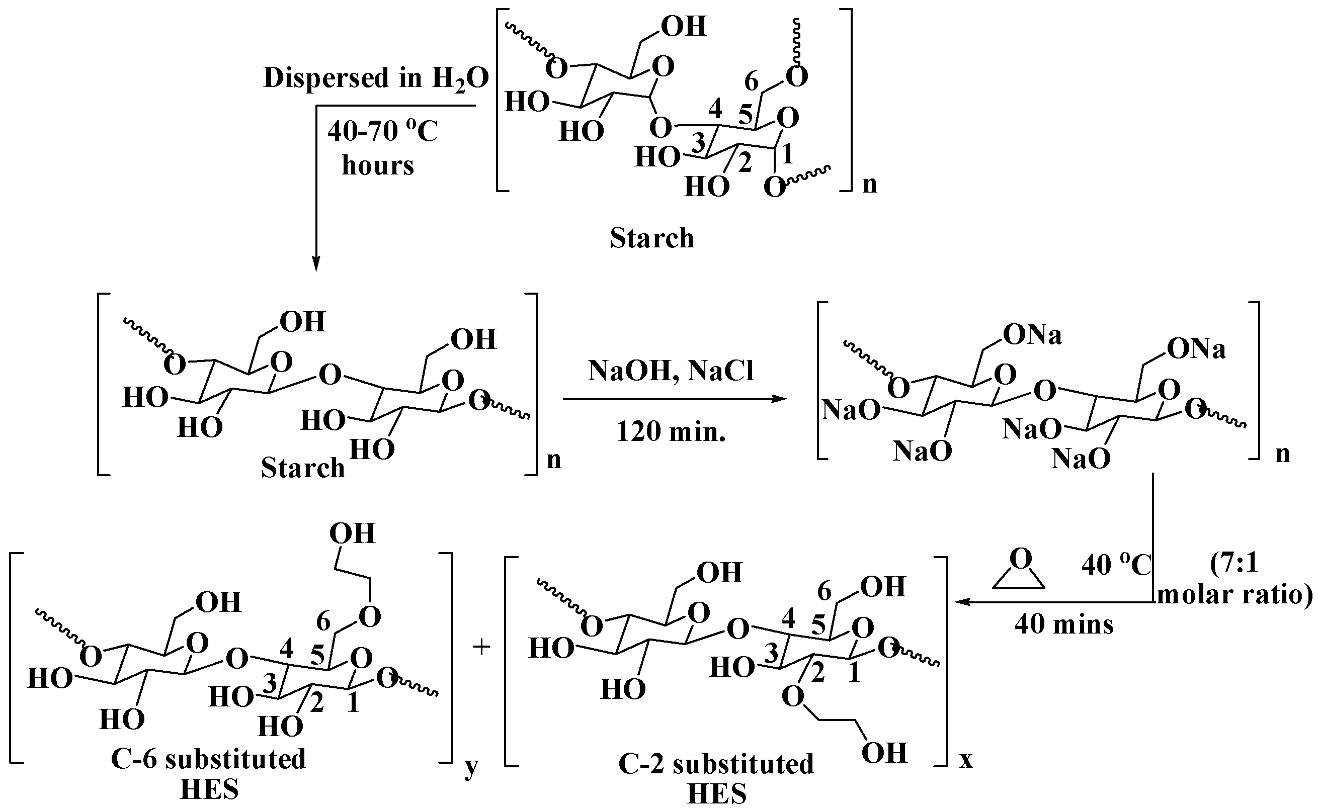
HES is a versatile non-ionic starch derivative with modified branched-chain glucose polymer from amylopectin sources such as potatoes, maize, and sorghum. The molecule contains α―(1,4)-glycosidic linkages to anhydroglucose units and a branching chain with (1,6)-glycosidic links, making it a flexible and functional polymer (Figure 1) [20]. HES is primarily used in intravenous therapy and fluid management for critically ill patients in intensive care units to maintain vascular volume in the circulatory system [24,25,26,27,28]. The polyhydroxylated nature of HES turns it into a potential replacement for polyethylene glycol (PEG). It is available in different formulations, including HES 200/0.5 [29,30] and HES 130/0.42 [31,32,33], which is represented as the molecular weight/degree of substitution [34]. The molecular weight of the HES solution plays a crucial role in determining its effects on blood coagulation during hemodilution [35]. HES (200/0.5) is known to impair coagulation and is used in large volumes as preoperative infusion. In contrast, lower substituted HES (130/0.4) causes less impact on coagulation in major orthopedic surgery than HES (200/0.5) [36]. The C2/C6 ratio of HES backbone also influences hemorheology and coagulation, with larger ratios causing higher serum concentration and plasma viscosity increment during hemodilution therapy [37].The goal of hemodilution therapy with HES infusion is to improve hemodynamics and rheology by providing better tissue oxygen tension (tpO2). HES 130/0.4 has been found to be a promising candidate for improving skeletal muscle tpO2 as compared to HES 70/0.5 or 200/0.5. In vivo studies have revealed that HES 130 with ideal molecular sizes improves rheology and tissue oxygenation within an hour after infusion [38].
In contrast to starch, HES has increased solubility, a longer in vivo half-life, and diverse structural variation. It can be ingested by the human body up to 1.20 g/kg per day and is degraded by endogenous amylase and excreted through the kidneys [39]. HES-based nanoparticles have been studied as potential eco-friendly materials in recent decades [40]. HES is now approved as a plasma substitute and medication for shock and low blood volume [41]. In clinical research, HES serves as a nano conjugate for a potential nanocarrier of various medicinal drugs [42,43,44,45].

2.1. Synthesis

The synthesis of HES entails a series of chemical reactions following structural modification of natural starch to improve solubility, flexibility, and other desirable properties. The synthesis of hydroxyethyl starch (HES) involves the reaction of potato or corn starch with ethylene oxide in the presence of NaOH and NaCl [46]. The process uses three molar ratios of starch and ethylene oxide, which are 7:1, 7:2, and 7:3 [47]. In this process, starch is first dispersed in water and heated to 40–70 °C. NaOH and NaCl are added to the mixture to adjust the pH and to enhance the reaction rate. Ethylene oxide is then added slowly to the mixture with continuous stirring (Scheme 1) [48]. The reaction is allowed to proceed for several hours until the desired degree of substitution is achieved. However, the degree of hydroxy ethyl substitution augments the solubility of HES. With this method, the hydroxyethyl groups in the HES molecule are acetylated using acetic anhydride and pyridine. The degree of substitution can then be calculated based on the amount of acetyl groups present in the molecule. Typically, the degree of substitution ranges from 0.02 to 0.20 per glucose unit, with the most probable position of attachment of the hydroxyethyl group being at the C2 position of the glucose unit [49]. In contrast to that at the C6 position, the substitution of hydroxyethyl groups at C2 position more efficiently inhibits the access of α-amylase toward the substrate molecule. Thus, the synthesis of HES with high C2/C6 ratio is expected to be stable under proteolytic degradation.

2.2. Synthesis of HES Based Polymers for Drug-delivery system

Hydroxyethyl starch (HES) can be synthesized through various methods, including self-assembly, layer-by-layer assembly, and nanoprecipitation [50,51,52,53,54,55].

2.2.1. Self-Assemble

The self-assembly is a feasible approach for creating 2D or 3D colloidal particles using gravity sedimentation, vertical deposition, electrophoresis, and crystallization. It is achieved by adjusting reaction parameters such as temperature, weight of reactants, solvent, stickiness, contact time, and high-quality colloidal particles [50,51]. HES can also be modified with hydrophobic particles or oligomers to create nanoparticles or nano-gels suitable for drug delivery [56]. Micelle-shaped thermo-responsive polymers in the aqueous medium have been studied for drug delivery [17,57,58,59,60,61,62]. Self-assemblies of twelve novel poly(allylamine) (PAA)-based, comb-shaped amphiphilic polymers with positive zeta potential have been reported in aqueous solution [63]. Esterified HES undergoes self-assemble with lauric, palmitic, and stearic acids using dicyclohexylcarbodiimide (DCC) and dimethylaminopyridine (DMAP) [64].

2.2.2. Layer-by-Layer Assembly

The layer-by-layer approach has been used in numerous therapeutics and biomaterials, by consolidating layer-by-layer films non-covalently under physiological conditions without compromising their natural properties. The layer-by-layer assembly of polyelectrolyte nanoparticles (NPs) plays a promising role in developing polyelectrolyte-based nano-sized delivery systems for biomedical applications. Layer-by-layer NPs have been utilized to deliver different active substances, allowing for the controlled discharge of each captured substance [52]. Initially, it was used for creating thin polyelectrolyte layers on solid surfaces. This approach has long been used in bio-sensing, regenerative medicine, tissue construction, and biomimetic research [52,53]. This strategy relies on electrostatic interactions and alternative adsorption among the opposite-charged particulate of polyelectrolytes. The film structure can be controlled to 1 nm accuracy with an extension from 5 to 1000 nm. The approach involves incubating charged solid support in an oppositely charged particle of polyelectrolyte solution [65]. The polyelectrolyte adsorption at each layer is accomplished to a level of immersion, and the terminal charge is rotated after each subsequent layer deposition [65]. It emerges during the adsorption of the primary layer, which acts to expel abundance-free polyelectrolytes. It is submerged in a solution containing a second polyelectrolyte to form the second layer. The method is repeated until the desired thickness is attained [52,65].

2.2.3. Nanoprecipitation

The nanoprecipitation method or solvent migration strategy for nanoparticle fabrication was patented by Fessi and co-workers [54] and then modified by Yadav et al. [66]. This strategy is advantageous for its direct, fast, and simple nanoprecipitation process. The nanoparticle solution is created instantly, and the whole method is carried out in one step. This method requires two miscible solvents in which both the polymer and the drug must be dissolved fast. Nanoprecipitation occurs via a rapid desolvation of the polymer when the polymer solution is added to the non-solvent. As long as the polymer-containing solvent condition has been diffused into the scattering medium, the polymer accelerates, including quick drug entanglement. The Marangoni effect represents the temporal nanoparticle arrangement through interfacial turbulences at the interface of the solvent and the non-solvent, resulting from stream dissemination, surface pressure variations, etc. [67]. Besheeret al. esterified distinct molar masses of HES with lauric acid to induce diverse molar substitutions to produce a fully biodegradable amphiphilic polymer. These polymers and Pluronic F68 and F127 were nano-precipitated into poly(lactic-co-glycolic acid) nano-spheres [25].

2.2.4. Graft Copolymerization

The development of functional polymeric materials for DDS often involves graft copolymerization, which allows for the combination of the best properties of two or more polymers in one backbone unit. There are different polymerization strategies that can be employed to synthesize tailor-made graft copolymers, depending on the specific needs and end-use [55]. One common method is “grafting-to”, which attaches espre-formed polymers to the backbone of a biopolymer. However, this approach is limited by the swarming of chains at the surface, which hinders the diffusion of the chains and subsequently limits the ultimate graft density. Another method is “grafting-from”, which initiates polymerization from reactive groups on the backbone of a biopolymer. This approach leads to higher graft densities and includes free radical polymerization (FRP), controlled radical polymerization (LRP), and ring-opening polymerization (ROP) [55,56]. The most widely used polymerization strategy is FRP, which includes three steps: initiation, propagation, and termination. In initiation, the initiator initiates chain-derived radicals and adds to monomers. Propagation occurs when the expanding monomers react with the propagating radical, which is a crucial step for chain propagation. Chain termination takes place when the propagating radicals react by combination, disproportionation, or transfer. Almost 60% of all available polymers are still obtained by this strategy due to its various attractive characteristics [68,69,70].
In the “grafting-from” approach, the development of polymer chains originates from the initiating sites on the biopolymer backbone. This approach allows for a higher graft density because the receptive groups are more accessible to the chain ends of the growing polymers. It can also involve an end-functional pre-formed polymer with its reactive end-group coupled with the valuable groups on the biopolymer backbone. A schematic representation of the “grafting-to” approach and “grafting-from” approach is shown in Figure 2.
In contrast to “grafting-to” and “grafting-from”, the “grafting-through” or macromonomer is a widely employed method in polymer chemistry for synthesizing graft copolymers with a well-defined side chain. In this approach, a preformed polymer backbone (macroinitiator) is prepared initially, which is then used as a platform for grafting polymer chains. For instance, a low-molecular-weight (LMW) monomer is typically copolymerized with a (meth)acrylate functionalized macromonomer via a radical pathway. By controlling the reaction conditions and monomer incorporation, the “grafting-through” approach allows for precise control over the length, density, and architecture of the grafted chains, enabling the creation of complex polymer structures or multifunctional macromonomers with tailored properties for various applications [71]. Naturally, both the homogeneous and the heterogeneous distributions of grafts are possible to synthesize, which turn out to be different characteristic physical properties of the materials. Macromonomers such as polyethylene, poly (ethylene oxide) and polysiloxanes can be inserted in a polystyrene or poly(methyl acrylate) backbone following the “grafting-through” technique. Despite analogous composition, the material properties invariably vary owing to phase separation as modified by polymer topology. In the context of NP synthesis, the “grafting-through” approach is useful where NPs anchor polymerizable groups onto their surface following the initiation of polymerization in the solution containing the initiator, monomers, and modified crosslink NPs.
Noga et al. synthesized a graft copolymer HES-PEI by using a coupling reaction with HES20 and HES70 involving direct polyamine PEI22 in PBS at pH 7.4. This involved using an excess of HES (molar proportion 25:1) to ensure Schiff’s base formation between a terminal aldehyde function of HES and the amine groups in PEI. Modified HES–PEI copolymers were also synthesized by grafting HES onto linear PEI by a condensation pathway through an unstable aminol intermediate that was rapidly converted to an enamine. The reduction of sodium cyanoborohydride of enamine transformed it to secondary or tertiary amine groups [72]. The synthesized HES-PEI is usually applied to produce polyplexes with the luciferase-expressing plasmid DNA pCMVluc [73]. Starch-graft-poly (N, N-diethyl amino ethyl methacrylate-co-poly (ethylene glycol) methyl ether methacrylate) [starch-g-P(DEAEMA-co-PEGMA)] was successfully synthesized by Wang et al. using living radical polymerization (LRP), and it is applied to a hydrophobic DDS like Doxorubicin(DOX). Yu et al. synthesized HES-g-PLA copolymers using an esterification reaction between terminal carboxyl groups of PLA and hydroxyl groups of HES to enhance Doxorubicin delivery towards tumor cells [74,75].

2.3. Commonly Used Anticancer Drugs

The World Health Organization (WHO) maintains a list of essential medicines for cancer treatment, which includes several crucial anticancer drugs. The WHO recommended list of essential medicine for cancer treatment (2021) are shown in Table 1.
Table 1 highlights that many drugs have not been recommended by expert committees or working groups due to their potential adverse effects on the human body. However, doxorubicin (DOX) is considered to be a valuable therapeutic option for non-metastatic rhabdomyosarcoma and may play a significant role in cases where standard chemotherapy regimens are unavailable. This has led researchers to investigate various ways to improve the effectiveness and minimize the side effects of the DDS for DOX. DOX is a cytotoxic anthracycline antibiotic produced by cultures of Streptomyces peucetius var. caesius. It works by selective binding of the planar anthracycline nucleus with the DNA double helix, thereby disrupting DNA replication and leading to cell death. However, DOX causes significant side effects, such as cardiotoxicity, myelosuppression, and mucositis.
To minimize these side effects, researchers have focused on developing DDS to improve the pharmacokinetics and bio-distribution of DOX by reducing toxicity. One approach is to encapsulate DOX into nanoparticles, such as liposomes, polymeric micelles, or dendrimers. These nanoparticles can improve drug stability, prolong drug circulation time, and enhance drug accumulation in tumor tissues by exploiting the enhanced permeability and retention (EPR) effect. Additionally, some nanoparticles can be functionalized with targeting ligands, such as antibodies or peptides, to improve the specificity of drug delivery to cancer cells.
Another approach is to modify DOX itself by conjugating it with different moieties, such as PEG, to improve its pharmacokinetic profile and reduce its immunogenicity. Furthermore, conjugating DOX with certain ligands, such as folic acid or hyaluronic acid, can enhance its specificity for cancer cells that overexpress the corresponding receptors.

2.4. Drug-Delivery Systems (DDSs)

Macromolecules offer a promising targeted drug delivery method for essential medications due to their significant drug-loading capacity, continuous drug-release capability, nontoxicity, biocompatibility, immunogenicity, and delayed pharmacokinetic activity. DDSs and other macromolecules increase the selectivity, efficiency, sustainability, and safety of therapeutic agents while controlling the rate and location of drug administration. Several DDS templates have been developed, including liposomes, dendrimers, cyclodextrin, micelles, calix[n] arenas, carbon nanotubes, azo polymers, and polymer–drug conjugates. Most of these DDS function as drug encapsulation strategies to accommodate hydrophobic drugs into the inner hydrophobic/hydrophilic core through non-covalent interactions.
The use of macromolecular DDSs in the non-invasive treatment of various cancers has recently increased, in contrast to the traditional use of chemotherapeutic drugs where a lack of selectivity in targeting tumor cells and dose-limiting toxicity are persistent. However, these macromolecules may disintegrate due to proteolysis or nucleases. Chemical modifications are necessary to keep them stable in the systemic circulation. A sustainable chemical modification is also required to allow the whole macromolecule-drug species to cross the membranes of the tumor cells without adverse effects. Diverse macromolecular DDS strategies based on cationic polymer, exosome, membrane-camouflage, lipid, smart patch, and nano-gel have been reported, and macromolecular drugs that inhibit tumor growth or destroy malignant cells directly have been rationally applied to treat cancers.
Selective chemotherapy using macromolecular prodrugs involves rearranging linkages between drug molecules and macromolecules. The advantage is not only that it keeps the drug stable in the circulation but also that it allows for fast release of the drug soon after accumulation in tumor locations or through tumor cell endocytosis. Polysaccharides such as dextran, chitosan, pullulan, and hyaluronic acid have been extensively studied for DDS or DCC by covalent or non-covalent chemical conjugation physical encapsulation inside polysaccharide backbones or as polysaccharide-oriented nanoparticles. Drug conjugation is considered the most reliable strategy for producing stable prodrugs with reduced systematic toxicity induced by other foreign substances [76,77].

EPR Effect on DDS

The macromolecular prodrug conjugated with polysaccharides can assemble in solid tumors during circulation in blood vessels following an ”enhanced permeability and retention effect” (EPR effect) [78]. The EPR effect is a unique pathophysiological mechanism of progressive accumulation of macromolecular compounds in the tumor-vascularized tissues’ resulting cancer-targeted drug delivery with the retention of anticancer agents in the solid tumors [79,80]. It is considered as a productive modality for ensuring cancer targeted delivery and entrapment inside tumor tissues for a prolonged period, with many macromolecular compounds, including drug-loaded nanocarriers. The EPR effect originates from the increased permeability of tumorous blood vessel, endothelium to macromolecules, coupled with limited lymphatic drainage within the tumor interstitium. EPR effect acts more efficiently via (1) strong irregular neovascularization in tumors with abnormalities in tumor blood vessels; (2) elevated expression of inflammatory factors; and (3) a lack of efficient drainage of lymphatic systems in solid tumor tissue [81]. When low-molecular-weight drugs are conjugated with high-molecular-weight carriers, their removal by lymphatic drainage becomes inefficient, resulting in their accumulation within tumors. Solid stress or interstitial fluid pressure often obstructs the anticancer penetration agent inside the center of the tumor tissue. However, it does not hamper macromolecular compounds in oozing out and accumulation in the peripheral region of the tumor tissue [82,83]. The EPR effect thus principally crops up in the peritumoral region, which potentially brings forth the suppression of the tumor or eliminating neoplasia even if it is not inside the tumor cell. Literally any water-soluble high-molecular-weight drug carrier, such as water-soluble polymers or liposomes, can exhibit this effect. The existence of the EPR effect has been experimentally confirmed in various macromolecular anticancer drug-delivery systems [84]. Furthermore, the EPR effect strongly depends upon the size, surface charge, and physico-chemical properties of drug-loaded nanocarriers [81]. NPs with a size of 100–200 nm; neutral or negative surface-charge; and cylindrical, discoidal, or ellipsoidal shape usually exhibit an optimal EPR effect with longer plasma half-life in circulation by accumulating in neoplastic tissues. It is worth noting that the targeted delivery of macromolecular drugs into neoplastic tissues can be augmented by modulating EPR effect by introducing antibody photosensitizers, adjuvants, or inflammatory factors. Despite, the nanomedicine delivery occasionally suffers from serious criticism owing to the choice of animal model for preclinical development, usually in a small mice xenograft model, which is dissimilar in human tumor tissues as a result of the knocking-in or knocking-out of certain genes for initiating tumors. Naturally, pharmacodynamic activities involving biodistribution in regard to nanodrug delivery into neoplastic cells are different in both mice and human model [82].
In current research, hydroxyethyl starch (HES) has replaced most polysaccharides, as it meets the criteria required for a suitable DCC. HES has been applied to conventional anticancer drugs and drugs undergoing clinical trials in various cancer treatments (as shown in Table 2). The therapeutic efficacy of drugs conjugated to polysaccharide macromolecules includes advantages such as improved antitumor activity and repressed systematic toxicity.
The HES-DOX prodrug has shown exceptional potential in the precise chemotherapy of cancer [86]. Amphiphilic HES conjugated with DOX can self-assemble into micellar nanoparticles in an aqueous solution, as shown in Figure 3. A recent study reported the synthesis and selective use of the HES-polycaprolactone-DOX-indocyanine green drug for the selective clinical treatment of liver cancer [88]. Additionally, polysaccharide-based stimuli-sensitive graft copolymers have been developed for drug delivery [93]. PEGs have been observed to effectively reduce the hyper-aggregation of red blood cells (RBC) in the presence of HES solution, with variations in RBC morphology and conglomeration as measured using a radiation microscope [94]. Amphiphilic HES grafted with polylactides (HES-g-PLA) was prepared using graft copolymerization procedures and used for lipophilic docetaxel drug delivery [95].
Owing to the use of artificial plasma and having a long circulation time by HES, the pH-responsive hydrazone bond linked to HES–DOX conjugates was explored for antitumor activity with a promising increase in the circulation time and minimization of side reactions. The percentage of the cumulative release of DOX conjugated to HES via pH-sensitive functional group was significantly lower at pH 7.4 than pH 5.3. pH-sensitive linkers such as hydrazone (hyd) and pH-insensitive linkers such as succinic anhydride (SAD) were triggered differently to render a faster release rate of DOX for self-assembled NPs of HES-hyd-DOX than HES-SAD-DOX. The DOX conjugated with amphiphilic HES with a Schiff base linkage can be assembled into polymeric micelles, which showed a rate of survival in mice about 30% higher than that of free DOX. HES-DOX-conjugated polymeric prodrug could self-assemble into well-organized structural micelles that was triggered by pH to convert charge from negative to positive.

2.5. Hydroxyethyl-Starch-Based Drug-Delivery System

Hydroxyethyl starch (HES) is a natural polymer commonly used as a plasma expander due to its ability to increase blood volume. However, HES has also been explored as a potential DDS for its biocompatibility, biodegradability, and ability to be functionalized with various groups for improved properties. One approach to functionalizing HES is through crosslinking with suitable groups to produce multifunctional polymers such as hydrogels or nanocapsules. Crosslinking involves the formation of covalent bonds between different polymer chains or within the same polymer chain, resulting in a three-dimensional network structure. This crosslink structure can improve the mechanical properties of HES, making it more suitable for various biomedical applications. Furthermore, by introducing crosslinks between HES and drug molecules, drug-release properties can also be improved. For example, crosslinked HES hydrogels have been studied as a potential DDS for the sustained release of various drugs, including anticancer drugs. The hydrogel structure allows for controlled drug release over an extended period, potentially reducing the frequency of drug administration and improving patient compliance. Similarly, HES nanocapsules have also been explored as a potential DDS. These nanocapsules can encapsulate drug molecules within their core and have exhibited sustained release properties under physiological conditions [96,97,98].

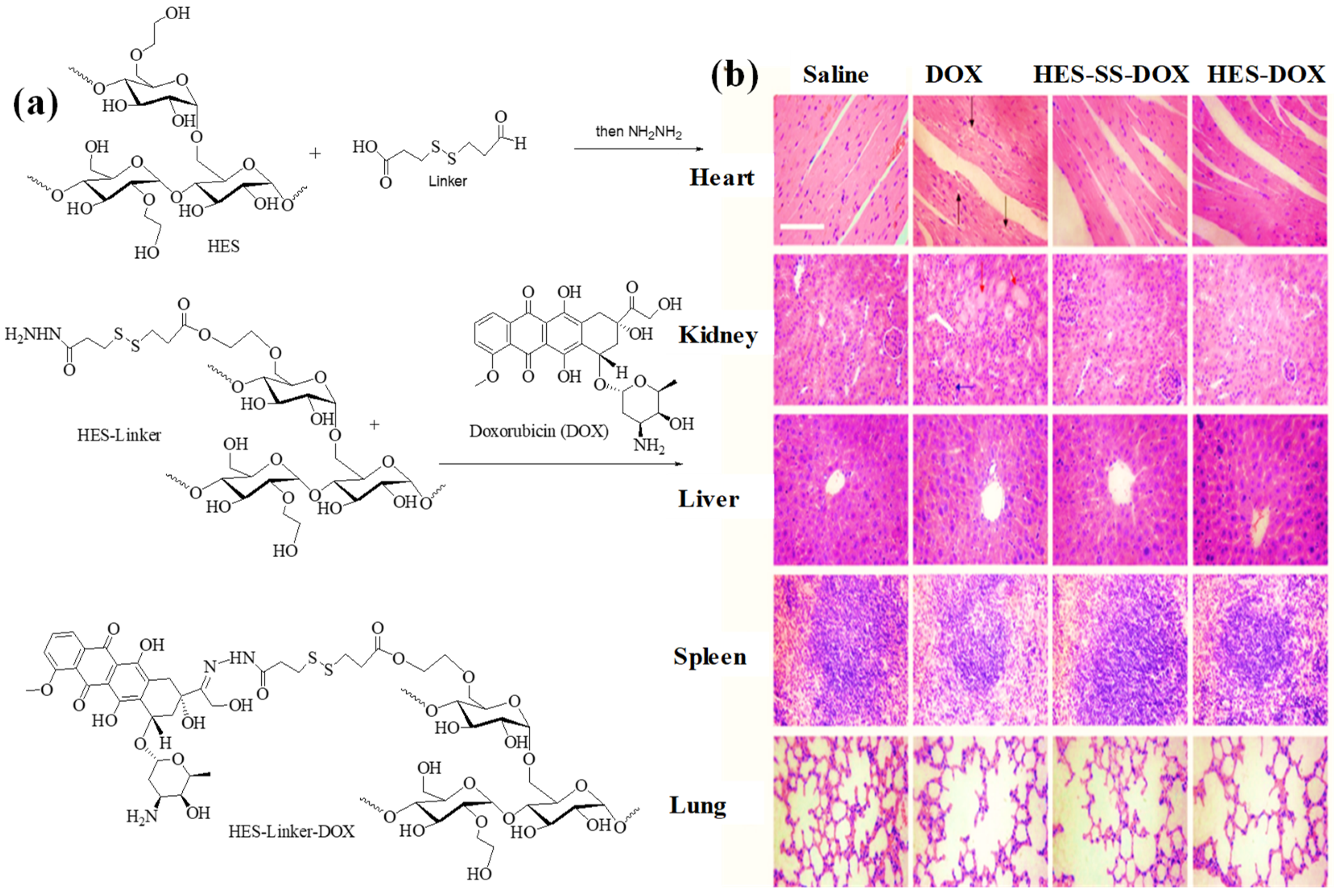
2.5.1. HES as DDS for Doxorubicin

Doxorubicin (DOX) is a type of anthracycline chemotherapy drug that is used to treat a wide range of cancers, including liver, bladder, breast, stomach, esophagus, endometrial, lymphoma, and lymphocytic leukemia [99]. DOX and its major metabolites such as doxorubicinol bind to plasma proteins and receptors and thus enter the cell via passive diffusion. DOX inhibits the function and genomic integrity of topoisomerase II (Top2) including DNA replication, DNA transcription, and chromosome segregation. DOX interferes Top2 through forming lesions via DNA strand-breaking, trapping the intermediate, and enhancing the level of Top2: DNA covalent complex, resulting in the deceleration of the growth of cancer cells. The discovery of DOX was an effective strategy for cancer chemotherapy as a result of enzyme-induced DNA damage. However, both Top1 inhibitors such as irinotecan and topotecan and Top2 inhibitors, including etoposide, teniposide, and anthracyclines (such as idarubicin, daunorubicin, and doxorubicin/DOX), exert inhibitory effects by targeting the activities of topoisomerases. These inhibitors disrupt the normal function of topoisomerases, leading to the formation of DNA strand-breaking and interfering with DNA replication processes and apoptosis [100]. DOX also demonstrates anticancer activity by acting as intercalator Top2 poison. However, the therapeutic use of DOX is limited to its nonspecific distribution, significant side effects such as gastrointestinal issues, hematopoietic depression, and cardiotoxicity. To address these limitations, a redox-sensitive hydroxyethyl starch doxorubicin (HES-DOX) conjugate has been developed. This conjugate can be used as a biodegradable prodrug, with a diameter of 19.9±0.4 nm, that can mediate safe intracellular DOX release in response to glutathione (GSH) levels in the target cell, which minimizes side effects and improves antitumor efficacy [87]. The HES-DOX conjugate is stable in the presence of extracellular GSH levels (~2 μM), but it rapidly releases DOX when exposed to intracellular GSH levels (2−10 mM). To modify HES, it was initially subjected to esterification reactions with the DTDPA-linker (Diethylenetriaminepentaacetic acid) to yield HES-DTDPA. This reaction involves the esterification of the carboxyl groups of DTDPA with the hydroxyl groups of HES. Subsequently, DOX (doxorubicin) was conjugated to HES-DTDPA to generate HES-SS-DOX through the formation of amide bonds. Compared to free DOX, HES-DOX or HES-SS-DOX exhibits a relatively longer plasma half-life and enhances tumor accumulation (Figure 4). In vitro cell studies have also shown that HES-DOX exhibits GSH-mediated cytotoxicity.
DOX binds to the HES backbone via a pH/redox responsive linker bearing disulfide and hydrazone groups. The formed conjugates (HES-Linker) were self-assembled into HES-Linker-DOX nanoparticles (NPs). HES-Linker-DOXNPs showed the ability to accumulate tumor-orientated drug and were also capable of releasing DOX into target cells. The tumor-containing mice model revealed that the application of pH/redox-responsive linkers instead enabled the HES-Linker-DOX NPs to bestow fast and sufficient release of DOX. The use of hydrazine bond and hydrazine-sulfide bond in conjugation were synergistically triggered for rapid and sufficient drug release. The lysosomal pH is 4.5–5.0, while the cytosolic pH is usually 7–7.5. The pH of early endosomes is maintained at about 6.5, while late endosomes are at about 5.5. When endosomes mature fully into lysosomes, the pH is about 4.5 [101]. Since endosomes (pH~5.5) and lysosomes (pH~5.0) in tumor matrix (pH~6.5) are moderately acidic, the photoresponsive HES-Linker-DOX in the form of NPs could effectively deliver DOX at pH~5 even when the given DL-dithiothreitol (DTT) dose was fixed as 0 mmol and 10 mmol during initial 24 h of release period in in vivo circulation. HES-Linker-DOXNPs exhibited a considerably advanced growth inhibition on HepG2 cells with IC50 2.22, 1.97, and 7.12 times lower than HES-hydrazine-DOX, HES-sulfur-DOX, and HES-DOX, respectively. Between 40 and 150 nm sizes of HES-Linker-DOXNPs were typically found to accumulate in tumors through the EPR effect and subsequently exerted adequate DOX release. In another study, the loading and encapsulation of drug efficiencies of DOX-loaded-micelles were found 9.96% and 55.31%, respectively. This result demonstrated that the antitumor property of HES-Linker-DOX NPs inhibits the progress of the advanced tumors with considerable efficacy in contrast to free DOX or unresponsive or single responsive bonds.
The tumor-containing mice model suggests that HES-Linker-DOX NPs exhibited adequate safety for other parts of the treated mice. The tissue sections of the histopathological examination of heart, kidney, liver, spleen, and lung from different groups were analyzed by hematoxylin and eosin (H&E) staining, and the results are shown in Figure 4b. The result is consistent with mice intravenous injection with free DOX in which histopathological alteration were observed in heart, kidney tubules, liver, and glomerulus. In contrast to free DOX, the conjugated HEX-DOX and HES-SS-DOX display insignificant pathological alteration on heart, kidney, spleen, and lungs underlying lowered cardiac and renal impairment. Considering the analogous distribution of free DOX and conjugated HES-DOX and HES-SS-DOX in the heart, the conjugated form can attenuate nonspecific cellular uptake than free DOX. Histopathological analysis confirms that the conjugated HES-DOX and HES-SS-DOX shows very low cardiotoxicity and nephrotoxicity compared with that of free DOX. The investigation of subcutaneously transplanted H22-tumor mice model indicated remarkable in vivo antitumor activity of conjugated HES-DOX and HES-SS-DOX with extended plasma half-life time, which enhanced the tumor-targeting capacity with GSH-mediated cytotoxicity.

2.5.2. HES as DDS for Methotrexate

Methotrexate (MTX) is an antifolate drug that has been commercially available for a long time and is widely used to treat cancer, rheumatoid arthritis, and many other diseases. To form a covalent linkage, the carboxyl groups of MTX are coupled with the hydroxyl group of HES, resulting in HES-MTX. The HES-MTX conjugate, which serves as a drug–carrier conjugate (DCC) of methotrexate, has been characterized in terms of hydrodynamic size, MTX content, zeta potential, and drug release kinetics. The negative charge on the surfaces of HES-MTX conjugates results in a prolonged half-life in plasma and, thus, greater tumor accumulation of HES-MTX conjugates via the enhanced permeability and retention (EPR) effect. In vivo investigations using the MV-4-11 leukemia model demonstrated that the HES-MTX conjugate had a higher antitumor activity than non-conjugated HES. Analogously, the in vivo antitumor effect was tested in NOD/SCID mice. It was inoculated subcutaneously both with MV-4-11human leukemia cells and CDF1 mice intraperitoneal with P388murine leukemia cells. The in vivo results revealed that a significantly high antitumor efficacy was obtained from the HES-MTX conjugate rather than unconjugated drug. The in vitro antiproliferative activities on human (MV4-11) and murine (P388) leukemia cell lines indicates about 10-fold weaker cytotoxic activity of HES-MTX than unbound MTX [91]. Similarly, in a murine leukemia P388 model, the controlled and sustained release of MTX in conjugation with HES was more pronounced than that of unbound MTX. Further, in vivo experiments in murine leukemia P388 model demonstrated that the survival time of leukemia-induced mice treated with conjugated HES-MTX DDS was essentially longer than that of mice treated with unbound MTX (ILS = 38%, p < 0.05) and untreated animals (ILS = 55%, p < 0.05). The HES-MTX conjugate diminished the volume of the MV4-11 human leukemia cell line to a considerable extent (p < 0.05) in contrast to MTX from day 8 to day 22. HES thus acts with a high level of anticancer-drug-carrier potential with improved treatment efficacy. Figure 5 shows the probable structure of the conjugated HES-MTX [20,91].

2.5.3. HES as DDS for Hydroxychloroquine

Chloroquine (CQ) and hydroxychloroquine (HCQ) are well-known antimalarial agents that have been used for several decades. These compounds are derivatives of 4-aminoquinoline and are weak bases. They undergo protonation in acidic vacuoles, which allows them to accumulate in lysosomes and neutralize lysosomal pH. The buffer effect of CQ and HCQ exhibits potential chemotherapeutics by disrupting endolysosomal trafficking and autophagy [102]. To enhance the therapeutic efficacy and selectivity of HCQ, it can be covalently attached to HES using a linker approach by adjusting the degree of substitution. Carbonyl diimidazole (CDI) can be used to create covalent bonds between the main hydroxyl groups of HCQ and HES, generating HES-HCQ conjugates. The resulting conjugates for pharmacokinetics and pharmacodynamics of HCQ prolong its half-life, enhance its solubility, and improve its bioavailability. The conjugation of HCQ with HES may also improve its selectivity towards cancer cells by exploiting the enhanced permeability and retention (EPR) effect, which leads to preferential accumulation of drugs in tumor tissues [102]. Figure 6 shows a possible structure of the HES-HCQ conjugate.
The stability of the HES backbone and carbonate linker was evaluated under various conditions such as incubation with PBS or in 0.01 N HCl, acetate buffer (pH 2, 5.0, 7.0, or 7.4, respectively), or water for 72 h at 37 °C, and they were found to be hydrolytically stable. The HES-HCQ conjugate, synthesized using the carbonyl diimidazole (CDI) linker approach, was shown to inhibit the autophagy, migration, and invasion of pancreatic cancer cells more effectively than isolated HCQ. The conjugate exhibited a sustained drug-release profile, indicating the presence of a polymeric drug. In vitro cytotoxicity studies using the CellTiter-Blue viability assay revealed that the polycationic HES-HCQ conjugate did not produce any discernable cytotoxicity.
The study on HCQ-DDS reveals that HCQ conjugated polymers retained autophagy-inhibitory activities of HCQ for better anticancer activity. HES-HCQ DDS-assembling moiety displays substantial autophagy inhibition in PC cells and safe drug HCQ release. In cells, CXCL 4, a chemokine ligand 4, assists intracellular trafficking of HES-HCQ conjugates through endocytosis to retain autophagy inhibition. At the molecular level, amine protons at moderately high concentrations of HCQ neutralize the acidic lysosome and thus inhibit protease activity followed by autophagosome fusion with lysosome and autophagy inhibition. The process can be corroborated with cytotoxicity studies using Cell Titer-Blue viability assay. The result suggests that there is no such difference in the cytotoxicity of HCQ and HES-HCQ and also no such discernible cytotoxicity of HES-HCQ until 300 μM [102]. The stable conjugation of HCQ to HES preserves the biological activity of HCQ, including CXCR4 antagonism and autophagy inhibition (Figure 7). The pH-dependent propensity of HES-HCQ to form rapid nanoparticles demonstrated a similar effect in blocking the local invasion of cancer cells, making it a potential multifunctional drug-delivery vehicle. Overall, these findings suggest that HES-HCQ conjugates hold promising effect and safe therapeutic option for the treatment of pancreatic cancer and other diseases.

2.5.4. HES as DDS for 5-Fluorouracil-1-acetic Acid

5-Fluorouracil (5-FU) is a widely used low-molecular-weight drug for the treatment of solid tumors. However, its low selectivity toward cancer cells leads to systemic toxicity, and its short half-life limits its therapeutic efficacy. To address these issues, a macromolecular prodrug carrier based on HES was developed to selectively deliver FU to solid tumor cells. Initially, FU was converted to 5-Fluorouracil-1-acetic acid (FUAC), which was covalently linked to HES through the appended hydroxyl group of the alkoxy glycosyl unit at the C-2 position, resulting in the formation of HES-FUAC conjugates (Figure 8). The coupling reaction was performed using a conventional DCC-DMAP coupling agent to yield a typical ester bond. The HES-FUAC conjugates were found to be stable in acidic buffer solutions (pH 5.8) and released FUAC slowly. However, FUAC was rapidly released upon hydrolysis under elevated temperature and pH. The conjugates were shown to improve the cytotoxicity and in vivo half-life of FUAC in human and rat plasma, thereby providing sustained therapeutic effects. In vitro and in vivo drug-release studies indicated that the sustained release of FUAC from HES-FUAC conjugates inhibited the colony formation of leukemia cells and demonstrated antitumor activity.
Where the delivery of most chemotherapeutic drugs results in systematic toxicity to critical organs in the form of different side reactions owing to their nonselective nature in an unbound state, the encapsulation of these agents in a vehicle acts as magic bullet to reduce systemic toxicity [103]. The Ns form of drug conjugate system delivers more drugs specifically into a tumor to reduce the drug systemic toxicity. Alternatively, NP systems can influence immune cells to foil tumor cells by reducing systemic toxicity in cancer treatment. HES-FUAC conjugates hold promise as a DDS for treating solid tumors, offering targeted therapy with reduced systemic toxicity [104]. The cell-penetrating peptide (CPP) of epithelial growth factor receptor (EGFR) displayed on hepatitis B virus-like nanoparticle (VLNP) exhibited targeted delivery of FUAC [105]. By leveraging the enhanced permeability and retention (EPR) effect, these conjugates can accumulate within the tumor microenvironment, potentially enhancing the effectiveness of FUAC release [104].

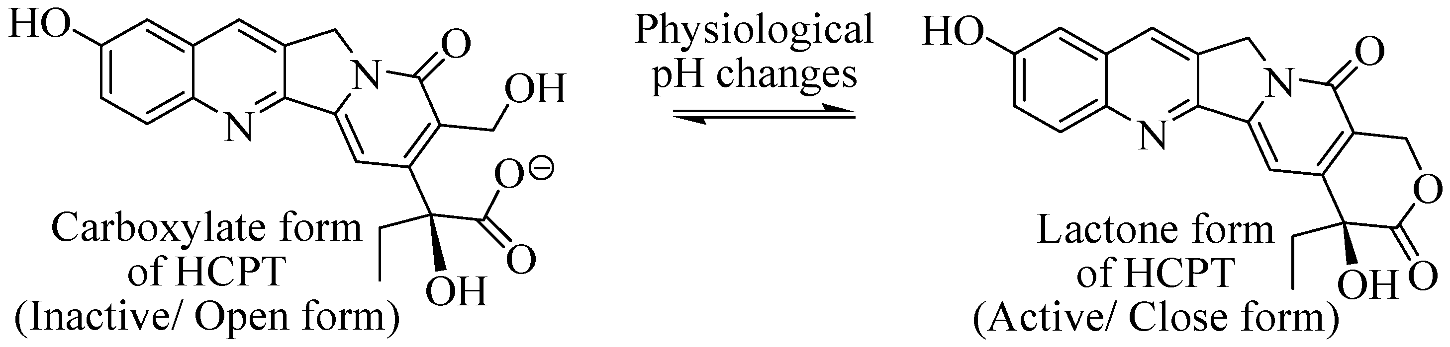
2.5.5. HES as DDS for 10-Hydroxy Camptothecin

HCPT is a highly potent and effective anticancer drug that is widely used for the treatment of various types of cancers. Its mode of action involves the inhibition of DNA topoisomerase I in cancer cells, which results in the arrest of DNA replication and cell division, ultimately leading to cancer cell death. However, the therapeutic potential of HCPT is limited by its low aqueous solubility and poor stability, particularly of its active lactone form. In PBS buffer and PBS-containing BSA, the rate of hydrolysis of free camptothecin consists of a half-life (t1/2) of about 24 and 13 min between carboxylate to lactone form to achieve an optimal equilibrium ratio of 83:17 and 99:1, respectively. Analogously, under physiological conditions (pH: 7.4, 37 °C), HCPT undergoes a pH-dependent equilibrium between its carboxylate and lactone forms, with the lactone form being the active one for its antitumor activity, though it is present in low percentage in the equilibrium. A preliminary experiment indicated that the lactone ring of camptothecin became partly stabilized by keeping the agents within lipid vesicles at lower internal pH of 5, in which camptothecin remained in lactone form and the lipid bilayers in gel phase exerted a certain barrier to interrupt drug–serum-albumin interactions. Naturally, HCPT remains predominantly in the form of active lactone structure in acidic tumor cells (endosome pH~5, lysosome pH~5.5, and inside tumor matrix pH~6.5) to wield its antitumor activity (Figure 9). The half-life (t1/2) of carboxylate form under physiological pH was reported to be a few minutes, indicating a lower proportion of the active lactone form of HCPT in plasma. Fugit et al. deduced a mechanism-based mathematical model citing liposomal release of Topotecan, whose permeability was relied on pH involving drug’s ionization state, binding to the membrane and kinetics of ring-opening interconversion [106]. They also demonstrated ring-closing and ring-opening reactions by varying pH from low to neutral pH (3.5–7.50) following the principle of microscopic reversibility. The delivery of the active lactone form of HCPT is thus challenging for its existence in pH-dependent equilibrium with an inactive open carboxylate. This lactone form is unstable and often converts to the inactive carboxylate form through ring opening, which reduces its therapeutic efficacy. Furthermore, serum albumin has the propensity to bind with the active lactone form of HCPT, thereby lowering the drug concentration. HCPT has low aqueous solubility, which further limits its clinical application. Consequently, there is a need to develop novel DDSs that can overcome these limitations and enhance the therapeutic potential of HCPT [40,107].
HES is a biocompatible polymer that has shown promising results in developing conjugates with different anticancer drugs. In the case of HCPT, HES conjugation has demonstrated a significant improvement in its pharmacological properties, such as enhanced biological half-life, improved drug absorption, distribution, and bioavailability. This conjugation also resulted in higher cytotoxicity effects on Hep-3B and SMMC-7721 cell lines compared to free 10-HCPT.The conjugation of HES with HCPT was achieved via covalent bonding through a glycine amino acid spacer, forming an amide bond. The aliphatic secondary hydroxyl group (10-OH) was involved in the conjugation process (Figure 10). Interestingly, 10-OH conjugation suggests water solubility, while 20-OH conjugation is responsible for the solubility and stability of the lactone. Despite the stereo protective effect of HES to 10-HCPT, the sustained release of 10-HCPT from the conjugate is exclusive to the variation in pH. In vitro studies have shown that the release of HCPT is faster in PBS solution or in rat plasma and liver homogenate, and in vivo release is predominant over in vitro due to faster esterase or starch hydrolysis enzyme degradation. These results suggest that HES-HCPT conjugates can potentially improve the therapeutic efficacy of HCPT by allowing sustained release and improving the drug delivery to target tumor cells [40].

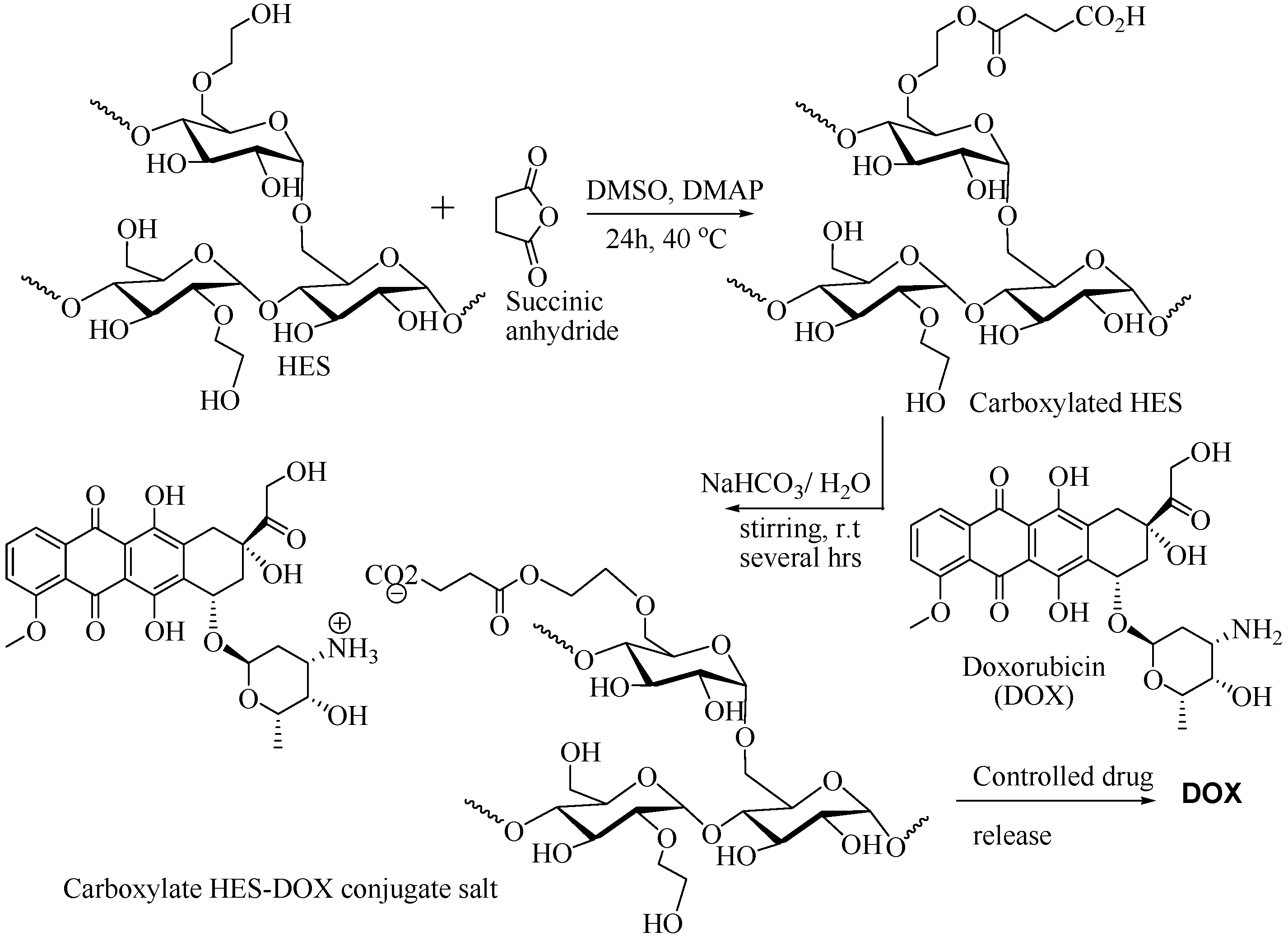
2.5.6. Carboxylated HES as DDS for Doxorubicin

HES is a useful hyperbranched DDS that can covalently be modified by functionalizing with carboxylic groups. Carboxylated HES can be used to accompany chemotherapeutic compounds through non-covalent interactions, with appended carboxylic groups forming either ionic salts or amides with the therapeutic compounds’ primary or secondary amino groups. This approach allows for the effective encapsulation of drugs such as doxorubicin (DOX), a well-known anticancer drug, within the carboxylated HES carrier. The encapsulated DOX in carboxylated HES has exhibited significant cytotoxicity against DU145 human prostate cancer cells in vitro. The carboxylated HES-DOX conjugate has also demonstrated controlled drug release, which is inevitable owing to solid electrostatic interactions between the drug and the carbohydrate scaffold of HES.
This result suggests that the HES polysaccharide can function as a DDS that not only conjugates but also modulates the transport of the drug across cell membranes. The use of carboxylated HES as a drug-delivery system demonstrates a great potential for improving the pharmacological activities of anticancer drugs through enhanced drug absorption, distribution, and bioavailability, as well as sustained release for better therapeutic efficacy (Figure 11) [108].

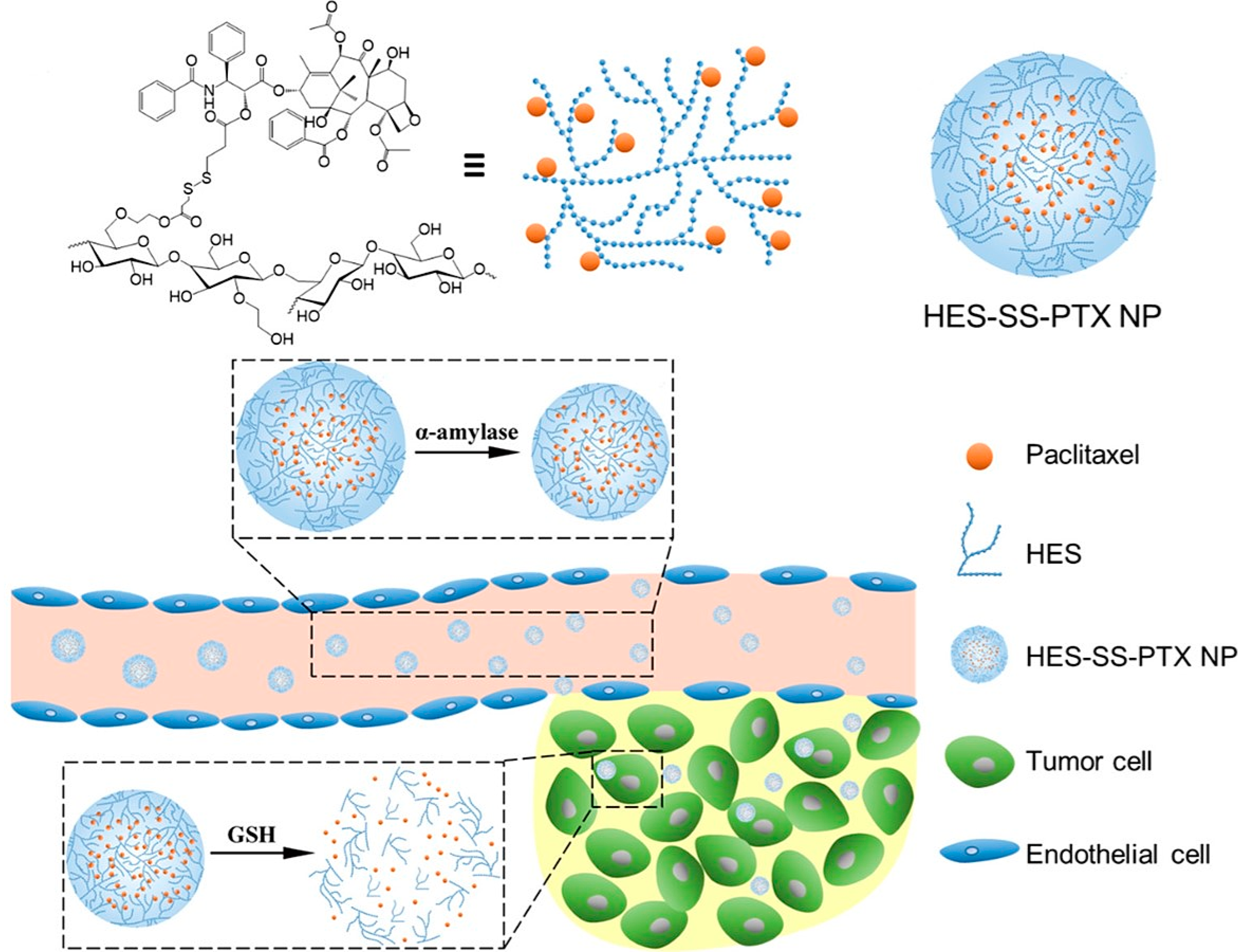
2.5.7. HES as DDS for Paclitaxel

Paclitaxel (PTX) is a highly potent antineoplastic agent that shows broad-spectrum antitumor activity in cancer therapy [109]. However, its clinical application is limited due to its hydrophobic nature and poor aqueous solubility, which leads to undesired neurotoxic side effects, such as hypersensitivity, peripheral neuropathy, and neutropenia. To overcome these limitations, various strategies have been developed, including the encapsulation method of DDS and water-soluble polymer-PTX conjugates with natural polymers, PEG, poly(L-glutamic acid), and poly N-(2-hydroxypropyl) methacrylamide (PHPMA) [110].
HES has also been explored as a promising tumor-targeted delivery system for PTX. In this approach, PTX is chemically conjugated onto HES through a redox-sensitive disulfide bond, leading to the formation of HES−SS-PTX (as shown in Figure 12). This conjugate is stable in the bloodstream due to the disulfide bond’s protection, which prevents PTX release in the blood plasma. However, in the presence of high levels of glutathione (GSH), which are typically found in tumor cells, the disulfide bond is cleaved, leading to the release of PTX. This results in targeted delivery of PTX to tumor cells while minimizing the drug’s toxicity to normal cells. Studies have revealed that HES−SS-PTX exhibits superior antitumor activity and lower toxicity than free PTX in various cancer cell lines [110].
The HES−SS-PTX conjugates have the ability to form monodispersed nanoparticles that are degraded by α-amylase in plasma, leading to the release of active drug at the tumor site. The redox-sensitive disulfide bond facilitates the extravasation and penetration of the HES−SS-PTX nanoparticles into the tumor microenvironment (as shown in Figure 13) [109]. These α-amylase and redox-responsive nanoparticles offer significant advantages in cancer chemotherapy and have great potential for clinical translation.
A pharmacokinetic study revealed that HES−SS-PTX nanoparticles exhibit an extended half-life, resulting in increased accumulation at the tumor site compared to free Taxol or commercial PTX formulations. The disassembly of HES−SS-PTX nanoparticles under reducing conditions results in burst drug release and increased cytotoxicity against 4T1 tumor cells. In addition to PTX, HES has been used to develop stimuli-responsive nanoplatforms for targeted delivery of various anticancer drugs or drug combinations. These drugs can be encapsulated or conjugated onto the nanoparticles to enable combination therapy for a wide range of cancers.

2.5.8. HES-HEMA as DDS for Fluorescein Isothiocyanate

HES can be combined with hydroxyethyl methacrylate (HEMA) to form the photocrosslink polymer HES-HEMA, which provides a favorable hydrophilic environment for biological drugs [111]. This is likely due to the formation of a sponge-shaped hydrogel structure. Such a structure can interact with proteins and drugs, leading to appreciable influences on drug release kinetics. Polyethylene glycol methacrylate (PEG-MA) has also been employed to obtain the HES-P(EG)n MA polymer, which displays higher hydrophilicity and biodegradability than the HES-MA polymer. The facile increase in the hydrophilicity of HES-MA conjugates originates in the presence of oligoethylene glycol spacer units to improve the inherent properties of the DDS, including its production with regard to clear aqueous solutions and the formulation of highly concentrated prepolymers. The introduction of PEG groups into HES-MA further increases the hydrophilicity and relatively lacks the hydrolytic cleavage, leading to an altered drug-release profile. The hydrophilic nature of HES-MA enables it to readily dissolve and interact with water and aqueous environments, making it suitable for various applications, including drug delivery. For example, HES-P(EG)nMA loaded with fluorescein isothiocyanate (FITC) imparts a higher rate of FITC release than HES-HEMA loaded with FITC [112]. FITC was attached to HES, producing HES-FITC conjugates as a model DCC-based drug delivery for detection (Figure 14). The rate of release is more significant when the conjugates were transformed into self-assembled NPs. The modified HES-PEG-methacrylate (HES-P(EG)6MA) has been reported to exhibit continuous and sustained release of protein drugs due to its smaller hydrodynamic diameter and smaller pores [113]. PEG-modified HES-HEMA hydrogel microparticles are expected to satisfy the requirements of a drug-delivery system considering biodegradability and biocompatibility [111].

2.5.9. HES-PLGA as DDS

A novel hydrogel composite was synthesized by grafting HES onto PLGA polymer through an acryloyl-mediated reaction. This resulted in the formation of a network of interconnected polymer chains with HES as a hydrophilic component, which provided a suitable environment for the encapsulation and release of protein drugs. The researchers found that this hydrogel composite exhibited improved drug-loading capacity and release kinetics compared to pure PLGA polymer. This is expected due to the hydrophilic nature of HES, which eventually increases the water uptake and swelling properties of the hydrogel, allowing for more efficient diffusion of the protein drug. Hydrogel microparticles derived from HES-grafted poly(2-hydroxethyl methacrylate) (PHEMA) were synthesized via typical free radical polymerization, and protein release was studied. The outcome of the study revealed the protein release to be dependent upon the size of the entrapped proteins and hydrogel network density. Moreover, the PLGA-HES-acryloyl composite showed good biocompatibility, making it a promising material for use in biomedical applications. It can potentially be used for the controlled and sustained delivery of protein drugs, since the rapid release of protein drugs can lead to a short therapeutic effect and potential side effects [114].

3. Limitations and Future Scope

Limitations:
  • Although HES-based DDSs have shown promise in preclinical studies, there are still some challenges that need to be addressed before they can be translated into clinical applications.
  • One limitation of HES-based DDS is associated with its immunogenicity. It might cause unknown or adverse immune response in some patients.
  • The relatively short half-life of HES-DDS in the bloodstream may limit its effectiveness as a drug carrier.
  • The variability in the properties of different HES formulations may affect their pharmacodynamic feasibility in the application of drug delivery.
Future scope:
  • Future research could focus on improving the biocompatibility of HES-based DDS to minimize potential immune reactions.
  • Efforts could also be made to optimize the properties of HES formulations to enhance their efficacy as drug carriers.
  • Future research could involve by exploring the use of HES-based DDSs for targeted drug delivery to specific tissues or cells.
  • The combination of HES with other natural or synthetic polymers can be investigated to develop more advanced delivery systems with improved properties.

4. Conclusions

In conclusion, the utilization of HES-mediated drug-delivery systems (DDSs) has shown great potential in addressing the limitations of the conventional use of anticancer therapies. HES-based polymers can serve as effective carriers for a wide range of therapeutic substances, which has been supported by numerous results from preclinical and clinical studies. However, there are still some limitations that need to be addressed, such as the immunogenicity of HES and the need for further optimization of the drug-loading and release kinetics. Nevertheless, the utility of HES-based drug carriers has been demonstrated in various applications, including hydrogels, micro- and nano-particle carriers, and transmucosal delivery systems. The standard protocol for HES-mediated drug-delivery frameworks has been outlined, providing a roadmap for designing and implementing HES-derived innovative DDS. HES-based drug carriers is promising, with potential applications in a broad range of therapeutic areas beyond cancer.

Author Contributions

K.C.: Writing—original draft, Writing—review and editing. S.D.: Writing—review and editing. C.-W.K.: Writing—review and editing. H.K.: Conceptualization, Writing—original draft, Writing—review and editing. All authors have read and agreed to the published version of the manuscript.

Funding

Dr. Chandra gratefully acknowledges the financial support from RUSA 2.0, Component 8: by Midnapore College (Autonomous) (355/MC/MRP/RUSA-2.0/21 (8/14) on 16 March 2021). Professor Kang is thankful to the Basic Science Research Program through the National Research Foundation of Korea, funded by the Ministry of Education (NRF-2019R1I1A3A02059471) and an international cooperation program managed by the National Research Foundation of Korea (NRF-2020K2A9A2A08000181).

Institutional Review Board Statement

Not applicable.

Informed Consent Statement

Not applicable.

Data Availability Statement

Not applicable.

Conflicts of Interest

The authors declare that they have no known competing financial interests or personal relationships that could have appeared to influence the work reported in this paper. Moreover, there is no involvement of animal studies or human participants in this report.

Abbreviations

CLRP: Controlled/living radical polymerization, DCC: Dicyclohexyl carbodiimide, DDS: Drug-delivery system, DMAP: Dimethylaminopyridine, DNA: Deoxyribonucleic acid, DOX: Doxorubicin, FRP: Free radical polymerization, FU: Fluorouracil, FUAC: 5-fluorouracil-1-acetic acid, GSH: Glutathione, HCPT: Hydroxycamptothecin, HCQ: Hydroxychloroquine, HES: Hydroxyethyl starch, LRP: Living radical polymerization, MA: Methacrylate, NPs: Nanoparticles, PAA: Poly (allylamine), PDDS: Pulsatile drug-delivery system, PEG: Poly(ethylene glycol), PEI: Polyethylenimine, PLA: Polylactide, PLGA: Poly (D, L-lactide-co-glycolide), RBC: Red blood cell, ROP: Ring opening polymerization, TODDS: Target oriented drug-delivery system.

References

  1. Bertram, J.S. The molecular biology of cancer. Mol. Aspects Med. 2000, 21, 167–223. [Google Scholar] [CrossRef] [PubMed]
  2. Jemal, A.; Bray, F.; Center, M.M.; Ferlay, J.; Ward, E.; Forman, D. Global cancer statistics. CA. Cancer J. Clin. 2011, 61, 69–90. [Google Scholar] [CrossRef] [PubMed] [Green Version]
  3. Sobue, T. Association of Indoor Air Pollution and Lifestyle with Lung Cancer in Osaka, Japan. Int. J. Epidemiol. 1990, 19, S62–S66. [Google Scholar] [CrossRef] [PubMed]
  4. Hemminki, K.; Pershagen, G. Cancer risk of air pollution: Epidemiological evidence. Environ. Health Perspect. 1994, 102, 187–192. [Google Scholar] [CrossRef]
  5. Ames, B.N.; Gold, L.S. Environmental pollution, pesticides, and the prevention of cancer: Misconceptions. FASEB J. 1997, 11, 1041–1052. [Google Scholar] [CrossRef] [Green Version]
  6. Drake, I.; Dias, J.A.; Teleka, S.; Stocks, T.; Orho-Melander, M. Lifestyle and cancer incidence and mortality risk depending on family history of cancer in two prospective cohorts. Int. J. Cancer 2019, 146, 1198–1207. [Google Scholar] [CrossRef] [PubMed]
  7. Sriraman, S.K.; Aryasomayajula, B.; Torchilin, V.P. Barriers to drug delivery in solid tumors. Tissue Barriers 2014, 2, e29528. [Google Scholar] [CrossRef] [Green Version]
  8. Longley, D.B.; Johnston, P.G. Molecular mechanisms of drug resistance. J. Pathol. 2005, 205, 275–292. [Google Scholar] [CrossRef]
  9. Bertino, J.R. Karnofsky memorial lecture. Ode to methotrexate. J. Clin. Oncol. 1993, 11, 5–14. [Google Scholar] [CrossRef]
  10. Kwon, G.S. Polymeric micelles for delivery of poorly water-soluble compounds. Crit. Rev. Ther. Drug Carr. Syst. 2003, 20, 357–403. [Google Scholar] [CrossRef]
  11. Cairns, R.; Papandreou, I.; Denko, N. Overcoming Physiologic Barriers to Cancer Treatment by Molecularly Targeting the Tumor Microenvironment. Mol. Cancer Res. 2006, 4, 61–70. [Google Scholar] [CrossRef] [PubMed] [Green Version]
  12. Lipinski, C.A.; Lombardo, F.; Dominy, B.W.; Feeney, P.J. Experimental and computational approaches to estimate solubility and permeability in drug discovery and development settings. Adv. Drug Deliv. Rev. 1997, 23, 3–25. [Google Scholar] [CrossRef]
  13. Avdeef, A. Physicochemical Profiling (Solubility, Permeability and Charge State). Curr. Top. Med. Chem. 2001, 1, 277–351. [Google Scholar] [CrossRef] [PubMed]
  14. Morgan, D.J.A. and L.R. Tumor Physiology and Charge Dynamics of Anticancer Drugs: Implications for Camptothecin-based Drug Development. Curr. Med. Chem. 2011, 18, 1367–1372. [Google Scholar]
  15. Jacob, J.; Haponiuk, J.T.; Thomas, S.; Gopi, S. Biopolymer based nanomaterials in drug delivery systems: A review. Mater. Today Chem. 2018, 9, 43–55. [Google Scholar] [CrossRef]
  16. Debele, T.A.; Mekuria, S.L.; Tsai, H.-C. Polysaccharide based nanogels in the drug delivery system: Application as the carrier of pharmaceutical agents. Mater. Sci. Eng. C 2016, 68, 964–981. [Google Scholar] [CrossRef]
  17. Yu, C.; Liu, C.; Wang, S.; Li, Z.; Hu, H.; Wan, Y.; Yang, X. Hydroxyethyl Starch-Based Nanoparticles Featured with Redox-Sensitivity and Chemo-Photothermal Therapy for Synergized Tumor Eradication. Cancers 2019, 11, 207. [Google Scholar] [CrossRef] [Green Version]
  18. Kang, B.; Opatz, T.; Landfester, K.; Wurm, F.R. Carbohydrate nanocarriers in biomedical applications: Functionalization and construction. Chem. Soc. Rev. 2015, 44, 8301–8325. [Google Scholar] [CrossRef] [Green Version]
  19. Knop, K.; Hoogenboom, R.; Fischer, D.; Schubert, U.S. Poly(ethylene glycol) in Drug Delivery: Pros and Cons as Well as Potential Alternatives. Angew. Chemie Int. Ed. 2010, 49, 6288–6308. [Google Scholar] [CrossRef]
  20. Wang, H.; Hu, H.; Yang, H.; Li, Z. Hydroxyethyl starch based smart nanomedicine. RSC Adv. 2021, 11, 3226–3240. [Google Scholar] [CrossRef]
  21. Tan, R.; Wan, Y.; Yang, X. Hydroxyethyl starch and its derivatives as nanocarriers for delivery of diagnostic and therapeutic agents towards cancers. Biomater. Transl. 2020, 1, 46. [Google Scholar] [PubMed]
  22. Devy, J.; Balasse, E.; Kaplan, H.; Madoulet, C.; Andry, M.-C. Hydroxyethylstarch microcapsules: A preliminary study for tumor immunotherapy application. Int. J. Pharm. 2006, 307, 194–200. [Google Scholar] [CrossRef] [PubMed]
  23. Lemarchand, C.; Gref, R.; Couvreur, P. Polysaccharide-decorated nanoparticles. Eur. J. Pharm. Biopharm. 2004, 58, 327–341. [Google Scholar] [CrossRef]
  24. Noga, M.; Edinger, D.; Kläger, R.; Wegner, S.V.; Spatz, J.P.; Wagner, E.; Winter, G.; Besheer, A. The effect of molar mass and degree of hydroxyethylation on the controlled shielding and deshielding of hydroxyethyl starch-coated polyplexes. Biomaterials 2013, 34, 2530–2538. [Google Scholar] [CrossRef]
  25. Besheer, A.; Vogel, J.; Glanz, D.; Kressler, J.; Groth, T.; Mäder, K. Characterization of PLGA Nanospheres Stabilized with Amphiphilic Polymers: Hydrophobically Modified Hydroxyethyl Starch vs Pluronics. Mol. Pharm. 2009, 6, 407–415. [Google Scholar] [CrossRef]
  26. Perner, A.; Haase, N.; Guttormsen, A.B.; Tenhunen, J.; Klemenzson, G.; Åneman, A.; Madsen, K.R.; Møller, M.H.; Elkjær, J.M.; Poulsen, L.M.; et al. Hydroxyethyl Starch 130/0.42 versus Ringer’s Acetate in Severe Sepsis. N. Engl. J. Med. 2012, 367, 124–134. [Google Scholar] [CrossRef] [PubMed] [Green Version]
  27. Ferguson, E.L.; Duncan, R. Dextrin−Phospholipase A2: Synthesis and Evaluation as a Bioresponsive Anticancer Conjugate. Biomacromolecules 2009, 10, 1358–1364. [Google Scholar] [CrossRef]
  28. Marchant, R.E.; Yuan, S.; Szakalas-Gratzl, G. Interactions of plasma proteins with a novel polysaccharide surfactant physisorbed to polyethylene. J. Biomater. Sci. Polym. Ed. 1995, 6, 549–564. [Google Scholar] [CrossRef]
  29. Ruttmann, T.G.; James, M.F.; Aronson, I. In vivo investigation into the effects of haemodilution with hydroxyethyl starch (200/0.5) and normal saline on coagulation. Br. J. Anaesth. 1998, 80, 612–616. [Google Scholar] [CrossRef]
  30. de Jonge, E.; Levi, M.; Büller, H.; Berends, F.; Kesecioglu, J. Decreased circulating levels of von Willebrand factor after intravenous administration of a rapidly degradable hydroxyethyl starch (HES 200/0.5/6) in healthy human subjects. Intensive Care Med. 2001, 27, 1825–1829. [Google Scholar] [CrossRef]
  31. Isoda, S.; Izubuchi, R.; Yamazaki, I.; Nakayama, Y.; Yano, Y.; Masuda, M. Priming and replenishment in cardiopulmonary bypass with hydroxyethyl starch 130/0.4 decreases fluid overbalance without renal dysfunction or bleeding in adult valve surgery. Gen. Thorac. Cardiovasc. Surg. 2019, 67, 374–376. [Google Scholar] [CrossRef]
  32. Trentini, A.; Murganti, F.; Rosta, V.; Cervellati, C.; Manfrinato, C.M.; Spadaro, S.; Dallocchio, F.; Volta, A.C.; Bellini, T. Hydroxyethyl Starch 130/0.4 Binds to Neutrophils Impairing Their Chemotaxis through a Mac-1 Dependent Interaction. Int. J. Mol. Sci. 2019, 20, 817. [Google Scholar] [CrossRef] [PubMed] [Green Version]
  33. Wong, Y.L.; Lautenschläger, I.; Zitta, K.; Hummitzsch, L.; Parczany, K.; Steinfath, M.; Weiler, N.; Albrecht, M. Effects of hydroxyethyl starch (HES 130/0.42) on endothelial and epithelial permeability in vitro. Toxicol. Vitr. 2019, 60, 36–43. [Google Scholar] [CrossRef] [PubMed]
  34. Kolya, H.; Tripathy, T. Hydroxyethyl Starch-g-Poly-(N,N-dimethylacrylamide-co-acrylic acid): An efficient dye removing agent. Eur. Polym. J. 2013, 49, 4265–4275. [Google Scholar] [CrossRef]
  35. Jamnicki, M.; Zollinger, A.; Seifert, B.; Popovic, D.; Pasch, T.; Spahn, D.R. Compromised blood coagulation: An in vitro comparison of hydroxyethyl starch 130/0.4 and hydroxyethyl starch 200/0.5 using thrombelastography. Anesth. Analg. 1998, 87, 989–993. [Google Scholar]
  36. Langeron, O.; Doelberg, M.; Ang, E.-T.; Bonnet, F.; Capdevila, X.; Coriat, P. Voluven®, a lower substituted novel hydroxyethyl starch (HES 130/0.4), causes fewer effects on coagulation in major orthopedic surgery than HES 200/0.5. Anesth. Analg. 2001, 92, 855–862. [Google Scholar] [CrossRef] [PubMed]
  37. Treib, J.; Haass, A.; Pindur, G.; Seyfert, U.T.; Treib, W.; Grauer, M.T.; Jung, F.; Wenzel, E.; Schimrigk, K. HES 200/0.5 Is not HES 200/0.5. Thromb. Haemost. 1995, 74, 1452–1456. [Google Scholar] [CrossRef]
  38. Standl, T.; Burmeister, M.-A.; Schroeder, F.; Currlin, E.; Esch, J.S.; Freitag, M.; Esch, J.S. Hydroxyethyl starch (HES) 130/0.4 provides larger and faster increases in tissue oxygen tension in comparison with prehemodilution values than HES 70/0.5 or HES 200/0.5 in volunteers undergoing acute normovolemic hemodilution. Anesth. Analg. 2003, 96, 936–943. [Google Scholar] [CrossRef]
  39. Chen, S.; Wu, J.; Tang, Q.; Xu, C.; Huang, Y.; Huang, D.; Luo, F.; Wu, Y.; Yan, F.; Weng, Z.; et al. Nano-micelles based on hydroxyethyl starch-curcumin conjugates for improved stability, antioxidant and anticancer activity of curcumin. Carbohydr. Polym. 2020, 228, 115398. [Google Scholar] [CrossRef]
  40. Li, G.; Li, Y.; Tang, Y.; Zhang, Y.; Zhang, Y.; Yin, T.; Xu, H.; Cai, C.; Tang, X. Hydroxyethyl starch conjugates for improving the stability, pharmacokinetic behavior and antitumor activity of 10-hydroxy camptothecin. Int. J. Pharm. 2014, 471, 234–244. [Google Scholar] [CrossRef]
  41. Treib, J.; Baron, J.-F.; Grauer, M.T.; Strauss, R.G. An international view of hydroxyethyl starches. Intensive Care Med. 1999, 25, 258–268. [Google Scholar] [CrossRef]
  42. Xiao, C.; Hu, H.; Yang, H.; Li, S.; Zhou, H.; Ruan, J.; Zhu, Y.; Yang, X.; Li, Z. Colloidal hydroxyethyl starch for tumor-targeted platinum delivery. Nanoscale Adv. 2019, 1, 1002–1012. [Google Scholar] [CrossRef] [PubMed] [Green Version]
  43. Baier, G.; Baumann, D.; Siebert, J.M.; Musyanovych, A.; Mailänder, V.; Landfester, K. Suppressing Unspecific Cell Uptake for Targeted Delivery Using Hydroxyethyl Starch Nanocapsules. Biomacromolecules 2012, 13, 2704–2715. [Google Scholar] [CrossRef]
  44. Xu, C.; Chen, S.; Chen, C.; Ming, Y.; Du, J.; Mu, J.; Luo, F.; Huang, D.; Wang, N.; Lin, Z.; et al. Colon-targeted oral nanoparticles based on ROS-scavenging hydroxyethyl starch-curcumin conjugates for efficient inflammatory bowel disease therapy. Int. J. Pharm. 2022, 623, 121884. [Google Scholar] [CrossRef] [PubMed]
  45. Liu, Y.; Lu, J.; Dong, C.; Zhu, L.; Zhou, L.; Zhu, K. Hydroxyethyl Starch Improves the Prognosis of Rats with Traumatic Shock via Activation of the ERK Signaling Pathway in Lymphocytes. Comput. Math. Methods Med. 2022, 2022, 5262189. [Google Scholar] [CrossRef] [PubMed]
  46. El-Hinnawy, S.I.; Fahmy, A.; El-Saied, H.M.; El-Shirbeeny, A.F.; El-Sahy, K.M. Preparation and evaluation of hydroxyethyl starch. Starch-Stärke 1982, 34, 65–68. [Google Scholar] [CrossRef]
  47. El-Sahy, K.M.; Ahmed, S.H.; Attia, R.M.; Fahmy, A.H. Studies on the kinetics of amyloglucosidase as affected by native and derivatized corn starch. Food Chem. 1984, 15, 45–50. [Google Scholar] [CrossRef]
  48. Öztürk, Y.S.; Dolaz, M. Synthesis and Characterization of Hydroxyethyl Starch from Chips Wastes Under Microwave Irradiation. J. Polym. Environ. 2021, 29, 948–957. [Google Scholar] [CrossRef]
  49. Glover, P.A.; Rudloff, E.; Kirby, R. Hydroxyethyl starch: A review of pharmacokinetics, pharmacodynamics, current products, and potential clinical risks, benefits, and use. J. Vet. Emerg. Crit. Care 2014, 24, 642–661. [Google Scholar] [CrossRef]
  50. Xia, Y.; Yin, Y.; Lu, Y.; McLellan, J. Template-Assisted Self-Assembly of Spherical Colloids into Complex and Controllable Structures. Adv. Funct. Mater. 2003, 13, 907–918. [Google Scholar] [CrossRef]
  51. Zhang, J.; Sun, Z.; Yang, B. Self-assembly of photonic crystals from polymer colloids. Curr. Opin. Colloid Interface Sci. 2009, 14, 103–114. [Google Scholar] [CrossRef]
  52. Poon, Z.; Lee, J.B.; Morton, S.W.; Hammond, P.T. Controlling in Vivo Stability and Biodistribution in Electrostatically Assembled Nanoparticles for Systemic Delivery. Nano Lett. 2011, 11, 2096–2103. [Google Scholar] [CrossRef] [PubMed]
  53. Mizrahy, S.; Peer, D. Polysaccharides as building blocks for nanotherapeutics. Chem. Soc. Rev. 2012, 41, 2623–2640. [Google Scholar] [CrossRef] [PubMed]
  54. Fessi, H.; Puisieux, F.; Devissaguet, J.P.; Ammoury, N.; Benita, S. Nanocapsule formation by interfacial polymer deposition following solvent displacement. Int. J. Pharm. 1989, 55, R1–R4. [Google Scholar] [CrossRef]
  55. Roy, D.; Semsarilar, M.; Guthrie, J.T.; Perrier, S. Cellulose modification by polymer grafting: A review. Chem. Soc. Rev. 2009, 38, 2046–2064. [Google Scholar] [CrossRef]
  56. Oh, J.K.; Lee, D.I.; Park, J.M. Biopolymer-based microgels/nanogels for drug delivery applications. Prog. Polym. Sci. 2009, 34, 1261–1282. [Google Scholar] [CrossRef]
  57. Wei, H.; Cheng, S.-X.; Zhang, X.-Z.; Zhuo, R.-X. Thermo-sensitive polymeric micelles based on poly(N-isopropylacrylamide) as drug carriers. Prog. Polym. Sci. 2009, 34, 893–910. [Google Scholar] [CrossRef]
  58. Ju, B.; Yan, D.; Zhang, S. Micelles self-assembled from thermoresponsive 2-hydroxy-3-butoxypropyl starches for drug delivery. Carbohydr. Polym. 2012, 87, 1404–1409. [Google Scholar] [CrossRef]
  59. Yang, J.; Gao, C.; Lü, S.; Zhang, X.; Yu, C.; Liu, M. Physicochemical characterization of amphiphilic nanoparticles based on the novel starch–deoxycholic acid conjugates and self-aggregates. Carbohydr. Polym. 2014, 102, 838–845. [Google Scholar] [CrossRef]
  60. Guo, Y.; Wang, X.; Shu, X.; Shen, Z.; Sun, R.-C. Self-Assembly and Paclitaxel Loading Capacity of Cellulose-graft-poly(lactide) Nanomicelles. J. Agric. Food Chem. 2012, 60, 3900–3908. [Google Scholar] [CrossRef]
  61. Liu, J.; Li, J.; Ma, Y.; Chen, F.; Zhao, G. Synthesis, Characterization, and Aqueous Self-Assembly of Octenylsuccinate Oat β-Glucan. J. Agric. Food Chem. 2013, 61, 12683–12691. [Google Scholar] [CrossRef] [PubMed]
  62. Liu, Z.; Chen, M.; Guo, Y.; Wang, X.; Zhang, L.; Zhou, J.; Li, H.; Shi, Q. Self-assembly of cationic amphiphilic cellulose-g-poly (p-dioxanone) copolymers. Carbohydr. Polym. 2019, 204, 214–222. [Google Scholar] [CrossRef] [PubMed]
  63. Thompson, C.J.; Ding, C.; Qu, X.; Yang, Z.; Uchegbu, I.F.; Tetley, L.; Cheng, W.P. The effect of polymer architecture on the nano self-assemblies based on novel comb-shaped amphiphilic poly(allylamine). Colloid Polym. Sci. 2008, 286, 1511–1526. [Google Scholar] [CrossRef]
  64. Besheer, A.; Hause, G.; Kressler, J.; Mäder, K. Hydrophobically Modified Hydroxyethyl Starch:  Synthesis, Characterization, and Aqueous Self-Assembly into Nano-Sized Polymeric Micelles and Vesicles. Biomacromolecules 2007, 8, 359–367. [Google Scholar] [CrossRef] [PubMed]
  65. Ai, H.; Jones, S.A.; Lvov, Y.M. Biomedical applications of electrostatic layer-by-layer nano-assembly of polymers, enzymes, and nanoparticles. Cell Biochem. Biophys. 2003, 39, 23. [Google Scholar] [CrossRef] [PubMed]
  66. Yadav, K.S.; Sawant, K.K. Modified Nanoprecipitation Method for Preparation of Cytarabine-Loaded PLGA Nanoparticles. AAPS PharmSciTech 2010, 11, 1456–1465. [Google Scholar] [CrossRef] [Green Version]
  67. Quintanar-Guerrero, D.; Allémann, E.; Fessi, H.; Doelker, E. Preparation Techniques and Mechanisms of Formation of Biodegradable Nanoparticles from Preformed Polymers. Drug Dev. Ind. Pharm. 1998, 24, 1113–1128. [Google Scholar] [CrossRef]
  68. Šebenik, A. Living free-radical block copolymerization using thio-iniferters. Prog. Polym. Sci. 1998, 23, 875–917. [Google Scholar] [CrossRef]
  69. Jenkins, D.W.; Hudson, S.M. Review of Vinyl Graft Copolymerization Featuring Recent Advances toward Controlled Radical-Based Reactions and Illustrated with Chitin/Chitosan Trunk Polymers. Chem. Rev. 2001, 101, 3245–3274. [Google Scholar] [CrossRef]
  70. Moad, G.; Rizzardo, E.; Thang, S.H. Living Radical Polymerization by the RAFT Process. Aust. J. Chem. 2005, 58, 379–410. [Google Scholar] [CrossRef]
  71. Lizundia, E.; Meaurio, E.; Vilas, J.L. Chapter 3-Grafting of Cellulose Nanocrystals. In Multifunctional Polymeric Nanocomposites Based on Cellulosic Reinforcements; Puglia, D., Fortunati, E., Kenny, J.M., Eds.; William Andrew Publishing: Norwich, NY, USA, 2016; pp. 61–113. ISBN 978-0-323-44248-0. [Google Scholar]
  72. Borch, R.F.; Bernstein, M.D.; Durst, H.D. Cyanohydridoborate anion as a selective reducing agent. J. Am. Chem. Soc. 1971, 93, 2897–2904. [Google Scholar] [CrossRef]
  73. Noga, M.; Edinger, D.; Rödl, W.; Wagner, E.; Winter, G.; Besheer, A. Controlled shielding and deshielding of gene delivery polyplexes using hydroxyethyl starch (HES) and alpha-amylase. J. Control. Release 2012, 159, 92–103. [Google Scholar] [CrossRef] [PubMed]
  74. Wang, X.; Liu, Z.; Huang, L. pH and thermo dual-responsive starch-g-P(DEAEMA-co-PEGMA): Synthesis via SET-LRP, self-assembly and drug release behaviors. React. Funct. Polym. 2019, 141, 165–171. [Google Scholar] [CrossRef]
  75. Yu, C.; Zhou, Q.; Xiao, F.; Li, Y.; Hu, H.; Wan, Y.; Li, Z.; Yang, X. Enhancing Doxorubicin Delivery toward Tumor by Hydroxyethyl Starch-g-Polylactide Partner Nanocarriers. ACS Appl. Mater. Interfaces 2017, 9, 10481–10493. [Google Scholar] [CrossRef]
  76. Singh, Y.; Palombo, M.; Sinko, P.J. Recent trends in targeted anticancer prodrug and conjugate design. Curr. Med. Chem. 2008, 15, 1802–1826. [Google Scholar] [CrossRef] [Green Version]
  77. Khandare, J.; Minko, T. Polymer–drug conjugates: Progress in polymeric prodrugs. Prog. Polym. Sci. 2006, 31, 359–397. [Google Scholar] [CrossRef]
  78. Matsumura, Y.; Maeda, H. A New Concept for Macromolecular Therapeutics in Cancer Chemotherapy: Mechanism of Tumoritropic Accumulation of Proteins and the Antitumor Agent Smancs1. Cancer Res. 1986, 46, 6387–6392. [Google Scholar]
  79. Nichols, J.W.; Bae, Y.H. EPR: Evidence and fallacy. J. Control. Release 2014, 190, 451–464. [Google Scholar] [CrossRef]
  80. Kwon, I.K.; Lee, S.C.; Han, B.; Park, K. Analysis on the current status of targeted drug delivery to tumors. J. Control. Release 2012, 164, 108–114. [Google Scholar] [CrossRef] [Green Version]
  81. Leporatti, S. Thinking about Enhanced Permeability and Retention Effect (EPR). J. Pers. Med. 2022, 12, 1259. [Google Scholar] [CrossRef]
  82. Danhier, F. To exploit the tumor microenvironment: Since the EPR effect fails in the clinic, what is the future of nanomedicine? J. Control. Release 2016, 244, 108–121. [Google Scholar] [CrossRef] [PubMed]
  83. Park, K. Questions on the role of the EPR effect in tumor targeting. J. Control. Release 2013, 172, 391. [Google Scholar] [CrossRef] [PubMed]
  84. David, A.; Kopečková, P.; Minko, T.; Rubinstein, A.; Kopeček, J. Design of a multivalent galactoside ligand for selective targeting of HPMA copolymer–doxorubicin conjugates to human colon cancer cells. Eur. J. Cancer 2004, 40, 148–157. [Google Scholar] [CrossRef] [PubMed]
  85. Wu, H.; Hu, H.; Wan, J.; Li, Y.; Wu, Y.; Tang, Y.; Xiao, C.; Xu, H.; Yang, X.; Li, Z. Hydroxyethyl starch stabilized polydopamine nanoparticles for cancer chemotherapy. Chem. Eng. J. 2018, 349, 129–145. [Google Scholar] [CrossRef]
  86. Li, D.; Ding, J.; Zhuang, X.; Chen, L.; Chen, X. Drug binding rate regulates the properties of polysaccharide prodrugs. J. Mater. Chem. B 2016, 4, 5167–5177. [Google Scholar] [CrossRef] [PubMed]
  87. Hu, H.; Li, Y.; Zhou, Q.; Ao, Y.; Yu, C.; Wan, Y.; Xu, H.; Li, Z.; Yang, X. Redox-Sensitive Hydroxyethyl Starch–Doxorubicin Conjugate for Tumor Targeted Drug Delivery. ACS Appl. Mater. Interfaces 2016, 8, 30833–30844. [Google Scholar] [CrossRef]
  88. Hu, H.; Xiao, C.; Wu, H.; Li, Y.; Zhou, Q.; Tang, Y.; Yu, C.; Yang, X.; Li, Z. Nanocolloidosomes with Selective Drug Release for Active Tumor-Targeted Imaging-Guided Photothermal/Chemo Combination Therapy. ACS Appl. Mater. Interfaces 2017, 9, 42225–42238. [Google Scholar] [CrossRef]
  89. Zhao, K.; Li, D.; Xu, W.; Ding, J.; Jiang, W.; Li, M.; Wang, C.; Chen, X. Targeted hydroxyethyl starch prodrug for inhibiting the growth and metastasis of prostate cancer. Biomaterials 2017, 116, 82–94. [Google Scholar] [CrossRef]
  90. Jiang, G.; Qiu, W.; DeLuca, P.P. Preparation and in Vitro/in Vivo Evaluation of Insulin-Loaded Poly(Acryloyl-Hydroxyethyl Starch)-PLGA Composite Microspheres. Pharm. Res. 2003, 20, 452–459. [Google Scholar] [CrossRef]
  91. Goszczyński, T.M.; Filip-Psurska, B.; Kempińska, K.; Wietrzyk, J.; Boratyński, J. Hydroxyethyl starch as an effective methotrexate carrier in anticancer therapy. Pharmacol. Res. Perspect. 2014, 2, e00047. [Google Scholar] [CrossRef]
  92. Li, G.; Zhao, M.; Zhao, L. Lysine-mediated hydroxyethyl starch-10-hydroxy camptothecin micelles for the treatment of liver cancer. Drug Deliv. 2020, 27, 519–529. [Google Scholar] [CrossRef] [PubMed] [Green Version]
  93. Kulkarni, R.V.; Inamdar, S.Z.; Das, K.K.; Biradar, M.S. 7-Polysaccharide-based stimuli-sensitive graft copolymers for drug delivery. In Polysaccharide Carriers for Drug Delivery; Maiti, S., Jana, S., Eds.; Woodhead Publishing: Cambridge, UK, 2019; pp. 155–177. ISBN 978-0-08-102553-6. [Google Scholar]
  94. Mosbah, I.B.; Franco-Gou, R.; Abdennebi, H.B.; Hernandez, R.; Escolar, G.; Saidane, D.; Rosello-Catafau, J.; Peralta, C. Effects of Polyethylene Glycol and Hydroxyethyl Starch in University of Wisconsin Preservation Solution on Human Red Blood Cell Aggregation and Viscosity. Transplant. Proc. 2006, 38, 1229–1235. [Google Scholar] [CrossRef] [PubMed]
  95. Liu, Q.; Yang, X.; Xu, H.; Pan, K.; Yang, Y. Novel nanomicelles originating from hydroxyethyl starch-g-polylactide and their release behavior of docetaxel modulated by the PLA chain length. Eur. Polym. J. 2013, 49, 3522–3529. [Google Scholar] [CrossRef]
  96. Kolya, H.; Sasmal, D.; Tripathy, T. Novel Biodegradable Flocculating Agents Based on Grafted Starch Family for the Industrial Effluent Treatment. J. Polym. Environ. 2017, 25, 408–418. [Google Scholar] [CrossRef]
  97. Kulicke, W.-M.; Heinze, T. Improvements in Polysaccharides for use as Blood Plasma Expanders. Macromol. Symp. 2005, 231, 47–59. [Google Scholar] [CrossRef]
  98. Ickx, B.E.; Bepperling, F.; Melot, C.; Schulman, C.; Van der Linden, P.J. Plasma substitution effects of a new hydroxyethyl starch HES 130/0.4 compared with HES 200/0.5 during and after extended acute normovolaemic haemodilution. Br. J. Anaesth. 2003, 91, 196–202. [Google Scholar] [CrossRef] [Green Version]
  99. Huang, K.; Shi, B.; Xu, W.; Ding, J.; Yang, Y.; Liu, H.; Zhuang, X.; Chen, X. Reduction-responsive polypeptide nanogel delivers antitumor drug for improved efficacy and safety. Acta Biomater. 2015, 27, 179–193. [Google Scholar] [CrossRef]
  100. Bukowski, K.; Kciuk, M.; Kontek, R. Mechanisms of Multidrug Resistance in Cancer Chemotherapy. Int. J. Mol. Sci. 2020, 21, 3233. [Google Scholar] [CrossRef]
  101. Hu, Y.-B.; Dammer, E.B.; Ren, R.-J.; Wang, G. The endosomal-lysosomal system: From acidification and cargo sorting to neurodegeneration. Transl. Neurodegener. 2015, 4, 18. [Google Scholar] [CrossRef] [Green Version]
  102. Sleightholm, R.; Yang, B.; Yu, F.; Xie, Y.; Oupický, D. Chloroquine-Modified Hydroxyethyl Starch as a Polymeric Drug for Cancer Therapy. Biomacromolecules 2017, 18, 2247–2257. [Google Scholar] [CrossRef]
  103. Wang, H.; Yu, J.; Lu, X.; He, X. Nanoparticle systems reduce systemic toxicity in cancer treatment. Nanomedicine 2015, 11, 103–106. [Google Scholar] [CrossRef] [PubMed]
  104. Luo, Q.; Wang, P.; Miao, Y.; He, H.; Tang, X. A novel 5-fluorouracil prodrug using hydroxyethyl starch as a macromolecular carrier for sustained release. Carbohydr. Polym. 2012, 87, 2642–2647. [Google Scholar] [CrossRef]
  105. Gan, B.K.; Rullah, K.; Yong, C.Y.; Ho, K.L.; Omar, A.R.; Alitheen, N.B.; Tan, W.S. Targeted delivery of 5-fluorouracil-1-acetic acid (5-FA) to cancer cells overexpressing epithelial growth factor receptor (EGFR) using virus-like nanoparticles. Sci. Rep. 2020, 10, 16867. [Google Scholar] [CrossRef] [PubMed]
  106. Fugit, K.D.; Anderson, B.D. The role of pH and ring-opening hydrolysis kinetics on liposomal release of topotecan. J. Control. Release 2014, 174, 88–97. [Google Scholar] [CrossRef] [Green Version]
  107. Li, G.; Cai, C.; Qi, Y.; Tang, X. Hydroxyethyl starch–10-hydroxy camptothecin conjugate: Synthesis, pharmacokinetics, cytotoxicity and pharmacodynamics research. Drug Deliv. 2016, 23, 277–284. [Google Scholar] [CrossRef]
  108. Paleos, C.M.; Sideratou, Z.; Theodossiou, T.A.; Tsiourvas, D. Carboxylated Hydroxyethyl Starch: A novel Polysaccharide for the Delivery of Doxorubicin. Chem. Biol. Drug Des. 2015, 85, 653–658. [Google Scholar] [CrossRef] [PubMed]
  109. Li, Y.; Hu, H.; Zhou, Q.; Ao, Y.; Xiao, C.; Wan, J.; Wan, Y.; Xu, H.; Li, Z.; Yang, X. α-Amylase- and Redox-Responsive Nanoparticles for Tumor-Targeted Drug Delivery. ACS Appl. Mater. Interfaces 2017, 9, 19215–19230. [Google Scholar] [CrossRef]
  110. Miller, K.; Eldar-Boock, A.; Polyak, D.; Segal, E.; Benayoun, L.; Shaked, Y.; Satchi-Fainaro, R. Antiangiogenic Antitumor Activity of HPMA Copolymer–Paclitaxel–Alendronate Conjugate on Breast Cancer Bone Metastasis Mouse Model. Mol. Pharm. 2011, 8, 1052–1062. [Google Scholar] [CrossRef]
  111. Harling, S.; Schwoerer, A.; Scheibe, K.; Daniels, R.; Menzel, H. A new hydrogel drug delivery system based on Hydroxyethylstarch derivatives. J. Microencapsul. 2010, 27, 400–408. [Google Scholar] [CrossRef]
  112. Wöhl-Bruhn, S.; Bertz, A.; Harling, S.; Menzel, H.; Bunjes, H. Hydroxyethyl starch-based polymers for the controlled release of biomacromolecules from hydrogel microspheres. Eur. J. Pharm. Biopharm. 2012, 81, 573–581. [Google Scholar] [CrossRef]
  113. Bertz, A.; Wöhl-Bruhn, S.; Miethe, S.; Tiersch, B.; Koetz, J.; Hust, M.; Bunjes, H.; Menzel, H. Encapsulation of proteins in hydrogel carrier systems for controlled drug delivery: Influence of network structure and drug size on release rate. J. Biotechnol. 2013, 163, 243–249. [Google Scholar] [CrossRef] [PubMed]
  114. Woo, B.H.; Jiang, G.; Jo, Y.W.; DeLuca, P.P. Preparation and Characterization of a Composite PLGA and Poly(Acryloyl Hydroxyethyl Starch) Microsphere System for Protein Delivery. Pharm. Res. 2001, 18, 1600–1606. [Google Scholar] [CrossRef] [PubMed]
Figure 1. A part of the probable structure of HES.
Scipharm 91 00032 g001
Scheme 1. Common synthetic route of hydroxyethyl starch.
Scipharm 91 00032 sch001
Figure 2. Schematic representation of the (a) grafting-to, (b) grafting-from, and (c) grafting-through approaches [71]. Copyright 2016; reproduced with permission from Elsevier Ltd.
Scipharm 91 00032 g002
Figure 3. Schematic representation of the self-assembly of pH-responsive HES-DOX conjugates and their in vivo circulation, accumulation in tumor tissue, and pH-triggered intracellular DOX release after intravenous injection [86]. Copyright 2012; reproduced with permission from Royal Society of Chemistry.
Scipharm 91 00032 g003
Figure 4. (a) The hypothetical formation of HES-MTX conjugates. (b) Histopathological examination of heart, kidney, liver, spleen, and lung sections from different groups using hematoxylin and eosin (H&E) staining [87]. Copyright 2016; reproduced with permission from American Chemical Society.
Scipharm 91 00032 g004
Figure 5. The hypothetical formation of HES-MTX conjugates.
Scipharm 91 00032 g005
Figure 6. The hypothetical formation of HES-HCQ conjugates.
Scipharm 91 00032 g006
Figure 7. The hypothetical pathway of HES-HCQ [102]. Copyright 2017; reproduced with permission from American Chemical Society.
Scipharm 91 00032 g007
Figure 8. The hypothetical structure of HES-FUAC conjugates.
Scipharm 91 00032 g008
Figure 9. A pH-dependent equilibrium between carboxylate and lactone form of HCPT.
Scipharm 91 00032 g009
Figure 10. The chemical synthesis of hypothetical HES-HCPT conjugates.
Scipharm 91 00032 g010
Figure 11. The hypothetical formation of carboxylated HES-DOX conjugate salt.
Scipharm 91 00032 g011
Figure 12. Synthesis of hypothetical HES-SS-PTX conjugates.
Scipharm 91 00032 g012
Figure 13. The fate of HES−SS-PTX NP in vivo [109]. Copyright 2017; reproduced with permission from American Chemical Society.
Scipharm 91 00032 g013
Figure 14. Synthesis of hypothetical HES-FITC conjugates.
Scipharm 91 00032 g014
Table 1. WHO-recommended list of essential medicine for cancer treatment (2021).
Medicines and TherapyApplicationsWorking Group Conclusions
1.AzacitidineAcute myeloid leukemiaNot support
2.Cancer medicines for children up to 12 years old, EMLc: carboplatin, cisplatin, cyclophosphamide, vinblastine and vincristineLow-grade gliomaSupports
3.CAR-T cell therapyAcute lymphoblastic leukemiaSupports
4.Cyclin-dependent kinase (CDK) 4/6 inhibitorsMetastatic breast cancerNot support
5.DaratumumabMultiple myelomaNot support
6.DoxorubicinRhabdomyosarcomaSupports
7.EnzalutamideMetastatic castration-resistant prostate cancerSupports
8.EverolimusSubependymal giant cell astrocytomaSupports
9.FulvestrantMetastatic breast cancerNot support
10.Ibrutinibchronic lymphocytic leukemia with 17p deletionsupports
11.OsimertinibLung cancerNot support
12.PembrolizumabLocally
advanced and metastatic non-small
cell lung cancer
Supports
13.PertuzumabHER2+ metastatic breast cancerSupports
14.RasburicaseTumor lysis syndromeSupports
15.TislelizumabHodgkin lymphomaNot support
16.TislelizumabUrothelial carcinomaNot support
17.Tyrosine kinase inhibitorsPh+ acute lymphoblastic leukemiaSupports
18.VinorelbineRhabdomyosarcomaSupports
19.Zanubrutinibchronic lymphocytic leukemia, small lymphocytic lymphomaNot support
20.ZanubrutinibMantle cell lymphomaNot support
Table 2. Recent report on biopolymer-based drug delivery.
Sl NoBiopolymers/Biopolymer-Based PolymersConjugated with DrugUtilize forIC50 (µg/mL)
(Cell Line)
References
1.HES-polydopamineDoxorubicinCancer chemotherapy0.89
(HepG2)
[85]
2.HES-DoxorubicinDoxorubicinChemotherapy of malignancy7.12
(HepG2)
[86]
3.HES-SS-DoxorubicinDoxorubicinSafe cancer chemotherapy1.97
(HepG2)
[87]
4.HES-polycaprolactone-Doxorubicin-indocyanine greenDoxorubicin and indocyanine green mixtureLiver cancer-[88]
5.LHRH-conjugated HES-doxorubicinDoxorubicinClinical chemotherapy of metastatic prostate cancer0.79
(RM-1)
[89]
6.HES-coated polydopamine nanoparticlesDoxorubicinAntitumor drug for cancer chemotherapy0.46
(HepG2)
[85]
7.HES-g-PolylactideDoxorubicinClinical cancer chemotherapy-[90]
8.HES-MTXMethotrexateClinical cancer chemotherapy106
(MV4-11)
[91]
9.HES-10-HCPT-SS-Lysine10-Hydroxy camptothecin (10-HCPT)Liver cancer chemotherapy9.9
(HepG2)
[92]
Disclaimer/Publisher’s Note: The statements, opinions and data contained in all publications are solely those of the individual author(s) and contributor(s) and not of MDPI and/or the editor(s). MDPI and/or the editor(s) disclaim responsibility for any injury to people or property resulting from any ideas, methods, instructions or products referred to in the content.

