
Streamlined Multimodal DESI and MALDI Mass Spectrometry Imaging on a Singular Dual-Source FT-ICR Mass Spectrometer


Abstract
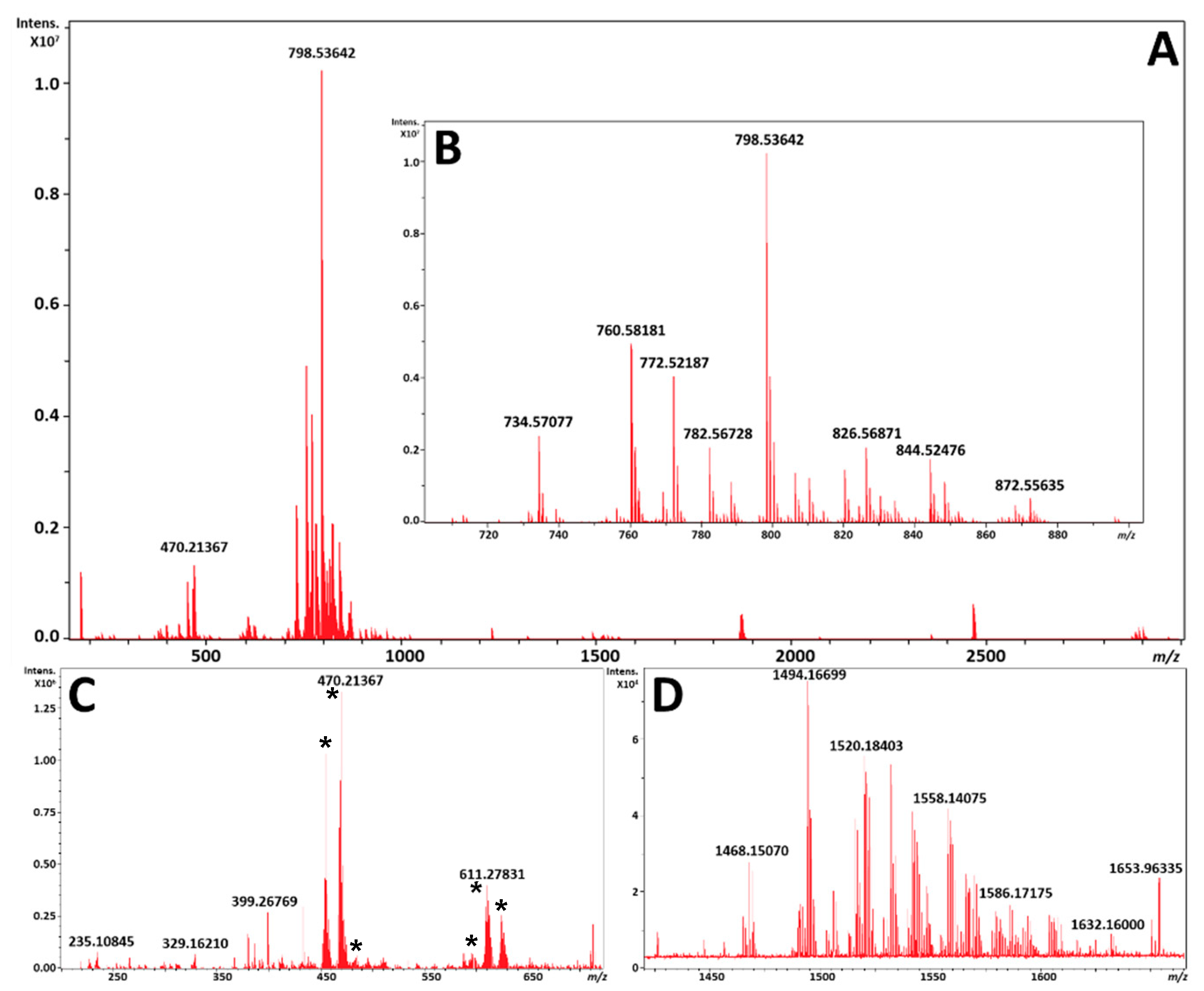
:
1. Introduction
2. Results
2.1. DESI FT-ICR MSI Image Generation
2.2. Comparison of DESI FT-ICR MSI to MALDI FT-ICR MSI
3. Discussion
4. Materials and Methods
4.1. Chemicals
4.2. Tissue Processing
4.3. Instrumentation
4.4. DESI-FT-ICR MSI Analysis
4.5. MALDI-FT-ICR MSI Analysis
4.6. Image Generation, and Data Analysis for MSI
5. Conclusions
Supplementary Materials
Author Contributions
Funding
Institutional Review Board Statement
Informed Consent Statement
Data Availability Statement
Acknowledgments
Conflicts of Interest
Abbreviations
| 3D | three dimensional |
| MS | mass spectrometry/spectrometer |
| FT-MS | Fourier transform mass spectrometer/spectrometry |
| FT-ICR | Fourier transform ion cyclotron resonance |
| ADD | accumulation during detection |
| ICR | ion cyclotron resonance |
| AMP | absorption-mode processing |
| aFT | absorption-mode Fourier transform |
| mFT | magnitude-mode Fourier transform |
| SNR | signal-to-noise ratio |
| RP | resolving power |
| FWHM | full width half maximum |
| HR/AM | high-resolution/accurate mass |
| TOF | time of flight |
| IMS | ion mobility spectrometry |
| MSI | mass spectrometry imaging |
| DESI | desorption electrospray ionization |
| MALDI | matrix-assisted laser desorption/ionization |
| 1,5-DAN | 1,5-diaminonapthalene |
| ITO | indium tin oxide |
| H&E | hematoxylin and eosin |
| PC | phosphatidylcholine |
| PS | phosphatidylserine |
| PE | phosphatidylethanolamine |
| SM | sphingomyelin |
| CL | cardiolipin |
References
- Norris, J.L.; Caprioli, R.M. Analysis of Tissue Specimens by Matrix-Assisted Laser Desorption/Ionization Imaging Mass Spectrometry in Biological and Clinical Research. Chem. Rev. 2013, 113, 2309–2342. [Google Scholar] [CrossRef] [PubMed] [Green Version]
- Scott, A.J.; Jones, J.W.; Orschell, C.M.; MacVittie, T.J.; Kane, M.A.; Ernst, R.K. Mass Spectrometry Imaging Enriches Biomarker Discovery Approaches with Candidate Mapping. Health Phys. 2014, 106, 120–128. [Google Scholar] [CrossRef] [PubMed] [Green Version]
- Wiseman, J.M.; Ifa, D.R.; Zhu, Y.; Kissinger, C.B.; Nicholas, E.; Manicke, N.E.; Peter, T.; Kissinger, P.T.; Cooks, R.G. Desorption Electrospray Ionization Mass Spectrometry: Imaging Drugs and Metabolites in Tissues. Proc. Natl. Acad. Sci. USA 2008, 105, 18120–18125. [Google Scholar] [CrossRef] [PubMed] [Green Version]
- Lietz, C.B.; Gemperline, E.; Li, L. Qualitative and Quantitative Mass Spectrometry Imaging of Drugs and Metabolites. Adv. Drug Deliv. Rev. 2013, 65, 1074–1085. [Google Scholar] [CrossRef] [Green Version]
- Treu, A.; Kokesch-Himmelreich, J.; Walter, K.; Hölscher, C.; Römpp, A. Integrating High-Resolution MALDI Imaging into the Development Pipeline of Anti-Tuberculosis Drugs. J. Am. Soc. Mass Spectrom. 2020, 31, 2277–2286. [Google Scholar] [CrossRef]
- Neumann, E.K.; Djambazova, K.V.; Caprioli, R.M.; Spraggins, J.M. Multimodal Imaging Mass Spectrometry: Next Generation Molecular Mapping in Biology and Medicine. J. Am. Soc. Mass Spectrom. 2020, 31, 2401–2415. [Google Scholar] [CrossRef]
- Heijs, B.; Holst, S.; Briaire-de Bruijn, I.H.; van Pelt, G.W.; de Ru, A.H.; van Veelen, P.A.; Drake, R.R.; Mehta, A.S.; Mesker, W.E.; Tollenaar, R.A.; et al. Multimodal Mass Spectrometry Imaging of N-Glycans and Proteins from the Same Tissue Section. Anal. Chem. 2016, 88, 7745–7753. [Google Scholar] [CrossRef]
- Holzlechner, M.; Bonta, M.; Lohninger, H.; Limbeck, A.; Marchetti-Deschmann, M. Multisensor Imaging—from Sample Preparation to Integrated Multimodal Interpretation of LA-ICPMS and MALDI MS Imaging Data. Anal. Chem. 2018, 90, 8831–8837. [Google Scholar] [CrossRef]
- Nguyen, S.N.; Kyle, J.E.; Dautel, S.E.; Sontag, R.; Luders, T.; Corley, R.; Ansong, C.; Carson, J.; Laskin, J. Lipid Coverage in Nanospray Desorption Electrospray Ionization Mass Spectrometry Imaging of Mouse Lung Tissues. Anal. Chem. 2019, 91, 11629–11635. [Google Scholar] [CrossRef]
- Van Nuffel, S.; Quatredeniers, M.; Pirkl, A.; Zakel, J.; Le Caer, J.-P.; Elie, N.; Vanbellingen, Q.P.; Dumas, S.J.; Nakhleh, M.K.; Ghigna, M.-R.; et al. Multimodal Imaging Mass Spectrometry to Identify Markers of Pulmonary Arterial Hypertension in Human Lung Tissue Using MALDI-TOF, TOF-SIMS, and Hybrid SIMS. Anal. Chem. 2020, 92, 12079–12087. [Google Scholar] [CrossRef]
- Van de Plas, R.; Yang, J.; Spraggins, J.; Caprioli, R.M. Image Fusion of Mass Spectrometry and Microscopy: A Multimodality Paradigm for Molecular Tissue Mapping. Nat. Methods 2015, 12, 366–372. [Google Scholar] [CrossRef] [Green Version]
- Liu, X.; Flinders, C.; Mumenthaler, S.M.; Hummon, A.B. Maldi Mass Spectrometry Imaging for Evaluation of Therapeutics in Colorectal Tumor Organoids. J. Am. Soc. Mass Spectrom. 2018, 29, 516–526. [Google Scholar] [CrossRef]
- Kriegsmann, K.; Longuespée, R.; Hundemer, M.; Zgorzelski, C.; Casadonte, R.; Schwamborn, K.; Weichert, W.; Schirmacher, P.; Harms, A.; Kazdal, D.; et al. Combined Immunohistochemistry after Mass Spectrometry Imaging for Superior Spatial Information. PROTEOM. Clin. Appl. 2019, 13, 1800035. [Google Scholar] [CrossRef] [Green Version]
- Lasch, P.; Noda, I. Two-Dimensional Correlation Spectroscopy for Multimodal Analysis of FT-IR, Raman, and MALDI-Tof Ms Hyperspectral Images with Hamster Brain Tissue. Anal. Chem. 2017, 89, 5008–5016. [Google Scholar] [CrossRef]
- Svirkova, A.; Turyanskaya, A.; Perneczky, L.; Streli, C.; Marchetti-Deschmann, M. Multimodal Imaging of Undecalcified Tissue Sections by Maldi Ms and Μxrf. Analyst 2018, 143, 2587–2595. [Google Scholar] [CrossRef] [Green Version]
- Tian, X.; Xie, B.; Zou, Z.; Jiao, Y.; Lin, L.-E.; Chen, C.-L.; Hsu, C.-C.; Peng, J.; Yang, Z. Multimodal Imaging of Amyloid Plaques: Fusion of the Single-Probe Mass Spectrometry Image and Fluorescence Microscopy Image. Anal. Chem. 2019, 91, 12882–12889. [Google Scholar] [CrossRef]
- Lorenz, M.; Ovchinnikova, O.S.; Kertesz, V.; Van Berkel, G.J. Laser Microdissection and Atmospheric Pressure Chemical Ionization Mass Spectrometry Coupled for Multimodal Imaging. Rapid Commun. Mass Spectrom. 2013, 27, 1429–1436. [Google Scholar] [CrossRef]
- Dewez, F.; Oejten, J.; Henkel, C.; Hebeler, R.; Neuweger, H.; De Pauw, E.; Heeren, R.M.A.; Balluff, B. MS Imaging-Guided Microproteomics for Spatial Omics on a Single Instrument. PROTEOMICS 2020, 20, 1900369. [Google Scholar] [CrossRef]
- Eberlin, L.S.; Ferreira, C.R.; Dill, A.L.; Ifa, D.R.; Cheng, L.; Cooks, R.G. Nondestructive, Histologically Compatible Tissue Imaging by Desorption Electrospray Ionization Mass Spectrometry. ChemBioChem 2011, 12, 2129–2132. [Google Scholar] [CrossRef]
- Shariatgorji, M.; Nilsson, A.; Fridjonsdottir, E.; Vallianatou, T.; Källback, P.; Katan, L.; Sävmarker, J.; Mantas, I.; Zhang, X.; Bezard, E.; et al. Comprehensive Mapping of Neurotransmitter Networks by Maldi–Ms Imaging. Nat. Methods 2019, 16, 1021–1028. [Google Scholar] [CrossRef]
- Hulme, H.; Fridjonsdottir, E.; Gunnarsdottir, H.; Vallianatou, T.; Zhang, X.; Wadensten, H.; Shariatgorji, R.; Nilsson, A.; Bezard, E.; Svenningsson, P.; et al. Simultaneous Mass Spectrometry Imaging of Multiple Neuropeptides in the Brain and Alterations Induced by Experimental Parkinsonism and L-Dopa Therapy. Neurobiol. Dis. 2020, 137, 104738. [Google Scholar] [CrossRef]
- Ryan, D.J.; Spraggins, J.M.; Caprioli, R.M. Protein Identification Strategies in MALDI Imaging Mass Spectrometry: A Brief Review. Curr. Opin. Chem. Biol. 2019, 48, 64–72. [Google Scholar] [CrossRef]
- Shariatgorji, M.; Nilsson, A.; Goodwin, R.J.A.; Källback, P.; Schintu, N.; Zhang, X.; Crossman, A.R.; Bezard, E.; Svenningsson, P.; Andren, P.E. Direct Targeted Quantitative Molecular Imaging of Neurotransmitters in Brain Tissue Sections. Neuron 2014, 84, 697–707. [Google Scholar] [CrossRef] [Green Version]
- Towers, M.W.; Karancsi, T.; Jones, E.A.; Pringle, S.D.; Claude, E. Optimised Desorption Electrospray Ionisation Mass Spectrometry Imaging (DESI-MSI) for the Analysis of Proteins/Peptides Directly from Tissue Sections on a Travelling Wave Ion Mobility Q-Tof. J. Am. Soc. Mass Spectrom. 2018, 29, 2456–2466. [Google Scholar] [CrossRef]
- Garza, K.Y.; Feider, C.L.; Klein, D.R.; Rosenberg, J.A.; Brodbelt, J.S.; Eberlin, L.S. Desorption Electrospray Ionization Mass Spectrometry Imaging of Proteins Directly from Biological Tissue Sections. Anal. Chem. 2018, 90, 7785–7789. [Google Scholar] [CrossRef]
- Škrášková, K.; Claude, E.; Jones, E.A.; Towers, M.; Ellis, S.R.; Heeren, R.M.A. Enhanced Capabilities for Imaging Gangliosides in Murine Brain with Matrix-Assisted Laser Desorption/Ionization and Desorption Electrospray Ionization Mass Spectrometry Coupled to Ion Mobility Separation. Methods 2016, 104, 69–78. [Google Scholar] [CrossRef]
- Eberlin, L.S.; Liu, X.; Ferreira, C.R.; Santagata, S.; Agar, N.Y.R.; Cooks, R.G. Desorption Electrospray Ionization Then Maldi Mass Spectrometry Imaging of Lipid and Protein Distributions in Single Tissue Sections. Anal. Chem. 2011, 83, 8366–8371. [Google Scholar] [CrossRef] [Green Version]
- Kooijman, P.C.; Nagornov, K.O.; Kozhinov, A.N.; Kilgour, D.P.A.; Tsybin, Y.O.; Heeren, R.M.A.; Ellis, S.R. Increased Throughput and Ultra-High Mass Resolution in DESI FT-ICR MS Imaging through New-Generation External Data Acquisition System and Advanced Data Processing Approaches. Sci. Rep. 2019, 9, 8. [Google Scholar] [CrossRef] [Green Version]
- Stopka, S.A.; Samarah, L.Z.; Shaw, J.B.; Liyu, A.V.; Veličković, D.; Agtuca, B.J.; Kukolj, C.; Koppenaal, D.W.; Stacey, G.; Paša-Tolić, L.; et al. Ambient Metabolic Profiling and Imaging of Biological Samples with Ultrahigh Molecular Resolution Using Laser Ablation Electrospray Ionization 21 Tesla FTICR Mass Spectrometry. Anal. Chem. 2019, 91, 5028–5035. [Google Scholar] [CrossRef]
- Bowman, A.P.; Blakney, G.T.; Hendrickson, C.L.; Ellis, S.R.; Heeren, R.M.A.; Smith, D.F. Ultra-High Mass Resolving Power, Mass Accuracy, and Dynamic Range Maldi Mass Spectrometry Imaging by 21-T FT-ICR Ms. Anal. Chem. 2020, 92, 3133–3142. [Google Scholar] [CrossRef] [Green Version]
- Clauser, K.R.; Baker, P.; Burlingame, A.L. Role of Accurate Mass Measurement (±10 ppm) in Protein Identification Strategies Employing Ms or Ms/Ms and Database Searching. Anal. Chem. 1999, 71, 2871–2882. [Google Scholar] [CrossRef]
- Züllig, T.; Köfeler, H.C. High Resolution Mass Spectrometry in Lipidomics. Mass Spectrom. Rev. 2020, 40, 162–176. [Google Scholar] [CrossRef] [PubMed] [Green Version]
- Smith, D.F.; Kilgour, D.P.A.; Konijnenburg, M.; O’Connor, P.B.; Heeren, R.M.A. Absorption Mode FTICR Mass Spectrometry Imaging. Anal. Chem. 2013, 85, 11180–11184. [Google Scholar] [CrossRef] [PubMed]
- Nicolardi, S.; Bogdanov, B.; Deelder, A.M.; Palmblad, M.; van der Burgt, Y.E. Developments in FTICR-MS and Its Potential for Body Fluid Signatures. Int. J. Mol. Sci. 2015, 16, 27133–27144. [Google Scholar] [CrossRef] [PubMed] [Green Version]
- Barry, J.A.; Groseclose, M.R.; Robichaud, G.; Castellino, S.; Muddiman, D.C. Assessing Drug and Metabolite Detection in Liver Tissue by UV-MALDI and IR-MALDESI Mass Spectrometry Imaging Coupled to Ft-Icr Ms. Int. J. Mass Spectrom. 2015, 377, 448–455. [Google Scholar] [CrossRef] [Green Version]
- Shaw, J.B.; Gorshkov, M.V.; Wu, Q.; Paša-Tolić, L. High Speed Intact Protein Characterization Using 4x Frequency Multiplication, Ion Trap Harmonization, and 21 Tesla FTICR-MS. Anal. Chem. 2018, 90, 5557–5562. [Google Scholar] [CrossRef]
- Bennet, R.V.; Gamage, C.M.; Fernández, F.M. Imaging of Biological Tissues by Desorption Electrospray Ionization Mass Spectrometry. J. Vis. Exp. 2013, 77, e50575. [Google Scholar] [CrossRef] [Green Version]
- Wei, J.; Wu, J.; Tang, Y.; Ridgeway, M.E.; Park, M.A.; Costello, C.E.; Zaia, J.; Lin, C. Characterization and Quantification of Highly Sulfated Glycosaminoglycan Isomers by Gated-Trapped Ion Mobility Spectrometry Negative Electron Transfer Dissociation Ms/Ms. Anal. Chem. 2019, 91, 2994–3001. [Google Scholar] [CrossRef]
- Zubarev, R.A.; Makarov, A. Orbitrap Mass Spectrometry. Anal. Chem. 2013, 85, 5288–5296. [Google Scholar] [CrossRef]
- Zhang, Y.; Wang, J.; Liu, J.; Han, J.; Xiong, S.; Yong, W.; Zhao, Z. Combination of ESI and MALDI Mass Spectrometry for Qualitative, Semi-Quantitative and in Situ Analysis of Gangliosides in Brain. Sci. Rep. 2016, 6, 25289. [Google Scholar] [CrossRef] [Green Version]
- Yang, H.; Jackson, S.N.; Woods, A.S.; Goodlett, D.R.; Ernst, R.K.; Scott, A.J. Streamlined Analysis of Cardiolipins in Prokaryotic and Eukaryotic Samples Using a Norharmane Matrix by MALDI-MSI. J. Am. Soc. Mass Spectrom. 2020, 31, 2495–2502. [Google Scholar] [CrossRef]
- Manicke, N.E.; Dill, A.L.; Ifa, D.R.; Cooks, R.G. High-Resolution Tissue Imaging on an Orbitrap Mass Spectrometer by Desorption Electrospray Ionization Mass Spectrometry. J. Mass Spectrom. 2010, 45, 223–226. [Google Scholar] [CrossRef]
- Dória, M.L.; McKenzie, J.S.; Mroz, A.; Phelps, D.L.; Speller, A.; Rosini, F.; Strittmatter, N.; Golf, O.; Veselkov, K.; Brown, R.; et al. Epithelial Ovarian Carcinoma Diagnosis by Desorption Electrospray Ionization Mass Spectrometry Imaging. Sci. Rep. 2016, 6, 39219. [Google Scholar] [CrossRef] [Green Version]
- Bagley, M.C.; Ekelöf, M.; Rock, K.; Patisaul, H.; Muddiman, D.C. IR-MALDESI Mass Spectrometry Imaging of Underivatized Neurotransmitters in Brain Tissue of Rats Exposed to Tetrabromobisphenol A. Anal. Bioanal. Chem. 2018, 410, 7979–7986. [Google Scholar] [CrossRef]
- Shaw, J.B.; Lin, T.-Y.; Leach, F.E.; Tolmachev, A.V.; Tolić, N.; Robinson, E.W.; Koppenaal, D.W.; Paša-Tolić, L. 21 Tesla Fourier Transform Ion Cyclotron Resonance Mass Spectrometer Greatly Expands Mass Spectrometry Toolbox. J. Am. Soc. Mass Spectrom. 2016, 27, 1929–1936. [Google Scholar] [CrossRef]
- Pól, J.; Vidová, V.; Kruppa, G.; Kobliha, V.; Novák, P.; Lemr, K.; Kotiaho, T.; Kostiainen, R.; Havlíček, V.; Volný, M. Automated Ambient Desorption−Ionization Platform for Surface Imaging Integrated with a Commercial Fourier Transform Ion Cyclotron Resonance Mass Spectrometer. Anal. Chem. 2009, 81, 8479–8487. [Google Scholar] [CrossRef]
- Robichaud, G.; Barry, J.A.; Garrard, K.P.; Muddiman, D.C. Infrared Matrix-Assisted Laser Desorption Electrospray Ionization (IR-MALDESI) Imaging Source Coupled to a FT-ICR Mass Spectrometer. J. Am. Soc. Mass Spectrom. 2013, 24, 92–100. [Google Scholar] [CrossRef] [Green Version]
- Amoscato, A.A.; Sparvero, L.J.; He, R.R.; Watkins, S.; Bayir, H.; Kagan, V.E. Imaging Mass Spectrometry of Diversified Cardiolipin Molecular Species in the Brain. Anal. Chem. 2014, 86, 6587–6595. [Google Scholar] [CrossRef] [Green Version]
- Martínez-Jarquín, S.; Moreno-Pedraza, A.; Guillén-Alonso, H.; Winkler, R. Template for 3D Printing a Low-Temperature Plasma Probe. Anal. Chem. 2016, 88, 6976–6980. [Google Scholar] [CrossRef]
- Guillén-Alonso, H.; Rosas-Román, I.; Winkler, R. The Emerging Role of 3D-Printing in Ion Mobility Spectrometry and Mass Spectrometry. Anal. Methods 2021, 13, 852–861. [Google Scholar] [CrossRef]
- Zemaitis, K.J.; Wood, T.D. Integration of 3D-Printing for a Desorption Electrospray Ionization Source for Mass Spectrometry. Rev. Sci. Instrum. 2020, 91, 104102. [Google Scholar] [CrossRef] [PubMed]
- Maser, T.L.; Honarvar, E.; Venter, A.R. Delayed Desorption Improves Protein Analysis by Desorption Electrospray Ionization Mass Spectrometry. J. Am. Soc. Mass Spectrom. 2020, 31, 803–811. [Google Scholar] [CrossRef] [PubMed]
- Nyadong, L.; Hohenstein, E.G.; Galhena, A.; Lane, A.L.; Kubanek, J.; Sherrill, C.D.; Fernández, F.M. Reactive Desorption Electrospray Ionization Mass Spectrometry (DESI-MS) of Natural Products of a Marine Alga. Anal. Bioanal. Chem. 2009, 394, 245–254. [Google Scholar] [CrossRef] [PubMed] [Green Version]
- Wu, C.; Ifa, D.R.; Manicke, N.E.; Cooks, R.G. Rapid, Direct Analysis of Cholesterol by Charge Labeling in Reactive Desorption Electrospray Ionization. Anal. Chem. 2009, 81, 7618–7624. [Google Scholar] [CrossRef] [Green Version]
- Lostun, D.; Perez, C.J.; Licence, P.; Barrett, D.A.; Ifa, D.R. Reactive DESI-MS Imaging of Biological Tissues with Dicationic Ion-Pairing Compounds. Anal. Chem. 2015, 87, 3286–3293. [Google Scholar] [CrossRef]
- Sud, M.; Fahy, E.; Cotter, D.; Brown, A.; Dennis, E.A.; Glass, C.K.; Merrill, A.H., Jr.; Raetz, C.R.; Russell, D.W.; Subramaniam, S. Lmsd: Lipid Maps Structure Database. Nucleic Acids Res. 2007, 35, D527–D532. [Google Scholar] [CrossRef] [Green Version]
- Smith, C.A.; O’Maille, G.; Want, E.J.; Qin, C.; Trauger, S.A.; Brandon, T.R.; Custodio, D.E.; Abagyan, R.; Siuzdak, G. Metlin: A Metabolite Mass Spectral Database. Ther. Drug. Monit. 2005, 27, 747–751. [Google Scholar] [CrossRef]


Publisher’s Note: MDPI stays neutral with regard to jurisdictional claims in published maps and institutional affiliations. |
© 2021 by the authors. Licensee MDPI, Basel, Switzerland. This article is an open access article distributed under the terms and conditions of the Creative Commons Attribution (CC BY) license (https://creativecommons.org/licenses/by/4.0/).
Share and Cite
Zemaitis, K.J.; Izydorczak, A.M.; Thompson, A.C.; Wood, T.D. Streamlined Multimodal DESI and MALDI Mass Spectrometry Imaging on a Singular Dual-Source FT-ICR Mass Spectrometer. Metabolites 2021, 11, 253. https://0-doi-org.brum.beds.ac.uk/10.3390/metabo11040253
Zemaitis KJ, Izydorczak AM, Thompson AC, Wood TD. Streamlined Multimodal DESI and MALDI Mass Spectrometry Imaging on a Singular Dual-Source FT-ICR Mass Spectrometer. Metabolites. 2021; 11(4):253. https://0-doi-org.brum.beds.ac.uk/10.3390/metabo11040253
Chicago/Turabian StyleZemaitis, Kevin J., Alexandra M. Izydorczak, Alexis C. Thompson, and Troy D. Wood. 2021. "Streamlined Multimodal DESI and MALDI Mass Spectrometry Imaging on a Singular Dual-Source FT-ICR Mass Spectrometer" Metabolites 11, no. 4: 253. https://0-doi-org.brum.beds.ac.uk/10.3390/metabo11040253