Article

The Influence of Drying Temperatures on the Metabolic Profiles and Antioxidant Activity of Manilkara zapota Leaves

by
Gloria I. Hernández-Bolio
1,
Rubí E. Dzul-Romero
2,
María G. Maldonado Velázquez
2,
Pedro Zamora Cresencio
3,
Emanuel Hernández-Núñez
1,4,* and
Francisco J. Aguirre-Crespo
2,*
1
Marine Resources Department, Center for Research and Advanced Studies -National Polytechnic Institute (CINVESTAV-IPN), Unidad Mérida 97310, Mexico
2
Laboratory of Pharmaceutical Biotechnology, Faculty of Biological Chemical Sciences, Autonomous University of Campeche, Campeche 24030, Mexico
3
Center for Historical and Social Research, Autonomous University of Campeche, Campeche 24030, Mexico
4
National Council of Science and Technology (CONACYT), Mexico City 03940, Mexico
*
Authors to whom correspondence should be addressed.
Submission received: 27 August 2019 / Revised: 26 September 2019 / Accepted: 30 September 2019 / Published: 6 October 2019
(This article belongs to the Special Issue Plant Metabolomics)

Abstract

:
In the present study, the leaves of Manilkara zapota (L.) P. Royen (Sapotaceae), an evergreen tree recognized for its medicinal properties in Southern Mexico, were used as a model to study the effect of different drying temperatures on its metabolic profile and therefore, its antioxidant potential. For this purpose, a methanol extraction of leaves dried at room temperature (25 °C) or by heat convection (50, 75 and 100 °C) were compared in terms of drying efficiency, yield of extraction, total phenol content, 1H-NMR metabolic profile, and DPPH antioxidant activity. The drying curves enabled the fact to be uncovered that drying efficiency improves with increase of temperature, as does the level of total phenols and antioxidant activity. A metabolomics approach using principal component analysis (PCA) and orthogonal projections to latent structures discriminant analysis (OPLS-DA) of the corresponding 1H-NMR profiles allowed the impact of the drying temperature on their metabolic profile to be documented and also, caffeic acid and epicatechin as main secondary metabolites contributing to the antioxidant activity of M. zapota to be identified.

1. Introduction

One of the simplest and inexpensive methods to preserve the medicinal properties of plants is by drying; also, it is a classical way to start the isolation of natural products [1]. By drying, the plant material becomes easier to handle and less prone to microbial degradation [2], although, it is known that drying temperature may affect the metabolic profile and antioxidant properties of the corresponding extracts [3,4,5]. Heat convection is a process commonly used in the elimination of water vapor during the drying process and the drying curves allow monitoring of the moisture loss as a function of time, thus enabling an estimate of the drying speed of a material [6]. In the context of medicinal plants, the identification of adequate drying conditions ensures the efficiency and drying time as well as the recovery performance of the secondary metabolites of interest.
Manilkara zapota (L.) P. Royen (Sapotaceae), also known as chicozapote or sapodilla, is an evergreen tree native to Southern Mexico, Central America, and part of South America [7]. It is mainly cultivated for its fruit, which is highly prized and considered one of the best in Central America [8], although, it is also recognized for its medicinal properties. A decoction of the leaves of M. zapota is used as an antipyretic, for treatment of hemorrhage, wounds, and ulcers; other uses include a remedy for diarrhea, intestinal inflammation, lack of appetite, and normalization of blood pressure [9]. Some pharmacological studies have confirmed specific medicinal properties of the leaves such as antidiabetic, antilipidemic, hypoglycemic, and antioxidant activity [10,11,12,13]. While the phytochemistry of the fruit has been explored, reporting the presence of bioactive and novel polyphenols [14], the leaves have only had qualitative studies suggesting the presence of phenols and flavonoids [10] and a single phytochemical study reporting the isolation of lupeol acetate, oleanolic acid, apigenin-7-O-α-l-rhamnoside, myricetin-3-O-α-l-rhamnoside, and caffeic acid [15].
In the present work, we used M. zapota leaf extracts as a model to explore the differences in its metabolic profile, and the effect on its antioxidant activity, as influenced by the temperature of the leaf drying. For this purpose, a metabolomics approach using principal component analysis (PCA) and orthogonal projections to latent structures discriminant analysis (OPLS-DA) was performed. The multivariate analysis of the corresponding 1H-NMR profiles both allowed the documentation of the impact of drying temperature on the metabolic profile and the detection of the main antioxidant- related metabolites. This method thus contributes to simplifying the pathway to the phytochemical knowledge of a medicinal plant species.

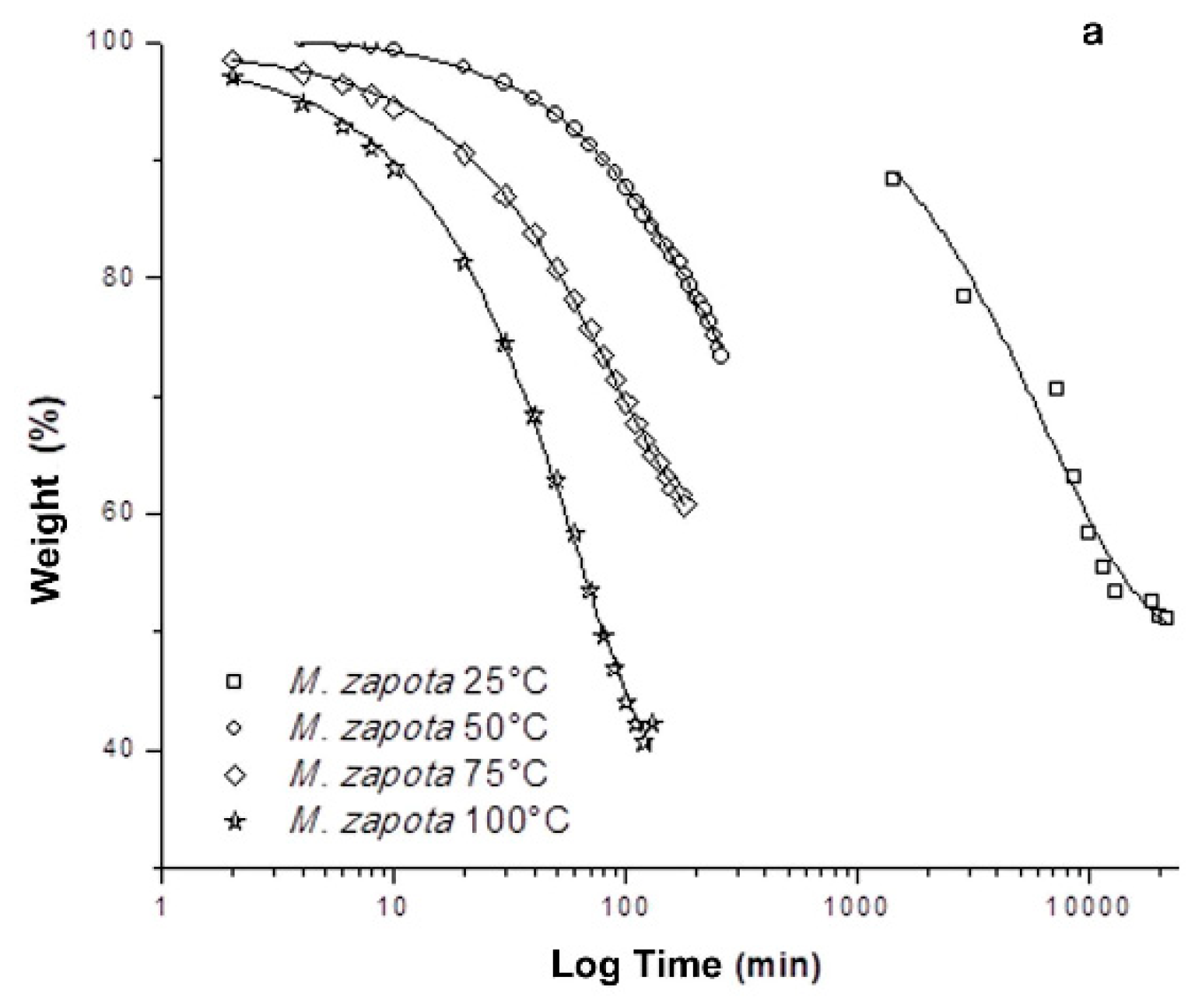
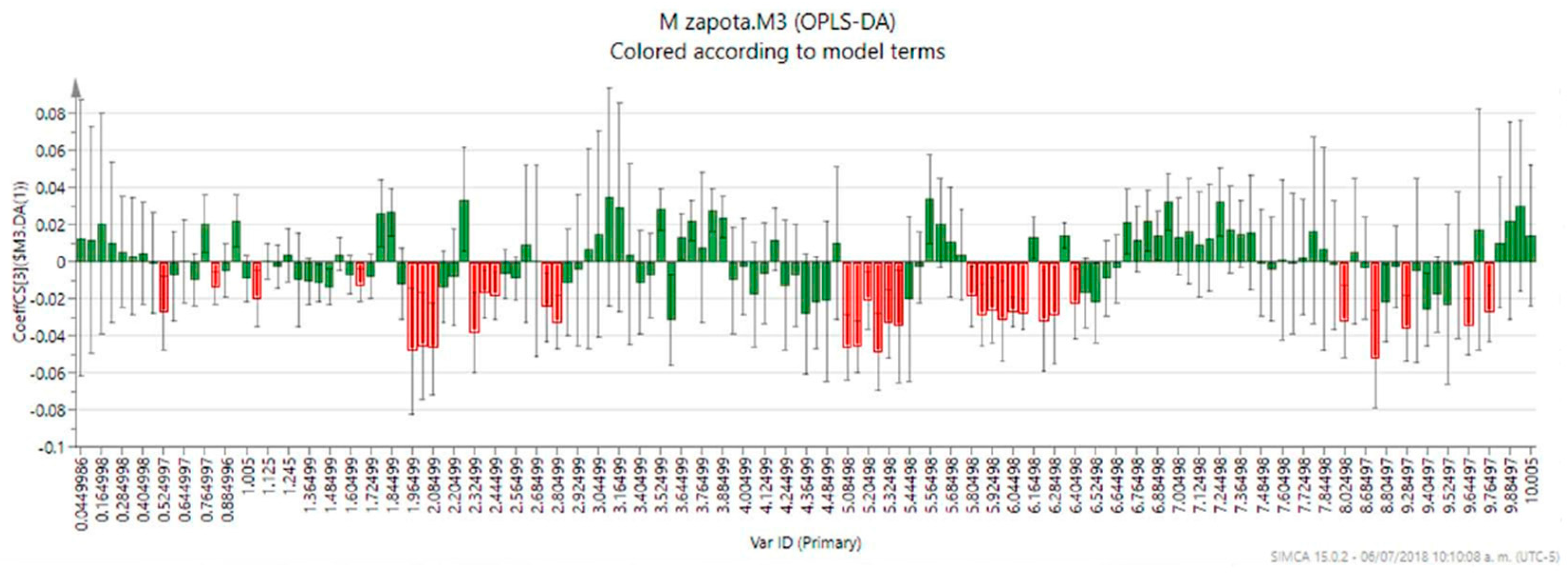
2. Results and Discussion

For handling and storage purposes, reducing the water content of freshly harvested medicinal plants is imperative [16]. Choosing a drying method includes taking into account features as maximum recovery of metabolites of interest and dry efficiency evaluation in order to obtain a higher yield in a reduced time. For that purpose, a comparison between four drying temperatures of M. zapota leaves was carried out. Monitoring of the leaf drying process revealed 100 °C as the most efficient temperature, showing the highest percentage of water loss (57.81 ± 1.48%) in the least time (1 h, 18 min) (Figure 1, Table 1), while the drying at room temperature (25 °C) required at least, 15 days in order to remove 50% of the humidity from the plant material. It is worth mentioning that at 50 °C the efficiency of drying decreases 54.60% when compared to room temperature (25 °C), resulting in a slower drying process and with a lower percentage of water loss.
Poor water removal at 50 °C could be related to stomatal closure in the leaves of M. zapota, a situation that prevents transpiration and loss of water in the plant material. With high water stress, the stomatal system closes with increasing temperature, a fact that prevents water loss due to transpiration [17]. It has been reported that in response to drying, the synthesis of abscisic acid (ABA) is increased and its interaction with PYR/PYL/RCAR ABA receptors increases the flow of Ca2+ and K+ as well as depolarization of the membrane of occlusive cells. These events decrease the turgor pressure, stomatal closure, and reduction of water loss due to transpiration [18]. On the other hand, the use of high drying temperature (75 and 100 °C) favors water vapor pressure as well as protein denaturation involved in the signal transduction of stomatal closure, among other pathways and proteins.
Regarding the methanol extraction of M. zapota leaves, it was found that the yield is reduced in a proportional way to the increment of the drying temperature (R2 = 0.985, m = –0.31%/°C) (Table 1). The data revealed 100 °C as the temperature with the lowest extraction yield (14.13 ± 0.29%), however, it has the highest simple phenol content (R2 = 0.992). On the other hand, it was also the extract with the highest antioxidant activity when tested in the DPPH assay (EC50 = 11.04 ± 0.01 µg/mL), thus showing its selectivity in the extraction of antioxidant metabolites (Table 1).
Conversely, few studies analyzing different drying temperatures of plant materials have reported that antioxidant activity decreases with temperature [3,19], and have attributed those changes in the antioxidant activity mainly to the polyphenol content of the plant extracts, which is also reduced with the increment of temperature. It has been shown that polyphenol content is affected by drying [20]. In fact, prolonged exposure at high temperatures reduced 30%, 32%, and 37% of gallic acid, vanillic acid and catequine content, respectively in grape seeds [21]. On the other hand, gallotannins derived from Caesalpinia spinosa enhance their antioxidant activity after thermal hydrolysis [22]. In this context, the drying of leaves of M. zapota could favor the hydrolysis of gallotannins and increase the content of simple phenols in the extracts as well as the antioxidant activity. Finally, it is reported that thermal stress degrades 11.1%–16.6% of the content of condensed tannins [19] while quercetin exhibits high thermal stability [23]. These facts suggest that simple phenols and other flavonoids could be involved in the antioxidant activity induced by extracts of M. zapota leaves.
The 1H-NMR profiles of methanol extracts from M. zapota leaves dried at different temperatures were also compared to detect the changes in the metabolic profile of the corresponding extracts and, furthermore, to explore the possible nature of the antioxidant metabolites. All the extracts showed a high abundance of both aliphatic and carbinolic proton resonances, with the exception of those extracts obtained from leaves dried at 50 °C (Figure 2), which showed a different and simpler profile regarding the carbinolic-proton region and a marked absence of the signals at δ 6.95, 6.36, and 6.21, which could be further assigned to the myricetin (3,5,7-trihydroxy-2-(3,4,5-trihydroxyphenyl)-4H-chromen-4-one) (Table S1). This flavonoid, previously reported from the leaves of M. zapota as rhamnoside [15] was thought to be one of the metabolites responsible for the antioxidant activity of the M. zapota extract, however, its absence in one of the extracts (MZ50) as shown by the 1H-NMR, and the fact that this extract possesses a higher antioxidant activity than MZ25, suggest that myricetin does not have an important contribution for the antioxidant activity of the M. zapota leaves extract. (Figure S1).
The analysis of the PCA score plot (PC1 vs PC3, 54.2% of the explained variance) confirmed the expected grouping according to the temperatures of leaf drying and, as well, showed a clear discrimination of the 50 °C extracts from the others (Figure 3). The PCA ‘contribution plot’ was analyzed in order to detect the signals influencing the discrimination of the 50 °C extracts, showing that these profiles are lacking or with low abundance of signals in the 7.8–5.6 ppm and 4.42–2.68 ppm regions (Figure S1). This change could be related to the poor drying and generation of a high concentration of hydrophilic glycosylated products and therefore, a decrease of organic and methanol-soluble metabolites [24].
As stated before, assessment of the DPPH antioxidant activity of M. zapota leaves dried at different temperatures revealed 100 °C as the best temperature for the extraction of antioxidant metabolites with a value almost comparable with C. sinensis leaf extract (Table 1), a plant well-known for its antioxidant properties. In order to detect the metabolites responsible for the antioxidant activity in the M. zapota leaf extracts, an OPLS-DA was carried out using the matrix produced by the PCA and adding as ‘discriminant variables’ the levels of antioxidant activity shown by the extracts. The variance (R2) and model predictability (Q2) for the OPLS-DA were calculated as 0.827 and 0.659, respectively [25]. While the OPLS-DA score plot showed the characteristic clustering observed earlier in the PCA (Figure 4), the coefficient plot allowed the detection of those bins related to the high antioxidant activity of the M. zapota leaf extracts (Figure 5).
In this plot, the coefficients express how strongly the variable Y (e.g., the level of antioxidant activity) is correlated to the systematic part of the X-variables (e.g., spectral bins), meanwhile, the error bars indicate the confidence intervals of the coefficients. If the coefficient is significant, the confidence interval does not include zero. Therefore, 32 bins in total were identified as related to the activity by the analysis of this plot. The analysis of the activity-related bins led to the successful assignation of two antioxidant metabolites as follows: the signals at δ 5.92, 5.86, 2.80, and 2.74 could be assigned to epicatechin, while the signal at δ 6.28 was assigned to caffeic acid.
The complete assignation of the 1H-NMR signals corresponding to antioxidant metabolites was carried out by analysis of their 1H, 13C and bidimensional (1H-1H COSY, 1H-13C HSQC, 1H-13C HMBC and TOCSY) NMR data, as well as comparison with data reported in the literature (Tables S2 and S3, respectively). Epicatechin and caffeic acid, phenolic metabolites that are widely distributed among the plant species, are well-known for their antioxidant effects, having a reported DPPH trolox equivalent activity of 2.7 and 2.2 mM, respectively [25]. Their high antioxidant activity (Table 1) thus validates the model created by OPLS-DA and confirm its proficiency to detect the antioxidant components from M. zapota. Regarding the contribution of these phenolic metabolites to the antioxidant activity of M. zapota, their absence in the extracts of leaves dried at 25, 50 and 75 °C, explains to a large extent the high level of antioxidant activity of the 100 °C dried leaf extract, however, the presence of unassigned, less abundant signals, suggests that other metabolites can be also displaying some level of antioxidant activity [26]. On the other hand, the fact that some signals corresponding to the antioxidant metabolites were not detected as activity-related can be explained by the overlapping of 1H-NMR signals, although, further approaches including J-resolved and 2D-NMR metabolomics methods can be further applied [27,28] in order to clearly detect the signals from the individual metabolites. Additionally, the information obtained in studies as the current investigation may contribute to dereplication of plant extracts by the identification and removal of known products in order to detect the minor abundant bioactive metabolites, activities which are currently underway.
The analysis of the 1H-NMR profiles of M. zapota by such a metabolomics approach allowed the documentation of the impact of drying temperature on their metabolic profile and the identification of caffeic acid and epicatechin as main secondary metabolites contributing to antioxidant activity exerted by methanolic extracts derived from leaves of M. zapota. Thus, this represents an efficient, rapid and reliable technique to begin the phytochemical study of medicinal plants and their preservation.

3. Materials and Methods

3.1. General Experimental Procedures

Deuterated methanol (MeOH-d4), trimethylsilane (TMS), gallic acid, and 1,1-diphenyl-2-picrylhydrazil (DPPH) were purchased from Sigma-Aldrich Co. (St. Louis, MO, USA), Camellia sinensis was purchased from Badia Spices, Inc. (Doral, FL, USA) and MeOH was analytical grade obtained from a local source. 1H-NMR experiments were conducted at 25 °C using a Varian 600 MHz AR Premium Compact (Varian-Agilent, Santa Clara, California, U.S.).

3.2. Plant Material and Drying

Fresh leaves of M. zapota were collected in March 2017 near Campeche, Campeche, México. A voucher specimen was identified taxonomically by MSc. Pedro Zamora Cresencio and deposited at the UACAM-herbarium under registration number 004727.
Four groups of leaves were processed individually to assess the influence of drying on the metabolic profiles and antioxidant activity of M. zapota. A first batch of leaves was room temperature dried (T = 25 °C; m = 15.24 ± 0.23 g) while the remaining groups were oven dried at different temperatures (T = 50 °C, m = 2.21 ± 0.29 g; T= 75 °C, m = 2.17 ± 0.23 g; T = 100 °C, m = 2.56 ± 0.07 g, respectively) using a thermobalance (Mettler-Toledo®). The loss of humidity and weight as a function of time were registered until the attainment of a constant weight. An additional analysis of the drying process was carried out by a sigmoidal and non-linear mathematical model. The weight of leaf samples and the loss of humidity are expressed as the mean ± standard error of three experiments with three replicates each (n = 9).

3.3. Preparation of Plant Extracts

A portion (1 g) of ground, previously dried M. zapota leaves was extracted three times by maceration with 28 mL of MeOH (24 h at 25 °C). The combined extractions of each group were concentrated in vacuo at 40 °C (Büchi) to produce the corresponding extracts MZ25, MZ50, MZ75, and MZ100. The yield extraction is expressed as the mean ± standard error of three experiments with three replicates each (n = 9).

3.4. Total Phenol Content

A calibration curve for gallic acid (GA, 1.04–8.2 µg/mL) was prepared. Briefly, 0.4 mL of GA solution or extract (10 µg/mL) was added to 0.4 mL of Folin-Ciocalteu reagent in 1 mL of distilled water and incubated at room temperature in the dark for 20 min. The reaction was stopped with 1.6 mL of 10% Na2CO3 solution and the absorbance was recorded at 765 nm. Total phenol content in the methanol extracts was expressed as equivalents (mg/g) of gallic acid present in the plant material [5].

3.5. DPPH Assay

The DPPH reducing activity of the M. zapota leaf extracts was determined following the method reported by ref. [29] with slight modifications. Briefly, 200 µL of each extract (1–50 µg/mL) was added to 1.8 mL of a 0.1 M DPPH solution in MeOH and vortexed. After 30 min of incubation in the dark, the absorbance at 517 nm was measured. The percentage of remaining DPPH was calculated using the formula: % (DPPH) = [(Abs DPPH sample)t=30/(Abs DPPH 0.1 M)t=0)] × 100. Spectrometric measurements were made using methanol as a blank. A MeOH extract of C. sinensis (1–50 µg/mL), as well as gallic acid (0.03–32 µg/mL), were used as positive control. The assay was run three times for each extract on different days. Finally, from the experimental data and the use of a non-linear model, the potency (EC50, [µg/mL]) and efficacy (Emax, (%)) of the antioxidant activity exerted by the extracts was calculated. The antioxidant activity is expressed as the mean ± standard error of three experiments with three replicates each (n = 9).

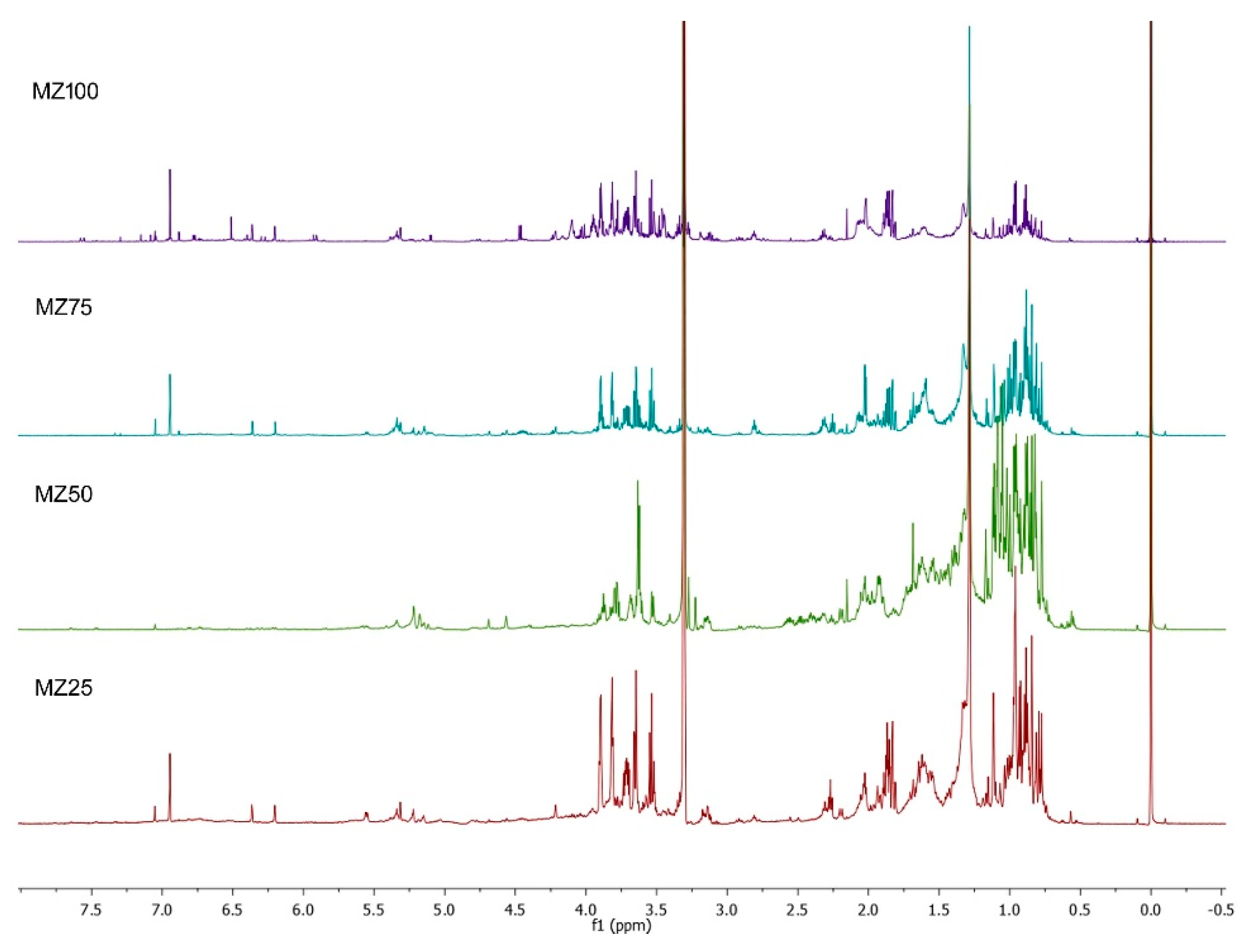
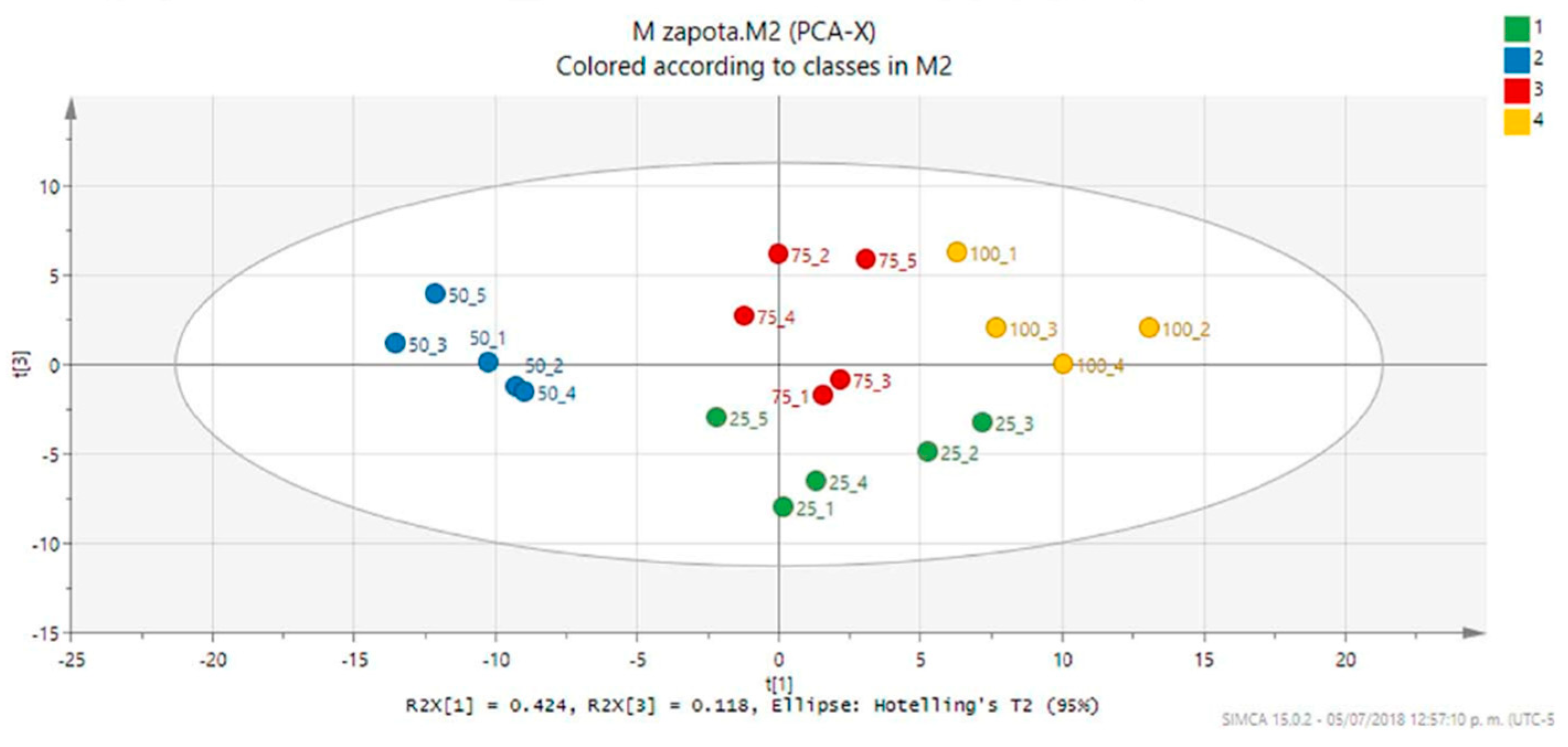
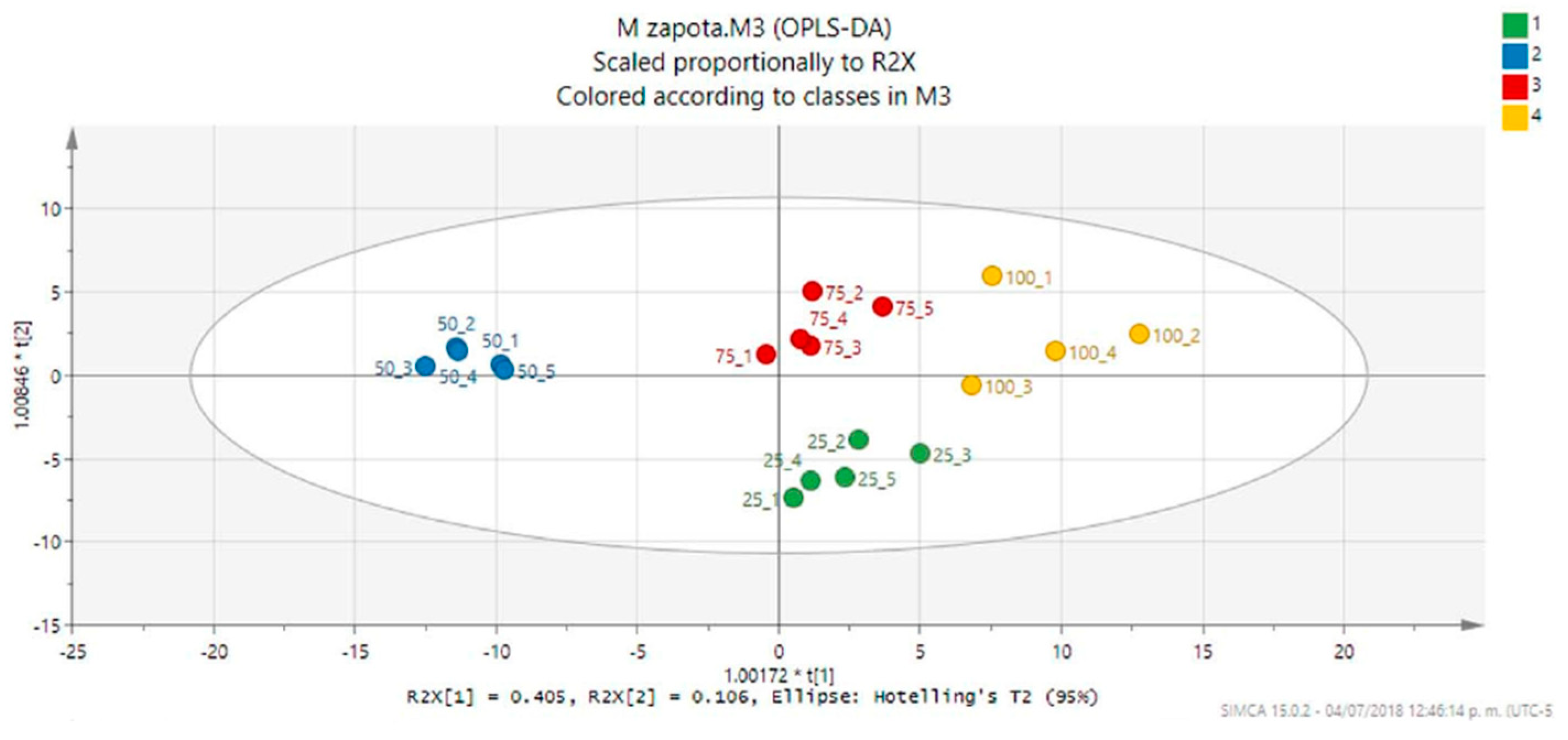
3.6. 1H-NMR Metabolic Profiles and Multivariate Analysis

A portion (approximately 5 mg) of each extract was dissolved in 600 µL of deuterated methanol (MeOH-d4) containing 0.05% v/v TMS and transferred to 5 mm NMR tubes. The measurements were conducted at a magnetic field of 14.1 T and a 1H resonance frequency of 599.77 Hz, using the PRESAT sequence with a total of 128 transients with data collected in 64k data points. The relaxation delay was set 1.0 s and the acquisition time was 3.0 s. The resulting spectra were manually phased, and baseline corrected using Mestrenova 12.0 (Demo version, Mestrelab Research) and referenced to TMS at 0.0 ppm. Additional NMR analyses including 13C-NMR, gCOSY, gHSQC, and gHMBC were conducted on a 100 °C dried sample in order to confirm the identification of the detected metabolites.

3.7. Statistical Analysis

The weight of leaf samples and the loss of humidity, together with the yield of extraction and the results from total phenol content and antioxidant activity are expressed as the mean ± standard error of three experiments with three replicates each (n = 9). The graphics from the drying process and antioxidant activity were obtained in the Origin® 8.0 software, applying ANOVA and significance values of p ˂ 0.05. Principal component analysis (PCA) and orthogonal projections to latent structures discriminant analysis (OPLS-DA) were performed using SIMCA 15 software (Demo version, Sartorius-Stedim). NMR spectra were normalized to the TMS signal (δ 0.0), divided into equally sized bins of 0.06 ppm within the region of 10.0 ppm to 0.44 ppm using the ‘average sum’ method in Mestrenova and Pareto scaled. A data matrix was constructed with each row representing a spectrum and each column a variable (e.g., bin). The spectral regions between 3.26–3.36 ppm and 4.5–4.9 ppm (residual solvent signals) as well as uninformative regions as 8.14–8.62 ppm and 8.86–9.16 ppm, were excluded in PCA to yield a total of 141 bins. The binning generated from PCA was used to perform the OPLS-DA. The quality of the model was described by R2 (percentage of variation of the training set explained by the Y-predicted components) and Q2 (percentage of variation of the training set predicted by the model according to cross validation).

Supplementary Materials

The following are available online at https://0-www-mdpi-com.brum.beds.ac.uk/2218-1989/9/10/217/s1, Table S1: Experimental 1H, 13C-NMR, COSY, HSQC and HMBC data of the putative myricetin and those reported for the myricetin aglycone, Table S2: Experimental 1H-NMR data of the putative epicatechin and those reported for the epicatechin, Table S3: Experimental 1H, 13C-NMR, COSY, HSQC and HMBC data of the putative caffeic acid and those reported for the caffeic acid, Figure S1: Representative 1H-NMR profiles (Region δ 5.0 to 8.1) of the MeOH extracts from M. zapota leaves dried at different temperatures (T = 25, 50, 75 and 100 °C) showing the characteristic resonances from the detected metabolites.

Author Contributions

G.I.H.-B. contributed to the acquisition, analysis and interpretation of NMR data, as well as drafting the paper. R.E.D.-R. and M.G.M.V. contributed to running the laboratory work and analysis of the data. P.Z.C. contributed to collection, identification and handling of plant material as well in the analysis of the results. E.H.-N. and F.J.A.-C. contributed equally in the conceptualization and design of the study and critical reading of the manuscript.

Funding

This work was supported by CONACYT (CB-2015-01) 254321.

Acknowledgments

G.I.H.B. wishes to thank Leopoldina Aguirre Macedo (CINVESTAV-IPN Unidad Mérida, Departamento de Recursos del Mar) and Laboratorio Nacional de Nano y Biomateriales, CINVESTAV-IPN, financed by the Projects: FOMIX Yucatán 2008-108160, CONACYT LAB-2009-01-123913, 292692, 294643, 188345 and 204822, for the access to the NMR spectrometer and laboratory facilities. F.J.A.-C. wishes to thank QFB Iliana Osorio Horta (FCQB-UACAM) and the PRODEP Incorporación Nuevos Profesores program.

Conflicts of Interest

The authors declare no conflict of interest.

References

  1. Bucar, F.; Wube, A.; Schmid, M. Natural product isolation - how to get from biological material to pure compounds. Nat. Prod. Rep. 2013, 30, 525–545. [Google Scholar] [CrossRef] [PubMed]
  2. Zubair, M.; Nybom, H.; Lindholm, C.; Rumpunen, K. Major polyphenols in aerial organs of greater plantain (Plantago major L.), and effects of drying temperature on polyphenol contents in the leaves. Sci. Hortic. 2011, 128, 523–529. [Google Scholar] [CrossRef]
  3. Katsube, T.; Tsurunaga, Y.; Sugiyama, M.; Furuno, T.; Yamasaki, Y. Effect of air-drying temperature on antioxidant capacity and stability of polyphenolic compounds in mulberry (Morus alba L.) leaves. Food Chem. 2009, 113, 964–969. [Google Scholar] [CrossRef]
  4. Darfour, B.; Asare, K.; Ofosu, O.; Daniel, G.A.; Elom, S.A.; Agbenyegah, S. The Effect of Different Drying Methods on the Phytochemicals and Radical Scavenging Activity of Ceylon Cinnamon (Cinnamomum zeylanicum) Plant Parts. Eur. J. Med. Plants 2014, 4, 1324–1335. [Google Scholar]
  5. Mediani, A.; Abas, F.; Tan, C.; Khatib, A. Effects of Different Drying Methods and Storage Time on Free Radical Scavenging Activity and Total Phenolic Content of Cosmos Caudatus. Antioxidants 2014, 3, 358–370. [Google Scholar] [CrossRef] [Green Version]
  6. Bennett, C.O.; Myers, J.E. Transferencia de Cantidad de Movimiento, Calor y Materia; Editorial Reverté: Bogota, Colombia, 1979; ISBN 9788429170528. [Google Scholar]
  7. Tropicos.org Missouri Botanical Garden. Available online: http://www.tropicos.org/Name/28700370 (accessed on 17 December 2018).
  8. Orwa, C.; Mutua, A.; Kindt, R.; Jamnadass, R.; Anthony, S. Agroforestree Database: A Tree Reference and Selection Guide Version 4.0. Available online: http://www.worldagroforestry.org/treedb/AFTPDFS/Manilkara_zapota.PDF (accessed on 11 February 2019).
  9. Biblioteca Digital de la Medicina Tradicional Mexicana Manilkara zapota. Available online: http://www.medicinatradicionalmexicana.unam.mx/monografia.php?l=3&t=Chicozapote&id=7519 (accessed on 20 January 2019).
  10. Chanda, S.V.; Nagani, K.V. Antioxidant Capacity of Manilkara zapota L. Leaves Extracts Evaluated by Four in vitro Methods. J. Biol. Sci. 2010, 8, 260–266. [Google Scholar]
  11. Kaneria, M.; Chanda, S. Evaluation of antioxidant and antimicrobial properties of Manilkara zapota L.(chiku) leaves by sequential soxhlet extraction method. Asian Pac. J. Trop. Biomed. 2012, 2, S1526–S1533. [Google Scholar] [CrossRef]
  12. Barbalho, S.M.; Bueno, P.C.D.S.; Delazari, D.S.; Guiguer, E.L.; Coqueiro, D.P.; Araújo, A.C.; de Souza, M.D.S.S.; Farinazzi-Machado, F.M.V.; Mendes, C.G.; Groppo, M. Antidiabetic and Antilipidemic Effects of Manilkara zapota. J. Med. Food 2015, 18, 385–391. [Google Scholar] [CrossRef]
  13. Paul, S.R.; Hakim, M.L. In vivo hypoglycemic study of Manilkara zapota leaf and seed extracts. Bangladesh J. Pharmacol. 2015, 10, 246–250. [Google Scholar] [CrossRef]
  14. Ma, J.; Luo, X.D.; Protiva, P.; Yang, H.; Ma, C.; Basile, M.J.; Weinstein, I.B.; Kennelly, E.J. Bioactive novel polyphenols from the fruit of Manilkara zapota (Sapodilla). J. Nat. Prod. 2003, 66, 983–986. [Google Scholar] [CrossRef]
  15. Fayek, N.M.; Monem, A.R.A.; Mossa, M.Y.; Meselhy, M.R.; Shazly, A.H. Chemical and biological study of Manilkara zapota (L.) Van Royen leaves (Sapotaceae) cultivated in Egypt. Pharmacogn. Res. 2012, 4, 85–91. [Google Scholar]
  16. Tanko, H.; Carrier, D.J.; Duan, L.; Clausen, E. Pre- and post-harvest processing of medicinal plants. Plant Genet. Resour. 2005, 3, 304–313. [Google Scholar] [CrossRef]
  17. Schulze, E.D.; Lange, O.L.; Kappen, L.; Buschbom, U.; Evenari, M. Stomatal responses to changes in temperature at increasing water stress. Planta 1973, 110, 29–42. [Google Scholar] [CrossRef] [PubMed]
  18. Munemasa, S.; Hauser, F.; Park, J.; Waadt, R.; Brandt, B.; Schroeder, J.I. Mechanisms of abscisic acid-mediated control of stomatal aperture. Curr. Opin. Plant Biol. 2015, 28, 154–162. [Google Scholar] [CrossRef] [PubMed] [Green Version]
  19. Larrauri, J.A.; Rupérez, P.; Saura-Calixto, F. Effect of Drying Temperature on the Stability of Polyphenols and Antioxidant Activity of Red Grape Pomace Peels. J. Agric. Food Chem. 1997, 45, 1390–1393. [Google Scholar] [CrossRef]
  20. Yu, J. Thermal stability of major classes of polyphenols in skins, seeds and stems of grape pomace. In Grapes: Production, Phenolic Composition and Potential Biomedical Effects; de Sousa Câmara, J., Ed.; Nova Science Publishers, Inc.: New York, NY, USA, 2014; pp. 273–285. ISBN 9781633214101. [Google Scholar]
  21. Volf, I.; Ignat, I.; Neamtu, M.; Popa, V.I. Thermal stability, antioxidant activity, and photo-oxidation of natural polyphenols. Chem. Pap. 2014, 68, 121–129. [Google Scholar] [CrossRef]
  22. Terán-Hilares, R.; Chirinos, R.; Pedreschi, R.; Campos, D. Enhanced antioxidant properties of tara (Caesalpinia spinosa) gallotannins by thermal hydrolysis and its synergistic effects with α-tocopherol, ascorbyl palmitate, and citric acid on sacha inchi (Plukenetia volubilis) oil. J. Food Process Eng. 2018, 41, e12613. [Google Scholar] [CrossRef]
  23. Elhamirad, A.H.; Zamanipoor, M.H. Thermal stability of some flavonoids and phenolic acids in sheep tallow olein. Eur. J. Lipid Sci. Technol. 2012, 114, 602–606. [Google Scholar] [CrossRef]
  24. Ju, Z.Y.; Howard, L.R. Effects of solvent and temperature on pressurized liquid extraction of anthocyanins and total phenolics from dried red grape skin. J. Agric. Food Chem. 2003, 51, 5207–5213. [Google Scholar] [CrossRef]
  25. Eriksson, L.; Johansson, E.; Kettaneh-Wold, N.; Trygg, J.; Wikström, C.; Wold, S. Multi-and Megavariate Data Analysis; Umetrics AB: Umeá, Sweden, 2006. [Google Scholar]
  26. Zafrilla, P.; Ferreres, F.; Tomás-Barberán, F.A. Effect of processing and storage on the antioxidant ellagic acid derivatives and flavonoids of red raspberry (Rubus idaeus) jams. J. Agric. Food Chem. 2001, 49, 3651–3655. [Google Scholar] [CrossRef]
  27. Khatib, A.; Wilson, E.G.; Kim, H.K.; Lefeber, A.W.M.; Erkelens, C.; Choi, Y.H.; Verpoorte, R. Application of two-dimensional J-resolved nuclear magnetic resonance spectroscopy to differentiation of beer. Anal. Chim. Acta 2006, 559, 264–270. [Google Scholar] [CrossRef]
  28. Mahrous, E.A.; Farag, M.A. Two dimensional NMR spectroscopic approaches for exploring plant metabolome: A review. J. Adv. Res. 2015, 6, 3–15. [Google Scholar] [CrossRef] [PubMed]
  29. Brand-Williams, W.; Cuvelier, M.E.; Berset, C. Use of a free radical method to evaluate antioxidant activity. LWT-Food Sci. Technol. 1995, 28, 25–30. [Google Scholar] [CrossRef]
Figure 1. Drying process of M. zapota leaves at different temperatures (T = 25, 50, 75 and 100 °C).
Metabolites 09 00217 g001
Figure 2. Representative 1H-NMR profiles (Region δ 0 to 8) of the MeOH extracts from M. zapota leaves dried at different temperatures (T = 25, 50, 75 and 100 °C).
Metabolites 09 00217 g002
Figure 3. Principal component analysis (PCA) score plot (PC1 vs. PC3, 54.2% of the explained variance) of the M. zapota leaf extracts (MZ25, green; MZ50, blue; MZ75, red; MZ100, yellow). The ellipse represents the Hotelling T2 with 95% confidence in the score plot.
Metabolites 09 00217 g003
Figure 4. Orthogonal projections to latent structures discriminant analysis (OPLS-DA) score plot (PC1 vs. PC2, 42% of the explained variance) of the M. zapota leaf extracts (MZ25, green; MZ50, blue; MZ75, red; MZ100, yellow). The ellipse represents the Hotelling T2 with 95% confidence in the score plot.
Metabolites 09 00217 g004
Figure 5. OPLS-DA coefficient plot showing the individual correlation of each bin to the model. Significant bins are shown as red columns.
Metabolites 09 00217 g005
Table 1. Experimental results of M. zapota leaves dried at 25, 50, 75 and 100 °C.
Drying Temperature (°C)Water Loss (%) aExtraction Yield (%) bTotal Phenol Content (mg/g) bDPPH Antioxidant Activity EC50 (µg/mL) b
2548.94 ± 9.373.67 ± 0.030.57 ± 0.00326.41 ± 0.180
5026.71 ± 3.612.88 ± 0.020.24 ± 0.00422.73 ± 0.050
7539.25 ± 2.001.90 ± 0.021.21 ± 0.00417.31 ± 0.010
10057.81 ± 1.481.41 ± 0.032.59 ± 0.00411.04 ± 0.010
C. sinensis--2.96 ± 0.0209.22 ± 0.060
Gallic acid---2.18 ± 0.01
a Until reaching of constant weight; b Results expressed as the mean ± standard error of six experiments.

