
Recent Advances in Pancreatic Cancer: Novel Prognostic Biomarkers and Targeted Therapy—A Review of the Literature


1
Oncologic Center, Department of Internal Medicine III with Haematology, Medical Oncology, Haemostaseology, Infectiology and Rheumatology, Paracelsus Medical University, 5020 Salzburg, Austria
2
Cancer Cluster Salzburg, 5020 Salzburg, Austria
3
Laboratory for Immunological and Molecular Cancer Research (SCRI-LIMCR), Salzburg Cancer Research Institute, 5020 Salzburg, Austria
*
Author to whom correspondence should be addressed.
Biomolecules 2021, 11(10), 1469; https://0-doi-org.brum.beds.ac.uk/10.3390/biom11101469
Submission received: 22 July 2021
/
Revised: 27 September 2021
/
Accepted: 1 October 2021
/
Published: 6 October 2021
(This article belongs to the Collection Recent Advances in Pancreatic Cancer)
Abstract
:Pancreatic adenocarcinoma carries a devastating prognosis. For locally advanced and metastatic disease, several chemotherapeutic regimens are currently being used. Over the past years, novel approaches have included targeting EGFR, NTRK, PARP, K-Ras as well as stroma and fibrosis, leading to approval of NTRK and PARP inhibitors. Moreover, immune check point inhibitors and different combinational approaches involving immunotherapeutic agents are being investigated in many clinical trials. MiRNAs represent a novel tool and are thought to greatly improve management by allowing for earlier diagnosis and for more precise guidance of treatment.
1. Introduction
For many years, chemotherapeutic regimens have been the cornerstone of therapy for inoperable pancreatic adenocarcinoma (PC) (i.e., locally advanced or metastatic) and evolved as adjuvant or neoadjuvant treatment for resectable pancreatic carcinoma [1,2]. Overall, however, pancreatic carcinoma shows only moderate sensitivity to chemotherapy [3]. This is reflected by the overall dismal prognosis, that has not improved over the last years [4]. In the following review of the available literature, we would like to provide a summary of novel approaches that specifically aim at targeting the malignant cells and their surroundings, i.e., the microenvironment and the stroma. We will discuss some approaches over the last years that failed and how the positive results for the PARP inhibitor olaparib in the POLO trial finally allow for targeted therapy to become clinically useful in PC [5,6]. Other well-investigated targets are EGFR, K-RAS, and more recently, NTRK 8 [2,7,8]. As extensive fibrosis is a prominent feature of PC, some of the most interesting approaches at targeting it are included in this review. Immunotherapy has revolutionized the treatment of many solid tumor entities, starting with melanoma, and expanding further since then. PC, however, is amongst the less responsive entities and so far, immunotherapy has not been successful. We summarize novel immunotherapeutic approaches with a focus on combinations of several agents.
In the 2nd part of our review we are going to discuss micro-RNAs as possible novel biomarkers and their prognostic and predictive roles in pancreatic cancer. MicroRNAs are small, approximately 22 nucleotides long non-coding single-stranded RNAs, regulating gene expression at a posttranscriptional level. MicroRNAs act as tumor suppressors by negatively regulating oncogenes, i.e., genes that promote aberrant cell proliferation, and thereby inhibit cell division [9]. Currently, microRNAs are of interest in various cancer entities for their diagnostic, prognostic and predictive roles as biomarkers.
In PC, a distinct microRNA expression profile compared to benign lesions has been observed [10]. By using microRNA expression signatures, a clear discrimination between healthy, inflamed and cancerous pancreatic tissue can be made [10,11,12]. These findings are of clinical relevance, because histological clarification of pancreatic lesions is often challenging, particularly in tissues obtained by endoscopic ultrasound-assisted fine-needle biopsies. Furthermore, an increasing number of publications in recent years correlate miRNA expression in PC with resistance or sensitivity towards various chemotherapeutic agents.
2. Targeted Therapy
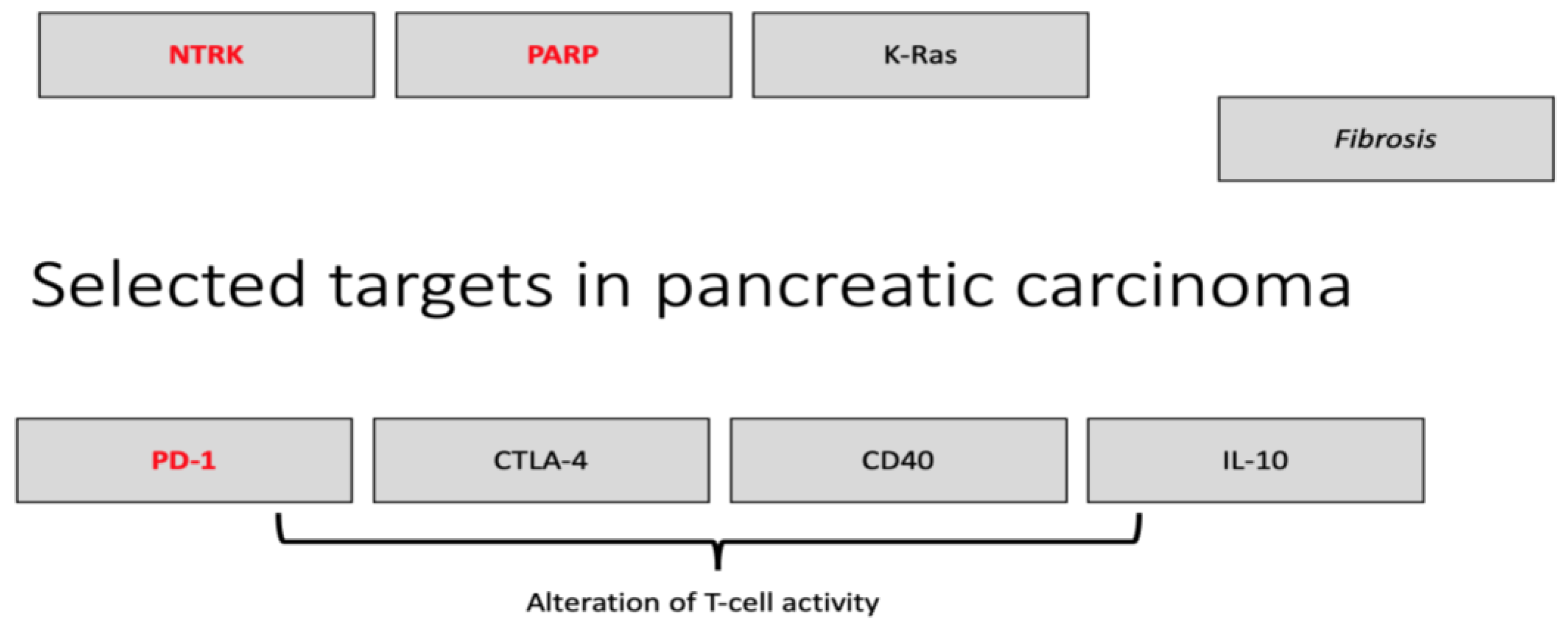
The following section will describe targeted therapy in depth. A summary of all the clinical trials discusses in Section 2 and Section 3 is provided in Table S1 in the Supplementary Materials. Figure 1 shows important (immuno-)therapeutic targets in PC.
2.1. Targeting EGFR
Discussion should start with the first approved targeted therapy for PC, the epidermal growth factor receptor (EGFR) tyrosine kinase inhibitor erlotinib. In the early 2000s, a phase III trial showed a statistically significant prolongation of overall survival (OS) by addition of erlotinib to gemcitabine (6.24 vs. 5.91 months) [13]. Arguably, this difference is too small to be clinically relevant and given the side effects of erlotinib that mainly affect the skin, erlotinib is used in routine clinical practice. A later phase III trial investigating the blockade of EGFR signaling by the monoclonal antibody cetuximab with gemcitabine did not result in any benefit, and another phase III trial exploring gemcitabine or capecitabine with erlotinib has reported negative results, too [14,15]. As a side note, addition of erlotinib to gemcitabine did not show any benefit in OS in the adjuvant setting [16].
2.2. Targeting NTRK
Recently, larotrectinib and entrectinib, inhibitors of the neurotrophic tyrosine receptor kinase (NTRK) fusion genes, have received approval in many countries for tumors harboring the mutation, independently of their origin [17,18,19]. Their gene products, tropomyosin receptor kinases, have been known protooncogenes for over 20 years due to their capability to induce signals of proliferation via activation of MAPK, PI3K, and PKC pathways [8]. A small fraction of patients with PC harbors this mutation (<5%) [8]. For larotrectinib, a pooled analysis of three phase 1 and 2 trials (NCT02122913, NCT02637687, NCT02576431) included 159 patients, two of them with PC, of which one had a positive response [20]. With an overall response rate of 79% for all tumor types, larotrectinib may become a valid treatment option for a subgroup of patients with PC. Similarly, an analysis of three phase 1 and 2 clinical trials for entrectinib (ALKA-372–001, STARTRK-1, STARTRK-2) analyzed 54 patients, three of which with PC [7]. The overall response rate was close to 60%.
2.3. Targeting PARP
Attention has also been brought to the poly(adenosine diphosphate–ribose) polymerase (PARP) inhibitors. Prevention of PARP to repair single-strand breaks eventually leads to accumulation of double strand breaks, to which cells with non-intact double strand repair, notably because of BCRA1/2 mutation, are sensitive [21]. In addition to being efficacious in BRCA1/2 positive ovarian carcinoma and breast cancer, olaparib has been successfully investigated for pretreated pancreatic carcinoma [22]. After a phase III trial has shown an increase in the median progression-free overall surival (PFS) from 3.8 to 7.4 months for patients receiving four cycles of olaparib as maintenance therapy compared to best supportive care after a platinum-based first line therapy, it has been approved in the U.S. for this indication [6]. Overall survival, however, might not be influenced by the administration of olaparib [5]. Several phase I and II clinical trials have investigated another PARP inhibitor, veliparib. In 2018, a phase II trial reported stable disease in 25% of patients for veliparib monotherapy [23]. Following this, veliparib was investigated in combination with gemcitabine and radiotherapy for locally advanced PC (phase 1) and a benefit in median OS of 5 months (19 versus 14 months) was reported for patients harboring mutations affecting DNA repair mechanisms as compared to individuals with wild-type status of DNA damage repair genes [24]. Veliparib with 5-FU and oxaliplatin for metastatic disease (phase I/II, single-arm trial) showed an overall response rate (ORR) of 57% for platinum-naïve patients with these mutations [25]. Another phase II trial, however, did not observe any improvement in response rates for the addition of veliparib to gemcitabine plus cisplatin [26]. Further phase III clinical trials are thus needed to clarify the role of PARP inhibitors in clinical management.
2.4. Targeting K-RAs
In approximately 95% of PC patients, an activating mutation in K-Ras is found and plays a central role in initiation, maintenance, and progression of disease [27]. For a long time, approaches to target Ras have failed and Ras was therefore considered to be undruggable [28]. Around a decade ago, adjuvant treatment with K-Ras vaccines have brought rather discouraging results in resectable PC [29,30]. One study observed an immunological response to vaccination, but the median OS in did not significantly increase for responders and the 5-year survival rate of 20% of the entire cohort increased to 24% for responders [29]. In another trial, no immunological response was induced by the vaccine [30]. In PC, also clinical trials to target downstream pathways of K-Ras have failed so far [27,31]. Recently, this paradigm has been changing. The development of the K-Ras inhibitor sotorasib demonstrated the possibility of directly targeting K-Ras (although effective only for the G12C mutation, not the much more common G12D in pancreatic carcinoma) [32]. The mDC3/8 dendritic cell vaccine designed to target mutant K-Ras is currently investigated in a phase I trial (NCT03592888).
2.5. Targeting the Stroma, Fibrosis, and Extracellular Matrix
The main factors leading to chemoresistance are to be found in the cancer microenvironment and the extensive fibrosis surrounding the tumor, as reviewed in depth by Schober and colleagues [33]. Naturally, attempts to target the cancer microenvironment have been made. More than a decade ago, an approach to increase drug delivery into the tumor included successful experiments with inhibition of hedgehog signaling in mice [34]. Subsequent clinical trials, however, failed to show any benefit [35,36,37] This includes a randomized phase Ib/II trial, where addition of vismogedib or placebo to gemcitabine did not alter OS [35]. For gemcitabine in combination with saridegib (another hedgehog inhibitor), a decreased OS compared to gemcitabine plus placebo at interim analysis led to a premature end of the study and subsequently also put a stop to a clinical trial of FOLFIRINOX and saridegib [35,36]. In a more recent phase II study, the addition of vismogedib to gemcitabine plus nab-paclitaxel as first-line therapy resulted in a median OS of 5.4 months, discouraging further research, as the median OS for the chemotherapeutic regimen alone has been reported to be equal or higher [37,38].
In a similar approach to facilitate chemotherapeutic penetration into tumor tissue, enzymatic lysis of extracellular hyaluronic acid by PEGPH20 plus gemcitabine was promising in vitro and in vivo [39]. Investigations carried on, until the HALO 109-301 phase III trial did not show any benefit in OS and PFS [40].
TGF-beta is a central cytokine in the tumorigenesis of PC, as well as its microenvironment, and three-dimensional in-vitro models for PC underline a direct relation between TGF-beta and the architecture [41,42,43]. TGF-beta has recently become of interest as a therapeutic target: Preclinical experiments demonstrated a reduction of the tumor cells’ aggressiveness, if secretion of TGF-beta in pancreatic stellate was abrogated [44]. Another interesting aspect lies in the fact that cancer-associated fibroblasts (CAFs), which are central for the desmoplastic stroma, rely on TGF-beta signaling and therefore, inhibition of TGF-beta has been explored as one strategy to target CAFs [45]. A phase Ib/II clinical trial in patients with unresectable PC reported an increase in OS from 7.1 to 8.9 months for the addition of the TGF-beta inhibitor galunisertib to gemcitabine [46].
Another attempt of stromal remodeling to increase chemotherapeutic delivery took advantage of the overexpression of the vitamin D receptor, which according to a mice model, serves as a transcriptional master regulator of the tumor stroma in this tumor entity, mainly by affecting PSCs [47]. As preclinical data were promising, many clinical trials have been started, more recently combining immune checkpoint therapy as well as oncolytic vaccine therapy with vitamin D analogues [48].
As PSCs also overexpress the vitamin A receptor, and its inhibition decreases the deposition of extracellular matrix, a phase II clinical trial is currently ongoing to explore the combination of gemcitabine/nab-paclitaxel and all-trans retinoic acid in patients with locally advanced and metastatic disease (NCT03307148) [49,50].
3. Combined Immunotherapies
In addition to fibrosis, the immunosuppressive properties of the microenvironment are considered crucial for disease progression and resistance to therapy [53,54,55]. It consists of a complex network of different immune cell populations, including myeloid-derived suppressor cells, macrophages, and regulatory T-cells, but not cytotoxic T-cells. As these are thought to exert their effect by expression of PD-1, CTLA-4, CD-40 as well as secretion of TGF-beta, many novel approaches have aimed at targeting these proteins, some of which we will discuss in the following paragraphs [54].
3.1. Targeting PD-L1/PD-1
Tumors characterized by high microsatellite instability (MSI-high) or mismatch-repair deficiency (dMMR) are susceptible to immune checkpoint therapy [56]. It is thought that the increased neoantigen expression by the tumor cells is recognized by the immune system [57,58]. In the phase II KEYNOTE-158 study investigating the PD-1 inhibitor pembrolizumab for dMMR/MSI-high tumors, 9.4% of the patients included suffered from PC, making it the third most included tumor entity. With an ORR of 18% and a PFS of 2.2 months, however, it was among the least responsive tumors, and of the six most included types of tumors, only brain tumors with an overall response rate of 0% performed worse [59]. Based on this trial, pembrolizumab has received FDA approval for MSI-high metastatic tumors for pretreated patients with no effective other line of treatment. For pancreatic carcinoma, several attempts at targeting PD-L1/PD-1 as part of a combination are ongoing.
The combination of pembrolizumab with the CXCR4 antagonist motixaforatide (and chemotherapy) has been investigated in the COMBAT trial (phase IIa) with promising results especially in the arm with additional chemotherapy: A disease control rate (DCR) of 32% was reported for motixaforatide plus pembrolizumab and a DCR of 77% and ORR of 32% with additional chemotherapy, encouraging further randomized trials [60]. Also, an increased cytotoxic T-cell infiltration into the tumor and a decrease in immunosuppressive cells were observed.
Another attempt to increase the efficacy of pembrolizumab by increasing T-cell infiltration and PD-L1 expression yielded into a phase Ib trial that combined pembrolizmab with chemotherapy and the oncolytic virus pelareorep. Due to promising results, a phase II trial (NCT03723915) is now ongoing [61]. The abovementioned PEGPH20 is currently being evaluated in combination with pembrolizumab as part of a phase II trial (NCT03634332). As recently reviewed by Arias-Pinilla and Modjtahedi, several other early phase trials investigating pembrolizumab or nivolumab in combination with olaparib, with paricalcitol plus gemcitabine and nab-paclitaxel, with a chemokine receptor agonist for CCR2/5, or with a cytokine receptor antagonist for CXCR1/2 are ongoing [62].
Two other ongoing trials involve the novel PD-1 inhibitor spartalizumab. For the combination of spartalizumab with the anti-IL-6 monoclonal antibody siltuximab, a phase Ib/II trial is ongoing (NCT04191421). Another phase I trial is investigating spartalizumab with nab-paclitaxel, gemcitabine, and the anti Il-1 beta monoclonal antibody canakinumab (NCT04581343) [62].
For the combination of the anti-PD-L1 monoclonal antibody durvalumab with the anti-CTLA-4 antibody tremelimumab, a phase II clinical trial reported a discouraging objective response rate of 3.1% only (and 0% for durvalumab monotherapy) [63].
3.2. Targeting CTLA-4
Ipilimumab, an inhibitory monoclonal antibody targeting CTLA-4, has been unsuccessfully investigated in a phase II clinical trial in advanced PC [64]. The combination of ipilimumab with the GM-CSF cell-based vaccines (GVAX), however, showed superior activity in a later trial, encouraging further studies [65]. Taking advantage of the T-cell priming by GVAX, an additional boost with the listeria monocytogenes-expressing mesothelin vaccine CRS-207 significantly increased overall survival from 4.6 to 9.7 months [66]. Therefore, the combination of GVAX and CRS-207 was evaluated together with ipilimumab, where, unfortunately, the addition CTLA-4 immune checkpoint inhibition did not prolong survival [67]. Other recent studies investigated the addition of ipilimumab to gemcitabine without increasing efficacy, or reported inferiority of ipilimumab with GVAX compared to continued administration of FOLFIRNOX [68,69]. Four ongoing trials investigate the combination of ipilimumab with nivolumab in PC (NCT04361162, NCT04258150, NCT03104439, NCT04247165).
3.3. Targeting CD40
Already in 2011, a study involving mice models and human tissue demonstrated that CD40 agonists could aid at tumor regression in patients with PC by activating macrophages to fight the tumor and by leading to regression of the stroma [70]. In combination with chemotherapeutic agents, CD40 agonists have also been shown to help overcome resistance to immune checkpoint inhibitors in mice models [71]. More recently, a proof-of concept study in a murine model showed that although CD40-agonist monotherapy did not improve OS, it increased susceptibility of PC to subsequent dendritic cell vaccinations, arguably by unleashing a T-cell response, and resulted in prolongation of the OS [72]. Despite a phase I trial investigating the combination of CP-870,893 (a CD40 monoclonal antibody) with gemcitabine showed antitumor activity, no further trials have been conducted so far [73].
3.4. Targeting IL-10
Another attempt to increase T-cell infiltration into the tumor took advantage of recombinant Il-10 (pegilodecakin) [74]. Whereas preclinical and early clinical data were promising, the phase III SEQUIOA trial failed to show a benefit in OS (5.8 vs. 6.3 months) and PFS (2.1 months both groups) for the addition of pegilodecakin to FOLFOX [75,76]. For an overview of selected studies discussed in the text, please refer to Table S1.
4. miRNA
Several efforts have been performed during the last years to enable physicians to diagnose cancer at an earlier stage of disease and to identify biomarkers to guide treatment. As pancreatic cancer remains asymptomatic over a long period of time, diagnoses at an early stage of disease is possible in 15–20% of patients only. Only these patients are potential candidates for curative surgical resection, resulting in a median survival of up to 24 months, if a R0 resection is achieved [77,78]. Micro RNAs (miRNAs) have been propeosed as biomarkers potentially allowing for earlier diagnosis and will be discussed in the following section. A summary of miRNAs can be found in Table S2 in the Supplementary Materials.
4.1. miRNA: Introduction
MicroRNAs are currently under investigation in various cancer entities for their diagnostic as well as prognostic and predictive roles [79]. MiRNAs are small, approximately 22 nucleotides long, non-coding single-stranded RNAs that regulate gene expression at a posttranscriptional level. The human genome may encode for more than 1000 microRNAs and approximately 60% of human genes are regulated by microRNAs. Besides other functions, they are known to be involved in tumor evolution including regulation of angiogenesis and development of treatment resistance [80,81,82]. Therefore, their respective roles as potential diagnostic and predictive biomarkers have been evaluated: In PC a distinct microRNA expression profile compared to benign lesions has been observe [10,11,83,84]. Distinct miRNA expression profiles correlate to stages of malignant pancreatic disease and hold potentials as biomarkers [85] There is an existing medical need of biomarkers for early diagnosis. CA19-9 is the only prognostic serum-based tumor marker approved in PC; however, it comes with several limitations such as a moderate sensitivity and specificity (estimated around 79% and 82%, respectively). Tumor markers do not always accurately reflect the disease burden; e.g., PC patients with Lewis blood antigen A do not express CA19-9, and false positive results are often seen with the co-existence of inflammation or cholestasis e.g., in case of biliary obstruction [86].
Normally, oncogenes and tumor suppressor genes are regulated at an optimal activation/inhibition equilibrium. If downregulation of a specific miRNA increases the activity of a corresponding oncogene, this is identified as a tumor suppressor miRNA. On the other hand, if upregulation, it will result in a continuous inhibition of the target tumor suppressor gene. An increase in the activity of an oncogene target of an miRNA after knockdown of that miRNA suggests that the miRNA in question acts as a tumor suppressor and conversely overexpression of that same miRNA would lead to increased inhibition of that locus An imbalance can result in the loss of controlling specific tumor formation pathways and can contribute to the development of malignencies [87].
4.2. miRNA in Precursor Lesions and Diagnoses
The development of PC is a multistage process of genetic mutations resulting in histological and morphological abnormalities within the ductal cells and the acinar cells of the pancreas [88,89]. These lesions have potential to transform into pre-neoplastic lesions known as pancreatic intraepithelial neoplasia (PanIN).
MiRNA expression profiles are promising as non-invasive diagnostic markers, as they can be obtained easily from peripheral blood, saliva, urine or feces in order to detect above mentioned pre-neoplastic lesions [90]:
Measuring the expression of only 20 to 32 miRNAs could help clinicians in discriminating between healthy, inflamed and cancerous pancreatic tissue [12]. This hypothesis is supported by the fact that upregulation (e.g., miR-21, 221 424-5p, 27a, 4295) as well as downregulation (e.g., miR-124, 203, 150, 218) of miRNAs have been reported to play significant roles during initiation on the one hand and progression on the other hand of pancreatic cancer. Yu et al. investigated the miRNA expression profile in PanIN lesions and reported the aberrant expression of 35 miRNAs. Among those, miR-196b surfaced as a potential biomarker in identifying PanIN-3 lesions [91].
Another microarray analysis study compared blood samples of PC patients and healthy individuals and reported that a distinct miRNA expression profile (miR-22, miR-642b, 10, 752 12 of 22 and miR-885-5p) identified early PC [92].
Furthermore, early stage K-RAS mutations observed in PanIN lesions can directly affect the levels of specific miRNAs as investigated by Humeau et al. [93]. An upregulation of miR-205, miR-200, and miR-21 was detected in early adenocarcinoma lesions in a K-RAS(G12D) mouse model, where miRNA production could be measured in pathological and nonpathological ducts [94].
Increased levels of miR-155 and miR-210 in the serum of pancreatic cancer patient have been reported in various studies [95,96] suggesting a potential role as biomarkers for the diagnosis of early pancreatic neoplasia [95].
Li et al. evaluated a total of 735 miRNAs in the serum of pancreatic cancer patient. This analysis led to the identification of miR-1290 as a promising biomarker [97]. Furthermore, it is reported that miR-1290 displays higher sensitivity (81%) and specificity (80%) when compared to healthy control groups [97,98].
Wang et al. demonstrated that aberrant expression of miR-21, miR-155, miR-196a and miR-210 in plasma can easily distinguish PC patients from healthy controls [99]. Higher than normal levels of miR-210, miR-192 and miR-18a in the serum of PC patients may also be exploited as diagnostic markers [96,100,101].
Another approach in the field of biomarker research is combining various biomarkers or combining them with tumor markers in order to enhance the sensitivity and specificity. Recently, it was reported that the combination of CA 19.9 with miR-16 and miR-196a allows to distinguish between PC patients and healthy controls [102,103]. Similarly, when the expression profile of miR-27a-3p was coupled with CA 19.9, PC patient and healthy controls could be differentiated with a sensitivity of 85.3% and specificity of 81.61% [103,104].
These findings are of utmost clinical relevance, because histological clarification of pancreatic lesions is often challenging, particularly if only small tissue can be obtained by endoscopic ultrasound-assisted fine-needle biopsies. However, no currently existing miRNA panel is endorsed by any guidelines for clinical use to assess response to therapy.
4.3. miRNA and Therapy Response
Beyond the diagnostic value of miRNA expression profiles, they might also play a role in the prediction of chemoresistance as well as responsiveness to systemic therapy. MiRNA could help clinicians to choose a combination of various therapies in order to overcome therapeutic resistance.
The predictive value of miRNAs for the response to a therapy with gemcitabine was extensively shown in vitro and in vivo. An overexpression of miR-21 and miR-10b and a downregulation of miR-34a has previously been linked to worse survival under gemcitabine chemotherapy [105,106,107,108,109]. In addition, miRNA-320c has been reported to be of predictive significance for a response to gemcitabine [110].
Furthermore, an irregular expression of miRNA was found in a gemcitabine-resistant cell line, including downregulation of miRNA-200b, miRNA-200c, let-7b, let-7c, let-7d and let-7e in gemcitabine-resistant cells [111]. Similarly, miRNA-33a is also downregulated in gemcitabine-resistant cells, and upon the restoration of normal levels, gemcitabine sensitivity could be restored [112].
In a recent meta-analysis Royam et al. studied a total of 48 miRNAs and reported a downregulation of 23 and upregulation of 25 miRNAs [113]. In particular, nine upregulated miRNAs (15b, 17-5p, 21, 155, 181c, 203, 221,320c and 1246) exhibited chemotherapeutic resistance and six upregulated miRNAs (21, 33a, 138-5p,509-5p, 1207 and 1243) exhibited chemotherapeutic sensitivity. In contrast, nine downregulated miRNAs (7, 100, 124, 210, 200c, 205, 220b, 374b-5p and 497) exhibited chemotherapeutic resistance and nine downregulated miRNAs (101, 101-3p, 153, 203, 205-5p, 494, 506, 3656, let-7a) exhibited chemotherapeutic sensitivity.
This above-mentioned meta-analysis included studies using gemcitabine, 5-FU, capecitabine, and erlotinib. The pooled hazard-ratio (HR) value for OS was 1.603; (95% confidence interval (CI) 1.2–2.143; p-value: 0.01), with the subgroup analysis for miR-21 showing a HR for resistance of 2.061; 95% CI 1.195–3.556; p-value: 0.09.
Altered expressions of several miRNAs including miR-21-5p, miR-10b-5p, and miR-34a-5p have previously been linked to a worse response to gemcitabine [105,106,107,108,109]. Our group, however, could show that high and low expressions of these three miRNAs have no influence on the outcome of treatment with FOLFIRINOX regarding PFS and OS, in contrast to treatment with gemcitabine [114].
Another study reported by Meijer et al. demonstrated that a decline in plasma miR-181a-5p levels after five to six cycles of FOLFIRINOX was associated with better prognosis [115]. This association was not observed in a second cohort of patients treated with gemcitabine plus nab-paclitaxel. In-vitro analyses detected an increased sensitivity of PC cells lines to oxaliplatin when miR-181a-5p was inhibited. However, to our knowledge there are no miRNA data currently available on response prediction of novel chemotherapy regimens such as nab-paclitaxel or nano-liposomal irinotecan.
A summary of all above-mentioned micro-RNA results can be found in Table S2 in the Supplementary Materials.
Supplementary Materials
The following are available online at https://0-www-mdpi-com.brum.beds.ac.uk/article/10.3390/biom11101469/s1, Table S1: Overview of selected studies, Table S2: Aberrant expression of micro- RNA.
Author Contributions
Conceptualization, K.S. and R.G.; writing—original draft preparation, K.S. and D.K.; writing—review and editing, K.S., D.K. and R.G.; All authors have read and agreed to the published version of the manuscript.
Funding
This research received no external funding.
Institutional Review Board Statement
Not applicable.
Informed Consent Statement
Not applicable.
Data Availability Statement
Not applicable.
Conflicts of Interest
The authors declare no conflict of interest.
References
- McGuigan, A.; Kelly, P.; Turkington, R.C.; Jones, C.; Coleman, H.G.; McCain, R.S. Pancreatic cancer, A review of clinical diagnosis, epidemiology, treatment and outcomes. World J. Gastroenterol. 2018, 24, 4846–4861. [Google Scholar] [CrossRef]
- Nevala-Plagemann, C.; Hidalgo, M.; Garrido-Laguna, I. From state-of-the-art treatments to novel therapies for advanced-stage pancreatic cancer. Nat. Rev. Clin. Oncol. 2020, 17, 108–123. [Google Scholar] [CrossRef]
- Zeng, S.; Pöttler, M.; Lan, B.; Grützmann, R.; Pilarsky, C.; Yang, H. Chemoresistance in Pancreatic Cancer. Int. J. Mol. Sci. 2019, 20, 4504. [Google Scholar] [CrossRef] [PubMed] [Green Version]
- Siegel, R.L.; Miller, K.D.; Jemal, A. Cancer statistics, 2020. CA Cancer J. Clin. 2020, 70, 7–30. [Google Scholar] [CrossRef]
- Vaishampayan, U.N. An evaluation of olaparib for the treatment of pancreatic cancer. Expert Opin. Pharmacother. 2021, 22, 521–526. [Google Scholar] [CrossRef] [PubMed]
- Golan, T.; Hammel, P.; Reni, M.; Van Cutsem, E.; Macarulla, T.; Hall, M.J.; Park, J.-O.; Hochhauser, D.; Arnold, D.; Oh, D.-Y.; et al. Maintenance Olaparib for Germline. N. Engl. J. Med. 2019, 381, 317–327. [Google Scholar] [CrossRef] [PubMed]
- Doebele, R.C.; Drilon, A.; Paz-Ares, L.; Siena, S.; Shaw, A.T.; Farago, A.F.; Blakely, C.M.; Seto, T.; Cho, B.C.; Tosi, D.; et al. Entrectinib in patients with advanced or metastatic NTRK fusion-positive solid tumours. Integrated analysis of three phase 1-2 trials. Lancet Oncol. 2020, 21, 271–282. [Google Scholar] [CrossRef]
- Cocco, E.; Scaltriti, M.; Drilon, A. NTRK fusion-positive cancers and TRK inhibitor therapy. Nat. Rev. Clin. Oncol. 2018, 15, 731–747. [Google Scholar] [CrossRef]
- Calin, G.A.; Croce, C.M. MicroRNA-cancer connection. The beginning of a new tale. Cancer Res. 2006, 66, 7390–7394. [Google Scholar] [CrossRef] [Green Version]
- Bloomston, M.; Frankel, W.L.; Petrocca, F.; Volinia, S.; Alder, H.; Hagan, J.P.; Liu, C.G.; Bhatt, D.; Taccioli, C.; Croce, C.M. MicroRNA expression patterns to differentiate pancreatic adenocarcinoma from normal pancreas and chronic pancreatitis. JAMA 2007, 297, 1901–1908. [Google Scholar] [CrossRef] [Green Version]
- Szafranska, A.E.; Davison, T.S.; John, J.; Cannon, T.; Sipos, B.; Maghnouj, A.; Labourier, E.; Hahn, S.A. MicroRNA expression alterations are linked to tumorigenesis and non-neoplastic processes in pancreatic ductal adenocarcinoma. Oncogene 2007, 26, 4442–4452. [Google Scholar] [CrossRef] [PubMed] [Green Version]
- Schultz, N.A.; Werner, J.; Willenbrock, H.; Roslind, A.; Giese, N.; Horn, T.; Wøjdemann, M.; Johansen, J.S. MicroRNA expression profiles associated with pancreatic adenocarcinoma and ampullary adenocarcinoma. Research Support, Non-U.S. Gov’t. Mod. Pathol. Off. J. United States Can. Acad. Pathol. Inc. 2012, 25, 1609–1622. [Google Scholar] [CrossRef] [PubMed] [Green Version]
- Moore, M.J.; Goldstein, D.; Hamm, J.; Figer, A.; Hecht, J.R.; Gallinger, S.; Au, H.J.; Murawa, P.; Walde, D.; Wolff, R.A.; et al. Erlotinib plus gemcitabine compared with gemcitabine alone in patients with advanced pancreatic cancer. A phase III trial of the National Cancer Institute of Canada Clinical Trials Group. J. Clin. Oncol. 2007, 25, 1960–1966. [Google Scholar] [CrossRef]
- Heinemann, V.; Vehling-Kaiser, U.; Waldschmidt, D.; Kettner, E.; Märten, A.; Winkelmann, C.; Klein, S.; Kojouharoff, G.; Gauler, T.C.; Von Weikersthal, L.F.; et al. Gemcitabine plus erlotinib followed by capecitabine versus capecitabine plus erlotinib followed by gemcitabine in advanced pancreatic cancer, Final results of a randomised phase 3 trial of the ’Arbeitsgemeinschaft Internistische Onkologie’ (AIO-PK0104). Gut 2013, 62, 751–759. [Google Scholar] [CrossRef]
- Philip, P.A.; Benedetti, J.; Corless, C.L.; Wong, R.; O’Reilly, E.M.; Flynn, P.J.; Rowland, K.M.; Atkins, J.N.; Mirtsching, B.C.; Rivkin, S.E.; et al. Phase III study comparing gemcitabine plus cetuximab versus gemcitabine in patients with advanced pancreatic adenocarcinoma. Southwest Oncology Group-directed intergroup trial S0205. J. Clin. Oncol. 2010, 28, 3605–3610. [Google Scholar] [CrossRef] [Green Version]
- Sinn, M.; Bahra, M.; Liersch, T.; Gellert, K.; Messmann, H.; Bechstein, W.; Waldschmidt, D.; Jacobasch, L.; Wilhelm, M.; Rau, B.M.; et al. CONKO-005: Adjuvant Chemotherapy With Gemcitabine Plus Erlotinib Versus Gemcitabine Alone in Patients After R0 Resection of Pancreatic Cancer. A Multicenter Randomized Phase III Trial. J. Clin. Oncol. 2017, 35, 3330–3337. [Google Scholar] [CrossRef]
- Drilon, A.; Laetsch, T.W.; Kummar, S.; Dubois, S.G.; Lassen, U.N.; Demetri, G.D.; Nathenson, M.; Doebele, R.C.; Farago, A.F.; Pappo, A.S.; et al. Efficacy of Larotrectinib in TRK Fusion-Positive Cancers in Adults and Children. N. Engl. J. Med. 2018, 378, 731–739. [Google Scholar] [CrossRef]
- Delgado, J.; Pean, E.; Melchiorri, D.; Migali, C.; Josephson, F.; Enzmann, H.; Pignatti, F. The European Medicines Agency review of entrectinib for the treatment of adult or paediatric patients with solid tumours who have a neurotrophic tyrosine receptor kinase gene fusions and adult patients with non-small-cell lung cancer harbouring ROS1 rearrangements. ESMO Open. 2021, 6, 100087. [Google Scholar] [CrossRef]
- Scott, L.J. Larotrectinib, First Global Approval. Drugs 2019, 79, 201–206. [Google Scholar] [CrossRef]
- Hong, D.S.; DuBois, S.G.; Kummar, S.; Farago, A.F.; Albert, C.M.; Rohrberg, K.S.; van Tilburg, C.M.; Nagasubramanian, R.; Berlin, J.D.; Federman, N.; et al. Larotrectinib in patients with TRK fusion-positive solid tumours. A pooled analysis of three phase 1/2 clinical trials. Lancet Oncol. 2020, 21, 531–540. [Google Scholar] [CrossRef]
- Bochum, S.; Berger, S.; Martens, U.M. Olaparib. Recent Results Cancer Res. 2018, 211, 217–233. [Google Scholar] [CrossRef] [PubMed]
- Kaufman, B.; Shapira-Frommer, R.; Schmutzler, R.K.; Audeh, M.W.; Friedlander, M.; Balmaña, J.; Mitchell, G.; Fried, G.; Stemmer, S.M.; Hubert, A.; et al. Olaparib monotherapy in patients with advanced cancer and a germline BRCA1/2 mutation. J. Clin. Oncol. 2015, 33, 244–250. [Google Scholar] [CrossRef] [PubMed]
- Lowery, M.A.; Kelsen, D.P.; Capanu, M.; Smith, S.C.; Lee, J.W.; Stadler, Z.K.; Moore, M.J.; Kindler, H.L.; Golan, T.; Segal, A.; et al. Phase II trial of veliparib in patients with previously treated BRCA-mutated pancreas ductal adenocarcinoma. Eur. J. Cancer. 2018, 89, 19–26. [Google Scholar] [CrossRef] [PubMed]
- Tuli, R.; Shiao, S.L.; Nissen, N.; Tighiouart, M.; Kim, S.; Osipov, A.; Bryant, M.; Ristow, L.; Placencio-Hickok, V.; Hoffman, D.; et al. A phase 1 study of veliparib, a PARP-1/2 inhibitor, with gemcitabine and radiotherapy in locally advanced pancreatic cancer. eBioMedicine 2019, 40, 375–381. [Google Scholar] [CrossRef] [Green Version]
- Pishvaian, M.J.; Hwang, J.J.; He, A.R.; Smaglo, B.G.; Kim, S.S.; Weinberg, B.A.; Weiner, L.M.; Marshall, J.L.; Brody, J.R. A Phase I/II Study of Veliparib (ABT-888) in Combination with 5-Fluorouracil and Oxaliplatin in Patients with Metastatic Pancreatic Cancer. Clin. Cancer Res. 2020, 26, 5092–5101. [Google Scholar] [CrossRef]
- O’Reilly, E.M.; Lee, J.W.; Zalupski, M.; Capanu, M.; Park, J.; Golan, T.; Tahover, E.; Lowery, M.; Chou, J.F.; Sahai, V.; et al. Randomized, Multicenter, Phase II Trial of Gemcitabine and Cisplatin With or Without Veliparib in Patients With Pancreas Adenocarcinoma and a Germline. J. Clin. Oncol. 2020, 38, 1378–1388. [Google Scholar] [CrossRef]
- Eser, S.; Schnieke, A.; Schneider, G.; Saur, D. Oncogenic KRAS signalling in pancreatic cancer. Br. J. Cancer 2014, 111, 817–822. [Google Scholar] [CrossRef]
- Gorfe, A.A.; Cho, K.J. Approaches to inhibiting oncogenic K-Ras. Small GTPases 2021, 12, 96–105. [Google Scholar] [CrossRef]
- Wedén, S.; Klemp, M.; Gladhaug, I.P.; Møller, M.; Eriksen, J.A.; Gaudernack, G.; Buanes, T. Long-term follow-up of patients with resected pancreatic cancer following vaccination against mutant K-ras. Int. J. Cancer 2011, 128, 1120–1128. [Google Scholar] [CrossRef]
- Abou-Alfa, G.K.; Chapman, P.B.; Feilchenfeldt, J.; Brennan, M.F.; Capanu, M.; Gansukh, B.; Jacobs, G.; Levin, A.; Neville, D.; Kelsen, D.P.; et al. Targeting mutated K-ras in pancreatic adenocarcinoma using an adjuvant vaccine. Am J. Clin. Oncol. 2011, 34, 321–325. [Google Scholar] [CrossRef]
- Choi, M.; Bien, H.; Mofunanya, A.; Powers, S. Challenges in Ras therapeutics in pancreatic cancer. Semin. Cancer Biol. 2019, 54, 101–108. [Google Scholar] [CrossRef] [PubMed]
- Hong, D.S.; Fakih, M.G.; Strickler, J.H.; Desai, J.; Durm, G.A.; Shapiro, G.I.; Falchook, G.S.; Price, T.J.; Sacher, A.; Denlinger, C.S.; et al. KRASG12C Inhibition with Sotorasib in Advanced Solid Tumors. N. Engl. J. Med. 2020, 383, 1207–1217. [Google Scholar] [CrossRef] [PubMed]
- Schober, M.; Jesenofsky, R.; Faissner, R.; Weidenauer, C.; Hagmann, W.; Michl, P.; Heuchel, R.L.; Haas, S.L.; Löhr, J.M. Desmoplasia and chemoresistance in pancreatic cancer. Cancers 2014, 6, 2137–2154. [Google Scholar] [CrossRef] [PubMed] [Green Version]
- Olive, K.P.; Jacobetz, M.A.; Davidson, C.J.; Gopinathan, A.; McIntyre, D.; Honess, D.; Madhu, B.; Goldgraben, M.A.; Caldwell, M.E.; Allard, D.; et al. Inhibition of Hedgehog signaling enhances delivery of chemotherapy in a mouse model of pancreatic cancer. Science 2009, 324, 1457–1461. [Google Scholar] [CrossRef] [PubMed] [Green Version]
- Catenacci, D.V.T.; Junttila, M.R.; Karrison, T.; Bahary, N.; Horiba, M.N.; Nattam, S.R.; Marsh, R.; Wallace, J.; Kozloff, M.; Rajdev, L.; et al. Randomized Phase Ib/II Study of Gemcitabine Plus Placebo or Vismodegib, a Hedgehog Pathway Inhibitor, in Patients with Metastatic Pancreatic Cancer. J. Clin. Oncol. 2015, 33, 4284–4292. [Google Scholar] [CrossRef] [PubMed]
- Ko, A.H.; LoConte, N.; Tempero, M.A.; Walker, E.J.; Kelley, R.K.; Lewis, S.; Chang, W.-C.; Kantoff, E.; Vannier, M.W.; Catenacci, D.V.; et al. A Phase I Study of FOLFIRINOX Plus IPI-926, a Hedgehog Pathway Inhibitor, for Advanced Pancreatic Adenocarcinoma. Pancreas 2016, 45, 370–375. [Google Scholar] [CrossRef] [Green Version]
- De Jesus-Acosta, A.; Sugar, E.A.; O’Dwyer, P.J.; Ramanathan, R.K.; Von Hoff, D.D.; Rasheed, Z.; Zheng, L.; Begum, A.; Anders, R.; Maitra, A.; et al. Phase 2 study of vismodegib, a hedgehog inhibitor, combined with gemcitabine and nab-paclitaxel in patients with untreated metastatic pancreatic adenocarcinoma. Br. J. Cancer 2020, 122, 498–505. [Google Scholar] [CrossRef]
- Von Hoff, D.D.; Ervin, T.; Arena, F.P.; Chiorean, E.G.; Infante, J.; Moore, M.; Seay, T.; Tjulandin, S.A.; Ma, W.W.; Saleh, M.N.; et al. Increased survival in pancreatic cancer with nab-paclitaxel plus gemcitabine. N. Engl. J. Med. 2013, 369, 1691–1703. [Google Scholar] [CrossRef] [PubMed] [Green Version]
- Provenzano, P.P.; Cuevas, C.; Chang, A.E.; Goel, V.K.; Von Hoff, D.D.; Hingorani, S.R. Enzymatic targeting of the stroma ablates physical barriers to treatment of pancreatic ductal adenocarcinoma. Cancer Cell. 2012, 21, 418–429. [Google Scholar] [CrossRef] [PubMed] [Green Version]
- Van Cutsem, E.; Tempero, M.A.; Sigal, D.; Oh, D.-Y.; Fazio, N.; Macarulla, T.; Hitre, E.; Hammel, P.; Hendifar, A.E.; Bates, S.E.; et al. Randomized Phase III Trial of Pegvorhyaluronidase Alfa With Nab-Paclitaxel Plus Gemcitabine for Patients With Hyaluronan-High Metastatic Pancreatic Adenocarcinoma. J. Clin. Oncol. 2020, 38, 3185–3194. [Google Scholar] [CrossRef]
- Delle Cave, D.; Rizzo, R.; Sainz, B.; Gigli, G.; Del Mercato, L.L.; Lonardo, E. The Revolutionary Roads to Study Cell-Cell Interactions in 3D In Vitro Pancreatic Cancer Models. Cancers 2021, 23, 930. [Google Scholar] [CrossRef]
- Cavo, M.; Delle Cave, D.; D’Amone, E.; Gigli, G.; Lonardo, E.; Del Mercato, L.L. A synergic approach to enhance long-term culture and manipulation of MiaPaCa-2 pancreatic cancer spheroids. Sci. Rep. 2020, 10, 10192. [Google Scholar] [CrossRef]
- Hao, Y.; Baker, D.; Ten Dijke, P. TGF-β-Mediated Epithelial-Mesenchymal Transition and Cancer Metastasis. Int. J. Mol. Sci. 2019, 20, 2767. [Google Scholar] [CrossRef] [Green Version]
- Delle Cave, D.; Di Guida, M.; Costa, V.; Sevillano, M.; Ferrante, L.; Heeschen, C.; Corona, M.; Cucciardi, A.; Lonardo, E. TGF-β1 secreted by pancreatic stellate cells promotes stemness and tumourigenicity in pancreatic cancer cells through L1CAM downregulation. Oncogene 2020, 39, 4271–4285. [Google Scholar] [CrossRef] [Green Version]
- Norton, J.; Foster, D.; Chinta, M.; Titan, A.; Longaker, M. Pancreatic Cancer Associated Fibroblasts (CAF), Under-Explored Target for Pancreatic Cancer Treatment. Cancers 2020, 12, 1347. [Google Scholar] [CrossRef] [PubMed]
- Melisi, D.; Garcia-Carbonero, R.; Macarulla, T.; Pezet, D.; Deplanque, G.; Fuchs, M.; Trojan, J.; Oettle, H.; Kozloff, M.; Cleverly, A.; et al. Galunisertib plus gemcitabine vs. gemcitabine for first-line treatment of patients with unresectable pancreatic cancer. Br. J. Cancer 2018, 119, 1208–1214. [Google Scholar] [CrossRef] [PubMed] [Green Version]
- Sherman, M.H.; Yu, R.T.; Engle, D.D.; Ding, N.; Atkins, A.R.; Tiriac, H.; Collisson, E.A.; Connor, F.; Van Dyke, T.; Kozlov, S.; et al. Vitamin D receptor-mediated stromal reprogramming suppresses pancreatitis and enhances pancreatic cancer therapy. Cell 2014, 159, 80–93. [Google Scholar] [CrossRef] [PubMed] [Green Version]
- LaRocca, C.J.; Warner, S.G. A New Role for Vitamin D, The Enhancement of Oncolytic Viral Therapy in Pancreatic Cancer. Biomedicines 2018, 6, 104. [Google Scholar] [CrossRef] [PubMed] [Green Version]
- Kocher, H.M.; Basu, B.; Froeling, F.E.M.; Sarker, D.; Slater, S.; Carlin, D.; Desouza, N.M.; De Paepe, K.N.; Goulart, M.R.; Hughes, C.; et al. Phase I clinical trial repurposing all-trans retinoic acid as a stromal targeting agent for pancreatic cancer. Nat. Commun. 2020, 11, 4841. [Google Scholar] [CrossRef] [PubMed]
- Weniger, M.; Honselmann, K.C.; Liss, A.S. The Extracellular Matrix and Pancreatic Cancer. A Complex Relationship. Cancers 2018, 10, 316. [Google Scholar] [CrossRef] [Green Version]
- Kindler, H.L.; Niedzwiecki, D.; Hollis, D.; Sutherland, S.; Schrag, D.; Hurwitz, H.; Innocenti, F.; Mulcahy, M.F.; O’Reilly, E.; Wozniak, T.F.; et al. Gemcitabine plus bevacizumab compared with gemcitabine plus placebo in patients with advanced pancreatic cancer. Phase III trial of the Cancer and Leukemia Group B (CALGB 80303). J. Clin. Oncol. 2010, 28, 3617–3622. [Google Scholar] [CrossRef] [PubMed] [Green Version]
- Van Cutsem, E.; Vervenne, W.L.; Bennouna, J.; Humblet, Y.; Gill, S.; Van Laethem, J.-L.; Verslype, C.; Scheithauer, W.; Shang, A.; Cosaert, J.; et al. Phase III trial of bevacizumab in combination with gemcitabine and erlotinib in patients with metastatic pancreatic cancer. J. Clin. Oncol. 2009, 27, 2231–2237. [Google Scholar] [CrossRef]
- Wörmann, S.M.; Diakopoulos, K.N.; Lesina, M.; Algül, H. The immune network in pancreatic cancer development and progression. Oncogene 2014, 33, 2956–2967. [Google Scholar] [CrossRef] [PubMed] [Green Version]
- Brunner, M.; Wu, Z.; Krautz, C.; Pilarsky, C.; Grützmann, R.; Weber, G.F. Current Clinical Strategies of Pancreatic Cancer Treatment and Open Molecular Questions. Int. J. Mol. Sci. 2019, 20, 4543. [Google Scholar] [CrossRef] [PubMed] [Green Version]
- Le, D.T.; Uram, J.N.; Wang, H.; Bartlett, B.R.; Kemberling, H.; Eyring, A.D.; Skora, A.D.; Luber, B.S.; Azad, N.S.; Laheru, D.; et al. PD-1 Blockade in Tumors with Mismatch-Repair Deficiency. N. Engl. J. Med. 2015, 372, 2509–2520. [Google Scholar] [CrossRef] [PubMed] [Green Version]
- Zhao, P.; Li, L.; Jiang, X.; Li, Q. Mismatch repair deficiency/microsatellite instability-high as a predictor for anti-PD-1/PD-L1 immunotherapy efficacy. J. Hematol. Oncol. 2019, 12, 54. [Google Scholar] [CrossRef]
- Lee, V.; Murphy, A.; Le, D.T.; Diaz, L.A. Mismatch Repair Deficiency and Response to Immune Checkpoint Blockade. Oncologist 2016, 21, 1200–1211. [Google Scholar] [CrossRef] [Green Version]
- Dudley, J.C.; Lin, M.T.; Le, D.T.; Eshleman, J.R. Microsatellite Instability as a Biomarker for PD-1 Blockade. Clin. Cancer Res. 2016, 22, 813–820. [Google Scholar] [CrossRef] [Green Version]
- Marabelle, A.; Le, D.T.; Ascierto, P.A.; Di Giacomo, A.M.; De Jesus-Acosta, A.; Delord, J.-P.; Geva, R.; Gottfried, M.; Penel, N.; Hansen, A.R.; et al. Efficacy of Pembrolizumab in Patients With Noncolorectal High Microsatellite Instability/Mismatch Repair-Deficient Cancer. Results From the Phase II KEYNOTE-158 Study. J. Clin. Oncol. 2020, 38, 1–10. [Google Scholar] [CrossRef]
- Bockorny, B.; Semenisty, V.; Macarulla, T.; Borazanci, E.; Wolpin, B.M.; Stemmer, S.M.; Golan, T.; Geva, R.; Borad, M.J.; Pedersen, K.S.; et al. BL-8040, a CXCR4 antagonist, in combination with pembrolizumab and chemotherapy for pancreatic cancer. The COMBAT trial. Nat. Med. 2020, 26, 878–885. [Google Scholar] [CrossRef]
- Mahalingam, D.; Wilkinson, G.A.; Eng, K.; Fields, P.; Raber, P.; Moseley, J.L.; Cheetham, K.; Coffey, M.; Nuovo, G.; Kalinski, P.; et al. Pembrolizumab in Combination with the Oncolytic Virus Pelareorep and Chemotherapy in Patients with Advanced Pancreatic Adenocarcinoma. A Phase Ib Study. Clin. Cancer Res. 2020, 26, 71–81. [Google Scholar] [CrossRef] [Green Version]
- Arias-Pinilla, G.A.; Modjtahedi, H. Therapeutic Application of Monoclonal Antibodies in Pancreatic Cancer. Advances, Challenges and Future Opportunities. Cancers 2021, 13, 1781. [Google Scholar] [CrossRef]
- O’Reilly, E.M.; Oh, D.-Y.; Dhani, N.; Renouf, D.J.; Lee, M.A.; Sun, W.; Fisher, G.; Hezel, A.; Chang, S.-C.; Vlahovic, G.; et al. Durvalumab With or Without Tremelimumab for Patients With Metastatic Pancreatic Ductal Adenocarcinoma. A Phase 2 Randomized Clinical Trial. JAMA Oncol. 2019, 5, 1431–1438. [Google Scholar] [CrossRef]
- Royal, R.E.; Levy, C.; Turner, K.; Mathur, A.; Hughes, M.; Kammula, U.S.; Sherry, R.M.; Topalian, S.L.; Yang, J.C.; Lowy, I.; et al. Phase 2 trial of single agent Ipilimumab (anti-CTLA-4) for locally advanced or metastatic pancreatic adenocarcinoma. J. Immunother. 2010, 33, 828–833. [Google Scholar] [CrossRef]
- Le, D.T.; Lutz, E.; Uram, J.N.; Sugar, E.A.; Onners, B.; Solt, S.; Zheng, L.; Diaz, L.A.; Donehower, R.C.; Jaffee, E.M.; et al. Evaluation of ipilimumab in combination with allogeneic pancreatic tumor cells transfected with a GM-CSF gene in previously treated pancreatic cancer. J. Immunother. 2013, 36, 382–389. [Google Scholar] [CrossRef] [Green Version]
- Le, D.T.; Wang-Gillam, A.; Picozzi, V.; Greten, T.F.; Crocenzi, T.; Springett, G.; Morse, M.; Zeh, H.; Cohen, D.; Fine, R.L.; et al. Safety and survival with GVAX pancreas prime and Listeria Monocytogenes-expressing mesothelin (CRS-207) boost vaccines for metastatic pancreatic cancer. J. Clin. Oncol. 2015, 33, 1325–1333. [Google Scholar] [CrossRef] [Green Version]
- Tsujikawa, T.; Crocenzi, T.; Durham, J.N.; Sugar, E.A.; Wu, A.A.; Onners, B.; Nauroth, J.M.; Anders, R.A.; Fertig, E.J.; Laheru, D.A.; et al. Evaluation of Cyclophosphamide/GVAX Pancreas Followed by Listeria-Mesothelin (CRS-207) with or without Nivolumab in Patients with Pancreatic Cancer. Clin. Cancer Res. 2020, 26, 3578–3588. [Google Scholar] [CrossRef] [Green Version]
- Kamath, S.D.; Kalyan, A.; Kircher, S.; Nimeiri, H.; Fought, A.J.; Benson, A.; Mulcahy, M. Ipilimumab and Gemcitabine for Advanced Pancreatic Cancer. A Phase Ib Study. Oncologist 2020, 25, e808–e815. [Google Scholar] [CrossRef] [PubMed] [Green Version]
- Wu, A.A.; Bever, K.M.; Ho, W.J.; Fertig, E.J.; Niu, N.; Zheng, L.; Parkinson, R.M.; Durham, J.N.; Onners, B.L.; Ferguson, A.K.; et al. A Phase II Study of Allogeneic GM-CSF-Transfected Pancreatic Tumor Vaccine (GVAX) with Ipilimumab as Maintenance Treatment for Metastatic Pancreatic Cancer. Clin. Cancer Res. 2020, 26, 5129–5139. [Google Scholar] [CrossRef] [PubMed]
- Beatty, G.; Chiorean, E.G.; Fishman, M.P.; Saboury, B.; Teitelbaum, U.R.; Sun, W.; Huhn, R.D.; Song, W.; Li, D.; Sharp, L.L.; et al. CD40 agonists alter tumor stroma and show efficacy against pancreatic carcinoma in mice and humans. Science 2011, 331, 1612–1616. [Google Scholar] [CrossRef] [PubMed] [Green Version]
- Winograd, R.; Byrne, K.; Evans, R.A.; Odorizzi, P.M.; Meyer, A.R.L.; Bajor, D.L.; Clendenin, C.; Stanger, B.Z.; Furth, E.E.; Wherry, E.J.; et al. Induction of T-cell Immunity Overcomes Complete Resistance to PD-1 and CTLA-4 Blockade and Improves Survival in Pancreatic Carcinoma. Cancer Immunol. Res. 2015, 3, 399–411. [Google Scholar] [CrossRef] [Green Version]
- Lau, S.P.; Van Montfoort, N.; Kinderman, P.; Lukkes, M.; Klaase, L.; Van Nimwegen, M.; Van Gulijk, M.; Dumas, J.; Mustafa, D.A.M.; Lievense, S.L.A.; et al. Dendritic cell vaccination and CD40-agonist combination therapy licenses T cell-dependent antitumor immunity in a pancreatic carcinoma murine model. J. Immunother. Cancer 2020, 8. [Google Scholar] [CrossRef]
- Beatty, G.L.; Torigian, D.A.; Chiorean, E.G.; Saboury, B.; Brothers, A.; Alavi, A.; Troxel, A.; Sun, W.; Teitelbaum, U.R.; Vonderheide, R.H.; et al. A phase I study of an agonist CD40 monoclonal antibody (CP-870,893) in combination with gemcitabine in patients with advanced pancreatic ductal adenocarcinoma. Clin. Cancer Res. 2013, 19, 6286–6295. [Google Scholar] [CrossRef] [Green Version]
- Mumm, J.B.; Emmerich, J.; Zhang, X.; Chan, I.; Wu, L.; Mauze, S.; Blaisdell, S.; Basham, B.; Dai, J.; Grein, J.; et al. IL-10 elicits IFNγ-dependent tumor immune surveillance. Cancer Cell. 2011, 20, 781–796. [Google Scholar] [CrossRef] [Green Version]
- Hecht, J.R.; Lonardi, S.; Bendell, J.; Sim, H.-W.; Macarulla, T.; Lopez, C.D.; Van Cutsem, E.; Martin, A.J.M.; Park, J.O.; Greil, R.; et al. Randomized Phase III Study of FOLFOX Alone or With Pegilodecakin as Second-Line Therapy in Patients With Metastatic Pancreatic Cancer That Progressed After Gemcitabine (SEQUOIA). J. Clin. Oncol. 2021, JCO2002232. [Google Scholar] [CrossRef]
- Hecht, J.R.; Papadopoulos, K.P.; Falchook, G.S.; Patel, M.R.; Infante, J.R.; Aljumaily, R.; Wong, D.J.; Autio, K.A.; Wainberg, Z.A.; Bauer, T.M.; et al. Immunologic and tumor responses of pegilodecakin with 5-FU/LV and oxaliplatin (FOLFOX) in pancreatic ductal adenocarcinoma (PDAC). Investig. New Drugs 2021, 39, 182–192. [Google Scholar] [CrossRef] [PubMed]
- de Braud, F.; Cascinu, S.; Gatta, G. Cancer of pancreas. Crit. Rev. Oncol. Hematol. 2004, 50, 147–155. [Google Scholar] [CrossRef]
- Wray, C.J.; Ahmad, S.A.; Matthews, J.B.; Lowy, A.M. Surgery for pancreatic cancer. Recent controversies and current practice. Gastroenterology 2005, 128, 1626–1641. [Google Scholar] [CrossRef]
- Biancur, D.E.; Kimmelman, A.C. The plasticity of pancreatic cancer metabolism in tumor progression and therapeutic resistance. Biochim. Biophys. Acta Rev. Cancer 2018, 1870, 67–75. [Google Scholar] [CrossRef]
- Niederhuber, J.E.; Brennan, M.F.; Menck, H.R. The National Cancer Data Base report on pancreatic cancer. Cancer 1995, 76, 1671–1677. [Google Scholar] [CrossRef]
- Rahib, L.; Smith, B.D.; Aizenberg, R.; Rosenzweig, A.B.; Fleshman, J.M.; Matrisian, L.M. Projecting Cancer Incidence and Deaths to 2030, The Unexpected Burden of Thyroid, Liver, and Pancreas Cancers in the United States. Cancer Res. 2014, 11, 2913–2921. [Google Scholar] [CrossRef] [PubMed] [Green Version]
- Conroy, T.; Hammel, P.; Hebbar, M.; Ben Abdelghani, M.; Wei, A.C.; Raoul, J.-L.; Choné, L.; Francois, E.; Artru, P.; Biagi, J.J.; et al. FOLFIRINOX or Gemcitabine as Adjuvant Therapy for Pancreatic Cancer. N. Engl. J. Med. 2018, 379, 2395–2406. [Google Scholar] [CrossRef]
- Ikenaga, N.; Ohuchida, K.; Mizumoto, K.; Yu, J.; Kayashima, T.; Sakai, H.; Fujita, H.; Nakata, K.; Tanaka, M. MicroRNA-203 expression as a new prognostic marker of pancreatic adenocarcinoma. Ann. Surg. Oncol. 2010, 17, 3120–3128. [Google Scholar] [CrossRef] [PubMed]
- Szafranska, A.E.; Doleshal, M.; Edmunds, H.S.; Gordon, S.; Luttges, J.; Munding, J.B.; Barth, R.J.; Gutmann, E.J.; Suriawinata, A.A.; Pipas, J.M.; et al. Analysis of microRNAs in pancreatic fine-needle aspirates can classify benign and malignant tissues. Clin. Chem. 2008, 54, 1716–1724. [Google Scholar] [CrossRef] [PubMed] [Green Version]
- Li, Y.; Sarkar, F.H. MicroRNA Targeted Therapeutic Approach for Pancreatic Cancer. Int. J. Biol. Sci. 2016, 12, 326–337. [Google Scholar] [CrossRef] [PubMed]
- Tempero, M.A.; Uchida, E.; Takasaki, H.; Burnett, D.A.; Steplewski, Z.; Pour, P.M. Relationship of carbohydrate antigen 19-9 and Lewis antigens in pancreatic cancer. Cancer Res. 1987, 47, 5501–5503. [Google Scholar]
- Iorio, M.V.; Croce, C.M. MicroRNA dysregulation in cancer, Diagnostics, monitoring and therapeutics. A comprehensive review. EMBO Mol. Med. 2012, 4, 143–159. [Google Scholar] [CrossRef]
- Ferreira, R.; Sancho, R.; Messal, H.A.; Nye, E.; Spencer-Dene, B.; Stone, R.K.; Stamp, G.; Rosewell, I.; Quaglia, A.; Behrens, A. Duct- and Acinar-Derived Pancreatic Ductal Adenocarcinomas Show Distinct Tumor Progression and Marker Expression. Cell. Rep. 2017, 21, 966–978. [Google Scholar] [CrossRef] [Green Version]
- Stelow, E.B.; Shaco-Levy, R.; Bao, F.; Garcia, J.; Klimstra, D.S. Pancreatic acinar cell carcinomas with prominent ductal differentiation, Mixed acinar ductal carcinoma and mixed acinar endocrine ductal carcinoma. Am. J. Surg. Pathol. 2010, 34, 510–518. [Google Scholar] [CrossRef]
- Słotwiński, R.; Lech, G.; Słotwińska, S.M. MicroRNAs in pancreatic cancer diagnosis and therapy. Cent. Eur. J. Immunol. 2018, 43, 314–324. [Google Scholar] [CrossRef] [Green Version]
- Yu, J.; Ohuchida, K.; Mizumoto, K.; Sato, N.; Kayashima, T.; Fujita, H.; Nakata, K.; Tanaka, M. MicroRNA, hsa-miR-200c, is an independent prognostic factor in pancreatic cancer and its upregulation inhibits pancreatic cancer invasion but increases cell proliferation. Mol. Cancer 2010, 9, 169. [Google Scholar] [CrossRef] [Green Version]
- Ganepola, G.A.; Rutledge, J.R.; Suman, P.; Yiengpruksawan, A.; Chang, D.H. Novel blood-based microRNA biomarker panel for early diagnosis of pancreatic cancer. World J. Gastrointest. Oncol. 2014, 6, 22–33. [Google Scholar] [CrossRef]
- Humeau, M.; Torrisani, J.; Cordelier, P. miRNA in clinical practice. Pancreatic cancer. Clin. Biochem. 2013, 46, 933–936. [Google Scholar] [CrossRef]
- du Rieu, M.C.; Torrisani, J.; Selves, J.; Al Saati, T.; Souque, A.; Dufresne, M.; Tsongalis, G.J.; Suriawinata, A.A.; Carrère, N.; Buscail, L.; et al. MicroRNA-21 is induced early in pancreatic ductal adenocarcinoma precursor lesions. Clin. Chem. 2010, 56, 603–612. [Google Scholar] [CrossRef] [Green Version]
- Habbe, N.; Koorstra, J.-B.M.; Mendell, J.T.; Offerhaus, G.J.; Ryu, J.K.; Feldmann, G.; Mullendore, M.E.; Goggins, M.G.; Hong, S.-M.; Maitra, A. MicroRNA miR-155 is a biomarker of early pancreatic neoplasia. Cancer Biol. Ther. 2009, 8, 340–346. [Google Scholar] [CrossRef] [PubMed] [Green Version]
- Ho, A.S.; Huang, X.; Cao, H.; Christman-Skieller, C.; Bennewith, K.; Le, Q.-T.; Koong, A.C. Circulating miR-210 as a Novel Hypoxia Marker in Pancreatic Cancer. Transl. Oncol. 2010, 3, 109–113. [Google Scholar] [CrossRef] [PubMed] [Green Version]
- Li, A.; Yu, J.; Kim, H.; Wolfgang, C.L.; Canto, M.I.; Hruban, R.H.; Goggins, M. MicroRNA array analysis finds elevated serum miR-1290 accurately distinguishes patients with low-stage pancreatic cancer from healthy and disease controls. Clin. Cancer Res. Off. J. Am. Assoc. Cancer Res. 2013, 19, 3600–3610. [Google Scholar] [CrossRef] [Green Version]
- E Frampton, A.; Giovannetti, E.; Jamieson, N.; Krell, J.; Gall, T.M.; Stebbing, J.; Jiao, L.R.; Castellano, L. A microRNA meta-signature for pancreatic ductal adenocarcinoma. Expert Rev. Mol. Diagn. 2014, 14, 267–271. [Google Scholar] [CrossRef] [PubMed]
- Wang, J.; Chen, J.; Chang, P.; Leblanc, A.; Li, D.; Abbruzzesse, J.L.; Frazier, M.L.; Killary, A.M.; Sen, S. MicroRNAs in plasma of pancreatic ductal adenocarcinoma patients as novel blood-based biomarkers of disease. Cancer Prev. Res. 2009, 2, 807–813. [Google Scholar] [CrossRef] [Green Version]
- Morimura, R.; Komatsu, S.; Ichikawa, D.; Takeshita, H.; Tsujiura, M.; Nagata, H.; Konishi, H.; Shiozaki, A.; Ikoma, H.; Okamoto, K.; et al. Novel diagnostic value of circulating miR-18a in plasma of patients with pancreatic cancer. Br. J. Cancer 2011, 105, 1733–1740. [Google Scholar] [CrossRef] [Green Version]
- Zhao, C.; Zhang, J.; Zhang, S.; Yu, D.; Chen, Y.; Liu, Q.; Shi, M.; Ni, C.; Zhu, M. Diagnostic and biological significance of microRNA-192 in pancreatic ductal adenocarcinoma. Oncol. Rep. 2013, 30, 276–284. [Google Scholar] [CrossRef] [Green Version]
- Liu, J.; Gao, J.; Du, Y.; Li, Z.; Ren, Y.; Gu, J.; Wang, X.; Gong, Y.; Wang, W.; Kong, X. Combination of plasma microRNAs with serum CA19-9 for early detection of pancreatic cancer. Int. J. Cancer 2012, 131, 683–691. [Google Scholar] [CrossRef]
- Halkova, T.; Cuperkova, R.; Minarik, M.; Benesova, L. MicroRNAs in Pancreatic Cancer, Involvement in Carcinogenesis and Potential Use for Diagnosis and Prognosis. Gastroenterol. Res. Pract. 2015, 2015, 892903. [Google Scholar] [CrossRef]
- Wang, W.-S.; Liu, L.-X.; Li, G.-P.; Chen, Y.; Li, C.-Y.; Jin, D.-Y.; Wang, X.-L. Combined serum CA19-9 and miR-27a-3p in peripheral blood mononuclear cells to diagnose pancreatic cancer. Cancer Prev. Res. 2013, 6, 331–338. [Google Scholar] [CrossRef] [Green Version]
- Giovannetti, E.; Funel, N.; Peters, G.J.; Del Chiaro, M.; Erozenci, L.A.; Vasile, E.; Leon, L.G.; Pollina, L.E.; Groen, A.; Falcone, A.; et al. MicroRNA-21 in pancreatic cancer. Correlation with clinical outcome and pharmacologic aspects underlying its role in the modulation of gemcitabine activity. Cancer Res. 2010, 70, 4528–4538. [Google Scholar] [CrossRef] [PubMed] [Green Version]
- Hwang, J.-H.; Voortman, J.; Giovannetti, E.; Steinberg, S.M.; Leon, L.G.; Kim, Y.-T.; Funel, N.; Park, J.K.; Kim, M.A.; Kang, G.H.; et al. Identification of microRNA-21 as a biomarker for chemoresistance and clinical outcome following adjuvant therapy in resectable pancreatic cancer. PLoS ONE 2010, 5, e10630. [Google Scholar] [CrossRef] [PubMed]
- Park, J.K.; Lee, E.J.; Esau, C.; Schmittgen, T.D. Antisense inhibition of microRNA-21 or -221 arrests cell cycle, induces apoptosis, and sensitizes the effects of gemcitabine in pancreatic adenocarcinoma. Pancreas 2009, 38, e190–e199. [Google Scholar] [CrossRef] [PubMed]
- Preis, M.; Gardner, T.B.; Gordon, S.R.; Pipas, J.M.; MacKenzie, T.A.; Klein, E.E.; Longnecker, D.S.; Gutmann, E.J.; Sempere, L.F.; Korc, M. MicroRNA-10b expression correlates with response to neoadjuvant therapy and survival in pancreatic ductal adenocarcinoma. Clin. Cancer Res. Off. J. Am. Assoc. Cancer Res. 2011, 17, 5812–5821. [Google Scholar] [CrossRef] [Green Version]
- Ji, Q.; Hao, X.; Zhang, M.; Tang, W.; Yang, M.; Li, L.; Xiang, D.; DeSano, J.T.; Bommer, G.; Fan, D.; et al. MicroRNA miR-34 inhibits human pancreatic cancer tumor-initiating cells. PLoS ONE 2009, 4, e6816. [Google Scholar] [CrossRef]
- Iwagami, Y.; Eguchi, H.; Nagano, H.; Akita, H.; Hama, N.; Wada, H.; Kawamoto, K.; Kobayashi, S.; Tomokuni, A.; Tomimaru, Y.; et al. miR-320c regulates gemcitabine-resistance in pancreatic cancer via SMARCC1. Br. J. Cancer 2013, 109, 502–511. [Google Scholar] [CrossRef] [Green Version]
- Li, Y.; Vandenboom, T.G.; Kong, D.; Wang, Z.; Ali, S.; Philip, P.A.; Sarkar, F.H. Up-regulation of miR-200 and let-7 by natural agents leads to the reversal of epithelial-to-mesenchymal transition in gemcitabine-resistant pancreatic cancer cells. Cancer Res. 2009, 69, 6704–6712. [Google Scholar] [CrossRef] [PubMed] [Green Version]
- Liang, C.; Wang, Z.; Li, Y.Y.; Yu, B.H.; Zhang, F.; Li, H.Y. miR-33a suppresses the nuclear translocation of β-catenin to enhance gemcitabine sensitivity in human pancreatic cancer cells. Tumour Biol. J. Int. Soc. Oncodev. Biol. Med. 2015, 36, 9395–9403. [Google Scholar] [CrossRef] [PubMed]
- Royam, M.M.; Ramesh, R.; Shanker, R.; Sabarimurugan, S.; Kumarasamy, C.; Gothandam, K.M.; Baxi, S.; Gupta, A.; Krishnan, S.; Jayaraj, R. miRNA Predictors of Pancreatic Cancer Chemotherapeutic Response. A Systematic Review and Meta-Analysis. Cancers 2019, 11, 900. [Google Scholar] [CrossRef] [PubMed] [Green Version]
- Schlick, K.; Hohla, F.; Hamacher, F.; Hackl, H.; Hufnagl, C.; Markus, S.; Magnes, T.; Gampenrieder, S.P.; Melchardt, T.; Stättner, S.; et al. Overcoming negative predictions of microRNA expressions to gemcitabine response with FOLFIRINOX in advanced pancreatic cancer patients. Future Sci. OA 2020, 7, fso644. [Google Scholar] [CrossRef] [PubMed]
- Meijer, L.L.; Garajová, I.; Caparello, C.; Le Large, T.Y.S.; Frampton, A.E.; Vasile, E.; Funel, N.; Kazemier, G.; Giovannetti, E. Plasma miR-181a-5p Downregulation Predicts Response and Improved Survival After FOLFIRINOX in Pancreatic Ductal Adenocarcinoma. Ann. Surg. 2020, 271, 1137–1147. [Google Scholar] [CrossRef] [PubMed]
Figure 1.
Overview of selected therapeutic targets in locally advanced/metastatic pancreatic carcinoma. Red indicates that therapies have already been approved for this tumor entity.
Figure 1.
Overview of selected therapeutic targets in locally advanced/metastatic pancreatic carcinoma. Red indicates that therapies have already been approved for this tumor entity.

Publisher’s Note: MDPI stays neutral with regard to jurisdictional claims in published maps and institutional affiliations. |
© 2021 by the authors. Licensee MDPI, Basel, Switzerland. This article is an open access article distributed under the terms and conditions of the Creative Commons Attribution (CC BY) license (https://creativecommons.org/licenses/by/4.0/).
Share and Cite
MDPI and ACS Style
Schlick, K.; Kiem, D.; Greil, R. Recent Advances in Pancreatic Cancer: Novel Prognostic Biomarkers and Targeted Therapy—A Review of the Literature. Biomolecules 2021, 11, 1469. https://0-doi-org.brum.beds.ac.uk/10.3390/biom11101469
AMA Style
Schlick K, Kiem D, Greil R. Recent Advances in Pancreatic Cancer: Novel Prognostic Biomarkers and Targeted Therapy—A Review of the Literature. Biomolecules. 2021; 11(10):1469. https://0-doi-org.brum.beds.ac.uk/10.3390/biom11101469
Chicago/Turabian StyleSchlick, Konstantin, Dominik Kiem, and Richard Greil. 2021. "Recent Advances in Pancreatic Cancer: Novel Prognostic Biomarkers and Targeted Therapy—A Review of the Literature" Biomolecules 11, no. 10: 1469. https://0-doi-org.brum.beds.ac.uk/10.3390/biom11101469
Note that from the first issue of 2016, this journal uses article numbers instead of page numbers. See further details here.