
The Expression of the Claudin Family of Proteins in Colorectal Cancer
by
 , ,
, ,
Kristin E. Cox
1,2,
Shanglei Liu
1,
Robert M. Hoffman
1,2,3,
Surinder K. Batra
4,
Punita Dhawan
4 and
Michael Bouvet
1,2,* 1
Department of Surgery, University of California San Diego, La Jolla, CA 92037, USA
2
VA San Diego Healthcare System, La Jolla, CA 92161, USA
3
AntiCancer, Inc., San Diego, CA 92111, USA
4
Department of Biochemistry and Molecular Biology, University of Nebraska Medical Center, Omaha, NE 68198, USA
*
Author to whom correspondence should be addressed.
Biomolecules 2024, 14(3), 272; https://0-doi-org.brum.beds.ac.uk/10.3390/biom14030272
Submission received: 5 February 2024
/
Revised: 20 February 2024
/
Accepted: 21 February 2024
/
Published: 24 February 2024
(This article belongs to the Collection Feature Papers in Section Molecular Medicine)
Abstract
:Claudins (CLDN1–CLDN24) are a family of tight junction proteins whose dysregulation has been implicated in tumorigeneses of many cancer types. In colorectal cancer (CRC), CLDN1, CLDN2, CLDN4, and CLDN18 have been shown to either be upregulated or aberrantly expressed. In the normal colon, CLDN1 and CLDN3–7 are expressed. Although a few claudins, such as CLDN6 and CLDN7, are expressed in CRC their levels are reduced compared to the normal colon. The present review outlines the expression profiles of claudin proteins in CRC and those that are potential biomarkers for prognostication.
1. Introduction
Colorectal cancer (CRC) is the second leading cause of cancer-related deaths in the United States [1]. There are many factors that play a role in the malignant transformation of normal colonic mucosa. A central component of tumorigenesis is the epithelial-to-mesenchymal transition (EMT), which was first described in 1982 by Greenburg et al. regarding its role in embryogenesis [2,3,4,5]. When a cell undergoes EMT, it loses its cell-to-cell adhesion and apical/basal polarity and instead gains the mesenchymal features of motility, invasiveness, and resistance to apoptosis [6]. However, this transition is incomplete, as it is exceedingly rare for carcinoma cells to lose all epithelial markers [7]. Additionally, this process can occur in the reverse, known as mesenchymal to epithelial transition (MET), and is thought to occur at the sites of distant metastases following dissemination [6].
EMT involves the loss of cell-to-cell adhesion and apical/basal polarity. Tight junctions are responsible for these epithelial characteristics. The primary functions of tight junctions are to maintain cell polarity (known as the fence function) and regulate paracellular transport (known as the gate function) [8,9]. Altered or disrupted tight junction proteins have been implicated in tumorigenesis [2,9,10,11].
Claudins and occludins are essential components of tight junctions, which are the most apical connection of epithelial and endothelial cells [2]. In 1998, Furuse et al. first discovered claudin-1 and claudin-2 and named them after the Latin word claudere, meaning “to close” [12]. The claudin family of proteins consists of twenty-four proteins, though the expression of claudin-13 and claudin-24 have yet to be found in human tissues. Claudins contain four transmembrane components, with the N- and C-termini residing within the cytoplasm [9]. Additionally, all human claudins (except claudin-12) contain a motif at the C-terminus that allows for binding to the PDZ (PSD-95/DLG/ZO-1) domains of scaffold proteins [9,13,14,15].
Claudin expression in malignancy is a heterogeneous phenomenon. Downregulation of claudin proteins has been reported in many cancer types, including claudin-2 and claudin-6 in breast cancer and claudin-18 in gastric cancer [16,17]. It is hypothesized that the downregulation of tight junction proteins seen in many cancer types promotes invasiveness, distortion of architecture, and poor differentiation [18]. Despite this apparent advantage of downregulating tight junction proteins, there are a tremendous number of reports on the upregulation of claudins in cancer [10]. This includes claudin-2 in oral squamous cell cancer, claudin-3 in ovarian and laryngeal cancers, and claudin-10 in papillary thyroid cancer [19,20,21,22]. While previous review articles have highlighted numerous claudin proteins’ dysregulation in cancer, there has yet to be a comprehensive report on the expression profiles of the claudin family of proteins in CRC and the ability to use levels of claudin expression for prognostication. The present article will review each claudin subtype and its known expression pattern and role in tumorigenesis.
2. Materials and Methods
PubMed was accessed from September 2023 to February 2024. Inclusion criteria included (1) reports of claudin genes in colorectal cancer or the normal colon, (2) non-retracted, and (3) accessible by the University of California, San Diego (UCSD) library. Exclusion criteria included (1) reporting on mouse genes/proteins and (2) expression within inflammatory bowel disease (IBD). For each claudin gene, the phrases “CLD#” OR “claudin #” AND “colon” OR “colorectal” were used as search terms (ex. CLDN2 colon). These criteria returned 562 entries, and each abstract was screened for possible inclusion, after which 245 papers remained and were examined further. Upon reviewing their citations, an additional 38 papers were identified and reviewed. An additional 223 papers were reviewed for background on claudin proteins as well as the expression of claudins within cancers other than colorectal cancer. In total, 172 papers were included in this review article.
3. Results
3.1. Claudin-1
Claudin-1 (CLDN1), first described by Furuse et al. in 1998, is strongly expressed in the liver and kidney with moderate expression in the lung and skeletal muscle [12]. CLDN1 has been shown to be regulated by the pro-inflammatory cytokine TNF-α and upregulated in areas of active inflammation [23,24]. In oral squamous cell cancer, CLDN1 has been shown to increase cancer cell invasion through the activation of matrix metalloproteinases [25,26].
In the normal colon, multiple groups have used immunohistochemistry (IHC) to determine the expression levels of CLDN1 and found that 76–100% of samples had strong membranous staining (Table 1) [27,28,29,30,31,32]. However, Wang et al. and Gröne et al. reported expression in only 20–25% of normal colon samples, though both had small sample sizes [33,34]. By western blotting, Bürgel et al. reported that 100% of normal colon specimens expressed CLDN1 (n = 5) [35].
Although there are conflicting reports regarding CLDN1 expression in CRC, the majority of data suggests that CLDN1 is upregulated in CRC. At the RNA level, seventeen independent groups reported the upregulation of CLDN1 in CRC compared to the normal colon [34,36,37,38,39,40,41,42,43,44,45,46,47,48,49,50,51,52,53]. This upregulation was found to occur at all stages of CRC, including metastases [41,42]. It should be noted, however, that most of these studies reported an upregulation based on the average CLDN1 level in CRC compared to the normal colon. When comparing the individual levels of paired samples, however, there is variability. For example, Gröne et al. evaluated thirty paired samples and found that twenty-two CRCs had a statistically significant upregulation, one was downregulated, and the remaining seven were not statistically different from the normal colon [34]. Sewda et al. evaluated over four hundred CRC samples and one hundred normal colon samples, and although the median RNA level of CLDN1 for CRC was greater than the normal colon, almost every value for the normal colon samples fit within the range of the CRC values [46].
At the protein level, CLDN1 was also found to be increased in CRC [34,47,54,55]. Using western blotting, Cherradi et al. found that 92.3% (n = 13) of paired samples showed a significant increase of CLDN1 in CRC compared to the normal colon [31]. Kim et al. also evaluated CRC liver metastases and found that they had the greatest expression of CLDN1, followed by the primary tumor, and then, the normal colon [55]. Shiou et al. reported that 70% (n = 30) of CRCs had high CLDN1 expression while the remainder were found to have low expression [25].
Using IHC, seventeen groups reported that 54–100% of CRCs exhibited CLDN1 staining (Table 2) [27,28,29,30,31,32,33,34,37,46,56,57,58,59,60,61,62]. Multiple groups also directly compared the staining patterns of CRC to the normal colon with two groups reporting that overall, staining patterns were stronger in CRC (Figure 1) [63]. Resnick et al. found that 39.8% (n = 128) of CRCs had increased staining, and Abdelzaher et al. found that 38% (n = 50) of CRC samples had equal staining compared to the normal colon [27,29,44,48].
3.1.1. Changes in the Location of CLDN1 Staining
As a tight junction protein, claudin staining is expected to be confined to cellular membranes. This was found to be true for CLDN1 in the normal colon, with four groups reporting zero cytoplasmic staining, while Bezdekova et al. reported cytoplasmic staining in 5.2% of samples [28,29,30,32,62]. However, in CRC, the incidence of cytoplasmic CLDN1 staining was greatly increased, with reports ranging from 19.4 to 87% [28,29,30,32]. CLDN1 staining was also found to be reduced at the invasive edge of tumors [59,63].
3.1.2. CLDN1 Expression in Colonic Polyps
At the RNA level, CLDN1 was found to be upregulated in adenomas compared to the normal colon (n = 42) [36]. By IHC, CLDN1 expression was found in 51–56% of adenomas [30,31], while Erlenbach-Wünsch et al. reported that 100% of hyperplastic polyps (n = 19) and sessile serrated adenomas (n = 4) exhibited CLDN1 staining [64].
3.1.3. CLDN1 Expression in CRC Metastases
CLDN1 expression was also seen in multiple types of metastases. In the liver, Dhawan et al. reported that 42% of CRC liver metastases had membranous staining while 83% had cytoplasmic staining (n = 12) [32]. Kinugasa et al. found that 92.9% of CRC liver metastases expressed CLDN1, and the only negative sample was from a carcinoid primary tumor (n = 14) [57]. Georges et al. reported that CLDN1 staining patterns in CRC liver metastases were reduced compared to the primary tumor, though 87.5% were still CLDN1 positive (n = 8) [61]. Strong CLDN1 staining was seen in CRC liver metastases (n = 20) compared to normal liver, though when comparing mRNA levels, no significant difference was observed [65]. By western blotting, Kim et al. found that CRC liver metastases had the highest levels of CLDN1, followed by the primary tumor, and then, the normal colon [55]. In CRC lymph node metastases, membranous CLDN1 staining was seen in 31% of cases, while cytoplasmic staining was seen in 38% (n = 13) [32].
3.1.4. CLDN1 Expression in CRC Cell Lines
By western blotting, the human CRC cell lines Caco2, Colo205, DiFi, HCT115, HT29, KM12, SW480, and SW620 were all found to express CLDN1 [25,31,32,40,41,46,55,66,67]. Those negative for CLDN1 included HCT116, HCT15, and RIE [25,31,32,67]. LS174T and DLD-1 had conflicting reports regarding CLDN1 expression [31,40,41,67,68]. Using an anti-CLDN1 antibody conjugated to a near infrared dye, Hollandsworth et al. were able to brightly label orthotopic CRC tumors grown from the LS174T human CRC cell line in nude mice (Figure 2) [68]. By IHC, Caco2, LoVo, SW480, and SW620 all stained positive for CLDN1 [34]. The overexpression of CLDN1 in both mouse and in vitro studies demonstrated increased tumor growth, development of metastases, and resistance to apoptosis [32].
3.1.5. Prognostication with CLDN1
Multiple groups reported that the loss of CLDN1 in CRC at either the mRNA or the protein level was associated with worse overall survival, while high CLDN1 levels were associated with improved overall survival [29,49,56,58,59,63,69,70]. Zuo et al. performed a meta-analysis and reported that high CLDN1 was associated with a greater overall survival (HR 0.46) [71]. Low CLDN1 staining was also associated with advanced stage, poor differentiation, and positive lymph nodes [58,62,69,72]. However, it was reported that non-responders to the first-line chemotherapeutic agent FOLFOX were more likely to have high CLDN1, while responders were likelier to have low CLDN1 levels [40,41].
![Biomolecules 14 00272 i001]()

3.2. Claudin-2
Claudin-2 (CLDN2), first described by Furuse et al. in 1998, has been found to be highly expressed in the liver and kidney [12,65]. In cancerous tissues, it has been shown to be downregulated in breast carcinoma and upregulated in oral squamous cell cancer compared to normal tissues [16,19].
In the normal colon, there are conflicting reports regarding CLDN2 expression with results ranging from 0 to 100%. Two groups reported complete absence of CLDN2 via western blotting, RT-PCR, or IHC [35,73]. However, Wei at al. reported CLDN2 expression in 10.6% (n = 85), and Dhawan et al. and Hahn-Stromberg et al. reported expression in 100% (n = 13 and n = 32, respectively) of normal colon samples via IHC [74,75,76].
When comparing CLDN2 expression in CRC to the normal colon, there is complete agreement among multiple groups that the average CLDN2 RNA levels are upregulated in CRC [36,39,42,43,48,73,74,75,77]. Similar RNA upregulation was also found to be true in adenomas (n = 42) compared to the normal colon [36]. Additionally, Tabariès et al. reported that CLDN2 expression was more likely to be present in samples with microsatellite instability (n = 377) [78].
At the protein level, CLDN2 was found to be upregulated in CRC compared to paired normal samples via western blotting, though sample sizes were small at five to nine in each study [74,75]. By IHC, significant intra- and inter-tumor variability in CLDN2 staining was seen (Figure 3) [79]. The reports on the percentage of CRCs expressing CLDN2 vary from 25 to 100% (Table 3) [73,74,75,80,81]. In adenomas, 27.1% (n = 13) were found to have high CLDN2 expression, while the remaining 76.9% had low expression [75]. When comparing the staining intensity of CRC liver metastases to the primary tumor, 59% (13 of 22) had equal staining, 27.3% (6 of 22) had reduced staining, and 13.6% (3 of 22) had increased CLDN2 staining compared to the primary tumor [78].
3.2.1. CLDN2 Expression in CRC Cell Lines
The CRC cell lines Caco2, DLD-1, HCA-7, HCT15, HT29, LoVo, and SK-CO15 were all reported to express CLDN2 [74,75,78,82]. While HCT116, NCM460, SW403, and SW620 do not express CLDN2 [74,75,78]. SW480 had conflicting reports of absent versus low CLDN2 expression [74,75,82]. Additionally, Ahmad et al. showed that with progressive days in culture, Caco2 no longer expressed CLDN2 while HT29 levels remained stable via western blotting [83]. When CLDN2 expression was induced via a cDNA vector in CLDN2-negative cell lines, they formed more colonies in vitro, and larger tumors grew when implanted into mice [75]. Conversely, when a CLDN2 knockout of HT29 was injected into the spleen of mice, a 2.37-fold reduction in liver metastases was observed [78].
3.2.2. Prognostication with CLDN2
An unpolarized pattern of CLDN2 staining compared to the normal basal pattern was associated with worse overall survival (median 22.8 months vs. 38.4 months), while disease-free survival did not meet statistical significance [79]. High CLDN2 mRNA levels from tumor samples were associated with worse overall survival in three independent datasets as well as a cohort of patient samples [74,82]. Additionally, patients that developed CRC liver metastases within 5 years of diagnosis had increased CLDN2 staining compared to those who did not develop CRC liver metastases [78].
![Biomolecules 14 00272 i002]()

3.3. Claudin-3
Claudin-3 (CLDN3) has been found to be upregulated in ovarian, breast, laryngeal, and intestinal-type gastric cancers [20,21,38,84,85,86]. In CRC, there is no clear consensus on CLDN3 expression levels. At the mRNA level, four independent groups were split as to the downregulation versus upregulation of CLDN3 in CRC compared to normal colon samples [43,44,45,87]. Ahmad et al. reported that CLDN3 levels progressively decreased from normal colon to adenoma to successive stages of CRC [88]. However, the range of values for every stage of CRC overlapped with the distribution of the normal colon samples, suggesting high variability among cancer samples.
At the protein level, Bürgel et al. found that CLDN3 was expressed in the normal colon by western blotting (n = 5) [35]. When comparing CRC to the normal colon, three groups showed upregulation of CLDN3 in CRC while Pérez et al. showed downregulation, and Dhawan et al. showed a stable level of CLDN3. It should be noted however that all five studies had modest sample sizes of nine to sixteen [54,75,87,89,90].
By IHC, 58–92.5% of CRC samples stained positive for CLDN3 [91,92,93], and de Mattos et al. reported decreased CLDN3 staining in CRC compared to normal colon samples [94]. There was variability in the reports of CLDN3 expression in the normal colon: Li et al. reported expression in 59% (n = 22), while Ishikawa et al. reported expression in 100% (n = 71) of normal colon samples by IHC [92,93]. There were also conflicting reports on how CRC grade correlated with CLDN3 staining. Li et al. found that CLDN3-positive CRC was more likely to be poorly differentiated, while Ishikawa et al. found that these cancers were more likely to be CLDN3-negative [92,93].
3.3.1. CLDN3 Expression in CRC Cell Lines
3.3.2. Prognostication with CLDN3
Low CLDN3 in CRC at the mRNA level was found to be associated with worse overall survival (n = 250) [88].
![Biomolecules 14 00272 i003]()

3.4. Claudin-4
Claudin-4 (CLDN4) was initially identified as the receptor in which Clostridium perfringens interfaces with the epithelial cells of the GI system, creating small pores that disrupt cell permeability and osmosis [96,97]. It has also been shown to be upregulated in ovarian, breast, gastric, cholangiocarcinoma, and pancreatic cancers [20,84,85,86,98,99,100,101,102,103].
In both the normal colon and CRC, there have been conflicting reports on the expression levels of CLDN4. By IHC, four independent groups reported that 100% of normal colon samples expressed CLDN4, though Wang et al. reported expression in only 30% [28,29,33,62,93]. Bürgel et al. found that CLDN4 was expressed in 100% of normal colon samples by western blotting (n = 5) [35].
In CRC, the reports of CLDN4 positivity range from 43 to 100% (Table 4) [28,29,33,59,61,62,93,96]. Resnick et al. reported that 24% of CRC tumors had increased staining compared to the normal colon [29]. Additionally, although Süren et al. found that 87% of the CRCs were found to express CLDN4, all 70 samples had areas within the tumors that lacked CLDN4 staining [62]. Intra- and inter-tumoral variability was also seen by Fujiwara-Tani et al. (Figure 4) [104]. Ueda et al. found that 43% of CRC samples had high CLDN4 staining, and the remaining 57% were categorized as “reduced staining” (n = 129), though no normal colon samples were analyzed for comparison [96].
At the protein level, de Oliveira et al. reported a 2.4-fold increase in CLDN4 in CRC compared to the normal colon [54]. In contrast, Tang et al. found a two-fold decrease of CLDN4 in CRC samples (n = 50) [44]. By RT-PCR, CLDN4 expression was found to be higher in CRC compared to normal colon samples (n = 205) [45].
3.4.1. CLDN4 Expression in CRC Metastases
In CRC metastases, there have been conflicting reports regarding CLDN4 expression. Ueda et al. reported that 68.2% (n = 44) of metastatic lesions had reduced CLDN4 staining compared to the primary tumor [96]. However, Fujiwara-Tani et al. found CLDN4 upregulation in 92.9% of metastatic samples (n = 14) [104]. Holczbauer et al. found strong CLDN4 staining of CRC liver metastases compared to the normal liver (n = 20), though when evaluating mRNA levels, no significant difference was observed [65].
3.4.2. Variation in CLDN4 Staining Patterns
Changes in CLDN4 staining patterns of CRC have also been reported. Matsuoka et al. found that at the invasive margin of tumors, CLDN4 staining was reduced compared to central parts of the tumor [63]. Hahn-Strömberg et al. found that while both normal colon and CRC samples had membranous staining, 25.8% (8 of 31) of CRC samples also had weak-to-moderate cytoplasmic staining [28].
3.4.3. CLDN4 Expression in CRC Cell Lines
3.4.4. Prognostication with CLDN4
Matsuoka et al. reported that marked loss of CLDN4 staining was associated with improved disease-free survival compared to mild loss [63]. However, four other groups found that reduced or lack of CLDN4 staining in CRC was associated with higher grade, advanced stage, and positive lymph nodes [62,72,93,96].
![Biomolecules 14 00272 i004]()

3.5. Claudin-5
Claudin-5 (CLDN5) has been found to be expressed in angiosarcomas and benign vascular tumors [80]. Bürgel et al. found that CLDN5 was expressed in the normal colon by western blotting (n = 5) [35]. Reports in CRC, however, are limited, and consist of data only at the RNA level. Using data from both The Cancer Genome Atlas (TCGA) and patient-derived samples, four independent groups reported downregulation of CLDN5 in CRC compared to normal colon samples [36,39,42,43]. Bujko et al. also reported downregulation of CLDN5 in adenoma samples compared to the normal colon [36].
![Biomolecules 14 00272 i005]()

3.6. Claudin-6
Claudin-6 (CLDN6) has been shown to be downregulated in breast carcinoma [16]. In CRC, there is no clear consensus regarding CLDN6 expression at either the mRNA or the protein level. Using a TCGA dataset, Alghamdi et al. found that CLDN6 mRNA levels were upregulated in CRC compared to the normal colon [39]. However, Dong et al. reported a reduction in CLDN6 RNA levels in CRC samples. They also compared ten paired samples of CRC and the normal colon by western blotting and found that four had large reductions in CLDN6, while the remaining six had either a minimal change or a slight increase in CLDN6 compared to the normal colon [105].
By IHC, Qu et al. found that 26.2% (n = 107) of CRCs expressed CLDN6, compared to 75.7% (n = 107) of the adjacent normal colon (Figure 5) [106].
3.6.1. CLDN6 Expression in CRC Cell Lines
3.6.2. Prognostication with CLDN6
Qu et al. reported that positive CLDN6 staining was associated with nodal metastases. Of the CRCs that expressed CLDN6, 75% (n = 28) had lymph node metastases, while only 46.8% (n = 79) of those negative for CLDN6 had nodal disease [106]. However, Dong et al. reported that higher levels of CLND6 were associated with improved disease-free survival, with a 5-year survival of ~81% for those with high CLDN6 expression compared to ~60% for those with low levels of expression [105].
![Biomolecules 14 00272 i006]()

3.7. Claudin-7
Claudin-7 (CLDN7) is a unique claudin protein in that it has a strong basolateral membrane distribution unlike other claudins, which are primarily located at the apical surface [107,108,109]. CLDN7 has been shown to be downregulated in lung cancer, and when knocked down in human lung cancer cell lines, cells showed accelerated growth both in vitro and when inoculated into nude mice [110,111]. Downregulation of CLDN7 has been linked to breast cancer as well as invasiveness of both endometrial cancer and esophageal squamous cell carcinoma [38,112,113,114], while the upregulation of CLDN7 has been found in ovarian cancer, chromophobe renal cell carcinoma, and gastric cancer [17,115,116,117,118].
In CRC, data at the mRNA level consistently demonstrate that CLDN7 is downregulated in cancerous tissues compared to the normal colon [39,40,42,43,44,77,119]. Bornholdt et al. also showed this downregulation in colonic tissues as early as mild-to-moderate dysplasia [119]. However, Oshima et al. did not find a statistically significant difference between CRC and normal tissues despite having a sample size of 205 [45].
At the protein level with western blotting, Bornholdt et al. demonstrated a reduction of CLDN7 in CRC compared to paired normal colon samples, though the sample size was only five [119].
By IHC, the majority of groups report that the normal colon expresses CLDN7 (Table 5) [28,62,120,121]. In CRC, reports of CLDN7 staining range from 27.3 to 100% [28,62,67,91,109,120,121,122,123,124,125]. However, when comparing the staining of CRC samples to the normal colon, multiple groups have reported decreased expression in CRC (Figure 6) [44,62,119,122,124,125]. This reduced staining was most pronounced at the invasive margin of tumors compared to the tumor core [122,123]. Nakayama et al. reported that 80% (n = 90) of CRC samples had low CLDN7 expression, with 30% or less of the tumor cells positive for CLDN7 [122]. Süren et al. found that 34.3% of CRC samples had loss of staining in more than two thirds of the tumor cells, 42.9% had loss of staining in less than one third of the tumor cells, and 22.8% had staining equal to normal colon tissue [62]. From this, one can reason that at least 65.7% of CRC samples had CLDN7 expression within at least two thirds of the cancer cells.
3.7.1. CLDN7 Serum Levels
Two groups reported reduced serum CLDN7 levels in patients with CRC. Karabulut et al. reported an average serum level in patients with CRC (n = 140) that was 2.3 times lower than healthy control patients (n = 40) (11.6 vs. 26.6 ng/mL) [50]. Xu et al. found that CLDN7 serum levels in patients with CRC (n = 27) were 4.7 times lower than in healthy control patients (n = 9) (15.3 vs. 72.1 pg/mL) [120].
3.7.2. CLDN7 Expression Based on Tumor Differentiation
CLDN7 expression has also been found to correlate with the degree of tumor differentiation (Table 6). Wang et al. found that high expression was seen in 90% of well-differentiated CRCs, 80% of moderately-differentiated CRCs, and 70% of poorly-differentiated CRCs (n = 20 for each group) [124]. Xu et al. found larger differences, with 85% of well-differentiated, 55% of moderately-differentiated, and 28% of poorly-differentiated CRCs exhibiting CLDN7 staining [120].
3.7.3. CLDN7 Expression in CRC Metastases
Holczbauer et al. reported strong CLDN7 staining in CRC liver metastases (n = 20) compared to nearby normal liver [65]. Wang et al. and Kuhn et al. found high CLDN7 expression in 40% (n = 20) and 62.2% (n = 66) of CRC liver metastases, respectively [124,126]. However, Xu et al. reported CLDN7 staining in only 22% [120]. Additionally, 11% (n = 11) of CRC lung metastases and 14% (n = 37) of CRC nodal metastases were found to have CLDN7 expression [120]. Primary CRCs with low CLDN7 mRNA levels were more likely to have liver metastases, 42.2% compared to 22.3% for those with high CLDN7 (n = 102 each) [45].
3.7.4. CLDN7 Expression in CRC Cell Lines
Numerous groups have reported CLDN7 expression via western blotting in CRC cell lines. Those expressing CLDN7 were Caco2, Colo201, DLD-1, HT29, HCT116, LoVo, MDCK, SW480, SW948, and TCO [40,67,75,91,122,124,127,128,129]. Meanwhile, those negative for CLDN7 were Colo320 and YAMC [122,127]. There is conflicting data regarding CLDN7 expression in SW620 [67,91].
3.7.5. Prognostication with CLDN7
Quan et al. found that low CLDN7 expression was associated with worse overall survival and disease-free recurrence [121]. Low or loss of CLDN7 expression was also associated with advanced stage, higher tumor grade, and positive lymph nodes [62,122]. Gowrikumar et al. reported that non-responders (n = 8) to first-line therapy FOLFOX were more likely to have low CLDN7 while responders (n = 8) had high CLDN7 expression [40]. Ianole et al. found that strong CLDN7 staining at the invasive margin, but not at the tumor core, was associated with a worse overall survival [123].
![Biomolecules 14 00272 i007]()

3.8. Claudin-8
Claudin-8 (CLDN8) has been implicated in many cancers, including laryngeal, prostate, and osteosarcoma [21,36,130,131]. In CRC, there have been conflicting reports regarding overexpression versus downregulation of CLDN8 compared to paired normal samples. Six independent groups reported that CLDN8 was downregulated in CRC at the RNA level compared to the normal colon [34,36,39,42,43,77]. Bujko et al. also found the same pattern of downregulation in 42 adenoma samples [36]. Gröne et al. showed that in 75% of patients (n = 30), there was at least a 10-fold downregulation of CLDN8. However, no statistically significant difference was seen at the protein level [34]. In contrast, Cheng et al. demonstrated elevated CLDN8 mRNA and protein levels in 20 patient CRC samples compared to normal colonic tissue [132].
CLDN8 Expression in CRC Cell Lines
Cheng et al. demonstrated elevated CLDN8 mRNA and protein levels in the CRC cell lines Caco2, HCT116, HT29, SW480, and SW620. With in vitro studies, they found that knockdown of CLDN8 led to reduced cell proliferation, while CLDN8 overexpression led to increased cell migration. Subcutaneous tumor models using the CLDN8 knockdown HT29 and SW480 cell lines showed that the tumors were approximately 50% smaller than those grown from the cell lines with normal CLDN8 expression [132].
![Biomolecules 14 00272 i008]()

3.9. Claudin-9
Claudin-9 (CLDN9) expression within non-neoplastic tissues is limited to the cochlea and the anterior pituitary [133,134,135]. Its upregulation has been reported in diffuse and intestinal-type gastric cancers, endometrial cancer, and hepatocellular carcinoma [118,133,136,137]. In CRC, reports are limited to a single TCGA analysis where CLND9 was found to be upregulated in CRC [39].
![Biomolecules 14 00272 i009]()

3.10. Claudin-10
Claudin-10 (CLDN10) has been reported to be upregulated in papillary thyroid cancer and KRAS mutant non-squamous cell lung cancer and reduced in clear cell renal cell carcinoma [22,138,139,140]. In CRC, a single report by Ahmad et al. indicates weak CLDN10 expression in the CRC cell line Caco2 [88].
![Biomolecules 14 00272 i010]()

3.11. Claudin-11
Claudin-11 (CLDN11) has been reported to be upregulated in breast carcinoma, squamous cell lung cancer, and gastric cancer [16,141,142,143]. In CRC, CLDN11 has been shown to be downregulated at the RNA level [39,43,144].
Prognostication with CLDN11
Although CLDN11 RNA levels were generally found to be decreased in CRC, a persistently high level was associated with worse overall survival [43]. Additionally, Li et al. reported that DNA hypermethylation leading to silencing of CLDN11 was associated with metastatic potential and worse progression-free survival [144].
![Biomolecules 14 00272 i011]()

3.12. Claudin-12
Claudin-12 (CLDN12) has been shown to be expressed in lung squamous cell cancer and osteosarcoma [145,146]. In CRC, CLDN12 has been shown to be upregulated at the RNA level [39,43]. When directly comparing RNA levels of paired normal colon and CRC samples, Gröne et al. found that 40% of CRCs had a two-fold increase in CLDN12 levels while only 6% showed a significant downregulation (n = 30) [34].
CLDN12 Expression in CRC Cell Lines
CLDN12 was found to be expressed in the CRC cell line SW620 [34].
![Biomolecules 14 00272 i012]()

3.13. Claudin-13

3.14. Claudin-14
Claudin-14 (CLDN14) has been shown to be downregulated in breast carcinoma [16]. In CRC, evidence suggests that CLDN14 is upregulated. Multiple groups used TCGA data on colon adenocarcinomas (n = 287) and found that at the RNA level, CLDN14 was upregulated compared to the normal colon [39,43,149].
Prognostication with CLDN14

3.15. Claudin-15
Claudin-15 (CLDN15) has been reported to be expressed in malignant mesothelioma [150]. In CRC, however, it has been found to be downregulated compared to the normal colon at the RNA level [36,39,43].
CLDN15 Expression in CRC Cell Lines
By western blotting, HCT116 was found to express CLDN15 [75].
![Biomolecules 14 00272 i015]()

3.16. Claudin-16

3.17. Claudin-18
Claudin-18 (CLDN18) has two alternatively spliced variants, 18.1 and 18.2 [152]. In normal tissues, CLDN18 expression is confined to the lungs for 18.1 and the stomach and duodenum for 18.2 [153,154,155]. In gastric cancers, CLDN18 has been shown to be reduced [17], while its upregulation has been reported in pre-neoplastic conditions such as Barrett’s esophagus, mucinous cystic neoplasms, and intraductal papillary mucinous neoplasms [156,157,158].
The normal colon, however, does not express CLDN18 [159]. When evaluating all subtypes of CRC by IHC, six groups (all with sample sizes greater than 55) reported 1–15% of CRCs expressed CLDN18, while Kim et al. reported expression in 42% of CRCs [152,153,160,161,162,163,164]. Within specific pathologic subtypes of CRC, CLDN18 expression has been reported to be higher: In signet-ring-type CRC, CLDN18 expression was reported at 37.5% while 27.8% of serrated adenocarcinomas expressed CLDN18 (n = 16 and n = 36, respectively) [159,162]. Expression of CLDN18 within the serrated adenocarcinoma subtype was associated with a greater degree of metastatic lymph nodes and advanced overall stage [162]. When evaluating CRC liver metastases, 0% were found to have CLDN18 expression, though the sample size was small at twelve [165].
3.17.1. CLDN18 Expression in Colonic Polyps
There are conflicting reports regarding CLDN18 staining within different polyp types: Sentani et al. reported no expression in either hyperplastic polyps (n = 66) or adenomas (n = 57), while 44.4% of sessile serrated adenomas (n = 45) and 12.8% of traditional serrated adenomas (n = 47) exhibited membranous CLDN18 staining [162]. Kim et al., however, reported higher expression in almost all polyp types. They found that 22.6% of hyperplastic polyps (n = 53), 1.6% of adenomas (n = 63), 35.5% of sessile serrated adenomas (n = 31), and 12.8% of traditional serrated adenomas expressed CLDN18 [164].
3.17.2. Prognostication with CLDN18
CLDN18 staining in CRC was found to be associated with worse overall survival, with a 5-year survival of 0% for those positive for CLDN18 (n = 5) and ~60% for those negative for CLDN18 (n = 92). In multivariate analysis, CLDN18 expression was also found to be an independent predictor of survival [152].
![Biomolecules 14 00272 i017]()

3.18. Claudin-19
Claudin-19 (CLDN19) reports in CRC are limited to a single TCGA analysis where it was found to be upregulated in CRC (n = 287) [39].
![Biomolecules 14 00272 i018]()

3.19. Claudin-23
Claudin-23 (CLDN23), which has been shown to be downregulated in gastric cancer, has also been implicated in CRC [166]. Multiple groups compared mRNA levels of CLDN23 in CRC and found it to be downregulated compared to normal colonic tissues [36,39,42,43,167,168,169]. Bujko et al. evaluated adenoma samples (n = 42) and found the same downregulation of CLDN23 compared to normal colon tissues [36].
Prognostication with CLDN23

3.20. Claudin-24
The Claudin-24 (CLDN24) gene has been found to be located on chromosome 4 in humans, though there are no reports of its expression in human tissues [170].
![Biomolecules 14 00272 i020]()

4. Discussion
Claudins play a role in normal cellular membrane function, malignant transformation, and tumor invasion. As a class of proteins associated with membrane tight junctions, they facilitate cellular barrier and selective paracellular permeability. In malignancy, however, they have heterogeneous expression profiles and functions. Although often studied in the context of intestinal epithelial cells, claudins also have wide expression in vital organs such as the kidney, skin, and lung [12].
While much work has been performed on this family of proteins, the complete story of their role in cancer remains poorly understood. For example, a single claudin protein (such as CLDN1) may be upregulated in early-stage cancer development only to be reported downregulated in late-stage or more aggressive cancer phenotypes [58,69]. Yet, there are still conflicting reports if some claudins are even expressed in normal colonic tissue, such as CLDN2, or in malignancy, such as CLDN3 [35,73,74,75,76]. There are also discordant findings regarding RNA expression and protein production in some cases, such as CLDN6 [39,105].
Based on the currently available body of work on the protein expression of claudins in colorectal cancer, CLDN1, CLDN2, CLDN4, and CLDN18 have all been reported to be expressed in CRC [27,28,29,30,31,32,33,34,37,46,56,57,58,59,60,61,62,63,73,74,75,80,81,93,96,152,153,160,161,162,163,164]. Although CLDN6 and CLDN7 are expressed in CRC, their levels are reduced compared to the normal colon [44,62,105,106,119,122,124,125]. The remainder of the claudin proteins have yet to be investigated in CRC at the protein level or have conflicting reports at present.
Claudins offer an attractive target for cancer therapeutic and diagnostic strategies due to their transmembrane nature and exposure to the extracellular environment. Despite conflicting reports, dysregulation of claudin proteins is involved in numerous aspects of tumor biology. For example, CLDN1 has recently been proposed as a target for immunofluorescence targeting for improved visualization of colon adenomas and cancers in mouse models [171]. Additionally, CLDN6 and CLDN18 have been proposed as targets for tumor inhibition in proof-of-concept studies [172]. These offer exciting new targets to expand our understanding and management of cancer while calling for highly individualized treatment strategies due to the variability in claudin expression between patients.
Author Contributions
Conceptualization, K.E.C. and M.B.; literature review, K.E.C.; writing—original draft preparation, K.E.C. and S.L.; writing—review and editing, K.E.C., S.L., R.M.H., S.K.B., P.D., and M.B. All authors have read and agreed to the published version of the manuscript.
Funding
This research was funded by the following grants: VA Merit Review: I01 CX0022281, I01 BX003856-01A1, and 1 I01 BX004494-01 (MB); National Institute of Health R01CA250383 (PD and MB); and National Institute of Health Training Grant: T32CA121938 (KEC).
Conflicts of Interest
Author Robert M. Hoffman was employed by the company AntiCancer, Inc. The remaining authors declare that the research was conducted in the absence of any commercial or financial relationships that could be construed as a potential conflict of interest.
References
- Siegel, R.L.; Miller, K.D.; Wagle, N.S.; Jemal, A. Cancer statistics, 2023. CA Cancer J. Clin. 2023, 73, 17–48. [Google Scholar] [CrossRef]
- Singh, A.B.; Sharma, A.; Dhawan, P. Claudin family of proteins and cancer: An overview. J. Oncol. 2010, 2010, 541957. [Google Scholar] [CrossRef]
- Ye, X.; Weinberg, R.A. Epithelial-Mesenchymal Plasticity: A Central Regulator of Cancer Progression. Trends Cell Biol. 2015, 25, 675–686. [Google Scholar] [CrossRef] [PubMed]
- Zhang, N.; Ng, A.S.; Cai, S.; Li, Q.; Yang, L.; Kerr, D. Novel therapeutic strategies: Targeting epithelial-mesenchymal transition in colorectal cancer. Lancet Oncol. 2021, 22, e358–e368. [Google Scholar] [CrossRef]
- Greenburg, G.; Hay, E.D. Epithelia suspended in collagen gels can lose polarity and express characteristics of migrating mesenchymal cells. J. Cell Biol. 1982, 95, 333–339. [Google Scholar] [CrossRef] [PubMed]
- Polyak, K.; Weinberg, R.A. Transitions between epithelial and mesenchymal states: Acquisition of malignant and stem cell traits. Nat. Rev. Cancer 2009, 9, 265–273. [Google Scholar] [CrossRef]
- Zhang, Y.; Weinberg, R.A. Epithelial-to-mesenchymal transition in cancer: Complexity and opportunities. Front. Med. 2018, 12, 361–373. [Google Scholar] [CrossRef]
- Hewitt, K.J.; Agarwal, R.; Morin, P.J. The claudin gene family: Expression in normal and neoplastic tissues. BMC Cancer 2006, 6, 186. [Google Scholar] [CrossRef]
- Ding, L.; Lu, Z.; Lu, Q.; Chen, Y.H. The claudin family of proteins in human malignancy: A clinical perspective. Cancer Manag. Res. 2013, 5, 367–375. [Google Scholar] [CrossRef] [PubMed]
- Morin, P.J. Claudin proteins in human cancer: Promising new targets for diagnosis and therapy. Cancer Res. 2005, 65, 9603–9606. [Google Scholar] [CrossRef]
- Soler, A.P.; Miller, R.D.; Laughlin, K.V.; Carp, N.Z.; Klurfeld, D.M.; Mullin, J.M. Increased tight junctional permeability is associated with the development of colon cancer. Carcinogenesis 1999, 20, 1425–1431. [Google Scholar] [CrossRef] [PubMed]
- Furuse, M.; Fujita, K.; Hiiragi, T.; Fujimoto, K.; Tsukita, S. Claudin-1 and -2: Novel integral membrane proteins localizing at tight junctions with no sequence similarity to occludin. J. Cell Biol. 1998, 141, 1539–1550. [Google Scholar] [CrossRef] [PubMed]
- Will, C.; Fromm, M.; Müller, D. Claudin tight junction proteins: Novel aspects in paracellular transport. Perit. Dial. Int. 2008, 28, 577–584. [Google Scholar] [CrossRef]
- Findley, M.K.; Koval, M. Regulation and roles for claudin-family tight junction proteins. IUBMB Life 2009, 61, 431–437. [Google Scholar] [CrossRef] [PubMed]
- Elkouby-Naor, L.; Ben-Yosef, T. Functions of claudin tight junction proteins and their complex interactions in various physiological systems. Int. Rev. Cell Mol. Biol. 2010, 279, 1–32. [Google Scholar] [CrossRef] [PubMed]
- Jia, H.; Chai, X.; Li, S.; Wu, D.; Fan, Z. Identification of claudin-2, -6, -11 and -14 as prognostic markers in human breast carcinoma. Int. J. Clin. Exp. Pathol. 2019, 12, 2195–2204. [Google Scholar] [PubMed]
- Jun, K.H.; Kim, J.H.; Jung, J.H.; Choi, H.J.; Chin, H.M. Expression of claudin-7 and loss of claudin-18 correlate with poor prognosis in gastric cancer. Int. J. Surg. 2014, 12, 156–162. [Google Scholar] [CrossRef]
- Li, D.; Mrsny, R.J. Oncogenic Raf-1 disrupts epithelial tight junctions via downregulation of occludin. J. Cell Biol. 2000, 148, 791–800. [Google Scholar] [CrossRef]
- Zejc, T.; Piontek, J.; Schulzke, J.D.; Fromm, M.; Ervens, J.; Rosenthal, R. Clinical Significance of Claudin Expression in Oral Squamous Cell Carcinoma. Int. J. Mol. Sci. 2022, 23, 11234. [Google Scholar] [CrossRef]
- Agarwal, R.; D’Souza, T.; Morin, P.J. Claudin-3 and claudin-4 expression in ovarian epithelial cells enhances invasion and is associated with increased matrix metalloproteinase-2 activity. Cancer Res. 2005, 65, 7378–7385. [Google Scholar] [CrossRef]
- Zhou, S.; Piao, X.; Wang, C.; Wang, R.; Song, Z. Identification of claudin-1, -3, -7 and -8 as prognostic markers in human laryngeal carcinoma. Mol. Med. Rep. 2019, 20, 393–400. [Google Scholar] [CrossRef] [PubMed]
- Zhou, Y.; Xiang, J.; Bhandari, A.; Guan, Y.; Xia, E.; Zhou, X.; Wang, Y.; Wang, O. CLDN10 is Associated with Papillary Thyroid Cancer Progression. J. Cancer 2018, 9, 4712–4717. [Google Scholar] [CrossRef] [PubMed]
- Weber, C.R.; Nalle, S.C.; Tretiakova, M.; Rubin, D.T.; Turner, J.R. Claudin-1 and claudin-2 expression is elevated in inflammatory bowel disease and may contribute to early neoplastic transformation. Lab. Investig. 2008, 88, 1110–1120. [Google Scholar] [CrossRef] [PubMed]
- Bhat, A.A.; Ahmad, R.; Uppada, S.B.; Singh, A.B.; Dhawan, P. Claudin-1 promotes TNF-α-induced epithelial-mesenchymal transition and migration in colorectal adenocarcinoma cells. Exp. Cell Res. 2016, 349, 119–127. [Google Scholar] [CrossRef]
- Shiou, S.R.; Singh, A.B.; Moorthy, K.; Datta, P.K.; Washington, M.K.; Beauchamp, R.D.; Dhawan, P. Smad4 regulates claudin-1 expression in a transforming growth factor-beta-independent manner in colon cancer cells. Cancer Res. 2007, 67, 1571–1579. [Google Scholar] [CrossRef] [PubMed]
- Oku, N.; Sasabe, E.; Ueta, E.; Yamamoto, T.; Osaki, T. Tight junction protein claudin-1 enhances the invasive activity of oral squamous cell carcinoma cells by promoting cleavage of laminin-5 gamma2 chain via matrix metalloproteinase (MMP)-2 and membrane-type MMP-1. Cancer Res. 2006, 66, 5251–5257. [Google Scholar] [CrossRef] [PubMed]
- Abdelzaher, E.; Rizk, A.M.; Bessa, S.S.; Omer, K.M. Predictive value of immunohistochemical expression of claudin-1 in colonic carcinoma. J. Egypt. Natl. Cancer Inst. 2011, 23, 123–131. [Google Scholar] [CrossRef]
- Hahn-Strömberg, V.; Askari, S.; Ahmad, A.; Befekadu, R.; Nilsson, T.K. Expression of claudin 1, claudin 4, and claudin 7 in colorectal cancer and its relation with CLDN DNA methylation patterns. Tumour Biol. 2017, 39, 1010428317697569. [Google Scholar] [CrossRef]
- Resnick, M.B.; Konkin, T.; Routhier, J.; Sabo, E.; Pricolo, V.E. Claudin-1 is a strong prognostic indicator in stage II colonic cancer: A tissue microarray study. Mod. Pathol. 2005, 18, 511–518. [Google Scholar] [CrossRef]
- Bezdekova, M.; Brychtova, S.; Sedlakova, E.; Langova, K.; Brychta, T.; Belej, K. Analysis of Snail-1, E-cadherin and claudin-1 expression in colorectal adenomas and carcinomas. Int. J. Mol. Sci. 2012, 13, 1632–1643. [Google Scholar] [CrossRef]
- Cherradi, S.; Ayrolles-Torro, A.; Vezzo-Vié, N.; Gueguinou, N.; Denis, V.; Combes, E.; Boissière, F.; Busson, M.; Canterel-Thouennon, L.; Mollevi, C.; et al. Antibody targeting of claudin-1 as a potential colorectal cancer therapy. J. Exp. Clin. Cancer Res. 2017, 36, 89. [Google Scholar] [CrossRef] [PubMed]
- Dhawan, P.; Singh, A.B.; Deane, N.G.; No, Y.; Shiou, S.R.; Schmidt, C.; Neff, J.; Washington, M.K.; Beauchamp, R.D. Claudin-1 regulates cellular transformation and metastatic behavior in colon cancer. J. Clin. Investig. 2005, 115, 1765–1776. [Google Scholar] [CrossRef]
- Wang, L.; Li, S.Y.; An, P.; Cai, H.Y. Expression and clinical significance of Claudin-1 and Claudin-4 in colorectal cancer tissues. Zhonghua Wei Chang. Wai Ke Za Zhi 2012, 15, 1073–1076. [Google Scholar] [PubMed]
- Gröne, J.; Weber, B.; Staub, E.; Heinze, M.; Klaman, I.; Pilarsky, C.; Hermann, K.; Castanos-Velez, E.; Röpcke, S.; Mann, B.; et al. Differential expression of genes encoding tight junction proteins in colorectal cancer: Frequent dysregulation of claudin-1, -8 and -12. Int. J. Color. Dis. 2007, 22, 651–659. [Google Scholar] [CrossRef] [PubMed]
- Bürgel, N.; Bojarski, C.; Mankertz, J.; Zeitz, M.; Fromm, M.; Schulzke, J.D. Mechanisms of diarrhea in collagenous colitis. Gastroenterology 2002, 123, 433–443. [Google Scholar] [CrossRef] [PubMed]
- Bujko, M.; Kober, P.; Mikula, M.; Ligaj, M.; Ostrowski, J.; Siedlecki, J.A. Expression changes of cell-cell adhesion-related genes in colorectal tumors. Oncol. Lett. 2015, 9, 2463–2470. [Google Scholar] [CrossRef]
- Miwa, N.; Furuse, M.; Tsukita, S.; Niikawa, N.; Nakamura, Y.; Furukawa, Y. Involvement of claudin-1 in the beta-catenin/Tcf signaling pathway and its frequent upregulation in human colorectal cancers. Oncol. Res. 2001, 12, 469–476. [Google Scholar] [CrossRef]
- Wang, X.; Tully, O.; Ngo, B.; Zitin, M.; Mullin, J.M. Epithelial tight junctional changes in colorectal cancer tissues. Sci. World J. 2011, 11, 826–841. [Google Scholar] [CrossRef]
- Alghamdi, R.A.; Al-Zahrani, M.H. Identification of key claudin genes associated with survival prognosis and diagnosis in colon cancer through integrated bioinformatic analysis. Front. Genet. 2023, 14, 1221815. [Google Scholar] [CrossRef]
- Gowrikumar, S.; Primeaux, M.; Pravoverov, K.; Wu, C.; Szeglin, B.C.; Sauvé, C.G.; Thapa, I.; Bastola, D.; Chen, X.S.; Smith, J.J.; et al. A Claudin-Based Molecular Signature Identifies High-Risk, Chemoresistant Colorectal Cancer Patients. Cells 2021, 10, 2211. [Google Scholar] [CrossRef]
- Primeaux, M.; Liu, X.; Gowrikumar, S.; Fatima, I.; Fisher, K.W.; Bastola, D.; Vecchio, A.J.; Singh, A.B.; Dhawan, P. Claudin-1 interacts with EPHA2 to promote cancer stemness and chemoresistance in colorectal cancer. Cancer Lett. 2023, 579, 216479. [Google Scholar] [CrossRef] [PubMed]
- Cherradi, S.; Martineau, P.; Gongora, C.; Del Rio, M. Claudin gene expression profiles and clinical value in colorectal tumors classified according to their molecular subtype. Cancer Manag. Res. 2019, 11, 1337–1348. [Google Scholar] [CrossRef] [PubMed]
- Yang, L.; Zhang, W.; Li, M.; Dam, J.; Huang, K.; Wang, Y.; Qiu, Z.; Sun, T.; Chen, P.; Zhang, Z.; et al. Evaluation of the Prognostic Relevance of Differential Claudin Gene Expression Highlights Claudin-4 as Being Suppressed by TGFβ1 Inhibitor in Colorectal Cancer. Front. Genet. 2022, 13, 783016. [Google Scholar] [CrossRef] [PubMed]
- Tang, W.; Dou, T.; Zhong, M.; Wu, Z. Dysregulation of Claudin family genes in colorectal cancer in a Chinese population. Biofactors 2011, 37, 65–73. [Google Scholar] [CrossRef] [PubMed]
- Oshima, T.; Kunisaki, C.; Yoshihara, K.; Yamada, R.; Yamamoto, N.; Sato, T.; Makino, H.; Yamagishi, S.; Nagano, Y.; Fujii, S.; et al. Reduced expression of the claudin-7 gene correlates with venous invasion and liver metastasis in colorectal cancer. Oncol. Rep. 2008, 19, 953–959. [Google Scholar] [CrossRef]
- Sewda, K.; Coppola, D.; Enkemann, S.; Yue, B.; Kim, J.; Lopez, A.S.; Wojtkowiak, J.W.; Stark, V.E.; Morse, B.; Shibata, D.; et al. Cell-surface markers for colon adenoma and adenocarcinoma. Oncotarget 2016, 7, 17773–17789. [Google Scholar] [CrossRef] [PubMed]
- Huo, Q.; Kinugasa, T.; Wang, L.; Huang, J.; Zhao, J.; Shibaguchi, H.; Kuroki, M.; Tanaka, T.; Yamashita, Y.; Nabeshima, K.; et al. Claudin-1 protein is a major factor involved in the tumorigenesis of colorectal cancer. Anticancer Res. 2009, 29, 851–857. [Google Scholar]
- Kinugasa, T.; Huo, Q.; Higashi, D.; Shibaguchi, H.; Kuroki, M.; Tanaka, T.; Futami, K.; Yamashita, Y.; Hachimine, K.; Maekawa, S.; et al. Selective up-regulation of claudin-1 and claudin-2 in colorectal cancer. Anticancer Res. 2007, 27, 3729–3734. [Google Scholar] [CrossRef]
- Nakagawa, S.; Miyoshi, N.; Ishii, H.; Mimori, K.; Tanaka, F.; Sekimoto, M.; Doki, Y.; Mori, M. Expression of CLDN1 in colorectal cancer: A novel marker for prognosis. Int. J. Oncol. 2011, 39, 791–796. [Google Scholar] [CrossRef]
- Karabulut, M.; Alis, H.; Bas, K.; Karabulut, S.; Afsar, C.U.; Oguz, H.; Gunaldi, M.; Akarsu, C.; Kones, O.; Aykan, N.F. Clinical significance of serum claudin-1 and claudin-7 levels in patients with colorectal cancer. Mol. Clin. Oncol. 2015, 3, 1255–1267. [Google Scholar] [CrossRef]
- Horaira, M.A.; Islam, M.A.; Kibria, M.K.; Alam, M.J.; Kabir, S.R.; Mollah, M.N.H. Bioinformatics screening of colorectal-cancer causing molecular signatures through gene expression profiles to discover therapeutic targets and candidate agents. BMC Med. Genom. 2023, 16, 64. [Google Scholar] [CrossRef] [PubMed]
- Wang, G.; Wang, F.; Meng, Z.; Wang, N.; Zhou, C.; Zhang, J.; Zhao, L.; Wang, G.; Shan, B. Uncovering potential genes in colorectal cancer based on integrated and DNA methylation analysis in the gene expression omnibus database. BMC Cancer 2022, 22, 138. [Google Scholar] [CrossRef] [PubMed]
- Zhao, Z.W.; Fan, X.X.; Yang, L.L.; Song, J.J.; Fang, S.J.; Tu, J.F.; Chen, M.J.; Zheng, L.Y.; Wu, F.Z.; Zhang, D.K.; et al. The identification of a common different gene expression signature in patients with colorectal cancer. Math. Biosci. Eng. 2019, 16, 2942–2958. [Google Scholar] [CrossRef]
- de Oliveira, S.S.; de Oliveira, I.M.; De Souza, W.; Morgado-Díaz, J.A. Claudins upregulation in human colorectal cancer. FEBS Lett. 2005, 579, 6179–6185. [Google Scholar] [CrossRef]
- Kim, J.C.; Ha, Y.J.; Tak, K.H.; Roh, S.A.; Kim, C.W.; Kim, T.W.; Kim, S.K.; Kim, S.Y.; Cho, D.H.; Kim, Y.S. Complex Behavior of ALDH1A1 and IGFBP1 in Liver Metastasis from a Colorectal Cancer. PLoS ONE 2016, 11, e0155160. [Google Scholar] [CrossRef]
- Kim, N.Y.; Pyo, J.S.; Kang, D.W.; Yoo, S.M. Loss of claudin-1 expression induces epithelial-mesenchymal transition through nuclear factor-κB activation in colorectal cancer. Pathol. Res. Pract. 2019, 215, 580–585. [Google Scholar] [CrossRef] [PubMed]
- Kinugasa, T.; Akagi, Y.; Ochi, T.; Tanaka, N.; Kawahara, A.; Ishibashi, Y.; Gotanda, Y.; Yamaguchi, K.; Shiratuchi, I.; Oka, Y.; et al. Increased claudin-1 protein expression in hepatic metastatic lesions of colorectal cancer. Anticancer Res. 2012, 32, 2309–2314. [Google Scholar]
- Shibutani, M.; Noda, E.; Maeda, K.; Nagahara, H.; Ohtani, H.; Hirakawa, K. Low expression of claudin-1 and presence of poorly-differentiated tumor clusters correlate with poor prognosis in colorectal cancer. Anticancer Res. 2013, 33, 3301–3306. [Google Scholar]
- Takahashi, S.; Kohashi, K.; Yamamoto, H.; Hirahashi, M.; Kumagai, R.; Takizawa, N.; Nakamura, K.; Maehara, Y.; Tanaka, M.; Takayanagi, R.; et al. Expression of adhesion molecules and epithelial-mesenchymal transition factors in medullary carcinoma of the colorectum. Hum. Pathol. 2015, 46, 1257–1266. [Google Scholar] [CrossRef]
- Dai, Y.C.; Fang, C.Y.; Yang, H.Y.; Jian, Y.J.; Wang, S.C.; Liu, Y.W. The correlation of epithelial-mesenchymal transition-related gene expression and the clinicopathologic features of colorectal cancer patients in Taiwan. PLoS ONE 2021, 16, e0254000. [Google Scholar] [CrossRef]
- Georges, R.; Bergmann, F.; Hamdi, H.; Zepp, M.; Eyol, E.; Hielscher, T.; Berger, M.R.; Adwan, H. Sequential biphasic changes in claudin1 and claudin4 expression are correlated to colorectal cancer progression and liver metastasis. J. Cell Mol. Med. 2012, 16, 260–272. [Google Scholar] [CrossRef]
- Süren, D.; Yıldırım, M.; Kaya, V.; Alikanoğlu, A.S.; Bülbüller, N.; Yıldız, M.; Sezer, C. Loss of tight junction proteins (Claudin 1, 4, and 7) correlates with aggressive behavior in colorectal carcinoma. Med. Sci. Monit. 2014, 20, 1255–1262. [Google Scholar] [CrossRef]
- Matsuoka, T.; Mitomi, H.; Fukui, N.; Kanazawa, H.; Saito, T.; Hayashi, T.; Yao, T. Cluster analysis of claudin-1 and -4, E-cadherin, and β-catenin expression in colorectal cancers. J. Surg. Oncol. 2011, 103, 674–686. [Google Scholar] [CrossRef]
- Erlenbach-Wünsch, K.; Bihl, M.; Hartmann, A.; Groisman, G.M.; Vieth, M.; Agaimy, A. Serrated epithelial colorectal polyps (hyperplastic polyps, sessile serrated adenomas) with perineurial stroma: Clinicopathological and molecular analysis of a new series. Ann. Diagn. Pathol. 2018, 35, 48–52. [Google Scholar] [CrossRef]
- Holczbauer, Á.; Gyöngyösi, B.; Lotz, G.; Szijártó, A.; Kupcsulik, P.; Schaff, Z.; Kiss, A. Distinct claudin expression profiles of hepatocellular carcinoma and metastatic colorectal and pancreatic carcinomas. J. Histochem. Cytochem. 2013, 61, 294–305. [Google Scholar] [CrossRef]
- de Souza, W.F.; Fortunato-Miranda, N.; Robbs, B.K.; de Araujo, W.M.; de-Freitas-Junior, J.C.; Bastos, L.G.; Viola, J.P.; Morgado-Díaz, J.A. Claudin-3 overexpression increases the malignant potential of colorectal cancer cells: Roles of ERK1/2 and PI3K-Akt as modulators of EGFR signaling. PLoS ONE 2013, 8, e74994. [Google Scholar] [CrossRef]
- Bhat, A.A.; Pope, J.L.; Smith, J.J.; Ahmad, R.; Chen, X.; Washington, M.K.; Beauchamp, R.D.; Singh, A.B.; Dhawan, P. Claudin-7 expression induces mesenchymal to epithelial transformation (MET) to inhibit colon tumorigenesis. Oncogene 2015, 34, 4570–4580. [Google Scholar] [CrossRef]
- Hollandsworth, H.M.; Lwin, T.M.; Amirfakhri, S.; Filemoni, F.; Batra, S.K.; Hoffman, R.M.; Dhawan, P.; Bouvet, M. Anti-Claudin-1 Conjugated to a Near-Infrared Fluorophore Targets Colon Cancer in PDOX Mouse Models. J. Surg. Res. 2019, 242, 145–150. [Google Scholar] [CrossRef] [PubMed]
- Jiang, L.; Yang, L.; Huang, H.; Liu, B.Y.; Zu, G. Prognostic and clinical significance of claudin-1 in colorectal cancer: A systemic review and meta-analysis. Int. J. Surg. 2017, 39, 214–220. [Google Scholar] [CrossRef] [PubMed]
- Hases, L.; Ibrahim, A.; Chen, X.; Liu, Y.; Hartman, J.; Williams, C. The Importance of Sex in the Discovery of Colorectal Cancer Prognostic Biomarkers. Int. J. Mol. Sci. 2021, 22, 1354. [Google Scholar] [CrossRef] [PubMed]
- Zuo, D.; Zhang, J.; Liu, T.; Li, C.; Ning, G. Claudin-1 Is a Valuable Prognostic Biomarker in Colorectal Cancer: A Meta-Analysis. Gastroenterol. Res. Pract. 2020, 2020, 4258035. [Google Scholar] [CrossRef]
- Ersoz, S.; Mungan, S.; Cobanoglu, U.; Turgutalp, H.; Ozoran, Y. Prognostic importance of Claudin-1 and Claudin-4 expression in colon carcinomas. Pathol. Res. Pract. 2011, 207, 285–289. [Google Scholar] [CrossRef]
- Aung, P.P.; Mitani, Y.; Sanada, Y.; Nakayama, H.; Matsusaki, K.; Yasui, W. Differential expression of claudin-2 in normal human tissues and gastrointestinal carcinomas. Virchows Arch. 2006, 448, 428–434. [Google Scholar] [CrossRef] [PubMed]
- Wei, M.; Zhang, Y.; Yang, X.; Ma, P.; Li, Y.; Wu, Y.; Chen, X.; Deng, X.; Yang, T.; Mao, X.; et al. Claudin-2 promotes colorectal cancer growth and metastasis by suppressing NDRG1 transcription. Clin. Transl. Med. 2021, 11, e667. [Google Scholar] [CrossRef] [PubMed]
- Dhawan, P.; Ahmad, R.; Chaturvedi, R.; Smith, J.J.; Midha, R.; Mittal, M.K.; Krishnan, M.; Chen, X.; Eschrich, S.; Yeatman, T.J.; et al. Claudin-2 expression increases tumorigenicity of colon cancer cells: Role of epidermal growth factor receptor activation. Oncogene 2011, 30, 3234–3247. [Google Scholar] [CrossRef]
- Hahn-Strömberg, V.; Edvardsson, H.; Bodin, L.; Franzén, L. Disturbed expression of E-cadherin, beta-catenin and tight junction proteins in colon carcinoma is unrelated to growth pattern and genetic polymorphisms. Apmis 2008, 116, 253–262. [Google Scholar] [CrossRef]
- Marincola Smith, P.; Choksi, Y.A.; Markham, N.O.; Hanna, D.N.; Zi, J.; Weaver, C.J.; Hamaamen, J.A.; Lewis, K.B.; Yang, J.; Liu, Q.; et al. Colon epithelial cell TGFβ signaling modulates the expression of tight junction proteins and barrier function in mice. Am. J. Physiol. Gastrointest. Liver Physiol. 2021, 320, G936–G957. [Google Scholar] [CrossRef] [PubMed]
- Tabariès, S.; Annis, M.G.; Lazaris, A.; Petrillo, S.K.; Huxham, J.; Abdellatif, A.; Palmieri, V.; Chabot, J.; Johnson, R.M.; Van Laere, S.; et al. Claudin-2 promotes colorectal cancer liver metastasis and is a biomarker of the replacement type growth pattern. Commun. Biol. 2021, 4, 657. [Google Scholar] [CrossRef]
- Mezheyeuski, A.; Strell, C.; Hrynchyk, I.; Guren, T.K.; Dragomir, A.; Doroshenko, T.; Pashkova, O.; Gorgun, J.; Ruksha, K.; Pfeiffer, P.; et al. Treatment-related survival associations of claudin-2 expression in fibroblasts of colorectal cancer. Virchows Arch. 2018, 472, 395–405. [Google Scholar] [CrossRef]
- Soini, Y. Expression of claudins 1, 2, 3, 4, 5 and 7 in various types of tumours. Histopathology 2005, 46, 551–560. [Google Scholar] [CrossRef]
- Hahn-Strömberg, V.; Edvardsson, H.; Bodin, L.; Franzén, L. Tumor volume of colon carcinoma is related to the invasive pattern but not to the expression of cell adhesion proteins. Apmis 2009, 117, 205–211. [Google Scholar] [CrossRef] [PubMed]
- Paquet-Fifield, S.; Koh, S.L.; Cheng, L.; Beyit, L.M.; Shembrey, C.; Mølck, C.; Behrenbruch, C.; Papin, M.; Gironella, M.; Guelfi, S.; et al. Tight Junction Protein Claudin-2 Promotes Self-Renewal of Human Colorectal Cancer Stem-like Cells. Cancer Res. 2018, 78, 2925–2938. [Google Scholar] [CrossRef]
- Ahmad, R.; Kumar, B.; Pan, K.; Dhawan, P.; Singh, A.B. HDAC-4 regulates claudin-2 expression in EGFR-ERK1/2 dependent manner to regulate colonic epithelial cell differentiation. Oncotarget 2017, 8, 87718–87736. [Google Scholar] [CrossRef] [PubMed]
- Rangel, L.B.; Agarwal, R.; D’Souza, T.; Pizer, E.S.; Alò, P.L.; Lancaster, W.D.; Gregoire, L.; Schwartz, D.R.; Cho, K.R.; Morin, P.J. Tight junction proteins claudin-3 and claudin-4 are frequently overexpressed in ovarian cancer but not in ovarian cystadenomas. Clin. Cancer Res. 2003, 9, 2567–2575. [Google Scholar] [PubMed]
- Kominsky, S.L.; Vali, M.; Korz, D.; Gabig, T.G.; Weitzman, S.A.; Argani, P.; Sukumar, S. Clostridium perfringens enterotoxin elicits rapid and specific cytolysis of breast carcinoma cells mediated through tight junction proteins claudin 3 and 4. Am. J. Pathol. 2004, 164, 1627–1633. [Google Scholar] [CrossRef] [PubMed]
- Resnick, M.B.; Gavilanez, M.; Newton, E.; Konkin, T.; Bhattacharya, B.; Britt, D.E.; Sabo, E.; Moss, S.F. Claudin expression in gastric adenocarcinomas: A tissue microarray study with prognostic correlation. Hum. Pathol. 2005, 36, 886–892. [Google Scholar] [CrossRef] [PubMed]
- Wang, Y.; Sun, T.; Sun, H.; Yang, S.; Li, D.; Zhou, D. SCF/C-Kit/JNK/AP-1 Signaling Pathway Promotes Claudin-3 Expression in Colonic Epithelium and Colorectal Carcinoma. Int. J. Mol. Sci. 2017, 18, 765. [Google Scholar] [CrossRef]
- Ahmad, R.; Kumar, B.; Chen, Z.; Chen, X.; Müller, D.; Lele, S.M.; Washington, M.K.; Batra, S.K.; Dhawan, P.; Singh, A.B. Loss of claudin-3 expression induces IL6/gp130/Stat3 signaling to promote colon cancer malignancy by hyperactivating Wnt/β-catenin signaling. Oncogene 2017, 36, 6592–6604. [Google Scholar] [CrossRef]
- Chen, J.S.; Chen, K.T.; Fan, C.W.; Han, C.L.; Chen, Y.J.; Yu, J.S.; Chang, Y.S.; Chien, C.W.; Wu, C.P.; Hung, R.P.; et al. Comparison of membrane fraction proteomic profiles of normal and cancerous human colorectal tissues with gel-assisted digestion and iTRAQ labeling mass spectrometry. FEBS J. 2010, 277, 3028–3038. [Google Scholar] [CrossRef]
- Pérez, A.G.; Andrade-Da-Costa, J.; De Souza, W.F.; De Souza Ferreira, M.; Boroni, M.; De Oliveira, I.M.; Freire-Neto, C.A.; Fernandes, P.V.; De Lanna, C.A.; Souza-Santos, P.T.; et al. N-glycosylation and receptor tyrosine kinase signaling affect claudin-3 levels in colorectal cancer cells. Oncol. Rep. 2020, 44, 1649–1661. [Google Scholar] [CrossRef]
- Tokuhara, Y.; Morinishi, T.; Matsunaga, T.; Sakai, M.; Sakai, T.; Ohsaki, H.; Kadota, K.; Kushida, Y.; Haba, R.; Hirakawa, E. Nuclear expression of claudin-3 in human colorectal adenocarcinoma cell lines and tissues. Oncol. Lett. 2018, 15, 99–108. [Google Scholar] [CrossRef] [PubMed]
- Li, J.Y.; Xie, F.; Xu, X.P.; Ma, J.J.; Zhou, D.C.; Liao, Y.; Tang, J.; Xie, Q.; Bai, L.; Nan, Q.Z. Claudin-3 expression in colorectal carcinoma and its significance. Nan Fang. Yi Ke Da Xue Xue Bao 2017, 37, 63–67. [Google Scholar] [CrossRef] [PubMed]
- Ishikawa, Y.; Akishima-Fukasawa, Y.; Ito, K.; Akasaka, Y.; Yokoo, T.; Ishii, T. Histopathologic determinants of regional lymph node metastasis in early colorectal cancer. Cancer 2008, 112, 924–933. [Google Scholar] [CrossRef]
- de Mattos, R.L.M.; Kanno, D.T.; Campos, F.G.; Pacciulli Pereira, G.; Magami Yoshitani, M.; de Godoy Delben, A.; Aires Pereira, J.; Augusto Real Martinez, C. Tissue Content and Pattern of Expression of Claudin-3 and Occludin in Normal and Neoplastic Tissues in Patients with Colorectal Cancer. J. Gastrointest. Surg. 2022, 26, 2351–2353. [Google Scholar] [CrossRef]
- Pahle, J.; Menzel, L.; Niesler, N.; Kobelt, D.; Aumann, J.; Rivera, M.; Walther, W. Rapid eradication of colon carcinoma by Clostridium perfringens Enterotoxin suicidal gene therapy. BMC Cancer 2017, 17, 129. [Google Scholar] [CrossRef]
- Ueda, J.; Semba, S.; Chiba, H.; Sawada, N.; Seo, Y.; Kasuga, M.; Yokozaki, H. Heterogeneous expression of claudin-4 in human colorectal cancer: Decreased claudin-4 expression at the invasive front correlates cancer invasion and metastasis. Pathobiology 2007, 74, 32–41. [Google Scholar] [CrossRef]
- Katahira, J.; Inoue, N.; Horiguchi, Y.; Matsuda, M.; Sugimoto, N. Molecular cloning and functional characterization of the receptor for Clostridium perfringens enterotoxin. J. Cell Biol. 1997, 136, 1239–1247. [Google Scholar] [CrossRef]
- Lanigan, F.; McKiernan, E.; Brennan, D.J.; Hegarty, S.; Millikan, R.C.; McBryan, J.; Jirstrom, K.; Landberg, G.; Martin, F.; Duffy, M.J.; et al. Increased claudin-4 expression is associated with poor prognosis and high tumour grade in breast cancer. Int. J. Cancer 2009, 124, 2088–2097. [Google Scholar] [CrossRef]
- Cunningham, S.C.; Kamangar, F.; Kim, M.P.; Hammoud, S.; Haque, R.; Iacobuzio-Donahue, C.A.; Maitra, A.; Ashfaq, R.; Hustinx, S.; Heitmiller, R.E.; et al. Claudin-4, mitogen-activated protein kinase kinase 4, and stratifin are markers of gastric adenocarcinoma precursor lesions. Cancer Epidemiol. Biomark. Prev. 2006, 15, 281–287. [Google Scholar] [CrossRef] [PubMed]
- Ono, Y.; Hiratsuka, Y.; Murata, M.; Takasawa, A.; Fukuda, R.; Nojima, M.; Tanaka, S.; Osanai, M.; Hirata, K.; Sawada, N. Claudins-4 and -7 might be valuable markers to distinguish hepatocellular carcinoma from cholangiocarcinoma. Virchows Arch. 2016, 469, 417–426. [Google Scholar] [CrossRef] [PubMed]
- Nishino, R.; Honda, M.; Yamashita, T.; Takatori, H.; Minato, H.; Zen, Y.; Sasaki, M.; Takamura, H.; Horimoto, K.; Ohta, T.; et al. Identification of novel candidate tumour marker genes for intrahepatic cholangiocarcinoma. J. Hepatol. 2008, 49, 207–216. [Google Scholar] [CrossRef] [PubMed]
- Michl, P.; Buchholz, M.; Rolke, M.; Kunsch, S.; Löhr, M.; McClane, B.; Tsukita, S.; Leder, G.; Adler, G.; Gress, T.M. Claudin-4: A new target for pancreatic cancer treatment using Clostridium perfringens enterotoxin. Gastroenterology 2001, 121, 678–684. [Google Scholar] [CrossRef]
- Facchetti, F.; Lonardi, S.; Gentili, F.; Bercich, L.; Falchetti, M.; Tardanico, R.; Baronchelli, C.; Lucini, L.; Santin, A.; Murer, B. Claudin 4 identifies a wide spectrum of epithelial neoplasms and represents a very useful marker for carcinoma versus mesothelioma diagnosis in pleural and peritoneal biopsies and effusions. Virchows Arch. 2007, 451, 669–680. [Google Scholar] [CrossRef] [PubMed]
- Fujiwara-Tani, R.; Sasaki, T.; Luo, Y.; Goto, K.; Kawahara, I.; Nishiguchi, Y.; Kishi, S.; Mori, S.; Ohmori, H.; Kondoh, M.; et al. Anti-claudin-4 extracellular domain antibody enhances the antitumoral effects of chemotherapeutic and antibody drugs in colorectal cancer. Oncotarget 2018, 9, 37367–37378. [Google Scholar] [CrossRef]
- Dong, Y.; Xu, W.; Qi, D.; Qu, H.; Jin, Q.; Sun, M.; Wang, X.; Quan, C. CLDN6 inhibits colorectal cancer proliferation dependent on restraining p53 ubiquitination via ZO-1/PTEN axis. Cell Signal 2023, 112, 110930. [Google Scholar] [CrossRef]
- Qu, H.; Wang, M.; Wang, M.; Liu, Y.; Quan, C. The expression and the tumor suppressor role of CLDN6 in colon cancer. Mol. Cell Biochem. 2022, 477, 2883–2893. [Google Scholar] [CrossRef]
- Li, W.; Xu, C.; Wang, K.; Ding, Y.; Ding, L. Non-tight junction-related function of claudin-7 in interacting with integrinβ1 to suppress colorectal cancer cell proliferation and migration. Cancer Manag. Res. 2019, 11, 1443–1451. [Google Scholar] [CrossRef] [PubMed]
- Ding, L.; Wang, L.; Sui, L.; Zhao, H.; Xu, X.; Li, T.; Wang, X.; Li, W.; Zhou, P.; Kong, L. Claudin-7 indirectly regulates the integrin/FAK signaling pathway in human colon cancer tissue. J. Hum. Genet. 2016, 61, 711–720. [Google Scholar] [CrossRef]
- Wang, K.; Xu, C.; Li, W.; Ding, L. Emerging clinical significance of claudin-7 in colorectal cancer: A review. Cancer Manag. Res. 2018, 10, 3741–3752. [Google Scholar] [CrossRef]
- Moldvay, J.; Jäckel, M.; Páska, C.; Soltész, I.; Schaff, Z.; Kiss, A. Distinct claudin expression profile in histologic subtypes of lung cancer. Lung Cancer 2007, 57, 159–167. [Google Scholar] [CrossRef]
- Lu, Z.; Kim, D.H.; Fan, J.; Lu, Q.; Verbanac, K.; Ding, L.; Renegar, R.; Chen, Y.H. A non-tight junction function of claudin-7-Interaction with integrin signaling in suppressing lung cancer cell proliferation and detachment. Mol. Cancer 2015, 14, 120. [Google Scholar] [CrossRef]
- Kominsky, S.L.; Argani, P.; Korz, D.; Evron, E.; Raman, V.; Garrett, E.; Rein, A.; Sauter, G.; Kallioniemi, O.P.; Sukumar, S. Loss of the tight junction protein claudin-7 correlates with histological grade in both ductal carcinoma in situ and invasive ductal carcinoma of the breast. Oncogene 2003, 22, 2021–2033. [Google Scholar] [CrossRef]
- Li, X.; Li, Y.; Qiu, H.; Wang, Y. Downregulation of claudin-7 potentiates cellular proliferation and invasion in endometrial cancer. Oncol. Lett. 2013, 6, 101–105. [Google Scholar] [CrossRef]
- Lioni, M.; Brafford, P.; Andl, C.; Rustgi, A.; El-Deiry, W.; Herlyn, M.; Smalley, K.S. Dysregulation of claudin-7 leads to loss of E-cadherin expression and the increased invasion of esophageal squamous cell carcinoma cells. Am. J. Pathol. 2007, 170, 709–721. [Google Scholar] [CrossRef]
- Dahiya, N.; Becker, K.G.; Wood, W.H., 3rd; Zhang, Y.; Morin, P.J. Claudin-7 is frequently overexpressed in ovarian cancer and promotes invasion. PLoS ONE 2011, 6, e22119. [Google Scholar] [CrossRef] [PubMed]
- Li, L.; Yao, J.L.; di Sant’Agnese, P.A.; Bourne, P.A.; Picken, M.M.; Young, A.N.; Shen, S.S.; Huang, J. Expression of claudin-7 in benign kidney and kidney tumors. Int. J. Clin. Exp. Pathol. 2008, 1, 57–64. [Google Scholar] [PubMed]
- Choi, Y.D.; Kim, K.S.; Ryu, S.; Park, Y.; Cho, N.H.; Rha, S.H.; Jang, J.J.; Ro, J.Y.; Juhng, S.W.; Choi, C. Claudin-7 is highly expressed in chromophobe renal cell carcinoma and renal oncocytoma. J. Korean Med. Sci. 2007, 22, 305–310. [Google Scholar] [CrossRef] [PubMed]
- Rendón-Huerta, E.; Teresa, F.; Teresa, G.M.; Xochitl, G.S.; Georgina, A.F.; Veronica, Z.Z.; Montaño, L.F. Distribution and expression pattern of claudins 6, 7, and 9 in diffuse- and intestinal-type gastric adenocarcinomas. J. Gastrointest. Cancer 2010, 41, 52–59. [Google Scholar] [CrossRef] [PubMed]
- Bornholdt, J.; Friis, S.; Godiksen, S.; Poulsen, S.S.; Santoni-Rugiu, E.; Bisgaard, H.C.; Lothe, I.M.; Ikdahl, T.; Tveit, K.M.; Johnson, E.; et al. The level of claudin-7 is reduced as an early event in colorectal carcinogenesis. BMC Cancer 2011, 11, 65. [Google Scholar] [CrossRef] [PubMed]
- Xu, C.; Wang, X.N.; Li, W.J.; Wang, K.; Ding, L. Expression and Clinical Significance of Claudin-7 in Patients with Colorectal Cancer. Technol. Cancer Res. Treat. 2018, 17. [Google Scholar] [CrossRef]
- Quan, J.C.; Peng, J.; Guan, X.; Liu, Z.; Jiang, Z.; Chen, H.P.; Zhuang, M.; Wang, S.; Sun, P.; Wang, H.Y.; et al. Evaluation of clinical significance of claudin 7 and construction of prognostic grading system for stage II colorectal cancer. World J. Clin. Cases 2020, 8, 2190–2200. [Google Scholar] [CrossRef] [PubMed]
- Nakayama, F.; Semba, S.; Usami, Y.; Chiba, H.; Sawada, N.; Yokozaki, H. Hypermethylation-modulated downregulation of claudin-7 expression promotes the progression of colorectal carcinoma. Pathobiology 2008, 75, 177–185. [Google Scholar] [CrossRef]
- Ianole, V.; Danciu, M.; Volovat, C.; Stefanescu, C.; Herghelegiu, P.C.; Leon, F.; Iftene, A.; Cusmuliuc, C.G.; Toma, B.; Drug, V.; et al. Is High Expression of Claudin-7 in Advanced Colorectal Carcinoma Associated with a Poor Survival Rate? A Comparative Statistical and Artificial Intelligence Study. Cancers 2022, 14, 2915. [Google Scholar] [CrossRef]
- Wang, K.; Li, T.; Xu, C.; Ding, Y.; Li, W.; Ding, L. Claudin-7 downregulation induces metastasis and invasion in colorectal cancer via the promotion of epithelial-mesenchymal transition. Biochem. Biophys. Res. Commun. 2019, 508, 797–804. [Google Scholar] [CrossRef]
- Hou, Y.; Hou, L.; Liang, Y.; Zhang, Q.; Hong, X.; Wang, Y.; Huang, X.; Zhong, T.; Pang, W.; Xu, C.; et al. The p53-inducible CLDN7 regulates colorectal tumorigenesis and has prognostic significance. Neoplasia 2020, 22, 590–603. [Google Scholar] [CrossRef] [PubMed]
- Kuhn, S.; Koch, M.; Nübel, T.; Ladwein, M.; Antolovic, D.; Klingbeil, P.; Hildebrand, D.; Moldenhauer, G.; Langbein, L.; Franke, W.W.; et al. A complex of EpCAM, claudin-7, CD44 variant isoforms, and tetraspanins promotes colorectal cancer progression. Mol. Cancer Res. 2007, 5, 553–567. [Google Scholar] [CrossRef] [PubMed]
- Darido, C.; Buchert, M.; Pannequin, J.; Bastide, P.; Zalzali, H.; Mantamadiotis, T.; Bourgaux, J.F.; Garambois, V.; Jay, P.; Blache, P.; et al. Defective claudin-7 regulation by Tcf-4 and Sox-9 disrupts the polarity and increases the tumorigenicity of colorectal cancer cells. Cancer Res. 2008, 68, 4258–4268. [Google Scholar] [CrossRef]
- Xu, C.; Ding, Y.H.; Wang, K.; Hao, M.; Li, H.; Ding, L. Claudin-7 deficiency promotes stemness properties in colorectal cancer through Sox9-mediated Wnt/β-catenin signalling. J. Transl. Med. 2021, 19, 311. [Google Scholar] [CrossRef]
- Philip, R.; Heiler, S.; Mu, W.; Büchler, M.W.; Zöller, M.; Thuma, F. Claudin-7 promotes the epithelial-mesenchymal transition in human colorectal cancer. Oncotarget 2015, 6, 2046–2063. [Google Scholar] [CrossRef]
- Ashikari, D.; Takayama, K.I.; Obinata, D.; Takahashi, S.; Inoue, S. CLDN8, an androgen-regulated gene, promotes prostate cancer cell proliferation and migration. Cancer Sci. 2017, 108, 1386–1393. [Google Scholar] [CrossRef]
- Xu, J.; Yang, Y.; Hao, P.; Ding, X. Claudin 8 Contributes to Malignant Proliferation in Human Osteosarcoma U2OS Cells. Cancer Biother. Radiopharm. 2015, 30, 400–404. [Google Scholar] [CrossRef] [PubMed]
- Cheng, B.; Rong, A.; Zhou, Q.; Li, W. CLDN8 promotes colorectal cancer cell proliferation, migration, and invasion by activating MAPK/ERK signaling. Cancer Manag. Res. 2019, 11, 3741–3751. [Google Scholar] [CrossRef]
- Endo, Y.; Sugimoto, K.; Kobayashi, M.; Kobayashi, Y.; Kojima, M.; Furukawa, S.; Soeda, S.; Watanabe, T.; Higashi, A.Y.; Higashi, T.; et al. Claudin-9 is a novel prognostic biomarker for endometrial cancer. Int. J. Oncol. 2022, 61, 135. [Google Scholar] [CrossRef] [PubMed]
- Nakano, Y.; Kim, S.H.; Kim, H.M.; Sanneman, J.D.; Zhang, Y.; Smith, R.J.; Marcus, D.C.; Wangemann, P.; Nessler, R.A.; Bánfi, B. A claudin-9-based ion permeability barrier is essential for hearing. PLoS Genet. 2009, 5, e1000610. [Google Scholar] [CrossRef] [PubMed]
- Higashi, A.Y.; Higashi, T.; Furuse, K.; Ozeki, K.; Furuse, M.; Chiba, H. Claudin-9 constitutes tight junctions of folliculo-stellate cells in the anterior pituitary gland. Sci. Rep. 2021, 11, 21642. [Google Scholar] [CrossRef]
- Liu, H.; Wang, M.; Liang, N.; Guan, L. Claudin-9 enhances the metastatic potential of hepatocytes via Tyk2/Stat3 signaling. Turk. J. Gastroenterol. 2019, 30, 722–731. [Google Scholar] [CrossRef]
- Wang, W.; Zhou, Y.; Li, W.; Quan, C.; Li, Y. Claudins and hepatocellular carcinoma. Biomed. Pharmacother. 2024, 171, 116109. [Google Scholar] [CrossRef] [PubMed]
- Aldred, M.A.; Huang, Y.; Liyanarachchi, S.; Pellegata, N.S.; Gimm, O.; Jhiang, S.; Davuluri, R.V.; de la Chapelle, A.; Eng, C. Papillary and follicular thyroid carcinomas show distinctly different microarray expression profiles and can be distinguished by a minimum of five genes. J. Clin. Oncol. 2004, 22, 3531–3539. [Google Scholar] [CrossRef]
- Alcaraz-Sanabria, A.; Cabañas Morafraile, E.; Fernández-Hinojal, G.; Velasco, G.; Pérez-Segura, P.; Pandiella, A.; Győrffy, B.; Ocaña, A. Transcriptomic Mapping of Non-Small Cell Lung Cancer K-RAS p.G12C Mutated Tumors: Identification of Surfaceome Targets and Immunologic Correlates. Front. Immunol. 2021, 12, 786069. [Google Scholar] [CrossRef]
- Yang, W.; Li, L.; Zhang, K.; Ma, K.; Gong, Y.; Zhou, J.; Gong, K. CLDN10 associated with immune infiltration is a novel prognostic biomarker for clear cell renal cell carcinoma. Epigenomics 2021, 13, 31–45. [Google Scholar] [CrossRef]
- Meng, L.; Xu, Y.; Xu, C.; Zhang, W. Biomarker discovery to improve prediction of breast cancer survival: Using gene expression profiling, meta-analysis, and tissue validation. Onco Targets Ther. 2016, 9, 6177–6185. [Google Scholar] [CrossRef]
- Maruhashi, R.; Eguchi, H.; Akizuki, R.; Hamada, S.; Furuta, T.; Matsunaga, T.; Endo, S.; Ichihara, K.; Ikari, A. Chrysin enhances anticancer drug-induced toxicity mediated by the reduction of claudin-1 and 11 expression in a spheroid culture model of lung squamous cell carcinoma cells. Sci. Rep. 2019, 9, 13753. [Google Scholar] [CrossRef]
- Lin, Z.; Zhang, X.; Liu, Z.; Liu, Q.; Wang, L.; Lu, Y.; Liu, Y.; Wang, M.; Yang, M.; Jin, X.; et al. The distinct expression patterns of claudin-2, -6, and -11 between human gastric neoplasms and adjacent non-neoplastic tissues. Diagn. Pathol. 2013, 8, 133. [Google Scholar] [CrossRef]
- Li, J.; Zhou, C.; Ni, S.; Wang, S.; Ni, C.; Yang, P.; Ye, M. Methylated claudin-11 associated with metastasis and poor survival of colorectal cancer. Oncotarget 2017, 8, 96249–96262. [Google Scholar] [CrossRef] [PubMed]
- Sun, L.; Feng, L.; Cui, J. Increased expression of claudin-12 promotes the metastatic phenotype of human bronchial epithelial cells and is associated with poor prognosis in lung squamous cell carcinoma. Exp. Ther. Med. 2019, 17, 165–174. [Google Scholar] [CrossRef] [PubMed]
- Tian, X.; He, Y.; Han, Z.; Su, H.; Chu, C. The Cytoplasmic Expression of CLDN12 Predicts an Unfavorable Prognosis and Promotes Proliferation and Migration of Osteosarcoma. Cancer Manag. Res. 2019, 11, 9339–9351. [Google Scholar] [CrossRef]
- Mineta, K.; Yamamoto, Y.; Yamazaki, Y.; Tanaka, H.; Tada, Y.; Saito, K.; Tamura, A.; Igarashi, M.; Endo, T.; Takeuchi, K.; et al. Predicted expansion of the claudin multigene family. FEBS Lett. 2011, 585, 606–612. [Google Scholar] [CrossRef] [PubMed]
- Yoshida, Y.; Ban, Y.; Kinoshita, S. Tight junction transmembrane protein claudin subtype expression and distribution in human corneal and conjunctival epithelium. Investig. Ophthalmol. Vis. Sci. 2009, 50, 2103–2108. [Google Scholar] [CrossRef] [PubMed]
- Qiao, T.Y.; Yuan, Z.M.; Ma, T.Y.; Hu, H.Q.; Zhu, Y.H.; Zhang, W.Y.; Zhang, Q.; Huang, R.; Tang, Q.C.; Wang, G.Y.; et al. Claudin14 promotes colorectal cancer progression via the PI3K/AKT/mTOR pathway. Neoplasma 2021, 68, 947–954. [Google Scholar] [CrossRef] [PubMed]
- Watanabe, M.; Higashi, T.; Ozeki, K.; Higashi, A.Y.; Sugimoto, K.; Mine, H.; Takagi, H.; Ozaki, Y.; Muto, S.; Okabe, N.; et al. CLDN15 is a novel diagnostic marker for malignant pleural mesothelioma. Sci. Rep. 2021, 11, 12554. [Google Scholar] [CrossRef]
- Kuo, S.J.; Chien, S.Y.; Lin, C.; Chan, S.E.; Tsai, H.T.; Chen, D.R. Significant elevation of CLDN16 and HAPLN3 gene expression in human breast cancer. Oncol. Rep. 2010, 24, 759–766. [Google Scholar] [CrossRef]
- Matsuda, M.; Sentani, K.; Noguchi, T.; Hinoi, T.; Okajima, M.; Matsusaki, K.; Sakamoto, N.; Anami, K.; Naito, Y.; Oue, N.; et al. Immunohistochemical analysis of colorectal cancer with gastric phenotype: Claudin-18 is associated with poor prognosis. Pathol. Int. 2010, 60, 673–680. [Google Scholar] [CrossRef]
- Hong, J.Y.; An, J.Y.; Lee, J.; Park, S.H.; Park, J.O.; Park, Y.S.; Lim, H.Y.; Kim, K.M.; Kang, W.K.; Kim, S.T. Claudin 18.2 expression in various tumor types and its role as a potential target in advanced gastric cancer. Transl. Cancer Res. 2020, 9, 3367–3374. [Google Scholar] [CrossRef]
- Sanada, Y.; Oue, N.; Mitani, Y.; Yoshida, K.; Nakayama, H.; Yasui, W. Down-regulation of the claudin-18 gene, identified through serial analysis of gene expression data analysis, in gastric cancer with an intestinal phenotype. J. Pathol. 2006, 208, 633–642. [Google Scholar] [CrossRef]
- Niimi, T.; Nagashima, K.; Ward, J.M.; Minoo, P.; Zimonjic, D.B.; Popescu, N.C.; Kimura, S. claudin-18, a novel downstream target gene for the T/EBP/NKX2.1 homeodomain transcription factor, encodes lung- and stomach-specific isoforms through alternative splicing. Mol. Cell Biol. 2001, 21, 7380–7390. [Google Scholar] [CrossRef]
- Ouban, A.; Arabi, T.Z. Expression of Claudins in Preneoplastic Conditions of the Gastrointestinal Tract: A Review. Cancers 2023, 15, 4095. [Google Scholar] [CrossRef] [PubMed]
- Jovov, B.; Van Itallie, C.M.; Shaheen, N.J.; Carson, J.L.; Gambling, T.M.; Anderson, J.M.; Orlando, R.C. Claudin-18: A dominant tight junction protein in Barrett’s esophagus and likely contributor to its acid resistance. Am. J. Physiol. Gastrointest. Liver Physiol. 2007, 293, G1106–G1113. [Google Scholar] [CrossRef]
- Lee, J.H.; Kim, K.S.; Kim, T.J.; Hong, S.P.; Song, S.Y.; Chung, J.B.; Park, S.W. Immunohistochemical analysis of claudin expression in pancreatic cystic tumors. Oncol. Rep. 2011, 25, 971–978. [Google Scholar] [CrossRef]
- Sentani, K.; Oue, N.; Tashiro, T.; Sakamoto, N.; Nishisaka, T.; Fukuhara, T.; Taniyama, K.; Matsuura, H.; Arihiro, K.; Ochiai, A.; et al. Immunohistochemical staining of Reg IV and claudin-18 is useful in the diagnosis of gastrointestinal signet ring cell carcinoma. Am. J. Surg. Pathol. 2008, 32, 1182–1189. [Google Scholar] [CrossRef] [PubMed]
- Sawada, T.; Yashiro, M.; Sentani, K.; Oue, N.; Yasui, W.; Miyazaki, K.; Kai, K.; Fujita, H.; Nakamura, K.; Maeda, K.; et al. New molecular staging with G-factors (VEGF-C and Reg IV) by supplementing TNM classification in colorectal cancers. Oncol. Rep. 2013, 30, 2609–2616. [Google Scholar] [CrossRef] [PubMed]
- Li, W.T.; Jeng, Y.M.; Yang, C.Y. Claudin-18 as a Marker for Identifying the Stomach and Pancreatobiliary Tract as the Primary Sites of Metastatic Adenocarcinoma. Am. J. Surg. Pathol. 2020, 44, 1643–1648. [Google Scholar] [CrossRef]
- Sentani, K.; Sakamoto, N.; Shimamoto, F.; Anami, K.; Oue, N.; Yasui, W. Expression of olfactomedin 4 and claudin-18 in serrated neoplasia of the colorectum: A characteristic pattern is associated with sessile serrated lesion. Histopathology 2013, 62, 1018–1027. [Google Scholar] [CrossRef] [PubMed]
- Iwaya, M.; Hayashi, H.; Nakajima, T.; Matsuda, K.; Kinugawa, Y.; Tobe, Y.; Tateishi, Y.; Iwaya, Y.; Uehara, T.; Ota, H. Colitis-associated colorectal adenocarcinomas frequently express claudin 18 isoform 2: Implications for claudin 18.2 monoclonal antibody therapy. Histopathology 2021, 79, 227–237. [Google Scholar] [CrossRef]
- Kim, J.H.; Kim, K.J.; Rhee, Y.Y.; Bae, J.M.; Cho, N.Y.; Lee, H.S.; Kang, G.H. Gastric-type expression signature in serrated pathway-associated colorectal tumors. Hum. Pathol. 2015, 46, 643–656. [Google Scholar] [CrossRef] [PubMed]
- Gelli, M.; Desterke, C.; Bani, M.A.; Boige, V.; Ferté, C.; Dartigues, P.; Job, B.; Perkins, G.; Laurent-Puig, P.; Goéré, D.; et al. Primary Colorectal Tumor Displays Differential Genomic Expression Profiles Associated with Hepatic and Peritoneal Metastases. Cancers 2023, 15, 4418. [Google Scholar] [CrossRef]
- Lu, Y.; Jing, J.; Sun, L.; Gong, Y.; Chen, M.; Wang, Z.; Sun, M.; Yuan, Y. Expression of claudin-11, -23 in different gastric tissues and its relationship with the risk and prognosis of gastric cancer. PLoS ONE 2017, 12, e0174476. [Google Scholar] [CrossRef] [PubMed]
- Maryan, N.; Statkiewicz, M.; Mikula, M.; Goryca, K.; Paziewska, A.; Strzałkowska, A.; Dabrowska, M.; Bujko, M.; Ostrowski, J. Regulation of the expression of claudin 23 by the enhancer of zeste 2 polycomb group protein in colorectal cancer. Mol. Med. Rep. 2015, 12, 728–736. [Google Scholar] [CrossRef]
- Pitule, P.; Vycital, O.; Bruha, J.; Novak, P.; Hosek, P.; Treska, V.; Hlavata, I.; Soucek, P.; Kralickova, M.; Liska, V. Differential expression and prognostic role of selected genes in colorectal cancer patients. Anticancer Res. 2013, 33, 4855–4865. [Google Scholar]
- Oh, B.Y.; Cho, J.; Hong, H.K.; Bae, J.S.; Park, W.Y.; Joung, J.G.; Cho, Y.B. Exome and transcriptome sequencing identifies loss of PDLIM2 in metastatic colorectal cancers. Cancer Manag. Res. 2017, 9, 581–589. [Google Scholar] [CrossRef]
- Osanai, M.; Takasawa, A.; Murata, M.; Sawada, N. Claudins in cancer: Bench to bedside. Pflug. Arch. 2017, 469, 55–67. [Google Scholar] [CrossRef]
- Rabinsky, E.F.; Joshi, B.P.; Pant, A.; Zhou, J.; Duan, X.; Smith, A.; Kuick, R.; Fan, S.; Nusrat, A.; Owens, S.R.; et al. Overexpressed Claudin-1 Can Be Visualized Endoscopically in Colonic Adenomas In Vivo. Cell Mol. Gastroenterol. Hepatol. 2016, 2, 222–237. [Google Scholar] [CrossRef] [PubMed]
- Singh, P.; Toom, S.; Huang, Y. Anti-claudin 18.2 antibody as new targeted therapy for advanced gastric cancer. J. Hematol. Oncol. 2017, 10, 105. [Google Scholar] [CrossRef] [PubMed]
Figure 1.
Immunohistochemistry of claudin-1 from Matsuoka et al., J Surg Oncol 2011, reprinted with permission [63]. (A) Membranous CLDN1 staining of normal colonic epithelium. (B) Strong membranous and cytoplasmic CLDN1 staining in colorectal carcinoma sample.
Figure 1.
Immunohistochemistry of claudin-1 from Matsuoka et al., J Surg Oncol 2011, reprinted with permission [63]. (A) Membranous CLDN1 staining of normal colonic epithelium. (B) Strong membranous and cytoplasmic CLDN1 staining in colorectal carcinoma sample.

Figure 2.
Orthotopic nude mouse model of LS174T labeled with CLDN1-IR800 by Hollandsworth et al., J Surg Res 2020, reprinted with permission [68]. (A). Bright light image of a large LS174T tumor growing from the cecum with two small metastases. (B). Fluorescence imaging of CLDN1-IR800 brightly labeling the tumor and metastases.

Figure 3.
Claudin-2 staining of colorectal cancer specimens from Mezheyeuski et al., Virchows Arch. 2018, reprinted with permission [79]. CRC samples without (A) and with (B) CLDN2 staining.
Figure 3.
Claudin-2 staining of colorectal cancer specimens from Mezheyeuski et al., Virchows Arch. 2018, reprinted with permission [79]. CRC samples without (A) and with (B) CLDN2 staining.

Figure 4.
Claudin-4 staining from Fujiwara-Tani et al., Oncotarget. 2018, reprinted with permission [104]. (A) CLDN4 membranous staining of the normal colon. (B) Strong diffuse CLDN4 staining and (C) absence of CLDN4 staining in colorectal carcinoma samples.
Figure 4.
Claudin-4 staining from Fujiwara-Tani et al., Oncotarget. 2018, reprinted with permission [104]. (A) CLDN4 membranous staining of the normal colon. (B) Strong diffuse CLDN4 staining and (C) absence of CLDN4 staining in colorectal carcinoma samples.

Figure 5.
Immunohistochemistry of CLDN6 from Qu et al., Mol Cell Biochem. 2022, reprinted with permission [106]. (A) Strong membranous CLDN6 staining of normal colonic epithelium. (B) Faint CLDN6 staining of CRC. Scale = 20 µm.
Figure 5.
Immunohistochemistry of CLDN6 from Qu et al., Mol Cell Biochem. 2022, reprinted with permission [106]. (A) Strong membranous CLDN6 staining of normal colonic epithelium. (B) Faint CLDN6 staining of CRC. Scale = 20 µm.

Figure 6.
CLDN7 staining of colorectal cancer specimens from Bornholdt et al., BMC Cancer 2011, reprinted with permission [119]. (A) Reduced CLDN7 staining in area of carcinoma noted by black arrow compared to staining of normal colonic epithelium noted by the white arrow. (B) Faint patchy staining of CLDN7 within a carcinoma sample at high magnification.


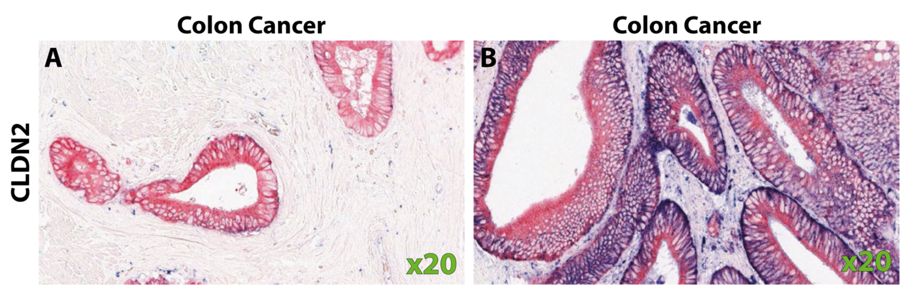
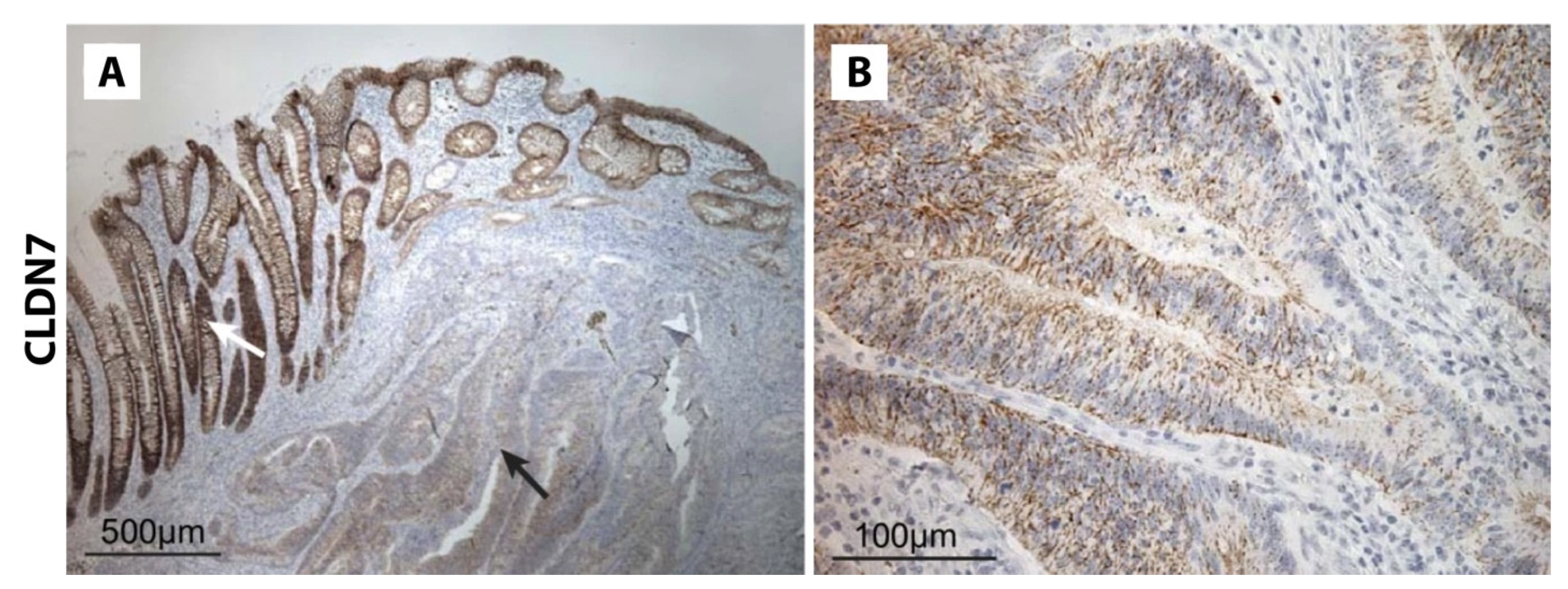
Table 1.
Claudin-1 expression in the normal colon by immunohistochemical staining.
| Normal Colon | Normal Cytoplasmic | Study Population | Study |
|---|---|---|---|
| 100% (n = 50) | - | Egyptian | Abdelzaher et al. [27] |
| 100% (n = 31) | 0% | Swedish | Hahn-Strömberg et al. [28] |
| 100% (n = 129) | - | American | Resnick et al. [29] |
| 94.8% (n = 120) | 5.2% | Czech | Bezdekova et al. [30] |
| 87% (n = 45) | - | French | Cherradi et al. [31] |
| 76% (n = 25) | 0% | American | Dhawan et al. [32] |
| 25% (n = 16) | - | German | Gröne et al. [34] |
| 20% (n = 20) | - | Chinese | Wang et al. [33] |
Table 2.
Claudin-1 expression in colorectal cancer by immunohistochemistry.
| CRC Membranous | CRC Cytoplasmic | Study Population | Study |
|---|---|---|---|
| 100% (n = 31) | 19.4% (n = 31) | Swedish | Hahn-Strömberg et al. [28] |
| 100% (n = 10) | - | Japanese | Miwa et al. [37] |
| 100% (n = 14) | - | Japanese | Kinugasa et al. [57] |
| 98.4% (n = 128) | - | American | Resnick et al. [29] |
| 96.9% (n = 32) | - | German | Georges et al. [61] |
| 95.7% (n = 23) | - | German | Gröne et al. [34] |
| 91.5% (n = 142) | - | Taiwanese | Dai et al. [60] |
| 87% (n = 120) | 87% (n = 120) | Czech | Bezdekova et al. [30] |
| 80% (n = 45) | - | French | Cherradi et al. [31] |
| 76.7% (n = 60) | - | Chinese | Wang et al. [33] |
| 68% (n = 91) | - | American | Sewda et al. [46] |
| 68% (n = 344) | - | Japanese | Shibutani et al. [58] |
| 63% (n = 30) | - | Japanese | Takahashi et al. [59] |
| 60% (n = 50) | - | Egyptian | Abdelzaher et al. [27] |
| 61.4% (n = 70) | - | Turkish | Süren et al. [62] |
| 56% (n = 25) | 60% (n = 25) | American | Dhawan et al. [32] |
| >54% (n = 260) | - | Korean | Kim et al. [56] |
Legend: >, data reported as degree of loss of expression (values taken from inverse of samples with “marked loss”).
Table 3.
Claudin-2 expression in colorectal cancer by immunohistochemistry.
| Colorectal Cancer | Study Population | Study |
|---|---|---|
| 100% (n = 10) | American | Dhawan et al. [75] |
| 100% (n = 33) | Swedish | Hahn-Strömberg [81] |
| 81.8% (n = 11) | Finish | Soini et al. [80] |
| 51% (n = 104) | Chinese | Wei at al. [74] |
| 25.3% (n = 99) | Japanese | Aung et al. [73] |
Table 4.
Claudin-4 expression in colorectal cancer by immunohistochemistry.
| Normal Colon | Colorectal Cancer | Study Population | Study |
|---|---|---|---|
| 100% (n = 31) | 100% (n = 31) | Swedish | Hahn-Strömberg et al. [28] |
| 100% | 96.9% (n = 127) | American | Resnick et al. [29] |
| - | 90.3% (n = 31) | German | Georges et al. [61] |
| 100% (n = 70) | at least 87% (n = 70) | Turkish | Süren et al. [62] |
| 30% (n = 20) | 85% (n = 60) | not specified | Wang et al. [33] |
| - | 67% (n = 30) | Japanese | Takahashi et al. [59] |
| 100% (n = 71) | 59.2% (n = 71) | Japanese | Ishikawa et al. [93] |
| - | 43% | Japanese | Ueda et al. [96] |
Table 5.
Claudin-7 expression in the normal colon and colorectal cancer.
| Normal Colon | Colorectal Cancer | Study Population | Study |
|---|---|---|---|
| 100% (n = 31) | 100% (n = 31) | Swedish | Hahn-Strömberg et al. [28] |
| - | 100% (n = 84) | Romanian | Ianole et al. [123] |
| 32.6% (n = 92) | 92.3% (n = 104) | German | Kuhn et al. [126] |
| - | at least 80% (n = 60) | Chinese | Wang et al. [124] |
| - | 80% (n = 10) | not specified | Hou et al. [125] |
| 100% (n = 72) | 74.5% (n = 231) | Chinese | Quan et al. [121] |
| 100% (n = 70) | at least 65.7% (n = 70) | Turkish | Süren et al. [62] |
| 96% (n = 75) | 52% (n = 75) | Chinese | Xu et al. [120] |
| - | 50% (n = 100) | Japanese | Tokuhara et al. [91] |
| - | 27.3% (n = 11) | American | Bhat et al. [67] |
| - | at least 20% (n = 90) | Japanese | Nakayama et al. [122] |
Table 6.
Percentage of colorectal samples with claudin-7 staining changes by degree of differentiation.
Disclaimer/Publisher’s Note: The statements, opinions and data contained in all publications are solely those of the individual author(s) and contributor(s) and not of MDPI and/or the editor(s). MDPI and/or the editor(s) disclaim responsibility for any injury to people or property resulting from any ideas, methods, instructions or products referred to in the content. |
© 2024 by the authors. Licensee MDPI, Basel, Switzerland. This article is an open access article distributed under the terms and conditions of the Creative Commons Attribution (CC BY) license (https://creativecommons.org/licenses/by/4.0/).
Share and Cite
MDPI and ACS Style
Cox, K.E.; Liu, S.; Hoffman, R.M.; Batra, S.K.; Dhawan, P.; Bouvet, M. The Expression of the Claudin Family of Proteins in Colorectal Cancer. Biomolecules 2024, 14, 272. https://0-doi-org.brum.beds.ac.uk/10.3390/biom14030272
AMA Style
Cox KE, Liu S, Hoffman RM, Batra SK, Dhawan P, Bouvet M. The Expression of the Claudin Family of Proteins in Colorectal Cancer. Biomolecules. 2024; 14(3):272. https://0-doi-org.brum.beds.ac.uk/10.3390/biom14030272
Chicago/Turabian StyleCox, Kristin E., Shanglei Liu, Robert M. Hoffman, Surinder K. Batra, Punita Dhawan, and Michael Bouvet. 2024. "The Expression of the Claudin Family of Proteins in Colorectal Cancer" Biomolecules 14, no. 3: 272. https://0-doi-org.brum.beds.ac.uk/10.3390/biom14030272
Note that from the first issue of 2016, this journal uses article numbers instead of page numbers. See further details here.