
A Novel Cytotoxic Steroidal Saponin from the Roots of Asparagus cochinchinensis


1
Department of Biomedical and Pharmaceutical Sciences, Graduate School, Kyung Hee University, Seoul 02447, Korea
2
Department of Life and Nanopharmaceutical Sciences, Graduate School, Kyung Hee University, Seoul 02447, Korea
3
College of Pharmacy, Kyung Hee University, Seoul 02447, Korea
*
Authors to whom correspondence should be addressed.
†
These authors contributed equally to this work and joint first authors.
Plants 2021, 10(10), 2067; https://0-doi-org.brum.beds.ac.uk/10.3390/plants10102067
Submission received: 14 September 2021
/
Revised: 28 September 2021
/
Accepted: 29 September 2021
/
Published: 30 September 2021
(This article belongs to the Topic Frontiers in Phytochemicals)
Abstract
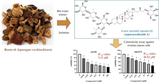
:A new steroidal saponin, 26-O-β-d-glucopyranosyl-(25R)-furost-5-ene-3β,22α,26-triol 3-O-(1−4)-β-d-glucopyranosyl-α-l-rhamnopyranosyl-(1−2)-[α-l-rhamnopyranosyl-(1−4)]-β-d-glucopyranoside [asparacochioside A (1)] was isolated from a hot water extract of the roots of Asparagus cochinchinensis, together with the known steroidal saponins protodioscin (2), methyl protodioscin (3), aspacochioside A (4), aspacochioside C (5), 15−hydroxypseudoprotodioscin (6), and chamaedroside E (7). The structure of the new compound 1 was determined by interpretation of its spectroscopic data (1D- and 2D-NMR and HR−Q−TOF−MS) and sugar analysis. The isolated compounds 1−7 were tested for their in vitro cytotoxicity against human ovarian cancer cell lines (A2780 and SKOV3). Asparacochioside A (1) exhibited a significant cytotoxicity against both A2780 and SKOV3 cells with IC50 values of 5.25 ± 2.2 and 46.82 ± 9.43 μM, respectively. Furthermore, asparacochioside A (1) significantly increased the percentage of Annexin V-positive cells (apoptotic cells), suggesting that asparacochioside A induces ovarian cancer cell death via apoptosis.
1. Introduction
The genus Asparagus (Liliaceae) comprises over three hundred species distributed around the world and used in salads and as herbs and vegetables. Asparagus cochinchinensis (Loureio) Merrill is a perennial herb distributed in Eastern Asia, including many provinces of China, Japan, and Korea [1]. The dried roots of A. cochinchinensis have been used in Traditional Chinese Medicine for more than two thousand years to treat fevers, renal failure, heart diseases, and lung cancer [2].
Moreover, extensive chemical studies on the roots of A. cochinchinensis have led to the identification of many compounds, such as steroidal saponins, mono-, oligo-, and polysaccharides, and amino acids [3,4]. The extracts of A. cochinchinensis and the corresponding constituents have been shown to have several pharmacological effects, including anti-oxidant [5] and anti-inflammatory effects [6]. Additionally, cytotoxicity against several human cancer cell lines such as cervical cancer HeLa, lung cancer NCI−H460, breast cancer MCF-7, and liver cancer HepG2 cells has been reported [5,7]. Steroidal saponins from A. cochinchinensis and other plants such as Smilax davidiana and Polygonautm sibiricum are also known to have anti−inflammatory effects by inhibiting cytokine production and cytotoxicity against tumor cell lines [8,9].
In our continuing search for bioactive compounds from medicinal plants, the roots of A. cochinchinensis were chosen for a phytochemical investigation, since its hot water extract was found to have significant cytotoxicity against human ovarian carcinoma (A2780) and human female ovarian adenocarcinoma (SKOV3) cell lines with 30 and 39% cell growth inhibition at 200 μg/mL, respectively.
In the present study one new (compound 1) and six known steroidal saponins 2−7 were purified from the roots of A. cochinchinensis by repeated chromatography. The structure of the new compound 1 was elucidated by interpreting 1D- and 2D-nuclear magnetic resonance (NMR) spectroscopic data analysis, acid hydrolysis, and high-resolution quadrupole time of flight mass (HR−Q−TOF−MS) spectrometric data analysis. All the isolated compounds 1−7 were evaluated for their cytotoxicity against ovarian cancer cell lines (A2780 and SKOV3). To summarize, in this paper, we describe the isolation of steroidal saponins 1−7 from the roots of A. cochinchinensis, structure elucidation of the new steroidal saponin 1, and cytotoxicity of the isolates against human ovarian cancer cells.
2. Results and Discussion
2.1. Structure Elucidation of Compound 1
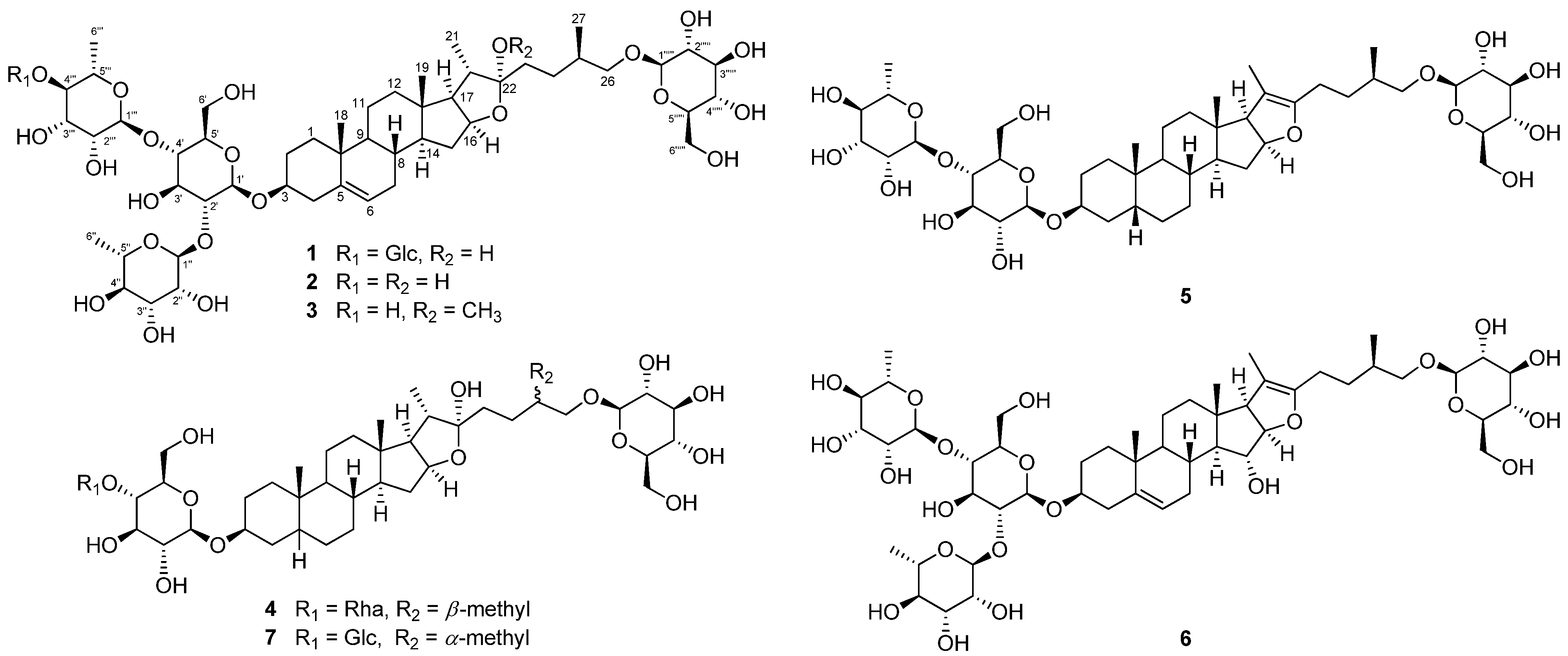
One new (compound 1) and six known furostane-type steroidal saponins 2−7 were isolated from the roots of A. cochinchinensis (Figure 1).
Compound 1 was isolated as a pale brown powder. The molecular formula was established as C57H94O27 through HR−Q−TOF−MS isotopic ion peaks at m/z = 1209.5892 [M − H]− (calcd for C57H93O27, 1209.5904) (Figure S1). The 1H-NMR spectrum of 1 exhibited six methyl signals at δH 0.89 (3H, s), 0.99 (3H, d, J = 6.5 Hz), 1.08 (3H, s), 1.34 (3H, d, J = 7.0 Hz), 1.67 (3H, d, J = 6.0 Hz), 1.77 (3H, d, J = 6.0 Hz), and five anomeric protons at δH 4.81 (1H, d, J = 7.5 Hz), 4.95 (1H, d, J = 7.5 Hz), 5.24 (1H, d, J = 7.5 Hz), 5.87 (1H, s), and 6.42 (1H, s) (Table 1, Figure S2) [10]. The 13C-NMR, distoriionless enhancement by polarization transfer (DEPT)135-NMR, and 1H-13C heteronuclear single quantum coherence spectroscopy (HSQC) spectra showed 57 signals with six methyl signals at δC 16.4 × 2, 17.4, 18.3, 18.6, and 19.3, 13 methylene carbon signals at δC 21.0, 28.3, 30.1, 32.2, 32.4, 37.1, 37.4, 38.9, 39.8, 78.5, 61.1, 62.3, and 62.7, nine methine carbons in aglycone moiety including an olefinic carbon at δC 31.6, 34.2, 40.6, 50.3, 56.6, 63.7, 78.0, 81.0, and 121.8, and four quaternary carbons at δC 37.1, 40.7, 110.6, and 140.7 (Table 1, Figures S3 and S4). According to the 1H- and 13C-NMR and HSQC data, compound 1 could be expected to have five sugars [δC/δH 100.3/4.95, 1H, d (7.5); 102.0/5.88, 1H, s; 101.9/6.42, 1H, s; 106.7/5.24, 1H, d (7.5); 104.8/4.81, 1H, d (7.5)]. Based on the 1H-1H correlation spectroscopy (COSY) and 1H-13C heteronuclear multiple bond correlation (HMBC) data (Figures S5 and S6), it was inferred that compound 1 is a furostane-type steroidal saponin.
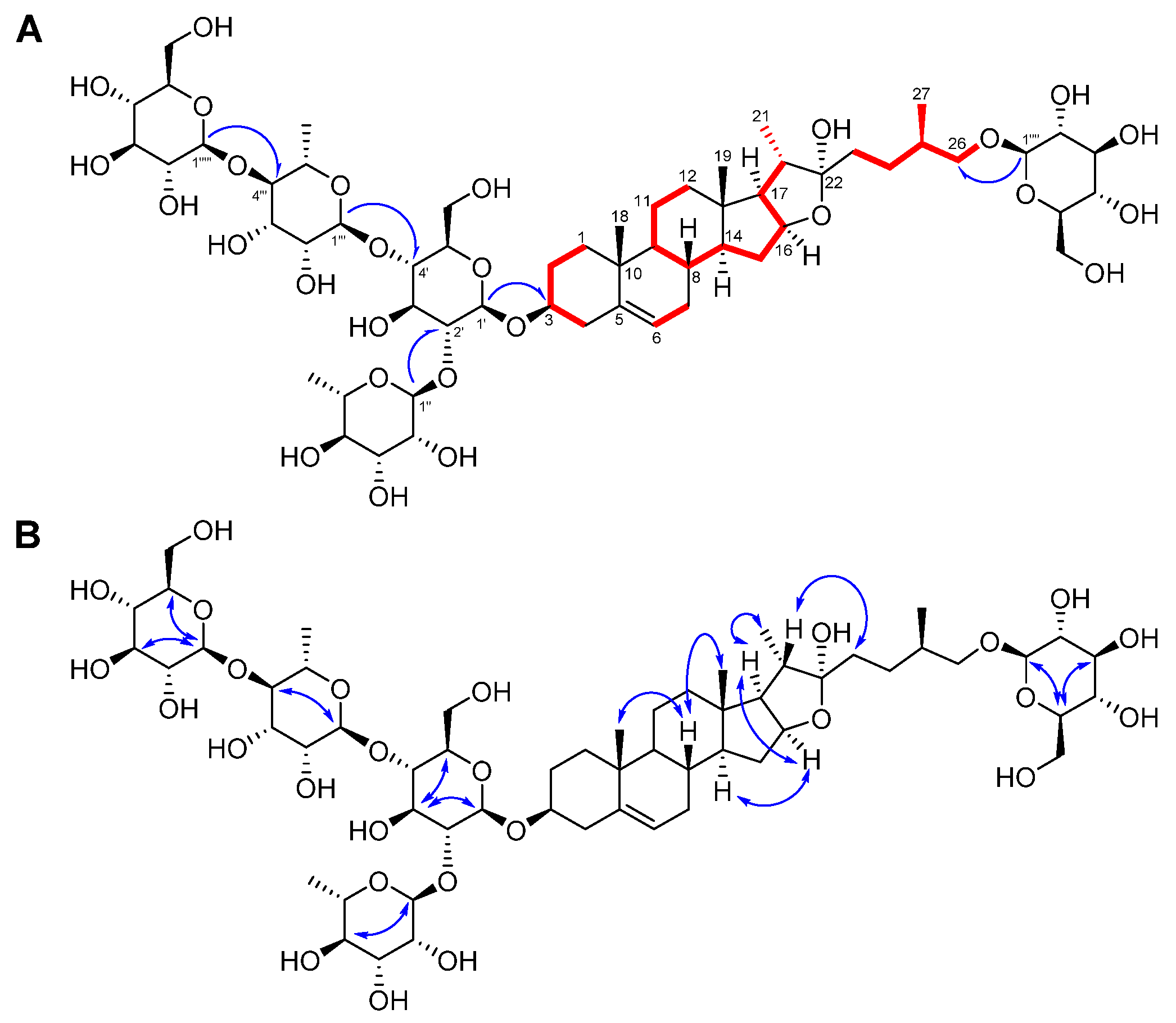
Through analysis of the HMBC and nuclear Overhauser effect correlation spectroscopy (NOESY) spectra (Figure 2, Figures S6 and S7), it was proposed that compound 1 has three glucoses and two rhamnoses [11]. The positions of the sugars were determined by analysis of the HMBC correlations; H−Glc−1′ [δ 4.95 (1H, d, J = 7.5 Hz)] with C−3 (δ 78.0), H−Rha−1′’ [δ 6.42 (1H, s)] with C−Glc−2′ (δ 78.0), H−Rha−1′’’ [δ 5.87 (1H, s)] with C−Glc−4′ (δ 78.4), and H−Glc−1′’’’’ [δ 5.24 (1H, d, J = 7.5 Hz)] with C−26 (δ 78.5) (Figure 2). The glucoses and rhamnoses in compound 1 were identified as β-d- and α-l-forms, respectively, based on the coupling constants of the anomeric protons and acid hydrolysis followed by high performance liquid chromatography (HPLC) analysis. The 1H- and 13C-NMR spectroscopic data of compound 1 were very similar to those of protodioscin (2), except for the presence of an additional β-glucose unit at Rha-4′’’ in 1 (Table 2 and Table 3, Figure 1) [10]. The chemical structure of compound 1 was very resemble to costucoside I, isolated from the seeds of Costus speciosus which only differ from the position of Glc−1′’’’at Rha−3′’’ [12]. The absolute configuration of the methyl group at C-25 was determined as R by the difference in proton NMR chemical shifts between H2−26α and H2−26β (∆ab = 0.32) [13,14]. Thus, the chemical structure of the new furostane-type steroidal saponin 1 was elucidated as 26-O-β-d-glucopyranosyl-(25R)-furost-5-ene-3β,22α,26-triol 3-O-(1−4)-β-d-glucopyranosyl-α-l-rhamnopyranosyl-(1−2)-[α-l-rhamnopyranosyl-(1−4)]-β-d-glucopyranoside and named asparacochioside A.
The other known compounds were identified as protodioscin (2) [10], methyl protodioscin (3) [15], aspacochioside A (4) [16], aspacochioside C (5) [16], 15-hydroxypseudoprotodioscin (6) [17], and chamaedroside E (7) [18] by comparison of their NMR spectroscopic data with those previously reported data.
2.2. The Cytotoxicity of the Compounds Isolated from A. cochinchinensis against Human Ovarian Cancer Cells
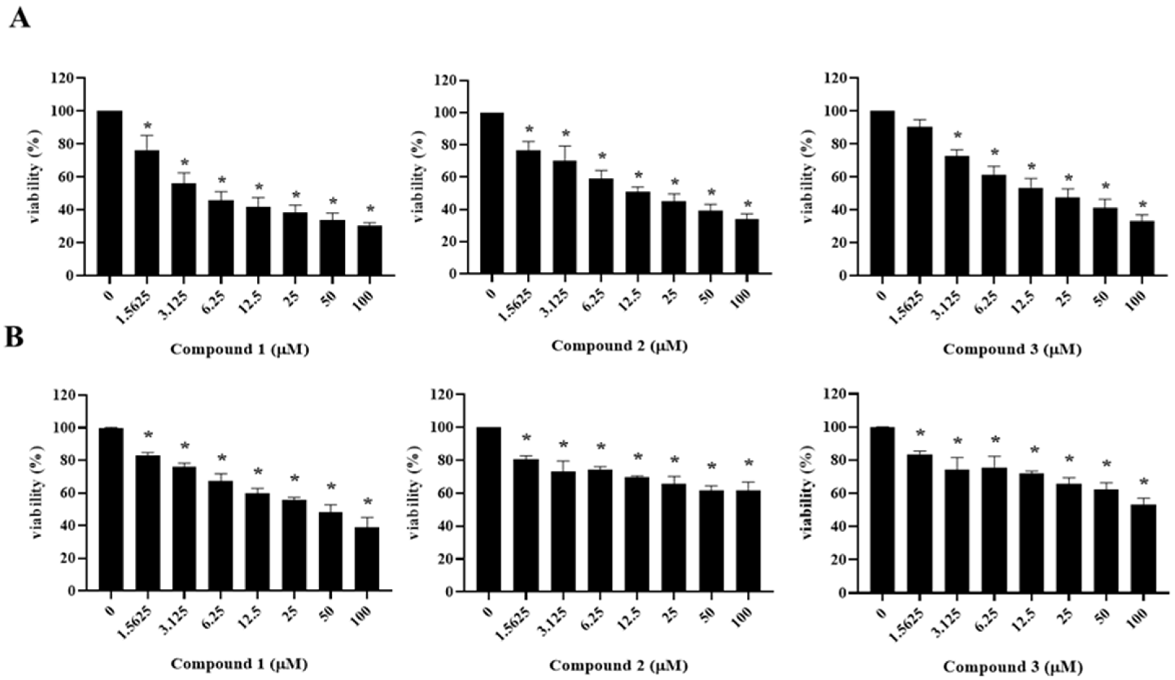
To evaluate the potential anti-tumor activity of the isolates from the hot water extract of A. cochinchinensis, the cytotoxicity of compounds 1−7 against human ovarian cancer cells (A2780 and SKOV3) were examined using a 3-[4,5-dimethylthiazol-2-yl]-2,5-diphenyl tetrazolium bromide (MTT) assay.
As shown in Table 3 and Figure 3, asparacochioside A (1), protodioscin (2), and methyl protodioscin (3) exhibited a significant cytotoxicity in A2780 cells with IC50 values of 5.25, 10.14, and 21.78 μM, respectively, in a dose dependent manner. In SKOV3 cells, only the new compound asparacochioside A (1) showed a significant cytotoxicity with observed IC50 value of 46.82 μM, while compounds 4−7 were not active against either A2780 or SKOV3 human ovarian cancer cells (>100 μM). Overall, among the seven compounds isolated from A. cochinchinensis, only the novel compound 1 showed a potent cytotoxicity against both A2780 and SKOV3, suggesting its potential anti−cancer activity in human ovarian cancer.
Both protodioscin (2) and methyl protodioscin (3) have been reported to show various biological activities such as anti-tumor and anti-inflammatory effects [19,20]. However, this is the first study to demonstrate their cytotoxicity against human ovarian cancer cells. Protodioscin (2) showed more potent cytotoxicity against A2780 cells than methyl protodioscin (3), suggesting that the methylation at C-22 hydroxy group may interrupt the activity of protodioscin (3). Although asapacochioside C (5) was cytotoxic against human lung adenocarcinoma A549 cells [21], it failed to show a significant cytotoxicity against human ovarian cancer cells in this study. Reportedly, chamaedroside E (7) was first isolated from Veronica chamaedrys [18]. However, the presence of 7 in A. cochinchinensis was found in this study, for the first time.
2.3. Induction of Apoptotic Cell Death by Asparacochioside A (1) in Human Ovarian Cancer Cells
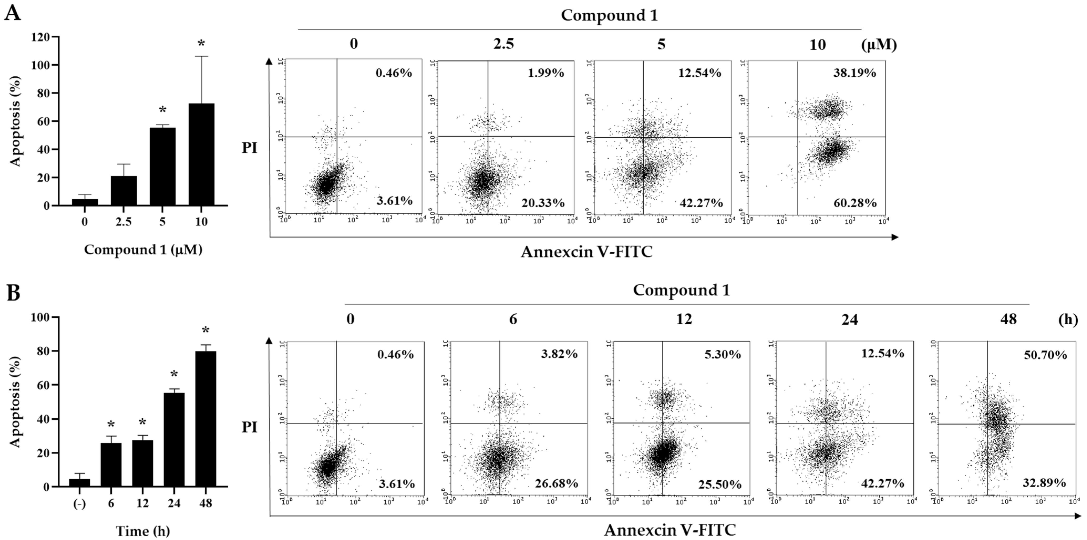
To further investigate whether the cytotoxicity of asparacochioside A (1) was associated with the induction of apoptosis, Annexin V-FITC staining was performed. Asparacochioside A (1) significantly increased the percentage of Annexin V positive cells (apoptotic cells) in a dose- and time-dependent manner (Figure 4). These results indicate that asparacochioside A-induced cytotoxicity was associated with apoptosis in human ovarian cancer cells.
3. Materials and Methods
3.1. Plant Material
The roots of Asparagus cochinchinensis (Loureio) Merrill (Liliaceae) were purchased from Nanuum pharmaceutical Co. (Youngcheon-si, Kyungsangbukdo, South Korea), in May 2017. The plant material was identified by D.S.J. and a voucher specimen (ASCO1-2017) has been deposited in the Lab. of Natural Product Medicine, Kyung Hee University.
3.2. General Experimental Procedures
General experimental procedures are provided in the Supplementary Materials.
3.3. Isolation of Compounds 1−7
Dried roots of A. cochinchinensis (800 g) were extracted with distilled water (8 L) at 100 °C for 2 h, and the solvent was removed using rotary evaporator. The extract (300.0 g) was subjected to Diaion HP column chromatography (CC, 9.6 × 56.0 cm), eluting with MeOH−H2O gradient system (from 0:1 to 1:0 v/v) to afford 12 fractions (C1~C12). Fraction C4 (3.8 g) was separated by Sephadex LH-20 CC (4.3 × 57.5 cm) with acetone−H2O (4:6 v/v) to make four fractions (C4−1~C4−4). Fraction C4−3 (2.2 g) was subjected further to silica gel CC (3.3 × 34.0 cm, 230−400 mesh) with EtOAc−acetone−H2O mixture (3:6:1 v/v/v), yielding seven subfractions (C4−3−1~C4−3−7). Chamaedroside E (7, 21.5 mg) was purified by HPLC with Gemini 5 µm NX-C18 110A column (acetonitirle–H2O = 25:75 to 40:60, v/v) from subfraction C4−3−4 (60.8 mg). Fraction C6 (12.0 g) was loaded to a silica gel column (230−400 mesh; 5.6 × 35.0 cm) and eluted with CH2Cl2−MeOH−H2O solvent system [14:6:1 v/v/v] to afford seven subfractions (C6−1~C6−7). Subfraction C6−4 (1.75 g) was subjected to silica gel CC (5.2 × 25.5 cm, 230−400 mesh) with EtOAc−acetone−H2O mixture (from 4:5:1 to 0:8:2 v/v/v), yielding protodioscin (2, 217.8 mg). Subfraction C6−5 (7.16 g) was subjected to silica gel CC (230−400 mesh; 5.2 × 32.0 cm) eluting with EtOAc−acetone−H2O [4:5:1 to 3.5:5:1.5 v/v/v] and afforded methylprotodioscin (3, 1.56 g). Aspacochioside A (4, 118.7 mg) was separated from subfraction C6−2 (392.0 mg) by silica gel CC (3.6 × 26.0 cm, 230−400 mesh) with EtOAc−acetone−H2O mixture (4:5:1 v/v/v). Subfraction C6−6 (2.11 g) was fractionated by silica gel CC (230−400 mesh; 4.4 × 30.0 cm), eluting with EtOAc−acetone−H2O gradient system [from 4:5:1 to 3.5:5:1.5 v/v/v] to yield compound 1 (724.2 mg).
Fraction C7 (16.3 g) was subjected to silica gel CC (5.8 × 40.0 cm, 230−400 mesh) with EtOAc−acetone−H2O mixture (4:5:1 v/v/v) to generate nine subfractions (C7−1~C7−9). 15−Hydroxy−pseudoprotodioscin (6, 211.0 mg) was purified by flash chromatography using a Redi Sep−C18 cartridge (130 g, acetonitrile−H2O, from 15:85 to 40:60 v/v) from C7−6 (1.6 g). Protodioscin (2, 2.08 g was obtained additionally from subfraction C7−8 (9.7 g) by flash chromatography using a Redi Sep−C18 cartridge (130 g, acetonitrile−H2O, from 15:85 to 45:55 v/v).
Fraction C9 (406.6 mg) was fractionated further by flash chromatography using a Redi Sep−C18 cartridge (130 g, acetonitrile−H2O, from 10:90 to 40:60 v/v) to afford aspacochioside C (5, 68.6 mg).
Asparacochioside A (1)
3.4. Acidic Hydrolysis of Compound 1
Compound 1 (10.0 mg) was hydrolyzed with 1N HCl at 80 °C for 3 h. Sodium bicarbonate was added to stop the reaction.
3.5. Absolute Configurations of the β-Glucose and α-Rhamnose in Compound 1
The absolute configurations of β-glucose and α-rhamnose in 1 was determined using the method reported by Tanaka et al. [21]. The hydrolysate was derivatized with pyridine (500 μL), l-cysteine methyl ester hydrochloride (1.2 mg), and σ-tolyl isothiocyanate (100 μL) and analyzed using HPLC. The glucose and rhamnose in the reaction mixture of 1 was detected at 30.7 and 35.5 min, respectively. The retention times of authentic d-glucoses and l-rhamnose were 30.8 and 35.4 min, respectively, under the same HPLC conditions. Therefore, the absolute configuration of β-glucose in 1 was confirmed as the d configuration, whereas α-rhamnose was confirmed as the l configuration.
3.6. Cell Culture
A2780 and SKOV3 human ovarian cancer cell lines were obtained from American Type Culture Collection (ATCC). The cells were maintained in Roswell Park Memorial Institute (RPMI) 1640 (Life Technologies Inc., Grand Island, NY, USA) medium with penicillin (100 U/mL), 5% fetal bovine serum (FBS), and streptomycin sulfate (100 μg/mL, Life Technologies Inc.). Cells with medium were kept in humidified atmosphere of 5% CO and 37 °C temperatures.
3.7. MTT Assay
The cell growth was assessed using the MTT assay. Briefly, Cells were seeded in a 96-well plate at a density of 1 × 10⁵/mL per well. After 24 h, cells were treated with different concentrations of extract or compounds for 48h. Following incubation, 50 μL of MTT (1 mg/mL stock solution, molecular probes Inc., Eugene, OR, USA) was added and incubated for 4 h in the 37 °C incubator. The medium was discarded and the formazan crystals were dissolved in DMSO. The absorbance was measured using a microplate spectrophotometer (SpectraMax; Molecular Devices, Sunnyvale, CA, USA) at 540 nm.
3.8. Annexin V−FITC Staining Assay for Apoptosis Analysis
Apoptotic cells were detected by the binding of fluorescent Annexin V (Annexin V-FITC). Cells were treated with asparacochioside A (1) for indicated time periods or concentrations. After incubation, both floating and adherent cells from each well collected in tubes and washed with PBS. The cells were then re-suspended in 500 μL of binding buffer and stained with 2.75 μL of FITC-conjugated Annexin V for 15min in darkness. Finally the stained cells were washed with binding buffer and stained with 5 μL of PI (50 mg/mL) prior to analysis using a Guava® easyCyte flow cytometer (Merck Millipore, Burlington, MA, USA).
3.9. Statistical Analysis
All statistical parameters were calculated using GraphPad Prism 5.0 (GraphPad Software, Inc., La Jolla, CA, USA). Data are presented as mean ± SD and evaluated by unpaired Student t-test or one-way ANOVA analysis. Difference with a p-value less than 0.05 was considered to be statistically significant.
4. Conclusions
Seven compounds 1‒7, including a new furostane-type steroidal saponin named asparacochioside A (1) were isolated from the roots of A. cochinchinensis in the present study. The chemical structure of the new compound was determined using their spectroscopic data (1H-NMR, 13C-NMR, DEPT, HSQC, HMBC, NOESY, and HR−Q−TOF−MS) measurement and by acidic hydrolysis. Isolation of chamaedroside E (7) from the roots of A. cochinchinensis was reported for the first time in this paper. Among the isolates, asparacochioside A (1), protodioscin (2), and methyl protodioscin (3) exhibited a significant cytotoxicity against A2780 cells. In SKOV3 cells, only the new compound asparacochioside A (1) showed a significant cytotoxicity. Compound 1 induced apoptotic cell death in human ovarian cancer cells and significantly increased the percentage of annexin V positive cells (apoptotic cells) in a dose- and time-dependent manner. Thus, asparacochioside A (1) is worthy of additional experiments for its potential as an anti-cancer agent.
Supplementary Materials
The following are available online at https://0-www-mdpi-com.brum.beds.ac.uk/article/10.3390/plants10102067/s1, General Experimental Procedure, The HR-ESI-MS, 1H-NMR (800 MHz, C5D5N), 13C-NMR (200 MHz, C5D5N), DEPT135-NMR, HSQC, COSY, HMBC, and NOESY spectra of 1 (Figures S1–S7).
Author Contributions
Conceptualization, J.-H.C. and D.S.J.; Funding acquisition, D.S.J.; Investigation, J.-Y.K., H.Y.C., H.M.K.; Supervision, J.-H.C. and D.S.J.; Writing—original draft, J.-Y.K. and H.Y.C.; Writing—review & editing, J.-H.C. and D.S.J. All authors have read and agreed to the published version of the manuscript.
Funding
This work was supported by Korea Institute of Planning and Evaluation for Technology in Food, Agriculture and Forestry (IPET) funded by Ministry of Agriculture, Food and Rural Affairs (MAFRA) (grant number: P430-117010-1) and by a grant from the National Research Foundation of Korea (NRF) funded by the Ministry of Science and ICT (MSIT), Republic of Korea (grant number: NRF−2019R1A2C1083945).
Institutional Review Board Statement
Not applicable.
Informed Consent Statement
Not applicable.
Conflicts of Interest
The authors declare no conflict of interest.
References
- Negi, J.S.; Singh, P.; Joshi, G.P.; Rawat, M.S.; Bisht, V.K. Chemical constituents of Asparagus. Phamacogn. Rev. 2010, 4, 215–220. [Google Scholar]
- Xiong, D.; Yu, L.X.; Yan, X.; Guo, C.; Xiong, Y. Effects of root and stem extracts of Asparagus cochinchinensis on biochemical indicators related to aging in the brain and liver of mice. Am. J. Chin. Med. 2011, 39, 719–726. [Google Scholar] [CrossRef]
- Shen, Y.; Xu, C.L.; Xuan, W.D.; Li, H.L.; Liu, R.H.; Xu, X.K.; Chen, H.S. A new furostanol saponin from Asparagus cochinchinensis. Arch. Pharm. Res. 2011, 34, 1587–1591. [Google Scholar] [CrossRef]
- Guo-Lei, Z.H.U.; Qian, H.A.O.; Rong-Tao, L.I.; Hai-Zhou, L.I. Steroidal saponins from the roots of Asparagus cochinchinensis. Chin. J. Nat. Med. 2014, 12, 213–217. [Google Scholar]
- Le Son, H.; Anh, N.P. Phytochemical composition, in vitro antioxidant and anticancer activities of quercetin from methanol extract of Asparagus cochinchinensis (Lour.) Merr. tuber. J. Med. Plant. Res. 2013, 7, 3360–3366. [Google Scholar]
- Samad, N.B.; Debnath, T.; Abul Hasnat, M.; Pervin, M.; Kim, D.H.; Jo, J.E.; Park, S.R.; Lim, B.O. Phenolic Contents, Antioxidant and Anti-inflammatory Activities of Asparagus cochinchinensis (Loureiro) Merrill. J. Food Biochem. 2014, 38, 83–91. [Google Scholar] [CrossRef]
- Park, M.; Cheon, M.S.; Kim, S.H.; Chun, J.M.; Lee, A.Y.; Moon, B.C.; Yoon, T.; Choo, B.K.; Kim, H.K. Anticancer activity of Asparagus cochinchinensis extract and fractions in HepG2 cells. J. Korean Soc. Appl. Bio Chem. 2011, 54, 188–193. [Google Scholar] [CrossRef]
- Zhou, M.; Huang, L.; Li, L.; Wei, Y.; Shu, J.; Liu, X.; Huang, H. New furostanol saponins with anti-inflammatory and cytotoxic activities from the rhizomes of Smilax davidiana. Steroids 2017, 127, 62–68. [Google Scholar] [CrossRef] [PubMed]
- Tang, C.; Yu, Y.M.; Qi, Q.L.; Wu, X.D.; Wang, J.; Tang, S.A. Steroidal saponins from the rhizome of Polygonatum sibiricum. J. Asian. Nat. Prod. Res. 2019, 21, 197–206. [Google Scholar] [CrossRef] [PubMed]
- Hu, K.; Dong, A.; Yao, X.; Kobayashi, H.; Iwasaki, S. Antineoplastic agents II: Four furostanol glycosides from rhizomes of Dioscorea collettii var. hypoglauca. Planta Med. 1997, 63, 161–165. [Google Scholar] [CrossRef] [PubMed]
- Zhu, G.L.; Hao, Q.; Xing, L.; Yang, X.Q.; Xie, S.D.; Zhao, P.; Li, H.Z. C21, C22 pregnane glycosides and cytotoxic C27 spriostanol steroids from Asparagus cochinchinesis. Steroids 2021, 172, 108874. [Google Scholar] [CrossRef]
- Singh, S.B.; Thakur, R.S. Costusoside-I and costusoside-J, two new furostanol saponins from the seeds of Costus speciosus. Phytochemistry 1982, 21, 911–915. [Google Scholar] [CrossRef]
- Agrawal, P.K. Dependence of 1H NMR chemical shifts of geminal protons of glycosyloxy methylene (H2-26) on the orientation of the 27-methyl group of furostane-type steroidal saponins. Magn. Reson. Chem. 2004, 42, 990–993. [Google Scholar] [CrossRef]
- Challinor, V.L.; Piacente, S.; De Voss, J.J. NMR assignment of the absolute configuration of C-25 in furostanol steroidal saponins. Steroids 2012, 77, 602–608. [Google Scholar] [CrossRef]
- Ju, Y.; Jia, Z.J. Steroidal saponins from the rhizomes of Smilax menispermoidea. Phytochemistry 1992, 31, 1349–1351. [Google Scholar] [CrossRef]
- Shi, J.G.; Li, G.Q.; Huang, S.Y.; Mo, S.Y.; Wang, Y.; Yang, Y.C.; Hu, W.Y. Furostanol oligoglycosides from Asparagus cochinchinensis. J. Asian Nat. Prod. Res. 2004, 6, 99–105. [Google Scholar] [CrossRef] [PubMed]
- Huang, H.L.; Liu, R.H.; Shao, F. Structural determination of two new steroidal saponins from Smilax china. Magn. Reson. Chem. 2009, 47, 741–745. [Google Scholar] [CrossRef] [PubMed]
- Marchenko, A.; Kintya, P.; Wyrzykiewicz, B.; Gorincioi, E. Steroidal glycosides from Veronica chamaedrys L. Part I. The structures of chamaedrosides C, C1, C2, E, E1 and E2. Nat. Prod. Commun. 2012, 7, 565–568. [Google Scholar] [CrossRef] [PubMed] [Green Version]
- Podolak, I.; Galanty, A.; Sobolewska, D. Saponins as cytotoxic agents: A review. Phytochem. Rev. 2010, 9, 425–474. [Google Scholar] [CrossRef] [PubMed] [Green Version]
- Sautour, M.; Mitaine-Offer, A.C.; Lacaille-Dubois, M.A. The Dioscorea genus: A review of bioactive steroid saponins. J. Nat. Med. 2007, 61, 91–101. [Google Scholar] [CrossRef]
- Tanaka, T.; Tatsuya, N.; Toshihisa, U.; Kenji, T.; Isao, K. Facile discrimination of aldose enantiomers by reversed-phase HPLC. Chem. Pharm. Bull. 2007, 55, 899–901. [Google Scholar] [CrossRef] [PubMed] [Green Version]
Figure 1.
Structures of compounds 1–7 isolated from the roots of A. cochinchiensis.

Figure 2.
Key 1H−1H COSY (![Plants 10 02067 i001]() ) and HMBC (
) and HMBC (![Plants 10 02067 i002]() ) correlations of compound 1 (A). Key 1H−1H NOESY (
) correlations of compound 1 (A). Key 1H−1H NOESY (![Plants 10 02067 i003]() : blue dashed arrows) correlations of compound 1 (B).
: blue dashed arrows) correlations of compound 1 (B).
 ) and HMBC (
 ) correlations of compound 1 (A). Key 1H−1H NOESY (
 : blue dashed arrows) correlations of compound 1 (B).
Figure 2.
Key 1H−1H COSY (![Plants 10 02067 i001]() ) and HMBC (
) and HMBC (![Plants 10 02067 i002]() ) correlations of compound 1 (A). Key 1H−1H NOESY (
) correlations of compound 1 (A). Key 1H−1H NOESY (![Plants 10 02067 i003]() : blue dashed arrows) correlations of compound 1 (B).
: blue dashed arrows) correlations of compound 1 (B).
) and HMBC () correlations of compound 1 (A). Key 1H−1H NOESY (: blue dashed arrows) correlations of compound 1 (B).
Figure 3.
The effects of compounds 1−3 on cell viability in human ovarian cancer cells A2780 (A) and SKOV3 (B). Ovarian cancer cells (A2780 and SKOV3) were treated with compounds 1−3 in the indicated concentrations for 48 h. Data are presented as mean ± SD and evaluated by one-way ANOVA analysis. * p < 0.05 as compared with the untreated group.

Figure 4.
The effect of compound 1 on apoptosis in A2780 cells. Cells were treated with compound 1 for the indicated concentrations (2.5, 5.0, and 10 μM) (A) and times (6, 12, 24, and 48 h) (B), and then co-stained with propidium iodide (PI) and Annexin V-FITC. The graph indicates the percentage of apoptosis. Data are presented as mean ± SD and evaluated by one-way ANOVA analysis. * p < 0.05 as compared with control.


Table 1.
1H- and 13C-NMR spectroscopic data for the aglycones of compounds 1 and 2 (δ in ppm, C5D5N, 800 and 200 MHz).
Table 1.
1H- and 13C-NMR spectroscopic data for the aglycones of compounds 1 and 2 (δ in ppm, C5D5N, 800 and 200 MHz).
| Position | 1 | Protodioscin (2) a | ||
|---|---|---|---|---|
| δH Multi (J in Hz) | δC | δH Multi (J in Hz) | δC | |
| 1 | 1.85, 1H, m/2.07, 1H, m | 30.1 | 1.86, 1H, m/2.08, 1H, m | 29.8 |
| 2 | 1.44, 2H, m | 21.0 | 1.54, 2H, m | 20.9 |
| 3 | 3.88, 1H, d (11.0, 6.5) | 78.0 | 3.81, 1H b | 78.4 |
| 4 | 2.73, 1H, d (11.0)/2.79, 1H, d (10.5) | 38.9 | 2.74, 1H, d (11.5)/2.79, 1H, d (10.5) | 38.8 |
| 5 | - | 140.7 | - | 140.6 |
| 6 | 5.30, 1H, s | 121.8 | 5.34, 1H, s | 121.8 |
| 7 | 1.88, 2H, m | 32.2 | 1.86, 2H, m | 32.2 |
| 8 | 1.57, 1H, m | 31.6 | 1.56, 1H, m | 31.5 |
| 9 | 0.90, 1H b | 50.3 | 0.90, 1H b | 50.1 |
| 10 | - | 37.1 | - | 37.1 |
| 11 | 0.98, 1H b/1.32, 1H b | 37.4 | 0.97, 1H b/1.30, 1H b | 37.8 |
| 12 | 1.12, 1H, m/1.74, 1H, m | 39.8 | 1.12, 1H, m/1.75, 1H, m | 39.7 |
| 13 | - | 40.7 | - | 40.6 |
| 14 | 1.07, 1H b | 56.6 | 1.07, 1H b | 56.4 |
| 15 | 1.48, 1H b / 2.02, 1H b | 32.4 | 1.48, 1H, m/2.03, 1H, m | 32.3 |
| 16 | 4.96, 1H, t (4.5) | 81.0 | 4.46, 1H, m | 81.1 |
| 17 | 1.93, 1H b | 63.7 | 1.94, 1H b | 63.7 |
| 18 | 1.08. 3H, s | 19.3 | 1.05, 3H, s | 19.2 |
| 19 | 0.89, 3H, s | 16.4 | 0.89, 3H, s | 16.3 |
| 20 | 2.25, 1H, q (2.0) | 40.6 | 2.24, 1H, m | 40.5 |
| 21 | 1.34, 3H, d (7.0) | 16.4 | 1.34, 3H, d (7.0) | 16.3 |
| 22 | - | 110.6 | - | 111.0 |
| 23 | 2.07, 2H b | 37.1 | 1.57, 2H, dd (10.5, 5.0) | 30.0 |
| 24 | 1.68, 1H, d (4.0)/2.05, 1H, m | 28.3 | 1.68, 1H, d (4.0)/2.05, 1H, m | 28.2 |
| 25 | 1.94, 1H b | 34.2 | 1.96, 1H b | 34.1 |
| 26 | 3.63, 1H, dd (9.5, 6.0)/3.95, 1H, dd (9.0, 6.5) | 78.5 | 3.63, 1H, d (4.0)/3.99, 1H, d (6.5) | 78.4 |
| 27 | 0.99, 3H, d (6.5) | 17.4 | 0.99, 3H, d (6.0) | 17.3 |
a NMR data of compound 2 isolated in this study. b signal is overlapped.
Table 2.
1H- and 13C-NMR spectroscopic data for the sugar moieties of compounds 1 and 2 (δ in ppm, C5D5N, 800 and 200 MHz).
Table 2.
1H- and 13C-NMR spectroscopic data for the sugar moieties of compounds 1 and 2 (δ in ppm, C5D5N, 800 and 200 MHz).
| Position | 1 | Protodioscin (2) a | ||
|---|---|---|---|---|
| δH Multi (J in Hz) | δC | δH Multi (J in Hz) | δC | |
| 3-O-Glc-1’ | 4.95, 1H, d (7.5) | 100.3 | 4.96, 1H, d (7.0) | 100.1 |
| 2’ | 3.88, 1H b | 78.0 | 3.78, 1H b | 77.9 |
| 3’ | 3.63, 1H b | 76.9 | 3.61, 1H b | 76.8 |
| 4’ | 3.95, 1H b | 78.4 | 3.92, 1H b | 78.3 |
| 5’ | 4.43, 1H b | 77.3 | 4.40, 1H b | 77.5 |
| 6’ | 4.06, 1H, t (8.0)/4.20, 1H b | 61.1 | 4.04, 1H, t (8.0)/4.23, 1H b | 61.1 |
| 2’-O-Rha-1” | 6.42, 1H, s | 101.9 | 6.41, 1H, s | 101.9 |
| 2” | 4.67, 1H b | 71.8 | 4.61, 1H b | 72.4 |
| 3” | 4.87, 1H, s | 72.5 | 4.75, 1H, s | 72.6 |
| 4” | 4.37, 1H b | 74.00 | 4.34, 1H b | 73.8 |
| 5” | 5.04, 1H b | 69.5 | 4.95, 1H b | 69.4 |
| 6” | 1.77, 3H, d (6.0) | 18.3 | 1.76, 3H, d (6.0) | 18.4 |
| 4’-O-Rha-1’” | 5.87, 1H, s | 102.0 | 5.87, 1H, s | 102.7 |
| 2’” | 4.64, 1H, dd (9.0, 3.0) | 72.4 | 4.61, 1H, dd (9.0, 3.0) | 72.4 |
| 3’” | 4.65, 1H, d (4.5) | 72.8 | 4.61, 1H, m | 72.7 |
| 4’” | 4.21, 1H b | 74.01 | 4.61, 1H b | 73.3 |
| 5’” | 4.96, 1H b | 71.2 | 4.92, 1H b | 70.2 |
| 6’” | 1.67, 3H, d (6.0) | 18.6 | 1.64, 3H, d (6.0) | 18.5 |
| 26-O-Glc-1”“ | 4.81, 1H, d (7.5) | 104.8 | 4.87, 1H, d (7.5) | 104.8 |
| 2”“ | 4.14, 1H, t (8.5) | 75.1 | 4.14, 1H, t (8.5) | 75.0 |
| 3”“ | 3.84, 1H b | 77.9 | 3.85, 1H, m | 77.8 |
| 4”“ | 4.24, 1H b | 71.6 | 4.23, 1H b | 71.5 |
| 5”“ | 4.29, 1H, t (9.5) | 78.5 | 4.30, 1H, t (9.0) | 78.4 |
| 6”“ | 4.36, 1H b/4.40, 1H b | 62.7 | 4.36, 1H b/4.41, 1H b | 62.6 |
| 4’”-O-Glc-1”“‘ | 5.24, 1H, d (7.5) | 106.7 | ||
| 2”“‘ | 4.03, 1H, m | 76.5 | ||
| 3”“‘ | 3.63, 1H, m/3.95, 1H, m | 75.3 | ||
| 4”“‘ | 4.23, 1H b | 78.5 | ||
| 5”“‘ | 4.30, 1H, t (9.0) | 71.2 | ||
| 6”“‘ | 4.37, 1H b 4.51/1H, d (11.5) | 62.3 | ||
a NMR data of compound 2 isolated in this study. b signal is overlapped.
Table 3.
The cytotoxicity of compounds 1−3 isolated from A. cochinchinensis in A2780 and SKOV3 human ovarian cancer cells.
Table 3.
The cytotoxicity of compounds 1−3 isolated from A. cochinchinensis in A2780 and SKOV3 human ovarian cancer cells.
| Compound | IC50 (μM) a | |
|---|---|---|
| A2780 | SKOV3 | |
| 1 | 5.25 ± 2.2d,e,f | 46.82 ± 9.43 f |
| 2 | 10.14 ± 0.12c, e | >100 |
| 3 | 21.78 ± 8.14 c,d,e | >100 |
| Cisplatin b | 10.82 ± 0.43 c,e | 17.55 ± 4.46c |
a IC50 is defined as the concentration that reduces cell number by 50% compared with control cultures. The data represents the means ± SD of the results from three independent experiments. Data are evaluated by two-tailed unpaired t-test. b Cisplatin was used as a positive control. c p < 0.05 as compared with the compound 1-treated group in the same cells. d p < 0.05 as compared with the compound 2-treated group in the same cells. e p < 0.05 as compared with the compound 3-treated group in the same cells. f p < 0.05 as compared with the cisplatin-treated group in the same cells.
Publisher’s Note: MDPI stays neutral with regard to jurisdictional claims in published maps and institutional affiliations. |
© 2021 by the authors. Licensee MDPI, Basel, Switzerland. This article is an open access article distributed under the terms and conditions of the Creative Commons Attribution (CC BY) license (https://creativecommons.org/licenses/by/4.0/).
Share and Cite
MDPI and ACS Style
Kim, J.-Y.; Choi, H.Y.; Kim, H.M.; Choi, J.-H.; Jang, D.S. A Novel Cytotoxic Steroidal Saponin from the Roots of Asparagus cochinchinensis. Plants 2021, 10, 2067. https://0-doi-org.brum.beds.ac.uk/10.3390/plants10102067
AMA Style
Kim J-Y, Choi HY, Kim HM, Choi J-H, Jang DS. A Novel Cytotoxic Steroidal Saponin from the Roots of Asparagus cochinchinensis. Plants. 2021; 10(10):2067. https://0-doi-org.brum.beds.ac.uk/10.3390/plants10102067
Chicago/Turabian StyleKim, Ji-Young, He Yun Choi, Hye Mi Kim, Jung-Hye Choi, and Dae Sik Jang. 2021. "A Novel Cytotoxic Steroidal Saponin from the Roots of Asparagus cochinchinensis" Plants 10, no. 10: 2067. https://0-doi-org.brum.beds.ac.uk/10.3390/plants10102067
Note that from the first issue of 2016, this journal uses article numbers instead of page numbers. See further details here.