Review

Comparative Effects of Different Light Sources on the Production of Key Secondary Metabolites in Plants In Vitro Cultures

1
Department of Biotechnology, Kinnaird College for Women, Jail Road, Lahore 54000, Pakistan
2
Shaheed Benazir Bhutto Women University, Peshawar 25000, Pakistan
3
Laboratoire de Biologie des Ligneux et des Grandes Cultures, INRAE USC1328, Eure & Loir Campus, University of Orleans, 28000 Chartres, France
4
Department of Biotechnology, Quaid-i-Azam University, Islamabad 15320, Pakistan
*
Author to whom correspondence should be addressed.
Submission received: 7 July 2021 / Revised: 18 July 2021 / Accepted: 23 July 2021 / Published: 26 July 2021
(This article belongs to the Special Issue The Effects of LED Light Spectra and Intensities on Plant Growth)

Abstract

:
Plant secondary metabolites are known to have a variety of biological activities beneficial to human health. They are becoming more popular as a result of their unique features and account for a major portion of the pharmacological industry. However, obtaining secondary metabolites directly from wild plants has substantial drawbacks, such as taking a long time, posing a risk of species extinction owing to over-exploitation, and producing a limited quantity. Thus, there is a paradigm shift towards the employment of plant tissue culture techniques for the production of key secondary metabolites in vitro. Elicitation appears to be a viable method for increasing phytochemical content and improving the quality of medicinal plants and fruits and vegetables. In vitro culture elicitation activates the plant’s defense response and increases the synthesis of secondary metabolites in larger proportions, which are helpful for therapeutic purposes. In this respect, light has emerged as a unique and efficient elicitor for enhancing the in vitro production of pharmacologically important secondary metabolites. Various types of light (UV, fluorescent, and LEDs) have been found as elicitors of secondary metabolites, which are described in this review.

1. Introduction

Plants are complex species and have gained importance due to their nutritional and pharmaceutical values. Apart from the production of primary metabolites such as carbohydrates, lipids, and proteins that plants need for their growth and development, low molecular weight organic compounds involved in defense against stress conditions called secondary metabolites are also synthesized by higher plants [1]. Secondary metabolites are involved in the production of pharmaceuticals, industrially important biochemicals, food additives, and flavors [2]. The production of secondary metabolites in the wild is limited to some re-gional and environmental constraints, which limit the production of compounds commercially [3]. Traditional cultivation of certain types of plants is often difficult and may take several years for their growth [4].Recent trends have focused on developing in vitro culture techniques as a convenient alternative to cope with the demand for medicinal plants, as more than 60% of anti-cancer drugs are manufactured directly or indirectly from plants [5,6]. In vitro cultures are an efficient means of production of biomass, leading to rapid growth and consistent metabolite productivity [7]. In addition, elicitation has proved beneficial in the production of in vitro cultures [2]. Usually, under stress conditions or environment variability, the output of these compounds is improved to cope with se-vere stress effects. For scaling up the development of these phytochemicals, in vitro techniques can prove beneficial.
Elicitation, where certain pathways are activated by introducing agents (elicitors) for triggering a plant’s defense mechanisms, is amongst the most relevant and effec-tive techniques [3,8]. To increase the production of secondary metabolites, many biotic and abiotic elicitors are used. Light is, however, an influential abiotic elicitor that af-fects the growth, development, and morphogenesis in plants [9,10]. Light also plays a critical role in controlling primary and secondary metabolism in order to achieve op-timum growth in plants [11,12,13]. Light stress has been designed to increase secondary metabolite production from various in vitro cultures of medicinally im-portant plants [12,14]. The signaling, regulatory, and metabolic mechanisms involved in eliciting secondary metabolites, as well as the mechanism of light precipitation, are not thoroughly characterized in the literature. However, it is reasonable to speculate that oxidative stress, in addition to other mechanisms, plays a significant role in light perception and signaling. Oxidative damage produced as a result of environmental stress leads to the production of highly reactive free radicals that halts the growth and development of plants [15,16,17]. To counteract the effect of these radicals, plants have natural antioxi-dant defense mechanisms that are involved in producing a wide range of secondary metabolites [18,19,20].
The improved production of various valuable secondary metabolites through light elicitation has unlocked a new area of research that could have significant economic benefits for the pharmaceutical and nutraceutical industry. To date, different sources of light such as ultraviolet (UV), light-emitting diodes (LED) and fluorescent lights have been reported as efficient elicitors of pharmacologically important secondary metabolites, as summarized in Figure 1 [21,22,23]. These light sources have been used either alone or in combination with each other in order to maximize the production of valuable metabolites in in vitro cultures of plants. In this review, in-depth literature on the role of light as an elicitor of valuable secondary metabolites has been critically reviewed. Furthermore, the mechanistic aspects of various sources of light as elicitors of secondary metabolites through activation/regulation of various genes are also discussed.

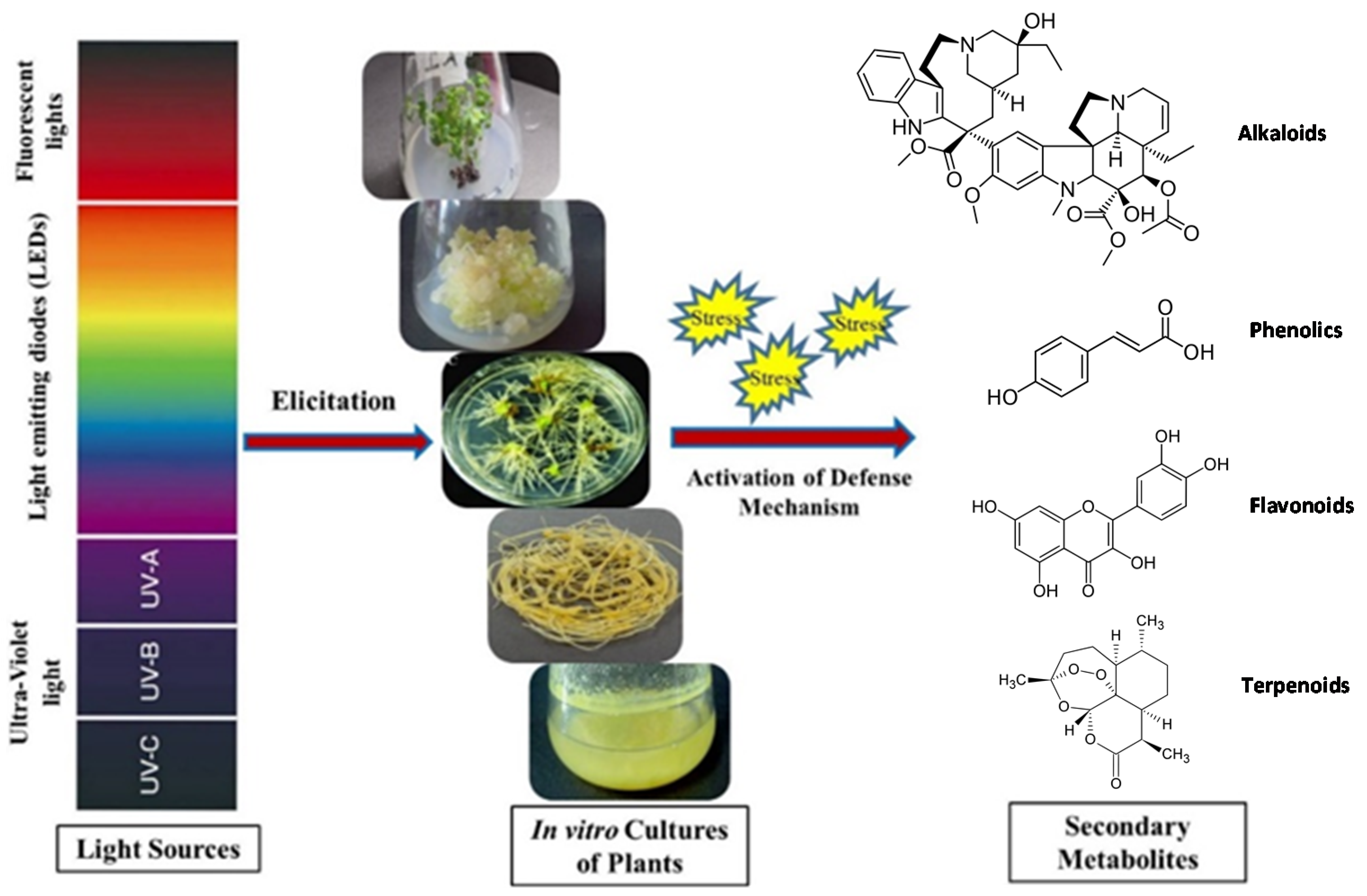
2. Light as an Elicitor

In vitro cultures of several plant species have been documented to elicit secondary metabolites using a variety of light sources in the past. These sources have been classified into three main categories in this review as UV lights, LED lights, and fluorescent lights, compared and discussed herein in detail.

2.1. UV Lights

A significant abiotic elicitor used in the past to boost the production of secondary metabolites in a variety of plant cultures is UV [24]. The wavelength of UV (400–200 nm) accounts for only a small por-tion of the solar radiation that reaches the earth’s surface, yet it has a significant bio-logical impact on living species, including plants. UV radiation is categorized into three parts: UV-A (320 to 390 nm), UV-B (280 to 320 nm), and UV-C (280 nm and be-low). The intensity of UV-A is greater than UV-B, but this difference is not biologically significant [25]. UV-B photons are perhaps the most intense wave that hits the surface of the earth, and even tiny changes in their quantity can have a huge impact on vital processes and properties at all scales, from species to ecosystems [26]. In addition to the rest of the UV groups, UV-C radiation has also proved to be the most effective in stimulating the production of plant secondary metabolites such as phenolics, alkaloids, or flavonoids [27,28,29] (Table 1).

2.1.1. UV-A and UV-B

Since UV light acts as an elicitor, it is basically involved in activating the defense mechanisms of plants which in turn produce secondary metabolites useful for humans for therapeutic purposes as they are not required by plants for their growth [28]. UV-A light can act as a potential elicitor to stimulate the production of secondary metabolites in plants grown under controlled conditions and/or in vitro cultures (Table 1).
Flavonols, also known as 3-hydroxylavones, are the most common flavonoids found in food. They are structurally similar to flavones, but they differ in that they have a hydroxyl group at the 3-position on the C-ring, while flavones have a ketone group with an unsaturated carbon–carbon bond [44]. Cynaroside (luteo-lin-7-glucoside), a flavone, is used for a variety of medical purposes; it may protect heart cells from apoptosis caused by reactive oxygen species (ROS). Cynaroside also reduces kidney damage caused by the chemotherapeutic drug cisplatin, which is used to treat cancer [45]. The production of cynaroside has been increased in in vitro culture of Capsicum annum (aka bell pepper plant) by elicitation of UVA/B light for a period of 16 days [30].
UV-B has also been reported as a potential elicitor candidate to induce various changes in the metabolism of plants [46]. This leads to the activation of plant protec-tion mechanism by the formation of secondary metabolites such as alkaloids and fla-vonoids [47,48]. Physical responses in plant tissues, such as increased amounts of spe-cific phenolics, make plants more resistant to UV-B radiation than other species, and improved levels of pigmentation are induced [49,50]. Plant secondary metabolites containing polyphenolic structure, i.e., flavonoids, can be found in a wide range of foods, including fruits and vegetables. They have antioxidant and biochemical properties that can help with disorders like cancer, Alzheimer, atherosclerosis, and many others [51,52,53]. In a recent study, elicitation of Nymphoides humboldtiana with UV-B reported the production of pharmaceutically important flavonoids such as phloroglucinol, chlorogenic acid, epicatechin, quercetin, and ferulic acid [31]. Similarly, shoots of Alternanthera species, i.e., A. sessilis and A. brasiliana, on elicitation with UV-B for 8 h showed a 51% and 62% increase in flavonoid content, respectively, in comparison with control [32]. Likewise, an increase in the production of flavonoids was also reported in Capsicum annum L. with the elicitation of UV- B [33]. Under UV-B exposure, the production of flavonols, particularly quercetin, kaempferol, and isorhamnetin, were also vastly improved in Ginkgo biloba leaves [34]. Vinblastine and vincristine are chemotherapy drugs that are made by linking the alkaloids catharanthine and vindoline and are used to treat a variety of cancers [54]. The impact of UV-B on cell suspension cultures of the Catharanthus roseus plant was investigated in which the production of these important alkaloids, catharanthine and vindoline, was improved to 3 and 12 fold, respectively [35]. Ocimum basilicum, also known as sweet basil, possesses a wide range of potent activities due to the presence of precious secondary metabolites. It is used in traditional medicine as a result of its bioactive dary metabolites. It is used in traditional medicine as a result of its bioactive metabo-lites [55,56]. Elicitation with UV-B irradiation at an intensity of 224 μmol m−2 s−1 dramatically raised the production of anthocyanin, phenolics, and flavonoids in leaves of Ocimum basilicum [21].
When aerobic or photosynthetic metabolism is disrupted by various environmental stresses, phenolic compounds can play an important role in plant development by functioning as protective substances and signal molecules in plants, as well as safeguarding them from ROS. According to several studies, whenever a plant is infected with a disease, phenolic compounds are produced in response to that infection [57]. Most phenolics were accumulated when UV-B radiation was applied at a rate of 20 W/cm2 to wheat seedlings. On day 4, total phenolics, DPPH, and ABTS levels increased by 26.3, 25.1, and 12.0%, respectively, as compared to un-irradiated wheat seedlings [37]. Likewise, in another study, seeds of Ocimum basilicum on irradiation of UV-B for three days showed enhanced production of phenolics [36].

2.1.2. UV-C

UV-C region of the spectrum includes wavelengths below 280 nm. These intense wavelengths are absorbed by ozone and do not reach the earth’s surface [58]. Thus, the application of UV-C on various plant cultures using artificial lamps may be a promising strategy in raising the production of valuable secondary metabolites. Trans-resveratrol (TR) and resveratrol are non-flavonoid phenolics of stilbenes and are classified under biologically active isomers found majorly in red grapes and berries. They are mainly employed in pharmaceutical industries due to their anti-carcinogenic, anti-diabetic, anti-acne, antioxidant and anti-inflammatory activities. These properties make them a promising candidate for novel drugs [59]. The elicited callus culture of Vitis vinifera L. showed 8–10 times higher production of resveratrol levels in the 48 h after UV-C application than in the first 24 h. The highest resveratrol accumulation was recorded to be 62.23 µg/g of FW in a 12-day old culture [38]. Likewise, in another study, calli that had been exposed to UV-C for 5 min showed higher content of TR (8.43 µg g−1) when harvested after 24 h. Catechin accumulation (8.89 mg/g) was also higher in calli exposed to UV-C for 10 min when harvested after 48 h. In conclusion, UV-C radiation aided the accumulation of secondary metabolites in the calli of the Okuzgozu grape cultivar [39].
Polyphenols found in Lepidium sativum L. have a diverse variety of medical and pharmaceutical purposes. Essential oils are also abundant in this therapeutic plant and exhibit exceptional anti-cancer properties in animal models as well as in various cell lines [60,61,62]. UV-C was irradiated on the callus of L. sativum for varying periods of time and different concentrations of melatonin. Phytochemical investigations revealed the production of three significant compounds: chlorogenic acid, kaempferol, and quercetin in callus culture. Cultures treated with melatonin (20 µM) showed three times higher production (36.36 mg/g DW), whereas cultures exposed to UV-C (60 min) showed a 2.5-fold increase (32.33 mg/g DW) in production in comparison with control (13.94 mg/g DW). This study compares both the elicitors and their effects on the initiation of physiological pathways in L. sativum for the formation of secondary metabolites [40].
Linum usitatissimum L. is considered a functional food and is widely used as oilseed crops in Europe [63,64]. It is a nutrient-dense diet that can be used as a substitute for food because it contains all dietary components [65,66]. When lignans and neolignans were quantified using reverse phase-high performance liquid chromatography (RP-HPLC), cultures of L. usitatissimum exposed to UV-C + photoperiod (16 h light/8 h dark) showed the presence of secoisolariciresinol diglucoside (SDG), dehydrodiconiferyl alcohol glucoside (DCG), guaiacylglycerol-β-coniferyl alcohol ether glucoside (GGCG), and lariciresinol diglucoside (LDG). UV-C radiation of 3.6 kJ/m2 resulted in a higher accumulation of SDG, LDG, and GGCG by 1.86-fold, followed by 2.25-fold and 1.33-fold in cell cultures grown under UV + photoperiod. Furthermore, the accumulation of DCG was raised by 1.36-fold in cell cultures grown under UV + dark under an intensity of 1.8 kJ/m2. Moreover, total phenolics, flavonoids, and antioxidants were also enhanced by 2.82-fold, 2.94-fold, and 1.04-fold, respectively, in cell cultures maintained under UV + photoperiod at a dosage of 3.6 kJ/m2 of UV-C radiation. These findings broadened the scope of what can be done in L. usitatasimum cell cultures to produce biologically active lignans and neolignans [41].
Ocimum basilicum L. var purpurascens (Lamiaceae) has gained popularity due to its ornamental and aromatic qualities as well as the presence of important volatile secondary metabolites such as rosmarinic acid, flavonoids, and anthocyanin s [67]. In a recent report, the effect of UV-C on the synthesis of phenylpropanoid metabolites was examined in in vitro culture of Ocimum basilicum L. When compared to the control, UV-C (10 min) exposure resulted in a 2.3-fold increase in rosmarinic acid (134.5 mg/g DW). Chichoric acid (51.52 mg/g DW) and anthocyanin (cyanide 0.50 mg/g DW) were about 4.1-fold greater after 50 min of UV-C exposure, whereas peonidin was 2.7-fold higher. Overall, UV-C proved to be efficient elicitors, as there was a positive association between induced phenolic compound synthesis and antioxidant activity of basil callus extracts [42]. Similarly, in another study formation of secondary metabolites in Echinacea purpurea callus and cell suspension cultures were observed after 24, 48, and 72 h of elicitation. Callus and cell suspension cultures were exposed to UV-C radiation for 15, 30, and 60 min, respectively. UV-C irradiation for 60 min was the most successful in promoting the aggregation of most secondary metabolites with varying incubation times depending on tissue culture meth ods [43]. This suggests that UV-C could be a potential elicitor of secondary metabolite synthesis in plants in vitro cultures.

2.2. Fluorescent Lights

When plants are grown under sole-source electric lighting, lamp spectral customization can be an approach for achieving desired plant characteristics [68]. Fluorescent lights are a major source of light energy for stimulating the production of secondary metabolites in in vitro cultures [9]. Previous studies have confirmed that, depending on plant species, light quality has a direct impact on morphological and physiological responses [9,69]. For the management of controlled-environment agriculture facilities, it is vital to conserve electrical energy expenditures. In this regard, fluorescent lamps are considered much cheaper than LEDs [70]. They are particularly appealing for a variety of applications due to their high efficiency, outstanding color rendering, and extended life [71]. As a result, their use could be beneficial in triggering various in vitro cultures for increased secondary metabolite production (Table 2).
In a study, enhanced production of secondary metabolites and biomass accumulation was observed in Stevia rebaudiana callus cultures on exposure to different fluorescent spectral lights. Maximum capability in enhancing total phenolic content (102.32 µg/g of DW), total antioxidant potential (11.63 µg/g DW), and total flavonoids content (22.07 µg/g DW) was shown under blue light [22]. Similarly, blue light increased total phenolic (23.9 mg/g DW) and flavonoids content (1.65 mg/g DW) in callus cultures of Prunella vulgaris [14]. Likewise, in another study, blue light promoted aggregation of metabolites to the greatest degree in shoot culture of Scutellaria lateriflora. The major flavonoids that showed the highest concentrations were baicalin, verbascoside, glucuronides, and wogonoside. Their levels were 1.49, 1.86, 1.54, and 2.05 times higher than the control under white light, respectiv ely [72]. Based on the findings of the above investigations, it can be hypothesized that blue light can operate as a potential elicitor in diverse in vitro cultures, promoting the formation of secondary metabolites.
Hypericin and pseudohypericin, which are derivates of naphtodianthrones, are structurally similar phenolic compounds that have gained importance commercially due to their unique activities [76]. However, conventional cultivation of these metabolites is unable to fulfill the fierce competition in the pharmaceutical industry in terms of both quantity and quality. Therefore, elicitation of root culture of Hypericum perforatum with red light showed the highest production of total hypericins (i.e., hyperin + pseudohypericin) (9.61 ± 0.3 µg/g), whereas the lowest on exposure to fluorescent light (7.12 ± 0.26 µg/g). Roots grown under red light also showed the highest content of flavonoids (41.17 ± 7.21 mg/g). In this study, the impact of blue light was also evaluated on the production of key secondary metabolites. A considerable increase in the production of metabolites was observed after one week of exposure; however, an inhibitory effect was detected after five weeks of incubation. Results also showed that the production of hypericin and total phenolic content were increased to 52% and 26%, respectively, in root culture after one-week exposure to blue ligh t [73].
Hyptis marrubioides has been traditionally used to cure infections related to gastrointestines, cramps, discomfort, and skin infections [77] in many regions. Effect of various fluorescent lights was observed on the production of important phenolic compounds in seed culture of H. marrubioides. White (0.308 mg/g of DW) and blue (0.298 mg/g of DW) light was shown to accumulate the highest amount of rutin, while red light improved plant development and increased dry weight and leaf number in in vitro-cultivated seeds of H. marrubioides [74]. Ginsenosides (saponins) found in the root extract of Panax ginseng are the most active components known to exhibit immunomodulatory properties and provide protection against heart and liver diseases [78]. Rb and Rg are formed from the structures of 20(S) protopanaxadiol and 20(S) protopanaxatriol, and are two important groups of ginsenosides [79]. The Rg group of ginsenosides (5.3–0.1 mg/g DW) accumulated more than the Rb group (3.7–0.7 mg/g DW) in hairy roots grown under fluorescent light. These findings imply that growing hairy roots in dim or bright settings can affect the Rb and Rg ginsenoside productions in in vitro cultures [75].
Artemisia absinthium L., often known as “Wormwood”, is referred to as a “universal treatment for all ailments” due to its therapeutic medical characteristics [80,81]. This plant has traditionally been used to treat diarrhea, cough, and common cold due [82] to its insecticidal, bitter [83,84], vermifuge, trematocidal [85], diuretic, and antispasmodic properties [86]. Total phenolics, total flavonoids, and antioxidant activity were found to be more supported by the green spectrum grown for three weeks under a photosynthetic photon flux density of 40–50 µmol m−2 s−1 in callus culture of A. absinthium [9]. As a result of the preceding studies, it is concluded that light elicitation (fluorescent, blue, red, green, and white light) has a positive influence and that different light regimes can aid in optimizing plant growth and developmental changes for the formation of commercially significant secondary metabolites in vitro.

2.3. LED Lights

The amount of light a plant receives has a big impact on its development, growth, and production. Agriculture employs traditional artificial light sources such as high-pressure sodium lamps (HPSLs), metal-halide lamps (MHLs), and fluorescent lamps (FLs) to provide a controlled atmosphere. Fluorescent lamps have risen in favor among these. However, the wavelengths of these lightning sources span from 350 to 750 nm, and for plant growth and development, it is considered of low quality. They possess a limited lifetime of activity and a low photosynthetic flux, limiting their use in plant illumination systems that require a large agricultural production. LEDs technology has been able to be employed in a rising variety of new sectors, including plant growth and development, due to the implementation of new types of semiconductor materials. As a substitute to conventional lighting systems, LEDs have proved to be a smarter source of artificial lighting to provide controlled conditions in agriculture and in vitro systems. Since LEDs can emit over specified spectral areas, they may be utilized to manage the amount of photosynthetically active and photomorphogenic radiation required for plant growth and development. Matching LED wavelengths to photoreceptors in plants can allow for optimal output while also altering plant shape and metabolism. As a result, these solid-state light sources can be used in developing lighting lamps for sustainable production and photo-morphogenesis research [87].
Plant growth, production, and secondary metabolism are all influenced by light in general and light quality in particular. LEDs exist in various colors, i.e., white, blue, green, red, yellow, violet, and far-red. Many scientists believe that red (600–700 nm) and blue (400–500 nm) light are more crucial for stimulating photosynthesis than other light wavelengths since they have the highest photosynthetic photon efficacy values. Green light (500–600 nm) can, on the other hand, penetrate deep inside the leaf due to its high transmittance and reflectance [88]. LED lights have been widely used to elicit the production of key secondary metabolites in various plant culture systems (Table 3).

2.3.1. Blue LED

Traditional lighting systems require color filters, but LED lighting systems may create the light of any desired color without them. LEDs are versatile enough to produce only the type of light that plants require. Plants require specific spectrums or colors for distinct morphogenic responses, and LED systems can be fine-tuned to provide only those. Blue light controls stomatal opening and transpiration, as well as preventing “red light syndrome” [103]. Thus, blue LEDs can play an efficient role in stimulating the production of pharmacologically active secondary metabolites, as demonstrated in Table 3.
Tomatoes (Solanum lycopersicum L.) are the world’s seventh most produced crop species and one of the year-round value crops grown in greenhouses. Tomato fruits are nutritionally dense because they include vital nutrients as well as phytochemicals that promote health [104]. LED was used as a source of artificial light to evaluate its effect on S. lycopersicum L. ‘Cuty’ (tomato seedlings). Results showed raised production of phenolics, flavonoids, and antioxidants under blue light when compared to control. This concludes that manipulating light quality using LED’s could stimulate the production of bioactive compounds and antioxidants [23].
The use of nutritional or therapeutic plant-based natural substances to treat disease has become a novel paradigm in clinical science. Flavonoids, particularly phenolic compounds, can be found in nearly all plants. Antioxidant, anti-cancer, anti-diabetic, and cardiovascular effects are observed in phenolics and flavonoids [105]. A study reported the highest concentrations of soluble protein and flavonoid in lettuce when exposed to blue light [89]. Another study found that leaves of synseed grown under blue LED had higher amounts of chlorophyll a, flavonoids, phenolics, and carotenoids than those grown under fluorescent or blue-red LED. Anti-oxidant activity was similarly boosted in synseed-derived seedlings grown in blue LED light [90]. Likewise, the overall polyphenol content in the blue LED treatment was also considerably higher than in the control treatment in Anoectochilus roxburghii [91,101]. Similarly, both blue and blue–violet light supplements boosted phenolic acid production in Ocimum basilicum, while in Eruca sativa, higher flavonoid synthesis was seen in response to both light supplements, but greater production was observed under blue– violet [92]. The above study concludes the potential of using blue LED as a promising elicitor in raising the production dramatically.

2.3.2. Red LED

Red LEDs has been widely employed as an alternative source of illumination for in vitro survival and improved production of metabolites in medicinally important plants. Red light is considered the most efficient spectral light in driving photosynthesis, and many studies reported it as an efficient elicitor in elevating the production of pharmacologically active constituents in in vitro systems of various plants [106]. Myrtus communis has long been used in medicine and possesses therapeutic characteristics [107,108]. M. communis (leaves, flowers, and fruits) contain various components that are critical to the pharmaceutical, food, liqueur, and cosmetic industries; hence, large-scale manufacturing of this plant is necessary [109]. M. communis leaf extracts are enriched sources of phenolic acids and flavonoids. In a recent study, the impact of red LED was evaluated on in vitro culture of M. communis. Results showed that out of all flavonoids, myricetin (347.02–1118.69 mg 100 g−1 DW) was the main constituent with the highest concentration and 6-benzyladenine (BA) level, whereas gallic acid had the highest concentrations of the phenolic acids (95.58 mg 100 g−1 DW on average) at a photosynthetic photon flux density of 35 µmol m−2 s−1 [93].
Milk thistle (Silybum marianum L.) is a well-known hepato-protective medicinal herb that has been extensively researched. The effects of various LEDs lights were investigated, and it was discovered that red light greatly increased phenolics, flavonoids, and superoxide dismutase (SOD) activities in this plant. Under red light, HPLC analysis revealed a substantially doubled total silymarin concentration (18.67 mg g−1 DW) when compared to the control (9.17 mg g−1 DW). When exposed to red light, the levels of isosilychristin, silybin A, silybin B, silychristin, and silydianin were found to be highest. This demonstrates that the quality of light has a significant impact on the callus culture of S. marianum morphological and biochemical properties [94].

2.3.3. Blue and Red LED in Combination

Several studies have reported the use of blue and red LEDs in combination on one or more plants simultaneously. Many reports suggested that the use of different LEDs in combination can enhance the production of bioactive compounds to several folds. Thus, their combination can be employed in eliciting various cultures that have gained importance pharmacologically [95,97,98]. Since ancient times, root extracts of Rehmannia glutinosa Libosch (Chinese foxglove) have been used to treat many disorders such as anemia and hypertension [96]. The therapeutic use of R. glutinosa roots is well known due to the presence of bioactive compounds such as catalpol, aucubin, and rehmaglutin [96,110]. In a study, plant development as well as phytochemicals with both defensive and prospective medicinal characteristics, such as phenolics and flavonoids, were influenced by spectral characteristics in both leaf and root tissues of R. glutinosa. On irradiation with blue LED, leaf extracts were found to have the highest total phenol concentration (35 ± 0.05 µg GAE/mg) as compared to root extracts. On the other hand, red LED exposure also increased the overall flavonoid content of the leaf extract by 33.6% and the root extracts by 61.7%. As a result, blue and red LEDs may be the most enticing light sources for R. glutinosa proliferation in in vitro conditions [95].
Bioactive compounds like phenolics and flavonoids are of significant importance in therapeutic plants because they can act as free radical scavengers [111]. The effects of red and blue LEDs on the accumulation of phenolic and flavonoids in Ocimum basilicum callus cultures were investigated in a recent study. Rosmarinic acid and eugenol were considerably enhanced under blue light (2.46 and 2.25 times greater than control). While under red light, the highest amounts of cyanidin (0.1216 mg/g DW) and peonidin (0.127 mg/g DW) were detected. Chicoric acid accumulation was about 4.52 times higher than callus grown under the control of continuous white light (81.40 mg/g DW) [97]. This suggests that employing LEDs to accelerate the production of physiologically active chemicals in vitro could be beneficial.
Fruits, nuts, medicinal herbs, and vegetables all contain phenylpropanoid, a type of secondary metabolite that is involved in reducing the risk of diabetes and heart disease through inhibiting carcinogenesis [112,113]. Plants are protected from bacteria by the antimicrobial activities of some phenylpropanoids during their interactions [114]. The generation of phenylpropanoid metabolites was examined using LEDs as an elicitor in wheat sprouts, and qRT-PCR and HPLC results revealed that white light (380 nm) was the best wavelength for epicatechin biosynthesis in wheat sprouts. Blue light (470 nm) was involved in the increased accumulation of gallic acid and quercetin, whereas red light (660 nm) increased the aggregation of ferulic acid on the 8th day and p-coumaric acid on the 12th day [98]. Similarly, flavonoids and anthocyanins, which also belong to a class of phenylpropanoids, were also enhanced in buckwheat, Fagopyrum sp. under red + blue (combination), and blue LED, respectively [99]. Likewise, phenolics and flavonoids were enhanced in seeds of Ocimum basilicum under red–blue (2R:1B) LED. In addition, the highest flavonoid content of 16.79 mmol/g FW was also achieved for protocorm-like bodies pre-treated with white LED for more than three cultures cycles under blue–red (1:1) LED [101]. Thus, red and blue LED ratios can be altered to generate improved growth and phenolic content in both red and green basil microgreens as a practical technique for generating superior quality foods [100]. Furthermore, in the callus culture of Cnidium officinale, mixed (red–blue) LED illumination enhanced the synthesis of phenolics and flavonoids [10]. In another study, in vitro generated Eclipta alba callus culture was subjected to multispectral lighting under regulated aseptic conditions. Results showed that the red light enhanced the production of phenolics (57.8 mg/g) and flavonoids (11.1 mg/g). whereas, on exposure to blue light, the production of four major compounds coumarin (1.26 mg/g), eclalbatin (5.00 mg/g), wedelolactone (32.54 mg/g), and demethylwedelolactone (23.67 mg/g), as well as two minor compounds β-amyrin (0.38 mg/g) and luteolin (0.39 mg/g) were increased [102].

3. Mechanistic Considerations and Future Prospects

Light is a key driver of plant secondary metabolite biosynthesis and accumulation [115]. When photons activate photoreceptors, they activate signaling pathways and cause changes in gene expression. The light-absorbing properties are defined by the interaction of a photoreceptor protein and a chromophore [116]. Figure 2 depicts the absorption bands of essential photoreceptors, as well as the quantities and processes that they affect. In comparison to photosynthesis, only a small fraction of photons absorbed are used to activate photoreceptors [117]. Plants have four major photoreceptors, absorbing photons not only in the photosynthetically active radiation (PAR) domain but also in the UV and far-red regions. They interact with many signal transduction factors and are in charge of initiating various physiological changes and adaptations, including secondary metabolite production, triggered by light.
To address secondary metabolites in plants, photosynthetically active radiation (PAR) is insufficient, and a broader wavelength range (physiologically active radiation) is required.
The protein cryptochrome absorption is comprised between the UV-A and blue light domains (340−520 nm). Phytochrome absorbs light primarily in the red/far-red area around 665 nm (Pr) and 730 nm (Pfr), but also in the blue/near-UV region. Phototropin light absorption is comprised between 350 and 520 nm, whereas UVR8 shows maximum absorption in the range between 280 and 350 nm.
Many significant players have been identified in the literature to explain how light affects secondary metabolite biosynthesis. The first is ROS; when exposed to large levels of UV-B radiation, plants preferentially produce a wide range of phenylpropanoid structures, including flavonoids, supporting the theory that flavonoids are principally engaged in photoprotection by absorbing the shortest solar wavelengths [118]. There is, however, substantial evidence that flavonoid production is similarly enhanced in response to high photosynthetically active radiation either in the presence or absence of UV radiation. For instance, the presence of phytochromes, at a modest level, as well as UVR8 and cryptochrome, are important for anthocyanin accumulation; therefore, wavelengths beyond UV also show impacts [115]. This lends credence to the idea that flavonoids can act as scavengers of ROS produced by excessive light [118].
Phytohormonal control is also an important regulation that could be linked to light. Phytohormones have been linked to the regulation of secondary metabolite synthesis under both developmental and stress circumstances [119,120]. Many phytochrome proteins have been connected to variations in phytohormone levels, including gibberellins and auxins, ethylene, jasmonates, and abscisic acid (ABA) [117,121], all of which are, for example, significant players in the control of plant secondary metabolism, including pigments [115].
Light may also regulate the expression of transcription factors, which are essential regulators of secondary metabolite biosynthesis. For instance, cryptochrome has been shown to regulate the accumulation of anthocyanins [122], whereas green light has been shown to reverse the effects of blue light in anthocyanin accumulation [123]. The expression of R2R3MYB transcription factors, including V1MYBA1-2 and VIMYBA2, which regulate the expression of anthocyanin synthesis-related genes like VvUFGT, increased in grape berries irradiated with blue light, which is consistent with this observation [124].
It all demonstrates how light can regulate the formation of secondary metabolites at various levels and via various signaling pathways. Yet, in vitro systems have received far too little attention at these levels of regulation, while in vitro systems could be appealing models for conducting this type of research since, unlike the whole plant, they allow for uniformity, accessibility, and reduced complexity [125].
Light allows for the manipulation of growth conditions and secondary metabolite synthesis in both controlled and in vitro cultures, but several elements must be addressed before it can be completely utilized. In recent years, LED systems appear to have a lot of promise, as light influences plant growth and development at every step. It affects morphogenesis, differentiation, and proliferation rates in the plant cell, tissue, and organ cultures, all of which are important for the biosynthesis of secondary metabolites. Conscious manipulation of light quality, along with the advantages of LED technology, will lead to increased biomass production with high secondary metabolite content under regulated in vitro settings. It will also ensure that homogenous material is obtained in a reasonably short amount of time without the need to cultivate the whole plant to the fully mature stage in the field. However, according to a review of the literature, the response appears to be depending on the plant species and the culture systems. Biosynthesis of plant secondary metabolites is regulated effectively by various signaling events, as the production of secondary metabolites depends upon the plant species and their respective phytochemical classes. As a result, proposing a general mechanism of light elicitation on the production of different secondary metabolites is complicated, and further research is needed to gain a comprehensive understanding. To uncover the molecular and cellular mechanism of light elicitation, an omics-based analysis could be a useful approach. Therefore, in conclusion, it may be inferred that different light regimes can aid in the improvement of growth and developmental alterations for the in vitro generation of safe secondary metabolites.

4. Conclusions

The current review deciphers the role of light as a promising elicitor in enhancing the production of key metabolites in different in vitro systems of plants. Among various abiotic elicitors, light has gained attention in enhancing secondary metabolite production due to its specified wavelengths, cost-effectiveness, and durability. A plethora of studies have reported the effect and quality of different types of light sources on enhanced secondary metabolite accumulation in several plant species in vitro. These findings pave the way for a more thorough examination of light as a plant elicitor. The fact that most studies have focused on plant growth and development, including the generation of primary metabolites, highlights the paucity of information about the different factors regulating light elicitation mechanisms. Thus, many studies still lack at depicting the mechanisms of elicitation promoting the production of pharmacologically important secondary metabolites since it may differ depending upon the type of plant species, culture conditions, and light source used.

Author Contributions

Conceptualization, S.A.; methodology, M.H., B.A., and S.D.; validation, C.H., B.H.A., and S.A.; formal analysis, S.A.; investigation, M.H., B.A., and S.D.; resources, C.H., B.H.A., and S.A.; data curation, C.H., B.H.A., and S.A.; writing—original draft preparation, M.H., B.A., and S.A.; writing—review and editing, C.H., B.H.A., and S.A.; supervision, C.H., B.H.A., and S.A.; project administration, S.A.; funding acquisition, C.H., B.H.A., and S.A. All authors have read and agreed to the published version of the manuscript.

Funding

This research was supported by Cosmetosciences, a global training and research program dedicated to the cosmetic industry. Located in the heart of the Cosmetic Valley, this program led by the University of Orleans is funded by the Region Centre-Val de Loire (VALBIOCOSM and INNOCOSM).

Institutional Review Board Statement

Not applicable.

Informed Consent Statement

Not applicable.

Data Availability Statement

All data are included in the present study.

Acknowledgments

B.H.A. and C.H. acknowledge the research fellowship of Le Studium-Institute for Advanced Studies, Loire Valley, Orléans, France. S.D. acknowledges Cosmetosciences and Région Centre Val de Loire for research fellowships. The authors would like to acknowledge networking support by Le Studium COSMENOVIC consortium and CNRS GDR3711 COSM’ACTIFS. The authors gratefully acknowledge the Région Centre Val de Loire for funding the project VALBIOCOSM and INNOCOSM. C.H. and B.H.A. gratefully acknowledge the support of Campus France through the PHC PERIDOT.

Conflicts of Interest

The authors declare no conflict of interest.

References

  1. Wang, J.W.; Wu, J.Y. Effective elicitors and process strategies for enhancement of secondary metabolite production in hairy root cultures. In Biotechnology of Hairy Root Systems; Springer: Berlin, Germany, 2013; pp. 55–89. [Google Scholar]
  2. Murthy, H.N.; Lee, E.-J.; Paek, K.-Y. Production of secondary metabolites from cell and organ cultures: Strategies and approaches for biomass improvement and metabolite accumulation. Plant Cell Tissue Organ. Cult. 2014, 118, 1–16. [Google Scholar] [CrossRef]
  3. Yue, W.; Ming, Q.-l.; Lin, B.; Rahman, K.; Zheng, C.-J.; Han, T.; Qin, L.-P. Medicinal plant cell suspension cultures: Pharmaceutical applications and high-yielding strategies for the desired secondary metabolites. Crit. Rev. Biotechnol. 2016, 36, 215–232. [Google Scholar] [CrossRef]
  4. Gonçalves, S.; Romano, A. Production of plant secondary metabolites by using biotechnological tools. In Secondary Metabolites, Sources and Applications; IntechOpen: London, UK, 2018; pp. 81–99. [Google Scholar]
  5. Hussain, M.S.; Fareed, S.; Saba Ansari, M.; Rahman, A.; Ahmad, I.Z.; Saeed, M. Current approaches toward production of secondary plant metabolites. J. Pharm. Bioall. Sci. 2012, 4, 10. [Google Scholar] [CrossRef] [PubMed]
  6. Cragg, G.M.; Newman, D.J. Plants as a source of anti-cancer agents. J. Ethnopharmacol. 2005, 100, 72–79. [Google Scholar] [CrossRef] [PubMed] [Green Version]
  7. Cui, X.-H.; Chakrabarty, D.; Lee, E.-J.; Paek, K.-Y. Production of adventitious roots and secondary metabolites by Hypericum perforatum L. in a bioreactor. Bioresour. Technol. 2010, 101, 4708–4716. [Google Scholar] [CrossRef]
  8. Yang, L.; Stöckigt, J. Trends for diverse production strategies of plant medicinal alkaloids. Nat. Prod. Rep. 2010, 27, 1469–1479. [Google Scholar] [CrossRef]
  9. Tariq, U.; Ali, M.; Abbasi, B.H. Morphogenic and biochemical variations under different spectral lights in callus cultures of Artemisia absinthium L. J. Photochem. Photobiol. B Biol. 2014, 130, 264–271. [Google Scholar] [CrossRef]
  10. Adil, M.; Ren, X.; Jeong, B.R. Light elicited growth, antioxidant enzymes activities and production of medicinal compounds in callus culture of Cnidium officinale Makino. J. Photochem. Photobiol. B Biol. 2019, 196, 111509. [Google Scholar] [CrossRef]
  11. Abbasi, B.H.; Tian, C.-L.; Murch, S.J.; Saxena, P.K.; Liu, C.-Z. Light-enhanced caffeic acid derivatives biosynthesis in hairy root cultures of Echinacea purpurea. Plant Cell Rep. 2007, 26, 1367–1372. [Google Scholar] [CrossRef] [PubMed]
  12. Shohael, A.; Ali, M.; Yu, K.; Hahn, E.; Islam, R.; Paek, K. Effect of light on oxidative stress, secondary metabolites and induction of antioxidant enzymes in Eleutherococcus senticosus somatic embryos in bioreactor. Process Biochem. 2006, 41, 1179–1185. [Google Scholar] [CrossRef]
  13. Liu, C.; Zhao, Y.; Wang, Y. Artemisinin: Current state and perspectives for biotechnological production of an antimalarial drug. Appl. Microbiol. Biotechnol. 2006, 72, 11–20. [Google Scholar] [CrossRef]
  14. Fazal, H.; Abbasi, B.H.; Ahmad, N.; Ali, S.S.; Akbar, F.; Kanwal, F. Correlation of different spectral lights with biomass accumulation and production of antioxidant secondary metabolites in callus cultures of medicinally important Prunella vulgaris L. J. Photochem. Photobiol. B Biol. 2016, 159, 1–7. [Google Scholar] [CrossRef] [PubMed]
  15. Moran, J.F.; Becana, M.; Iturbe-Ormaetxe, I.; Frechilla, S.; Klucas, R.V.; Aparicio-Tejo, P. Drought induces oxidative stress in pea plants. Planta 1994, 194, 346–352. [Google Scholar] [CrossRef] [Green Version]
  16. Alexieva, V.; Sergiev, I.; Mapelli, S.; Karanov, E. The effect of drought and ultraviolet radiation on growth and stress markers in pea and wheat. Plant Cell Environ. 2001, 24, 1337–1344. [Google Scholar] [CrossRef]
  17. Yu, T.-W.; Anderson, D. Reactive oxygen species-induced DNA damage and its modification: A chemical investigation. Mutat. Res. Fundam. Mol. Mech. Mutagenesis 1997, 379, 201–210. [Google Scholar] [CrossRef]
  18. Mittler, R. Oxidative stress, antioxidants and stress tolerance. Trends Plant Sci. 2002, 7, 405–410. [Google Scholar] [CrossRef]
  19. Ashry, N.A.; Mohamed, H.I. Impact of secondary metabolites and related enzymes in flax resistance and or susceptibility to powdery mildew. World J. Agric. Sci 2011, 7, 78–85. [Google Scholar]
  20. Samuolienė, G.; Brazaitytė, A.; Urbonavičiūtė, A.; Šabajevienė, G.; Duchovskis, P. The effect of red and blue light component on the growth and development of frigo strawberries. Zemdirbyste Agric. 2010, 97, 99–104. [Google Scholar]
  21. Dou, H.; Niu, G.; Gu, M. Pre-harvest UV-B radiation and photosynthetic photon flux density interactively affect plant photosynthesis, growth, and secondary metabolites accumulation in basil (Ocimum basilicum) plants. Agronomy 2019, 9, 434. [Google Scholar] [CrossRef] [Green Version]
  22. Ahmad, N.; Rab, A.; Ahmad, N. Light-induced biochemical variations in secondary metabolite production and antioxidant activity in callus cultures of Stevia rebaudiana (Bert). J. Photochem. Photobiol. B Biol. 2016, 154, 51–56. [Google Scholar] [CrossRef] [PubMed]
  23. Kim, E.-Y.; Park, S.-A.; Park, B.-J.; Lee, Y.; Oh, M.-M. Growth and antioxidant phenolic compounds in cherry tomato seedlings grown under monochromatic light-emitting diodes. Hortic. Environ. Biotechnol. 2014, 55, 506–513. [Google Scholar] [CrossRef]
  24. Xuan, T.D.; Khanh, T.D.; Khang, D.T.; Quan, N.T.; Elzaawely, A.A. Changes in chemical composition, total phenolics and antioxidant activity of Alpinia (Alpinia zerumbet) leaves exposed to UV. Int. Lett. Nat. Sci. 2016, 55, 25–34. [Google Scholar] [CrossRef] [Green Version]
  25. Stapleton, A.E. Ultraviolet radiation and plants: Burning questions. Plant Cell 1992, 4, 1353. [Google Scholar] [CrossRef] [PubMed] [Green Version]
  26. Parihar, P.; Singh, S.; Singh, R.; Singh, V.P.; Prasad, S.M. Changing scenario in plant UV-B research: UV-B from a generic stressor to a specific regulator. J. Photochem. Photobiol. B Biol. 2015, 153, 334–343. [Google Scholar] [CrossRef]
  27. Freitas, A.; Moldão-Martins, M.; Costa, H.S.; Albuquerque, T.G.; Valente, A.; Sanches-Silva, A. Effect of UV-C radiation on bioactive compounds of pineapple (Ananas comosus L. Merr.) by-products. J. Sci. Food Agric. 2015, 95, 44–52. [Google Scholar] [CrossRef]
  28. Yin, X.; Singer, S.D.; Qiao, H.; Liu, Y.; Jiao, C.; Wang, H.; Li, Z.; Fei, Z.; Wang, Y.; Fan, C. Insights into the mechanisms underlying ultraviolet-C induced resveratrol metabolism in grapevine (V. amurensis Rupr.) cv.“Tonghua-3”. Front. Plant Sci. 2016, 7, 503. [Google Scholar] [CrossRef] [PubMed] [Green Version]
  29. Abbasi, B.H.; Khan, T.; Khurshid, R.; Nadeem, M.; Drouet, S.; Hano, C. UV-C mediated accumulation of pharmacologically significant phytochemicals under light regimes in in vitro culture of Fagonia indica (L.). Sci. Rep. 2021, 11, 1–16. [Google Scholar] [CrossRef] [PubMed]
  30. Ellenberger, J.; Siefen, N.; Krefting, P.; Schulze Lutum, J.-B.; Pfarr, D.; Remmel, M.; Schröder, L.; Röhlen-Schmittgen, S. Effect of UV Radiation and Salt Stress on the Accumulation of Economically Relevant Secondary Metabolites in Bell Pepper Plants. Agronomy 2020, 10, 142. [Google Scholar] [CrossRef] [Green Version]
  31. Nocchi, N.; Duarte, H.M.; Pereira, R.C.; Konno, T.U.P.; Soares, A.R. Effects of UV-B radiation on secondary metabolite production, antioxidant activity, photosynthesis and herbivory interactions in Nymphoides humboldtiana (Menyanthaceae). J. Photochem. Photobiol. B Biol. 2020, 212, 112021. [Google Scholar] [CrossRef] [PubMed]
  32. Klein, F.R.S.; Reis, A.; Kleinowski, A.M.; Telles, R.T.; Amarante, L.d.; Peters, J.A.; Braga, E.J.B. UV-B radiation as an elicitor of secondary metabolite production in plants of the genus Alternanthera. Acta Bot. Bras. 2018, 32, 615–623. [Google Scholar] [CrossRef]
  33. Rodríguez-Calzada, T.; Qian, M.; Strid, Å.; Neugart, S.; Schreiner, M.; Torres-Pacheco, I.; Guevara-González, R.G. Effect of UV-B radiation on morphology, phenolic compound production, gene expression, and subsequent drought stress responses in chili pepper (Capsicum annuum L.). Plant Physiol. Biochem. 2019, 134, 94–102. [Google Scholar] [CrossRef] [PubMed]
  34. Zhao, B.; Wang, L.; Pang, S.; Jia, Z.; Wang, L.; Li, W.; Jin, B. UV-B promotes flavonoid synthesis in Ginkgo biloba leaves. Ind. Crops Prod. 2020, 151, 112483. [Google Scholar] [CrossRef]
  35. Ramani, S.; Jayabaskaran, C. Enhanced catharanthine and vindoline production in suspension cultures of Catharanthus roseus by ultraviolet-B light. J. Mol. Signal. 2008, 3, 1–6. [Google Scholar] [CrossRef] [Green Version]
  36. Mosadegh, H.; Trivellini, A.; Ferrante, A.; Lucchesini, M.; Vernieri, P.; Mensuali, A. Applications of UV-B lighting to enhance phenolic accumulation of sweet basil. Sci. Hortic. 2018, 229, 107–116. [Google Scholar] [CrossRef]
  37. Chen, Z.; Ma, Y.; Weng, Y.; Yang, R.; Gu, Z.; Wang, P. Effects of UV-B radiation on phenolic accumulation, antioxidant activity and physiological changes in wheat (Triticum aestivum L.) seedlings. Food Biosci. 2019, 30, 100409. [Google Scholar] [CrossRef]
  38. Keskin, N.; Kunter, B. Production of trans-resveratrol in callus tissue of Öküzgözü (Vitis vinifera L.) in response to ultraviolet-C irradiation. J. Anim Plant Sci. 2010, 20, 197–200. [Google Scholar]
  39. Cetin, E.S. Induction of secondary metabolite production by UV-C radiation in Vitis vinifera L. Öküzgözü callus cultures. Biol. Res. 2014, 47, 1–7. [Google Scholar] [CrossRef] [Green Version]
  40. Ullah, M.A.; Tungmunnithum, D.; Garros, L.; Drouet, S.; Hano, C.; Abbasi, B.H. Effect of ultraviolet-C radiation and melatonin stress on biosynthesis of antioxidant and antidiabetic metabolites produced in in vitro callus cultures of Lepidium sativum L. Int. J. Mol. Sci. 2019, 20, 1787. [Google Scholar] [CrossRef] [Green Version]
  41. Anjum, S.; Abbasi, B.H.; Doussot, J.; Favre-Réguillon, A.; Hano, C. Effects of photoperiod regimes and ultraviolet-C radiations on biosynthesis of industrially important lignans and neolignans in cell cultures of Linum usitatissimum L. (Flax). J. Photochem. Photobiol. B Biol. 2017, 167, 216–227. [Google Scholar] [CrossRef]
  42. Nazir, M.; Asad Ullah, M.; Mumtaz, S.; Siddiquah, A.; Shah, M.; Drouet, S.; Hano, C.; Abbasi, B.H. Interactive effect of melatonin and UV-C on phenylpropanoid metabolite production and antioxidant potential in callus cultures of purple basil (Ocimum basilicum L. var. purpurascens). Molecules 2020, 25, 1072. [Google Scholar] [CrossRef] [Green Version]
  43. Abd El-Aal, M.S.; Rabie, K.A.; Manaf, H.H. The effect of UV-C on secondary metabolites production of Echinacea purpurea culture in vitro. J. Biol. Chem. Environ. Sci. 2016, 11, 465–483. [Google Scholar]
  44. Zuiter, A. Proanthocyanidin: Chemistry and Biology: From Phenolic Compounds to Proanthocyanidins; Elsevier: Amsterdam, The Netherlands, 2014. [Google Scholar]
  45. Sun, X.; Sun, G.B.; Wang, M.; Xiao, J.; Sun, X.B. Protective effects of cynaroside against H2O2-induced apoptosis in H9c2 cardiomyoblasts. J. Cell. Biochem. 2011, 112, 2019–2029. [Google Scholar] [CrossRef]
  46. Schreiner, M.; Krumbein, A.; Mewis, I.; Ulrichs, C.; Huyskens-Keil, S. Short-term and moderate UV-B radiation effects on secondary plant metabolism in different organs of nasturtium (Tropaeolum majus L.). Innov. Food Sci. Emerg. Technol. 2009, 10, 93–96. [Google Scholar] [CrossRef]
  47. Zhang, W.J.; Björn, L.O. The effect of ultraviolet radiation on the accumulation of medicinal compounds in plants. Fitoterapia 2009, 80, 207–218. [Google Scholar] [CrossRef]
  48. Schreiner, M.; Mewis, I.; Huyskens-Keil, S.; Jansen, M.; Zrenner, R.; Winkler, J.; O’brien, N.; Krumbein, A. UV-B-induced secondary plant metabolites-potential benefits for plant and human health. Crit. Rev. Plant Sci. 2012, 31, 229–240. [Google Scholar] [CrossRef]
  49. Flint, S.D.; Ryel, R.J.; Caldwell, M.M. Ecosystem UV-B experiments in terrestrial communities: A review of recent findings and methodologies. Agric. For. Meteorol. 2003, 120, 177–189. [Google Scholar] [CrossRef]
  50. Caldwell, M.M.; Bornman, J.; Ballaré, C.; Flint, S.D.; Kulandaivelu, G. Terrestrial ecosystems, increased solar ultraviolet radiation, and interactions with other climate change factors. Photochem. Photobiol. Sci. 2007, 6, 252–266. [Google Scholar] [CrossRef]
  51. Burak, M.; Imen, Y. Flavonoids and their antioxidant properties. Turkiye Klin. Tip. Bil. Derg. 1999, 19, 296–304. [Google Scholar]
  52. Castañeda-Ovando, A.; de Lourdes Pacheco-Hernández, M.; Páez-Hernández, M.E.; Rodríguez, J.A.; Galán-Vidal, C.A. Chemical studies of anthocyanins: A review. Food Chem. 2009, 113, 859–871. [Google Scholar] [CrossRef]
  53. Lee, Y.K.; Yuk, D.Y.; Lee, J.W.; Lee, S.Y.; Ha, T.Y.; Oh, K.W.; Yun, Y.P.; Hong, J.T. (−)-Epigallocatechin-3-gallate prevents lipopolysaccharide-induced elevation of beta-amyloid generation and memory deficiency. Brain Res. 2009, 1250, 164–174. [Google Scholar] [CrossRef] [PubMed]
  54. Nisar, A.; Mamat, A.S.; Hatim, M.I.; Aslam, M.S.; Ahmad, M.S. An updated review on Catharanthus roseus: Phytochemical and pharmacological analysis. Indian Res. J. Pharm. Sci. 2016, 3, 631–653. [Google Scholar]
  55. Bora, K.S.; Arora, S.; Shri, R. Role of Ocimum basilicum L. in prevention of ischemia and reperfusion-induced cerebral damage, and motor dysfunctions in mice brain. J. Ethnopharmacol. 2011, 137, 1360–1365. [Google Scholar] [CrossRef]
  56. Loughrin, J.H.; Kasperbauer, M.J. Light reflected from colored mulches affects aroma and phenol content of sweet basil (Ocimum basilicum L.) leaves. J. Agric. Food Chem. 2001, 49, 1331–1335. [Google Scholar] [CrossRef]
  57. Petkovšek, M.M.; Štampar, F.; Veberič, R. Accumulation of phenolic compounds in apple in response to infection by the scab pathogen, Venturia inaequalis. Physiol. Mol. Plant Pathol. 2009, 74, 60–67. [Google Scholar] [CrossRef]
  58. Bridgen, M. Using ultraviolet-C (UV-C) irradiation on greenhouse ornamental plants for growth regulation. In Proceedings of the VIII International Symposium on Light in Horticulture 1134, East Lansing, MI, USA, 22–26 May 2016; pp. 49–56. [Google Scholar]
  59. Fiod Riccio, B.V.; Fonseca-Santos, B.; Colerato Ferrari, P.; Chorilli, M. Characteristics, biological properties and analytical methods of trans-resveratrol: A review. Crit. Rev. Anal. Chem. 2020, 50, 339–358. [Google Scholar] [CrossRef] [PubMed]
  60. Hardman, W.E.; Avula, C.R.; Fernandes, G.; Cameron, I.L. Three percent dietary fish oil concentrate increased efficacy of doxorubicin against MDA-MB 231 breast cancer xenografts. Clin. Cancer Res. 2001, 7, 2041–2049. [Google Scholar] [PubMed]
  61. Kassie, F.; Pool-Zobel, B.; Parzefall, W.; Knasmüller, S. Genotoxic effects of benzyl isothiocyanate, a natural chemopreventive agent. Mutagenesis 1999, 14, 595–604. [Google Scholar] [CrossRef] [Green Version]
  62. Kassie, F.; Laky, B.; Gminski, R.; Mersch-Sundermann, V.; Scharf, G.; Lhoste, E.; Kansmüller, S. Effects of garden and water cress juices and their constituents, benzyl and phenethyl isothiocyanates, towards benzo (a) pyrene-induced DNA damage: A model study with the single cell gel electrophoresis/Hep G2 assay. Chem. Biol. Interact. 2003, 142, 285–296. [Google Scholar] [CrossRef]
  63. Schmidt, T.J.; Klaes, M.; Sendker, J. Lignans in seeds of Linum species. Phytochemistry 2012, 82, 89–99. [Google Scholar] [CrossRef] [PubMed]
  64. Wang, H.; Wang, J.; Qiu, C.; Ye, Y.; Guo, X.; Chen, G.; Li, T.; Wang, Y.; Fu, X.; Liu, R.H. Comparison of phytochemical profiles and health benefits in fiber and oil flaxseeds (Linum usitatissimum L.). Food Chem. 2017, 214, 227–233. [Google Scholar] [CrossRef]
  65. Omar, K.A.; Shan, L.; Zou, X.; Song, Z.; Wang, X. Effects of two emulsifiers on yield and storage of flaxseed oil powder by response surface methodology. Pak. J. Nutr. 2009, 8, 1316–1324. [Google Scholar] [CrossRef] [Green Version]
  66. Waszkowiak, K.; Gliszczyńska-Świgło, A. Binary ethanol–water solvents affect phenolic profile and antioxidant capacity of flaxseed extracts. Eur. Food Res. Technol. 2016, 242, 777–786. [Google Scholar] [CrossRef] [Green Version]
  67. Zheng, W.; Wang, S.Y. Antioxidant activity and phenolic compounds in selected herbs. J. Agric. Food Chem. 2001, 49, 5165–5170. [Google Scholar] [CrossRef] [PubMed]
  68. Hernández, R.; Eguchi, T.; Deveci, M.; Kubota, C. Tomato seedling physiological responses under different percentages of blue and red photon flux ratios using LEDs and cool white fluorescent lamps. Sci. Hortic. 2016, 213, 270–280. [Google Scholar] [CrossRef] [Green Version]
  69. Ali, M.; Abbasi, B.H. Light-induced fluctuations in biomass accumulation, secondary metabolites production and antioxidant activity in cell suspension cultures of Artemisia absinthium L. J. Photochem. Photobiol. B Biol. 2014, 140, 223–227. [Google Scholar] [CrossRef]
  70. Ohashi-Kaneko, K.; Takase, M.; Kon, N.; Fujiwara, K.; Kurata, K. Effect of light quality on growth and vegetable quality in leaf lettuce, spinach and komatsuna. Environ. Control. Biol. 2007, 45, 189–198. [Google Scholar] [CrossRef] [Green Version]
  71. Jack, A.; Vrenken, L. Fluorescent lamps and low pressure sodium lamps. IEE Proc. A 1980, 127, 149–157. [Google Scholar] [CrossRef]
  72. Kawka, B.; Kwiecień, I.; Ekiert, H. Influence of culture medium composition and light conditions on the accumulation of bioactive compounds in shoot cultures of Scutellaria lateriflora L. (American Skullcap) grown in vitro. Appl. Biochem. Biotechnol. 2017, 183, 1414–1425. [Google Scholar] [CrossRef] [PubMed] [Green Version]
  73. Sobhani Najafabadi, A.; Khanahmadi, M.; Ebrahimi, M.; Moradi, K.; Behroozi, P.; Noormohammadi, N. Effect of different quality of light on growth and production of secondary metabolites in adventitious root cultivation of Hypericum perforatum. Plant Signal. Behav. 2019, 14, 1640561. [Google Scholar] [CrossRef]
  74. Pedroso, R.C.N.; Branquinho, N.A.A.; Hara, A.C.; Costa, A.C.; Silva, F.G.; Pimenta, L.P.; Silva, M.L.A.; Cunha, W.R.; Pauletti, P.M.; Januario, A.H. Impact of light quality on flavonoid production and growth of Hyptis marrubioides seedlings cultivated in vitro. Rev. Bras. Farmacogn. 2017, 27, 466–470. [Google Scholar] [CrossRef]
  75. Yu, K.-W.; Murthy, H.N.; Hahn, E.-J.; Paek, K.-Y. Ginsenoside production by hairy root cultures of Panax ginseng: Influence of temperature and light quality. Biochem. Eng. J. 2005, 23, 53–56. [Google Scholar] [CrossRef]
  76. Murthy, H.N.; Kim, Y.-S.; Park, S.-Y.; Paek, K.-Y. Hypericins: Biotechnological production from cell and organ cultures. Appl. Microbiol. Biotechnol. 2014, 98, 9187–9198. [Google Scholar] [CrossRef] [PubMed]
  77. McNeil, M.; Facey, P.; Porter, R. Essential oils from the Hyptis genus—A review (1909–2009). Nat. Prod. Commun. 2011, 6, 1934578X1100601149. [Google Scholar] [CrossRef] [Green Version]
  78. Coon, J.T.; Ernst, E. Panax ginseng. Drug Saf. 2002, 25, 323–344. [Google Scholar] [CrossRef] [PubMed]
  79. Palazón, J.; Cusidó, R.M.; Bonfill, M.; Mallol, A.; Moyano, E.; Morales, C.; Piñol, M.T. Elicitation of different Panax ginseng transformed root phenotypes for an improved ginsenoside production. Plant Physiol. Biochem. 2003, 41, 1019–1025. [Google Scholar] [CrossRef]
  80. Sharopov, F.S.; Sulaimonova, V.A.; Setzer, W.N. Composition of the Essential oil of Artemisia absinthium from Tajikistan. Rec. Nat. Prod. 2012, 6, 127–134. [Google Scholar]
  81. Baker, P. The Book of Absinthe: A Cultural History; Grove Press: Greenwich Village, NY, USA, 2001. [Google Scholar]
  82. Hayat, M.Q.; Khan, M.A.; Ashraf, M.; Jabeen, S. Ethnobotany of the Genus Artemisia L. (Asteraceae) in Pakistan; University of Hawaii at Manoa: Honolulu, HI, USA, 2009. [Google Scholar]
  83. Anderson, F.J. An Illustrated History of the Herbals; iUniverse: Bloomington, IN, USA, 1997. [Google Scholar]
  84. Smith, A.; Secoy, D. Plants used for agricultural pest control in western Europe before 1850. Chem. Ind. 1981, 70, 12–17. [Google Scholar]
  85. Ferreira, J.F.; Peaden, P.; Keiser, J. In vitro trematocidal effects of crude alcoholic extracts of Artemisia annua, A. absinthium, Asimina triloba, and Fumaria officinalis. Parasitol. Res. 2011, 109, 1585–1592. [Google Scholar] [CrossRef]
  86. Mohamed, A.E.-H.H.; El-Sayed, M.; Hegazy, M.E.; Helaly, S.E.; Esmail, A.M.; Mohamed, N.S. Chemical constituents and biological activities of Artemisia herba-alba. Rec. Nat. Prod. 2010, 4, 1–25. [Google Scholar]
  87. Gupta, S.D.; Agarwal, A. Light Emitting Diodes for Agriculture; Springer: Berlin, Germany, 2017. [Google Scholar]
  88. Terashima, I.; Fujita, T.; Inoue, T.; Chow, W.S.; Oguchi, R. Green light drives leaf photosynthesis more efficiently than red light in strong white light: Revisiting the enigmatic question of why leaves are green. Plant Cell Physiol. 2009, 50, 684–697. [Google Scholar] [CrossRef] [Green Version]
  89. Zhang, T.; Shi, Y.; Piao, F.; Sun, Z. Effects of different LED sources on the growth and nitrogen metabolism of lettuce. Plant Cell Tissue Organ. Cult. 2018, 134, 231–240. [Google Scholar] [CrossRef]
  90. Gupta, S.D.; Kumar, A.; Agarwal, A. Impact of light-emitting diodes (LEDs) on the growth and morphogenesis of encapsulated shoot buds of Curculigo orchioides Gaertn., an endangered medicinal herb. Acta Physiol. Plant 2019, 41, 50. [Google Scholar] [CrossRef]
  91. Wang, W.; Su, M.; Li, H.; Zeng, B.; Chang, Q.; Lai, Z. Effects of supplemental lighting with different light qualities on growth and secondary metabolite content of Anoectochilus roxburghii. PeerJ 2018, 6, e5274. [Google Scholar] [CrossRef] [PubMed] [Green Version]
  92. Taulavuori, K.; Pyysalo, A.; Taulavuori, E.; Julkunen-, R. Responses of phenolic acid and flavonoid synthesis to blue and blue-violet light depends on plant species. Environ. Exp. Bot. 2018, 150, 183–187. [Google Scholar] [CrossRef] [Green Version]
  93. Cioć, M.; Szewczyk, A.; Żupnik, M.; Kalisz, A.; Pawłowska, B. LED lighting affects plant growth, morphogenesis and phytochemical contents of Myrtus communis L. in vitro. Plant Cell Tissue Organ. Cult. 2018, 132, 433–447. [Google Scholar] [CrossRef] [Green Version]
  94. Younas, M.; Drouet, S.; Nadeem, M.; Giglioli-Guivarc’h, N.; Hano, C.; Abbasi, B.H. Differential accumulation of silymarin induced by exposure of Silybum marianum L. callus cultures to several spectres of monochromatic lights. J. Photochem. Photobiol. B Biol. 2018, 184, 61–70. [Google Scholar] [CrossRef]
  95. Manivannan, A.; Soundararajan, P.; Halimah, N.; Ko, C.H.; Jeong, B.R. Blue LED light enhances growth, phytochemical contents, and antioxidant enzyme activities of Rehmannia glutinosa cultured in vitro. Hortic. Environ. Biotechnol. 2015, 56, 105–113. [Google Scholar] [CrossRef]
  96. Zhang, R.-X.; Li, M.-X.; Jia, Z.-P. Rehmannia glutinosa: Review of botany, chemistry and pharmacology. J. Ethnopharmacol. 2008, 117, 199–214. [Google Scholar] [CrossRef]
  97. Nadeem, M.; Abbasi, B.H.; Younas, M.; Ahmad, W.; Zahir, A.; Hano, C. LED-enhanced biosynthesis of biologically active ingredients in callus cultures of Ocimum basilicum. J. Photochem. Photobiol. B Biol. 2019, 190, 172–178. [Google Scholar] [CrossRef] [PubMed]
  98. Cuong, D.M.; Ha, T.W.; Park, C.H.; Kim, N.S.; Yeo, H.J.; Chun, S.W.; Kim, C.; Park, S.U. Effects of LED lights on expression of genes involved in phenylpropanoid biosynthesis and accumulation of phenylpropanoids in wheat sprout. Agronomy 2019, 9, 307. [Google Scholar] [CrossRef] [Green Version]
  99. Seo, J.-M.; Arasu, M.V.; Kim, Y.-B.; Park, S.U.; Kim, S.-J. Phenylalanine and LED lights enhance phenolic compound production in Tartary buckwheat sprouts. Food Chem. 2015, 177, 204–213. [Google Scholar] [CrossRef]
  100. Lobiuc, A.; Vasilache, V.; Oroian, M.; Stoleru, T.; Burducea, M.; Pintilie, O.; Zamfirache, M.-M. Blue and red LED illumination improves growth and bioactive compounds contents in acyanic and cyanic Ocimum basilicum L. microgreens. Molecules 2017, 22, 2111. [Google Scholar] [CrossRef] [PubMed] [Green Version]
  101. Yeow, L.C.; Chew, B.L.; Sreeramanan, S. Elevation of secondary metabolites production through light-emitting diodes (LEDs) illumination in protocorm-like bodies (PLBs) of Dendrobium hybrid orchid rich in phytochemicals with therapeutic effects. Biotechnol. Rep. 2020, 27, e00497. [Google Scholar] [CrossRef]
  102. Khurshid, R.; Ullah, M.A.; Tungmunnithum, D.; Drouet, S.; Shah, M.; Zaeem, A.; Hameed, S.; Hano, C.; Abbasi, B.H. Lights triggered differential accumulation of antioxidant and antidiabetic secondary metabolites in callus culture of Eclipta alba L. PLoS ONE 2020, 15, e0233963. [Google Scholar] [CrossRef] [PubMed]
  103. Davis, P.A.; Burns, C. Photobiology in protected horticulture. Food Energy Secur. 2016, 5, 223–238. [Google Scholar] [CrossRef]
  104. García-Closas, R.; Berenguer, A.; Tormo, M.J.; Sánchez, M.J.; Quiros, J.R.; Navarro, C.; Arnaud, R.; Dorronsoro, M.; Chirlaque, M.D.; Barricarte, A. Dietary sources of vitamin C, vitamin E and specific carotenoids in Spain. Br. J. Nutr. 2004, 91, 1005–1011. [Google Scholar] [CrossRef] [PubMed] [Green Version]
  105. Al-Snafi, A.E. Phenolics and flavonoids contents of medicinal plants, as natural ingredients for many therapeutic purposes-A review. IOSR J. Pharm. 2020, 10, 42–81. [Google Scholar]
  106. Hogewoning, S.W.; Trouwborst, G.; Maljaars, H.; Poorter, H.; van Ieperen, W.; Harbinson, J. Blue light dose–responses of leaf photosynthesis, morphology, and chemical composition of Cucumis sativus grown under different combinations of red and blue light. J. Exp. Bot. 2010, 61, 3107–3117. [Google Scholar] [CrossRef] [PubMed]
  107. Yildirim, F.; Şan, B.; Yildirim, A.; Polat, M.; Ercişli, S. Mineral composition of leaves and fruit in some myrtle (Myrtus communis L.) genotypes. Erwerbs Obstbau 2015, 57, 149–152. [Google Scholar] [CrossRef]
  108. Bouaziz, A.; Abdalla, S.; Baghiani, A.; Charef, N. Phytochemical analysis, hypotensive effect and antioxidant properties of Myrtus communis L. growing in Algeria. Asian Pac. J. Trop. Biomed. 2015, 5, 19–28. [Google Scholar] [CrossRef] [Green Version]
  109. Shah, A.; Kazi, T.; Baig, J.; Afridi, H.; Kandhro, G.; Khan, S.; Kolachi, N.; Wadhwa, S. Determination of total mercury in chicken feed, its translocation to different tissues of chicken and their manure using cold vapour atomic absorption spectrometer. Food Chem. Toxicol. 2010, 48, 1550–1554. [Google Scholar] [CrossRef] [PubMed]
  110. Chung, I.M.; Kim, J.J.; Lim, J.D.; Yu, C.Y.; Kim, S.H.; Hahn, S.J. Comparison of resveratrol, SOD activity, phenolic compounds and free amino acids in Rehmannia glutinosa under temperature and water stress. Environ. Exp. Bot. 2006, 56, 44–53. [Google Scholar] [CrossRef]
  111. Machlin, L.J.; Bendich, A. Free radical tissue damage: Protective role of antioxidant nutrients 1. FASEB J. 1987, 1, 441–445. [Google Scholar] [CrossRef] [PubMed]
  112. Yang, C.S.; Landau, J.M.; Huang, M.-T.; Newmark, H.L. Inhibition of carcinogenesis by dietary polyphenolic compounds. Annu. Rev. Nutr. 2001, 21, 381–406. [Google Scholar] [CrossRef] [Green Version]
  113. Yao, L.H.; Jiang, Y.-M.; Shi, J.; Tomas-Barberan, F.; Datta, N.; Singanusong, R.; Chen, S. Flavonoids in food and their health benefits. Plant Foods Hum. Nutr. 2004, 59, 113–122. [Google Scholar] [CrossRef]
  114. Naoumkina, M.A.; Zhao, Q.; Gallego-Giraldo, L.; Dai, X.; Zhao, P.X.; Dixon, R.A. Genome-wide analysis of phenylpropanoid defence pathways. Mol. Plant Pathol. 2010, 11, 829–846. [Google Scholar] [CrossRef]
  115. Thoma, F.; Somborn-Schulz, A.; Schlehuber, D.; Keuter, V.; Deerberg, G. Effects of light on secondary metabolites in selected leafy greens: A review. Front. Plant Sci. 2020, 11, 497. [Google Scholar] [CrossRef]
  116. Folta, K.M.; Carvalho, S.D. Photoreceptors and control of horticultural plant traits. HortScience 2015, 50, 1274–1280. [Google Scholar] [CrossRef] [Green Version]
  117. Brennicke, A.; Schopfer, P. Pflanzenphysiologie; Spektrum Akademischer Verlag: Berlin, Germany, 2010. [Google Scholar]
  118. Brunetti, C.; Fini, A.; Sebastiani, F.; Gori, A.; Tattini, M. Modulation of phytohormone signaling: A primary function of flavonoids in plant–environment interactions. Front. Plant Sci. 2018, 9, 1042. [Google Scholar] [CrossRef] [Green Version]
  119. Renouard, S.; Corbin, C.; Lopez, T.; Montguillon, J.; Gutierrez, L.; Lamblin, F.; Lainé, E.; Hano, C. Abscisic acid regulates pinoresinol–lariciresinol reductase gene expression and secoisolariciresinol accumulation in developing flax (Linum usitatissimum L.) seeds. Planta 2012, 235, 85–98. [Google Scholar] [CrossRef]
  120. Markulin, L.; Corbin, C.; Renouard, S.; Drouet, S.; Durpoix, C.; Mathieu, C.; Lopez, T.; Auguin, D.; Hano, C.; Lainé, É. Characterization of LuWRKY36, a flax transcription factor promoting secoisolariciresinol biosynthesis in response to Fusarium oxysporum elicitors in Linum usitatissimum L. hairy roots. Planta 2019, 250, 347–366. [Google Scholar] [CrossRef] [PubMed]
  121. Casal, J.J. Phytochromes, cryptochromes, phototropin: Photoreceptor interactions in plants. Photochem. Photobiol. 2000, 71, 1–11. [Google Scholar] [CrossRef]
  122. Fox, A.R.; Soto, G.C.; Jones, A.M.; Casal, J.J.; Muschietti, J.P.; Mazzella, M.A. cry1 and GPA1 signaling genetically interact in hook opening and anthocyanin synthesis in Arabidopsis. Plant Mol. Biol. 2012, 80, 315–324. [Google Scholar] [CrossRef] [PubMed] [Green Version]
  123. Folta, K.M. Green light stimulates early stem elongation, antagonizing light-mediated growth inhibition. Plant Physiol. 2004, 135, 1407–1416. [Google Scholar] [CrossRef] [PubMed] [Green Version]
  124. Rodyoung, A.; Masuda, Y.; Tomiyama, H.; Saito, T.; Okawa, K.; Ohara, H.; Kondo, S. Effects of light emitting diode irradiation at night on abscisic acid metabolism and anthocyanin synthesis in grapes in different growing seasons. Plant Growth Regul. 2016, 79, 39–46. [Google Scholar] [CrossRef]
  125. Hano, C.; Addi, M.; Fliniaux, O.; Bensaddek, L.; Duverger, E.; Mesnard, F.; Lamblin, F.; Lainé, E. Molecular characterization of cell death induced by a compatible interaction between Fusarium oxysporum f. sp. linii and flax (Linum usitatissimum) cells. Plant Physiol. Biochem. 2008, 46, 590–600. [Google Scholar] [CrossRef]
Figure 1. An overview of light’s function as an elicitor of important secondary metabolites in various in vitro plant cultures maintained under controlled conditions, including shoot, callus hairy root, adventitious root, and cell suspension cultures (from top to bottom). Different light sources, including UV light but also excessive light, can cause stress and activate the defense response, resulting in the production of a variety of bioactive plant secondary metabolites such as alkaloids (e.g., vinblastine), phenolics (e.g., p-coumaric acid), flavonoids (e.g., quercetin), or terpenoids (e.g., artemisinin).
Plants 10 01521 g001
Figure 2. Photoreceptor absorption bands in plants and their processes (adapted from [115]). Absorbing photons activate different types of photoreceptors not just in the PAR domain but also in the UV and far-red regions. Each absorption band and photoreceptor couple are engaged in a different physiological response that may be essential for biomass and secondary metabolite synthesis, such as pigmentation, morphology, phototropism, circadian clock process, stomata opening, or photosynthetic activity.
Plants 10 01521 g002
Table 1. Effects of UV light as an elicitor of key secondary metabolites in controlled and in vitro plant culture systems.
Light
Sources
IntensityExposure TimePlant SpeciesCulture
System
Secondary
Metabolite
Yield
Increase
References
UV-A/B4–5 Wm−2/10–14 Wm−23 h per day for 16 daysCapsicum annumCGC (leaf)Cynaroside-[30]
UV-B73.08 kJ/m2/day7 h per day for 13 daysNymphoides humboldtianaCGC (leaf)Flavonoids-[31]
UV-B40 J/cm28 hAlternanthera SessilisCGC (shoot)Flavonoids51%[32]
Alternanthera Brasiliana62%
UV-B1.14 kJ/m2/day4 h per day for 14 daysCapsicum annuumCGC (leaf)Flavonoids-[33]
UV-B313 nm7–14 daysGinkgo bilobaCGC (leaf)Quercetin, kaempferol,
and isorhamnetin
2.05- to 2.4-fold and
16.67- to −42-fold, respectively
[34]
UV-B1.26 μW/cm2 5 minCatharanthus roseusCell suspension cultureCatharanthine and vindoline3-fold and 12-fold[35]
UV-B224
μmol m−2 s −1
1 h for 2 days and 2 h for 2 daysOcimum basilicum
Green basil
CGC (leaf)Anthocyanin, phenolics, and flavonoids9–23%, 28–126% and 80–169%, respectively[21]
2 h for 2 days and 2 h for five daysOcimum basilicum
Purple Basil
Phenolics and flavonoids29–63% and 37–79%
UV-B102 kJ/m2/day3 daysOcimum basilicumCGC (plantlet)Phenolics [36]
UV-B20 µW/cm24 daysTriticum aestiviumCGC (seedling)Phenolics26.3%[37]
UV-C254 nm15 minVitis viniferaCallus culturetrans-resveratrol26-fold[38]
UV-C254 nm5 min after 24 hVitis viniferaCallus culture trans-resveratrol8-fold[39]
10 min after 48 hCatechin-
UV-C3 W/m260 minLepidium sativumCallus cultureChlorogenic acid, kaemferol, and quercetin2.5-fold[40]
UV-C + Photoperiod UV-C + Dark3.6 kJ/m2 + 16/8 h
1.8 kJ/m2 + 24 h dark
10–60 minLinum usitatissimumCallus cultureSecoisolariciresinol diglucoside (SDG)1.86-fold[41]
Lariciresinol diglucoside (LDG)2.25-fold
Guaiacylglycerol-β-coniferyl alcohol ether glucoside (GGCG)1.33-fold
Total phenolic production2.82-fold
Total flavonoid production2.94-fold
Dehydrodiconiferyl alcohol glucoside (DCG)1.36-fold
UV-C3 W/m210 minOcimum basilicumCallus cultureRosmarinic acid2.3-fold[42]
Chichoric acid and cyanide4.1-fold
Peonidin2.7-fold
UV-C254 nm60 minEchinacea purpureaCallus culturePhenolics-[43]
Cell suspension culture
CGC = controlled growth chamber.
Table 2. Role of fluorescent light as an elicitor of key secondary metabolites in various in vitro systems of plants.
Light
Type
Light CharacteristicExposure
Time
Plant SpeciesCulture
System
Secondary
Metabolite
Yield
Increase
References
Blue
light
380–560 nm30 daysStevia rebaudianaCallus
culture
Phenolics and Flavonoids-[22]
Blue
light
40–50 µmol m−2 s−1 3 weeksPrunella vulgarisCallus culturePhenolics and Flavonoids-[14]
Blue
light
60 µmol m−2 s−16 weeksScutellaria lateriflora L.Shoot
culture
Glucuronides, Baicalin,
Wogonoside, Verbascoside
1.54-, 1.49-, 2.05- and 1.86-fold, respectively[72]
Red
light
660 nm5 weeksHypericum perforatumRoot
culture
Hypericins, Flavonoids-[73]
Blue
light
470 nm1 weekHypericins, Phenolics52% and 26%
Blue
light
50 µmol m−2 s−130 daysHyptis marrubioidesMicro
propagation
Rutin-[74]
White
light
Fluores cent
light
50 µmol m−2 s−14 weeksPanax ginsengHairy root
culture
Ginsenoside-[75]
Green light40–50 µmol m−2 s−13 weeksArtemisia absinthiumCallus culturePhenolics and Flavonoids-[9]
Table 3. Role of LED lights as an elicitor of key secondary metabolites in plant culture systems.
Light
Types
Light CharacteristicExposure TimePlant SpeciesCulture
System
Secondary
Metabolite
Yield
Increase
References
Monochromatic
Blue LED
456 nm27 daysSolanum lycopersicumClosed-type plant production system (seedling)Phenolics and flavonoids-[23]
Blue
LED
200 µmol m−2 s−114/10 hLettuceCGC (leaf)Flavonoids2.07-fold[89]
Blue
LED
50 µmol m−2 s−128 daysCurculigo OrchioidesCGC (shoot bud)Phenolics and flavonoids-[90]
Blue
LED
30 ±1 µmol m−2 s−18 h per day 40 daysAnoectochilus roxburghiiCGC (leaf)Flavonoids and polyphenols24.2%[91]
Blue (+B) and Blue-violet (+BV)
LED
450 nm and 420–440 nm10 daysOcimum basilicumCGC (leaf)Phenolics [92]
Eruca sativaFlavonoids
Red LED35 µmol m−2 s−16 weeksMyrtus communisIn vitro shoot cultureGallic acid and Myricetin-[93]
Red LED40–50 μmol m−2 s −124 hSilybum marianumCallus cultureSilymarin2-fold[94]
Blue
LED
50 µmol m−2 s−14 weeksRehmannia glutinosaIn vitro shoot culturePhenolics and flavonoids39.3%,
33.6%
[95]
Red
LED
450 nmCGC (root and leaf)Phenolics, Flavonoids33.6%, 61.7%[96]
Blue LED40–50 µmol m−2 s−14 weeksOcimum basilicumCallus cultureRosmarinic acid and eugenol2.46- and 2.25-fold[97]
Red LEDPeonidin and cyanidin3.5- and 4.53-fold
White LEDChicoric acid4.52-fold
White LED380 nm4 daysTriticum aestiviumCGC (seedling)Epicatechin1.3- to 1.46-fold[98]
Blue LED470 nm8 daysGallic acid and quercetin1.2–1.5 fold
Red LED660 nm8 daysFerulic acid-
12 daysp-coumaric acid1.27- to 1.77-fold
Red Blue light137 µmol m−2 s−196 hFagopyrum sp.CGC (seedling)Flavonoids-[99]
Blue LED177 µmol m−2 s−1Anthocyanins-
Red and Blue
2R:1B
120 µmol m−2 s−112 h each day for 17 daysOcimum basilicumCGC (seedling)Phenolics and flavonoids1.63–1.87-fold and 2.06-fold[100]
Blue–red (1:1)
LED
44.80 µmol/s30 days for 16 hDendrobiumIn vitro protocorn-like body cultureFlavonoids-[101]
Red: Blue LED33 µmol m−2 s−14 weeksCnidium officinaleCallus culturePhenolics and flavonoids-[10]
Red LED660 nm24 hEclipta albaCallus culturePhenolics and flavonoids2-fold[102]
Blue LED460 nm
Publisher’s Note: MDPI stays neutral with regard to jurisdictional claims in published maps and institutional affiliations.

